Note: Descriptions are shown in the official language in which they were submitted.
CA 02888984 2015-04-20
WO 2014/061824 PCT/JP2013/079053
DESCRIPTION
METHODS FOR TREATING EYE DISORDERS
BACKGROUND OF THE DISCLOSURE
[0001] The Wnt gene family encodes a large class of secreted proteins related
to the Intl/Wntl
proto-oncogene and Drosophila wingless ("Wg"), a Drosophila Wntl homologue
(Cadigan et al.
(1997) Genes & Development 11:3286-3305). Wnts are expressed in a variety of
tissues and
organs and are required for many developmental processes, including
segmentation in
Drosophila; endoderm development in C. elegans; and establishment of limb
polarity, neural
crest differentiation, kidney morphogenesis, sex determination, and brain
development in
mammals (Parr, et al. (1994) Curr. Opinion Genetics & Devel. 4:523-528). The
Wnt pathway is a
master regulator in development, both during embryogenesis and in the mature
organism
(Eastman, et al. (1999) Curr Opin Cell Biol 11: 233-240; Peifer, et al. (2000)
Science 287: 1606-
1609).
[0002] Wnt signals are transduced by the Frizzled ("Fz") family of seven
transmembrane domain
receptors (Bhanot et al. (1996) Nature 382:225-230). Frizzled cell-surface
receptors (Fzd) play
an essential role in both canonical and non-canonical Wnt signaling. In the
canonical pathway,
upon activation of Fzd and LRP5/6 (low-density-lipoprotein receptor-related
protein 5 and 6) by
Wnt proteins, a signal is generated that prevents the phosphorylation and
degradation of (3-
catenin by the "13-catenin destruction complex," permitting stable 13-catenin
translocation and
accumulation in the nucleus, and therefore Wnt signal transduction. (Perrimon
(1994) Cell
76:781-784)(Miller, J. R. (2001) Genome Biology; 3(1):1-15). The non-canonical
Wnt signaling
pathway is less well defined: there are at least two non-canonical Wnt
signaling pathways that
have been proposed, including the planar cell polarity (PCP) pathway, the
Wnt/Ca++ pathway,
and the convergence extension pathway.
[0003] Glycogen synthase kinase 3 (GSK3), the tumor suppressor gene product
APC
(adenomatous polyposis coli) (Gumbiner (1997) Cum Biol. 7:R443-436), and the
scaffolding
protein Axin, are all negative regulators of the Wnt pathway, and together
form the "13-catenin
destruction complex." In the absence of a Wnt ligand, these proteins form a
complex and
promote phosphorylation and degradation of 13-catenin, whereas Wnt signaling
inactivates the
complex and prevents 13-catenin degradation. Stabilized 13-catenin
translocates to the nucleus as a
1
CA 02888984 2015-04-20
WO 2014/061824 PCT/JP2013/079053
result, where it binds TCF (T cell factor) transcription factors (also known
as lymphoid
enhancer-binding factor-1 (LEF1)) and serves as a coactivator of TCF/LEF-
induced transcription
(Bienz, et al. (2000) Cell 103: 311-320; Polakis, et al. (2000) Genes Dev 14:
1837-1851).
[0004] Wnt signaling occurs via canonical and non-canonical mechanisms. In the
canonical
pathway, upon activation of Fzd and LRP5/6 by Wnt proteins, stabilized r3-
catenin accumulates
in the nucleus and leads to activation of TCF target genes (as described
above; Miller, J. R.
(2001) Genome Biology; 3(1):1-15). The non-canonical Wnt signaling pathway is
less well
defined: at least two non-canonical Wnt signaling pathways have been proposed,
including the
planar cell polarity (PCP) pathway and the Wnt/Ca++ pathway.
[0005] Diseases and degenerative conditions of the optic nerve and retina are
the leading causes
of blindness in the world. Macular degeneration (MD) is the loss of
photoreceptors in the portion
of the central retina, termed the macula, responsible for high-acuity vision.
Age-related macular
degeneration (AMD) is described as either "dry" or "wet." The wet, exudative,
neovascular form
of AMD affects about 10% of those with AMD and is characterized by abnormal
blood vessels
growing through the retinal pigment epithelium (RPE), resulting in hemorrhage,
exudation,
scarring, or serous retinal detachment. Ninety percent of AMD patients have
the dry form
characterized by atrophy of the retinal pigment epithelium and loss of macular
photoreceptors.
At present there is no cure for any form of MD or AMD, although some success
in attenuation
has been obtained with photodynamic therapy.
[0006] Glaucoma is a condition resulting from several distinct eye diseases
that cause vision loss
by damage to the optic nerve. Elevated intraocular pressure (I0P) due to
inadequate ocular
drainage is the most frequent cause of glaucoma. Glaucoma often develops as
the eye ages, or it
can occur as the result of an eye injury, inflammation, tumor or in advanced
cases of cataract or
diabetes. It can also be caused by the increase in TOP caused by treatment
with steroids. Drug
therapies that are proven to be effective in glaucoma reduce IOP either by
decreasing vitreous
humor production or by facilitating ocular draining. Such agents are often
vasodilators and as
such act on the sympathetic nervous system and include adrenergic antagonists.
2
CA 02888984 2015-04-20
WO 2014/061824 PCT/JP2013/079053
[0007] There is an urgent need for new treatments for ophthalmic disorders
such as macular
degeneration (MD), age-related macular degeneration (AMD), glaucoma,
cataracts, retinitis
pigmentosa, choroidal neovascularization, retinal degeneration, and oxygen-
induced retinopathy.
BRIEF SUMMARY OF THE DISCLOSURE
[0008] The present disclosure relates generally to alpha-helix mimetic
structures and specifically
to alpha-helix mimetic structures that are inhibitors of (3-catenin. The
disclosure also relates to
applications in the treatment of ophthalmic conditions, such as macular
degeneration and
glaucoma, and pharmaceutical compositions comprising such alpha helix mimetic
13-catenin
inhibitors.
BRIEF DESCRIPTION OF THE FIGURES
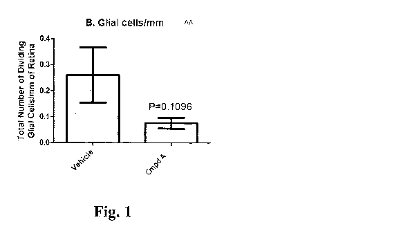
[0009] FIGS. 1A-1D. Number of dividing immune cells, glial cells, astrocytes
and Muller cells,
following treatment with Compound A. Compound A is 4-(((6S,9S,9a5)-1-
(benzylcarbamoy1)-
2,9-dimethy1-4,7-dioxo-8-(quinolin-8-ylmethyl)octahydro-1H-pyrazino[2,1-
c][1,2,4]triazin-6-
yOmethyl)phenyl dihydrogen phosphate. (A), Compound A treatment did not affect
immune cell
response. (B-D), Increase in number of proliferating glial cells (A), Muller
cells (B), and
astrocytes (D) following retinal detachment is attenuated in eyes treated with
Compound A
relative to control vehicle levels.
[0010] FIGS. 2A-2B. Quantitative analysis of glial scar frequency and size
following treatment
with Compound A. (A), Frequency of glial scars is significantly reduced
following treatment
with Compound A. (B), Average glial scar length is significantly reduced
following treatment
with Compound A.
[0011] FIGS. 3A-3D. Immunohistochemistry identifies subretinal gliosis (or
scarring) seen as
the presence of vimentin labeled Muller cell processes extending into the
subretinal space. OS,
outer layer/subretinal space; ONL, outer nuclear layer; GCL, ganglion cell
layer. (A-B), Vehicle
treated eyes. Following detachment, vimentin expression increases in Muller
cells, and Muller
cell processes are seen extending into the subretinal space (arrows).
Arrowheads point to
3
CA 02888984 2015-04-20
WO 2014/061824 PCT/JP2013/079053
dividing Muller cells. (C-D), Compound A treated eyes. No Muller cell growth
into the
subretinal space was observed. One dividing cell (astrocyte) is present in the
GCL (arrow).
[0012] FIGS. 4A-4B. CNV lesion size following treatment with Compound A or
Compound C.
Compound C is (6S,9S,9aS)-N-benzy1-6-(4-hydroxybenzy1)-2,9-dimethyl-4,7-dioxo-
8-(quinolin-
8-ylmethypoctahydro-1H-pyrazino[2,1-c][1,2,4]triazine-1-carboxamide. (A),
Average CNV
lesion size at day 15. (B), Average CNV lesion size at day 22.
DETAILED DESCRIPTION OF THE DISCLOSURE
[0013] Recently, non-peptide compounds have been developed which mimic the
secondary
structure of reverse-turns found in biologically active proteins or peptides.
For example, U.S.
Pat. No. 5,440,013 and published PCT Applications Nos. W094/03494,
W001/00210A1, and
W001/16135A2 each disclose conformationally constrained, non-peptidic
compounds, which
mimic the three-dimensional structure of reverse-turns. In addition, U.S. Pat.
No. 5,929,237 and
its continuation-in-part U.S. Pat. No. 6,013,458, disclose conformationally
constrained
compounds which mimic the secondary structure of reverse-turn regions of
biologically active
peptides and proteins. In relation to reverse-turn mimetics, conformationally
constrained
compounds have been disclosed which mimic the secondary structure of alpha-
helix regions of
biologically active peptide and proteins in W02007/056513 and W02007/056593.
[0014] The relevant structures and compounds of the alpha helix mimetic 13-
catenin inhibitors of
this invention are disclosed in WO 2010/044485, WO 2010/128685, WO
2009/148192, and US
2011/0092459, each of which is incorporated herein by reference in its
entirety. These
compounds have now been found to be useful in the treatment of ophthalmic
conditions and
disorders, such as macular degeneration and glaucoma. While not wishing to be
bound, the
effectiveness of these compounds in treating these conditions is based in part
on the ability of
these compounds to inhibit 13-catenin, thus altering Wnt pathway signaling,
which has been
found to improve various ophthalmic diseases and conditions.
4
CA 02888984 2015-04-20
WO 2014/061824 PCT/JP2013/079053
[0015] The preferable structure of the alpha helix mimetic p-catenin
inhibitors of this invention
have the following formula (I):
R2 R3
1
N
N AO
NR
wherein
A is -CHR7-,
wherein
R7 is optionally substituted arylalkyl, optionally substituted
heteroarylalkyl, optionally
substituted cycloalkylalkyl or optionally substituted heterocycloalkylalkyl;
G is -NH-, -NR6-, or -0-
wherein
R6 is lower alkyl or lower alkenyl;
R1 is -Ra-R1 ;
wherein
Ra is optionally substituted lower alkylene and
R10 is optionally substituted bicyclic fused aryl or optionally substituted
bicyclic fused
heteroaryl;
R2 is ¨(C0)-NH-Rb-R20,
wherein
Rb is bond or optionally substituted lower alkylene; and
TN20
is optionally substituted aryl or optionally substituted heteroaryl; and
R3 is C1-4 alkyl.
These compounds are especially useful in the prevention and/or treatment of
ophthalmic
conditions, such as macular degeneration and glaucoma.
CA 02888984 2015-04-20
WO 2014/061824 PCT/JP2013/079053
[0016] The more preferable structure of the alpha helix mimetic f3-catenin
inhibitors of this
invention have the following substituents in the above-mentioned formula (I):
A is -CHR7-,
wherein
R7 is arylalkyl optionally substituted with hydroxyl or C1-4 alkyl;
G is -NH-, -NR6-, or -0-
wherein
R6 is C1-4 alkyl or C1-4 alkenyl;
RI is -Ra-R' ;
wherein
Ra is C14 alkylene and
Rl is bicyclic fused aryl or bicyclic fused heteroaryl, optionally
substituted with halogen
or amino;
R2 is ¨(C0)-NH-Rb-R20,
wherein
Rb is bond or C1-4 alkylene; and
1,20
K is aryl or heteroaryl; and
R3 is C14 alkyl.
These compounds are especially useful in the prevention and/or treatment of
ophthalmic
conditions, such as macular degeneration and glaucoma.
[0017] The most preferable alpha helix mimetic P-catenin inhibitors of this
invention are as
follows:
(6 S,9S)-N-benzy1-6-(4-hydroxybenzy1)-2,9-dimethyl-8-(naphthalen-1-ylmethyl)-
4,7-
dioxooctahydro-1H-pyrazino[2,1-c][1,2,4]triazine-l-carboxamide,
(6S,9S)-2-allyl-N-benzy1-6-(4-hydroxybenzy1)-9-methyl-8-(naphthalen-l-
ylmethyl)-4,7-
dioxooctahydro-1H-pyrazino[2,1-c][1,2,4]triazine-l-carboxamide,
(6S,9S)-N-benzy1-6-(4-hydroxybenzy1)-9-methyl-8-(naphthalen-1-ylmethyl)-4,7-
dioxohexahydropyrazino[2,1-c][1,2,4]oxadiazine-1(6H)-carboxamide,
(6S,9S)-8-((2-aminobenzo[d]thiazol-4-yl)methyl)-N-benzyl-6-(4-hydroxybenzyl)-
2,9-
dimethyl-4,7-dioxooctahydro-1H-pyrazino[2,1-c][1,2,4]triazine-l-carboxamide,
6
CA 02888984 2015-04-20
WO 2014/061824 PCT/JP2013/079053
(6S,9S)-N-benzy1-6-(4-hydroxybenzy1)-2,9-dimethyl-4,7-dioxo-8-(quinolin-8-
ylmethypoctahydro-1H-pyrazino[2,1-c][1,2,4]triazine-1-carboxamide,
(6S,9S)-2-allyl-N-benzy1-6-(4-hydroxybenzy1)-9-methyl-4,7-dioxo-8-(quinolin-8-
ylmethypoctahydro-1H-pyrazino[2,1-c][1,2,4]triazine-1-carboxamide,
4-(((6S,9S)-1-(benzylcarbamoy1)-2,9-dimethy1-4,7-dioxo-8-(quinolin-8-
ylmethyl)octahydro-1H-pyrazino[2,1-c][1,2,4]triazin-6-yl)methyl)phenyl
dihydrogen phosphate,
4-(((6S,9S)-1-(benzylcarbamoy1)-2,9-dimethy1-8-(naphthalen-1-ylmethyl)-4,7-
dioxooctahydro-1H-pyrazino[2,1-c][1,2,4]triazin-6-yl)methyl)phenyl dihydrogen
phosphate,
sodium 4-(((6S,9S)-1-(benzylcarbamoy1)-2,9-dimethy1-4,7-dioxo-8-(quinolin-8-
ylmethypoctahydro-1H-pyrazino[2,1-c][1,2,4]triazin-6-yl)methyl)phenyl
phosphate,
sodium 4-(((6S,9S)-1-(benzylcarbamoy1)-2,9-dimethy1-4,7-dioxo-8-(naphthalen-8-
ylmethypoctahydro-1H-pyrazino[2,1-c][1,2,4]triazin-6-yl)methyl)phenyl
phosphate,
(6S,9S)-2-ally1-6-(4-hydroxybenzy1)-9-methy1-4,7-dioxo-N-((R)-1-phenylethyl)-8-
(quinolin-8-ylmethyl)octahydro-1H-pyrazino[2,1-c][1,2,4]triazine-1-
carboxamide,
(6S,9S)-2-ally1-6-(4-hydroxybenzy1)-9-methy1-4,7-dioxo-N-((S)-1-phenylethyl)-8-
(quinolin-8-ylmethyl)octahydro-1H-pyrazino[2,1-c][1,2,4]triazine-1-
carboxamide,
(6S,9S)-N-benzy1-6-(4-hydroxy-2,6-dimethylbenzy1)-2,9-dimethyl-4,7-dioxo-8-
(quinolin-8-ylmethypoctahydro-1H-pyrazino[2,1-c][1,2,4]triazine-1-carboxamide,
(6S,9S)-8-(benzo[b]thiophen-3-ylmethyl)-N-benzy1-6-(4-hydroxybenzy1)-2,9-
dimethyl-
4,7-dioxooctahydro-1H-pyrazino[2,1-c][.1,2,4]triazine-1-carboxamide,
(6S,9S)-8-(benzo[c][1,2,5]thiadiazol-4-ylmethyl)-N-benzyl-6-(4-hydroxybenzyl)-
2,9-
dimethyl-4,7-dioxooctahydro-1H-pyrazino[2,1-c][1,2,4]triazine-1-carboxamide,
(6S,9S)-N-benzy1-6-(4-hydroxybenzy1)-8-(isoquinolin-5-ylmethyl)-2,9-dimethyl-
4,7-
dioxooctahydro-1H-pyrazino[2,1-c][1,2,4]triazine-1-carboxamide,
(6S,9S)-N-benzy1-84(5-chlorothieno[3,2-b]pyridin-3-yOmethyl)-6-(4-
hydroxybenzy1)-
2,9-dimethyl-4,7-dioxooctahydro-1H-pyrazino[2,1-c][1,2,4]triazine-l-
carboxamide,
(6S,9S)-N-benzy1-6-(4-hydroxybenzy1)-2,9-dimethyl-4,7-dioxo-8-(quinoxalin-5-
ylmethyDoctahydro-1H-pyrazino[2,1-c][1,2,4]triazine-1-carboxamide, and
(6S,9S)-6-(4-hydroxybenzy1)-2,9-dimethyl-4,7-dioxo-8-(quinolin-8-ylmethyl)-N-
(thiophen-2-ylmethyDoctahydro-1H-pyrazino[2,1-c][1,2,4]triazine-1-carboxamide.
7
CA 02888984 2015-04-20
WO 2014/061824 PCT/JP2013/079053
These compounds are especially useful in the prevention and/or treatment of
ophthalmic
conditions, such as macular degeneration and glaucoma.
[0018] In a most preferred embodiment, the compound is:
4-(((6S,9S,9aS)-1-(benzylcarbamoy1)-2,9-dimethy1-4,7-dioxo-8-(quinolin-8-
ylmethypoctahydro-1H-pyrazino[2,1-c][1,2,4]triazin-6-yDrnethyl)phenyl
dihydrogen phosphate
(Compound A), or
(6S,9S,9aS)-N-benzy1-6-(4-hydroxybenzy1)-2,9-dimethyl-4,7-dioxo-8-(quinolin-8-
ylmethypoctahydro-1H-pyrazino[2,1-c][1,2,4]triazine-1-carboxamide (Compound
C).
These compounds are especially useful in the prevention and/or treatment of
ophthalmic
conditions, such as macular degeneration and glaucoma.
[0019] In particular, the alpha helix mimetics of the invention have been
found to be useful as
inhibitors of P-catenin. Disclosed herein are alpha helix mimetic 13-catenin
inhibitor compounds
for treatment of ophthalmic diseases and conditions.
[0020] A "13-catenin inhibitor" is a substance that can reduce or prevent 0-
catenin activity. 13-
catenin activities include translocation to the nucleus, binding with TCF (T
cell factor)
transcription factors, and coactivating TCF transcription factor-induced
transcription of TCF
target genes.
[0021] An "ophthalmic disease" or "ophthalmic condition" can be any disease,
condition or
disorder that affects the eye and eye area, including but not limited to
macular degeneration
(MD), age-related macular degeneration (AMID), glaucoma, cataracts, retinitis
pigmentosa,
choroidal neovascularization, retinal degeneration, and oxygen-induced
retinopathy.
[0022] As used herein, "treatment" refers to clinical intervention in an
attempt to alter the disease
course of the individual or cell being treated, and can be performed during
the course of clinical
pathology. Therapeutic effects of treatment include without limitation,
preventing recurrence of
disease, alleviation of symptoms, diminishment of any direct or indirect
pathological
consequences of the disease, decreasing the rate of disease progression,
amelioration or palliation
of the disease state, and remission or improved prognosis.
8
CA 02888984 2015-04-20
WO 2014/061824 PCT/JP2013/079053
[0023] As used herein, the terms "therapeutically effective amount" and
"effective amount" are
used interchangeably to refer to an amount of a composition of the invention
that is sufficient to
result in the prevention of the development or onset of an ophthalmic disease,
or one or more
symptoms thereof, to enhance or improve the effect(s) of another therapy,
and/or to ameliorate
one or more symptoms of an ophthalmic disease.
[0024] A therapeutically effective amount can be administered to a patient in
one or more doses
sufficient to palliate, ameliorate, stabilize, reverse or slow the progression
of the disease, or
otherwise reduce the pathological consequences of the disease, or reduce the
symptoms of the
disease. The amelioration or reduction need not be permanent, but may be for a
period of time
ranging from at least one hour, at least one day, or at least one week or
more. The effective
amount is generally determined by the physician on a case-by-case basis and is
within the skill of
one in the art. Several factors are typically taken into account when
determining an appropriate
dosage to achieve an effective amount. These factors include age, sex and
weight of the patient,
the condition being treated, the severity of the condition, as well as the
route of administration,
dosage form and regimen and the desired result.
[0025] As used herein, the terms "subject" and "patient" are used
interchangeably and refer to an
animal, preferably a mammal such as a non-primate (e.g., cows, pigs, horses,
cats, dogs, rats etc.)
and a primate (e.g., monkey and human), and most preferably a human.
[0026] The alpha helix mimetic [3-catenin inhibitors described herein can be
incorporated into
pharmaceutical compositions for administration, singly or in combination, to a
subject for the
treatment or prevention of a disorder described herein. Such compositions
typically include the
active agent and a pharmaceutically acceptable carrier. As used herein the
term
"pharmaceutically acceptable carrier" includes saline, solvents, dispersion
media, coatings,
antibacterial and antifungal agents, isotonic and absorption delaying agents,
and the like,
compatible with pharmaceutical administration. Supplementary active compounds
can also be
incorporated into the compositions.
[0027] The compounds and compositions described herein are useful for
treatment of ophthalmic
conditions and diseases, such as macular degeneration and glaucoma.
9
CA 02888984 2015-04-20
WO 2014/061824 PCT/JP2013/079053
[0028] The alpha helix mimetic I3-catenin inhibitors described herein are
useful to prevent or
treat disease. Specifically, the disclosure provides for both prophylactic and
therapeutic methods
of treating a subject at risk of (or susceptible to) an ophthalmic disease or
condition. Accordingly,
the present methods provide for the prevention and/or treatment of an
ophthalmic condition in a
subject by administering an effective amount of an alpha helix mimetic P-
catenin inhibitor to a
subject in need thereof. For example, a subject can be administered a 0-
catenin inhibitor
composition in an effort to improve one or more of the factors contributing to
an ophthalmic
disease or condition.
[0029] One aspect of the technology includes methods of reducing an ophthalmic
condition in a
subject for therapeutic purposes. In therapeutic applications, compositions or
medicaments are
administered to a subject suspected of, or already suffering from such a
disease in an amount
sufficient to cure, or at least partially arrest, the symptoms of the disease,
including its
complications and intermediate pathological phenotypes in development of the
disease. As such,
the disclosure provides methods of treating an individual afflicted with an
ophthalmic condition.
In some embodiments, the technology provides a method of treating or
preventing specific
ophthalmic disorders, such as cataracts, retinitis pigmentosa, glaucoma,
choroidal
neovascularization, retinal degeneration, and oxygen-induced retinopathy, in a
mammal by
administering an alpha helix mimetic 13-catenin inhibitor.
[0030] In one embodiment, the 13-catenin inhibitor is administered to a
subject to treat or prevent
cataracts. Cataracts is a congenital or acquired disease characterized by a
reduction in natural
lens clarity. Individuals with cataracts may exhibit one or more symptoms,
including, but not
limited to, cloudiness on the surface of the lens, cloudiness on the inside of
the lens, and/or
swelling of the lens. Typical examples of congenital cataract-associated
diseases are pseudo-
cataracts, membrane cataracts, coronary cataracts, lamellar cataracts,
punctuate cataracts, and
filamentary cataracts. Typical examples of acquired cataract-associated
diseases are geriatric
cataracts, secondary cataracts, browning cataracts, complicated cataracts,
diabetic cataracts, and
traumatic cataracts. Acquired cataracts is also inducible by electric shock,
radiation, ultrasound,
drugs, systemic diseases, and nutritional disorders. Acquired cataracts
further includes
postoperative cataracts.
CA 02888984 2015-04-20
PCT/JP2013/079053
WO 2014/061824
[0031] In one embodiment, the 13-catenin inhibitor is administered to a
subject to treat or prevent
retinitis pigmentosa. Retinitis pigmentosa is a disorder that is characterized
by rod and/or cone
cell damage. The presence of dark lines in the retina is typical in
individuals suffering from
retinitis pigmentosa. Individuals with retinitis pigmentosa also present with
a variety of
symptoms including, but not limited to, headaches, numbness or tingling in the
extremities, light
flashes, and/or visual changes. See, e.g., Heckenlively et al., Am J.
Ophthalmol. 105(5): 504-511
(1988).
[0032] In one embodiment, the 13-catenin inhibitor is administered to a
subject to treat or prevent
glaucoma. Glaucoma is a genetic disease characterized by an increase in
intraocular pressure,
which leads to a decrease in vision. Glaucoma may emanate from various
ophthalmologic
conditions that are already present in an individual, such as, wounds,
surgery, and other
structural malformations. Although glaucoma can occur at any age, it
frequently develops in
elderly individuals and leads to blindness. Glaucoma patients typically have
an intraocular
pressure in excess of 21 mmHg. However, normal tension glaucoma, where
glaucomatous
alterations are found in the visual field and optic papilla, can occur in the
absence of such
increased intraocular pressures, i.e., greater than 21 mmHg. Symptoms of
glaucoma include, but
are not limited to, blurred vision, severe eye pain, headache, seeing haloes
around lights, nausea,
and/or vomiting.
[0033] In one embodiment, the 13-catenin inhibitor is administered to a
subject to treat or prevent
macular degeneration. Macular degeneration is typically an age-related
disease. The general
categories of macular degeneration include wet, dry, and non-aged related
macular degeneration.
Dry macular degeneration, which accounts for about 80-90 percent of all cases,
is also known as
atrophic, nonexudative, or drusenoid macular degeneration. With dry macular
degeneration,
drusen typically accumulate beneath the retinal pigment epithelium tissue.
Vision loss
subsequently occurs when drusen interfere with the function of photoreceptors
in the macula.
Symptoms of dry macular generation include, but are not limited to, distorted
vision, center-
vision distortion, light or dark distortion, and/or changes in color
perception. Dry macular
degeneration can result in the gradual loss of vision.
11
CA 02888984 2015-04-20
WO 2014/061824 PCT/JP2013/079053
[0034] Wet macular degeneration is also known as neovascularization,
subretinal
neovascularization, exudative, or disciform degeneration. With wet macular
degeneration,
abnormal blood vessels grow beneath the macula. The blood vessels leak fluid
into the macula
and damage photoreceptor cells. Wet macular degeneration can progress rapidly
and cause
severe damage to central vision. Wet and dry macular degeneration have
identical symptoms.
Non-age related macular degeneration, however, is rare and may be linked to
heredity, diabetes,
nutritional deficits, injury, infection, or other factors. The symptoms of non-
age related macular
degeneration also include, but are not limited to, distorted vision, center-
vision distortion, light
or dark distortion, and/or changes in color perception.
[0035] In one embodiment, the I3-catenin inhibitor is administered to a
subject to treat or prevent
choroidal neovascularization. Choroidal neovascularization (CNV) is a disease
characterized by
the development of new blood vessels in the choroid layer of the eye. The
newly formed blood
vessels grow in the choroid, through the Bruch membrane, and invade the
subretinal space. CNV
can lead to the impairment of sight or complete loss of vision. Symptoms of
CNV include, but
are not limited to, seeing flickering, blinking lights, or gray spots in the
affected eye or eyes,
blurred vision, distorted vision, and/or loss of vision.
[0036] In one embodiment, the 13-catenin inhibitor is administered to a
subject to treat or prevent
retinal degeneration. Retinal degeneration is a genetic disease that relates
to the break-down of
the retina. Retinal tissue may degenerate for various reasons, such as, artery
or vein occlusion,
diabetic retinopathy, retinopathy of prematurity, and/or retrolental
fibroplasia. Retinal
degradation generally includes retinoschisis, lattic degeneration, and is
related to progressive
macular degeneration. The symptoms of retina degradation include, but are not
limited to,
impaired vision, loss of vision, night blindness, tunnel vision, loss of
peripheral vision, retinal
detachment, and/or light sensitivity.
[0037] In one embodiment, the 13-catenin inhibitor is administered to a
subject to treat or prevent
oxygen-induced retinopathy. Oxygen-induced retinopathy (01R) is a disease
characterized by
microvascular degeneration. OM is an established model for studying
retinopathy of prematurity.
OW is associated with vascular cell damage that culminates in abnormal
neovascularization.
Microvascular degeneration leads to ischemia which contributes to the physical
changes
12
CA 02888984 2015-04-20
WO 2014/061824 PCT/JP2013/079053
associated with OIR. Oxidative stress also plays an important role in the
vasoobliteration of OIR
where endothelial cells are prone to peroxidative damage. Pericytes, smooth
muscle cells, and
perivascular astrocytes, however, are generally resistant to peroxidative
injury. See, e.g.,
Beauchamp et al., Role of thromboxane in retinal microvascular degeneration in
oxygen-induced
retinopathy, J Appl Physiol. 90: 2279-2288 (2001). OIR, including retinopathy
of prematurity, is
generally asymptomatic. However, abnormal eye movements, crossed eyes, severe
nearsightedness, and/or leukocoria, can be a sign of OIR or retinopathy of
prematurity.
[0038] In one aspect, the invention provides a method for preventing, in a
subject, an ophthalmic
condition by administering to the subject an alpha-helix mimetic 0-catenin
inhibitor that
modulates one or more signs or markers of an ophthalmic condition. Subjects at
risk for an
ophthalmic condition can be identified by, e.g., any or a combination of
diagnostic or prognostic
assays. In prophylactic applications, pharmaceutical compositions or
medicaments of the alpha
helix mimetic f3-catenin inhibitors are administered to a subject susceptible
to, or otherwise at
risk of a disease or condition in an amount sufficient to eliminate or reduce
the risk, lessen the
severity, or delay the outset of the disease, including biochemical,
histologic and/or behavioral
symptoms of the disease, its complications and intermediate pathological
phenotypes presenting
during development of the disease. Administration of the 13-catenin inhibitors
can occur prior to
the manifestation of symptoms characteristic of the aberrancy, such that a
disease or disorder is
prevented or, alternatively, delayed in its progression.
[0039] Any suitable route of administration may be employed for providing a
mammal,
especially a human, with an effective dose of a compound described herein. For
example, oral,
rectal, topical, parenteral, ocular, pulmonary, nasal, and the like may be
employed. Dosage forms
include tablets, troches, dispersions, suspensions, solutions, capsules,
creams, ointments,
aerosols, and the like. Preferably compounds described herein are administered
orally.
[0040] The effective dosage of active ingredient employed may vary depending
on the particular
compound employed, the mode of administration, the condition being treated and
the severity of
the condition being treated. Such dosage may be ascertained readily by a
person skilled in the art.
[0041] When treating or controlling ophthalmic conditions and diseases for
which compounds
described herein are indicated, generally satisfactory results are obtained
when the compounds
13
CA 02888984 2015-04-20
WO 2014/061824 PCT/JP2013/079053
described herein are administered at a daily dosage of from about 0.1
milligram to about 100
milligram per kilogram of animal body weight, preferably given as a single
daily dose or in
divided doses two to six times a day, or in sustained release form. For most
large mammals, the
total daily dosage is from about 1.0 milligrams to about 1000 milligrams. In
the case of a 70 kg
adult human, the total daily dose will generally be from about 1 milligram to
about 500
milligrams. For a particularly potent compound, the dosage for an adult human
may be as low as
0.1 mg. In some cases, the daily dose may be as high as 1 gram. The dosage
regimen may be
adjusted within this range or even outside of this range to provide the
optimal therapeutic
response.
[0042] Oral administration will usually be carried out using tablets or
capsules. Examples of
doses in tablets and capsules are 0.1 mg, 0.25 mg, 0.5 mg, 1 mg, 2 mg, 5 mg,
10 mg, 15 mg, 20
mg, 25 mg, 30 mg, 40 mg, 50 mg, 100 mg, 200 mg, 250 mg, 300 mg, 400 mg, 500
mg, and 750
mg. Other oral forms may also have the same or similar dosages.
[0043] Also described herein are pharmaceutical compositions which comprise a
compound
described herein and a pharmaceutically acceptable carrier. The pharmaceutical
compositions
described herein comprise a compound described herein or a pharmaceutically
acceptable salt as
an active ingredient, as well as a pharmaceutically acceptable carrier and
optionally other
therapeutic ingredients. A pharmaceutical composition may also comprise a
prodrug, or a
pharmaceutically acceptable salt thereof, if a prodrug is administered.
[0044] The compositions can be suitable for oral, rectal, topical, parenteral
(including
subcutaneous, intramuscular, and intravenous), ocular (ophthalmic), pulmonary
(nasal or buccal
inhalation), or nasal administration, although the most suitable route in any
given case will
depend on the nature and severity of the conditions being treated and on the
nature of the active
ingredient. They may be conveniently presented in unit dosage form and
prepared by any of the
methods well-known in the art of pharmacy.
[0045] In practical use, the compounds described herein can be combined as the
active
ingredient in intimate admixture with a pharmaceutical carrier according to
conventional
pharmaceutical compounding techniques. The carrier may take a wide variety of
forms
depending on the form of preparation desired for administration, e.g., oral or
parenteral
14
CA 02888984 2015-04-20
WO 2014/061824 PCT/JP2013/079053
(including intravenous). In preparing the compositions as oral dosage form,
any of the usual
pharmaceutical media may be employed, such as, for example, water, glycols,
oils, alcohols,
flavoring agents, preservatives, coloring agents and the like in the case of
oral liquid preparations,
such as, for example, suspensions, elixirs and solutions; or carriers such as
starches, sugars,
microcrystalline cellulose, diluents, granulating agents, lubricants, binders,
disintegrating agents
and the like in the case of oral solid preparations such as, for example,
powders, hard and soft
capsules and tablets, with the solid oral preparations being preferred over
the liquid preparations.
[0046] Because of their ease of administration, tablets and capsules represent
the most
advantageous oral dosage unit form in which case solid pharmaceutical carriers
are employed. If
desired, tablets may be coated by standard aqueous or nonaqueous techniques.
Such
compositions and preparations should contain at least 0.1 percent of active
compound. The
percentage of active compound in these compositions may, of course, be varied
and may
conveniently be between about 2 percent to about 60 percent of the weight of
the unit. The
amount of active compound in such therapeutically useful compositions is such
that an effective
dosage will be obtained. The active compounds can also be administered
intranasally as, for
example, liquid drops or spray.
[0047] The tablets, pills, capsules, and the like may also contain a binder
such as gum tragacanth,
acacia, corn starch or gelatin; excipients such as dicalcium phosphate; a
disintegrating agent such
as corn starch, potato starch, alginic acid; a lubricant such as magnesium
stearate; and a
sweetening agent such as sucrose, lactose or saccharin. When a dosage unit
form is a capsule, it
may contain, in addition to materials of the above type, a liquid carrier such
as a fatty oil.
[0048] Various other materials may be present as coatings or to modify the
physical form of the
dosage unit. For instance, tablets may be coated with shellac, sugar or both.
A syrup or elixir
may contain, in addition to the active ingredient, sucrose as a sweetening
agent, methyl and
propylparabens as preservatives, a dye and a flavoring such as cherry or
orange flavor.
[0049] For ophthalmic applications, the therapeutic compound is formulated
into solutions,
suspensions, and ointments appropriate for use in the eye. For ophthalmic
formulations generally,
see Mitra (ed.), Ophthalmic Drug Delivery Systems, Marcel Dekker, Inc., New
York, N.Y.
(1993) and also Havener, W. H., Ocular Pharmacology, C.V. Mosby Co., St. Louis
(1983).
CA 02888984 2015-04-20
WO 2014/061824 PCT/JP2013/079053
Ophthalmic pharmaceutical compositions may be adapted for topical
administration to the eye in
the form of solutions, suspensions, ointments, creams or as a solid insert.
For a single dose, from
between 0.1 ng to 5000 g, 1 ng to 500 g, or 10 ng to 100 g of the aromatic-
cationic peptides
can be applied to the human eye.
[0050] The ophthalmic preparation may contain non-toxic auxiliary substances
such as
antibacterial components which are non-injurious in use, for example,
thimerosal, benzalkonium
chloride, methyl and propyl paraben, benzyldodecinium bromide, benzyl alcohol,
or
phenylethanol; buffering ingredients such as sodium chloride, sodium borate,
sodium acetate,
sodium citrate, or gluconate buffers; and other conventional ingredients such
as sorbitan
monolaurate, triethanolamine, polyoxyethylene sorbitan monopalmitylate,
ethylenediamine
tetraacetic acid, and the like.
[0051] The ophthalmic solution or suspension may be administered as often as
necessary to
maintain an acceptable level of the alpha helix mimetic 13-catenin inhibitor
in the eye.
Administration to the mammalian eye may be about once or twice daily.
[0052] Compounds described herein may also be administered parenterally.
Solutions or
suspensions of these active compounds can be prepared in water suitably mixed
with a surfactant
or mixture of surfactants such as hydroxypropylcellulose, polysorbate 80, and
mono and
diglycerides of medium and long chain fatty acids. Dispersions can also be
prepared in glycerol,
liquid polyethylene glycols and mixtures thereof in oils. Under ordinary
conditions of storage
and use, these preparations contain a preservative to prevent the growth of
microorganisms.
[0053] The pharmaceutical forms suitable for injectable use include sterile
aqueous solutions or
dispersions and sterile powders for the extemporaneous preparation of sterile
injectable solutions
or dispersions. In all cases, the form must be sterile and must be fluid to
the extent that easy
syringability exists. It must be stable under the conditions of manufacture
and storage and must
be preserved against the contaminating action of microorganisms such as
bacteria and fungi. The
carrier can be a solvent or dispersion medium containing, for example, water,
ethanol, polyol
(e.g. glycerol, propylene glycol and liquid polyethylene glycol), suitable
mixtures thereof, and
vegetable oils.
16
CA 02888984 2015-04-20
WO 2014/061824 PCT/JP2013/079053
[0054] The present disclosure is further illustrated by the following non-
limiting examples.
EXAMPLES
Example 1. PVR Study.
[0055] The objective of this study was to assess the anti-fibrotic efficacy of
Compound A, an
alpha helix mimetic P-catenin inhibitor compound, in a rat model of
proliferative
vitreoretinopathy (PVR) following retinal detachment. Compound A is 4-
(((6S,9S,9aS)-1-
(benzylcarbamoy1)-2,9-dimethy1-4,7-dioxo-8-(quinolin-8-ylmethyl)octahydro-1H-
pyrazino[2,1-
c][1,2,4]triazin-6-yl)methyl)phenyl dihydrogen phosphate.
[0056] The well defined normal consequences of retinal detachment in this
animal model are the
hyperproliferation of retinal glial cells (primarily Muller cells), the
recruitment of immune cells,
and the formation of glial scars. An effective treatment would result in less
glial scarring.
[0057] Retinal detachments were created by infusing a dilute solution (0.25%)
of Healon into the
subretinal space of the right eyes in 16 Long Evans rats. Twenty (20) mg/ml of
Compound A in
microliters were injected intravitreally immediately after the detachment
surgery in 8 animals.
The other 8 animals received an intravitreal injection of the vehicle as a
control. The left eyes
served as naive controls. Seven days after detachment, settling of the retina
occurs causing folds
to form in the retina. All animals were euthanized using CO2, 7 days after
retinal detachment.
[0058] Following euthanasia, the retinas were fixed in 4% paraformaldehyde for
24 hours. Three
retinal regions approximately 3mm square were sampled from within each
detached retina as
well as from control retinas. The retinas were embedded in agarose and
vibratomed at 100
microns in thickness. Sections were immunolabeled with antibodies to
intermediate filament
proteins (vimentin) and proliferating cells (phosphohistone H3). A marker for
immune cells
(isolectin B4) and a nuclear stain (Hoescht) was also used. All 4 probes were
added to the same
sections (i.e. quadruple labeling).
[0059] The sections were imaged using an Olympus FV1000 confocal microscope.
Digital
images were aquired and used to determine 1) the number and size of subretinal
glial scars 2) the
17
CA 02888984 2015-04-20
WO 2014/061824 PCT/JP2013/079053
number of dividing cells and their cell type e.g. of immune or glial origin 3)
whether microglia
were "activated" and 4) if macrophages were present.
[0060] The data were analyzed using a two-tailed T-test. Mean differences with
a P values less
than 0.05 were considered significant.
[0061] Specific histological stains were utilized in this study to determine
the number of
dividing immune cells present in the areas of retinal detachment as well as
the number of
dividing glial cells (astrocytes and Muller cells). The results from this
analysis are summarized
in Figures 1A-1D. There was an increase in dividing immune cells following
retinal detachment
when compared to naïve retina (data not shown). Compound A treatment did not
have any effect
on this immune cell response (Figure 1A). Retinal detachment typically results
in an increased
number of proliferating glial cells, Muller cells, and astrocytes. This
increase was largely
attenuated in eyes treated with Compound A (Figures 1B-1D). The number of
Muller cells/mm
was significantly affected by Compound A treatment (Figure 1C).
[0062] The biological significance of this reduction on proliferating Muller
cells is further
exhibited in Compound A's effect on glial scar formation (Figures 2A-2B). Scar
frequency was
calculated by dividing the total number of scars noted in each retina by the
total area examined.
The average scar size/length was calculated by dividing the total scar length
in a given retina by
the total number of scars counted in that retina. Compound A significantly
reduced the
frequency of glial scar formation (Figure 2A) and also resulted in a
significant reduction in
average scar size (Figure 2B).
[0063] Immunohistochemistry. Qualitative assessment of animals (4 saline and 4
Compound A
treated) were evaluated for glial scarring, cell proliferation, and immune
cell infiltrates. Three
retinal regions from rats 5-8 (PVR + saline) and 13-16 (PVR + Compound A) were
excised from
the eye, sectioned, and labeled with antibodies (-25 sections from each eye
were surveyed).
[0064] Subretinal gliosis (or scarring), defined as the presence of vimentin
labeled Muller cell
processes extending into the subretinal space, was observed in saline treated
eyes (Figures 3A-
3B), while no glial scarring was noted in any of the Compound A treated
animals examined
(Figures 3C-3D).
18
CA 02888984 2015-04-20
WO 2014/061824 PCT/JP2013/079053
[0065] Thus, administration of Compound A treats or prevents glial scarring.
Example 2. CNV Study.
[0066] The objective of this study was to assess the anti-angiogenic/vascular
disrupting effects
of Compound A, an alpha helix mimetic p-catenin inhibitor compound, and
Compound C, the
active metabolite of Compound A, in a rat model of laser-induced choroidal
neovascularization.
Compound A is 4-(((6S,9S,9aS)-1-(benzylcarbamoy1)-2,9-dimethy1-4,7-dioxo-8-
(quinolin-8-
ylmethyl)octahydro-1H-pyrazino[2,1-c][1,2,4]triazin-6-yl)methyl)phenyl
dihydrogen phosphate.
Compound C is (6S,9S,9aS)-N-benzy1-6-(4-hydroxybenzy1)-2,9-dimethyl-4,7-dioxo-
8-(quinolin-
8-ylmethypoctahydro-1H-pyrazino[2,1-c][1,2,4]triazine-l-carboxamide.
[0067] For compound A, an 80 mg/ml solution of 4-(R6S,9S,9aS)-1-
(benzylcarbamoy1)-2,9-
dimethyl-4,7-dioxo-8-(quinolin-8-ylmethypoctahydro-1H-pyrazino[2,1-
c][1,2,4]triazin-6-
yOmethyl)phenyl dihydrogen phosphate was prepared in sterile PBS. This
solution was further
diluted 1:4 to make a 20 mg/ml solution. The 20 mg/ml solution was diluted 1:4
to make a 5
mg/ml solution.
[0068] For compound C, a solution containing 0.5% NaCMC and 0.5% Polysorbate
80 (Tween
80) was prepared in USP grade water. 20 mg of (6S,9S,9aS)-N-benzy1-6-(4-
hydroxybenzy1)-2,9-
dimethyl-4,7-dioxo-8-(quinolin-8-ylmethypoctahydro-1H-pyrazino[2,1-
c][1,2,4]triazine-1-
carboxamide was dissolved in 1 ml (using displacement pipette) of USP grade
PEG400 in a glass
vial. Slight heating and sonication/vortexing were performed if necessary. The
glass vial
containing the 20 mg/ml compound C solution was placed on a magnetic stir
plate and an equal
volume (1m1) of the CMC/Tween 80 solution was added to the compound C/PEG400
solution
(slowly in drop-wise fashion during continuous mixing with a stir bar). The
resultant
formulation was a clear solution containing 10 mg/ml compound C, 50 % PEG400,
0.25%
NaCMC, and 0.25% Tween 80.
[0069] Laser application to produce CNV lesions. Animals were dilated with 1%
Cyclogyl
solution and protected from light. Following observable dilation, the animals
were sedated with
ketamine/xylazine. The fundus of sedated animals was observed and recorded
using a Micron III
small animal funduscope (Phoenix Research). Laser treatments were performed
using a thermal
19
CA 02888984 2015-04-20
WO 2014/061824 PCT/JP2013/079053
laser which is connected through the Micron III custom laser attachment. A
total of 3 lesions per
eye were placed using a wavelength of 520 nm.
[0070] Fundus images were recorded to confirm that the laser had successfully
produced a
bubble through the Bruch's membrane. It was expected that 5-10% of all laser
spots would not
develop any quantifiable CNV.
[0071] Intravitreal injections. Animals were anesthetized with
ketamine/xylazine and the test
compound was then injected in a volume of 5 I into the vitreous through the
pars plana using a
Hamilton syringe and a 32 gauge needle. Following injection, the animals
received an equal
amount of topical antibiotic ointment on both eyes. Any eyes displaying signs
of hemorrhage
following laser application or intravitreal injection were excluded from
analysis.
[0072] Fluorescein angiography. Animals were anesthetized with
ketamine/xylazine and then
received an IP injection of 10% Fluorescein Sodium at 1 I / gram of body
weight. Fundus
images were then captured as 8-bitt TIFF files using the Micron III and
exciter/barrier filters for
a target wavelength of 488 nm. Standard color fundus photos were also captured
for each eye.
[0073] Imaging and lesion quantification. All TIFF images were quantified
using computerized
image-analysis software (ImageJ, NIH, USA). Lesions were then individually
traced free-hand in
order to quantify the area in pixels and the color fundus photos were used as
a reference for
lesion location. Areas of avascularization in the center of lesions were
excluded from area
calculations. In the case of a hemorrhage or two lesions overlapping these
lesions were excluded
from analysis.
[0074] Statistical Analyses. Statistical Analyses were performed with Graphpad
Prism software
(version 5) using one-way analysis of variance (ANOVA) with a Tukey's post-hoc
test for
significance. Only changes with a p-value <0.05 were deemed statistically
significant.
[0075] Animals. Female Brown Norway rats, 8 weeks-old at time of Laser-
treatment.
[0076] Results. The effect of two bilateral intravitreal administrations (on
Days 3 and 10) of
vehicle (PBS), anti-VEGF Ab (positive control), Compound A (at three different
doses; 25 Kg,
100 g and 400 g), or Compound C (50 g) was evaluated in a rat model of
laser-induced
CA 02888984 2015-04-20
WO 2014/061824 PCT/JP2013/079053
choroidal neovascularization (CNV). On Days 15 and 22 (two-and three-weeks
post laser
treatment) fundus imaging and fluorescein angiography were performed to
quantify the size
(area) of the CNV lesions in these rats. Average lesion size was smaller for
all treatments at Day
15 (compared to vehicle controls), and was statistically significant for
treatment groups
administered the anti-VEGF antibody (positive control; p<0.001), 50 jig
Compound C (p<0.05),
or 400 lag Compound A (p<0.01) (Figure 4A). At Day 22, only anti-VEGF
(p<0.001) and 100 jig
Compound A (p<0.01) demonstrated significance (Figure 4B).
[0077] Both tested formulations (Compound A and Compound C) demonstrated
efficacy in
terms of anti-angiogenic or vascular disruption activity in a rat model of
CNV. For Compound A
there was a dose effect as only the two higher tested doses (100 jig or 400
jig) demonstrated
statistical significance. Compound C (50 jig) also had a significant impact on
the size of the
lesions.
[0078] Thus, Compound A and Compound C are effective for treating and
preventing neo-
vascularization.
21