Note: Descriptions are shown in the official language in which they were submitted.
CA 02890056 2015-04-30
WO 2014/072831 PCT/1B2013/003099
FLUORESCENCE COLORING FOR EYE SURGERY
CROSS REFERENCE
[0001] This application claims the benefit of USSN 61/721,715, filed November
2, 2012; and
USSN 61/754,487, filed January 18, 2013; the contents of both applications are
incorporated by
reference in their entirety.
SUMMARY OF THE INVENTION
[0002] The present invention relates to formulations and methods incorporating
coloration in
ophthalmic surgical procedures.
[0003] In one embodiment, an ophthalmic solution comprising a therapeutically
effective
amount of a viscous or viscoelastic material and a coloring dye is provided
during eye surgical
procedure.
[0004] In another embodiment the viscoelastic material comprises a
viscoelastic gel.
[0005] In one embodiment the coloration is a fluorescent viscoelastic gel.
[0006] In another embodiment a fluorophore is provided with viscoelastic gel
with sodium
hyaluronate structure.
[0007] In another embodiment a fluorophore is provided with viscoelastic gel
with
methylcellulose structure.
[0008] In one embodiment the fluorophore is fluorescein.
[0009] In one embodiment the ophthalmic solution is a fluorescent viscoelastic
gel comprising
hydroxypropylmethylcellulose and fluorescein.
[0010] In another embodiment the ophthalmic solution is a fluorescent gel
comprising sodium
hyaluronate and fluorescein.
[0011] In one embodiment the fluorescent viscoelastic gel is used in anterior
segment eye
surgery.
[0012] In one embodiment the viscoelastic gel and the fluorophore are
formulated together.
[0013] In another embodiment the viscoelastic gel and the fluorophore are
formulated
separately.
[0014] In another embodiment the viscoelastic gel and the fluorophore are
combined during
surgery.
[0015] In another embodiment the colored viscoelastic gel is provided in
phacoemulsification
surgery such as cataract surgery.
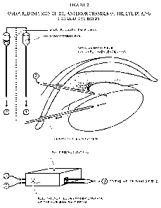
[0016] In one embodiment the coloration is provided during infusion in
phacoemulsification
surgery.
1
CA 02890056 2015-04-30
WO 2014/072831 PCT/1B2013/003099
[0017] In another embodiment the coloration is provided for irrigation of the
anterior segment
of the eye during phacoemulsification surgery.
[0018] In another embodiment several dyes will alternate during various step
of
phacoemulsification in particular for Hydro dissection lens nucleus.
[0019] In another embodiment each one of the dye is visualized with a suitable
filter during the
surgery.
[0020] In another embodiment fluorescent viscoelastic gel is provided in
intraocular lens
implant.
[0021] In another embodiment the fluorescent viscoelastic gel is provided
during surgery for
traumatic injury to the anterior segment of the eye.
[0022] In another embodiment the fluorescent viscoelastic gel is provided
suring ciliary
sclerotomy for the treatment of presbiopia.
[0023] In another embodiment the fluorescent viscoelastic gel is provided
during glaucoma
surgery.
[0024] One embodiment provides for illuminating the field of surgery with a
portable
microfibroscope providing monochromatic light, wherein the light of the
microfibroscope is
generated by fiber-optic.
[0025] In another embodiment the fiber-optic is incorporated in the surgery
instrument.
[0026] In one embodiment the surgery instrument allows for anterior chamber
eye surgery.
[0027] In another embodiment the monochromatic light is white.
[0028] In yet another embodiment the monochromatic light is blue.
INCORPORATION BY REFERENCE
[0029] All publications, patents, and patent applications mentioned in this
specification are
herein incorporated by reference to the same extent as if each individual
publication, patent, or
patent application was specifically and individually indicated to be
incorporated by reference.
BRIEF DESCRIPTION OF DRAWINGS
[0030] Figure 1 illustrates the use of a microfibroscope for anterior chamber
eye surgery.
[0031] Figure 2 presents the use of colored infusion liquid (BSS) during
cataract surgery.
[0032] Figure 3 describes the use of micro eye endoscopy with the
microfibroscope for
aspiration of colored viscoelastic gel from the anterior chamber of the eye.
[0033] Figure 4 demonstrates addition of fluorescent viscoelastic gel in a
tubular mode,
wherein the external coating is the fluorescent BSS.
[0034] Figures 5A and 5B illustrates the wave mode application of fluorescent
viscoelastic gel.
2
CA 02890056 2015-04-30
WO 2014/072831 PCT/1B2013/003099
DETAILED DESCRIPTION OF THE INVENTION
[0035] There are a number of ophthalmic musculoskeletal and nerve surgical
procedures
performed by skilled surgeons which require or are facilitated by the use of a
viscoelastic
medium.
[0036] The anterior chamber of the eye is filled with a circulating liquid
called aqueous humor
or aqueous, whereas its posterior chamber is filled with vitreous humor or
vitreous. The
endothelial cell layer of the cornea is easily damaged and, once lost, these
cells do not
regenerate. The surgical procedures used in cataract surgery, corneal
transplants and other types
of ophthalmic surgery are likely to result in damage to these delicate cells
unless measures are
taken to protect them in the manner in which the aqueous does naturally.
[0037] In ophthalmic surgical procedures, except for non penetrating
keratoplasty in which the
corneal tissue is not fully penetrated, the recommended practice is to use an
intraocular
viscoelastic fluid for protecting the inner endothelial corneal surface and
the delicate inner eye
structures. Solutions that have been used in ophthalmologic surgical
irrigation include normal
saline, lactated Ringer's solution and Hartmann's lactated Ringer's solution,
but these are not
optimal due to potential unfavorable corneal and endothelial effects. Other
aqueous solutions
that include agents such as electrolytes, buffering agents for pH adjustment,
glutathione and/or
energy sources such as dextrose, better protect the tissues of the eye, but do
not address other
physiologic processes associated with surgery. One commonly used solution for
ophthalmologic
irrigation is a two part buffered electrolyte and glutathione solution
disclosed in U.S. Pat. No.
4,550,022 to Garabedian et al., the disclosure of which is hereby expressly
incorporated by
reference. The two parts of this solution are mixed just prior to
administration to ensure stability.
These solutions are formulated with a goal of maintaining the health of ocular
tissues during
surgery.
[0038] Of the several substances that have been developed as substitutes for
aqueous and
vitreous, both as a protective layer covering the endothelial cells and as a
coating on the surgical
instruments and implanted material, sodium hyaluronate extracted from rooster
combs, mixtures
thereof or bioengineered forms of the naturally-occurring substance are widely
employed. Once
the surgical procedure is completed, the remaining vitreous/aqueous substitute
is aspirated from
the site using a syringe while remaining amounts are merely reabsorbed by the
body in time
without ill effects.
[0039] Methylcellulose has a long history of safe and effective use for
ophthalmic applications.
In 1945, Dr. Kenneth C. Swan studied the effects of methylcellulose on the
ocular tissues of
rabbit eyes. He suggested its use as a vehicle for ophthalmic drugs, to treat
keratoconjunctivitis
sicca and as an emollient. Then in 1959, Flemming, Merrill and Girard reported
on further
3
CA 02890056 2015-04-30
WO 2014/072831 PCT/1B2013/003099
studies of methylcellulose in relation to irritation, hypersensitivity and its
outflow from the
anterior chamber of the rabbit eye.
[0040] The first reported use of methylcellulose as an intraocular lens
coating serving to protect
the corneal endothelium in rabbits was made by Drs. Kaufman and Katz in 1976.
In the
following year Dr. Paul Fechner reported upon the first human clinical use of
methylcellulose to
coat an intraocular lens prior to implantation.
[0041] Then in November of 1982, Dr. Danielle Aron-Rosa reported using
methylcellulose in
extracapsular surgery instead of high molecular weight sodium hyaluronate
extracted from
rooster combs which is very expensive. Shortly thereafter, Dr. Fechner
amplified upon his
earlier findings describing the use of methylcellulose as an intraocular
viscous cushioning
material in ophthalmic surgery.
[0042] The composition of the viscoelastic mixed gel slurries can vary within
broad limits. The
polymer solution in the mixture can constitute from 0.1 to 99.5%, preferably,
from 0.5 to 99%,
more preferably, from 1 to 95%, the rest being the gel phase. The choice of
the proper
composition of the mixture depends on the properties and composition of the
two components
and is governed by the desirable properties of the slurry and its final use.
[0043] The viscoelastic gel with varied density is used to protect the cornea
by maintaining
constant volume of the anterior chamber in place of the Aqueous humor. The
surgical
procedures using the phacoemulsification or small incision technique is
performed in modern
cataract surgery. Phacoemulsification surgery involves the use of a machine
with
microprocessor-controlled fluid dynamic. The phaco probe is a sophisticated
microscopic,
ultrasonic jack hammer which vibrates thousands at ultrasonic frequency
pulverizes and
liquidizes the cloudy cataract material. As the phacoemulsification probe is
hollow, the debris
created by this technique is aspirated through the tube of the
phacoemulsification probe and led
into a disposable chamber. Following complete removal of all the cataract
material; the
periphery of the capsular bag often has remnants left behind which are cleaned
in an intervening
stage called 'I-A' which stands for 'irrigation-aspiration' further
viscoelastic gel is injected to
the eye.
[0044] However, the disadvantage of using viscoelastic gel is the transparency
of the gel and
the difficulty to visualize the presence of the gel after surgery and the
decrease of the
transparency of the operative area; the use of a colored dye can alleviate
these issues, however,
such a dye should not decrease the transparency in the surgery; the color of
the dye would allow
for seeing the flows.
4
CA 02890056 2015-04-30
WO 2014/072831 PCT/1B2013/003099
[0045] As disclosed herein, the combination of a dye and the monochromatic
light would make
it possible to intensify the visualization, for example the fluorescein
diluted in the liquid infusion
will be more visible on the blue light, filter that can easily be interposed
at the source.
[0046] Lighting of the anterior chamber of the eye during surgery may depend
on the
microscope employed in the surgery and the retro lighting known as a pupillary
gleam. To have
effective lighting, the pupil needs to be dilated and the lighting center
needs to be positioned in
the visual center, which causes the phenomena of Purkinje. The system can be
sophisticated
comprising, for example, the addition of a fiber-optic connected to the
infusion probe, this
microfibroscope with either a white or blue light source illuminates with a
tangential beam of
light. Figure 1 is a representation of the illumination field using the
microfibroscope in the
anterior chamber, allowing to clarify the flow of liquid from the inside of
the probe.
[0047] The fiber-optic can also be added onto the probe of emulsification to
view the suction of
the liquid and the masses.
[0048] The variation in density of the dye also allows modulating the effect
obtained with the
dye into the anterior chamber helping to visualize the surgery site. A pulsed
mode will allow for
waves of dye, alternating clear phases and dense phases. The final washing of
the anterior
chamber will eliminate all of the dye. Several dyes can be alternatively used
during surgery. A
suitable filter downstream of the microscope will help to visualize the dye.
The colorant will be
biocompatible with the anterior segment of the eye with no toxicity.
[0049] A double electrical gallows controls the vials of the infusion liquid
(BSS) which has a
composition similar to that of aqueous humor and dyes independently: the
height of each vial
defines the density of the dye in the irrigation solution. The release of the
alternate colors,
lighting and fiber optic will be programed and controlled by solenoid valves
allowing the
irrigation solution in the anterior chamber of the eye. The visualization of
the flow at the output
of the infuser, its density, the laminar or turbulent aspect allows to better
use the surgical tool
and maximize the effectiveness during the surgery procedure.
[0050] Illustrations of the methods disclosed herein are further illustrated
by the appended
fogures. Figure 2, illustrates the use of a microfibroscope during cataract
surgery, during the
PHAKO infusion, emulsification or aspiration. Figure 3 describes the use of
micro eye
endoscopy with the microfibroscope for aspiration of colored viscoelastic gel
from the anterior
chamber of the eye. The suction flow will also be viewed by its appearance,
allowing to access
any pre occlusion with crystalline or any reflux in case of occlusion.
[0051] The microfibroscope as an instrument with fiber-optics which conveys
light may be
advantageously employed in any type of surgery helping the surgeon avoid the
zones of shades
during the surgery.
CA 02890056 2015-04-30
WO 2014/072831 PCT/1B2013/003099
[0052] The viscoelastic gel mixture according to the invention, contains a
fluorophore in
addition to the two major components namely, the polymeric gel slurry and the
polymer
solution.
[0053] One preferred fluorophore used with the viscoelastic gel is
fluorescein, a synthetic
organic compound. Fluorescein was first synthesized by Adolf von Baeyer in
1871; the sodium
salt of fluorescein, is used extensively as a diagnostic tool in the field of
ophthalmology and
optometry, where topical fluorescein is used in the diagnosis of corneal
abrasions, corneal ulcers
and herpetic corneal infections. It is also used in rigid gas permeable
contact lens fitting to
evaluate the tear layer under the lens. It is available as sterile single-use
sachets containing lint-
free paper applicators soaked in fluorescein sodium. Intravenous or oral
fluorescein is used in
fluorescein angiography in research and to diagnose and categorize vascular
disorders in e.g.
legs, including retinal disease macular degeneration, diabetic retinopathy,
inflammatory
intraocular conditions, and intraocular tumors, and, increasingly, during
surgery for brain
tumors.
[0054] The fluorescence yield of the fluorescein molecule is very high and
excitation occurs at
494 nm and emission at 521 nm for "Fluorescein sodium"; this allows for the
detection of
fluorescein at a very low concentration. The green fluorescence is detected
under Ultraviolet
light, using a blue emission filter (red-free).
[0055] The use of a fluorescent dye with commercially available viscoelastic
gel will allow to
visualize the smallest amount of the gel. In the case that the viscoelastic
gel is colored, the
presence of fluorescein will be easily detected using a blue emission filter
placed downstream of
the light source from the microscope. In operations such as iridio ciliary
angle and space of
implant of post capsule, where it is generally difficult to control and
evaluate the presence of
viscoelastic gel, the gel with fluorescein will reduce or eliminate such
complications.
[0056] After using the fluorescent viscoelastic gel, the gel will be reduced
or completely
eliminated after surgery, and this in turn will decrease the risk for
hypertonia , inflammation
after surgery and accelerate visual recovery, resulting in overall less post
operative stress for the
patient.
[0057] For the surgeon the use of the fluorescein viscoelastic gel helps to
visualize the liquid
in the anterior chamber, which allows determination of the volume of the gel
during different
operational steps; aspiration of the gel at the end of the surgery; also
allowing the use of
ultraviolet light during the surgery by using a blue, red-free filter
downstream of the microscope
used for the surgery ( example of a ZEISS microscope); reduction of surgery
time; no major
investment compared to transparent viscoelastic gel, by the addition of a red-
free emission filter.
6
CA 02890056 2015-04-30
WO 2014/072831 PCT/1B2013/003099
[0058] Various products are contemplated within the present disclosure which
can be produced
in different form to be used in different ophthalmologic treatments.
EXAMPLES
[0059] Example 1: Ampule containing the fluorescent viscoealstic gel reserved
for intraocular
lens implant to treat aphakia or other type of refractive disorders using a
phakic ICL.
[0060] All type of viscoelastic gel can be used; dispersive; cohesive or
joint. The name of each
ampule can be clarifying the type of viscoelastic gel.
Visco D fluo : dispersive viscoelastic gel with fluorophore
Visco C fluo : cohesive viscoelastic gel with fluorophore
Visco M fluo : joint (mix) viscoelastic gel with fluorophore
[0061] The concentration of fluoresceine is at minimum and the fluorescence
can just be seen
in the presence of ultraviolet light.
[0062] The final formulation can also be as two separate ampoules; one ampule
being the
viscoalastic gel used for the first step of the surgery the second one is used
for the IOL implant
with the fluorescent dye.
BiviscoC/ D fluo : dispersive viscoelastic gel with fluorophore
Bivisco D/C fluo : cohesive viscoelastic gel with fluorophore
BiviscoC/ M fluo : joint (mix) viscoelastic gel with fluorophore
And so on
[0063] Example 2: A gauge needle with fluorophore dye used with an ampule of
standard
viscoelastic gel. Having the needle with the fluorescein or Trypan blue allows
the use of all
commercial viscoelastic gels.
[0064] Example 3: The walls of a catheter or cannula coated with a film of dry
fluorescein
that will dissolve with the viscoelastic gel during the injection.
[0065] Example 4: An ampule of fluorescein diluted at some preferred
concentration with
viscoelastic gel.
7
