Note: Descriptions are shown in the official language in which they were submitted.
CA 02890706 2015-05-07
WO 2014/074815
PCT/US2013/069141
SMALL MOLECULE INHIBITORS OF MALT1
CROSS-REFERENCE TO RELATED APPLICATIONS
This application claims the priority of U.S. provisional patent application
Serial Number 61/724,650, filed Nov. 9, 2012, the disclosure of which is
incorporated herein by reference in its entirety.
BACKGROUND
Non-Hodgkin lymphoma (NHL) is the 7th most frequent cancer (Siegel et
al., 2012). Diffuse large B-cell lymphoma (DLBCL) is the most common
subtype of NHL accounting for ¨25% of all lymphoma cases (Swerdlow, 2008).
Gene expression profiling allowed subclassification of DLBCL into distinct
molecular subtypes including: germinal center B-cell-like (GCB) DLBCL,
activated B-cell-like (ABC) DLBCL and primary mediastinal B-cell lymphoma
(PMBL) (Alizadeh et al., 2000; Rosenwald et al., 2003). These subtypes differ
significantly in their spectrum of recurrent somatic mutations, dependence on
different signaling pathways and response to current standard therapies (Lenz
et
al., 2008b; Wright et al., 2003). Patients with the GCB subtype have a
significantly better overall survival compared to those with the ABC subtype
(Alizadeh et al., 2000; Rosenwald et al., 2002). Improved therapies are needed
for all DLBCLs but most urgently for ABC-DLBCLs, which are the most
chemo-resistant.
ABC-DLBCL is characterized by its reliance on the oncogenic activation
of the NF-KB pathway through several different mechanisms. These mostly
involve somatic mutations in molecules participating in signaling downstream
of
the B-cell receptor (BCR) including: activating mutations of CARMAl/CARD 11
(Lenz et al., 2008a) and CD79A/B (Davis et al., 2010), homozygous
deletion/inactivating mutations of TNFAIP3/A20 (Compagno et al., 2009;
Honma et al., 2009) or activating mutations of MYD88 downstream of the Toll-
like receptor (Ngo et al., 2011). CARMA1 forms part of the CBM complex
(CARMA1-BCL10-MALT1) and mediates NF-KB activation downstream of the
B-cell receptor, T-cell receptor (Ruefli-Brasse et al., 2003; Ruland et al.,
2003)
and ITAM-coupled NK cell receptors (Gross et al., 2008). The MALT1 subunit
is the active signaling component of the CBM complex (Lucas et al., 2001) and
1
CA 02890706 2015-05-07
WO 2014/074815
PCT/US2013/069141
features protease activity that cleaves and inactivates inhibitors of the NF-
KB
signaling pathway such as TNFAIP3/A20 (Coornaert et al., 2008), CYLD (Staal
et al., 2011) and RELB (Hailfinger et al., 2011) or the BCL10 protein (Rebeaud
et al., 2008), indirectly activating NF-KB signaling. MALT1 translocations
(t(11;18)(q21;q21) which produces an APP-MALT1 fusion and the
t(14;18)(q32;q21) that results in the IGH-MALT] translocation) are detected in
up to 55% of patients with MALT-type lymphomas (Farinha and Gascoyne,
2005). This translocations lead to overexpression of MALT] and, in the case of
the API2-MALT1 translocation, constitutive activation of the pathway
(Dierlamm et al., 1999; Sanchez-Izquierdo et al., 2003; Streubel et al.,
2003).
Constitutive expression of MALT1 in mice induces a disease that is similar to
MALT lymphomas in humans, and induces ABC-like DLBCLs in a p53 null
background (Vicente-Duenas et al., 2012). MALT] has not been found mutated
or translocated in DLBCL, but is gained along with BCL2 and this low copy
number amplification is associated with an ABC-DLBCL phenotype (Dierlamm
et al., 2008). Moreover, ABC-DLBCL cell lines have been shown to be
dependent on the MALT1 catalytic activity (Ferch et al., 2009; Hailfinger et
al.,
2009; Ngo et al., 2006).
MALT1 is a paracaspase, related to the caspase (cysteine-aspartic
proteases) family of proteases but which cleaves after arginine or lysine
residues
instead of aspartate (Rebeaud et al., 2008). MALT1 null animals display
defects
in B and T cell function but are otherwise healthy (Ruefli-Brasse et al.,
2003;
Ruland et al., 2003), and MALT1 is the only paracaspase in the human genome.
These factors suggest that MALT1 targeted therapy would likely be well
tolerated with little or manageable toxicity. Consequently, MALT1 represents a
potentially important therapeutic target for ABC-DLBCL and MALT
lymphoma.
SUMMARY
MALT1 is a unique paracaspase protein that transduces aberrant
oncogenic signaling in ABC-DLBCL. The inventors disclose herein the
development of a constitutively activated form of MALT1 that enabled a screen
for small molecule inhibitors, and claim MALT1 inhibitory compounds and their
use for treatment of medical disorders such as B-cell lymphomas. The
2
CA 02890706 2015-05-07
WO 2014/074815
PCT/US2013/069141
compound MI-2, an irreversible MALT1 protease inhibitor, was identified as a
lead compound with nanomolar activity in cell-based assays and selective
activity against ABC-DLBCLs. Importantly we show that MALT1 inhibitors kill
ABC-DLBCLs in vitro and in vivo, are non-toxic to animals and also suppress
primary human non GCB-DLBCL specimens. Hence we demonstrate that
MALT1 is a bona fide therapeutic target, and provide a lead compound that
forms the basis of a new class of therapeutic agents for B-cell lymphomas.
The invention provides, in various embodiments, a method of modulating
MALT1, comprising contacting MALT1 with an effective amount or
concentration of a compound of formula (I)
__
y1 Arl
'' '
0 ---------< --..----...- \ 2
,
,
R1 N . 3 0õ,õõõ,..--
..õ....................,
Y Ar2
,
,
,
,
,
Ar3 (I)
wherein
a dashed bond indicates that a bond can be present or absent;
when a double bond is present between Y1 and Y2, Y1 is N or CR, Y2 is
C, and Ari is present; when a single bond is present between Y1 and Y2, Y1 is
CR2, Y2 is 0 or S, and Ari is absent, and each independently selected R is H
or
(C1-C6)alkyl;
20R1 =
is alkyl, alkoxyalkyl, or arylalkyl, wherein any alkyl, alkoxyalkyl, or
arylalkyl, can be mono- or independently multi-substituted with halo or (C1-
C6)alkoxy, provided that when a double bond is present between the oxygen
atom and the ring comprising Y3, R1 is absent and Ar3 is present, and when a
single bond is present between the oxygen atom and the ring, R1 is present, a
double bond between Y3 and the carbon atom bearing the oxygen atom is
present, and Ar3 is absent;
Ari is phenyl substituted with 1-3 J1 groups; J1 is halo or (C1-C6)alkoxy;
3
CA 02890706 2015-05-07
WO 2014/074815
PCT/US2013/069141
Ar2 is phenyl substituted with 1-3 J2 groups; J2 is a group of formula
-N(R)C(0)-R2 and R2 is alkyl, aryl, or arylamino, wherein any alkyl, aryl, or
arylamino is substituted with 0-2 halo, nitro, or (C1-C6)alkoxy groups;
Ar3 is phenyl substituted with 1-3 J3 groups; J3 is halo or (C1-C6)alkoxy;
or any salt, hydrate, tautomer, or stereoisomer thereof.
The invention further provides, in various embodiments, a method of
treating or preventing cancer comprising administering to a patient an
effective
dose of a compound of formula (I) as defined above. More specifically, the
cancer can be a lymphoma, such as a diffuse large B-cell lymphoma (DLBCL).
The invention further provides, in various embodiments, a method of
identifying a small molecule modulator of MALT1, comprising contacting a
recombinant form of MALT1 (340-789) fused with a leucine zipper dimerization
motif (LZ-MALT1) and a candidate modulator compound, using the MALT1
substrate peptide LRSR linked to the fluorogen AMC (7-amino-4-
methylcoumarin), such that cleavage of the Ac-LRSR-AMC substrate by
MALT1 results in release of AMC and a fluorescent signal, wherein a decrease
in the cleavage of the Ac-LRSR-AMC substrate by the recombinant form of
MALT1 in the presence of the candidate modulator indicates that the candidate
modulator is a small molecule modulator of MALT1.
BRIEF DESCRIPTION OF THE FIGURES
Figure lA depicts two perspective views of the structure of a
recombinant form of MALT1 (340-789) fused with a leucine zipper dimerization
motif (LZ-MALT1), which promotes its dimerization and activation.
Figure 1B is a graphic representation of the results by which 324
candidate compounds were selected from a compound library for validation in a
concentration response assay using LZ-MALT1
Figure 1C is a graphic representation of the results by which nineteen
compounds were selected for further validation based on their biochemical
activity (IC50 <20 [iM).
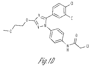
Figure 1D shows the chemical structure of compound MI-2.
Figure lE shows a photograph of a Western blot of gel electrophoresis
results demonstrating that MI-2 caused a dose-dependent decrease in MALT1-
mediated cleavage, noted by an increase in the uncleaved CYLD protein and a
4
CA 02890706 2015-05-07
WO 2014/074815
PCT/US2013/069141
decrease of the cleaved form of the protein as shown in the graphic
representation of the Western blot data.
Figure 2A is a graphic representation of the results by which nineteen
analogs displaying equal or higher activity than MI-2 were selected.
Figure 2B shows the chemical structures of five analogs (MI-2A1
through MI-2A5) of MI-2 with biochemical IC50s within a similar range as MI-2
selected for further characterization in cell proliferation assays and two
analog
compounds with no LZ-MALT1 inhibitory activity in vitro (MI-2A6 and MI-
2A7) used as chemical controls that had no effect on cell proliferation over
the
same dose range.
Figure 2C is a graphic representation of the results of bioassays of
compounds MI-2A1 through MI-2A5.
Figure 2D is a graphic representation of the results obtained from the five
compounds MI-2A1 through MI-2A5, administered at 5 [LM for 8 hr, with
respect to cleavage inhibition, with the Z-VRPR-FMK MALT1 blocking peptide
(50 [LM) used as positive control.
Figure 3A is a Heteronuclear Single Quantum Coherence (HSQC)
Nuclear Magnetic Resonance (NMR) spectrogram of MI-2 binding the
paracaspase domain of MALT1 (residues 329-728).
Figure 3B shows NMR spectrograms evidencing the absence of binding
of the paracaspase domain of MALT1 (residues 329-728).by the inactive
analogs MI-2A6 and MI-2A7.
Figure 3C shows mass spectometric data indicating that the MALT1
paracaspase domain (329-728) presented a major peak at 55,988.4 Da, and that
upon incubation with compound MI-2, the major peak of MALT1 was shifted to
56,407.5 Da, an increase of 419.1 Da.
Figure 3D shows an image of the potential mode of binding of MI-2 to
the MALT1 paracaspase domain, as calculated by the molecular docking routine
of molecular modeling program AutoDock 4.2, wherein MI-2 appears to bind
the active site cleft with its chloromethyl group close to the active site
C464 in
the paracaspase domain.
Figure 3E shows the time course of enzymatic activity when LZ-
MALT1 was pre-incubated with different concentrations of MI-2 (irreversible
5
CA 02890706 2015-05-07
WO 2014/074815
PCT/US2013/069141
inhibition) versus MI-2A2 (reversible inhibition) for 5 to 80 minutes followed
by
addition of the fluorescent reporter substrate Ac-LRSR-AMC.
Figure 4A shows a photograph of a Western blot of gel electrophoresis
results using proteasome inhibitor MG-132 to facilitate visualization of
cleavage
products in HBL-1 and TMD8 cell lines exposed to either MI-2 (2 [iM) or
vehicle, for 30 minutes followed by 5 [iM MG-132 for an additional one (lanes
2,3), or two hour (lanes 4, 5) in order to allow cleaved forms of MALT1
substrates to accumulate during exposure to MI-2.
Figure 4B shows results of experiments wherein HBL-1 cells were
exposed to 200 nM MI-2, 50 [iM Z-VRPR-FMK (positive control) or vehicle for
24 hr, followed by c-REL flow cytometry of whole cells or isolated nuclei.
Both
MI-2 and Z-VRPR-FMK reduced nuclear c-REL to a similar extent, without
affecting whole cell levels of this protein.
Figure 4C shows Western blots for c-REL and p65 in nuclear extracts of
HBL-1 and TMD8 cells treated for 24 hr with GI50 concentrations of MI-2 (200
nM for HBL-1 and 500 nM for TMD8). In both cell lines exposure to MI-2
caused a clear reduction of nuclear c-REL while it did not affect p65 levels.
Figure 4D is a graphical representation of data on the effect of MI-2 on
attenuating NF-KB activation induced by PMA/ionomycin, wherein 293T cells
were transfected with the NF-KB reporter vector (NF-KB)5-luc2CP-pGL4 and
TK-pRL control together with plasmids expressing BCL10 and either MALT1 WT
or MALT1 C464A (inactive mutant).
Figure 4E is a graphical representation of data on the effect of MI-2 on
attenuating NF-KB activation induced by PMA/ionomycin, wherein HBL-1 cells
were transfected with the NF-KB reporter vector (NF-KB)5-luc2CP-pGL4 and
TK-pRL control.
Figure 4F shows results of gene set enrichment analysis (GSEA) of the
Z-VRPR-FMK signature against the differential expression of all genes pre-
ranked by fold change between MI-2 and vehicle-treated cells for each cell
line.
The Z-VRPR-FMK signature was significantly enriched among genes
downregulated after MI-2-treatment for both cell lines (HBL-1: FDR<0.0001;
and TMD8: FDR<0.0001).
Figure 5A shows a graphical representation of results from experiments
wherein eight cell lines were exposed to increasing concentrations of MI-2
6
CA 02890706 2015-05-07
WO 2014/074815
PCT/US2013/069141
(single dose) and cell proliferation measured at 48 hr using an ATP-based
metabolic luminescent assay.
Figure 5B is a graphical representation of results of MI-2 intracellular
concentration experiments where HBL-1 cells were exposed to 0.02, 0.2 or 2
1..i1VI
MI-2 for 2 hr, washed three times, and MI-2 measured by LC-MS.
Figure 5C1, 5C2, and 5C3, show results of experiments wherein HBL-1,
TMD8, OCI-Ly10 and the GCB-DLBCL cell line OCI-Lyl were treated with
increasing concentrations of MI-2. Cell proliferation was examined using the
CFSE dilution assay by flow cytometry on viable cells at 48, 72 and 96 h. MI-2
substantially inhibited proliferation in HBL-1, TMD8 and OCI-Ly10 while it did
not affect OCI-Lyl.
Figure 5D shows graphical representation of results of experiments
wherein, using BrdU incorporation - DAPI staining and flow cytometry to assess
cell cycle, it was evident that MI-2 induced a dose-dependent decrease in S
phase, with reciprocal increment in the proportion of cells in G1-0 and Sub-
GO.
Figure 5E shows graphical results of experiments demonstrating that
whereas MI-2 had no effect on OCI-Lyl cells, it profoundly suppressed both
HBL-1 and TMD8 cells, with the former exhibiting earlier and higher abundance
of apoptotic cells.
Figure 6 shows results of experiments wherein five C57BL/6 mice were
exposed to daily intraperitoneal (IP) administration of increasing doses of MI-
2
ranging from 0.05 to 25 mg/kg over the course of 10 days to a cumulative dose
of 51.1 mg/kg and another five mice were exposed to vehicle only (5% DMSO,
n=5) (Figure 6A, Toxicity 1). There was no evidence of lethargy, weight loss
(Figure 6B, Toxicity 1) or other physical indicators of sickness. To ascertain
if
the maximal administered dose of 25 mg/kg is safe in a 14-day schedule, we
exposed ten mice to daily IP administration of 25 mg/kg of MI-2 over 14 days
to
a cumulative dose of 350 mg/kg, using as controls five mice injected with
vehicle only (Figure 6A, Toxicity 2). Five mice were sacrificed after the 14-
day
course of MI-2 administration (together with the 5 controls) and the other 5
mice
were sacrificed after a 10-day washout period to assess delayed toxicity. No
toxic effects or other indicators of sickness, including weight loss (Figure
6B,
Toxicity 2) or tissue damage (macroscopic or microscopic), were noted (Figures
7
CA 02890706 2015-05-07
WO 2014/074815
PCT/US2013/069141
6C1 and 6C2). Brain, heart, lung, liver, kidney, bowel, spleen, thymus and
bone
marrow tissues were examined.
Figure 7A shows graphical data demonstrating that MI-2 profoundly
suppressed the growth of both the TMD8 (p=0.015, t-test) and HBL1 (p=0.014,
t-test) ABC-DLBCL xenografts vs. vehicle, whereas it had no effect on the
growth of the OCI-Lyl tumors (p=0.47, t-test).
Figure 7B shows graphical results of histological examination using the
TUNEL assay to detect apoptotic cells, that showed a significant increase in
apoptotic cells in MI-2-treated HBL-1 (p=0.0008, t-test) and TMD8 (p<0.0001,
t-test) xenografts relative to vehicle but not in OCI-Lyl xenografts
(p=0.5580, t-
test).
Figure 7C shows graphical results of evidence of a significant decrease in
proliferation as measured by Ki-67 staining in HBL-1 (p<0.0001, t-test) and
TMD8 xenografts (p=0.0006, t-test) compared to vehicle, but observed no
difference in OCI-Lyl xenografts (p=1.0, t-test).
Figure 7D shows stained microphotographs indicating that MI-2 treated
tumors exhibited reduced c-REL nuclear protein.
Figure 7E shows graphical data obtained from single cell suspensions
from lymph node biopsies of five DLBCL patients for whom their GCB vs. non-
GCB status could be ascertained by immunohistochemistry using the Hans
criteria, wherein lymphoma cells were isolated and exposed to 0.8 [iM MI-2 or
vehicle in four replicates. After 48 hr exposure, cell number and viability
were
determined using Trypan blue.
DETAILED DESCRIPTION
Overview
In various embodiments, the present invention provides a method of
modulating MALT1, comprising contacting MALT1 with an effective amount or
concentration of a compound of formula (I)
8
CA 02890706 2015-05-07
WO 2014/074815
PCT/US2013/069141
,Arl
yi ,--
- - ...---:"----- 2 -
--, Y
0 \
\
,,' ....... \
,
R1 N
, .......õõ0õ.- ,õ........õ....N.
Y3
Ar2
,
,
,
,
,
Ar3 (I)
wherein
a dashed bond indicates that a bond can be present or absent;
when a double bond is present between Y1 and Y2, Y1 is N or CR, Y2 is
C, and Ari is present; when a single bond is present between Y1 and Y2, Y1 is
CR2, Y2 is 0 or S, and Ari is absent, and each independently selected R is H
or
(C1-C6)alkyl;
R1 is alkyl, alkoxyalkyl, or arylalkyl, wherein any alkyl, alkoxyalkyl, or
arylalkyl, can be mono- or independently multi-substituted with halo or (C1-
C6)alkoxy, provided that when a double bond is present between the oxygen
atom and the ring comprising Y3, R1 is absent and Ar3 is present, and when a
single bond is present between the oxygen atom and the ring, R1 is present, a
double bond between Y3 and the carbon atom bearing the oxygen atom is
present, and Ar3 is absent;
Ari is phenyl substituted with 1-3 J1 groups; J1 is halo or (C1-C6)alkoxy;
Ar2 is phenyl substituted with 1-3 J2 groups; J2 is a group of formula -
N(R)C(0)-R2 and R2 is alkyl, aryl, or arylamino, wherein any alkyl, aryl, or
arylamino is substituted with 0-2 halo, nitro, or (C1-C6)alkoxy groups;
Ar3 is phenyl substituted with 1-3 J3 groups; J3 is halo or (C1-C6)alkoxy;
or any salt, hydrate, tautomer, or stereoisomer thereof.
More specifically, the compound of formula (I) can be a compound of
formula (IA)
R1 N Arl
0.----7 y
\\
N-N\
Ar2 (IA)
9
CA 02890706 2015-05-07
WO 2014/074815
PCT/US2013/069141
wherein R1, Ari, and Ar2 are as defined for the compound of formula (I), or
any
salt, hydrate, tautomer, or stereoisomer thereof.
More specifically, the compound of formula (I) can be a compound of
formula (IB)
0 /------S
N Ar2
Ar3 (IB)
wherein Ar2 and Ar3 are as defined for the compound of formula (I), or any
salt,
hydrate, tautomer, or stereoisomer thereof.
For instance, the compound of formula (I) used to carry out a method of
the invention can be any of
CI
0 CI
N
0 --
--...,.0
N--N1
ili 0
N
H -k-- C I (MI-2)
OCH3
N .0_..../
, \\
N--N
lit 0
N
H O
02N (MI-2A1)
ocH3
N
0 ----
0 N-N
fi 0
N
H fik
02N (MI-2A2)
CA 02890706 2015-05-07
WO 2014/074815
PCT/US2013/069141
F
I.
N
N-N
0 OCH3
* r(N =
H (MI-2A3)
OS
N
H3C0 . 40
HN
0
NO2 (MI-2A4), or
. ocH3
N
, /0---
0 N-N
0
Nd = F
H N
H
F (MI-2A5),
or any salt, hydrate, tautomer, or stereoisomer thereof.
5 For example, in
carrying out a method of the invention, the MALT1 can
be disposed within a living animal, such as when the living animal is a human
being afflicted with cancer, such as a diffuse large B-cell lymphoma.
Accordingly, the invention further provides, in various embodiments, a
method of treating or preventing cancer comprising administering to a patient
an
10 effective dose of
a compound of formula (I) as defined above; e.g., a compound
of formula (I), formula (IA), formula (IB), or any of the specific examples of
compounds that can be used.
For example, the cancer can be a lymphoma, such as a diffuse large B-
cell lymphoma.
15 As used in the
specification and the appended claims, the singular forms
"a," "an" and "the" include plural referents unless the context clearly
dictates
otherwise.
11
CA 02890706 2015-05-07
WO 2014/074815
PCT/US2013/069141
The term "about" as used herein, when referring to a numerical value or
range, allows for a degree of variability in the value or range, for example,
within 10%, or within 5% of a stated value or of a stated limit of a range.
As used herein, "individual" (as in the subject of the treatment) or
"patient" means both mammals and non-mammals. Mammals include, for
example, humans; non-human primates, e.g. apes and monkeys; and non-
primates, e.g. dogs, cats, cattle, horses, sheep, and goats. Non-mammals
include, for example, fish and birds.
The term "disease" or "disorder" or "malcondition" are used
interchangeably, and are used to refer to diseases or conditions wherein MALT1
plays a role in the biochemical mechanisms involved in the disease or
malcondition or symptom(s) thereof such that a therapeutically beneficial
effect
can be achieved by acting on MALT1. "Acting on" MALT1, or "modulating"
MALT1, can include binding to MALT1 and/or inhibiting the bioactivity of
MALT1 and/or allosterically regulating the bioactivity of MALT1 in vivo.
The expression "effective amount", when used to describe therapy to an
individual suffering from a disorder, refers to the amount of a compound of
the
invention that is effective to inhibit or otherwise act on MALT1 in the
individual's tissues wherein MALT1 involved in the disorder is active, wherein
such inhibition or other action occurs to an extent sufficient to produce a
beneficial therapeutic effect.
"Substantially" as the term is used herein means completely or almost
completely; for example, a composition that is "substantially free" of a
component either has none of the component or contains such a trace amount
that any relevant functional property of the composition is unaffected by the
presence of the trace amount, or a compound is "substantially pure" is there
are
only negligible traces of impurities present.
"Treating" or "treatment" within the meaning herein refers to an
alleviation of symptoms associated with a disorder or disease, or inhibition
of
further progression or worsening of those symptoms, or prevention or
prophylaxis of the disease Of disorder, Of curing the disease Of disorder.
Similarly, as used herein, an "effective amount" or a "therapeutically
effective
amount" of a compound of the invention refers to an amount of the compound
that alleviates, in whole or in part, symptoms associated with the disorder or
12
CA 02890706 2015-05-07
WO 2014/074815
PCT/US2013/069141
condition, or halts or slows further progression or worsening of those
symptoms,
or prevents or provides prophylaxis for the disorder or condition. In
particular, a
"therapeutically effective amount" refers to an amount effective, at dosages
and
for periods of time necessary, to achieve the desired therapeutic result. A
therapeutically effective amount is also one in which any toxic or detrimental
effects of compounds of the invention are outweighed by the therapeutically
beneficial effects.
Phrases such as "under conditions suitable to provide" or "under
conditions sufficient to yield" or the like, in the context of methods of
synthesis,
as used herein refers to reaction conditions, such as time, temperature,
solvent,
reactant concentrations, and the like, that are within ordinary skill for an
experimenter to vary, that provide a useful quantity or yield of a reaction
product. It is not necessary that the desired reaction product be the only
reaction
product or that the starting materials be entirely consumed, provided the
desired
reaction product can be isolated or otherwise further used.
By "chemically feasible" is meant a bonding arrangement or a compound
where the generally understood rules of organic structure are not violated;
for
example a structure within a definition of a claim that would contain in
certain
situations a pentavalent carbon atom that would not exist in nature would be
understood to not be within the claim. The structures disclosed herein, in all
of
their embodiments are intended to include only "chemically feasible"
structures,
and any recited structures that are not chemically feasible, for example in a
structure shown with variable atoms or groups, are not intended to be
disclosed
or claimed herein.
An "analog" of a chemical structure, as the term is used herein, refers to
a chemical structure that preserves substantial similarity with the parent
structure, although it may not be readily derived synthetically from the
parent
structure. A related chemical structure that is readily derived synthetically
from
a parent chemical structure is referred to as a "derivative."
When a substituent is specified to be an atom or atoms of specified
identity, "or a bond", a configuration is referred to when the substituent is
"a
bond" that the groups that are immediately adjacent to the specified
substituent
are directly connected to each other in a chemically feasible bonding
configuration.
13
CA 02890706 2015-05-07
WO 2014/074815
PCT/US2013/069141
All single enantiomer, diastereomeric, and racemic forms of a structure
are intended, unless a particular stereochemistry or isomeric form is
specifically
indicated. In several instances though an individual stereoisomer is described
among specifically claimed compounds, the stereochemical designation does not
imply that alternate isomeric forms are less preferred, undesired, or not
claimed.
Compounds used in the present invention can include enriched or resolved
optical isomers at any or all asymmetric atoms as are apparent from the
depictions, at any degree of enrichment. Both racemic and diastereomeric
mixtures, as well as the individual optical isomers can be isolated or
synthesized
so as to be substantially free of their enantiomeric or diastereomeric
partners,
and these are all within the scope of the invention.
A "small molecule" refers to an organic compound, including an
organometallic compound, of a molecular weight less than about 2 kDa, that is
not a polynucleotide, a polypeptide, a polysaccharide, or a synthetic polymer
composed of a plurality of repeating units.
As to any of the groups described herein, which contain one or more
substituents, it is understood that such groups do not contain any
substitution or
substitution patterns which are sterically impractical and/or synthetically
non¨
feasible. In addition, the compounds of this disclosed subject matter include
all
stereochemical isomers arising from the substitution of these compounds.
As used herein, the terms "stable compound" and "stable structure" are
meant to indicate a compound that is sufficiently robust to survive isolation
to a
useful degree of purity from a reaction mixture, and formulation into an
efficacious therapeutic agent. Only stable compounds are contemplated herein.
When a group is recited, wherein the group can be present in more than a
single orientation within a structure resulting in more than single molecular
structure, e.g., a carboxamide group C(=0)NR, it is understood that the group
can be present in any possible orientation, e.g., X-C(=0)N(R)-Y or X-
N(R)C(=0)-Y, unless the context clearly limits the orientation of the group
within the molecular structure.
The inclusion of an isotopic form of one or more atoms in a
molecule that is different from the naturally occurring isotopic distribution
of the
atom in nature is referred to as an "isotopically labeled form" of the
molecule.
All isotopic forms of atoms are included as options in the composition of any
14
CA 02890706 2015-05-07
WO 2014/074815
PCT/US2013/069141
molecule, unless a specific isotopic form of an atom is indicated. For
example,
any hydrogen atom or set thereof in a molecule can be any of the isotopic
forms
of hydrogen, i.e., protium (1H), deuterium (2H), or tritium (3H) in any
combination. Similarly, any carbon atom or set thereof in a molecule can be
any
of the isotopic form of carbons, such as 11C, 12C, 13C, or , 14u¨ or any
nitrogen
atom or set thereof in a molecule can be any of the isotopic forms of
nitrogen,
such as 13N, , 14¨IN 1
or 5N. A molecule can include any combination of isotopic
forms in the component atoms making up the molecule, the isotopic form of
every atom forming the molecule being independently selected. In a multi-
molecular sample of a compound, not every individual molecule necessarily has
the same isotopic composition. For example, a sample of a compound can
include molecules containing various different isotopic compositions, such as
in
a tritium or 14C radiolabeled sample where only some fraction of the set of
molecules making up the macroscopic sample contains a radioactive atom. It is
also understood that many elements that are not artificially isotopically
enriched
themselves are mixtures of naturally occurring isotopic forms, such as 14N and
15N, 32S and 34, and so forth. A molecule as recited herein is defined as
including isotopic forms of all its constituent elements at each position in
the
molecule. As is well known in the art, isotopically labeled compounds can be
prepared by the usual methods of chemical synthesis, except substituting an
isotopically labeled precursor molecule. The isotopes, radiolabeled or stable,
can be obtained by any method known in the art, such as generation by neutron
absorption of a precursor nuclide in a nuclear reactor, by cyclotron
reactions, or
by isotopic separation such as by mass spectrometry. The isotopic forms are
incorporated into precursors as required for use in any particular synthetic
route.
For example, 14C and 3H can be prepared using neutrons generated in a nuclear
reactor. Following nuclear transformation, 14C and 3H are incorporated into
precursor molecules, followed by further elaboration as needed.
In general, "substituted" refers to an organic group as defined herein in
which one or more bonds to a hydrogen atom contained therein are replaced by
one or more bonds to a non-hydrogen atom such as, but not limited to, a
halogen
(i.e., F, Cl, Br, and I); an oxygen atom in groups such as hydroxyl groups,
alkoxy groups, aryloxy groups, aralkyloxy groups, oxo(carbonyl) groups,
carboxyl groups including carboxylic acids, carboxylates, and carboxylate
esters;
CA 02890706 2015-05-07
WO 2014/074815
PCT/US2013/069141
a sulfur atom in groups such as thiol groups, alkyl and aryl sulfide groups,
sulfoxide groups, sulfone groups, sulfonyl groups, and sulfonamide groups; a
nitrogen atom in groups such as amines, hydroxylamines, nitriles, nitro
groups,
N-oxides, hydrazides, azides, and enamines; and other heteroatoms in various
other groups. Non-limiting examples of substituents J1, J2, and J3 that can be
bonded to a substituted carbon (or other) atom include F, Cl, Br, I, OR',
OC(0)N(R')2, CN, NO, NO2, 0NO2, azido, CF3, OCF3, W, 0 (oxo), S (thiono),
methylenedioxy, ethylenedioxy, N(R)2, SR', SOR', SO2R', SO2N(W)2, SO3R',
C(0)R', C(0)C(0)R', C(0)CH2C(0)R', C(S)W, C(0)OR', OC(0)R', C(0)N(R)2,
OC(0)N(R')2, C(S)N(R)2, (CH2)0_2N(W)C(0)R', (CH2)0_2N(W)N(W)2,
N(R)N(R)C(0)R, N(R)N(R)C(0)OR, N(R)N(R)CON(R)2, N(W)S02R',
N(W)S02N(W)2, N(W)C(0)OR', N(R)C(0)R, N(R)C(S)R, N(W)C(0)N(W)2,
N(R')C(S)N(R')2, N(COR')COR', N(OR')R', C(NH)N(R)2, C(0)N(OR)R, or
C(=NOR')W wherein R' can be hydrogen or a carbon-based moiety, and wherein
the carbon-based moiety can itself be further substituted; for example,
wherein
R' can be hydrogen, alkyl, acyl, cycloalkyl, aryl, aralkyl, heterocyclyl,
heteroaryl, or heteroarylalkyl, wherein any alkyl, acyl, cycloalkyl, aryl,
aralkyl,
heterocyclyl, heteroaryl, or heteroarylalkyl; or wherein two R' groups bonded
to
a nitrogen atom or to adjacent nitrogen atoms can together with the nitrogen
atom or atoms form a heterocyclyl, which can be mono- or independently multi-
substituted with J.
In various embodiments, J1, J2, and J3 can each independently be halo,
nitro, cyano, OR, NR'2, or R', or is C(0)OR', C(0)NR'2, OC(0)OR',
OC(0)NR'2, N(R')C(0)OR', N(R')C(0)NR'2 or thio/thiono analogs thereof.
By "thio/thiono analogs thereof', with respect to a group containing an 0, is
meant that any or all 0 atoms in the group can be replaced by an S atom; e.g.,
for group C(0)0R, a "thio/thiono analog thereof' includes C(S)OR, C(0)SR,
and C(S)SR; e.g., for group OC(0)NR2, a "thio/thiono analog thereof' includes
SC(0)NR2, OC(S)NR2, and SC(S)NR2; and so forth.
In various embodiments, J1, J2, and J3 is any of halo, (C1-C6)alkyl, (C1-
C6)alkoxy, (C1-C6)halo alkyl, hydroxy(C1-C6)alkyl, alkoxy(C1-C6)alkyl, (C1-
C6)alkanoyl, (C1-C6)alkanoyloxy, cyano, nitro, azido, R'2N, R2NC(0),
R'2NC(0)0, R'2NC(0)NR, (C1-C6)alkenyl, (C1-C6)alkynyl, (C6-C10)aryl,
(C6-C10)aryloxy, (C6-C10)aroyl, (C6-C10)aryl(C1-C6)alkyl, (C6-C10)aryl(C1-
16
CA 02890706 2015-05-07
WO 2014/074815
PCT/US2013/069141
C6)alkoxy, (C6-C10)aryloxy(C1-C6)alkyl, (C6-C10)aryloxy(C1-C6)alkoxy, (3-
to 9-membered)heterocyclyl, (3- to 9-membered)heterocyclyl(C1-C6)alkyl, (3-
to 9-membered)heterocyclyl(C1-C6)alkoxy, (5- to 10-membered)heteroaryl, (5-
to 10-membered)heteroaryl(C1-C6)alkyl, (5- to 10-membered)heteroaryl(C1-
C6)alkoxy, or (5- to 10-membered)heteroaroyl. For example, R' independently
at each occurrence can be H, (C1-C6)alkyl, or (C6-C10)aryl, wherein any alkyl
or aryl group is substituted with 0-3 J.
When a substituent is monovalent, such as, for example, F or Cl, it is
bonded to the atom it is substituting by a single bond. When a substituent is
more than monovalent, such as 0, which is divalent, it can be bonded to the
atom it is substituting by more than one bond, i.e., a divalent substituent is
bonded by a double bond; for example, a C substituted with 0 forms a carbonyl
group, C=0, which can also be written as "CO", "C(0)", or "C(=0)", wherein
the C and the 0 are double bonded. When a carbon atom is substituted with a
double-bonded oxygen (=0) group, the oxygen substituent is termed an "oxo"
group. When a divalent substituent such as NR' is double-bonded to a carbon
atom, the resulting C(=NR') group is termed an "imino" group. When a divalent
substituent such as S is double-bonded to a carbon atom, the results C(=S)
group
is termed a "thiocarbonyl" or "thiono" group.
Alternatively, a divalent substituent such as 0 or S can be connected by
two single bonds to two different carbon atoms. For example, 0, a divalent
substituent, can be bonded to each of two adjacent carbon atoms to provide an
epoxide group, or the 0 can form a bridging ether group, termed an "oxy"
group,
between adjacent or non-adjacent carbon atoms, for example bridging the 1,4-
carbons of a cyclohexyl group to form a [2.2.1]-oxabicyclo system. Further,
any
substituent can be bonded to a carbon or other atom by a linker, such as
(CH2)n
or (CR'2)õ wherein n is 1, 2, 3, or more, and each R' is independently
selected.
Another divalent substituent is an alkylidene carbon, represented as C=
and signifying that the carbon atom so indicated, which also bears two
additional
groups, is double bonded to a third group. For example, (CH3)2C= indicates an
isopropylidene group bonded to another carbon or nitrogen atom.
C(0) and S(0)2 groups can also be bound to one or two heteroatoms, such as
nitrogen or oxygen, rather than to a carbon atom. For example, when a C(0)
group is bound to one carbon and one nitrogen atom, the resulting group is
17
CA 02890706 2015-05-07
WO 2014/074815
PCT/US2013/069141
called an "amide" or "carboxamide." When a C(0) group is bound to two
nitrogen atoms, the functional group is termed a "urea." When a C(0) is bonded
to one oxygen and one nitrogen atom, the resulting group is termed a
"carbamate" or "urethane." When a S(0)2 group is bound to one carbon and one
nitrogen atom, the resulting unit is termed a "sulfonamide." When a
S(0)2 group is bound to two nitrogen atoms, the resulting unit is termed a
"sulfamide."
Substituted alkyl, alkenyl, alkynyl, cycloalkyl, and cycloalkenyl groups
as well as other substituted groups also include groups in which one or more
bonds to a hydrogen atom are replaced by one or more bonds, including double
or triple bonds, to a carbon atom, or to a heteroatom such as, but not limited
to,
oxygen in carbonyl (oxo), carboxyl, ester, amide, imide, urethane, and urea
groups; and nitrogen in imines, hydroxyimines, oximes, hydrazones, amidines,
guanidines, and nitriles.
Substituted ring groups such as substituted cycloalkyl, aryl, heterocyclyl
and heteroaryl groups also include rings and fused ring systems in which a
bond
to a hydrogen atom is replaced with a bond to a carbon atom. Therefore,
substituted cycloalkyl, aryl, heterocyclyl and heteroaryl groups can also be
substituted with alkyl, alkenyl, and alkynyl groups as defined herein.
When a number of carbon atoms in a group, e.g., an alkyl, alkenyl,
alkynyl, cycloalkyl, aryl, etc., is specified as a range, each individual
integral
number representing the number of carbon atoms is intended. For example,
recitation of a (C1-C4)alkyl group indicates that the alkyl group can be any
of
methyl, ethyl, propyl, isopropyl, butyl, sec-butyl, isobutyl, or tert-butyl.
It is
understood that a specification of a number of carbon atoms must be an
integer.
Alkyl groups include straight chain and branched alkyl groups and
cycloalkyl groups having from 1 to about 20 carbon atoms, and typically from 1
to 12 carbons or, in some embodiments, from 1 to 8 carbon atoms. Examples of
straight chain alkyl groups include those with from 1 to 8 carbon atoms such
as
methyl, ethyl, n-propyl, n-butyl, n-pentyl, n-hexyl, n-heptyl, and n-octyl
groups.
Examples of branched alkyl groups include, but are not limited to, isopropyl,
iso-butyl, sec-butyl, t-butyl, neopentyl, isopentyl, and 2,2-dimethylpropyl
groups. As used herein, the term "alkyl" encompasses n-alkyl, isoalkyl, and
anteisoalkyl groups as well as other branched chain forms of alkyl.
18
CA 02890706 2015-05-07
WO 2014/074815
PCT/US2013/069141
Representative substituted alkyl groups can be substituted one or more
times with any of the groups listed above, for example, amino, hydroxy, cyano,
carboxy, nitro, thio, alkoxy, and halogen groups. Exemplary alkyl groups
include, but are not limited to, straight or branched hydrocarbons of 1-6, 1-
4, or
1-3 carbon atoms, referred to herein as Ci_6alkyl, Ci_4alkyl, and Ci_3alkyl,
respectively. Exemplary alkyl groups include, but are not limited to, methyl,
ethyl, propyl, isopropyl, 2-methyl-l-butyl, 3-methyl-2-butyl, 2-methyl-l-
pentyl,
3-methyl-l-pentyl, 4-methyl-l-pentyl, 2-methyl-2-pentyl, 3-methyl-2-pentyl, 4-
methy1-2-pentyl, 2,2-dimethyl-1-butyl, 3,3-dimethyl-1-butyl, 2-ethyl-l-butyl,
butyl, isobutyl, t-butyl, pentyl, isopentyl, neopentyl, hexyl, etc.
Aryl groups are cyclic aromatic hydrocarbons that do not contain
heteroatoms in the ring. Thus aryl groups include, but are not limited to,
phenyl,
azulenyl, heptalenyl, biphenyl, indacenyl, fluorenyl, phenanthrenyl,
triphenylenyl, pyrenyl, naphthacenyl, chrysenyl, biphenylenyl, anthracenyl,
and
naphthyl groups. In some embodiments, aryl groups contain about 6 to about 14
carbons in the ring portions of the groups. Aryl groups can be unsubstituted
or
substituted, as defined above. Representative substituted aryl groups can be
mono-substituted or substituted more than once, such as, but not limited to, 2-
,
3-, 4-, 5-, or 6-substituted phenyl or 2-8 substituted naphthyl groups, which
can
be substituted with carbon or non-carbon groups such as those listed above.
Aralkyl groups are alkyl groups as defined above in which a hydrogen or
carbon bond of an alkyl group is replaced with a bond to an aryl group as
defined above. Representative aralkyl groups include benzyl and phenylethyl
groups and fused (cycloalkylaryl)alkyl groups such as 4-ethyl-indanyl.
Aralkenyl group are alkenyl groups as defined above in which a hydrogen or
carbon bond of an alkyl group is replaced with a bond to an aryl group as
defined above.
The term "alkoxy" or "alkoxyl" refers to an oxygen atom connected to an
alkyl group, including a cycloalkyl group, as are defined above. Examples of
linear alkoxy groups include but are not limited to methoxy, ethoxy, propoxy,
butoxy, pentyloxy, hexyloxy, and the like. Examples of branched alkoxy
include but are not limited to isopropoxy, sec-butoxy, tert-butoxy,
isopentyloxy,
isohexyloxy, and the like. Exemplary alkoxy groups include, but are not
limited
to, alkoxy groups of 1-6 or 2-6 carbon atoms, referred to herein as
Ci_6alkoxy,
19
CA 02890706 2015-05-07
WO 2014/074815
PCT/US2013/069141
and C2_6alkoxy, respectively. Exemplary alkoxy groups include, but are not
limited to methoxy, ethoxy, isopropoxy, etc.
An alkoxy group can include one to about 12-20 carbon atoms bonded to
the oxygen atom, and can further include double or triple bonds, and can also
include heteroatoms. For example, an allyloxy group is an alkoxy group within
the meaning herein. A methoxyethoxy group is also an alkoxy group within the
meaning herein, as is a methylenedioxy group in a context where two adjacent
atoms of a structures are substituted therewith.
The terms "halo" or "halogen" or "halide" by themselves or as part of
another substituent mean, unless otherwise stated, a fluorine, chlorine,
bromine,
or iodine atom, preferably, fluorine, chlorine, or bromine.
A "haloalkyl" group includes mono-halo alkyl groups, poly-halo alkyl
groups wherein all halo atoms can be the same or different, and per-halo alkyl
groups, wherein all hydrogen atoms are replaced by halogen atoms, such as
fluoro. Examples of haloalkyl include trifluoromethyl, 1,1-dichloroethyl, 1,2-
dichloroethyl, 1,3-dibromo-3,3-difluoropropyl, perfluorobutyl, and the like.
A "haloalkoxy" group includes mono-halo alkoxy groups, poly-halo
alkoxy groups wherein all halo atoms can be the same or different, and per-
halo
alkoxy groups, wherein all hydrogen atoms are replaced by halogen atoms, such
as fluoro. Examples of haloalkoxy include trifluoromethoxy, 1,1-
dichloroethoxy, 1,2-dichloroethoxy, 1,3-dibromo-3,3-difluoropropoxy,
perfluorobutoxy, and the like.
The term "amine" or "amino" includes primary, secondary, and tertiary
amines having, e.g., the formula N(group)3 wherein each group can
independently be H or non-H, such as alkyl, aryl, and the like. Amines include
but are not limited to R'-NH2, for example, alkylamines, arylamines,
alkylarylamines; R'2NH wherein each R is independently selected, such as
dialkylamines, diarylamines, aralkylamines, heterocyclylamines and the like;
and R'3N wherein each R is independently selected, such as trialkylamines,
dialkylarylamines, alkyldiarylamines, triarylamines, and the like. The term
"amine" also includes ammonium ions as used herein.
An "amino" group is a substituent of the form -NH2, -NHR', -NR'2, -
NR'3', wherein each R' is independently selected, and protonated forms of
each,
except for -NR'3', which cannot be protonated. Accordingly, any compound
CA 02890706 2015-05-07
WO 2014/074815
PCT/US2013/069141
substituted with an amino group can be viewed as an amine. An "amino group"
within the meaning herein can be a primary, secondary, tertiary or quaternary
amino group. An "alkylamino" group includes a monoalkylamino,
dialkylamino, and trialkylamino group.
An "ammonium" ion includes the unsubstituted ammonium ion NH4',
but unless otherwise specified, it also includes any protonated or
quaternarized
forms of amines. Thus, trimethylammonium hydrochloride and
tetramethylammonium chloride are both ammonium ions, and amines, within the
meaning herein.
The term "amide" (or "amido") includes C- and N-amide groups, i.e.,
-C(0)NR'2, and ¨NR'C(0)R' groups, respectively. Amide groups therefore
include but are not limited to primary carboxamide groups (-C(0)NH2) and
formamide groups (-NHC(0)H). A "carboxamido" group is a group of the
formula C(0)NR'2, wherein R' can be H, alkyl, aryl, etc.
A "salt" as is well known in the art includes an organic compound such
as a carboxylic acid, a sulfonic acid, or an amine, in ionic form, in
combination
with a counterion. For example, acids in their anionic form can form salts
with
cations such as metal cations, for example sodium, potassium, and the like;
with
ammonium salts such as NH4 or the cations of various amines, including
tetraalkyl ammonium salts such as tetramethylammonium, or other cations such
as trimethylsulfonium, and the like. A "pharmaceutically acceptable" or
"pharmacologically acceptable" salt is a salt formed from an ion that has been
approved for human consumption and is generally non-toxic, such as a chloride
salt or a sodium salt. A "zwitterion" is an internal salt such as can be
formed in a
molecule that has at least two ionizable groups, one forming an anion and the
other a cation, which serve to balance each other. For example, amino acids
such as glycine can exist in a zwitterionic form. A "zwitterion" is a salt
within
the meaning herein. The compounds of the present invention may take the form
of salts. The term "salts" embraces addition salts of free acids or free bases
which are compounds of the invention. Salts can be "pharmaceutically-
acceptable salts." The term "pharmaceutically-acceptable salt" refers to salts
which possess toxicity profiles within a range that affords utility in
pharmaceutical applications. Pharmaceutically unacceptable salts may
nonetheless possess properties such as high crystallinity, which have utility
in
21
CA 02890706 2015-05-07
WO 2014/074815
PCT/US2013/069141
the practice of the present invention, such as for example utility in process
of
synthesis, purification or formulation of compounds of the invention.
"Pharmaceutically or pharmacologically acceptable" include molecular
entities and compositions that do not produce an adverse, allergic or other
untoward reaction when administered to an animal, or a human, as appropriate.
For human administration, preparations should meet sterility, pyrogenicity,
and
general safety and purity standards as required by FDA Office of Biologics
standards.
A "hydrate" is a compound that exists in a composition with water
molecules. The composition can include water in stoichiometric quantities,
such
as a monohydrate or a dihydrate, or can include water in random amounts. As
the term is used herein a "hydrate" refers to a solid form, i.e., a compound
in
water solution, while it may be hydrated, is not a hydrate as the term is used
herein.
In addition, where features or aspects of the invention are described in
terms of Markush groups, those skilled in the art will recognize that the
invention is also thereby described in terms of any individual member or
subgroup of members of the Markush group. For example, if X is described as
selected from the group consisting of bromine, chlorine, and iodine, claims
for X
being bromine and claims for X being bromine and chlorine are fully described.
Moreover, where features or aspects of the invention are described in terms of
Markush groups, those skilled in the art will recognize that the invention is
also
thereby described in terms of any combination of individual members or
subgroups of members of Markush groups. Thus, for example, if X is described
as selected from the group consisting of bromine, chlorine, and iodine, and Y
is
described as selected from the group consisting of methyl, ethyl, and propyl,
claims for X being bromine and Y being methyl are fully described.
If a value of a variable that is necessarily an integer, e.g., the number of
carbon atoms in an alkyl group or the number of substituents on a ring, is
described as a range, e.g., 0-4, what is meant is that the value can be any
integer
between 0 and 4 inclusive, i.e., 0, 1, 2, 3, or 4.
In various embodiments, the compound or set of compounds, such as are
used in the inventive methods, can be any one of any of the combinations
and/or
sub-combinations of the above-listed embodiments.
22
CA 02890706 2015-05-07
WO 2014/074815
PCT/US2013/069141
In various embodiments, a compound as shown in any of the Examples,
or among the exemplary compounds, is provided. Provisos may apply to any of
the disclosed categories or embodiments wherein any one or more of the other
above disclosed embodiments or species may be excluded from such categories
or embodiments.
The compounds described herein for use in a method of the invention can
be prepared in a number of ways based on the teachings contained herein and
synthetic procedures known in the art. In the description of the synthetic
methods described below, it is to be understood that all proposed reaction
conditions, including choice of solvent, reaction atmosphere, reaction
temperature, duration of the experiment and workup procedures, can be chosen
to be the conditions standard for that reaction, unless otherwise indicated.
It is
understood by one skilled in the art of organic synthesis that the
functionality
present on various portions of the molecule should be compatible with the
reagents and reactions proposed. Substituents not compatible with the reaction
conditions will be apparent to one skilled in the art, and alternate methods
are
therefore indicated. The starting materials for the examples are either
commercially available or are readily prepared by standard methods from known
materials. All commercially available chemicals were obtained from Aldrich,
Alfa Aesare, Wako, Acros, Fisher, Fluka, Maybridge or the like and were used
without further purification, except where noted. Dry solvents are obtained,
for
example, by passing these through activated alumina columns.
The present invention further embraces isolated compounds of the
invention. The expression "isolated compound" refers to a preparation of a
compound of the invention, or a mixture of compounds the invention, wherein
the isolated compound has been separated from the reagents used, and/or
byproducts formed, in the synthesis of the compound or compounds. "Isolated"
does not mean that the preparation is technically pure (homogeneous), but it
is
sufficiently pure to compound in a form in which it can be used
therapeutically.
Preferably an "isolated compound" refers to a preparation of a compound of the
invention or a mixture of compounds of the invention, which contains the named
compound or mixture of compounds of the invention in an amount of at least 10
percent by weight of the total weight. Preferably the preparation contains the
named compound or mixture of compounds in an amount of at least 50 percent
23
CA 02890706 2015-05-07
WO 2014/074815
PCT/US2013/069141
by weight of the total weight; more preferably at least 80 percent by weight
of
the total weight; and most preferably at least 90 percent, at least 95 percent
or at
least 98 percent by weight of the total weight of the preparation.
The compounds of the invention and intermediates may be isolated from
their reaction mixtures and purified by standard techniques such as
filtration,
liquid-liquid extraction, solid phase extraction, distillation,
recrystallization or
chromatography, including flash column chromatography, or HPLC.
It will be understood that when compounds of the present invention
contain one or more chiral centers, the compounds may exist in, and may be
isolated as single and substantially pure enantiomeric or diastereomeric forms
or
as racemic mixtures. The present invention therefore includes any possible
enantiomers, diastereomers, racemates or mixtures thereof of the compounds of
the invention.
The compounds of the invention, or compounds used in practicing
methods of the invention, may contain one or more chiral centers and,
therefore,
exist as stereoisomers. The term "stereoisomers" when used herein consist of
all
enantiomers or diastereomers. These compounds may be designated by the
symbols "(+)," "(-)," "R" or "S," depending on the configuration of
substituents
around the stereogenic carbon atom, but the skilled artisan will recognize
that a
structure may denote a chiral center implicitly. The present invention
encompasses various stereoisomers of these compounds and mixtures thereof.
Mixtures of enantiomers or diastereomers may be designated "( )" in
nomenclature, but the skilled artisan will recognize that a structure may
denote a
chiral center implicitly.
The compounds of the disclosure may contain one or more double bonds
and, therefore, exist as geometric isomers resulting from the arrangement of
substituents around a carbon-carbon double bond. The symbol ¨ denotes a
bond that may be a single, double or triple bond as described herein.
Substituents around a carbon-carbon double bond are designated as being in the
"Z" or "E" configuration wherein the terms "Z" and "E" are used in accordance
with IUPAC standards. Unless otherwise specified, structures depicting double
bonds encompass both the "E" and "Z" isomers. Substituents around a carbon-
carbon double bond alternatively can be referred to as "cis" or "trans," where
24
CA 02890706 2015-05-07
WO 2014/074815
PCT/US2013/069141
"cis" represents substituents on the same side of the double bond and "trans"
represents substituents on opposite sides of the double bond.
Compounds of the invention, or compounds used in practicing methods
of the invention, may contain a carbocyclic or heterocyclic ring and
therefore,
exist as geometric isomers resulting from the arrangement of substituents
around
the ring. The arrangement of substituents around a carbocyclic or heterocyclic
ring are designated as being in the "Z" or "E" configuration wherein the terms
"Z" and "E" are used in accordance with IUPAC standards. Unless otherwise
specified, structures depicting carbocyclic or heterocyclic rings encompass
both
"Z" and "E" isomers. Substituents around a carbocyclic or heterocyclic rings
may also be referred to as "cis" or "trans", where the term "cis" represents
substituents on the same side of the plane of the ring and the term "trans"
represents substituents on opposite sides of the plane of the ring. Mixtures
of
compounds wherein the substituents are disposed on both the same and opposite
sides of plane of the ring are designated "cis/trans."
Individual enantiomers and diastereomers of contemplated compounds
can be prepared synthetically from commercially available starting materials
that
contain asymmetric or stereogenic centers, or by preparation of racemic
mixtures
followed by resolution methods well known to those of ordinary skill in the
art.
These methods of resolution are exemplified by (1) attachment of a mixture of
enantiomers to a chiral auxiliary, separation of the resulting mixture of
diastereomers by recrystallization or chromatography and liberation of the
optically pure product from the auxiliary, (2) salt formation employing an
optically active resolving agent, (3) direct separation of the mixture of
optical
enantiomers on chiral liquid chromatographic columns or (4) kinetic resolution
using stereoselective chemical or enzymatic reagents. Racemic mixtures can
also be resolved into their component enantiomers by well known methods, such
as chiral-phase liquid chromatography or crystallizing the compound in a
chiral
solvent. Stereoselective syntheses, a chemical or enzymatic reaction in which
a
single reactant forms an unequal mixture of stereoisomers during the creation
of
a new stereocenter or during the transformation of a pre-existing one, are
well
known in the art. Stereoselective syntheses encompass both enantio- and
diastereoselective transformations, and may involve the use of chiral
auxiliaries.
CA 02890706 2015-05-07
WO 2014/074815 PCT/US2013/069141
For examples, see Carreira and Kvaerno, Classics in Stereoselective Synthesis,
Wiley-VCH: Weinheim, 2009.
The isomers resulting from the presence of a chiral center comprise a pair
of non-superimposable isomers that are called "enantiomers." Single
enantiomers of a pure compound are optically active, i.e., they are capable of
rotating the plane of plane polarized light. Single enantiomers are designated
according to the Cahn-Ingold-Prelog system. The priority of substituents is
ranked based on atomic weights, a higher atomic weight, as determined by the
systematic procedure, having a higher priority ranking. Once the priority
ranking of the four groups is determined, the molecule is oriented so that the
lowest ranking group is pointed away from the viewer. Then, if the descending
rank order of the other groups proceeds clockwise, the molecule is designated
as
having an (R) absolute configuration, and if the descending rank of the other
groups proceeds counterclockwise, the molecule is designated as having an (S)
absolute configuration. In the example in the Scheme below, the
Cahn-Ingold-Prelog ranking is A> B > C > D. The lowest ranking atom, D is
oriented away from the viewer.
A A
B .
000 D
C
B C
(R) configuration (S) configuration
A carbon atom bearing the A-D atoms as shown above is known as a
"chiral" carbon atom, and the position of such a carbon atom in a molecule is
termed a "chiral center." Compounds of the invention may contain more than
one chiral center, and the configuration at each chiral center is described in
the
same fashion.
The present invention is meant to encompass diastereomers as well as
their racemic and resolved, diastereomerically and enantiomerically pure forms
and salts thereof. Diastereomeric pairs may be resolved by known separation
techniques including normal and reverse phase chromatography, and
crystallization.
"Isolated optical isomer" or "isolated enantiomer" means a compound
which has been substantially purified from the corresponding optical isomer(s)
of the same formula. Preferably, the isolated isomer is at least about 80%,
more
26
CA 02890706 2015-05-07
WO 2014/074815
PCT/US2013/069141
preferably at least 90% enantiomerically pure, even more preferably at least
98%
enantiomerically pure, most preferably at least about 99% enantiomerically
pure,
by weight. By "enantiomeric purity" is meant the percent of the predominant
enantiomer in an enantiomeric mixture of optical isomers of a compound. A
pure single enantiomer has an enantiomeric purity of 100%.
Isolated optical isomers may be purified from racemic mixtures by
well-known chiral separation techniques. According to one such method, a
racemic mixture of a compound of the invention, or a chiral intermediate
thereof,
is separated into 99% wt.% pure optical isomers by HPLC using a suitable
chiral
column, such as a member of the series of DAICEL CHIRALPAK family of
columns (Daicel Chemical Industries, Ltd., Tokyo, Japan). The column is
operated according to the manufacturer's instructions.
Another well-known method of obtaining separate and substantially pure
optical isomers is classic resolution, whereby a chiral racemic compound
containing an ionized functional group, such as a protonated amine or
carboxylate group, forms diastereomeric salts with an oppositely ionized
chiral
nonracemic additive. The resultant diastereomeric salt forms can then be
separated by standard physical means, such as differential solubility, and
then
the chiral nonracemic additive may be either removed or exchanged with an
alternate counter ion by standard chemical means, or alternatively the
diastereomeric salt form may retained as a salt to be used as a therapeutic
agent
or as a precursor to a therapeutic agent.
Another aspect of an embodiment of the invention provides compositions
of the compounds of the invention, alone or in combination with another
medicament. As set forth herein, compounds of the invention include
stereoisomers, tautomers, solvates, prodrugs, pharmaceutically acceptable
salts
and mixtures thereof. Compositions containing a compound of the invention can
be prepared by conventional techniques, e.g. as described in Remington: The
Science and Practice of Pharmacy, 19th Ed., 1995, or later versions thereof,
incorporated by reference herein. The compositions can appear in conventional
forms, for example capsules, tablets, aerosols, solutions, suspensions or
topical
applications.
Typical compositions include a compound of the invention and a
pharmaceutically acceptable excipient which can be a carrier or a diluent. For
27
CA 02890706 2015-05-07
WO 2014/074815
PCT/US2013/069141
example, the active compound will usually be mixed with a carrier, or diluted
by
a carrier, or enclosed within a carrier which can be in the form of an
ampoule,
capsule, sachet, paper, or other container. When the active compound is mixed
with a carrier, or when the carrier serves as a diluent, it can be solid, semi-
solid,
or liquid material that acts as a vehicle, excipient, or medium for the active
compound. The active compound can be adsorbed on a granular solid carrier,
for example contained in a sachet. Some examples of suitable carriers are
water,
salt solutions, alcohols, polyethylene glycols, polyhydroxyethoxylated castor
oil,
peanut oil, olive oil, gelatin, lactose, terra alba, sucrose, dextrin,
magnesium
carbonate, sugar, cyclodextrin, amylose, magnesium stearate, talc, gelatin,
agar,
pectin, acacia, stearic acid or lower alkyl ethers of cellulose, silicic acid,
fatty
acids, fatty acid amines, fatty acid monoglycerides and diglycerides,
pentaerythritol fatty acid esters, polyoxyethylene, hydroxymethylcellulose and
polyvinylpyrrolidone. Similarly, the carrier or diluent can include any
sustained
release material known in the art, such as glyceryl monostearate or glyceryl
distearate, alone or mixed with a wax.
The formulations can be mixed with auxiliary agents which do not
deleteriously react with the active compounds. Such additives can include
wetting agents, emulsifying and suspending agents, salt for influencing
osmotic
pressure, buffers and/or coloring substances preserving agents, sweetening
agents or flavoring agents. The compositions can also be sterilized if
desired.
The route of administration can be any route which effectively transports
the active compound of the invention to the appropriate or desired site of
action,
such as oral, nasal, pulmonary, buccal, subdermal, intradermal, transdermal or
parenteral, e.g., rectal, depot, subcutaneous, intravenous, intraurethral,
intramuscular, intranasal, ophthalmic solution or an ointment, the oral route
being preferred.
If a solid carrier is used for oral administration, the preparation can be
tableted, placed in a hard gelatin capsule in powder or pellet form or it can
be in
the form of a troche or lozenge. If a liquid carrier is used, the preparation
can be
in the form of a syrup, emulsion, soft gelatin capsule or sterile injectable
liquid
such as an aqueous or non-aqueous liquid suspension or solution.
Injectable dosage forms generally include aqueous suspensions or oil
suspensions which can be prepared using a suitable dispersant or wetting agent
28
CA 02890706 2015-05-07
WO 2014/074815
PCT/US2013/069141
and a suspending agent Injectable forms can be in solution phase or in the
form
of a suspension, which is prepared with a solvent or diluent. Acceptable
solvents or vehicles include sterilized water, Ringer's solution, or an
isotonic
aqueous saline solution. Alternatively, sterile oils can be employed as
solvents
or suspending agents. Preferably, the oil or fatty acid is non-volatile,
including
natural or synthetic oils, fatty acids, mono-, di- or tri-glycerides.
For injection, the formulation can also be a powder suitable for
reconstitution with an appropriate solution as described above. Examples of
these include, but are not limited to, freeze dried, rotary dried or spray
dried
powders, amorphous powders, granules, precipitates, or particulates. For
injection, the formulations can optionally contain stabilizers, pH modifiers,
surfactants, bioavailability modifiers and combinations of these. The
compounds can be formulated for parenteral administration by injection such as
by bolus injection or continuous infusion. A unit dosage form for injection
can
be in ampoules or in multi-dose containers.
The formulations of the invention can be designed to provide quick,
sustained, or delayed release of the active ingredient after administration to
the
patient by employing procedures well known in the art. Thus, the formulations
can also be formulated for controlled release or for slow release.
Compositions contemplated by the present invention can include, for
example, micelles or liposomes, or some other encapsulated form, or can be
administered in an extended release form to provide a prolonged storage and/or
delivery effect. Therefore, the formulations can be compressed into pellets or
cylinders and implanted intramuscularly or subcutaneously as depot injections.
Such implants can employ known inert materials such as silicones and
biodegradable polymers, e.g., polylactide-polyglycolide. Examples of other
biodegradable polymers include poly(orthoesters) and poly(anhydrides).
For nasal administration, the preparation can contain a compound of the
invention, dissolved or suspended in a liquid carrier, preferably an aqueous
carrier, for aerosol application. The carrier can contain additives such as
solubilizing agents, e.g., propylene glycol, surfactants, absorption enhancers
such as lecithin (phosphatidylcholine) or cyclodextrin, or preservatives such
as
parabens.
29
CA 02890706 2015-05-07
WO 2014/074815
PCT/US2013/069141
For parenteral application, particularly suitable are injectable solutions or
suspensions, preferably aqueous solutions with the active compound dissolved
in
polyhydroxylated castor oil.
Tablets, dragees, or capsules having talc and/or a carbohydrate carrier or
binder or the like are particularly suitable for oral application. Preferable
carriers for tablets, dragees, or capsules include lactose, corn starch,
and/or
potato starch. A syrup or elixir can be used in cases where a sweetened
vehicle
can be employed.
A typical tablet that can be prepared by conventional tableting techniques
can contain:
Core:
Active compound (as free compound or salt thereof)250 mg
Colloidal silicon dioxide (Aerosi10) 1.5 mg
Cellulose, microcryst. (Avice10) 70 mg
Modified cellulose gum (Ac-Di-Solt) 7.5 mg
Magnesium stearate Ad.
Coating:
HPMC approx. 9 mg
*Mywacett 9-40 T approx. 0.9 mg
*Acylated monoglyceride used as plasticizer for film coating.
A typical capsule for oral administration contains compounds of the
invention (250 mg), lactose (75 mg) and magnesium stearate (15 mg). The
mixture is passed through a 60 mesh sieve and packed into a No. 1 gelatin
capsule. A typical injectable preparation is produced by aseptically placing
250
mg of compounds of the invention into a vial, aseptically freeze-drying and
sealing. For use, the contents of the vial are mixed with 2 mL of sterile
physiological saline, to produce an injectable preparation.
This disclosure provides pharmaceutical compositions comprising
compounds as disclosed herein formulated together with a pharmaceutically
acceptable carrier for use in practice of a method of the invention. In
particular,
the present disclosure provides for these methods pharmaceutical compositions
comprising compounds as disclosed herein formulated together with one or more
CA 02890706 2015-05-07
WO 2014/074815
PCT/US2013/069141
pharmaceutically acceptable carriers. These formulations include those
suitable
for oral, rectal, topical, buccal, parenteral (e.g., subcutaneous,
intramuscular,
intradermal, or intravenous) rectal, vaginal, or aerosol administration,
although
the most suitable form of administration in any given case will depend on the
degree and severity of the condition being treated and on the nature of the
particular compound being used. For example, disclosed compositions may be
formulated as a unit dose, and/or may be formulated for oral or subcutaneous
administration.
The compounds of the invention can be administered to a mammal,
especially a human in need of such treatment, prevention, elimination,
alleviation or amelioration of a malcondition. Such mammals include also
animals, both domestic animals, e.g. household pets, farm animals, and non-
domestic animals such as wildlife.
The compounds of the invention are effective over a wide dosage range.
For example, in the treatment of adult humans, dosages from about 0.05 to
about
5000 mg, preferably from about 1 to about 2000 mg, and more preferably
between about 2 and about 2000 mg per day can be used. A typical dosage is
about 10 mg to about 1000 mg per day. In choosing a regimen for patients it
can
frequently be necessary to begin with a higher dosage and when the condition
is
under control to reduce the dosage. The exact dosage will depend upon the
activity of the compound, mode of administration, on the therapy desired, form
in which administered, the subject to be treated and the body weight of the
subject to be treated, and the preference and experience of the physician or
veterinarian in charge.
Generally, the compounds of the invention are dispensed in unit dosage
form including from about 0.05 mg to about 1000 mg of active ingredient
together with a pharmaceutically acceptable carrier per unit dosage.
Usually, dosage forms suitable for oral, nasal, pulmonal or transdermal
administration include from about 125 [tg to about 1250 mg, preferably from
about 250 pg to about 500 mg, and more preferably from about 2.5 mg to about
250 mg, of the compounds admixed with a pharmaceutically acceptable carrier
or diluent.
Dosage forms can be administered daily, or more than once a day, such
as twice or thrice daily. Alternatively dosage forms can be administered less
31
CA 02890706 2015-05-07
WO 2014/074815
PCT/US2013/069141
frequently than daily, such as every other day, or weekly, if found to be
advisable by a prescribing physician.
It is within ordinary skill to evaluate any compound disclosed and
claimed herein for effectiveness in inhibition of MALT1 and in the various
cellular assays using the procedures described above or found in the
scientific
literature. Accordingly, the person of ordinary skill can prepare and evaluate
any of the claimed compounds without undue experimentation.
Any compound found to be an effective inhibitor of MALT1 can
likewise be tested in animal models and in human clinical studies using the
skill
and experience of the investigator to guide the selection of dosages and
treatment regimens.
Biochemical Screening Identifies Low-Molecular-Weight Inhibitors of MALT1
Proteolytic Activity
We reasoned that MALT1 small-molecule inhibitors might be useful
chemical tools for studying MALT1 biology and treating MALT1 addicted
tumors. However, full-length MALT1 and its paracaspase domain (amino acids
340-789) are naturally present in physiological solutions as a monomer, which
has very low proteolytic activity. Caspases generally must homodimerize for
maximal catalytic activity (Pop et al., 2006; Walker et al., 1994; Yin et al.,
2006)
and accordingly the recently reported structures of the paracaspase domain of
MALT1 in complex with a peptide inhibitor are dimeric (Wiesmann et al., 2012;
Yu et al., 2011). In order to generate catalytically active MALT1 for an
effective
assay to screen for inhibitors, we biochemically engineered a recombinant form
of MALT1 (340-789) fused with a leucine zipper dimerization motif (LZ-
MALT1), which promotes its dimerization and activation (Figure 1A). We
developed a MALT1 activity assay using the MALT1 substrate peptide LRSR
linked to the fluorogen AMC (7-amino-4-methylcoumarin). Cleavage of the Ac-
LRSR-AMC substrate by MALT1 resulted in release of AMC and a fluorescent
signal.
The optimal conditions for high throughput screening were determined
by systematic variation of the enzyme and the substrate in a two-dimensional
grid. Fluorescence measurements were taken every 45 seconds for 60 minutes.
The measurements were plotted as a function of time. Conditions with a linear
relationship between fluorescence and time were considered appropriate for
32
CA 02890706 2015-05-07
WO 2014/074815
PCT/US2013/069141
screening. Quality was assessed using Z'-factor, a coefficient reflective of
the
dynamic range of the assay and variance of the data (Zhang et al., 1999),
calculated by the formula Z'-factor = 1-3*(Gp+45tplinl) where 6p/n, standard
deviation for positive and negative control; pin, mean for positive and
negative
control. The Z'-factor for this screen was 0.738, which is within the optimal
range 0.5-1 (Zhang et al., 1999). A total of 46,464 compounds were screened.
The compound library was obtained from Albany Molecular Research,
Inc. (AMRI), of Albany, New York.
For MI-2, the ID number from AMRI is ALB-H03200218;
MI-2A1: CGX-01216062
MI-2A2: CGX-01216044
MI-2A3: CGX-01207032
MI-2A4: ALB-H09612295
MI-2A5: ALB-H01205459
Using 40% inhibition as a threshold, 324 candidate compounds were
selected for validation in a concentration response assay (Figure 1B). Of
these,
nineteen compounds were selected for further validation based on their
biochemical activity (IC50 <20 M, Figure 1C).
Candidate Inhibitors Selectively Suppress ABC-DLBCL Cell Lines and MALT1
Catalytic Activity.
MALT1 activity plays an important role in selectively maintaining
proliferation of ABC-DLBCL cell lines (Ngo et al., 2006). Accordingly ABC
and GCB-DLBCL cell lines present differential sensitivity to MALT1 cleavage
inhibition by the peptide Z-VRPR-FMK (Ferch et al., 2009; Hailfinger et al.,
2009; Rebeaud et al., 2008). To determine whether candidate small molecules
display a similar profile two ABC-DLBCL cell lines, HBL-1 and TMD8, and
one GCB-DLBCL cell line, OCI-Lyl, were exposed to increasing concentrations
of the nineteen selected molecules. Cell proliferation was measured 48 hr
after
exposure to a single dose of compound using an ATP-based metabolic
luminescent assay (summarized in Figure 1C). Three compounds consistently
induced significant selective dose-dependent suppression of ABC-DLBCL cells
(MI-2, p<0.0001; MI-4, p=0.006 and MI-11, p<0.0001 - Regression extra sum-
of-squares F test). Hence these compounds were active in cells, selective for
ABC-DLBLs and lack non-specific cellular toxicity. MI-6 and MI-15 also
33
CA 02890706 2015-05-07
WO 2014/074815
PCT/US2013/069141
showed differential inhibition of ABC-DLBCL cells but did not reach
statistical
significance.
Compound MI-2 was the most potent in cell-based assays, with GI25
concentrations in the high nanomolar range. MI-2 (Figure 1D) was therefore
next assayed for inhibition of MALT1-mediated substrate cleavage in lymphoma
cells. HBL-1 cells were treated with increasing concentrations of MI-2 for 24
hr
and cleavage of the MALT1 target protein CYLD measured by Western blotting
and densitometry. MI-2 caused a dose-dependent decrease in MALT1-mediated
cleavage, noted by an increase in the uncleaved CYLD protein and a decrease of
the cleaved form of the protein (Figure 1E). MI-2 was selective as a MALT1
paracaspase inhibitor since it displayed little activity against the
structurally
related caspase family members Caspase-3, -8 and -9. Moreover MI-2 did not
inhibit Caspase-3/7 activity or apoptosis in cell-based assays at
concentrations
which suppress MALT1. Hence MI-2 is a potential lead compound as a
therapeutic MALT1 inhibitor.
MI-2 Analogs Display MALT1 Inhibitory Activity
To establish whether compound MI-2 represented a potential scaffold for
development of MALT1 inhibitors we compared MI-2 with other chemical
compounds in silico to identify potential analogs. A total of 704 analog
compounds from available libraries with similarity score 70% (Tanimoto
similarity functions) were screened by LZ-MALT1 fluorescence assay. Nineteen
analogs displaying equal or higher activity than MI-2 were selected (Figure
2A).
Five analogs with biochemical IC5os within a similar range as MI-2 were
selected for further characterization in cell proliferation assays (Figure 2B
and
2C). All five analogs (MI-2A1-5) exhibited the same trend towards selective
suppression of the ABC-DLBCL cell lines, with GI25 concentrations in the
micromolar range (Figure 2C). Two analog compounds with no LZ-MALT1
inhibitory activity in vitro (MI-2A6-7) used as chemical controls had no
effect
on cell proliferation over the same dose range. The five active MI-2 analogs
were assayed for inhibition of MALT1 cleavage of CYLD. All five compounds,
administered at 5 [iM for 8 hr showed cleavage inhibition similar to the Z-
VRPR-FMK MALT1 blocking peptide (50 M) used as positive control (Figure
2D), although MI-2 itself remained the most potent compound. Collectively the
conservation of MALT1 inhibitor activity in vitro and in cell-based assays
34
CA 02890706 2015-05-07
WO 2014/074815
PCT/US2013/069141
among chemically related compounds points towards the suitability of MI-2 and
its analogs as lead compound inhibitors of MALT1.
MI-2 Directly Binds and Irreversibly Inhibits MALT1
We next investigated whether MI-2 directly bound to MALT1 or
indirectly affected MALT1 activity, for example through binding to the LZ
region of the fusion protein. Heteronuclear Single Quantum Coherence (HSQC)
Nuclear Magnetic Resonance (NMR) spectroscopy was used to characterize the
binding of MI-2 to the paracaspase domain of MALT1 (residues 329-728). As
MI-2 was titrated in, resonances corresponding to the unbound state of the
MALT1 decreased in intensity, while another set of resonances corresponding to
the MALT1-MI-2 complex gradually appeared (Figure 3A). This pattern of
chemical shift changes is characteristic of slow exchange on the NMR time
scale
and is indicative of a robust interaction between MALT1 and MI-2. In contrast,
NMR spectroscopy studies showed no evidence of binding by the inactive
analogs MI-2A6 and MI-2A7 (Figure 3B).
Because MI-2 contains a reactive chloromethyl amide, we investigated
whether MI-2 could modify MALT1 covalently using liquid chromatography¨
mass spectrometry (LC-MS). As shown in Figure 3C, MALT1 paracaspase
domain (329-728) presented a major peak at 55,988.4 Da. Upon incubation with
compound MI-2, the major peak of MALT1 was shifted to 56,407.5 Da, an
increase of 419.1 Da. This corresponds to addition of MI-2 minus the chloride
group, indicating that MI-2 can bind covalently to MALT1 and potentially act
as
an irreversible inhibitor. Because the chloromethyl amide group is not
conserved
in the active MI-2 analogs (Figure 2B), it is most likely the common chemical
scaffold in the MI-2 series that provide specificity to MALT1. Notably, LC-MS
performed with MI-2 and the MALT1 active site mutant C464A revealed
markedly reduced covalent binding, suggesting that the active site C464
residue
is the main target of modification by MI-2 (Figure 3C). To further explore the
potential mode of binding of MI-2 to the MALT1 paracaspase domain, we
employed molecular docking using AutoDock 4.2 (Morris et al., 2009). The
crystal structure of MALT1 (Wiesmann et al., 2012; Yu et al., 2011) was kept
as
a rigid body while allowing conformational flexibility of MI-2. The final
results
were ranked on the predicted binding free energy and the cluster size for each
docking conformation. The top 5 poses were selected, all of which had similar
CA 02890706 2015-05-07
WO 2014/074815
PCT/US2013/069141
docking scores with slight changes in their orientations. As shown for the
first
top hit, MI-2 appears to bind the active site cleft with its chloromethyl
group
close to the active site C464 in the paracaspase domain (Figure 3D),
consistent
with a covalent bond formation between these two groups. Collectively the data
suggest that MI-2 engages and irreversibly binds the MALT1 active site.
To examine whether MI-2 inhibition of MALT1 is consistent with
irreversible binding kinetics LZ-MALT1 was pre-incubated with different
concentrations of MI-2 for 5 to 80 minutes followed by addition of the
substrate
Ac-LRSR-AMC to determine enzymatic activity (Figure 3E). Notably, the
percent MALT1 inactivation increased with time, reaching plateaus near the end
of the test, consistent with covalent and irreversible inhibition. Inhibition
was
concentration-dependent, with higher concentrations showing greater
inactivation and faster rates of saturation. In contrast the active MI-2
analog MI-
2A2, which does not have the chloromethyl amide group, showed no evidence of
cumulative inhibition of MALT1, consistent with reversible inhibition. It
should
be noted that MI-2 reached close to 100% inhibition while MI-2A2 with lower
IC50 only reached ¨50% inhibition (Figure 3E). The irreversible kinetics might
contribute to the more potent effects of MI-2 in cell-based assays vs. its
analogs
which lack the chloromethyl amide group and only bind reversibly, as has been
noted in the case of peptidyl halomethyl ketone protease inhibitors (Powers et
al., 2002).
MI-2 Inhibits MALT1 Functions in ABC-DLBCL Cell Lines
Having confirmed MI-2 as a lead compound we next explored its effects
on MALT1 signaling in ABC-DLBCL cells. We first examined the impact of
MI-2 on cleavage of additional MALT1 substrates such as A20, BCL10 and
RELB. As these proteins are directed to proteasomal degradation after cleavage
(Coornaert et al., 2008; Hailfinger et al., 2011; Rebeaud et al., 2008), we
used
the proteasome inhibitor MG-132 to facilitate visualization of cleavage
products
(Figure 4A). HBL-1 and TMD8 cell lines were exposed to either MI-2 (2 [tM) or
vehicle, for 30 minutes followed by 5 [tM MG-132 for an additional one (lanes
2,3), or two hour (lanes 4, 5) in order to allow cleaved forms of MALT1
substrates to accumulate during exposure to MI-2. As expected MG-132
exposure revealed the accumulation of A20, BCL10 and RELB cleavage
products due to the constitutive activity of MALT1 in these DLBCL cells.
36
CA 02890706 2015-05-07
WO 2014/074815
PCT/US2013/069141
However exposure to MI-2 diminished the abundance of cleaved forms and/or
increased the abundance of full-length proteins consistent with the loss of
MALT1 enzymatic activity (Figure 4A).
MALT1 mediates c-REL translocation to the nucleus following BCR
stimulation (Ferch et al., 2007). Therefore HBL-1 cells were exposed to 200 nM
MI-2, 50 [LM Z-VRPR-FMK (positive control) or vehicle for 24 hr, followed by
c-REL flow cytometry of whole cells or isolated nuclei. Both MI-2 and Z-
VRPR-FMK reduced nuclear c-REL to a similar extent, without affecting whole
cell levels of this protein (Figure 4B). To further confirm this result, we
also
performed Western blots for c-REL and p65 in nuclear extracts of HBL-1 and
TMD8 cells treated for 24 hr with GI50 concentrations of MI-2 (200 nM for
HBL-1 and 500 nM for TMD8). In both cell lines exposure to MI-2 caused a
clear reduction of nuclear c-REL while it did not affect p65 levels (Figure
4C).
This selectivity towards c-REL had also been previously shown in MALT1
knockout mice and after MALT1 cleavage inhibition by the MALT1 blocking
peptide Z-VRPR-FMK (Ferch et al., 2009; Ferch et al., 2007; Hailfinger et al.,
2011).
Antigen receptor-mediated NF-KB signaling partly depends on MALT1
activity (Ruefli-Brasse et al., 2003; Ruland et al., 2003). Hence we tested
the
effect of MI-2 on attenuating NF-KB activation induced by PMA/ionomycin,
which mimics BCR activation and activates MALT1-dependent cleavage
(Coornaert et al., 2008; Rebeaud et al., 2008). First, 293T cells were
transfected
with the NF-KB reporter vector (NF-KB)5-luc2CP-pGL4 (harboring 5 copies of
the NF-KB consensus response element and a destabilized Firefly luciferase)
and
TK-pRL control together with plasmids expressing BCL10 and either MALT1 WT
or MALT1C464A (inactive mutant). Exposure to PMA/ionomycin significantly
increased luciferase activity in 293T cells when MALT1wT was transfected
(p<0.001; ANOVA and Bonferroni post-test), but not with the mutant
mALT1 C464A. Pre-treatment with MI-2 significantly inhibited NF-KB induction
by PMA/ionomycin stimulation (p<0.01; ANOVA and Bonferroni post-test)
similarly to Z-VRPR-FMK (p<0.05), while it did not significantly affect that
of
mALTi C464A
(Figure 4D). HBL-1 cells are reported to exhibit chronic active B-
cell receptor signaling with consequent NF-KB activation (Davis et al., 2010).
HBL-1 was transfected with the reporter construct (NF-KB)5-luc2CP-pGL4 and
37
CA 02890706 2015-05-07
WO 2014/074815
PCT/US2013/069141
TK-pRL control. Treatment with MI-2 promoted a 20% and 50% reduction in
NF-KB reporter activity at 8 and 24 hr, respectively. A similar result was
observed for Z-VRPR-FMK 50 liM (Figure 4E). This reduction in NF-KB
reporter activity was significant at 24 hr for MI-2 (p<0.001, ANOVA and
Bonferroni post-test) and the blocking peptide Z-VRPR-FMK (p<0.05).
The impact of MI-2 on NF-KB signaling was further characterized by
gene expression profiling. For these experiments the HBL-1 and TMD8 cell
lines were treated with GI50 concentrations of MI-2 (HBL-1, 200 nM; TMD8,
500 nM) or 50 liM Z-VRPR-FMK for 8 hr, and RNA was extracted for gene
expression studies using oligonucleotide microarrays. Z-VRPR-FMK was
previously shown to attenuate the NF-KB signature in ABC-DLBCL cell lines
(Hailfinger et al., 2009). MI-2 would be expected to exhibit a similar
profile. For
this study we assigned Z-VRPR-FMK signatures by capturing the top 200
downregulated genes by Z-VRPR-FMK treatment compared to vehicle for each
cell line. We next performed gene set enrichment analysis (GSEA) of this Z-
VRPR-FMK signature against the differential expression of all genes pre-ranked
by fold change between MI-2 and vehicle-treated cells for each cell line. The
Z-
VRPR-FMK signature was significantly enriched among genes downregulated
after MI-2-treatment for both cell lines (HBL-1: FDR<0.0001; and TMD8:
FDR<0.0001, Figure 4F). GSEA was next performed using two independent
ABC-DLBCL NF-KB gene expression signatures derived from either OCI-Ly3
and OCI-Lyl 0 or HBL-1 cell lines. We observed significant enrichment of these
NF-KB gene sets among genes downregulated after MI-2-treatment in both cell
lines (NF-KB OCI-Ly3/OCI-Ly10, HBL-1: FDR=0.015 and TMD8:
FDR<0.0001) and (NF-KB HBL-1, HBL-1: FDR=0.051 and TMD8:
FDR<0.0001). Collectively these data suggest that MI-2 suppresses NF-KB
activity induced by MALT1, similar to the effect observed with Z-VRPR-FMK.
MI-2 Selectively Suppresses MALT1-Dependent DLBCL Cell Lines
To further explore the spectrum of MI-2-mediated MALT1 inhibition
effects we turned to a larger panel of six ABC-DLBCL and two GCB-DLBCL
cell lines. Genetic features affecting B cell receptor and NF-KB pathway in
these
cell lines are summarized in Table 1.
Table 1: ABC-DLBCL NF-KB activating mutations present in the cell lines used
in this study.
38
CA 02890706 2015-05-07
WO 2014/074815
PCT/US2013/069141
,
t6,,t6e.;1 so, k-,;\ s , s
H B L-1 YI9OFIIM L265P wt wt wt
TMD V 196H111 I L265P \A t t \A t
OCI-Ly3 wt L2651' L251P Hem del wt
OCI-Ly10 A4275- L265P wt Hem del wt
4316'ET
U2932 wt wt wt Hem de. I S417A
H LY- 1 wt S210C E634Q Hum del wt
OC I- Ly7 wt wt wt wt wt
OCI-Lyl wt wt wt wt wt
Endogenous MALT1 activity was evaluated by Western blotting for A20,
BCL10 and CYLD; and NF-KB activation, by phospho-IKB-ot and total IKB-CL.
Dependence on MALT1 proteolytic activity for proliferation was tested by
50E M Z-VRPR-FMK treatment for 48 hr. As expected the two GCB-DLBCL
cell lines (OCI-Ly7 and OCI-Lyl) did not display evidence of MALT1 or NF-
KB signaling and did not respond to Z-VRPR-FMK. The U2932 and HLY1
ABC-DLBCL cell lines harbor mutations in TAK1 and A20, which activate NF-
KB signaling downstream of MALT1. Hence these two cell lines displayed
relatively little response to Z-VRPR-FMK. In contrast the ABC-DLBCL cells
HBL-1, TMD8, OCI-Ly3 and OCI-Ly10 displayed evidence of MALT1 activity
and inhibition of proliferation by Z-VRPR-FMK, indicating that these four cell
lines are MALT1 dependent.
All eight cell lines were exposed to increasing concentrations of MI-2
(single dose) and cell proliferation measured at 48 hr using an ATP-based
metabolic luminescent assay (Figure 5A). Growth inhibition by MI-2 was
selective for MALT1 dependent cell lines while the ABC-DLBCL MALT1
independent cell lines, U2932 and HLY-1, and the two GCB-DLBCL cell lines
were resistant. The GI50 for MI-2 in HBL-1, TMD8, OCI-Ly3 and OCI-Lyl 0
cells was 0.2, 0.5, 0.4, and 0.4 [tM, respectively, which is lower than its
IC50 in
vitro (Figure 1). This is likely explained by the irreversible binding of MI-2
to
MALT1 as shown in Figure 3 but could also be due to intracellular accumulation
39
CA 02890706 2015-05-07
WO 2014/074815
PCT/US2013/069141
of the compound. Indeed we observed an 18 to 30-fold increase in MI-2
intracellular concentration in experiments where HBL-1 cells were exposed to
0.02, 0.2 or 2 [tM MI-2 for 2 hr, washed three times, and MI-2 measured by LC-
MS (Figure 5B). The intracellular concentration in the 0.2 [LM MI-2 treated
cells
was 5 [LM, similar to the calculated in vitro IC50 (Figure 5B). To determine
the
kinetics of accumulation of free drug we measured the intracellular
concentration of MI-2 at the GI50 concentration of 0.2 [tM at 30 min, 2, 6,
12, 24
and 48 hr. By 12 hr there was virtually no detectable free MI-2 within the
cells.
However, after exposure of HBL-1 cells to increasing concentrations of a
single
dose of MI-2, recovery of cells only started to become evident after 48 hr (of
the
0.2 [LM dose level). These data suggest that the potent biological effects of
MI-2
are due at least in part to its irreversible binding to MALT1 aided by its
tendency
to concentrate in cells.
To explore in more detail the biological effects of MALT1 inhibition
HBL-1, TMD8, OCI-Ly10 and the GCB-DLBCL cell line OCI-Lyl were treated
with increasing concentrations of MI-2. Cell proliferation was examined using
the CFSE dilution assay by flow cytometry on viable cells at 48, 72 and 96 h.
MI-2 substantially inhibited proliferation in HBL-1, TMD8 and OCI-Ly10 while
it did not affect OCI-Lyl (Figures 5C1, 5C2, 5C3). Using BrdU incorporation -
DAPI staining and flow cytometry to assess cell cycle, it was evident that MI-
2
induced a dose-dependent decrease in S phase, with reciprocal increment in the
proportion of cells in G1-0 and Sub-GO (Figure 5D). To determine whether
MALT1 inhibitors induced apoptosis the ABC-DLBCL cell lines HBL1 and
TMD8 were treated daily with MI-2 at their respective GI25 and GI50, and the
control OCI-Lyl cell line at the higher doses used for TMD8. Trypan blue
exclusion and apoptosis assessed by Annexin V ' DAN- flow cytometry was
measured every 48 hr for a period of 14 days. Whereas MI-2 had no effect on
OCI-Lyl cells, it profoundly suppressed both HBL-1 and TMD8 cells, with the
former exhibiting earlier and higher abundance of apoptotic cells (Figure 5E).
Using the more sensitive Caspase-3/7 cleavage assay we observed evidence of
dose dependent apoptosis within 48 hr in both ABC-DLBCL cell lines. Hence
MI-2 powerfully suppresses the growth and survival of ABC-DLBLC cell lines.
Compound MI-2 is Non-Toxic to Animals
CA 02890706 2015-05-07
WO 2014/074815
PCT/US2013/069141
To determine its suitability as a MALT1 lead compound for in vivo
studies we examined whether MI-2 induced toxic effects in mice. Five C57BL/6
mice were exposed to daily intraperitoneal (IP) administration of increasing
doses of MI-2 ranging from 0.05 to 25 mg/kg over the course of 10 days to a
cumulative dose of 51.1 mg/kg and another five mice were exposed to vehicle
only (5% DMSO, n=5) (Figure 6A, Toxicity 1). There was no evidence of
lethargy, weight loss (Figure 6B, Toxicity 1) or other physical indicators of
sickness. To ascertain if the maximal administered dose of 25 mg/kg is safe in
a
14-day schedule, we exposed ten mice to daily IP administration of 25 mg/kg of
MI-2 over 14 days to a cumulative dose of 350 mg/kg, using as controls five
mice injected with vehicle only (Figure 6A, Toxicity 2). Five mice were
sacrificed after the 14-day course of MI-2 administration (together with the 5
controls) and the other 5 mice were sacrificed after a 10-day washout period
to
assess delayed toxicity. No toxic effects or other indicators of sickness,
including weight loss (Figure 6B, Toxicity 2) or tissue damage (macroscopic or
microscopic), were noted (Figures 6C1, 6C2). Brain, heart, lung, liver,
kidney,
bowel, spleen, thymus and bone marrow tissues were examined. Bone marrow
was normocellular with trilineage maturing hematopoiesis. Myeloid to erythroid
ratio was 4-5:1. Megakaryocytes were normal in number and distribution. There
was no fibrosis nor increased number of blasts or lymphocytes. Complete
peripheral blood counts, biochemistry and liver function tests were normal
(Table 2).
Table 2: Cell blood count and serum chemistry results from the Toxicity 2
experiment (25 mg/kg IP daily administration of MI-2 or equivalent volume of
vehicle for 14 days).
ALP 92.6 83 100 23-181 U, L
ALT 29.6 25 23 16-58 U/L
AST 98.4 70 49.5 36-102 U, L
C K 885. )0).8 119.5 358-1119 U, L
GOT U 0 0 U, L
ALBUMIN 3.26 3.12 3.15 2.5-3.9 g/dL
TOTAL PROTEIN 5.64 5.32 5.30 4.1-6.4
GLOBULIN 2.3 8 2.20 2.15 1.3-2.8 (r,(1L
41
CA 02890706 2015-05-07
WO 2014/074815 PCT/US2013/069141
"lbtAttftlittg*f" At16" :=.:0=====:::=.:1==.:s=====.:0=====::=.:
:=.:0=====:::=1=:=.:1=====5.====.::=.: ,w61: :milk
DIRECT BILIRUBIN 0.04 0.10 0.10 mg/dL
INDIRECT BILIRUBIN 0.18 0.08 0.07 nigidL
BUN 26.6 23.4 27.0 14-32 ni12-AIL
CREATININE 0.24 0.22 0.20 0.1-0.6 ingidL
CHOLESTEROL 87.8 85.4 91.75 70-100 mg/dL
GLUCOSE' 320.6 313.8 288.5 76-222 mg/dL
CALCIUM 11.06 10.88 10.80 7.6-10.7 mg/dL
PHOSPHORUS 10.62 9.46 9.65 4.6-10.5 nigic1L
CHLORIDE 107.4 108.4 106.7 103-115 mall
SODIUM 154.2 152.8 153 148-154 niEq/L
WBC 7.98 7.83 9.03 5.4-16 K.: it L
R BC 7.44 8.60 9.38 6.7-9.7 I\11.11_.
HEMOGLOBIN 12.15 12.94 13.55 10.2-10.6 2,-.41L
HEMATOC'RIT 35.77 40.86 45.02 32-54 ('4)
NEUTROPHILS 1.19 1.18 1.85 0-1.8 K.: !.LL
LYMPHOCYTES 6.26 6.29 6.76 2.5-10 kittl_.
MONOCYTES 0.27 0.32 0.27 0-0.2 Kftt L
EOSINOPHILS 0.21 0.03 0.11 0-0.5 Kij.iL
BASOPHILS 0.04 0.01 0.04 0-0.4 K./pL
PLATELETS 468 981 1275 799-1300 K1l.t1_,
a There was a mild increase in glucose in both vehicle and M1-2 treated
animals perhaps due to
administration of dextrose as an excipient, or because mice were not fasting.
These studies established the safety of MI-2 for use in anti-lymphoma
efficacy studies.
MI-2 Suppresses Human ABC-DLBCL Xenografts and Primary Human
DLBCLs ex vivo.
In order to determine whether MI-2 could suppress DLBCLs in vivo we
engrafted HBL-1 and TMD8 (MALT1-dependent) and OCI-Lyl (MALT1-
independent) DLBCL cells into the right flank region of NOD-SCID mice. Once
tumors reached an average of 120 mm3 in volume, mice were randomized to
receive IP injection of MI-2 25 mg/kg/day (n=10 for TMD8, n=5 for HBL1 and
n=10 for OCI-Lyl) or vehicle (5% DMSO, n=10 for TMD8, n=4 for HBL1 and
n=10 for OCI-Lyl). Animals were sacrificed 24 hr after the fourteenth
injection.
42
CA 02890706 2015-05-07
WO 2014/074815
PCT/US2013/069141
Remarkably, MI-2 profoundly suppressed the growth of both the TMD8
(p=0.015, t-test) and HBL1 (p=0.014, t-test) ABC-DLBCL xenografts vs.
vehicle, whereas it had no effect on the growth of the OCI-Lyl tumors (p=0.47,
t-test) (Figure 7A). The fact that OCI-Lyl tumors were unaffected suggests
that
MI-2 activity is due to its effects on lymphoma cells rather than the host
microenvironment. Histological examination using the TUNEL assay to detect
apoptotic cells showed a significant increase in apoptotic cells in MI-2-
treated
HBL-1 (p=0.0008, t-test) and TMD8 (p<0.0001, t-test) xenografts relative to
vehicle but not in OCI-Lyl xenografts (p=0.5580, t-test) (Figure 7B). We also
observed a significant decrease in proliferation as measured by Ki-67 staining
in
HBL-1 (p<0.0001, t-test) and TMD8 xenografts (p=0.0006, t-test) compared to
vehicle, but observed no difference in OCI-Lyl xenografts (p=1.0, t-test;
Figure
7C). To evaluate the effect of MI-2 treatment on NF-KB signaling in
xenografts,
c-REL immunofluorescence was performed in paraffinized tumor sections.
Consistent with data shown in Figure 4B and 4C, MI-2 treated tumors exhibited
reduced c-REL nuclear protein (Figure 7D). Therefore the MI-2 small molecule
MALT1 inhibitor specifically suppresses proliferation, survival and NF-KB
activity in ABC-DLBCLs in vivo in a lymphoma cell autonomous manner.
Finally, to determine whether MI-2 could also suppress primary human
DLBCLs we obtained single cell suspensions from lymph node biopsies of five
DLBCL patients for whom their GCB vs. non-GCB status could be ascertained
by immunohistochemistry using the Hans criteria (Hans et al., 2004), as a
surrogate for the GCB vs. ABC classification. Lymphoma cells were isolated
and exposed to 0.8 [LM MI-2 or vehicle in four replicates. After 48 hr
exposure,
cell number and viability were determined using Trypan blue. Notably, two of
the non-GCB cases responded to MI-2 (p=0.04 and 0.003 vs. vehicle
respectively), whereas none of the GCBs did (Figure 7E). One of the non-GCB
did not respond to MI-2, perhaps this case was not accurately classified by
Hans's criteria. Overall these studies indicate that therapeutic targeting of
MALT1 using the MI-2 small molecule inhibitor has powerful suppressive
effects on human ABC-DLBCL cells and warrants translation for use in clinical
trials.
The CARMA1 -BCL10-MALT1 (CBM) complex assembles after antigen
receptor activation leading to MALT1 dimerization and induction of its
43
CA 02890706 2015-05-07
WO 2014/074815
PCT/US2013/069141
paracaspase activity. Cleavage of substrate proteins A20, BCL10, CYLD and
RELB by MALT1 enhances NF-KB signaling through different mechanisms
(Coornaert et al., 2008; Hailfinger et al., 2011; Rebeaud et al., 2008; Staal
et al.,
2011). BCR signaling is chronically active in a subset of ABC-DLBCLs due to
somatic mutations of various genes leading to constitutive MALT1 signaling and
NF-KB activation (Davis et al., 2010). Moreover constitutive expression of
MALT1 in mice mimics MALT lymphomas and ABC-DLBCL (Vicente-Duenas
et al., 2012). A small molecule inhibitor of the MALT1 proteolytic activity
could
therefore represent a very useful therapeutic agent for the treatment of ABC-
DLBCL or MALT-lymphoma, and a variety of inflammatory and autoimmune
disorders.
The catalytic activity of MALT1 is well defined and involves substrate
features such as peptide length and amino acid composition and position
(Hachmann et al., 2012). Purified MALT1, either the full-length protein or the
paracaspase domain, is not very active in solution since it is present as a
monomer instead of its active dimeric form. Dimerization can be induced by
high salt concentrations, 1 M sodium citrate (Boatright et al., 2003). However
these high salt conditions are non-physiological thus hindering biochemical
screening for physiologically relevant small molecule inhibitors. To avoid
this,
we engineered a recombinant MALT1 protein fused to a leucine zipper domain,
so that the paracaspase domain of MALT1 (340-789) is in its dimeric active
conformation (Figure 1A), allowing us to screen using more physiological
conditions. Using this method, we identified 19 compounds able to inhibit
MALT1 in vitro with ICso at the micromolar range. We focused on MI-2, which
was the most potent inhibitor. We show that MI-2 is a covalent irreversible
and
selective inhibitor of MALT1, analogous to protease inhibitor drugs such as
Telaprevir against the NS3/4A protease of Hepatitis C virus (Klibanov et al.,
2011), the proteasome inhibitor Carfilzomib (Genin et al., 2010) and others
(Powers et al., 2002). Although the peptide inhibitor Z-VRPR-FMK has been
useful as a research tool, it is not suitable as a MALT1 therapeutic agent
given
its relatively large size, charge and consequent lower cell permeability.
Accordingly MI-2 displayed superior activity in cell based assays with
excellent
cell penetration and indeed featured high concentration within cells, and yet
was
still highly selective for MALT1 vs. other caspases. Notably, a selective and
44
CA 02890706 2015-05-07
WO 2014/074815
PCT/US2013/069141
irreversible small molecule inhibitor of the tyrosine kinase BTK, PCI-32765
(Ibrutinib) is currently under clinical development in patients with B-cell
non-
Hodgkin lymphoma (Honigberg et al., 2010). Irreversibility of MI-2 may
provide pharmacokinetic advantages. As ABC-DLBCL have chronically active
BCR signaling, prolonged suppression of MALT1-cleavage would likely be
necessary for maximal anti-lymphoma activity. Using an irreversible inhibitor,
activity will only return when new enzyme is synthesized. This may allow drug
to be effective at a lower plasma concentration thus reducing dosing level and
frequency, limiting the requirement for long plasma half-life without
compromising efficacy, and minimizing potential toxic effects related to
prolonged exposure to circulating drug. Indeed our detailed studies indicated
that
MI-2 was non-toxic in animals. This result is consistent with the fact that
MALT1 is the only paracaspase in humans and MALT1-deficient mice are
relatively healthy (Ruefli-Brasse et al., 2003; Ruland et al., 2003).
Chronic activation of the BCR pathway in ABC-DLBCL is mediated by
several different mechanisms, many of them upstream of MALT1. ABC-
DLBCL is addicted to this pathway and is often specifically addicted to MALT1
cleavage activity (Ferch et al., 2009; Hailfinger et al., 2009; Ngo et al.,
2006).
Notably, MI-2 selectively killed ABC-DLBCL cell lines with CD79A/B,
CARMA1 and/or MYD88 mutations but not those occurring in proteins
downstream of MALT1 including those with A20 homozygous deletion or
TAK1 mutation (Figure 5A and Table 1). These findings underline the
importance of targeted resequencing of recurrently mutated alleles in lymphoma
for the rational deployment of targeted therapeutics. Although the full
spectrum
of lymphomas that can be targeted with MALT1 inhibitors is not fully clear
yet,
using an ex vivo system we were able to show for the first time that primary
human non GCB-DLBCL specimens are also addicted to MALT1 and are
suppressed by MI-2.
As single agents are generally not curative and rapidly generate
resistance (Misale et al., 2012), there is a growing interest in combinatorial-
targeted therapy. Rational combination of MALT1 cleavage inhibition could
include combination with tyrosine kinase inhibitors targeting Src family
(dasatinib, saracatinib, bosutinib, and KX01), Syk (fostamatinib disodium) or
Btk (PCI-32765). These drugs would likely synergize with MALT1 cleavage
CA 02890706 2015-05-07
WO 2014/074815
PCT/US2013/069141
inhibition of NF-KB by further inhibiting BCR signaling, including Mitogen-
Activated Protein (MAP) Kinases and Phosphatidylinositol (PI) 3-kinase (Lim et
al., 2012). PKC inhibition would also be a potentially beneficial combination
as
it could further inhibit the NF-KB pathway, including those activities
dependent
on MALT1, but independent of its proteolytic activity. The PKC inhibitor
sotrastaurin, in clinical trials for prevention of transplantation rejection
and
treatment of psoriasis (Manicassamy, 2009; Matz et al., 2011), has been
recently
shown to inhibit growth of ABC-DLBCL xenografted tumors (Naylor et al.,
2011), pointing to its potential use as anti-lymphoma therapy for this
lymphoma
subtype. ABC-DLBCLs also feature BCL6 translocation, SPI-B amplification or
PRDM1 deletion or mutation (Lenz and Staudt, 2010). BCL6 inhibitors promote
apoptosis and cell cycle arrest through release of critical checkpoint genes
(Cerchietti et al., 2010; Cerchietti et al., 2009; Polo et al., 2004).
Combination of
MI-2 and BCL6 inhibitors would thus suppress two critical pathways in ABC-
DLBCLs (BCL6 and NF-KB) potentially leading to therapeutic synergy. Taken
together, the results reported here identify MI-2 as a lead compound targeting
MALT1 and demonstrate the significance, safety and efficacy of MALT1 as a
therapeutic target and MI-2 as a therapeutic agent for the treatment of
aggressive
NHLs that are both dependent on NF-KB signals and resistant to conventional
chemotherapeutic regimens.
Examples
High-Throughput Screening for MALT1 Proteolytic Activity Inhibitors
Ac-LRSR-AMC was used as substrate and reactions were measured with
excitation/emission wavelengths of 360/465 nm. Average of control values was
used in the calculation of percent inhibition. The final percent inhibition
was
calculated with the formula: {[fluorescence,
-,est compound(T2¨T1) ¨ fluorescenceneg
ctrl (T2-T1)] / [fluorescencepos ctrI(T2-T1) ¨ fluorescenceneg ctrl (T2-T1)]}
x 100. Z-
VRPR-FMK (300 nM) was used as positive control and, buffer only as negative
control.
Growth-Inhibition Determination
Cell proliferation was determined by ATP quantification using a
luminescent method (CellTiter-Glo, Promega, Madison, WI) and Trypan blue
dye-exclusion (Sigma, St. Louis, MO). Cell viability in drug-treated cells was
46
CA 02890706 2015-05-07
WO 2014/074815
PCT/US2013/069141
normalized to their respective controls (fractional viability) and results are
given
as 1-fractional viability. CompuSyn software (Biosoft, Cambridge, UK) was
used to determine drug concentrations inhibiting fractional growth compared to
controls.
Mouse Xenograft Experiments
Eight-week old male SCID NOD.CB17-Prkdesc1d/J mice were
subcutaneously injected with low-passage 107 human HBL-1, TMD8 or OCI-
Ly1 cells. Treatment was administered by intra-peritoneal injection. Tumor
volume was monitored by three-weekly digital calipering. All procedures
followed US NIH protocols and were approved by the Animal Institute
Committee of the Weill Cornell Medical College.
Accession Number
Microarray data: G5E40003.
High-Throughput Screening for MALT1 Proteolytie Activity Inhibitors
The screening consisted of a 20 [il reaction in 384-well black plates
(Greiner Bio One, Wemmel, Belgium, catalogue # 784076) with 100 nM LZ-
MALT1, 200 1..i1\4 Ac-LRSR-AMC, and 12.5 liM test compound in buffer A (20
mM HEPES pH 7.5, 10 mM KC1, 1.5 mM MgC12, 1 mM EDTA, 1 mM DTT,
0.01% Triton X-100). The reactions were measured with excitation/emission
wavelengths of 360/465 nm using Envision Multilabel Reader (Perkin-Elmer,
Waltham, MA). Two time points were measured for each reaction; the
fluorescence difference between time points (T2-T1) was considered as MALT1
activity to eliminate false positives due to compound autofluoreseence.
Average
of control values was used in the calculation of percent inhibition. The final
percent inhibition was calculated with the formula: {[fluoreseeneetest
compound(T2¨T1) ¨ fillOreSeeTICeneg ctrl (T2-T1)] / [fillOr CSCCTICepos
ctrl(T2-T1) ¨
fluoreseeneeneg ctrl (T2-T1)] } X 100. Z-VRPR-FMK (300 nM) was used as
positive
control, buffer only was used as negative control. Using 40% inhibition as a
threshold, 324 compounds were identified as potential MALT1 inhibitors. The
positive hits were validated in concentration-response experiments within a
dose
range of 0.122 [iM to 62.5 [iM to determine ICso (50% of inhibition) of the
compounds. Activity was also validated using recombinant full-length wild-type
MALT1 in addition to the LZ-MALT1 used for screening.
Protein Expression and Purification
47
CA 02890706 2015-05-07
WO 2014/074815
PCT/US2013/069141
MALT1 (340-789) was fused with leucine-zipper sequence from GCN4
(251-281) in the N-terminus (LZ-MALT1). N-terminal his-tagged LZ-MALT1
was expressed in E. Coli and purified by Ni-NTA affinity chromatography
(Qiagen, Valencia, CA) followed by gel filtration chromatography with
Superdex 200 HR 10/300 (GE Healthcare, UK) in buffer containing 20 mM Tris
(pH 7.5), 150 mM NaC1, and 5 mM DTT.
Cell Culture
DLBCL cell lines OCI-Lyl, OCI-Ly7 and OCI-Ly10 were grown in 80%
Iscove's medium, 20% FBS and penicillin G/streptomycin. DLBCL cell lines
HBL-1, TMD8, U2932 were cultured in 90% RPMI medium, 10% FBS, 2 mM
glutamine, 10 mM Hepes and penicillin G/streptomycin. DLBCL cell lines OCI-
Ly3 and HLY1 were cultured in 80% RPMI medium, 20% FBS, 2 mM
glutamine, 10 mM Hepes and penicillin G/streptomycin. 293T cells were
cultured in 90% D-MEM, 10% FBS and penicillin G/streptomycin. All cell lines
were cultured at 37 C in a humidified atmosphere of 5% CO2. Cell lines were
authenticated by single nucleotide polymorphism profiling (fingerprinting).
Growth-Inhibition Determination
DLBCL cell lines were grown in exponential growth conditions during
the 48 hr of treatment. Cell proliferation was determined by ATP
quantification
using a luminescent method (CellTiter-Glo, Promega, Madison, WI) and Trypan
blue dye-exclusion (Sigma, St. Louis, MO). Luminescence was measured using
the Synergy4 microplate reader (BioTek Instruments, Winooski, VT). Standard
curves for each cell line were calculated by plotting the cell number
(determined
by the Trypan blue method) against their luminescence values and number of
cells was calculated accordingly. Cell viability in drug-treated cells was
normalized to their respective controls (fractional viability) and results are
given
as 1-fractional viability. CompuSyn software (Biosoft, Cambridge, UK) was
used to determine the drug concentration that inhibits the growth of cell
lines by
25% compared to control (GI25). Experiments were performed in triplicate.
Analog Screening Based on the Lead Compound MI-2
Similarity searching was set to a 0.7 cutoff and performed using the
Collaborative Drug Discovery (CDD, Burlingame, CA) Database's
(www.collaborativedrug.com)similarity search function. The CDD search
function is based on ChemAxon's (www.chemaxon.com, Budapest, Hungary)
48
CA 02890706 2015-05-07
WO 2014/074815
PCT/US2013/069141
standard Tanimoto similarity functions of hashed fingerprints as described in
(http://www.chemaxon.com/jchem/doc/dev/search/index.html#simil). Briefly,
hashed fingerprints of every query structure are calculated and then the
dissimilarity formula is applied as: 1-(NA&B/NA+NB-NA&B), Where NA and NB
are the number of bits set in the fingerprint of molecule A and B,
respectively,
NA&B is the numbers of bits that are set in both fingerprints. The
dissimilarity
threshold is a number between 0 and 1, which specifies a cutoff limit in the
similarity calculation. If the dissimilarity value is less than the threshold,
then
the query structure and the given database structure are considered similar.
Analogue screening was performed using the same methods as in primary
screening, except it was done in triplicates. 19 compounds with higher
activity
than MI-2 were selected for further validation.
NMR
Uniformly 15N and 13C labeled MALT1 (329-728) was expressed in
BL21 (DE3) E. coli growing in M9 medium containing 1g/1 [15N] ammonium
chloride and 3g/1 [13C] glucose (Cambridge Isotope Labs, Andover, MA) and
purified from E. coli cell lysate as described in (Wiesmann et al., 2012).
Standard 2D 1H 13C NMR spectra of MALT1 (329-728) were recorded in
samples containing 10.0 mg/ml protein in 50 mM HEPES (pH 7.5), 50 mM
NaC1, and 10 % D20. NMR spectroscopy experiments were recorded on Bruker
AV 500 MHz spectrometer (Bruker, Billerica, MA), at 310K.
Standard 2D 1H 15N HSQC spectra of MALT1 (329-728) were recorded
in samples containing 70 [tM protein in 25 mM Tris pH 7.5, 250 mM NaC1, 5
mM DTT, 10% D20, 0.02% NaN3, 2% DMSO. HSQCs were run on a 600 MHz
Varian (Varian, Palo Alto, CA) at 37 C.
HPLC/ESI-MS
HPLC/ESI-MS experiments were carried out on 5 [L1 sample at 1 mg/ml
protein concentration. Separation of proteins was performed on a HP1100
system (Hewlett Packard, Palo Alto, CA, USA) employing a 1 mm x 150 mm
LC Packings column packed with POROS RUH (Perseptive Biosystems, Foster
City, CA, USA). The column was kept at 80 C. Samples were injected onto the
column using a CTC PAL autosampler (CTC, Zwingen Switzerland) fitted with
a Valco model C6UW HPLC valve (Valco, Houston, TX, USA) and a 10 ul
injection loop. HPLC was controlled by MassLynx software (Micromass,
49
CA 02890706 2015-05-07
WO 2014/074815
PCT/US2013/069141
Manchester, UK). UV detection was performed at 214 nm. Eluent A was water
containing 0.05% TFA. Eluent B was a 1:9 mixture of water:acetonitrile
containing 0.045% TFA. A gradient from 20% B to 90% B was run in 20
minutes. The flow rate was typically 60 ial/min. The total flow from the LC
system was introduced into the UV detection cell prior to introduction in the
ESI
source. The HPLC system was controlled and the signal from the UV detector
was processed using MassLynx software. Mass spectroscopy was carried out
using a Q-tof (Micromass, Manchester, UK) quadrupole time-of-flight hybrid
tandem mass spectrometer equipped with a Micromass Z-type electrospray
ionization source. Acquisition mass range was typically m/z 500-2000. Data
were recorded and processed using MassLynx software. Calibration of the 500-
2000 m/z scale was achieved by using the multiple-charged ion peaks of horse
heart myoglobin (MW 16,951.5 Da).
Dose-Effect and Time-Course of MALT1 Inhibition
In a 384 well black plate (Greiner Bio One, Wemmel Belgium, catalogue
# 784076), 8 pMole of purified LZ-MALT1 were incubated with compound MI-
2 at different concentrations (125, 62.5, 31.25, 15.625, 7.8125 or 0 [EM) for
indicated time (from 5 minutes to 80 minutes) at room temperature in buffer
containing 5% DMSO, 20 mM HEPES pH 7.5, 10 mM KC1, 1.5 mM MgCL2, 1
mM EDTA, 1 mM DTT, 0.01% TritonX-100, then 4 [tMol of Ac-LRSR-AMC
were then added into each mixture to initiate reactions. The reactions were
monitored in a SpectraMax M5 plate reader (Molecular Devices, Sunnyvale,
California USA) with excitation/emission wavelength at 360/465 nm and 20
seconds intervals. Normalized percentage of inhibition was calculated with the
following formula: (M(T2-T1)-N(T2_T1))/(P(T2-T1) ¨N(T2-T1))* 1 0 0, where M(T2-
T1) is
the difference signal of compound at time point 200s and Os, N(T2-T1) is the
difference signal of negative control buffer only, P(T2-T1) is the difference
signal
of positive control Z-VRPR-FMK.
MI-2 Docking to MALT1
The structure of MI-2 was generated and its geometry was optimized.
The atomic coordinates of MALT1 containing its paracaspase and Ig3 domains
in complex with the Z-VRPR-FMK peptide inhibitor (PDB ID: 3U0A)
(Wiesmann et al., 2012; Yu et al., 2011) were chosen for inhibitor docking.
After removing the peptide inhibitor and solvent molecules, hydrogen atoms
CA 02890706 2015-05-07
WO 2014/074815
PCT/US2013/069141
were added to the MALT1 structure. The docking simulation started with
defining 3D potential grids for MALT1 against MI-2. The calculated grid maps
were of dimensions 60 x 40 x 40 points with the spacing of 0.375 A/point. The
generic algorithm in AutoDock 4.2 (Morris et al., 2009) was used and the
docking was performed with MALT1 as a rigid molecule while allowing
flexibility in the MI-2 inhibitor. The final results were ranked based on the
predicted binding free energy.
Western Blot
Equal amounts of total protein (20-75 [ig) were separated on sodium
dodecyl sulfate¨polyacrylamide gel electrophoresis (SDS-PAGE), and
electrotransferred onto nitrocellulose membranes. Membranes were incubated
with primary antibodies (MALT1, BCL-10, CYLD from Santa Cruz
Biotechnologies, Santa Cruz, CA; A20 from eBioscience, San Diego, CA;
phospho-IKB-a, IKB-CL, c-REL, RELB from Cell Signaling, Danvers, MA and CL-
Tubulin from Sigma), followed by secondary antibodies conjugated to
horseradish peroxidase, which were detected by chemiluminescence (Pierce,
Thermo Scientific, Rockford, IL).
Flow Cytometry
To study the effect of MI-2 in cell proliferation, cells were labeled with
carboxyfluorescein diacetate succinimidyl ester (CFSE, Invitrogen, Life
Technologies, Grand Island, NY) at 0.5 [iM and 37 C for 10 minutes. CFSE
covalently labels long-lived intracellular molecules with carboxyfluorescein.
Following each cell division, fluorescent molecules dilute in daughter cells,
allowing comparative study of the kinetics of cell division. Cells were
stained
with DAPI (Sigma), followed by flow cytometry. DAPIneg cells were gated for
analysis.
To determine cell-cycle distribution, cells were analyzed by flow
cytometry using pulse-BrdU (bromodeoxyuridine) incorporation with the APC
BrdU Flow Kit (BD Pharmingen, San Jose, CA).
Apoptosis was assessed by AnnexinV-APC/DAPI (BD Pharmingen)
staining followed by flow cytometry.
Nuclear export of c-REL was studied by flow cytometry. Cells were
treated as indicated and total cells or isolated nuclei were prepared and
stained
for c-REL. Total cells were fixed and permeabilized using the Intrastain kit
from
51
CA 02890706 2015-05-07
WO 2014/074815
PCT/US2013/069141
Dako (Glostrup, Denmark). For nuclei extraction, cells were resuspended in
cold
nuclei extraction buffer (320 mM sucrose, 5 mM MgC12, 10 mM HEPES, 1%
Triton X-100 at pH 7.4), incubated for 10 min on ice and washed twice with
nuclei wash buffer (320 mM sucrose, 5 mM MgC12, 10 mM HEPES at pH 7.4,
no Triton X-100). Nuclei yield and integrity were confirmed by microscopic
examination with trypan blue staining. For labeling, nuclei wash buffer was
supplemented with 1% BSA, 0.1% sodium azide and 1:100 c-REL antibody
(Cell Signaling). Cells were washed then incubated with Alexa Fluor-488
conjugated secondary antibodies from Invitrogen. Cells were washed again and
stained with DAPI followed by flow cytometry.
Luciferase Assays
Reporter assays were performed in 293T cells seeded at a density of 2 x
105 cells per well of a 12-well dish. 100 ng of (NF-KB)5-Luc2CP-pGL4 and 10
ng of TK-Renilla internal control plasmid were cotransfected along with 25 ng
of the indicated plasmids (MIGR1-MALT1 WT or MIGRl_mALTi C464A and
pcDNA4-Flag-Bc110) using Lipofectamine 2000 (Invitrogen). Lysates were
submitted to dual luciferase assays following manufacturer's protocol
(Promega).
In HBL-1, 5 jig of (NF-KB)5-Luc2CP-pGL4 and 50 ng of TK-Renilla internal
control plasmid per 5 x 10⁶ cells were cotransfected using nucleofection
(Amaxa, Lonza, Basel, Switzerland). Forty-eight hours after transfection,
cells
were plated at a density of 5 x 10⁴ cells per well of a 24-well plate and
treated as
indicated. Lysates were submitted to dual luciferase assays following
manufacturer's protocol (Promega).
Microarray Data Analysis
RNA from HBL-1 and TMD8 cells treated for 8 hours with compound
MI-2 or vehicle at indicated concentrations and mRNA was isolated using the
RNeasy Plus kit (Qiagen, Valencia, CA) followed by DNase treatment using the
RNase-Free DNase reagent (Qiagen). RNA integrity was determined using the
RNA 6000 Nano LabChip Kit on an Agilent 2100 Bioanalyzer (Agilent
Technologies, Santa Clara, CA). Samples were processed following Illumina
recommendations and cRNA was hybridized to the HumanHT-12 v4 Expression
BeadChip (Illumina, San Diego, CA). Arrays were scanned on the iScan system.
Data pre-processing and quality control were performed using GenomeStudio.
52
CA 02890706 2015-05-07
WO 2014/074815
PCT/US2013/069141
The data were log2-transformed combined with quantile normalization (Du et
al.,
2008). GEO accession number GSE40003.
In order to determine the biological significance of the results,
enrichment tests with respect to sets of related genes were carried out. To
this
end, we used GSEA (Gene Set Enrichment Analysis) software, and datasets
were pre-ranked by fold change value (Subramanian et al., 2005). The p-values
for each gene-set were computed on the basis of 1,000 iterations and multiple
hypotheses testing correction for FDR calculation (Storey and Tibshirani,
2003).
Mouse Xenograft Experiments
Eight-week old male SCID NOD.CB17-Prkdc'd/J mice were purchased
from Jackson Laboratories (Bar Harbor, MN) and housed in a clean
environment. Mice were subcutaneously injected with low-passage 107 human
TMD8 or OCI-Lyl cells in 50% matrigel (BD Biosciences, #354234). Treatment
was initiated when tumors reached an average size of 120 mm3 (17 days post-
transplantation). Drugs were reconstituted in DMSO and stored at -80 C until
used and were administered by intra-peritoneal injection. Tumor volume was
monitored by three-weekly digital calipering (Fisher Scientific, Thermo
Scientific, Rockford, IL) and calculated using the formula (smallest diameter2
x
largest diameter)/2. Data were expressed as mean SEM, and differences were
considered statistically significant at p<0.05 by paired Student's t-test. All
procedures involving animals followed US NIH protocols and were approved by
the Animal Institute Committee of the Weill Cornell Medical College of Cornell
University.
Immunofluoresce in Paraffin (IF-P) and Immunohistochemistry (IHC)
Paraffin-embedded tumor xenografts were sectioned, dewaxed and
submitted to antigen retrieval. For IF-P, Alexa Fluor-488 conjugated secondary
antibodies from Invitrogen where used and cell nuclei where counterstained
with
DAPI. Fluorescent images were taken using an Axiovert 200M fluorescent
microscope (Carl Zeiss Inc., Thornwood, NY).0berkochen, Germany). For IHC,
biotin-conjugated secondary antibodies where used. Then avidin/biotin
peroxidase was applied to the slides (Vector Laboratories). Color was
developed
with diaminobenzoate chromogen peroxidase substrate (Vector) and
counterstained with hematoxilin-eosyn. Pictures were obtained using an
53
CA 02890706 2015-05-07
WO 2014/074815
PCT/US2013/069141
AxioCam (Carl Zeiss Inc.) camera attached to an AxioSkop II light microscope
(Carl Zeiss Inc.). Samples were reviewed by a pathologist.
TUNEL
Terminal deoxynucleotidyl transferase dUTP nick end labeling, TUNEL
assay (ApopTag, Chemicon, Temecula, CA), was used to detect apoptotic DNA
fragmentation (Gavrieli et al.. 1992). Briefly, formalin-fixed paraffin-
embedded
xenografted tumors were deparaffinized and pre-treated with trypsin (Zymed,
San Francisco, CA) to expose DNA. Endogenous peroxidase was quenched
using 3% hydrogen peroxide (Sigma) followed by incubation with TdT enzyme
for 1 hour. Then, anti-digoxigenin-peroxidase was applied to the slides. Color
was developed with diaminobenzoate chromogen peroxidase substrate (Vector
Laboratories, Burlingame, CA) and counterstained with methyl green (Fisher
Scientific, Thermo Scientific, Rockford, IL). Pictures were obtained using an
AxioCam (Carl Zeiss Inc.) camera attached to an AxioSkop II light microscope
(Carl Zeiss Inc.). Samples were reviewed by pathologist.
Primary Cells
Patient de-identified tissues were obtained in accordance with the
guidelines and approval of the Weill Cornell Medical College Review Board.
Patient samples were processed as previously described (Cerchietti et al..
2010).
Briefly, single cell suspensions from lymph node biopsies were obtained by
physical disruption of tissues followed by cell density gradient separation
(Fico/Lite LymphoH, Atlanta Biologicals, Lawrenceville, Georgia). Cell number
and viability were determined by Trypan blue exclusion. Primary DLBCL cells
were cultured in 96-well plates. Cells were grown in advanced RPMI medium
with 20% FBS supplemented with antibiotics for 48 hours. Cells were exposed
to 0.8 [iM of MI-2 (1 [iM for Pt.2) or control (DMSO) in quadruplicates. After
48 hr of exposure, viability was determined by using Trypan blue (Sigma). All
samples were normalized to their own replicate control. Statistical
significance
was calculated using paired T-test.
Documents Cited
Alizadeh, A. A., Eisen, M. B., Davis, R. E., Ma, C., Lossos, I. S., Rosenwald,
A., Boldrick, J. C., Sabet, H., Tran, T., Yu, X., et al. (2000). Distinct
types of
54
CA 02890706 2015-05-07
WO 2014/074815
PCT/US2013/069141
diffuse large B-cell lymphoma identified by gene expression profiling. Nature
403, 503-511.
Boatright, K. M., Renatus, M., Scott, F. L., Sperandio, S., Shin, H.,
Pedersen, I.
M., Ricci, J. E., Edris, W. A., Sutherlin, D. P., Green, D. R., and Salvesen,
G. S.
(2003). A unified model for apical caspase activation. Molecular cell //, 529-
541.
Cerchietti, L. C., Ghetu, A. F., Zhu, X., Da Silva, G. F., Zhong, S.,
Matthews,
M., Bunting, K. L., Polo, J. M., Fares, C., Arrowsmith, C. H., et al. (2010).
A
small-molecule inhibitor of BCL6 kills DLBCL cells in vitro and in vivo.
Cancer
cell /7, 400-411.
Cerchietti, L. C., Yang, S. N., Shaknovich, R., Hatzi, K., Polo, J. M.,
Chadburn,
A., Dowdy, S. F., and Melnick, A. (2009). A peptomimetic inhibitor of BCL6
with potent antilymphoma effects in vitro and in vivo. Blood 113, 3397-3405.
Compagno, M., Lim, W. K., Grunn, A., Nandula, S. V., Brahmachary, M., Shen,
Q., Bertoni, F., Ponzoni, M., Scandurra, M., Califano, A., et al. (2009).
Mutations of multiple genes cause deregulation of NF-kappaB in diffuse large
B-cell lymphoma. Nature 459, 717-721.
Coomaert, B., Baens, M., Heyninck, K., Bekaert, T., Haegman, M., Staal, J.,
Sun, L., Chen, Z. J., Marynen, P., and Beyaert, R. (2008). T cell antigen
receptor
stimulation induces MALT1 paracaspase-mediated cleavage of the NF-kappaB
inhibitor A20. Nat Immunol 9, 263-271.
Davis, R. E., Ngo, V. N., Lenz, G., Tolar, P., Young, R. M., Romesser, P. B.,
Kohlhammer, H., Lamy, L., Zhao, H., Yang, Y., et al. (2010). Chronic active B-
cell-receptor signalling in diffuse large B-cell lymphoma. Nature 463, 88-92.
Dierlamm, J., Baens, M., Wlodarska, I., Stefanova-Ouzounova, M., Hernandez,
J. M., Hossfeld, D. K., De Wolf-Peeters, C., Hagemeijer, A., Van den Berghe,
H., and Marynen, P. (1999). The apoptosis inhibitor gene API2 and a novel 18q
gene, MLT, are recurrently rearranged in the t(11;18)(q21;q21) associated with
mucosa-associated lymphoid tissue lymphomas. Blood 93, 3601-3609.
Dierlamm, J., Murga Penas, E. M., Bentink, S., Wessendorf, S., Berger, H.,
Hummel, M., Klapper, W., Lenze, D., Rosenwald, A., Haralambieva, E., et al.
(2008). Gain of chromosome region 18q21 including the MALT1 gene is
associated with the activated B-cell-like gene expression subtype and
increased
CA 02890706 2015-05-07
WO 2014/074815
PCT/US2013/069141
BCL2 gene dosage and protein expression in diffuse large B-cell lymphoma.
Haematologica 93, 688-696.
Farinha, P., and Gascoyne, R. D. (2005). Molecular pathogenesis of mucosa-
associated lymphoid tissue lymphoma. Journal of clinical oncology: official
journal of the American Society of Clinical Oncology 23, 6370-6378.
Ferch, U., Kloo, B., Gewies, A., Pfander, V., Duwel, M., Peschel, C.,
Krappmann, D., and Ruland, J. (2009). Inhibition of MALT1 protease activity is
selectively toxic for activated B cell-like diffuse large B cell lymphoma
cells.
The Journal of experimental medicine 206, 2313-2320.
Ferch, U., zum Buschenfelde, C. M., Gewies, A., Wegener, E., Rauser, S.,
Peschel, C., Krappmann, D., and Ruland, J. (2007). MALT1 directs B cell
receptor-induced canonical nuclear factor-kappaB signaling selectively to the
c-
Rel subunit. Nat Immunol 8, 984-991.
Genin, E., Reboud-Ravaux, M., and Vidal, J. (2010). Proteasome inhibitors:
recent advances and new perspectives in medicinal chemistry. Current topics in
medicinal chemistry /0, 232-256.
Gross, 0., Grupp, C., Steinberg, C., Zimmermann, S., Strasser, D.,
Hannesschlager, N., Reindl, W., Jonsson, H., Huo, H., Littman, D. R., et al.
(2008). Multiple ITAM-coupled NK-cell receptors engage the Bc110/Malt1
complex via Carmal for NF-kappaB and MAPK activation to selectively control
cytokine production. Blood 112, 2421-2428.
Hachmann, J., Snipas, S. J., van Raam, B. J., Cancino, E. M., Houlihan, E. J.,
Poreba, M., Kasperkiewicz, P., Drag, M., and Salvesen, G. S. (2012).
Mechanism and specificity of the human paracaspase MALT1. The Biochemical
journal 443, 287-295.
Hailfinger, S., Lenz, G., Ngo, V., Posvitz-Fejfar, A., Rebeaud, F., Guzzardi,
M.,
Penas, E. M., Dierlamm, J., Chan, W. C., Staudt, L. M., and Thome, M. (2009).
Essential role of MALT1 protease activity in activated B cell-like diffuse
large
B-cell lymphoma. Proceedings of the National Academy of Sciences of the
United States of America 106, 19946-19951.
Hailfinger, S., Nogai, H., Pelzer, C., Jaworski, M., Cabalzar, K., Charton, J.
E.,
Guzzardi, M., Decaillet, C., Grau, M., Dorken, B., et al. (2011). Maltl-
dependent RelB cleavage promotes canonical NF-kappaB activation in
56
CA 02890706 2015-05-07
WO 2014/074815
PCT/US2013/069141
lymphocytes and lymphoma cell lines. Proceedings of the National Academy of
Sciences of the United States of America 108, 14596-14601.
Hans, C. P., Weisenburger, D. D., Greiner, T. C., Gascoyne, R. D., Delabie,
J.,
Ott, G., Muller-Hermelink, H. K., Campo, E., Braziel, R. M., Jaffe, E. S., et
al.
(2004). Confirmation of the molecular classification of diffuse large B-cell
lymphoma by immunohistochemistry using a tissue microarray. Blood 103, 275-
282.
Honigberg, L. A., Smith, A. M., Sirisawad, M., Verner, E., Loury, D., Chang,
B., Li, S., Pan, Z., Thamm, D. H., Miller, R. A., and Buggy, J. J. (2010). The
Bruton tyrosine kinase inhibitor PCI-32765 blocks B-cell activation and is
efficacious in models of autoimmune disease and B-cell malignancy.
Proceedings of the National Academy of Sciences of the United States of
America 107, 13075-13080.
Honma, K., Tsuzuki, S., Nakagawa, M., Tagawa, H., Nakamura, S., Morishima,
Y., and Seto, M. (2009). TNFAIP3/A20 functions as a novel tumor suppressor
gene in several subtypes of non-Hodgkin lymphomas. Blood 114, 2467-2475.
Klibanov, 0. M., Williams, S. H., Smith, L. S., Olin, J. L., and Vickery, S.
B.
(2011). Telaprevir: a novel N53/4 protease inhibitor for the treatment of
hepatitis
C. Pharmacotherapy 3/, 951-974.
Lenz, G., Davis, R. E., Ngo, V. N., Lam, L., George, T. C., Wright, G. W.,
Dave, S. S., Zhao, H., Xu, W., Rosenwald, A., et al. (2008a). Oncogenic
CARD11 mutations in human diffuse large B cell lymphoma. Science 319,
1676-1679.
Lenz, G., and Staudt, L. M. (2010). Aggressive lymphomas. The New England
journal of medicine 362, 1417-1429.
Lenz, G., Wright, G. W., Emre, N. C., Kohlhammer, H., Dave, S. S., Davis, R.
E., Carty, S., Lam, L. T., Shaffer, A. L., Xiao, W., et al. (2008b). Molecular
subtypes of diffuse large B-cell lymphoma arise by distinct genetic pathways.
Proceedings of the National Academy of Sciences of the United States of
America 105, 13520-13525.
Lim, K. H., Yang, Y., and Staudt, L. M. (2012). Pathogenetic importance and
therapeutic implications of NF-kappaB in lymphoid malignancies.
Immunological reviews 246, 359-378.
57
CA 02890706 2015-05-07
WO 2014/074815
PCT/US2013/069141
Lucas, P. C., Yonezumi, M., Inohara, N., McAllister-Lucas, L. M., Abazeed, M.
E., Chen, F. F., Yamaoka, S., Seto, M., and Nunez, G. (2001). Bc110 and
MALT1, independent targets of chromosomal translocation in malt lymphoma,
cooperate in a novel NF-kappa B signaling pathway. The Journal of biological
chemistry 276, 19012-19019.
Manicassamy, S. (2009). Sotrastaurin, a protein kinase C inhibitor for the
prevention of transplant rejection and treatment of psoriasis. CLUT Opin
Investig
Drugs 10, 1225-1235.
Matz, M., Naik, M., Mashreghi, M. F., Glander, P., Neumayer, H. H., and
Budde, K. (2011). Evaluation of the novel protein kinase C inhibitor
sotrastaurin
as immunosuppressive therapy after renal transplantation. Expert opinion on
drug metabolism & toxicology 7, 103-113.
Misale, S., Yaeger, R., Hobor, S., Scala, E., Janakiraman, M., Liska, D.,
Valtorta, E., Schiavo, R., Buscarino, M., Siravegna, G., et al. (2012).
Emergence
of KRAS mutations and acquired resistance to anti-EGFR therapy in colorectal
cancer. Nature 486, 532-536.
Morris, G. M., Huey, R., Lindstrom, W., Sanner, M. F., Belew, R. K., Goodsell,
D. S., and Olson, A. J. (2009). AutoDock4 and AutoDockTools4: Automated
docking with selective receptor flexibility. Journal of computational
chemistry
30, 2785-2791.
Naylor, T. L., Tang, H., Ratsch, B. A., Enns, A., Loo, A., Chen, L., Lenz, P.,
Waters, N. J., Schuler, W., Dorken, B., et al. (2011). Protein kinase C
inhibitor
sotrastaurin selectively inhibits the growth of CD79 mutant diffuse large B-
cell
lymphomas. Cancer research 7/, 2643-2653.
Ngo, V. N., Davis, R. E., Lamy, L., Yu, X., Zhao, H., Lenz, G., Lam, L. T.,
Dave, S., Yang, L., Powell, J., and Staudt, L. M. (2006). A loss-of-function
RNA
interference screen for molecular targets in cancer. Nature 441, 106-110.
Ngo, V. N., Young, R. M., Schmitz, R., Jhavar, S., Xiao, W., Lim, K. H.,
Kohlhammer, H., Xu, W., Yang, Y., Zhao, H., et al. (2011). Oncogenically
active MYD88 mutations in human lymphoma. Nature 470, 115-119.
Polo, J. M., Dell'Oso, T., Ranuncolo, S. M., Cerchietti, L., Beck, D., Da
Silva,
G. F., Prive, G. G., Licht, J. D., and Melnick, A. (2004). Specific peptide
interference reveals BCL6 transcriptional and oncogenic mechanisms in B-cell
lymphoma cells. Nature medicine 10, 1329-1335.
58
CA 02890706 2015-05-07
WO 2014/074815
PCT/US2013/069141
Pop, C., Timmer, J., Sperandio, S., and Salvesen, G. S. (2006). The apoptosome
activates caspase-9 by dimerization. Molecular cell 22, 269-275.
Powers, J. C., Asgian, J. L., Ekici, 0. D., and James, K. E. (2002).
Irreversible
inhibitors of serine, cysteine, and threonine proteases. Chemical reviews 102,
4639-4750.
Rebeaud, F., Hailfinger, S., Posevitz-Fejfar, A., Tapernoux, M., Moser, R.,
Rueda, D., Gaide, 0., Guzzardi, M., Iancu, E. M., Rufer, N., et al. (2008).
The
proteolytic activity of the paracaspase MALT1 is key in T cell activation. Nat
Immunol 9, 272-281.
Rosenwald, A., Wright, G., Chan, W. C., Connors, J. M., Campo, E., Fisher, R.
I., Gascoyne, R. D., Muller-Hermelink, H. K., Smeland, E. B., Giltnane, J. M.,
et
al. (2002). The use of molecular profiling to predict survival after
chemotherapy
for diffuse large-B-cell lymphoma. The New England journal of medicine 346,
1937-1947.
Rosenwald, A., Wright, G., Leroy, K., Yu, X., Gaulard, P., Gascoyne, R. D.,
Chan, W. C., Zhao, T., Haioun, C., Greiner, T. C., et al. (2003). Molecular
diagnosis of primary mediastinal B cell lymphoma identifies a clinically
favorable subgroup of diffuse large B cell lymphoma related to Hodgkin
lymphoma. The Journal of experimental medicine 198, 851-862.
Ruefli-Brasse, A. A., French, D. M., and Dixit, V. M. (2003). Regulation of NF-
kappaB-dependent lymphocyte activation and development by paracaspase.
Science 302, 1581-1584.
Ruland, J., Duncan, G. S., Wakeham, A., and Mak, T. W. (2003). Differential
requirement for Maltl in T and B cell antigen receptor signaling. Immunity 19,
749-758.
Sanchez-Izquierdo, D., Buchonnet, G., Siebert, R., Gascoyne, R. D., Climent,
J.,
Karran, L., Mann, M., Blesa, D., Horsman, D., Rosenwald, A., et al. (2003).
MALT1 is deregulated by both chromosomal translocation and amplification in
B-cell non-Hodgkin lymphoma. Blood 101, 4539-4546.
Siegel, R., Naishadham, D., and Jemal, A. (2012). Cancer statistics, 2012. CA:
a
cancer journal for clinicians 62, 10-29.
Staal, J., Driege, Y., Bekaert, T., Demeyer, A., Muyllaert, D., Van Damme, P.,
Gevaert, K., and Beyaert, R. (2011). T-cell receptor-induced JNK activation
requires proteolytic inactivation of CYLD by MALT1. EMBO J 30, 1742-1752.
59
CA 02890706 2015-05-07
WO 2014/074815
PCT/US2013/069141
Streubel, B., Lamprecht, A., Dierlamm, J., Cerroni, L., Stolte, M., Ott, G.,
Raderer, M., and Chott, A. (2003). T(14;18)(q32;q21) involving IGH and
MALT1 is a frequent chromosomal aberration in MALT lymphoma. Blood 101,
2335-2339.
Swerdlow, S. H., Campo, E., Harris, N.L., Jaffe, E.S., Pileri, S.A., Stein,
H.,
Thiele, J., Vardiman, J.W (2008). World Health Organization Classification of
Tumours of Haematopoietic and Lymphoid Tissues, (Lyon: IARC Press).
Vicente-Duenas, C., Fontan, L., Gonzalez-Herrero, I., Romero-Camarero, I.,
Segura, V., Aznar, M. A., Alonso-Escudero, E., Campos-Sanchez, E., Ruiz-
Roca, L., Barajas-Diego, M., et al. (2012). Expression of MALT1 oncogene in
hematopoietic stem/progenitor cells recapitulates the pathogenesis of human
lymphoma in mice. Proceedings of the National Academy of Sciences of the
United States of America 109, 10534-10539.
Walker, N. P., Talanian, R. V., Brady, K. D., Dang, L. C., Bump, N. J.,
Ferenz,
C. R., Franklin, S., Ghayur, T., Hackett, M. C., Hammill, L. D., and et al.
(1994).
Crystal structure of the cysteine protease interleukin-1 beta-converting
enzyme:
a (p20/p10)2 homodimer. Cell 78, 343-352.
Wiesmann, C., Leder, L., Blank, J., Bernardi, A., Melkko, S., Decock, A.,
D'Arcy, A., Villard, F., Erbel, P., Hughes, N., et al. (2012). Structural
determinants of MALT1 protease activity. Journal of molecular biology 419, 4-
21.
Wright, G., Tan, B., Rosenwald, A., Hurt, E. H., Wiestner, A., and Staudt, L.
M.
(2003). A gene expression-based method to diagnose clinically distinct
subgroups of diffuse large B cell lymphoma. Proceedings of the National
Academy of Sciences of the United States of America 100, 9991-9996.
Yin, Q., Park, H. H., Chung, J. Y., Lin, S. C., Lo, Y. C., da Graca, L. S.,
Jiang,
X., and Wu, H. (2006). Caspase-9 holoenzyme is a specific and optimal
procaspase-3 processing machine. Molecular cell 22, 259-268.
Yu, J. W., Jeffrey, P. D., Ha, J. Y., Yang, X., and Shi, Y. (2011). Crystal
structure of the mucosa-associated lymphoid tissue lymphoma translocation 1
(MALT1) paracaspase region. Proceedings of the National Academy of Sciences
of the United States of America 108, 21004-21009.
CA 02890706 2015-05-07
WO 2014/074815
PCT/US2013/069141
Zhang, J. H., Chung, T. D., and Oldenburg, K. R. (1999). A Simple Statistical
Parameter for Use in Evaluation and Validation of High Throughput Screening
Assays. Journal of biomolecular screening 4, 67-73.
Cerchietti, L. C., Hatzi, K., Caldas-Lopes, E., Yang, S. N., Figueroa, M. E.,
Morin, R. D., Hirst, M., Mendez, L., Shaknovich, R., Cole, P. A., et al.
(2010).
BCL6 repression of EP300 in human diffuse large B cell lymphoma cells
provides a basis for rational combinatorial therapy. The Journal of clinical
investigation.
Du, P., Kibbe, W. A., and Lin, S. M. (2008). lumi: a pipeline for processing
Illumina microarray. Bioinformatics 24, 1547-1548.
Gavrieli, Y., Sherman, Y., and Ben-Sasson, S. A. (1992). Identification of
programmed cell death in situ via specific labeling of nuclear DNA
fragmentation. The Journal of cell biology 119, 493-501.
Lam, L. T., Davis, R. E., Pierce, J., Hepperle, M., Xu, Y., Hottelet, M.,
Nong,
Y., Wen, D., Adams, J., Dang, L., and Staudt, L. M. (2005). Small molecule
inhibitors of IkappaB kinase are selectively toxic for subgroups of diffuse
large
B-cell lymphoma defined by gene expression profiling. Clinical cancer research
: an official journal of the American Association for Cancer Research 11, 28-
40.
Storey, J. D., and Tibshirani, R. (2003). Statistical significance for
genomewide
studies. Proceedings of the National Academy of Sciences of the United States
of America 100, 9440-9445.
Subramanian, A., Tamayo, P., Mootha, V. K., Mukherjee, S., Ebert, B. L.,
Gillette, M. A., Paulovich, A., Pomeroy, S. L., Golub, T. R., Lander, E. S.,
and
Mesirov, J. P. (2005). Gene set enrichment analysis: a knowledge-based
approach for interpreting genome-wide expression profiles. Proceedings of the
National Academy of Sciences of the United States of America 102, 15545-
15550.
All patents and publications referred to herein are incorporated by
reference herein to the same extent as if each individual publication was
specifically and individually indicated to be incorporated by reference in its
entirety.
The terms and expressions which have been employed are used as terms
of description and not of limitation, and there is no intention that in the
use of
61
CA 02890706 2015-05-07
WO 2014/074815
PCT/US2013/069141
such terms and expressions of excluding any equivalents of the features shown
and described or portions thereof, but it is recognized that various
modifications
are possible within the scope of the invention claimed. Thus, it should be
understood that although the present invention has been specifically disclosed
by
preferred embodiments and optional features, modification and variation of the
concepts herein disclosed may be resorted to by those skilled in the art, and
that
such modifications and variations are considered to be within the scope of
this
invention as defined by the appended claims.
62