Note: Descriptions are shown in the official language in which they were submitted.
CA 02891327 2015-05-12
1
WO 2014/078306 PCT/US2013/069673
ANTI-PROKINETICIN RECEPTOR (PROKR) ANTIBODIES AND USES THEREOF
FIELD OF THE INVENTION
[0001] The present invention relates to antibodies, and antigen-binding
fragments thereof,
which bind to and/or block a prokineticin receptor (PROKR1 and/or PROKR2), and
methods of
use thereof.
BACKGROUND
[0002] Prokineticins (PK1 and PK2) are secreted, multifunctional chemokine-
like peptides.
Prokineticins exert their biological functions through the interaction with
two G-protein coupled
receptors (GPCRs) referred to as prokineticin receptor 1 (PROKR1 or PKR1) and
prokineticin
receptor 2 (PROKR2 or PKR2). PROKR1 and PROKR2 share approximately 87% overall
amino acid sequence identity with one another. PK1 and PK2 each interact with
PROKR1 and
PROKR2. At the cellular level, activation of prokineticin receptors leads to
calcium mobilization,
stimulation of phosphoinositide turnover and activation of the MAP kinase
signaling pathway. At
the multicellular level, prokineticins exhibit angiogenic activity and induce
cell proliferation and
migration. Prokineticins and their receptors are associated with the
development of several
human cancers and have also been shown to participate in nociception and the
transmission of
pain. (See, e.g., Monnier and Samson, 2010, Cancer Letters 296:144-149; and
Negri etal.,
2009, Int. Rev. Neurobiol. 85:145-157).
[0003] The prokineticin signaling system represents a potential target for the
treatment and/or
prevention of a variety of diseases and disorders. Accordingly, a need exists
in the art for novel
therapeutic agents which target and modulate one or more components of the
prokineticin
signaling pathway, such as the prokineticin receptors (PROKR1 and PROKR2).
BRIEF SUMMARY OF THE INVENTION
[0004] The present invention provides antibodies that bind prokineticin
receptors (PROKRs).
The antibodies of the invention are useful, inter alia, for inhibiting PROKR-
mediated signal
transduction and for treating diseases and disorders caused by or related to
PROKR activity or
prokineticin signaling. According to certain embodiments, the anti-PROKR
antibodies of the
present invention may be used to treat or attenuate pain in a subject in need
thereof.
[0005] The antibodies of the invention can be full-length (for example, an
IgG1 or IgG4
antibody) or may comprise only an antigen-binding portion (for example, a Fab,
F(alp')2 or scFv
fragment), and may be modified to affect functionality, e.g., to eliminate
residual effector
functions (Reddy et al., 2000, J. Immunol. 164:1925-1933).
[0006] The present invention provides anti-PROKR antibodies or antigen-binding
fragments
thereof comprising a heavy chain variable region (HCVR) having an amino acid
sequence
selected from the group consisting of SEQ ID NO: 2, 18, 34, 50, 66, 82, 98,
114, 130, 146, and
CA 02891327 2015-05-12
2
WO 2014/078306 PCT/US2013/069673
162, or a substantially similar sequence thereof having at least 90%, at least
95%, at least 98%
or at least 99% sequence identity.
[0007] The present invention also provides an antibody or antigen-binding
fragment of an
antibody comprising a light chain variable region (LCVR) having an amino acid
sequence
selected from the group consisting of SEQ ID NO: 10, 26, 42, 58, 74, 90, 106,
122, 138, 154,
and 170, or a substantially similar sequence thereof having at least 90%, at
least 95%, at least
98% or at least 99% sequence identity.
[0008] The present invention also provides an antibody or antigen-binding
fragment thereof
comprising a HCVR and LCVR (HCVR/LCVR) sequence pair selected from the group
consisting
of SEQ ID NO: 2/10, 18/26, 34/42, 50/58, 66/74, 82/90, 98/106, 114/122,
130/138, 146/154, and
162/170.
[0009] The present invention also provides an antibody or antigen-binding
fragment of an
antibody comprising a heavy chain CDR3 (HCDR3) domain having an amino acid
sequence
selected from the group consisting of SEQ ID NO: 8, 24, 40, 56, 72, 88, 104,
120, 136, 152, and
168, or a substantially similar sequence thereof having at least 90%, at least
95%, at least 98%
or at least 99% sequence identity; and a light chain CDR3 (LCDR3) domain
having an amino
acid sequence selected from the group consisting of SEQ ID NO: 16, 32, 48, 64,
80, 96, 112,
128, 144, 160, and 176, or a substantially similar sequence thereof having at
least 90%, at least
95%, at least 98% or at least 99% sequence identity.
[0010] In certain embodiments, the antibody or antigen-binding portion of an
antibody
comprises a HCDR3/LCDR3 amino acid sequence pair selected from the group
consisting of
SEQ ID NO: 8/16, 24/32, 40/48, 56/64, 72/80, 88/96, 104/112, 120/128, 136/144,
152/160, and
168/176.
[0011] The present invention also provides an antibody or fragment thereof
further comprising
a heavy chain CDR1 (HCDR1) domain having an amino acid sequence selected from
the group
consisting of SEQ ID NO: 4, 20, 36, 52, 68, 84, 100, 116, 132, 148, and 164,
or a substantially
similar sequence thereof having at least 90%, at least 95%, at least 98% or at
least 99%
sequence identity; a heavy chain CDR2 (HCDR2) domain having an amino acid
sequence
selected from the group consisting of SEQ ID NO: 6, 22, 38, 54, 70, 86, 102,
118, 134, 150, and
166, or a substantially similar sequence thereof having at least 90%, at least
95%, at least 98%
or at least 99% sequence identity; a light chain CDR1 (LCDR1) domain having an
amino acid
sequence selected from the group consisting of SEQ ID NO: 12, 28, 44, 60, 76,
92, 108, 124,
140, 156, and 172, or a substantially similar sequence thereof having at least
90%, at least
95%, at least 98% or at least 99% sequence identity; and a light chain CDR2
(LCDR2) domain
having an amino acid sequence selected from the group consisting of SEQ ID NO:
14, 30, 46,
62, 78, 94, 110, 126, 142, 158, and 174, or a substantially similar sequence
thereof having at
least 90%, at least 95%, at least 98% or at least 99% sequence identity.
[0012] Certain non-limiting, exemplary antibodies and antigen-binding
fragments of the
CA 02891327 2015-05-12
WO 2014/078306 PCT/US2013/069673
3
invention comprise HCDR1-HCDR2-HCDR3-LCDR1-LCDR2-LCDR3 domains, respectively,
having the amino acid sequences selected from the group consisting of: SEQ ID
NOs: 4-6-8-12-
14-16 (e.g. H1M6386N); 20-22-24-28-30-32 (e.g. H2M6385N); 36-38-40-44-46-48
(e.g.
H4H6663P); 52-54-56-60-62-64 (e.g. H4H6669P); 68-70-72-76-78-80 (e.g.
H4H6671P); 84-86-
88-92-94-96 (e.g. H4H6680P); 100-102-104-108-110-112 (e.g. H4H6690P); 116-118-
120-124-
126-128 (e.g. H4H6696P); 132-134-136-140-142-144 (e.g. H4H6698P); 148-150-152-
156-158-
160 (e.g., H4H6701P); and 164-166-168-172-174-176 (e.g. H4H6706P).
[0013] In a related embodiment, the invention includes an anti-PROKR antibody
or antigen-
binding fragment thereof, wherein the antibody or fragment comprises the heavy
and light chain
CDR domains derived from heavy and light chain variable region (HCVR/LCVR)
sequences
selected from the group consisting of SEQ ID NO: 2/10, 18/26, 34/42, 50/58,
66/74, 82/90,
98/106, 114/122, 130/138, 146/154, and 162/170. Methods and techniques for
identifying
CDRs within HCVR and LCVR amino acid sequences are well known in the art and
can be used
to identify CDRs within the specified HCVR and/or LCVR amino acid sequences
disclosed
herein. Exemplary conventions that can be used to identify the boundaries of
CDRs include,
e.g., the Kabat definition, the Chothia definition, and the AbM definition. In
general terms, the
Kabat definition is based on sequence variability, the Chothia definition is
based on the location
of the structural loop regions, and the AbM definition is a compromise between
the Kabat and
Chothia approaches. See, e.g., Kabat, "Sequences of Proteins of Immunological
Interest,"
National Institutes of Health, Bethesda, Md. (1991); Al-Lazikani etal., J.
Mol. Biol. 273:927-948
(1997); and Martin etal., Proc. Natl. Acad. Sci. USA 86:9268-9272 (1989).
Public databases
are also available for identifying CDR sequences within an antibody. Once the
CDRs of a
reference antibody are identified, new antibodies exhibiting identical or
substantially similar
binding properties as the reference antibody can be easily made using routine
methods in the
art.
[0014] In another aspect, the invention provides nucleic acid molecules
encoding anti-PROKR
antibodies or antigen-binding fragments thereof. Recombinant expression
vectors carrying the
nucleic acids of the invention, and host cells into which such vectors have
been introduced, are
also encompassed by the invention, as are methods of producing the antibodies
by culturing the
host cells under conditions permitting production of the antibodies, and
recovering the
antibodies produced.
[0015] In one embodiment, the invention provides an antibody or fragment
thereof comprising
a HCVR encoded by a nucleic acid sequence selected from the group consisting
of SEQ ID NO:
1, 17, 33, 49, 65, 81, 97, 113, 129, 145, and 161, or a substantially
identical sequence having at
least 90%, at least 95%, at least 98%, or at least 99% homology thereof.
[0016] The present invention also provides an antibody or fragment thereof
comprising a
LCVR encoded by a nucleic acid sequence selected from the group consisting of
SEQ ID NO:
9, 25, 41, 57, 73, 89, 105, 121, 137, 153, and 169, or a substantially
identical sequence having
CA 02891327 2015-05-12
WO 2014/078306 PCT/US2013/069673
4
at least 90%, at least 95%, at least 98%, or at least 99% homology thereof.
[0017] The present invention also provides an antibody or antigen-binding
fragment of an
antibody comprising a HCDR3 domain encoded by a nucleotide sequence selected
from the
group consisting of SEQ ID NO: 7, 23, 39, 55, 71, 87, 103, 119, 135, 151, and
167, or a
substantially identical sequence having at least 90%, at least 95%, at least
98%, or at least 99%
homology thereof; and a LCDR3 domain encoded by a nucleotide sequence selected
from the
group consisting of SEQ ID NO: 15, 31, 47, 63, 79, 95, 111, 127, 143, 159, and
175, or a
substantially identical sequence having at least 90%, at least 95%, at least
98%, or at least 99%
homology thereof.
[0018] The present invention also provides an antibody or fragment thereof
which further
comprises a HCDR1 domain encoded by a nucleotide sequence selected from the
group
consisting of SEQ ID NO: 3, 19, 35, 51, 67, 83, 99, 115, 131, 147, and 163, or
a substantially
identical sequence having at least 90%, at least 95%, at least 98%, or at
least 99% homology
thereof; a HCDR2 domain encoded by a nucleotide sequence selected from the
group
consisting of SEQ ID NO: 5, 21, 37, 53, 69, 85, 101, 117, 133, 149, and 165,
or a substantially
identical sequence having at least 90%, at least 95%, at least 98%, or at
least 99% homology
thereof; a LCDR1 domain encoded by a nucleotide sequence selected from the
group
consisting of SEQ ID NO: 11, 27, 43, 59, 75, 91, 107, 123, 139, 155, and 171,
or a substantially
identical sequence having at least 90%, at least 95%, at least 98%, or at
least 99% homology
thereof; and a LCDR2 domain encoded by a nucleotide sequence selected from the
group
consisting of SEQ ID NO: 13, 29, 45, 61, 77, 93, 109, 125, 141, 157, and 173,
or a substantially
identical sequence having at least 90%, at least 95%, at least 98%, or at
least 99% homology
thereof.
[0019] According to certain embodiments, the antibody or fragment thereof
comprises the
heavy and light chain CDR sequences encoded by the nucleic acid sequences of
SEQ ID NOs:
1 and 9 (e.g. H1M6386N), 17 and 25 (e.g. H2M6385N), 33 and 41 (e.g. H4H6663P),
49 and 57
(e.g. H4H6669P), 65 and 73 (e.g. H4H6671P), 81 and 89 (e.g. H4H6680P), 97 and
105 (e.g.
H4H6690P), 113 and 121 (e.g. H4H6696P), 129 and 137 (e.g. H4H6698P), 145 and
153 (e.g.
H4H6701P), or 161 and 169 (e.g. H4H6706P).
[0020] The present invention includes anti-PROKR antibodies having a modified
glycosylation
pattern. In some applications, modification to remove undesirable
glycosylation sites may be
useful, or an antibody lacking a fucose moiety present on the oligosaccharide
chain.
[0021] In another aspect, the invention provides a pharmaceutical composition
comprising an
anti-PROKR antibody or antigen-binding fragment thereof and a pharmaceutically
acceptable
carrier. In a related aspect, the invention features a composition comprising
a combination of
an anti-PROKR antibody and a second therapeutic agent. In one embodiment, the
second
therapeutic agent is any agent that is advantageously combined with an anti-
PROKR antibody.
Exemplary agents that may be advantageously combined with an anti-PROKR
antibody include,
CA 02891327 2015-05-12
WO 2014/078306 PCT/US2013/069673
without limitation, other agents that inhibit PROKR activity (including other
antibodies or antigen-
binding fragments thereof, peptide inhibitors, small molecule antagonists,
natural products, etc.)
and/or agents which do not directly bind PROKR but nonetheless interfere with,
block or
attenuate PROKR-mediated activity or signal transduction.
[0022] In yet another aspect, the invention provides methods for inhibiting
PROKR activity
using an anti-PROKR antibody or antigen-binding portion of an antibody of the
invention,
wherein the therapeutic methods comprise administering a therapeutically
effective amount of a
pharmaceutical composition comprising an antibody or antigen-binding fragment
of an antibody
of the invention. The disorder treated is any disease or condition which is
improved,
ameliorated, inhibited or prevented by removal, inhibition or reduction of
PROKR activity. The
anti-PROKR antibodies or antibody fragments of the invention may function to
block the
interaction of a PROKR with one or more prokineticin.
[0023] The present invention also includes the use of an anti-PROKR antibody
or antigen
binding portion of an antibody of the invention in the manufacture of a
medicament for the
treatment of a disease or disorder related to or caused by PROKR activity in a
patient.
[0024] Other embodiments will become apparent from a review of the ensuing
detailed
description.
BRIEF DESCRIPTION OF THE FIGURE
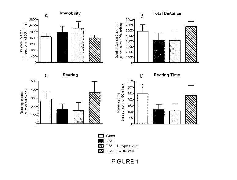
[0025] Figure 1 is a summary of the open field behaviors of mice subjected to
a DSS-induced
colitis model. Mice were treated with either: water alone, DSS alone, DSS +
isotype control
antibody, or DSS + antibody H4H6385N, as indicated. Figure 1A depicts the
extent of
immobility time in seconds; Figure 1B depicts the total distance traveled in
cm; Figure 1C
depicts the rearing counts; and Figure 1D depicts the rearing time in seconds.
All parameters
were measured over a 60 minute test period.
DETAILED DESCRIPTION
[0026] Before the present invention is described, it is to be understood that
this invention is
not limited to particular methods and experimental conditions described, as
such methods and
conditions may vary. It is also to be understood that the terminology used
herein is for the
purpose of describing particular embodiments only, and is not intended to be
limiting, since the
scope of the present invention will be limited only by the appended claims.
[0027] Unless defined otherwise, all technical and scientific terms used
herein have the same
meaning as commonly understood by one of ordinary skill in the art to which
this invention
belongs. As used herein, the term "about," when used in reference to a
particular recited
numerical value, means that the value may vary from the recited value by no
more than 1%.
For example, as used herein, the expression "about 100" includes 99 and 101
and all values in
between (e.g., 99.1, 99.2, 99.3, 99.4, etc.).
[0028] Although any methods and materials similar or equivalent to those
described herein
CA 02891327 2015-05-12
WO 2014/078306 PCT/US2013/069673
6
can be used in the practice or testing of the present invention, the preferred
methods and
materials are now described.
Definitions
[0029] The expressions "prokineticin receptor," "PROKR," "PKR," and the like,
as used herein,
are intended to encompass both PROKR1 and PROKR2. The terms "PROKR1" and
"PROKR2"
refer to the human PROKR1 or PROKR2 proteins unless specified as being from a
non-human
species (e.g., "mouse PROKR1," "mouse PROKR2," "monkey PROKR1," "monkey
PROKR2,"
etc.). Human PROKR1 has the amino acid sequence of SEQ ID NO:177. Human PROKR2
has
the amino acid sequence of SEQ ID NO:178. Mouse (Mus muscu/us) PROKR1 has the
amino
acid sequence as set forth in NCB! reference sequence number NP_067356.2;
mouse
PROKR2 has the amino acid sequence as set forth in NCB! reference sequence
number
NP_659193.3; rat (Rattus norvegicus) PROKR1 has the amino acid sequence as set
forth in
NCB! reference sequence number NP_620433.1; rat PROKR2 has the amino acid
sequence as
set forth in NCB! reference sequence number NP_620434.1; cynomolgus monkey
(Macaca
fascicularis) PROKR1 has the amino acid sequence as set forth in GenBank
accession number
EHH55625.1; and cynomolgus monkey PROKR2 has the amino acid sequence as set
forth in
GenBank accession number EHH65528.1.
[0030] An "anti-PROKR antibody" means an antibody that specifically binds
either PROKR1 or
PROKR2, or both PROKR1 and PROKR2. As used herein, "an antibody that binds
PROKR" or
an "anti-PROKR antibody" includes antibodies, and antigen-binding fragments
thereof, that bind
a soluble fragment of PROKR protein (e.g., a polypeptide comprising the N-
terminal
extracellular portion of PROKR1 or PROKR2 [see, e.g., Example 4 herein], or
one or more
extracellular loops thereof). The expressions "an antibody that binds PROKR"
or an "anti-
PROKR antibody" also include antibodies that bind cell surface-expressed
PROKR1 and/or
PROKR2. The expression "cell surface-expressed PROKR" means one or more PROKR
protein(s) that is/are expressed on the surface of a cell in vitro or in vivo,
such that at least a
portion of the PROKR protein (e.g., the N-terminal extracellular portion
and/or one or more
extracellular loops) is/are exposed to the extracellular side of the cell
membrane and accessible
to an antigen-binding portion of an antibody. "Cell surface-expressed PROKRs"
include
PROKRs that are naturally expressed on the surface of a cell as well as PROKRs
that are
artificially engineered to be expressed on the surface of a cell.
[0031] An antibody that "specifically binds cell surface-expressed PROKR1"
means an
antibody that detectably binds cells that express PROKR1 on the cell surface
but does not
detectably bind equivalent cells that do not express PROKR1 on the cell
surface. Likewise, an
antibody that "specifically binds cell surface-expressed PROKR2" means an
antibody that
detectably binds cells that express PROKR2 on the cell surface but does not
detectably bind
equivalent cells that do not express PROKR2 on the cell surface. An exemplary
method for
assessing whether an antibody binds cell surface-expressed PROKR1 and/or
PROKR2 is flow
CA 02891327 2015-05-12
WO 2014/078306 PCT/US2013/069673
7
cytometry (FACS), as illustrated in Example 3 herein.
[0032] The term "antibody", as used herein, means any antigen-binding molecule
or molecular
complex comprising at least one complementarity determining region (CDR) that
specifically
binds to or interacts with a particular antigen (e.g., a PROKR). The term
"antibody" includes
immunoglobulin molecules comprising four polypeptide chains, two heavy (H)
chains and two
light (L) chains inter-connected by disulfide bonds, as well as multimers
thereof (e.g., IgM).
Each heavy chain comprises a heavy chain variable region (abbreviated herein
as HCVR or VH)
and a heavy chain constant region. The heavy chain constant region comprises
three domains,
CH1, CH2 and CH3. Each light chain comprises a light chain variable region
(abbreviated herein
as LCVR or VL) and a light chain constant region. The light chain constant
region comprises
one domain (CO). The VH and VI_ regions can be further subdivided into regions
of
hypervariability, termed complementarity determining regions (CDRs),
interspersed with regions
that are more conserved, termed framework regions (FR). Each VH and VI_ is
composed of
three CDRs and four FRs, arranged from amino-terminus to carboxy-terminus in
the following
order: FR1, CDR1, FR2, CDR2, FR3, CDR3, FR4. In different embodiments of the
invention,
the FRs of the anti-PROKR antibody (or antigen-binding portion thereof) may be
identical to the
human germline sequences, or may be naturally or artificially modified. An
amino acid
consensus sequence may be defined based on a side-by-side analysis of two or
more CDRs.
[0033] The term "antibody", as used herein, also includes antigen-binding
fragments of full
antibody molecules. The terms "antigen-binding portion" of an antibody,
"antigen-binding
fragment" of an antibody, and the like, as used herein, include any naturally
occurring,
enzymatically obtainable, synthetic, or genetically engineered polypeptide or
glycoprotein that
specifically binds an antigen to form a complex. Antigen-binding fragments of
an antibody may
be derived, e.g., from full antibody molecules using any suitable standard
techniques such as
proteolytic digestion or recombinant genetic engineering techniques involving
the manipulation
and expression of DNA encoding antibody variable and optionally constant
domains. Such
DNA is known and/or is readily available from, e.g., commercial sources, DNA
libraries
(including, e.g., phage-antibody libraries), or can be synthesized. The DNA
may be sequenced
and manipulated chemically or by using molecular biology techniques, for
example, to arrange
one or more variable and/or constant domains into a suitable configuration, or
to introduce
codons, create cysteine residues, modify, add or delete amino acids, etc.
[0034] Non-limiting examples of antigen-binding fragments include: (i) Fab
fragments; (ii)
F(ab')2 fragments; (iii) Fd fragments; (iv) Fv fragments; (v) single-chain Fv
(scFv) molecules;
(vi) dAb fragments; and (vii) minimal recognition units consisting of the
amino acid residues that
mimic the hypervariable region of an antibody (e.g., an isolated
complementarity determining
region (CDR) such as a CDR3 peptide), or a constrained FR3-CDR3-FR4 peptide.
Other
engineered molecules, such as domain-specific antibodies, single domain
antibodies, domain-
deleted antibodies, chimeric antibodies, CDR-grafted antibodies, diabodies,
triabodies,
CA 02891327 2015-05-12
WO 2014/078306 PCT/US2013/069673
8
tetrabodies, minibodies, nanobodies (e.g. monovalent nanobodies, bivalent
nanobodies, etc.),
small modular immunopharmaceuticals (SMIPs), and shark variable IgNAR domains,
are also
encompassed within the expression "antigen-binding fragment," as used herein.
[0035] An antigen-binding fragment of an antibody will typically comprise at
least one variable
domain. The variable domain may be of any size or amino acid composition and
will generally
comprise at least one CDR which is adjacent to or in frame with one or more
framework
sequences. In antigen-binding fragments having a VH domain associated with a
VL domain, the
VH and VI_ domains may be situated relative to one another in any suitable
arrangement. For
example, the variable region may be dimeric and contain VH-VH, VH-VL or V[-V[
dimers.
Alternatively, the antigen-binding fragment of an antibody may contain a
monomeric VH or VL
domain.
[0036] In certain embodiments, an antigen-binding fragment of an antibody may
contain at
least one variable domain covalently linked to at least one constant domain.
Non-limiting,
exemplary configurations of variable and constant domains that may be found
within an antigen-
binding fragment of an antibody of the present invention include: (i) VH-CH1;
(ii) VH-CH2; (iii) VI-I-
CH3; (iv) VH-CH1-CH2, (V) VH-CH1-CH2-CH3; ND VH-CH2-CH3; Nip VH-CL, MO V[-CH1;
(ix) VL-CH2;
(X) VL-CH3; (Xi) VL-CH1-CH2; (Xii) VL-CH1-CH2-CH3; (Xiii) VL-CH2-CH3, and
(xiv) VL-CL. In any
configuration of variable and constant domains, including any of the exemplary
configurations
listed above, the variable and constant domains may be either directly linked
to one another or
may be linked by a full or partial hinge or linker region. A hinge region may
consist of at least 2
(e.g., 5, 10, 15, 20, 40, 60 or more) amino acids which result in a flexible
or semi-flexible linkage
between adjacent variable and/or constant domains in a single polypeptide
molecule.
Moreover, an antigen-binding fragment of an antibody of the present invention
may comprise a
homo-dimer or hetero-dimer (or other multimer) of any of the variable and
constant domain
configurations listed above in non-covalent association with one another
and/or with one or
more monomeric VH or VI_ domain (e.g., by disulfide bond(s)).
[0037] As with full antibody molecules, antigen-binding fragments may be
monospecific or
multispecific (e.g., bispecific). A multispecific antigen-binding fragment of
an antibody will
typically comprise at least two different variable domains, wherein each
variable domain is
capable of specifically binding to a separate antigen or to a different
epitope on the same
antigen. Any multispecific antibody format, including the exemplary bispecific
antibody formats
disclosed herein, may be adapted for use in the context of an antigen-binding
fragment of an
antibody of the present invention using routine techniques available in the
art.
[0038] The term "human antibody", as used herein, is intended to include
antibodies having
variable and constant regions derived from human germline immunoglobulin
sequences. The
human antibodies of the invention may include amino acid residues not encoded
by human
germline immunoglobulin sequences (e.g., mutations introduced by random or
site-specific
mutagenesis in vitro or by somatic mutation in vivo), for example in the CDRs
and in particular
CA 02891327 2015-05-12
WO 2014/078306 PCT/US2013/069673
9
CDR3. However, the term "human antibody", as used herein, is not intended to
include
antibodies in which CDR sequences derived from the germline of another
mammalian species,
such as a mouse, have been grafted onto human framework sequences.
[0039] The term "recombinant human antibody", as used herein, is intended to
include all
human antibodies that are prepared, expressed, created or isolated by
recombinant means,
such as antibodies expressed using a recombinant expression vector transfected
into a host cell
(described further below), antibodies isolated from a recombinant,
combinatorial human
antibody library (described further below), antibodies isolated from an animal
(e.g., a mouse)
that is transgenic for human immunoglobulin genes (see e.g., Taylor et al.
(1992) Nucl. Acids
Res. 20:6287-6295) or antibodies prepared, expressed, created or isolated by
any other means
that involves splicing of human immunoglobulin gene sequences to other DNA
sequences.
Such recombinant human antibodies have variable and constant regions derived
from human
germline immunoglobulin sequences. In certain embodiments, however, such
recombinant
human antibodies are subjected to in vitro mutagenesis (or, when an animal
transgenic for
human Ig sequences is used, in vivo somatic mutagenesis) and thus the amino
acid sequences
of the VH and VI_ regions of the recombinant antibodies are sequences that,
while derived from
and related to human germline VH and VI_ sequences, may not naturally exist
within the human
antibody germline repertoire in vivo.
[0040] Human antibodies can exist in two forms that are associated with hinge
heterogeneity.
In one form, an immunoglobulin molecule comprises a stable four chain
construct of
approximately 150-160 kDa in which the dimers are held together by an
interchain heavy chain
disulfide bond. In a second form, the dimers are not linked via inter-chain
disulfide bonds and a
molecule of about 75-80 kDa is formed composed of a covalently coupled light
and heavy chain
(half-antibody). These forms have been extremely difficult to separate, even
after affinity
purification.
[0041] The frequency of appearance of the second form in various intact IgG
isotypes is due
to, but not limited to, structural differences associated with the hinge
region isotype of the
antibody. A single amino acid substitution in the hinge region of the human
IgG4 hinge can
significantly reduce the appearance of the second form (Angal et al. (1993)
Molecular
Immunology 30:105) to levels typically observed using a human IgG1 hinge. The
instant
invention encompasses antibodies having one or more mutations in the hinge,
CH2 or CH3
region which may be desirable, for example, in production, to improve the
yield of the desired
antibody form.
[0042] An "isolated antibody," as used herein, means an antibody that has been
identified and
separated and/or recovered from at least one component of its natural
environment. For
example, an antibody that has been separated or removed from at least one
component of an
organism, or from a tissue or cell in which the antibody naturally exists or
is naturally produced,
is an "isolated antibody" for purposes of the present invention. An isolated
antibody also
CA 02891327 2015-05-12
WO 2014/078306 PCT/US2013/069673
includes an antibody in situ within a recombinant cell. Isolated antibodies
are antibodies that
have been subjected to at least one purification or isolation step. According
to certain
embodiments, an isolated antibody may be substantially free of other cellular
material and/or
chemicals.
[0043] A "neutralizing" or "blocking" antibody, as used herein, is intended to
refer to an
antibody whose binding to a PROKR: (i) inhibits the interaction between a
prokineticin (e.g.,
PK1 or PK2) and the PROKR; (ii) inhibits or attenuates prokineticin-mediated
activation of the
PROKR; and/or (iii) results in inhibition of at least one biological function
of the PROKR. The
inhibition caused by a PROKR-neutralizing or blocking antibody need not be
complete so long
as it is detectable using an appropriate assay. Exemplary assays for detecting
PROKR
inhibition are known in the art and are illustrated in the working Examples
herein.
[0044] The anti-PROKR antibodies disclosed herein may comprise one or more
amino acid
substitutions, insertions and/or deletions in the framework and/or CDR regions
of the heavy and
light chain variable domains as compared to the corresponding germline
sequences from which
the antibodies were derived. Such mutations can be readily ascertained by
comparing the
amino acid sequences disclosed herein to germline sequences available from,
for example,
public antibody sequence databases. The present invention includes antibodies,
and antigen-
binding fragments thereof, which are derived from any of the amino acid
sequences disclosed
herein, wherein one or more amino acids within one or more framework and/or
CDR regions are
mutated to the corresponding residue(s) of the germline sequence from which
the antibody was
derived, or to the corresponding residue(s) of another human germline
sequence, or to a
conservative amino acid substitution of the corresponding germline residue(s)
(such sequence
changes are referred to herein collectively as "germline mutations"). A person
of ordinary skill in
the art, starting with the heavy and light chain variable region sequences
disclosed herein, can
easily produce numerous antibodies and antigen-binding fragments which
comprise one or
more individual germline mutations or combinations thereof. In certain
embodiments, all of the
framework and/or CDR residues within the VH and/or VL domains are mutated back
to the
residues found in the original germline sequence from which the antibody was
derived. In other
embodiments, only certain residues are mutated back to the original germline
sequence, e.g.,
only the mutated residues found within the first 8 amino acids of FR1 or
within the last 8 amino
acids of FR4, or only the mutated residues found within CDR1, CDR2 or CDR3. In
other
embodiments, one or more of the framework and/or CDR residue(s) are mutated to
the
corresponding residue(s) of a different germline sequence (i.e., a germline
sequence that is
different from the germline sequence from which the antibody was originally
derived).
Furthermore, the antibodies of the present invention may contain any
combination of two or
more germline mutations within the framework and/or CDR regions, e.g., wherein
certain
individual residues are mutated to the corresponding residue of a particular
germline sequence
while certain other residues that differ from the original germline sequence
are maintained or
CA 02891327 2015-05-12
WO 2014/078306 PCT/US2013/069673
11
are mutated to the corresponding residue of a different germline sequence.
Once obtained,
antibodies and antigen-binding fragments that contain one or more germline
mutations can be
easily tested for one or more desired property such as, improved binding
specificity, increased
binding affinity, improved or enhanced antagonistic or agonistic biological
properties (as the
case may be), reduced immunogennicity, etc. Antibodies and antigen-binding
fragments
obtained in this general manner are encompassed within the present invention.
[0045] The present invention also includes anti-PROKR antibodies comprising
variants of any
of the HCVR, LCVR, and/or CDR amino acid sequences disclosed herein having one
or more
conservative substitutions. For example, the present invention includes anti-
PROKR antibodies
having HCVR, LCVR, and/or CDR amino acid sequences with, e.g., 10 or fewer, 8
or fewer, 6 or
fewer, 4 or fewer, etc. conservative amino acid substitutions relative to any
of the HCVR, LCVR,
and/or CDR amino acid sequences disclosed herein.
[0046] The term "epitope" refers to an antigenic determinant that interacts
with a specific
antigen binding site in the variable region of an antibody molecule known as a
paratope. A
single antigen may have more than one epitope. Thus, different antibodies may
bind to different
areas on an antigen and may have different biological effects. Epitopes may be
either
conformational or linear. A conformational epitope is produced by spatially
juxtaposed amino
acids from different segments of the linear polypeptide chain. A linear
epitope is one produced
by adjacent amino acid residues in a polypeptide chain. In certain
circumstance, an epitope
may include moieties of saccharides, phosphoryl groups, or sulfonyl groups on
the antigen.
[0047] The term "substantial identity" or "substantially identical," when
referring to a nucleic
acid or fragment thereof, indicates that, when optimally aligned with
appropriate nucleotide
insertions or deletions with another nucleic acid (or its complementary
strand), there is
nucleotide sequence identity in at least about 95%, and more preferably at
least about 96%,
97%, 98% or 99% of the nucleotide bases, as measured by any well-known
algorithm of
sequence identity, such as FASTA, BLAST or Gap, as discussed below. A nucleic
acid
molecule having substantial identity to a reference nucleic acid molecule may,
in certain
instances, encode a polypeptide having the same or substantially similar amino
acid sequence
as the polypeptide encoded by the reference nucleic acid molecule.
[0048] As applied to polypeptides, the term "substantial similarity" or
"substantially similar"
means that two peptide sequences, when optimally aligned, such as by the
programs GAP or
BESTFIT using default gap weights, share at least 95% sequence identity, even
more
preferably at least 98% or 99% sequence identity. Preferably, residue
positions which are not
identical differ by conservative amino acid substitutions. A "conservative
amino acid
substitution" is one in which an amino acid residue is substituted by another
amino acid residue
having a side chain (R group) with similar chemical properties (e.g., charge
or hydrophobicity).
In general, a conservative amino acid substitution will not substantially
change the functional
properties of a protein. In cases where two or more amino acid sequences
differ from each
CA 02891327 2015-05-12
WO 2014/078306 PCT/US2013/069673
12
other by conservative substitutions, the percent sequence identity or degree
of similarity may be
adjusted upwards to correct for the conservative nature of the substitution.
Means for making
this adjustment are well-known to those of skill in the art. See, e.g.,
Pearson (1994) Methods
Mol. Biol. 24: 307-331. Examples of groups of amino acids that have side
chains with similar
chemical properties include (1) aliphatic side chains: glycine, alanine,
valine, leucine and
isoleucine; (2) aliphatic-hydroxyl side chains: serine and threonine; (3)
amide-containing side
chains: asparagine and glutamine; (4) aromatic side chains: phenylalanine,
tyrosine, and
tryptophan; (5) basic side chains: lysine, arginine, and histidine; (6) acidic
side chains: aspartate
and glutamate, and (7) sulfur-containing side chains are cysteine and
methionine. Preferred
conservative amino acids substitution groups are: valine-leucine-isoleucine,
phenylalanine-
tyrosine, lysine-arginine, alanine-valine, glutamate-aspartate, and asparagine-
glutamine.
Alternatively, a conservative replacement is any change having a positive
value in the PAM250
log-likelihood matrix disclosed in Gonnet etal. (1992) Science 256: 1443-1445.
A "moderately
conservative" replacement is any change having a nonnegative value in the
PAM250 log-
likelihood matrix.
[0049] Sequence similarity for polypeptides, which is also referred to as
sequence identity, is
typically measured using sequence analysis software. Protein analysis software
matches
similar sequences using measures of similarity assigned to various
substitutions, deletions and
other modifications, including conservative amino acid substitutions. For
instance, GCG
software contains programs such as Gap and Bestfit which can be used with
default parameters
to determine sequence homology or sequence identity between closely related
polypeptides,
such as homologous polypeptides from different species of organisms or between
a wild type
protein and a mutein thereof. See, e.g., GCG Version 6.1. Polypeptide
sequences also can be
compared using FASTA using default or recommended parameters, a program in GCG
Version
6.1. FASTA (e.g., FASTA2 and FASTA3) provides alignments and percent sequence
identity of
the regions of the best overlap between the query and search sequences
(Pearson (2000)
supra). Another preferred algorithm when comparing a sequence of the invention
to a database
containing a large number of sequences from different organisms is the
computer program
BLAST, especially BLASTP or TBLASTN, using default parameters. See, e.g.,
Altschul etal.
(1990) J. Mol. Biol. 215:403-410 and Altschul etal. (1997) Nucleic Acids Res.
25:3389-402.
Biological Characteristics of the Antibodies
[0050] The present invention includes anti-PROKR antibodies and antigen-
binding fragments
thereof that specifically bind cell surface-expressed PROKR1 and/or cell
surface-expressed
PROKR2. For example, the present invention provides anti-PROKR antibodies
that: (a)
specifically bind cell surface-expressed PROKR1 but not cell surface-expressed
PROKR2; (b)
specifically bind cell surface-expressed PROKR2 but not cell surface-expressed
PROKR1; or
(c) specifically bind cell surface-expressed PROKR1 and cell surface-expressed
PROKR2.
CA 02891327 2015-05-12
WO 2014/078306 PCT/US2013/069673
13
Anti-PROKR antibodies can be tested and evaluated for the ability to
specifically bind a cell
surface-expressed PROKR using any assay format that allows for the detection
of antibody
binding to cells that express a PROKR. An exemplary assay format that can be
used to
determine whether an antibody specifically binds a cell surface-expressed
PROKR is illustrated
in Example 3 herein. In this Example, cells that normally do not express
PROKRs (e.g.,
HEK293 cells) are engineered to express PROKR1 or PROKR2, and antibody binding
to the
PROKR-expressing cells is determined by flow cytometry with detectably labeled
secondary
antibodies. "Specific antibody binding" to a cell surface-expressed PROKR
means that the
percentage of cells that exhibit detectable binding by flow cytometry is
greater than 1%. An
antibody that exhibits a binding percentage of between 1% and 10% in this
assay format is
generally regarded as having "weak" binding, but is nonetheless considered an
antibody that
"specifically binds a cell surface-expressed PROKR" for purposes of the
present disclosure.
According to certain embodiments, however, specific antibody binding to a cell
surface-
expressed PROKR means that the percentage of cells that exhibit detectable
binding by flow
cytometry is greater than 10%, greater than 20%, greater than 30%, greater
than 40%, greater
than 50%, or more.
[0051] The present invention also includes anti-PROKR antibodies that bind one
or more
soluble fragments of PROKR1 and/or PROKR2. For example, antibodies are
provided herein
which specifically bind a soluble fragment of PROKR1 or PROKR2 comprising all
or part of the
N-terminal extracellular portion of the PROKR protein. Exemplary soluble
PROKR1 and
PROKR2 constructs of this type are illustrated in Example 4 herein. As shown
in Example 4,
fusion proteins comprising amino acids 1-62 of PROKR1 (SEQ ID NO:177) or amino
acids 1-53
or PROKR2 (SEQ ID NO:178), fused to a human Fc component, were tested for
binding to anti-
PROKR antibodies by surface plasmon resonance (at 25 C and pH 7.4). Using an
assay format
of Example 4, or a similar assay, the binding of anti-PROKR antibodies to
soluble PROKR
molecules can be quantified, e.g., in terms of equilibrium dissociation
constant (KD) and/or
dissociation half-life (t1/2).
[0052] Thus, the present invention provides anti-PROKR antibodies that bind
soluble human
PROKR1 (e.g., N-terminal portion) with a KD of less than about 5 nM, less than
about 3 nM, less
than about 2 nM, less than about 1.5 nM, less than about 600 pM, less than
about 550 pM, less
than about 500 pM, less than about 450 pM, less than about 400 pM, less than
about 350 pM,
less than about 300 pM, less than about 250 pM, less than about 200 pM, less
than about 150
pM, or less than about 100 pM as measured by surface plasmon resonance, e.g.,
using the
assay format as defined in Example 4 herein or a substantially similar assay.
[0053] The present invention also includes anti-PROKR antibodies and antigen-
binding
fragments thereof that bind soluble human PROKR1 (e.g., N-terminal portion)
with a
dissociation half-life (t%) of greater than about 5 minutes, greater than
about 10 minutes,
greater than about 15 minutes, greater than about 20 minutes, greater than
about 25 minutes,
CA 02891327 2015-05-12
WO 2014/078306 PCT/US2013/069673
14
greater than about 30 minutes, greater than about 35 minutes, greater than
about 40 minutes,
greater than about 45 minutes, greater than about 50 minutes, greater than
about 55 minutes,
greater than about 60 minutes, greater than about 75 minutes, greater than
about 100 minutes,
greater than about 150 minutes, greater than about 200 minutes, or greater
than about 250
minutes, or more, as measured by surface plasmon resonance, e.g., using the
assay format as
defined in Example 4 herein or a substantially similar assay.
[0054] The present invention also provides anti-PROKR antibodies that bind
soluble human
PROKR2 (e.g., N-terminal portion) with a KD of less than about 150 nM, less
than about 130
nM, less than about 100 nM, less than about 90 nM, less than about 80 nM, less
than about 75
nM, less than about 70 nM, less than about 65 nM, less than about 60 nM, less
than about 55
nM, less than about 50 nM, less than about 45 nM, less than about 40 nM, less
than about 35
nM, less than about 30 nM, less than about 25 nM, less than about 20 nM, less
than about 15
nM, less than about 10 nM, less than about 5 nM, less than about 3 nM, or
less, as measured
by surface plasmon resonance, e.g., using the assay format as defined in
Example 4 herein or a
substantially similar assay.
[0055] The present invention also includes anti-PROKR antibodies and antigen-
binding
fragments thereof that bind soluble human PROKR2 (e.g., N-terminal portion)
with a
dissociation half-life (t1/2) of greater than about 1 minute, greater than
about 2 minutes, greater
than about 3 minutes, greater than about 4 minutes, greater than about 5
minutes, greater than
about 10 minutes, greater than about 20 minutes, greater than about 30
minutes, greater than
about 40 minutes, or more, as measured by surface plasmon resonance, e.g.,
using the assay
format as defined in Example 4 herein or a substantially similar assay.
[0056] The present invention also includes anti-PROKR antibodies and antigen-
binding
fragments thereof that block prokineticin-mediated activation of PROKR1 and/or
PROKR2. The
ability of anti-PROKR antibodies to block prokineticin-mediated activation of
PROKR1 and/or
PROKR2 can be measured, e.g., using the assay format illustrated in Example 5
herein. In this
assay, cells that do not normally express human PROKRs (i.e., HEK293 cells)
are engineered
to express PROKR1 or PROKR2. In this assay format, the extent of PROKR
activation is
indicated by calcium mobilization following treatment with prokineticin-1
(PK1) or prokineticin-2
(PK2) (e.g. using a concentration of about 1 to 20 nM or PK1 or PK2), in the
presence or
absence of an anti-PROKR antibody. Inhibition of prokineticin-mediated PROKR
activation in
this assay format is calculated as an 1050 value (i.e., the concentration of
antibody needed to
inhibit PK-mediated calcium flux by 50%) or as a blocking percentage. The
present invention
includes anti-PROKR antibodies that block: (a) PK1-mediated activation of
PROKR1; (b) PK2-
mediated activation of PROKR1; (c) PK1-mediated activation of PROKR2; and/or
(d) PK2-
mediated activation of PROKR2. For example, the present invention includes
anti-PROKR
antibodies that block PK1-mediated activation of PROKR1 with an 1050 of less
than about 20
nM, less than about 18 nM, less than about 16 nM, less than about 14 nM, less
than about 12
CA 02891327 2015-05-12
WO 2014/078306 PCT/US2013/069673
nM, less than about 10 nM, less than about 9 nM, less than about 8 nM, less
than about 7 nM,
or less than about 6 nM, or less, as measured using the assay format of
Example 5 (e.g., using
about 1 nM to about 20 nM of PK1), or a substantially similar assay. The
present invention also
includes anti-PROKR antibodies that block PK2-mediated activation of PROKR1
with an 1050 of
less than about 60 nM, less than about 50 nM, less than about 20 nM, or less
than about 20 nM,
as measured using the assay format of Example 5 (e.g., using about 1 nM to
about 20 nM of
PK2), or a substantially similar assay.
[0057] The present invention also includes anti-PROKR antibodies that inhibit
or reduce pain
response(s) in various animal pain models.
[0058] Other biological activities of the anti-PROKR antibodies of the present
invention will be
apparent to persons of ordinary skill in the art in light of the working
Examples set forth herein.
Epitope Mapping and Related Technologies
[0059] The present invention includes anti-PROKR antibodies which interact
with one or more
amino acids located within one or more regions or segments of the PROKR1
molecule selected
from the group consisting of: (a) the N-terminal extracellular region (amino
acids 1 to 62 of SEQ
ID NO:177); (b) extracellular loop 1 (amino acids 120 to 146 of SEQ ID
NO:177); (c)
extracellular loop 2 (amino acids 201 to 232 of SEQ ID NO:177); and/or
extracellular loop 3
(amino acids 304 to 322 of SEQ ID NO:177).
[0060] The present invention also includes anti-PROKR antibodies which
interact with one or
more amino acids located within one or more regions or segments of the PROKR2
molecule
selected from the group consisting of: (a) the N-terminal extracellular region
(amino acids 1 to
54 of SEQ ID NO:178); (b) extracellular loop 1 (amino acids 110 to 137 of SEQ
ID NO:178); (c)
extracellular loop 2 (amino acids 193 to 221 of SEQ ID NO:178); and/or
extracellular loop 3
(amino acids 297 to 310 of SEQ ID NO:178).
[0061] The epitope to which the antibodies bind may consist of a single
contiguous sequence
of 3 or more (e.g., 3, 4, 5, 6, 7, 8, 9, 10, 11, 12, 13, 14, 15, 16, 17, 18,
19, 20 or more) amino
acids located within any of the aforementioned regions or segments of PROKR1
and/or
PROKR2. Alternatively, the epitope may consist of a plurality of non-
contiguous amino acids (or
amino acid sequences) located within one or more of the aforementioned regions
or segments
of a PROKR molecule. For example, the antibodies of the present invention may
interact with
one or more amino acids located within the N-terminal extracellular region of
PROKR1 as well
as one or more amino acids located within one or more extracellular loops of
PROKR1.
[0062] Various techniques known to persons of ordinary skill in the art can be
used to
determine whether an antibody "interacts with one or more amino acids" within
a polypeptide or
protein. Exemplary techniques include, e.g., routine cross-blocking assay such
as that
described Antibodies, Harlow and Lane (Cold Spring Harbor Press, Cold Spring
Harb., NY),
alanine scanning mutational analysis, peptide blots analysis (Reineke, 2004,
Methods Mol Biol
248:443-463), and peptide cleavage analysis. In addition, methods such as
epitope excision,
CA 02891327 2015-05-12
WO 2014/078306 PCT/US2013/069673
16
epitope extraction and chemical modification of antigens can be employed
(Tomer, 2000,
Protein Science 9:487-496). Another method that can be used to identify the
amino acids within
a polypeptide with which an antibody interacts is hydrogen/deuterium exchange
detected by
mass spectrometry. In general terms, the hydrogen/deuterium exchange method
involves
deuterium-labeling the protein of interest, followed by binding the antibody
to the deuterium-
labeled protein. Next, the protein/antibody complex is transferred to water to
allow hydrogen-
deuterium exchange to occur at all residues except for the residues protected
by the antibody
(which remain deuterium-labeled). After dissociation of the antibody, the
target protein is
subjected to protease cleavage and mass spectrometry analysis, thereby
revealing the
deuterium-labeled residues which correspond to the specific amino acids with
which the
antibody interacts. See, e.g., Ehring (1999) Analytical Biochemistry
267(2):252-259; Engen and
Smith (2001) Anal. Chem. 73:256A-265A. X-ray crystallography of the
antigen/antibody
complex may also be used for epitope mapping purposes.
[0063] The present invention further includes anti-PROKR antibodies that bind
to the same
epitope as any of the specific exemplary antibodies described herein (e.g. H1
M6386N,
H2M6385N, H4H6663P, H4H6669P, H4H6671P, H4H6680P, H4H6690P, H4H6696P,
H4H6698P, H4H6701P, H4H6706P, etc.). Likewise, the present invention also
includes anti-
PROKR antibodies that compete for binding to PROKR1 and/or PROKR2 with any of
the
specific exemplary antibodies described herein (e.g. H1 M6386N, H2M6385N,
H4H6663P,
H4H6669P, H4H6671P, H4H6680P, H4H6690P, H4H6696P, H4H6698P, H4H6701P,
H4H6706P, etc.).
[0064] One can easily determine whether an antibody binds to the same epitope
as, or
competes for binding with, a reference anti-PROKR antibody by using routine
methods known in
the art. For example, to determine if a test antibody binds to the same
epitope as a reference
anti-PROKR antibody of the invention, the reference antibody is allowed to
bind to a PROKR
protein. Next, the ability of a test antibody to bind to the PROKR molecule is
assessed. If the
test antibody is able to bind to the PROKR following saturation binding with
the reference anti-
PROKR antibody, it can be concluded that the test antibody binds to a
different epitope than the
reference anti-PROKR antibody. On the other hand, if the test antibody is not
able to bind to
the PROKR molecule following saturation binding with the reference anti-PROKR
antibody, then
the test antibody may bind to the same epitope as the epitope bound by the
reference anti-
PROKR antibody of the invention. Additional routine experimentation (e.g.,
peptide mutation
and binding analyses) can then be carried out to confirm whether the observed
lack of binding
of the test antibody is in fact due to binding to the same epitope as the
reference antibody or if
steric blocking (or another phenomenon) is responsible for the lack of
observed binding.
Experiments of this sort can be performed using ELISA, RIA, Biacore, flow
cytometry or any
other quantitative or qualitative antibody-binding assay available in the art.
In accordance with
certain embodiments of the present invention, two antibodies bind to the same
(or overlapping)
CA 02891327 2015-05-12
WO 2014/078306 PCT/US2013/069673
17
epitope if, e.g., a 1-, 5-, 10-, 20- or 100-fold excess of one antibody
inhibits binding of the other
by at least 50% but preferably 75%, 90% or even 99% as measured in a
competitive binding
assay (see, e.g., Junghans etal., Cancer Res. 1990:50:1495-1502).
Alternatively, two
antibodies are deemed to bind to the same epitope if essentially all amino
acid mutations in the
antigen that reduce or eliminate binding of one antibody reduce or eliminate
binding of the
other. Two antibodies are deemed to have "overlapping epitopes" if only a
subset of the amino
acid mutations that reduce or eliminate binding of one antibody reduce or
eliminate binding of
the other.
[0065] To determine if an antibody competes for binding with a reference anti-
PROKR
antibody, the above-described binding methodology is performed in two
orientations: In a first
orientation, the reference antibody is allowed to bind to a PROKR protein
under saturating
conditions followed by assessment of binding of the test antibody to the PROKR
molecule. In a
second orientation, the test antibody is allowed to bind to a PROKR molecule
under saturating
conditions followed by assessment of binding of the reference antibody to the
PROKR
molecule. If, in both orientations, only the first (saturating) antibody is
capable of binding to the
PROKR molecule, then it is concluded that the test antibody and the reference
antibody
compete for binding to the PROKR. As will be appreciated by a person of
ordinary skill in the
art, an antibody that competes for binding with a reference antibody may not
necessarily bind to
the same epitope as the reference antibody, but may sterically block binding
of the reference
antibody by binding an overlapping or adjacent epitope.
Preparation of Human Antibodies
[0066] Methods for generating monoclonal antibodies, including fully human
monoclonal
antibodies are known in the art. Any such known methods can be used in the
context of the
present invention to make human antibodies that specifically bind to a human
PROKR.
[0067] Using VELOCIMMUNETm technology or other similar methods for generating
monoclonal antibodies, high affinity chimeric antibodies to PROKR are
initially isolated having a
human variable region and a mouse constant region. As in the experimental
section below, the
antibodies are characterized and selected for desirable characteristics,
including affinity,
selectivity, epitope, etc. The mouse constant regions are replaced with a
desired human
constant region to generate the fully human antibody of the invention, for
example wild-type or
modified IgG1 or IgG4. While the constant region selected may vary according
to specific use,
high affinity antigen-binding and target specificity characteristics reside in
the variable region.
Bioequivalents
[0068] The anti-PROKR antibodies and antibody fragments of the present
invention
encompass proteins having amino acid sequences that vary from those of the
described
antibodies but that retain the ability to bind a human PROKR. Such variant
antibodies and
antibody fragments comprise one or more additions, deletions, or substitutions
of amino acids
CA 02891327 2015-05-12
WO 2014/078306 PCT/US2013/069673
18
when compared to parent sequence, but exhibit biological activity that is
essentially equivalent
to that of the described antibodies. Likewise, the anti-PROKR antibody-
encoding DNA
sequences of the present invention encompass sequences that comprise one or
more
additions, deletions, or substitutions of nucleotides when compared to the
disclosed sequence,
but that encode an anti-PROKR antibody or antibody fragment that is
essentially bioequivalent
to an anti-PROKR antibody or antibody fragment of the invention. Examples of
such variant
amino acid and DNA sequences are discussed above.
[0069] Two antigen-binding proteins, or antibodies, are considered
bioequivalent if, for
example, they are pharmaceutical equivalents or pharmaceutical alternatives
whose rate and
extent of absorption do not show a significant difference when administered at
the same molar
dose under similar experimental conditions, either single does or multiple
dose. Some
antibodies will be considered equivalents or pharmaceutical alternatives if
they are equivalent in
the extent of their absorption but not in their rate of absorption and yet may
be considered
bioequivalent because such differences in the rate of absorption are
intentional and are
reflected in the labeling, are not essential to the attainment of effective
body drug
concentrations on, e.g., chronic use, and are considered medically
insignificant for the particular
drug product studied.
[0070] In one embodiment, two antigen-binding proteins are bioequivalent if
there are no
clinically meaningful differences in their safety, purity, and potency.
[0071] In one embodiment, two antigen-binding proteins are bioequivalent if a
patient can be
switched one or more times between the reference product and the biological
product without
an expected increase in the risk of adverse effects, including a clinically
significant change in
immunogenicity, or diminished effectiveness, as compared to continued therapy
without such
switching.
[0072] In one embodiment, two antigen-binding proteins are bioequivalent if
they both act by a
common mechanism or mechanisms of action for the condition or conditions of
use, to the
extent that such mechanisms are known.
[0073] Bioequivalence may be demonstrated by in vivo and in vitro methods.
Bioequivalence
measures include, e.g., (a) an in vivo test in humans or other mammals, in
which the
concentration of the antibody or its metabolites is measured in blood, plasma,
serum, or other
biological fluid as a function of time; (b) an in vitro test that has been
correlated with and is
reasonably predictive of human in vivo bioavailability data; (c) an in vivo
test in humans or other
mammals in which the appropriate acute pharmacological effect of the antibody
(or its target) is
measured as a function of time; and (d) in a well-controlled clinical trial
that establishes safety,
efficacy, or bioavailability or bioequivalence of an antibody.
[0074] Bioequivalent variants of anti-PROKR antibodies of the invention may be
constructed
by, for example, making various substitutions of residues or sequences or
deleting terminal or
internal residues or sequences not needed for biological activity. For
example, cysteine
CA 02891327 2015-05-12
WO 2014/078306 PCT/US2013/069673
19
residues not essential for biological activity can be deleted or replaced with
other amino acids to
prevent formation of unnecessary or incorrect intramolecular disulfide bridges
upon
renaturation. In other contexts, bioequivalent antibodies may include anti-
PROKR antibody
variants comprising amino acid changes which modify the glycosylation
characteristics of the
antibodies, e.g., mutations which eliminate or remove glycosylation.
Species Selectivity and Species Cross-Reactivity
[0075] The present invention includes anti-PROKR antibodies that bind to a
human PROKR
(e.g., cell surface-expressed human PROKR1 and/or cell surface expressed human
PROKR2)
but not to PROKRs from other species. The present invention also includes anti-
PROKR
antibodies that bind to a human PROKR (e.g., cell surface-expressed human
PROKR1 and/or
cell surface expressed human PROKR2) and also bind to one or more PROKR
proteins from
one or more non-human species. The present invention also includes anti-PROKR
antibodies
that block prokineticin-mediated activation of human PROKR1 and/or human
PROKR2 but do
not block prokineticin-mediated activation of one or more non-human PROKRs.
The present
invention also includes anti-PROKR antibodies that block prokineticin-mediated
activation of
human PROKR1 and/or human PROKR2 and also block prokineticin-mediated
activation of one
or more non-human PROKRs.
[0076] For example, the anti-PROKR antibodies of the invention may bind to
and/or block
human PROKR1 and/or human PROKR2, and may bind and/or block (or not bind or
not block
as the case may be) one or more of mouse, rat, guinea pig, hamster, gerbil,
pig, cat, dog, rabbit,
goat, sheep, cow, horse, camel, cynomologous, marmoset, rhesus or chimpanzee
PROKR1 or
PROKR2. For example, as shown in Example 5 herein, certain exemplary
antibodies of the
present invention block PK1-mediated activation of human PROKR1 as well as PK1-
mediated
activation of monkey PROKR1 (e.g., H4H6696, H4H6698, H4H6701 and H4H6385). On
the
other hand, antibody H1M6386 exhibited potent blocking of PK1-mediated
activation of human
PROKR1 but did not exhibit any detectable blocking of PK1-mediated activation
of monkey
PROKR1. Other cross-reactivity/cross-blocking patterns of the exemplary anti-
PROKR
antibodies of the present invention will be apparent to a person of ordinary
skill in the art upon
review of the working examples provided herein.
Multispecific Antibodies
[0077] The antibodies of the present invention may be monospecific, bi-
specific, or
multispecific. Multispecific antibodies may be specific for different epitopes
of one target
polypeptide or may contain antigen-binding domains specific for more than one
target
polypeptide. See, e.g., Tutt et al., 1991, J. lmmunol. 147:60-69; Kufer etal.,
2004, Trends
Biotechnol. 22:238-244. The anti-PROKR antibodies of the present invention can
be linked to
or co-expressed with another functional molecule, e.g., another peptide or
protein. For
example, an antibody or fragment thereof can be functionally linked (e.g., by
chemical coupling,
CA 02891327 2015-05-12
WO 2014/078306 PCT/US2013/069673
genetic fusion, noncovalent association or otherwise) to one or more other
molecular entities,
such as another antibody or antibody fragment to produce a bi-specific or a
multispecific
antibody with a second binding specificity. For example, the present invention
includes bi-
specific antibodies wherein one arm of an immunoglobulin is specific for a
human PROKR or a
fragment thereof, and the other arm of the immunoglobulin is specific for a
second therapeutic
target or is conjugated to a therapeutic moiety. The present invention
includes bispecific
antibodies comprising a first antigen-binding domain that specifically binds
PROKR1 and a
second antigen-binding domain that specifically binds PROKR2.
[0078] An exemplary bi-specific antibody format that can be used in the
context of the present
invention involves the use of a first immunoglobulin (Ig) CH3 domain and a
second Ig CH3
domain, wherein the first and second Ig CH3 domains differ from one another by
at least one
amino acid, and wherein at least one amino acid difference reduces binding of
the bispecific
antibody to Protein A as compared to a bi-specific antibody lacking the amino
acid difference.
In one embodiment, the first Ig CH3 domain binds Protein A and the second Ig
CH3 domain
contains a mutation that reduces or abolishes Protein A binding such as an
H95R modification
(by IMGT exon numbering; H435R by EU numbering). The second CH3 may further
comprise a
Y96F modification (by IMGT; Y436F by EU). Further modifications that may be
found within the
second CH3 include: D16E, L18M, N44S, K52N, V57M, and V82I (by IMGT; D356E,
L358M,
N384S, K392N, V397M, and V422I by EU) in the case of IgG1 antibodies; N44S,
K52N, and
V82I (IMGT; N384S, K392N, and V422I by EU) in the case of IgG2 antibodies; and
Q15R,
N44S, K52N, V57M, R69K, E79Q, and V82I (by IMGT; Q355R, N384S, K392N, V397M,
R409K,
E419Q, and V422I by EU) in the case of IgG4 antibodies. Variations on the bi-
specific antibody
format described above are contemplated within the scope of the present
invention.
Therapeutic Formulation and Administration
[0079] The present invention provides pharmaceutical compositions comprising
the anti-
PRO KR antibodies or antigen-binding fragments thereof of the present
invention. The
pharmaceutical compositions of the invention are formulated with suitable
carriers, excipients,
and other agents that provide improved transfer, delivery, tolerance, and the
like. A multitude of
appropriate formulations can be found in the formulary known to all
pharmaceutical chemists:
Remington's Pharmaceutical Sciences, Mack Publishing Company, Easton, PA.
These
formulations include, for example, powders, pastes, ointments, jellies, waxes,
oils, lipids, lipid
(cationic or anionic) containing vesicles (such as LIPOFECTIN TM, Life
Technologies, Carlsbad,
CA), DNA conjugates, anhydrous absorption pastes, oil-in-water and water-in-
oil emulsions,
emulsions carbowax (polyethylene glycols of various molecular weights), semi-
solid gels, and
semi-solid mixtures containing carbowax. See also Powell et al. "Compendium of
excipients for
parenteral formulations" PDA (1998) J Pharm Sci Technol 52:238-311.
[0080] The dose of antibody administered to a patient may vary depending upon
the age and
the size of the patient, target disease, conditions, route of administration,
and the like. The
CA 02891327 2015-05-12
WO 2014/078306 PCT/US2013/069673
21
preferred dose is typically calculated according to body weight or body
surface area. When an
antibody of the present invention is used for treating a condition or disease
associated with
PROKR activity in an adult patient, it may be advantageous to intravenously
administer the
antibody of the present invention normally at a single dose of about 0.01 to
about 20 mg/kg
body weight, more preferably about 0.02 to about 7, about 0.03 to about 5, or
about 0.05 to
about 3 mg/kg body weight. Depending on the severity of the condition, the
frequency and the
duration of the treatment can be adjusted. Effective dosages and schedules for
administering
anti-PROKR antibodies may be determined empirically; for example, patient
progress can be
monitored by periodic assessment, and the dose adjusted accordingly. Moreover,
interspecies
scaling of dosages can be performed using well-known methods in the art (e.g.,
Mordenti etal.,
1991, Pharmaceut. Res. 8:1351).
[0081] Various delivery systems are known and can be used to administer the
pharmaceutical
composition of the invention, e.g., encapsulation in liposomes,
microparticles, microcapsules,
recombinant cells capable of expressing the mutant viruses, receptor mediated
endocytosis
(see, e.g., Wu et al., 1987, J. Biol. Chem. 262:4429-4432). Methods of
introduction include, but
are not limited to, intradermal, intramuscular, intraperitoneal, intravenous,
subcutaneous,
intranasal, epidural, and oral routes. The composition may be administered by
any convenient
route, for example by infusion or bolus injection, by absorption through
epithelial or
mucocutaneous linings (e.g., oral mucosa, rectal and intestinal mucosa, etc.)
and may be
administered together with other biologically active agents. Administration
can be systemic or
local.
[0082] A pharmaceutical composition of the present invention can be delivered
subcutaneously or intravenously with a standard needle and syringe. In
addition, with respect
to subcutaneous delivery, a pen delivery device readily has applications in
delivering a
pharmaceutical composition of the present invention. Such a pen delivery
device can be
reusable or disposable. A reusable pen delivery device generally utilizes a
replaceable
cartridge that contains a pharmaceutical composition. Once all of the
pharmaceutical
composition within the cartridge has been administered and the cartridge is
empty, the empty
cartridge can readily be discarded and replaced with a new cartridge that
contains the
pharmaceutical composition. The pen delivery device can then be reused. In a
disposable pen
delivery device, there is no replaceable cartridge. Rather, the disposable pen
delivery device
comes prefilled with the pharmaceutical composition held in a reservoir within
the device. Once
the reservoir is emptied of the pharmaceutical composition, the entire device
is discarded.
[0083] Numerous reusable pen and autoinjector delivery devices have
applications in the
subcutaneous delivery of a pharmaceutical composition of the present
invention. Examples
include, but are not limited to AUTOPEN TM (Owen Mumford, Inc., Woodstock,
UK),
DISETRONICTm pen (Disetronic Medical Systems, Bergdorf, Switzerland), HUMALOG
MIX
75/25TM pen, HUMALOGTm pen, HUMALIN 7Q/3QTM pen (Eli Lilly and Co.,
Indianapolis, IN),
CA 02891327 2015-05-12
WO 2014/078306 PCT/US2013/069673
22
NOVOPENTM I, ll and III (Novo Nordisk, Copenhagen, Denmark), NOVOPEN JUNIORTM
(Novo
Nordisk, Copenhagen, Denmark), BDTM pen (Becton Dickinson, Franklin Lakes,
NJ),
OPTIPENTm, OPTIPEN PROTM, OPTIPEN STARLETTm, and OPTICLIKTm (sanofi-aventis,
Frankfurt, Germany), to name only a few. Examples of disposable pen delivery
devices having
applications in subcutaneous delivery of a pharmaceutical composition of the
present invention
include, but are not limited to the SOLOSTARTm pen (sanofi-aventis), the
FLEXPEN TM (Novo
Nordisk), and the KWIKPEN TM (Eli Lilly), the SURECLICKTM Autoinjector (Amgen,
Thousand
Oaks, CA), the PENLETTm (Haselmeier, Stuttgart, Germany), the EPIPEN (Dey,
L.P.), and the
HUMIRATm Pen (Abbott Labs, Abbott Park IL), to name only a few.
[0084] In certain situations, the pharmaceutical composition can be delivered
in a controlled
release system. In one embodiment, a pump may be used (see Langer, supra;
Sefton, 1987,
CRC Crit. Ref. Biomed. Eng. 14:201). In another embodiment, polymeric
materials can be
used; see, Medical Applications of Controlled Release, Langer and Wise (eds.),
1974, CRC
Pres., Boca Raton, Florida. In yet another embodiment, a controlled release
system can be
placed in proximity of the composition's target, thus requiring only a
fraction of the systemic
dose (see, e.g., Goodson, 1984, in Medical Applications of Controlled Release,
supra, vol. 2,
pp. 115-138). Other controlled release systems are discussed in the review by
Langer, 1990,
Science 249:1527-1533.
[0085] The injectable preparations may include dosage forms for intravenous,
subcutaneous,
intracutaneous and intramuscular injections, drip infusions, etc. These
injectable preparations
may be prepared by methods publicly known. For example, the injectable
preparations may be
prepared, e.g., by dissolving, suspending or emulsifying the antibody or its
salt described above
in a sterile aqueous medium or an oily medium conventionally used for
injections. As the
aqueous medium for injections, there are, for example, physiological saline,
an isotonic solution
containing glucose and other auxiliary agents, etc., which may be used in
combination with an
appropriate solubilizing agent such as an alcohol (e.g., ethanol), a
polyalcohol (e.g., propylene
glycol, polyethylene glycol), a nonionic surfactant [e.g., polysorbate 80, HCO-
50
(polyoxyethylene (50 mol) adduct of hydrogenated castor oil)], etc. As the
oily medium, there
are employed, e.g., sesame oil, soybean oil, etc., which may be used in
combination with a
solubilizing agent such as benzyl benzoate, benzyl alcohol, etc. The injection
thus prepared is
preferably filled in an appropriate ampoule.
[0086] Advantageously, the pharmaceutical compositions for oral or parenteral
use described
above are prepared into dosage forms in a unit dose suited to fit a dose of
the active
ingredients. Such dosage forms in a unit dose include, for example, tablets,
pills, capsules,
injections (ampoules), suppositories, etc. The amount of the aforesaid
antibody contained is
generally about 5 to about 500 mg per dosage form in a unit dose; especially
in the form of
injection, it is preferred that the aforesaid antibody is contained in about 5
to about 100 mg and
in about 10 to about 250 mg for the other dosage forms.
CA 02891327 2015-05-12
WO 2014/078306 PCT/US2013/069673
23
Therapeutic Uses of the Antibodies
[0087] The present invention includes methods comprising administering to a
subject in need
thereof a therapeutic composition comprising an anti-PROKR antibody. The
therapeutic
composition can comprise any of the anti-PROKR antibodies, or fragments
thereof, as disclosed
herein. As used herein, the expression "a subject in need thereof" means a
human or non-
human animal that exhibits one or more symptoms or indicia of a disease or
disorder associated
with or caused by PROKR activity, or who otherwise would benefit from an
inhibition or
reduction PRO KR signaling.
[0088] Exemplary diseases and disorders that can be treated with the anti-
PROKR antibodies
of the present invention include pain conditions (e.g., acute, chronic, or
breakthrough pain).
Exemplary types of pain conditions that are treatable with the anti-PROKR
antibodies of the
present invention include nociceptive pain, visceral pain (e.g., pain from
inflammatory bowel
disease/irritable bowel syndrome, interstitial cystitis, pancreatitis,
endometriosis, chronic pelvic
pain syndrome, etc.), as well as pain associated with inflammation (e.g.,
inflammatory muscle
pain), post-operative incision (e.g., post-surgical pain), neuropathy (e.g.,
diabetic neuropathy),
sciatica, post-herpetic neuralgia, myofascial pain syndromes, arthritis,
sickle cell, enteric nerve
ischemia, claudication pain, bone fracture, burn, osteoporotic fracture, gout,
migraine headache,
fibromyalgia, complex regional pain syndrome, acute herpetic pain, etc.
[0089] The anti-PROKR antibodies of the present invention are also useful for
treating or
preventing cancer-associated pain. "Cancer-associated pain" includes, e.g.,
bone cancer pain,
including pain from cancer that has metastasized to bone (e.g., breast cancer,
prostate cancer,
lung cancer, sarcoma, kidney cancer, multiple myeloma, etc.). "Cancer-
associated pain" also
includes pain more generally associated with cancerous conditions such as,
e.g., renal cell
carcinoma, pancreatic carcinoma, breast cancer, head and neck cancer, prostate
cancer,
malignant gliomas, osteosarcoma, colorectal cancer, gastric cancer, malignant
mesothelioma,
multiple myeloma, ovarian cancer, small cell lung cancer, non-small cell lung
cancer, synovial
sarcoma, thyroid cancer, or melanoma.
[0090] The antibodies of the present invention may also be useful in treating
diseases and
disorders associated with and/or caused by pathological angiogenesis (e.g.,
tumors, angiogenic
eye disorders, etc.). Other diseases and disorders that may be treated using
the anti-PROKR
antibodies of the present invention include, e.g., disorders of the
gastrointestinal tract (e.g.,
involving smooth muscle contraction), Hirschsprung disease, polycystic ovarian
syndrome,
Kal!man syndrome, rheumatoid arthritis, and osteoarthritis. The anti-PROKR
antibodies of the
present invention may also be used for fertility applications.
Combination Therapies and Formulations
[0091] The present invention provides methods which comprise administering a
pharmaceutical composition comprising any of the exemplary anti-PROKR
antibodies described
herein in combination with one or more additional therapeutic agents.
Exemplary additional
CA 02891327 2015-05-12
WO 2014/078306 PCT/US2013/069673
24
therapeutic agents that may be combined with or administered in combination
with an anti-
PROKR antibody of the present invention include, e.g., other pain-attenuating
biologics such as
anti-NGF antibodies, anti-PAR2 antibodies, anti-ASIC antibodies (e.g., anti-
ASIC1, anti-ASIC2,
anti-ASIC3, and anti-ASIC4 antibodies), anti-GFRa antibodies, as well as non-
biologic
therapeutic agents such as antivirals, antibiotics, analgesics,
corticosteroids, opioids, and/or
NSAIDs.
[0092] The anti-PROKR antibodies of the present invention may also be combined
with or
administered in combination with one or more cancer therapeutic agent(s) such
as, e.g., an
EGFR antagonist (e.g., an anti-EGFR antibody [e.g., cetuximab or panitumumab]
or small
molecule inhibitor of EGFR [e.g., gefitinib or erlotinib]), an antagonist of
another EGFR family
member such as Her2/ErbB2, ErbB3 or ErbB4 (e.g., anti-ErbB2, anti-ErbB3 or
anti-ErbB4
antibody or small molecule inhibitor of ErbB2, ErbB3 or ErbB4 activity), an
antagonist of
EGFRvIll (e.g., an antibody that specifically binds EGFRy111), a cMET
anagonist (e.g., an anti-
cMET antibody), an IGF1R antagonist (e.g., an anti-IGF1R antibody), a B-raf
inhibitor (e.g.,
vemurafenib, sorafenib, GDC-0879, PLX-4720), a PDGFR-a inhibitor (e.g., an
anti-PDGFR-a
antibody), a PDGFR-B inhibitor (e.g., an anti-PDGFR-B antibody), a VEGF
antagonist (e.g., a
VEGF-Trap, see, e.g., US 7,087,411 (also referred to herein as a "VEGF-
inhibiting fusion
protein"), anti-VEGF antibody (e.g., bevacizumab), a small molecule kinase
inhibitor of VEGF
receptor (e.g., sunitinib, sorafenib or pazopanib)), a DLL4 antagonist (e.g.,
an anti-DLL4
antibody disclosed in US 2009/0142354 such as REGN421), an Ang2 antagonist
(e.g., an anti-
Ang2 antibody disclosed in US 2011/0027286 such as H1H685P), a FOLH1
antagonist (e.g., an
anti-FOLH1 antibody), a PRLR antagonist (e.g., an anti-PRLR antibody), a
STEAP1 or STEAP2
antagonist (e.g., an anti-STEAP1 antibody or an anti-STEAP2 antibody), a
TMPRSS2
antagonist (e.g., an anti-TMPRSS2 antibody), a MSLN antagonist (e.g., an anti-
MSLN
antibody), a CA9 antagonist (e.g., an anti-CA9 antibody), a uroplakin
antagonist (e.g., an anti-
uroplakin antibody), a CD20 antagonist (e.g., an anti-CD20 antibody such as
rituximab), etc.
[0093] Other agents that may be beneficially administered in combination with
the anti-
PROKR antibodies of the invention include cytokine inhibitors, including small-
molecule cytokine
inhibitors and antibodies that bind to cytokines such as IL-1, IL-2, IL-3, IL-
4, IL-5, IL-6, IL-8, IL-9,
IL-11, IL-12, IL-13, IL-17, IL-18, or antagonists of their respective
receptors, as well as anti-IgE
antibodies, anti-TNF antibodies, etc.
[0094] The additional therapeutically active component(s) may be administered
just prior to,
concurrent with, or shortly after the administration of an anti-PRO KR
antibody of the present
invention; (for purposes of the present disclosure, such administration
regimens are considered
the administration of an anti-PROKR antibody "in combination with" an
additional therapeutically
active component). The present invention includes pharmaceutical compositions
in which an
anti-PROKR antibody of the present invention is co-formulated with one or more
of the
additional therapeutically active component(s) as described elsewhere herein.
CA 02891327 2015-05-12
WO 2014/078306 PCT/US2013/069673
Administration Regimens
[0095] According to certain embodiments of the present invention, multiple
doses of an anti-
PROKR antibody may be administered to a subject over a defined time course.
The methods
according to this aspect of the invention comprise sequentially administering
to a subject
multiple doses of an anti-PROKR antibody of the invention. As used herein,
"sequentially
administering" means that each dose of anti-PROKR antibody is administered to
the subject at
a different point in time, e.g., on different days separated by a
predetermined interval (e.g.,
hours, days, weeks or months). The present invention includes methods which
comprise
sequentially administering to the patient a single initial dose of an anti-
PROKR antibody,
followed by one or more secondary doses of the anti-PROKR antibody, and
optionally followed
by one or more tertiary doses of the anti-PROKR antibody.
[0096] The terms "initial dose," "secondary doses," and "tertiary doses,"
refer to the temporal
sequence of administration of the anti-PROKR antibody of the invention. Thus,
the "initial dose"
is the dose which is administered at the beginning of the treatment regimen
(also referred to as
the "baseline dose"); the "secondary doses" are the doses which are
administered after the
initial dose; and the "tertiary doses" are the doses which are administered
after the secondary
doses. The initial, secondary, and tertiary doses may all contain the same
amount of anti-
PROKR antibody, but generally may differ from one another in terms of
frequency of
administration. In certain embodiments, however, the amount of anti-PROKR
antibody
contained in the initial, secondary and/or tertiary doses varies from one
another (e.g., adjusted
up or down as appropriate) during the course of treatment. In certain
embodiments, two or
more (e.g., 2, 3, 4, or 5) doses are administered at the beginning of the
treatment regimen as
"loading doses" followed by subsequent doses that are administered on a less
frequent basis
(e.g., "maintenance doses").
[0097] In one exemplary embodiment of the present invention, each secondary
and/or tertiary
dose is administered 1 to 26 (e.g., 1, 1%, 2, 21/2, 3, 31/2, 4, 4%, 5, 5%, 6,
6%, 7, 7%, 8, 8%, 9,
9%, 10, 10%, 11, 11%, 12, 12%, 13, 13%, 14, 14%, 15, 15%, 16, 16%, 17, 17%,
18, 18%, 19,
19%, 20, 20%, 21, 21%, 22, 22%, 23, 23%, 24, 24%, 25, 25%, 26, 26%, or more)
weeks after
the immediately preceding dose. The phrase "the immediately preceding dose,"
as used herein,
means, in a sequence of multiple administrations, the dose of anti-PROKR
antibody which is
administered to a patient prior to the administration of the very next dose in
the sequence with
no intervening doses.
[0098] The methods according to this aspect of the invention may comprise
administering to a
patient any number of secondary and/or tertiary doses of an anti-PROKR
antibody. For
example, in certain embodiments, only a single secondary dose is administered
to the patient.
In other embodiments, two or more (e.g., 2, 3, 4, 5, 6, 7, 8, or more)
secondary doses are
administered to the patient. Likewise, in certain embodiments, only a single
tertiary dose is
CA 02891327 2015-05-12
WO 2014/078306 PCT/US2013/069673
26
administered to the patient. In other embodiments, two or more (e.g., 2, 3, 4,
5, 6, 7, 8, or more)
tertiary doses are administered to the patient.
[0099] In embodiments involving multiple secondary doses, each secondary dose
may be
administered at the same frequency as the other secondary doses. For example,
each
secondary dose may be administered to the patient 1 to 2 weeks after the
immediately
preceding dose. Similarly, in embodiments involving multiple tertiary doses,
each tertiary dose
may be administered at the same frequency as the other tertiary doses. For
example, each
tertiary dose may be administered to the patient 2 to 4 weeks after the
immediately preceding
dose. Alternatively, the frequency at which the secondary and/or tertiary
doses are
administered to a patient can vary over the course of the treatment regimen.
The frequency of
administration may also be adjusted during the course of treatment by a
physician depending
on the needs of the individual patient following clinical examination.
Diagnostic Uses of the Antibodies
[0100] The anti-PROKR antibodies of the present invention may also be used to
detect and/or
measure one or more PROKR protein(s), or PROKR-expressing cells in a sample,
e.g., for
diagnostic purposes. For example, an anti-PROKR antibody, or fragment thereof,
may be used
to diagnose a condition or disease characterized by aberrant expression (e.g.,
over-expression,
under-expression, lack of expression, etc.) of PROKR1 or PROKR2. Exemplary
diagnostic
assays for PROKR may comprise, e.g., contacting a sample, obtained from a
patient, with an
anti-PROKR antibody of the invention, wherein the anti-PROKR antibody is
labeled with a
detectable label or reporter molecule. Alternatively, an unlabeled anti-PROKR
antibody can be
used in diagnostic applications in combination with a secondary antibody which
is itself
detectably labeled. The detectable label or reporter molecule can be a
radioisotope, such as
3H, 140, 32p, 35.-s,
or 1251; a fluorescent or chemiluminescent moiety such as fluorescein
isothiocyanate, or rhodamine; or an enzyme such as alkaline phosphatase, beta-
galactosidase,
horseradish peroxidase, or luciferase. Specific exemplary assays that can be
used to detect or
measure PROKR in a sample include enzyme-linked immunosorbent assay (ELISA),
radioimmunoassay (RIA), and fluorescence-activated cell sorting (FACS).
[0101] Samples that can be used in PROKR diagnostic assays according to the
present
invention include any tissue or fluid sample obtainable from a patient which
contains detectable
quantities of PROKR protein, or fragments thereof, under normal or
pathological conditions.
Generally, levels of PROKR in a particular sample obtained from a healthy
patient (e.g., a
patient not afflicted with a disease or condition associated with abnormal
PROKR levels or
activity) will be measured to initially establish a baseline, or standard,
level of PROKR. This
baseline level of PROKR can then be compared against the levels of PROKR
measured in
samples obtained from individuals suspected of having a PROKR related disease
or condition.
CA 02891327 2015-05-12
WO 2014/078306 PCT/US2013/069673
27
EXAMPLES
[0102] The following examples are put forth so as to provide those of ordinary
skill in the art
with a complete disclosure and description of how to make and use the methods
and
compositions of the invention, and are not intended to limit the scope of what
the inventors
regard as their invention. Efforts have been made to ensure accuracy with
respect to numbers
used (e.g., amounts, temperature, etc.) but some experimental errors and
deviations should be
accounted for. Unless indicated otherwise, parts are parts by weight,
molecular weight is
average molecular weight, temperature is in degrees Centigrade, and pressure
is at or near
atmospheric.
Example 1. Generation of Human Antibodies to Prokineticin Receptors
[0103] To generate anti-PROKR antibodies, a VELOCIMMUNE mouse, comprising DNA
encoding human lmmunoglobulin heavy and kappa light chain variable regions,
was immunized
with a mouse fibroblast cell line (MG87) engineered to express either human
PROKR1 or
human PROKR2. The antibody immune response was monitored by a cell binding
assay using
cells engineered to express human PROKRs. When a desired immune response was
achieved,
splenocytes were harvested and fused with mouse myeloma cells to preserve
their viability and
form hybridoma cell lines. The hybridoma cell lines were screened and selected
to identify cell
lines that produce PROKR-specific antibodies. Using this technique anti-PROKR
chimeric
antibodies (i.e., antibodies possessing human variable domains and mouse
constant domains)
were obtained, including antibodies designated H1M6386N and H2M6385N.
[0104] Anti-PROKR antibodies were also isolated directly from antigen-positive
B cells without
fusion to myeloma cells, as described in US 2007/0280945A1. Using this method,
several fully
human anti-PROKR antibodies (i.e., antibodies possessing human variable
domains and human
constant domains) were obtained; exemplary antibodies generated in this manner
were
designated as follows: H4H6663P, H4H6669P, H4H6671P, H4H6680P, H4H6690P,
H4H6696P, H4H6698P, H4H6701P, and H4H6706P.
[0105] Certain biological properties of the exemplary anti-PROKR antibodies
generated in
accordance with the methods of this Example are described in detail in the
Examples set forth
below.
Example 2. Heavy and Light Chain Variable Region Amino Acid Sequences
[0106] Table 1 sets forth the heavy and light chain variable region amino acid
sequence pairs
of selected anti-PROKR antibodies and their corresponding antibody
identifiers.
CA 02891327 2015-05-12
WO 2014/078306 PCT/US2013/069673
28
Table 1
SEQ ID NOs:
Antibody
Designation HCVR HCDR1 HCDR2 HCDR3 LCVR LCDR1 LCDR2 LCDR3
6386N 2 4 6 8 10 12 14 16
6385N 18 20 22 24 26 28 30 32
6663P 34 36 38 40 42 44 46 48
6669P 50 52 54 56 58 60 62 64
6671P 66 68 70 72 74 76 78 80
6680P 82 84 86 88 90 92 94 96
6690P 98 100 102 104 106 108 110 112
6696P 114 116 118 120 122 124 126 128
6698P 130 132 134 136 138 140 142 144
6701P 146 148 150 152 154 156 158 160
6706P 162 164 166 168 170 172 174 176
[0107] Antibodies are typically referred to herein according to the following
nomenclature: Fc
prefix (e.g. "H1 M," "H4H"), followed by a numerical identifier (e.g. "6386,"
"6385," "6663," etc. as
shown in Table 1), followed by a "P" or "N" suffix. Thus, according to this
nomenclature, an
antibody may be referred to herein as, e.g., "H1M6386N," "H2M6385N,"
"H4H6663P," etc. The
Fc prefixes on the antibody designations used herein indicate the particular
Fc region of the
antibody. For example, an "Hi M" antibody has a mouse IgG1 Fc, whereas an
"H4H" antibody
has a human IgG4 Fc. As will be appreciated by a person of ordinary skill in
the art, an
antibody with a particular IgG isotype (e.g., "H4H") can be converted to an
antibody with a
different IgG isotype (e.g., H1H, H1 M, H2M, etc.) using routine methods; but
in any event, the
variable domains (including the CDRs) ¨ which are indicated by the numerical
identifiers shown
in Table 1 ¨ will remain the same, and the binding properties are expected to
be identical or
substantially similar regardless of the nature of the Fc domain.
Example 3. Determination of the Binding of Anti-PROKR Antibodies to Cell-
Surface
PROKR1 and PROKR2 by Flow Cytometry
[0108] Anti-PROKR antibodies generated in accordance with Example 1 were
tested for the
ability to bind to human, mouse, and monkey PROKR1 and PROKR2. For these
experiments,
HEK293 cells were engineered to express: human PROKR1; human PROKR2; mouse
PROKR1; mouse PROKR2; cynomolgus monkey PROKR1; or cynomolgus monkey PROKR2.
Binding of anti-human PROKR antibodies to the PROKR-expressing cell lines was
measured by
flow cytometry. The experimental protocol is set forth below, and the results
are summarized in
Table 2.
[0109] Adherent cells were collected using 1mM EDTA in PBS, then washed, and
re-
suspended in cold PBS containing 5% FBS. For the binding experiments, each
anti-PROKR
antibody was added to 250,000 cells in 500 pl of PBS with 5% FBS (final
antibody concentration
of 13nM). After incubation for 20 minutes at room temperature, the cells were
washed with PBS
CA 02891327 2015-05-12
WO 2014/078306 PCT/US2013/069673
29
containing 5% FBS. A secondary antibody, recognizing either human (Jackson
lmmuno
Research, # 115-135-205) or mouse Fc (BD Pharmigen, # 550826) and conjugated
to
allophycyanin, was then added to the cell mixture at a final concentration of
13.3 nM. After
incubating for 20 minutes on ice, the cells were washed and resuspended in PBS
containing 5%
FBS and then sorted and analyzed on a FACSCalibur (BD Biosciences) flow
cytometer to
determine relative binding by the candidate antibodies. The cell samples
containing secondary
antibody alone were used as negative control for FACS gating. Histograms of
cells stained with
anti-human PROKR antibodies were compared with cells stained with secondary
alone. The
percentage of cells exhibiting a PROKR FACS binding signal greater than the
signal observed
with secondary antibody alone ("percentage binding") was calculated by FlowJo
software (Tree
Star). The samples stained with anti-PROKR antibodies were recorded as FACS
positive
("Pos" in Table 1) when percentage binding was greater than 10%. The samples
stained with
anti-human PROKR antibodies were recorded as FACS negative ("Neg" in Table 1)
when
percentage binding was lower than 1% or when signal was detected on parental
cells (i.e.,
background signal). The samples stained with anti-human PROKR antibody were
recorded as
weak ("Weak" in Table 1) when percentage binding was between 1%-10%. ND = not
determined.
Table 2: Binding of Anti-PROKR Antibodies to HEK293 Cells Expressing Human,
Mouse
or Monkey PROKR1 or PROKR2 Measured by Flow Cytometry
A ntibody Human Human Mouse Mouse Monkey
Monkey
PROKR1 PROKR2 PROKR1 PROKR2 PROKR1 PROKR2
H4H6663P Pos Pos ND ND ND ND
H4H6669P Neg Neg ND ND ND ND
H4H6671P Neg Neg ND ND ND ND
H4H6680P Neg Neg ND ND ND ND
H4H6690P Pos Neg ND ND ND ND
H4H6696P Pos Neg ND ND ND ND
H4H6698P Pos Pos Pos Pos Pos Pos
H4H6701P Pos Pos Pos Pos Pos Pos
H4H6706P Neg Neg ND ND ND ND
H2M6385N Weak Pos Neg Pos Neg Pos
H1M6386N Weak Pos Neg Neg Neg Neg
[0110] As shown in Table 2, five antibodies bound to cells expressing human
PROKR1 and to
cells expressing human PROKR2: H4H6663P, H4H6698P, H4H6701P, H2M6385N and
H1M6386N (with H2M6385N and H1M6386N binding only weakly to cells expressing
human
CA 02891327 2015-05-12
WO 2014/078306 PCT/US2013/069673
PROKR1). Two of the antibodies tested bound to cells expressing human PROKR1
but not to
cells expressing human PROKR2: H4H6690P and H4H6696P.
[0111] Antibodies H4H6698P and H4H6701P, which bound to cells expressing human
PROKR1 and cell expressing human PROKR2, also exhibited positive binding to
cells
expressing mouse and monkey PROKR1 and PROKR2. Antibody H2M6385N, which bound
only weakly to cells expressing human PROKR1, was negative for binding to
mouse and
monkey PROKR1 but was positive for binding to mouse and monkey PROKR2.
[0112] Thus, as illustrated by this example, the present invention includes:
(a) antibodies that
specifically bind human PROKR1 and human PROKR2; (b) antibodies that
specifically bind
human PROKR1 and human PROKR2, as well as PROKR1 and PROKR2 from non-human
species (e.g., mouse and monkey); and (c) antibodies that specifically bind
human PROKR1 but
not human PROKR2. Anti-PROKR antibodies with binding specificity patterns,
other than those
specifically illustrated in this Example, are also contemplated within the
scope of the present
invention.
Example 4. Determination of the Equilibrium Binding Constants for Anti-PROKR
Antibodies Binding to PROKR1 and PROKR2 by Surface Plasmon Resonance (Biacore)
[0113] Equilibrium dissociation constants (KD values) were determined for
antigen binding to
selected purified anti-PROKR antibodies generated in accordance with Example 1
by surface
kinetics using a real-time surface plasmon resonance biosensor (Biacore 4000)
assay at 25 C.
For these experiments, antibody was captured on either a goat anti-mouse IgG
polyclonal
antibody (GE Healthcare, # BR-1008-38) or a mouse anti-human IgG (Fc)
monoclonal antibody
(GE Healthcare, # BR-1008-39) surface created through direct amine coupling to
a Biacore
CM5 sensor chip. Kinetic experiments were carried out using HBS-EP (10mM
HEPES, 150mM
NaCI, 3mM EDTA, 0.05% Surfactant P20, pH 7.4) as both the running buffer and
the sample
buffer. Antigen-antibody association rates were measured by injecting 2
concentrations (25 and
100 nM) of both human PROKR1(1-62)-hFc (SEQ ID NO:179) and human PROKR2(1-53)-
hFc
(SEQ ID NO:180) over the captured antibody surface. Antibody-antigen
association was
monitored for 90 seconds while dissociation was monitored for 360 seconds.
Kinetic on-rate
(ka) and off-rate (kd) constants were determined from the data using Scrubber
software version
2.0c. Binding dissociation equilibrium constants (KD) and dissociative half-
lives (t112) were
calculated from the kinetic rate constants as: KD = kdka and t112 = In(2)/kd.
Results are shown in
Tables 3 (PROKR1 binding) and 4 (PROKR2 binding) (NB = no binding observed).
Table 3: Binding Kinetics of Anti-PROKR Antibodies binding to Human PROKR1
Antibody ka (1/MS) kd (us) KD (M) t1/2 (min)
H1M6386N 2.12 x 10⁵ 1.03 x 10⁻³ 4.85 x 10⁻⁹ 11
H4H6385N 2.94 x 10⁵ 1.36 x 10⁴ 4.61 x 10⁻¹ 85
CA 02891327 2015-05-12
WO 2014/078306 PCT/US2013/069673
31
H4H6663P 5.83 x 10⁵ 6.20 x10⁻³ 1.06 x 10⁻⁸ 2
H4H6669P 2.36 x 10⁵ 6.23 x 10⁻⁵ 2.64 x 10⁻¹⁰ 186
H4H6671P 1.98 x 10⁵ 3.62 x 10⁻⁴ 1.83 x 10⁻⁹ 32
H4H6680P 2.31 x 10⁵ 4.55 x 10⁻⁵ 1.97 x 10⁻¹⁰ 254
H4H6690P 5.45 x 10⁵ 1.59 x 10⁻³ 2.92 x 10⁻⁹ 7
H4H6696P 3.17 x 10⁵ 7.27 x 10⁻⁴ 2.30 x 10⁻⁹ 16
H4H6698P 2.55 x 10⁵ 2.87 x 10⁻⁴ 1.12 x 10⁻⁹ 40
H4H6701P 3.85 x 10⁵ 4.12 x 10⁻⁴ 1.07 x 10⁻⁹ 28
H4H6706P 1.96 x 10⁵ 2.13 x 10⁻⁴ 1.09 x 10⁻⁹ 54
Table 4: Binding Kinetics of Anti-PROKR Antibodies binding to Human PROKR2
Antibody ka (1/MS) kd (1/s) KD (M) t1/2 (min)
H1M6386N NB NB NB NB
H4H6385N 4.89 x 10⁵ 5.39 x10⁻³ 1.10 x 10⁻⁸ 2
H4H6663P 1.04 x 10⁵ 1.34 x 10⁻² 1.29 x 10⁻⁷ 1
H4H6669P NB NB NB NB
H4H6671P NB NB NB NB
H4H6680P 5.99 x 10⁴ 2.53 x 10⁻⁴ 4.23 x 10⁻⁹ 46
H4H6690P NB NB NB NB
H4H6696P 7.63 x 10⁵ 1.63 x 10⁻² 2.14 x 10⁻⁸ 1
H4H6698P 5.33 x 10⁵ 8.57 x10⁻³ 1.61 x 10⁻⁸ 1
H4H6701P 8.91 x 10⁵ 2.15 x10⁻³ 2.41 x 10⁻⁹ 5
H4H6706P NB NB NB NB
[0114] As shown in Table 3, eleven anti-PROKR antibodies demonstrated binding
to the
human PROKR1(1-62)-hFc protein with KD values ranging from 197 pM to 10.6 nM.
As shown
in Table 4, six of the 11 anti-PROKR antibodies demonstrated binding to the
human
PROKR2(1-53)-hFc protein with KD values ranging from 2.41M to 129nM. Five
antibodies
(H1M6386N, H4H6669P, H4H6671P, H4H6690P, and H4H6706P) did not demonstrate any
measurable binding to the human PROKR2(1-53)-hFc protein in this assay format.
Example 5. Ability of Anti-PROKR Antibodies to Inhibit Prokineticin-Mediated
Calcium
Mobilization in Cells Engineered to Stably Express PROKR1 or PROKR2
[0115] The ability of anti-PROKR antibodies to block activation of PROKR1 and
PROKR2 by
their ligands prokineticin 1 (PK1) and prokineticin 2 (PK2) in vitro was
determined using a cell-
CA 02891327 2015-05-12
WO 2014/078306
PCT/US2013/069673
based assay as described below. 32
[0116] HEK293 cells were modified to stably express either human PROKR1
(293/hPROKR1), human PROKR2 (293/hPROKR2), mouse PROKR1 (293/mPROKR1), rat
PROKR1 (293/rPROKR1), cynomolgus monkey PROKR1 (293/mfPROKR1) or cynomolgus
monkey PROKR2 (293/mfPROKR2). For these experiments, the PROKR-expressing cell
lines
were maintained in complete growth medium [DME High Glucose (Irvine
Scientific, # 9033),
10% fetal bovine serum (Irvine Scientific, # 3000A), 1%
pencillin/streptomycin/glutamine
(GIBCO, #10378), and 500 pg/ml G418 (GIBCO, #11811-098)].
[0117] Intracellular calcium levels were measured using a Fluo-4 NW Calcium
Assay Kit
(Invitrogen, # F36206). To assess the ability of anti-PROKR antibodies to
inhibit human PK1- or
human PK2-dependent calcium mobilization, cells expressing PROKR1 or PROKR2
were
seeded in 96 well assay plates at 20,000 - 50,000 cells per well in complete
growth medium and
allowed to grow overnight at 37 C in 5% CO2. The next day the cell culture
medium was
replaced with Fluo-4 NW kit assay buffer plus calcium indicator dye as per
manufacturer's
specifications. For the inhibition curves, anti-human PROKR antibodies were
added to the cells
at final concentrations ranging from 17pM to 1pM and incubated for 1 hour (30
minute
incubation at 37 C followed by 30 minute incubation at room temperature).
Human PK1 (Cell
Sciences; # CRV015B) or hPK2 (ProSci Inc.; # 40-190) were then added for each
antibody
dose response to achieve a constant ligand concentration (as shown in the
corresponding
tables). Relative fluorescence units (RFU) were measured every second for at
least 50 seconds
using FLIPR Tetra high throughput cellular screening system (Molecular
Devices). For hPK1
and hPK2 dose-response curves, each ligand was added to cells without antibody
at
concentrations ranging from 70 pM to 500 nM, and then the RFU values were
measured. The
max ¨ min RFU was calculated for each well and EC50/1050 values were
determined from a four-
parameter logistic equation over an 8 or 12-point response curve (GraphPad
Prism). Results
are shown in Tables 5 - 8. For the experiments depicted in Table 8, 1 nM of
hPK1 was the
constant concentration used in both assays.
Table 5: EC50 Values for hPK1 or hPK2 Stimulation of HEK293 Cells Expressing
Human
and Monkey PROKR1 and PROKR2 And Constant Ligand Concentrations Used for IC50
Determinations
EC50 (nM) / Ligand Constant (nM)
Human Human Monkey Monkey
PROKR1 PROKR2 PROKR1 PROKR2
Experiment 1 6.2 / 20 ND 69 / 20 ND
hPK1 ligand Experiment 2 14.3 /20 8.7 / 10 ND ND
Experiment 3 2.2 / 1 12.7 / 20 5 / 5 22.6 / 25
Experiment 1 30.7 /20 ND ND ND
hPK2 ligand Experiment 2 ND 156 / 9 ND ND
Experiment 3 0.6 / 1 3.7 / 5 0.1 / 0.5 0.3 / 0.5
CA 02891327 2015-05-12
WO 2014/078306 PCT/US2013/069673
33
Table 6: IC50 Values for Anti-PROKR Antibody Inhibition of Calcium Flux in
hPK1- or
hPK2-Stimulated HEK293 Cells Expressing Human PROKR1 and Human PROKR2
hPROKR1 + PK1 hPROKR1 + PK2 hPROKR2 + PK1 hPROKR2 + PK2
A ntibody IC50 Block IC50 Block IC50 Block
IC50 Block
(nM) (yo) (nM) (%) (nM) (%) (nM) (yo)
H1M6386N 14.9* 92.03* 16.2* 66.60* ND ND ND ND
H4H6663P >1000** 15.48** ND ND NB** 0** NB** 0"
H4H6669P 14** 52.42** ND ND NB** 0** NB** 0**
H4H6671P 14.5** 45.00** ND ND NB** 0** NB** 0**
H4H6680P 16.4** 51.05** ND ND NB** 0** NB** 0**
H4H6690P >1000** 24.70** ND ND NB** 0** NB** 0**
H4H6696P 13.1*** 101.04*** >300' 32.85*** NB*** 0' NB***
0***
H4H6698P 8.5*** 100.58*** >300' 40.03*** NB*** 0*** NB***
0***
H4H6701P 6.6*** 98.55*** >300*** 56.62*** >300*** 48.56*** >300*** 60.80***
H4H6706P 11.2** 59.12** ND ND NB** 0** NB** 0**
H4H6385N 6.3*** 100.89*** 55.1' 77.52*** NB*** 0*** NB*** 0***
ND: not determined
NB: no blocking observed
*= Used constant ligand concentration shown for Experiment 1 in Table 5
** = Used constant ligand concentration shown for Experiment 2 in Table 5
***= Used constant ligand concentration shown for Experiment 3 in Table 5
Table 7: IC50 Values for Anti-PROKR Antibody Inhibition of Calcium Flux in
hPK1- or
hPK2-Stimulated HEK293 Cells Expressing Monkey PROKR1 and Monkey PROKR2
MfPROKR1 + PK1 MfPROKR1 + MfPROKR2 + MfPROKR2 +
PK2 PK1 PK2
A ntibody IC50 Block IC50 Block IC50 Block
IC50 Block
(nM) (yo) (nM) (0/0) (nM) (yo) (nM) (yo)
H1M6386N NB* 0* ND ND ND ND ND ND
H4H6696P 31*** 93.91*** >300' 67.92*** NB*** 0*** NB***
0***
H4H6698P 32.9*** 99.34*** 63.9*** 85.74*** NB*** 0*** NB*** 0***
H4H6701P 6.4*** 100.67*** >300*** 50.53*** >300*** 21.36*** >300*** 61.21***
H4H6385N 6.7*** 100.82*** 51.8*** 93.70*** NB*** 0*** NB*** 0'
ND: not determined
NB: no blocking observed
*= Used constant ligand concentration shown for Experiment 1 in Table 5
***= Used constant ligand concentration shown for Experiment 3 in Table 5
CA 02891327 2015-05-12
WO 2014/078306 PCT/US2013/069673
34
Table 8: IC50 Values for Anti-PROKR Antibody Inhibition of Calcium Flux in
hPK1-
Stimulated HEK293 Cells Expressing Mouse PROKR1 and Rat PROKR1
mPROKR1 + PK1 rPROKR1 + PK1
Antibody IC50 (nM)* Block (%) IC50 (nM)** Block (%)
H4H6669P NB 0 ND ND
H4H6671P NB 0 ND ND
H4H6680P NB 0 ND ND
H4H6696P NB 0 NB 0
H4H6698P NB 0 NB 0
H4H6701P >300 20.85 NB 0
H4H6385N NB 0 NB 0
ND: not determined
NB: no blocking observed
* = Observed hPK1 EC50 value (no antibody) was 0.9 nM
** = Observed hPK1 EC50 value (no antibody) was 0.7 nM
[0118] Eleven anti-PROKR1 antibodies were tested for inhibition of hPK1-
mediated calcium
mobilization in 293/hPROKR1 cells. As shown Table 6, four antibodies
(H4HM6385N,
H4H6701P, H4H6698P, and H4H6696P) blocked >98% of the PROKR1 activity with
1050 values
ranging from 6.3 nM to 13.1 nM. Another anti-PROKR antibody, H1M6386N, blocked
>92% of
the hPK1 mediated calcium mobilization of 293/hPROKR1 cells with an 1050 value
of 14.9 nM.
Four anti-PROKR antibodies (H4H6669P, H4H6671P, H4H6680P, H4H6706P) blocked
between 45% to 60% of the hPK1-mediated calcium mobilization in 293/hPROKR1
cells with
1050 values ranging from 11.2nM to 16.4nM. Two anti-PROKR antibodies (H4H6663P
and
H4H6690P) blocked less than 25% of the hPK1-mediated calcium mobilization in
293/hPROKR1 cells.
[0119] Five anti-PROKR antibodies were also tested for inhibition of hPK2-
mediated calcium
mobilization in 293/hPROKR1 cells as shown in Table 6. One anti-PROKR
antibody,
H4HM6385N, blocked >77% of the hPK2-mediated calcium mobilization in
293/hPROKR1 cells
with an IC50 value of 55.1 nM, while 2 anti-PROKR antibodies (H1M6386N and
H4H6701P)
blocked approximately 67% to 57% of the calcium mobilization with IC50 values
of 16.2nM and
>300 nM. Two anti-PROKR antibodies (H4H6696P and H4H6698P) blocked
approximately
40% and 33% of the hPK2-mediated calcium mobilization in 293/hPROKR1 cells
with IC50
values >300nM.
[0120] Ten anti-PROKR antibodies were also tested for inhibition of human
PROKR2 function,
as shown in Table 6. H4H6701P blocked hPK1- or hPK2-mediated calcium
mobilization of
293/hPROKR2 cells under these assay conditions. This antibody blocked
approximately 49%
of the hPK1- mediated calcium mobilization and approximately 61% of the hPK2-
mediated
calcium mobilization with IC50 values >300nM for both. None of the other
tested antibodies
blocked hPK1- or hPK2-mediated calcium mobilization of 293/hPROKR2 cells.
CA 02891327 2015-05-12
WO 2014/078306 PCT/US2013/069673
[0121] Five of the anti-PROKR antibodies were further tested for their ability
to inhibit hPK1-
mediated calcium flux in cells expressing cynomolgus monkey PROKR1 as shown in
Table 7.
Four antibodies (H4H6385N, H4H6701P, H4H6696P, and H4H6698P) blocked >90% of
the
hPK1-mediated calcium mobilization in 293/MfPROKR1 cells with 1050 values
ranging from
6.4nM to 32.9nM. One antibody, H1M6386N, did not measurably block hPK1-
mediated calcium
mobilization in 293/MfPROKR1 cells. Four of the anti-PROKR antibodies were
also tested for
their ability to inhibit hPK2-mediated calcium flux in cells expressing
cynomolgus monkey
PROKR1 as shown in Table 7. One anti-PROKR antibody, H4H6385N blocked
approximately
94% of hPK2-mediated calcium mobilization in 293/MfPROKR1 cells with an IC50
value of
51.8nM. Another anti-PRO KR antibody, H4H6698P, blocked approximated 86% of
the hPK2-
mediated calcium mobilization in 293/MfPROKR1 cells with an IC50 value of
63.9nM. The two
other anti-PRO KR antibodies tested (H4H6696P and H4H6701P) blocked
approximately 68%
and 51% of the hPK2-mediated calcium mobilization in 293/MfPROKR1 cells with
an IC50 values
>300nM. These four antibodies were also tested for their ability to inhibit
hPK1- or hPK2-
mediated calcium flux in cells expressing monkey PROKR2 as shown in Table 7.
H4H6701P
blocked approximately 21% of the hPK1- mediated calcium mobilization and
blocked 61% of the
hPK2-mediated calcium mobilization with IC50 values >300nM for both. The other
three tested
antibodies did not block hPK1- or hPK2-mediated calcium mobilization of
293/MfPROKR2 cells
under these assay conditions.
[0122] Seven of the anti-PROKR antibodies were tested for their ability to
block hPK1-
mediated calcium flux in cells expressing mouse PROKR1 as shown in Table 8.
H4H6701P
blocked approximately 21% of the hPK1-mediated calcium mobilization of
293/mPROKR1 cells
with an IC50 value >300nM, and the other 6 tested antibodies did not block in
this assay. Four
of the anti-PROKR antibodies were tested for their ability to block hPK1-
mediated calcium flux in
cells expressing rat PROKR1 as shown in Table 8. None of the tested anti-PROKR
antibodies
blocked stimulation of 293/rPROKR1 by hPK1 under these assay conditions.
Example 6. Ability of Anti-PROKR Antibodies to Inhibit Prokineticin-Mediated
Calcium
Mobilization in Cells Engineered For Inducible Expression of PROKR1 or PROKR2
[0123] The ability of anti-human PROKR antibodies to block activation of
prokineticin receptor
1 (PROKR1) and prokineticin receptor 2 (PROKR2) by their ligands prokineticin
1 (PK1) and
prokineticin 2 (PK2) in vitro was determined using a cell-based assay as
described below.
[0124] CHO cells were modified for inducible expression of either human PROKR1
(CHO/hPROKR1), human PROKR2 (CHO/hPROKR2), mouse PROKR1 (CHO/mPROKR1), or
mouse PROKR2 (CHO/mPROKR2). For these experiments, the PROKR-expressing cell
lines
were generated and maintained in complete growth medium [DME High Glucose
(Irvine
Scientific, #9033), 10`)/0 fetal bovine serum (Irvine Scientific, # 3000A),
1`)/0
penicillin/streptomycin/glutamine (GIBCO, # 10378), and 500 pg/mL G418 (GIBCO,
# 11811-
098)1 To induce PRO KR expression, CHO cell lines were grown in the presence
of 0.5 mg/mL
CA 02891327 2015-05-12
WO 2014/078306 PCT/US2013/069673
36
doxycycline for 16 to 24 hours. Non-induced cells were handled in an identical
manner, but in
the absence of doxycycline. As determined by FACS, PROKR surface staining was
present on
CHO cells even in the absence of the inducer, but at a lower level than in the
presence of the
inducer.
[0125] Intracellular calcium levels were measured using a Fluo-4 NW Calcium
Assay Kit
(Invitrogen, # F36206) as per the manufacturer's specifications. To assess the
ability of anti-
PROKR antibodies to inhibit human PK1- or human PK2-dependent calcium
mobilization, CHO
cell lines were grown in the presence or absence of doxycycline. Cells were
then seeded in 96
well assay plates at 125,000 cells per well in Fluo-4 NM assay buffer,
incubated for 1 hour at
37 C in 5% CO2, and an equivalent volume of Fluo-4 NW kit assay buffer plus
calcium indicator
dye was then added to each well. For the inhibition curves, anti-human PROKR
antibodies
were added to the cells at final concentrations ranging from 1.0 pM to 1.3 nM
and incubated for
1 hour (30 minute incubation at 37 C followed by 30 minute incubation at room
temperature).
Constant concentrations of hPK1 or hPK2 (as shown in the corresponding
figures) were then
added to cells that had been pre-incubated with antibody, and relative
fluorescence units (RFU)
were measured every second for at least 50 seconds using FLIPR Tetra
(Molecular Devices).
For hPK1 and hPK2 dose-response curves, each ligand was added to cells without
antibody at
concentrations ranging from 10 pM to 300 nM, and RFU were measured as for the
antibody
inhibition curves. The max ¨ min RFU was calculated for each concentration and
EC50/1050
values were determined from a four-parameter logistic equation over an 8 or 12-
point response
curve (Graph Pad Prism). Ligand EC50 values or mean EC50 values ( SEM) are
shown in Table
9.
Table 9: Ligand EC50 Values on Non-induced and Induced CHO/hPROKR1,
CHO/hPROKR2, CHO/mPROKR1, and CHO/mPROKR2 Cell Lines
Ligand EC50 (nM)
Cell line hPK1 hPK2
CHO/hPROKR1 (non-induced) 48 ( 14.3) 1.4 ( 0.58)
CHO/hPROKR1 (induced) 2.0( 0.82) 1.6 ( 0.96)
CHO/hPROKR2 (induced) 40 ( 34.95) 9.8 ( 9.4)
CHO/mPROKR1 (non-induced) 8.3 0.8
CHO/mPROKR1 (induced) 1.5 9.6
CHO/mPROKR2 (non-induced) 112 18
CHO/mPROKR2 (induced) 4.5 2.2
[0126] Four anti-PROKR1 antibodies were tested for inhibition of hPK1 and hPK2-
mediated
calcium mobilization in non-induced or induced CHO/hPROKR1, CHO/hPROKR2,
CHO/mPROKR1, and CHO/mPROKR2 cell lines. Blocking results in the non-induced
CHO/hPROKR1 cell line are shown in Table 10.
CA 02891327 2015-05-12
WO 2014/078306 PCT/US2013/069673
37
Table 10: Blocking of hPK1 and hPK2 Induced Calcium Flux on
Non-Induced CHO/hPROKR1 Cell Line
CHO/hPROKR1 non-induced IC50
Antibody (nM)
PK1 PK2
H4H6385N 14.2 ( 9.43)* 20.0 ( 10.4)***
H4H6701P 4.1** 21.9***
H4H6696P 3.8** 19.1***
H4H6698P 3.1** 27.4***
*1 050 values for H4H6385N are an average of experiments that used either
40nM or 80nM constant concentration of hPK1
**Experiment used 40nM constant concentration of hPK1
***Experiment used 4nM constant concentration of hPK2
[0127] As shown in Table 10, all four antibodies tested (H4HM6385N, H4H6701P,
H4H6696P
, and H4H6698P) blocked hPK1 mediated calcium flux in the non-induced
CHO/hPROKR1 cell
line (low hPROKR1 expressing cells) to near baseline levels with IC50 values
ranging from 3.1
nM to 14.2 nM. These same antibodies also blocked hPK2-mediated calcium flux
in the non-
induced CHO/hPROKR1 cell line to near baseline line levels with IC50 values
ranging from 19.1
nM to 27.4 nM. No blocking was observed in the induced CHO/hPROKR1 cell line
(high
expressing hPROKR1 cells) following ligand activation with either PK1 or PK2.
None of the
antibodies tested demonstrated blockade of hPK1 or hPK2-mediated calcium flux
in the induced
CHO/hPROKR2 cell line. Ligand activation of non-induced CHO/hPROKR2 cells was
weak and
inconsistent (data not shown), so antibody blockade could not be evaluated.
None of the anti-
PROKR antibodies tested blocked stimulation of non-induced or induced
CHO/mPROKR1 and
CHO/mPROKR2 cell lines with hPK1 or hPK2 under identical assay conditions.
[0128] The results of this Example confirm that anti-PROKR antibodies
H4H6385N,
H4H6701P, H4H6696P and H4H6698P effectively block PK1- and PK2-mediated PROKR1
signaling.
Example 7. Efficacy of an Anti-PROKR Antibody in a Model of DSS-Induced
Colitis
[0129] In this Example, the ability of the anti-PROKR antibody H4H6385N to
attenuate
changes in open field behaviors following 7 days of oral dextran sodium
sulfate administration
(DSS, 4% w/v in drinking water) or water control was assessed.
[0130] Humanized Prokr1 mice, in which the coding sequence of the mouse Prokr1
gene was
replaced with the corresponding human PROKR1 sequence, were used in this
experiment
(mixed male and female, 21-31 weeks of age). Separate cohorts of mice received
30 mg/kg
(s.c.) of an isotype control antibody, 30 mg/kg (s.c.) of H4H6385N, or no
injection. DSS
administration was initiated 24 hours after antibody dosing. All mice were
then tested in an
automated open field apparatus (Kinder Scientific SmartFrame, Poway, CA).
[0131] It has been previously observed that DSS-induced colitis reliably
alters 4 parameters of
CA 02891327 2015-05-12
WO 2014/078306 PCT/US2013/069673
38
the open field assay: time spent immobile (immobility), the total amount of
exploratory activity
(total distance), the number of times that the mouse rears onto its hind limbs
(rearing) and the
amount of time it spends rearing onto its hind limbs (rearing time). The
results of this
experiment, expressed as the sum of the total time spent performing each
behavior or the total
counts of each behavior (as appropriate) of over the test period of 60
minutes, are shown in
Figures 1A-1D (all data are represented as group mean SEM. Each cohort is
n=8-9/group).
[0132] As summarized in Figures 1A-1D, mice treated with the exemplary anti-
PROKR
antibody H4H6385N prior to DSS administration exhibited improved open field
behaviors (i.e.,
reduced immobility, increased total distance, and increased rearing) as
compared to untreated
and isotype control-treated mice subjected to equivalent DSS administration
conditions.
[0133] The present invention is not to be limited in scope by the specific
embodiments
described herein. Indeed, various modifications of the invention in addition
to those described
herein will become apparent to those skilled in the art from the foregoing
description and the
accompanying figures. Such modifications are intended to fall within the scope
of the appended
claims.