Note: Descriptions are shown in the official language in which they were submitted.
CA 02893154 2015-05-28
CATHETER WITH DISTAL SECTION HAVING SIDE-BY-SIDE LOOPS
FIELD OF INVENTION
[0001] This invention relates to catheters, in particular, pulmonary
catheters for ablation
and tissue diagnostics.
BACKGROUND
100021 Cardiac arrhythmia, such as atrial fibrillation, occurs when
regions of cardiac
tissue abnormally conduct electric signals to adjacent tissue, thereby
disrupting the normal
cardiac cycle and causing asynchronous rhythm. Important sources of undesired
signals are
located in the tissue region along the pulmonary veins of the left atrium and
in the superior
pulmonary veins. In this condition, after unwanted signals are generated in
the pulmonary
veins or conducted through the pulmonary veins from other sources, they are
conducted into
the left atrium where they can initiate or continue arrhythmia.
100031 Procedures for treating arrhythmia include surgically disrupting
the origin of the
signals causing the arrhythmia, as well as disrupting the conducting pathway
for such
signals. More recently, it has been found that by mapping the electrical
properties of the
endocardium and the heart volume, and selectively ablating cardiac tissue by
application of
energy, it is possible to cease or modify the propagation of unwanted
electrical signals from
one portion of the heart to another. The ablation process destroys the
unwanted electrical
pathways by formation of non-conducting lesions.
[0004] In this two-step procedure--mapping followed by ablation--electrical
activity at
points in the heart is typically sensed and measured by advancing a catheter
containing one
-1-
CA 02893154 2015-05-28
or more electrical sensors into the heart, and acquiring data at a
multiplicity of points. These
data are then utilized to select the target areas at which ablation is to be
performed.
[0005] A lasso catheter is disclosed in commonly assigned U.S. Pat. No.
6,973,339,
which is herein incorporated by reference. Particularly adapted for mapping
and ablation a
pulmonary vein or its ostium, the lasso catheter can decrease diagnostic time,
but its use is
limited to mapping one vein or ostium at a time. There being four pulmonary
veins in the
left atrium, there is a desire for a catheter to be able to simultaneously map
and/or ablate
more than a single PV ostium.
SUMMARY OF THE INVENTION
[0006] The catheter of the present invention is intended to allow mapping
and/or
ablation of the area around two or more PV ostia at the same time, with a
single placement
of a distal section of the catheter having a 2D configuration resembling an
infinity or and
upright or lazy "8" symbol.
[0007] In one embodiment, the catheter has an elongated catheter body, a
distal section
having at least a flexible elongated member with shape memory, the member
being
configured to assume a 2D configuration resembling an infinity symbol, and at
least one
electrode mounted on the member. The 2D configuration resembles a first loop
and a
second loop, wherein the first and second loops are side-by-side, generally
extending in a
common plane.
[0008] In a detailed embodiment, flexible elongated member has a distal S
configuration
and a proximal S configuration, wherein the S configurations are stacked on
each other, and
one of the S configurations is reversed. In another detailed embodiment, the
flexible
elongated member has a distal 0 configuration, a first proximal C
configuration and a
-2-
CA 02893154 2015-05-28
second proximal C configuration, wherein the first and second C configurations
face each
other. Alternatively, the flexible elongated member has a distal C
configuration, a less
proximal 0 configuration and a more proximal C configuration, wherein the
distal and
proximal C configurations form a first loop and the proximal 0 configuration
forms the
second loop. In yet another detailed embodiment, the member has a distal 0
configuration
that forms the first loop and a proximal 0 configuration forms the second
loop.
[0009]
The distal section may also have two flexible elongated members, each
extending
from the deflection section and defining an angle of about 180 degrees from
each other to
form the 2D configuration of a first loop and a second loop extending
generally in a
common plane.
BRIEF DESCRIPTION OF THE DRAWINGS
[0010]
These and other features and advantages of the present invention will be
better
understood by reference to the following detailed description when considered
in
conjunction with the accompanying drawings wherein:
[0011] FIG. 1 is a perspective view of a catheter of the present invention,
in accordance
with one embodiment.
[0012]
FIG. 2A is a side cross-sectional view of the catheter of FIG. 1, including a
junction between a catheter body and a deflection section, taken along a first
diameter.
[0013]
FIG. 2B is a side cross-sectional view of the catheter of FIG. 1, including
the
junction of FIG. 2A, taken along a second diameter generally perpendicular to
the first
diameter.
[0014]
FIG. 2C is an end cross-sectional view of the deflection section of FIGS. 2A
and
2B, taken along line C¨C.
-3-
CA 02893154 2015-05-28
[0015] FIG. 3A is a side cross-sectional view of the catheter of FIG. 1,
including a
junction between the deflection section and a distal section, taken along a
first diameter.
[0016] FIG. 3B is a side cross-sectional view of the junction of FIG.
3A, taken along a
second diameter generally perpendicular to the first diameter.
[0017] FIG. 3C is an end cross-sectional view of the distal section of FIGS
3A and 3B,
taken along line C--C.
[0018] FIG. 4A is a side cross-sectional view of a distal section, in
accordance with an
embodiment of the present invention, extending through a guiding sheath.
[0019] FIG. 4B is a side cross-sectional view of the distal section of
FIG. 4A partially
deployed from the guiding sheath.
[0020] FIG. 4C is a top side cross-sectional view of the distal section
of FIG. 4B.
[0021] FIG. 4D is a side cross-sectional view of the distal section
further deployed from
the guiding sheath.
[0022] FIG. 4E is a side view of the distal section fully deployed from
the guiding
sheath.
[0023] FIG. 4F is a top view of the distal section of FIG. 4E
[0024] FIG. 4G is a top view of the distal section of FIG. 4E in a
deflected
configuration.
[0025] FIG. 5 is an illustration of a catheter of the present invention
in use in the left
atrium of the heart.
[0026] FIG. 6A is top plan view of a distal section, in accordance with
another
embodiment of the present invention, partially deployed from a guiding sheath.
[0027] FIG. 6B is a side view of the distal section of FIG. 6A.
-4-
CA 02893154 2015-05-28
[0028] FIG. 6C is a side view of the distal section further deployed
from the guiding
sheath.
[0029] FIG. 6D is a side view of the distal section approaching full
deployment from the
guiding sheath.
[0030] FIG. 6E is a side view of the distal section fully deployed from the
guiding
sheath.
[0031] FIG. 6F is a front prospective view of the distal section of FIG.
6E.
[0032] FIG. 6G is a side view of a distal section, in accordance with
another
embodiment of the present invention.
[0033] FIG. 7A is a perspective view of a distal section, in accordance
with another
embodiment of the present invention.
[0034] FIG. 7B is a perspective view of a distal section, in accordance
with yet another
embodiment of the present invention.
[0035] FIG. 8A is a top view of a distal section, in accordance with
another embodiment
of the present invention.
[0036] FIG. 8B is a side view of the distal section of FIG. 8A,
partially deployed from
the guiding sheath.
[0037] FIG. 8C is a side view of the distal section approaching full
deployment from the
guiding sheath.
[0038] FIG. 8D is a front view of the distal section fully deployed from
the guiding
sheath.
[0039] FIG. 8E is a top view of the distal section of FIG. 8D.
[0040] FIG. 9A is a side cross-sectional view of the distal section of
FIG. 8A, including
a junction with a deflection section.
-5-
CA 02893154 2015-05-28
[0041] FIG. 9B is an end cross-sectional view of the deflection section
of FIG. 9A.
[0042] FIG. 10 is a side cross-sectional view of a tip electrode, in
accordance with one
embodiment of the present invention.
[0043] FIG. 11A is a side view of a distal section in accordance with
another
embodiment of the present invention, partially deployed from a guiding sheath.
[0044] FIG. 11B is a side view of the distal section of FIG. 11A,
further deployed from
the guiding sheath.
[0045] FIG. 11C is a side view of the distal section approaching full
deployment from
the guiding sheath.
[0046] FIG. 11D is a side view of the distal section fully deployed from
the guiding
sheath.
[0047] FIG. 12A is a side view of a distal section, in accordance with
yet another
embodiment of the present invention, partially deployed from a guiding sheath.
[0048] FIG. 12B is a side view of the distal section of FIG. 12A
approaching full
deployment from the guiding sheath.
[0049] FIG. 12C is a side view of the distal section fully deployed from
the guiding
sheath.
DETAILED DESCRIPTION OF THE INVENTION
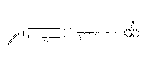
[0050] As shown in FIG. 1, the catheter 10 comprises an elongated
catheter body 12, an
intermediate deflection section 14, a distal section 15, and a deflection
control handle 16
attached to the proximal end of the catheter body 12. In accordance with a
feature of the
present invention, the distal section 15 has at least one flexible tubular
member, with shape
memory of a predetermined two-dimensional (2D) configuration, which when
deployed, or
-6-
CA 02893154 2015-05-28
otherwise released from external force(s) that can generally straighten the
flexible tubular
distal section, assumes the 2D configuration comprising at least two, side-by-
side loops
resembling an "infinity" symbol or a "lazy 8." The deployed 2D configuration
of two or
more, side-by-side loops enables the distal section 15 to contact two or more
pulmonary vein
(PV) regions, including the ostia, in a generally simultaneous manner. Each
loop carries one
or more electrodes, for example, a tip electrode 17 and at least one ring
electrode 19 for
obtaining electrical data from the PV regions and/or ablating the same.
[0051] With reference to FIGS. 2A and 2B, the catheter body 12 comprises
an elongated
tubular construction having a single, axial or central lumen 18. The catheter
body 12 is
flexible, i.e., bendable, but substantially non-compressible along its length.
The catheter
body 12 can be of any suitable construction and made of any suitable material.
A presently
preferred construction comprises an outer wall 20 made of polyurethane or
PEBAX. The
outer wall 20 comprises an imbedded braided mesh of stainless steel or the
like to increase
torsional stiffness of the catheter body 12 so that, when the control handle
16 is rotated, the
intermediate section 14 of the catheter 10 will rotate in a corresponding
manner.
[0052] The outer diameter of the catheter body 12 is not critical, but
is preferably no
more than about 8 french, more preferably about 7 french. Likewise, the
thickness of the
outer wall 20 is not critical, but is thin enough so that the central lumen 18
can
accommodate a puller wire, one or more lead wires, and any other desired
wires, cables or
tubes. If desired, the inner surface of the outer wall 20 is lined with a
stiffening tube 25 to
provide improved torsional stability. A particularly preferred catheter has an
outer wall 20
with an outer diameter of from about 0.090 inch to about 0.94 inch and an
inner diameter of
from about 0.061 inch to about 0.065 inch.
-7-
CA 02893154 2015-05-28
[0053] As shown in FIGS. 2A, 2B and 2C, the intermediate section 14
comprises a short
section of tubing 22 having multiple lumens, for example, four lumens 31, 32,
33 and 34.
The first lumen 31 carries one or more lead wires 40 or other wires discussed
further below,
the second lumen 32 carries a puller wire 24, and the third lumen 33 near its
distal end
carries a proximal end of a shape-memory support member 38. The fourth lumen
34 carries
a cable 26 for an electromagnetic position sensor 30. The tubing 22 is made of
a suitable
non-toxic material that is preferably more flexible than the catheter body 12.
One suitable
material for the tubing 22 is braided polyurethane, i.e., polyurethane with an
embedded
mesh of braided stainless steel or the like. The size of each lumen is not
critical, but is
sufficient to house the lead wires, puller wire or support member.
[0054] The useful length of the catheter, i.e., that portion that can be
inserted into the
body excluding the distal section 15, can vary as desired. Preferably the
useful length ranges
from about 110 cm to about 120 cm. The length of the intermediate section 14
is a relatively
smaller portion of the useful length, and preferably ranges from about 3.5 cm
to about 10
cm, more preferably from about 5 cm to about 6.5 cm.
[0055] A preferred means for attaching the catheter body 12 to the
intermediate section
14 is illustrated in FIGS. 2A and 2B. The proximal end of the intermediate
section 14
comprises an outer circumferential notch 27 that receives the inner surface of
the catheter
body 12. The intermediate section 14 and catheter body 12 are attached by glue
or the like.
[0056] If desired, a spacer (not shown) can be located within the catheter
body between
the distal end of the stiffening tube (if provided) and the proximal end of
the intermediate
section. The spacer provides a transition in flexibility at the junction of
the catheter body
and intermediate section, which allows this junction to bend smoothly without
folding or
-8-
CA 02893154 2015-05-28
kinking. A catheter having such a spacer is described in U.S. Pat. No.
5,964,757, the
disclosure of which is incorporated herein by reference.
[0057] Extending from the distal end of the intermediate section 14 is
the distal section
15, as shown in FIGS. 3A, 3B and 3C. Each flexible elongated member 42 of the
distal
section 15 comprises an elongated support member 38 with shape memory, and a
non-
conductive covering or tubing 28 covering the support member 38. The member 42
has a
length ranging from about 20 mm to about 300 mm, more preferably about 100 mm
to about
200 mm, still more preferably about 120 mm, but can vary as desired. Each loop
formed by
a respective member 42 has a diameter or width ranging from about 4 mm to
about 40 mm,
more preferably about 10 mm to about 25 mm, still more preferably about 17 mm,
but can
vary as desired. The support member 38 is made of a material having shape-
memory, i.e.,
that can be temporarily straightened or bent out of its original shape upon
exertion of a force
and is capable of substantially returning to its original shape upon removal
of the force. One
suitable material for the support member 38 is a nickel/titanium alloy. Such
alloys typically
comprise about 55% nickel and 45% titanium, but may comprise from about 54% to
about
57% nickel with the balance being titanium. A nickel/titanium alloy is
nitinol, which has
excellent shape memory, together with ductility, strength, corrosion
resistance, electrical
resistivity and temperature stability. The non-conductive covering 28 can be
made of any
suitable material, and is preferably made of a biocompatible plastic such as
polyurethane or
PEBAX. If desired, the support member 38 can be eliminated and the distal end
of the non-
conductive covering 28 can be pre-formed to have the desired curvature or
configuration.
[0058] At a junction of the intermediate section 14 and the distal
section 15, the non-
conductive covering 28 is attached to the tubing 22 of the intermediate
section by glue 37 or
the like. The support member 38 extends from the third lumen 33 into the non-
conductive
-9-
CA 02893154 2015-05-28
covering 28. The proximal end of the support member 38 terminates within the
third lumen
34, approximately about 5 mm from the distal end of the tubing 22, so as not
to adversely
affect the ability of the intermediate section 14 to deflect. However, if
desired, the proximal
end of the support member 38 can extend into the catheter body 12.
[0059] With reference to FIGS. 4A-4E, the distal section 15 (illustrated
without any tip
or ring electrodes for clarity) comprises a generally linear (one dimensional)
but flexible
tubular member 42 with shape memory, as provided by the support member 38, of
a
predetermined 2D configuration (FIG. 4E) comprising at least two side-by-side
loops A and
B that are generally coplanar with each other and are generally joined or
intersecting (or
having an appearance of being joined or intersecting) with each other at their
closest
location X to resemble an "infinity" symbol or a lazy 8 (used interchangeably
herein). It is
understood that the term "loop" as used herein does not necessarily mean a
closed
configuration accomplished with a single continuous flexible tubular member,
but rather that
one or more flexible tubular members may be configured to resemble or have the
appearance of a loop for purposes of enabling contact with tissue surface in a
pattern of a
generally closed configuration so as to surround or encircle a respective
region, for example,
a respective PV ostium. In other words, it is understood that the term "loop"
is used with
respect to the resulting "stamp" or "imprint" pattern of one or more flexible
tubular
members on tissue surface of purposes of mapping and/or ablation a region, for
example, a
PV ostium.
[0060] The tubular member 42 may be described as comprising a distal
portion 42D and
a proximal portion 42P. The flexible construction of the member 42 allows it
to be
generally straightened and advanced distally through a tube, for example, a
guiding sheath
36 (FIG. 4A). As the distal portion 42D passes and exits the distal end of the
guiding sheath
-10-
CA 02893154 2015-05-28
36, the distal portion 42D assumes a distal or first "S" configuration under
its shape-memory
(FIG. 4B). As the member 42 of the distal section 15 continues to be deployed,
shape-
memory of the member 42 causes the distal portion 42D to fold or flip back on
the proximal
portion 42P (FIG. 4C) via an angle 0 equal to about 180 degrees. As the
proximal portion
42P passes and exits the distal end of the guiding sheath 36, the proximal
portion 42P
assumes a proximal or second "S" configuration under its shape memory (FIGS.
and ).
When both the distal and the proximal portions 42D and 42P are fully deployed,
the first and
second "S" configurations are stacked one above the other (FIG. 4F), with one
"S" being
upside down relative to the other, whereupon the member 42 assumes the
predetermined 2D
configuration resembling an infinity symbol effectively forming loop A and
loop B in a
side-by-side configuration (FIG. 4E). Loops A and B (and the distal and
proximal portions
42D and 42B) lie generally in a common plane, which is also occupied by the
intermediate
deflection section 14 when in a neutral state (FIG. 4F), or the 2D
configuration of the
member 42 of the distal section 15 may be deflected at an angle a by the
intermediate
deflection section 14 so that they lie in different planes (FIG. 4G).
[0061] In another embodiment, as shown in FIGS. 11A-11D, the distal
portion 42D
forming the distal "S" configuration does not flip or fold back onto the
proximal portion 42P
forming the proximal "S" configuration. Rather, the curvature of the distal
"S"
configuration 42D is continuous with the curvature of the proximal "S"
configuration 42P
such that the two remain generally in the same plane as the distal section 15
is deployed.
[0062] Regardless of the manner by which loops A and B are achieved, the
2D
configuration of the distal section 15 is effectively positioned over a pair
of adjacent ostia
80A and 80B of the pulmonary veins in the left atrium LA via a transceptal
approach from
the right atrium RA, as shown in FIG. 5. With loop A positioned over ostium
80A and loop
-11-
CA 02893154 2015-05-28
B over ostium 80B, electrical signals in the regions of ostia A and B can be
read
simultaneously with the use of catheter 10 without the need for a second
catheter. Where
the catheter 10 is adapted for ablation, regions of ostia A and B may be
ablated
simultaneously with the catheter 10. The catheter 10 may be relocated to map
and/or ablate
pulmonary vein ostia 82A and 82B.
[0063] The 2D configuration resembling the infinity symbol may be
achieved with a
variety of different constructions, including those described below. In FIGS.
6A-6G, the
member 42 of the distal section 15 has a different shape memory configuration
to achieve
the 2D configuration resembling the infinity symbol. In this embodiment, the
member 42
has a distal portion 42D providing a generally closed loop or "0"
configuration (FIG. 6A)
and a first proximal portion 42P providing a first proximal "C" configuration
extending
generally in a common plane with the distal "0" configuration (FIG. 6A). As
the portion
42P is fully deployed outside of the guiding sheath 36, the portions 42D and
42P fold over
(under the shape memory of the member 42) via angle O of about 180 degrees
onto a more
proximal portion 42P' (FIG. 6B) which provides a second "C" configuration
opposite in
orientation to the less proximal "C" configuration of the portion 42P, so as
to achieve the 2D
configuration resembling the infinity symbol. As the portion 42P' is fully
deployed outside
of the guiding sheath 36, the 2D configuration folds or flips under (under the
shape memory
of the distal section 15) via angle a of about 90 degrees to be generally
perpendicular to the
intermediate deflection section 14 (FIG. 6F). It is understood that the angle
at which the 2D
configuration extends from the intermediate deflection section 14 may be at
any angle as
desired or appropriate. For example, 2D configuration and the deflection
section 14 can lie
generally in the same common plane (FIG. 6G). That is, while the 2D
configuration is
transverse to the deflection section 14 thereby adopting a "T" configuration,
the 2D
-12-
CA 02893154 2015-05-28
configuration can lie in a different plane from the deflection section 14
(FIG. 6F) or lie in
the same plane as the deflection section (FIG. 6G).
[0064] In another embodiment, as shown in FIGS. 12A-12C, the distal
portion 42D
forming loop A does not flip or fold back. Rather, the curvature of the
proximal portion 42P
configuration 42D continues to form loop B. Comparing and contrasting the
embodiments
of FIGS. 11A-11D and FIGS. 12A-12D, the loops A and B of both embodiments
remain in
generally the same plane during deployment of the distal section 15,whereas a
proximal end
of 2D configuration can be at a location along an outer segment or arc of a
loop (FIG. 11D)
or at a location along an inner segment or arc of a loop (FIG. 12C).
[0065] FIG. 7A illustrate another embodiment wherein the distal section 15
has a distal
portion 15D providing a first loop or "0" configuration in one direction
(e.g., clockwise)
(FIG. 6A) and a proximal portion 15P providing a second loop or "0"
configuration in the
opposite direction (e.g., counterclockwise), both of which lie generally in a
common plane
as the intermediate deflection section 14, where the common location X of
Loops A and B is
generally aligned longitudinally with the deflection section 14.
[0066] FIG. 7B illustrates another embodiment wherein the distal section
15 has a distal
portion 15D providing a first, loop A or "0" configuration in one direction
(e.g.,
counterclockwise) (FIG. 6A) and a proximal portion 15P providing a second loop
B or "0"
configuration in the same direction (e.g., counterclockwise), both of which
lie generally in a
common plane that is generally perpendicular to the intermediate deflection
section 14.
Moreover, the common location X of Loops A and B is offset longitudinally from
the
deflection section 14.
[0067] FIGS. 8A-8E illustrate yet another embodiment wherein the distal
section 15
comprises at least two generally parallel elongated flexible tubular members
42A and 42B
-13-
CA 02893154 2015-05-28
extending from the distal end of the intermediate deflection section 14. As
shown in FIGS.
10A and 10B, each member 42A and 42B has a respective elongated shape memory
support
member 38A and 38B and a respective nonconductive covering or tubing 28A and
28B.
The flexible construction of the members 42A and 42B allows them each to be
generally
straightened and advanced distally through a tube, for example, a guiding
sheath 36 (FIG.
8A). As the members 42A and 42B are fully deployed upon exiting the distal end
of the
guiding sheath 36 (FIG. 8B), their shape-memory begins to curve each of the
members 42A
and 42B back on itself into a loop or "0" configuration, and to separate from
each other by
pivoting outwardly from their proximal ends in opposite directions (FIGS. D
and E).
Separated by the angle of about 180 degrees, loops A and B forming the 2D
configuration
generally resembling an infinity symbol lie generally in a common plane, with
the deflection
section 14 being generally perpendicular to the 2D configuration of distal
section 15 (FIG.
8F). Again, it is understood that depending on the configuration of the
junction between the
intermediate deflection section 14 and the distal section 15, the 2D
configuration of the
distal section 15 and the deflection section 14 may be in a common plane, be
perpendicular
to each other, or, in fact, be at any angular orientation to each other, as
needed or desired.
[0068] It is understood that the loops of 2D configuration of the distal
section 15 need
not be of the same size or same shape to each other. One loop may be smaller
than the
other(s). One loop may be more circular and the other(s) more oval. Each loop
does not
need to form a completely closed loop, but should be at least about 270
degrees, more
preferably at least about 320 degrees, and more preferably at least about 340
degrees. Each
loop carries one or more electrodes, for example, at least one ring electrode,
for obtaining
electrical data from the PV regions and/or ablating the same, and if desired
or appropriate, a
tip electrode.
-14-
CA 02893154 2015-05-28
[0069] The tip electrode 17 for any member 42 of the 2D configuration of
the distal
section 15 is mounted on a distal end of the member 42. As shown in FIG. 7,
the tip
electrode 17 has an exposed distal portion 17D, and a proximal stem 17P that
extends into
the non-conductive covering 28 and is fixed therein by polyurethane glue or
the like.
[0070] The electrode lead wire 40T is connected at its distal end to the
tip electrode 17.
The distal end of the lead wire 40T is soldered in a first blind hole 51 in
the proximal end of
the tip electrode 17. The lead wire 40T extends between the non-conductive
covering 28
and the support member 38. The proximal end of the lead wire 40T is
electrically connected
to a suitable connector (not shown) in the distal end of the control handle
16, which is
connected to the source of ablation energy, e.g., RF energy, as is known in
the art. The lead
wire 40T extends through the lumen of the nonconductive covering 28, the first
lumen 31 of
the intermediate section 14, the central lumen 18 of the catheter body 12, and
the control
handle 16. In the depicted embodiment, the portion of the lead wire 40T
extending through
the central lumen 18 of the catheter body 12 and the lumen 31 of the
intermediate section 14
may be enclosed within a protective sheath 84 to prevent contact with other
components in
the catheter. The protective sheath can be made of any suitable material,
preferably
polyimide. The protective sheath may be anchored at its distal end to the
proximal end of the
intermediate section 14 by gluing it in the first lumen 31 with polyurethane
glue or the like.
As would be recognized by one skilled in the art, the protective sheath can be
eliminated if
desired.
[0071] One or more ring electrodes 19 are mounted on the non-conductive
covering 28
of the distal section 15 for mapping the region to be ablated before ablation,
conducting
ablation, and/or after ablation to assure that the resulting lesions blocked
the electrical
activity as desired. A description of a catheter including such ring
electrodes is described in
-15-
CA 02893154 2015-05-28
U.S. Patent No. 8545495, entitled A Catheter Having Circular Ablation
Assembly, the entire
disclosure of which is incorporated herein by reference.
[0072] The ring electrodes 19 can be made of any suitable solid
conductive material,
such as platinum or gold, preferably a combination of platinum and iridium,
and mounted
onto the non-conductive cover 28 with glue or the like. Alternatively, the
ring electrodes 19
can be formed by coating the non-conductive cover 28 with an electrically
conducting
material, like platinum, gold and/or iridium. The coating can be applied using
sputtering, ion
beam deposition or an equivalent technique.
[0073] In the embodiment of FIG. 3B, each ring electrode 19 is mounted
by first
forming a hole 62 in the non-conductive cover 28. A respective electrode lead
wire 40R is
fed through the hole 62, and the ring electrode 19 is welded in place over the
lead wire 40R
and the non-conductive cover 28. The lead wires 40R extend through the lumen
of the non-
conductive cover 28, the first lumen 31 of the intermediate deflection section
14, and
through the central lumen 18 of the catheter body 12. The proximal end of each
lead wire
40R is electrically connected to the suitable connector (not shown) in the
control handle 16.
[0074] The number of ring electrodes 19 on the distal section 15 can
vary as desired.
Preferably the number of ring electrodes ranges from about six to about
twenty, more
preferably from about eight to about twelve. In one embodiment, the distal
section 15 carries
ten ring electrodes. The ring electrodes 19 can be approximately evenly spaced
along the
distal section 15. In one embodiment, a distance of approximately 5 mm is
provided
between the centers of adjacent ring electrodes 19.
[0075] In another embodiment, the distal section 15 includes a series of
ring electrode
pairs. Each ring electrode pair comprises two closely-spaced ring electrodes.
As used herein,
the term "ring electrode pair" refers to a pair of ring electrodes that are
arranged closer to
-16-
CA 02893154 2015-05-28
each other than they are to the other adjacent ring electrodes. Preferably the
distance
between two electrodes of an electrode pair is less than about 3 mm, more
preferably less
than about 2 mm, still more preferably from about 0.5 mm to about 1.5 mm. The
number of
electrode pairs can vary as desired, and preferably ranges from 6 to 14 pairs,
more
preferably 10 pairs.
[0076] The distal section 15 may carry 10 pairs of electrodes with a
space of
approximately 1 mm between the two electrodes of each pair. Preferably each
ring electrode
is relatively short, having a length ranging from about 0.4 mm to about 0.75
mm, with the
most distal ring electrode being longer than the other ring electrodes,
preferably having a
length ranging from about 1 mm to about 1.5 mm. The longer ring electrode
provides a
visual reference to the user when the catheter is being viewed under
fluoroscopy. By having
one ring electrode, such as the most distal ring electrode, sized differently
from the other
ring electrodes, the user has a reference point when viewing the catheter
under fluoroscopy.
[0077] Regardless of the size and number of the ring electrodes, the
electrode pairs are
preferably approximately evenly spaced along the distal section 15. The
closely-spaced
electrode pairs allow for more accurate detection of near field pulmonary vein
potential
versus far field atrial signals, which is very important when trying to treat
atrial fibrillation.
Specifically, the near field pulmonary vein potentials are very small signals
whereas the
atria, located very close to the pulmonary vein, provides much larger signals.
Accordingly,
even when the mapping array is placed in the region of a pulmonary vein, it
can be difficult
for the physician to determine whether the signal is a small, close potential
(from the
pulmonary vein) or a larger, farther potential (from the atria). Closely-
spaced bipoles permit
the physician to more accurately determine whether he is looking at a close
signal or a far
signal. Accordingly, by having closely-spaced electrodes, one is able to
target exactly the
-17-
CA 02893154 2015-05-28
locations of myocardial tissue that have pulmonary vein potentials and
therefore allows the
clinician to deliver therapy to the specific tissue. Moreover, the closely-
spaced electrodes
allow the physician to determine the exact anatomical location of the
ostium/ostia by the
electrical signal.
[0078] The pair of thermocouple wires 53 and 54 are provided for monitoring
the
temperature of any tip electrode 17. Any conventional temperature sensor,
e.g., a
thermocouple or thermistor, may be used. In the embodiment shown in FIG. 7,
the
thermocouple is formed by an enameled wire pair. One wire of the wire pair is
a copper
wire 53, e.g., a number "40 AWG" copper wire. The other wire of the wire pair
is a
constantan wire 54. The wires 53 and 54 of the wire pair are electrically
isolated from each
other except at their distal ends where they are twisted together, covered
with a short piece
of plastic tubing 55, e.g., polyimide, and covered with epoxy. The plastic
tubing 55 is then
attached in a second blind hole 56 of the tip electrode 17, by polyurethane
glue or the like.
Alternatively, the wires 53 and 54 can be soldered into the second blind hole
56 or otherwise
attached to the tip electrode 17. The wires 53 and 54 extend through the first
lumen 31 in
the intermediate section 14 (FIG. 3C) and through the central lumen 18 of the
catheter body
12 along with the lead wire 40T and 40R (FIG. 2A). The wires 53 and 54 then
extend out
through the control handle 16 and to a connector (not shown) connectable to a
temperature
monitor (not shown).
[0079] Additionally, a safety wire 57 is provided to further secure the tip
electrode 17 to
the distal section 15 and assure that the tip electrode does not detach from
the catheter. The
safety wire is preferably a metal wire having its distal end soldered in a
third blind hole 58
in the tip electrode 17 and its proximal end soldered or otherwise attached in
the control
handle 16. In the depicted embodiment, the safety wire 57 extends through the
first lumen
-18-
CA 02893154 2015-05-28
31 in the intermediate section 14 (FIG. 3C) and through the central lumen 18
of the catheter
body 12 (FIG. 2A) along with the lead wires 40T and 40R and thermocouple wires
53 and
54. Other arrangements for attaching the safety wire can be provided, as would
be
recognized by one skilled in the art, or the safety wire can be eliminated.
[0080] An electromagnetic position sensor 30 is housed in the lumen of the
nonconductive covering 28 at or near its distal end, just proximal of the tip
electrode 17.
The sensor cable 26 extends from the sensor 30 and through the lumen of the
covering 28
(FIG. 3B), the lumen 34 of the tubing 22 of the deflection section 14(FIG.
2C), the lumen 18
of the catheter body 12 (FIG. 2A) and into the control handle 16
[0081] The puller wire 24 is provided for deflection of the intermediate
section 14. The
puller wire 24 extends through the catheter body 12, is anchored at its
proximal end to the
control handle 16, and is anchored at its distal end to a distal end of the
intermediate section
14. The puller wire 24 is made of any suitable metal, such as stainless steel
or Nitinol, and is
preferably coated with TEFLON or the like. The coating imparts lubricity to
the puller wire
24. The puller wire 24 preferably has a diameter ranging from about 0.006 to
about 0.010
inch.
[0082] A compression coil 66 is situated within the catheter body 12 in
surrounding
relation to the puller wire 24, as shown in FIG. 2B. The compression coil 66
extends from
the proximal end of the catheter body 12 to the proximal end of the
intermediate section 14.
The compression coil 66 is made of any suitable metal, preferably stainless
steel. The
compression coil 66 is tightly wound on itself to provide flexibility, i.e.,
bending, but to
resist compression. The inner diameter of the compression coil 66 is
preferably slightly
larger than the diameter of the puller wire 24. The Teflon coating on the
puller wire 24
-19-
CA 02893154 2015-05-28
allows it to slide freely within the compression coil 66. The outer surface of
the compression
coil 66 is covered by a flexible, non-conductive sheath 68, e.g., made of
polyimide tubing.
[0083] The compression coil 66 is anchored at its proximal end to the
outer wall 20 of
the catheter body 12 by a proximal glue joint (not shown) and at its distal
end to the
intermediate section 14 by a distal glue joint 72. Both glue joints may
comprise
polyurethane glue or the like. The glue may be applied by means of a syringe
or the like
through a hole made between the outer surface of the catheter body 12 and the
central lumen
18. Such a hole may be formed, for example, by a needle or the like that
punctures the outer
wall 20 of the catheter body 12 which is heated sufficiently to form a
permanent hole. The
glue is then introduced through the hole to the outer surface of the
compression coil 66 and
wicks around the outer circumference to form a glue joint about the entire
circumference of
the compression coil.
[0084] The puller wire 24 extends into the second lumen 32 of the
intermediate section
14. In the illustrated embodiment, the puller wire 24 is anchored at its
distal end to the distal
end of the intermediate section 14, as shown in FIG. 3. Specifically, a T-
shaped anchor is
formed, which comprises a short piece of tubular stainless steel 80, e.g.,
hypodermic stock,
which is fitted over the distal end of the puller wire 64 and crimped to
fixedly secure it to the
puller wire. The distal end of the tubular stainless steel 80 is fixedly
attached, e.g., by
welding, to a cross-piece 82 formed of stainless steel ribbon or the like. The
cross-piece 82
sits beyond the distal end of the second lumen 32. The cross-piece 82 is
larger than the
lumen opening and, therefore, cannot be pulled through the opening. The distal
end of the
second lumen 32 is then filled with glue or the like, preferably a
polyurethane glue 37.
Within the second lumen 32 of the intermediate section 14, the puller wire 24
extends
through a plastic, preferably Teflon, puller wire sheath 39, which prevents
the puller wire 24
-20-
CA 02893154 2015-05-28
from cutting into the wall of the tubing 22 of the deflection section 14 when
the deflection
section is deflected.
[0085] Longitudinal movement of the puller wire 24 relative to the
catheter body 12,
which results in deflection of the intermediate section 14, is accomplished by
suitable
manipulation of the control handle 16. Examples of suitable control handles
for use in the
present invention are disclosed, for example, in U.S. Pat. Nos. Re 34,502 and
5,897,529, the
entire disclosures of which are incorporated herein by reference. It is
understood that where
bi-directional deflection is desired, the catheter may be configured to
provide a second
puller wire that passes through a lumen (generally diametrically opposite of
the lumen 32 for
the first puller wire 24) in the deflection section 14 and is responsive to
the control handle
16.
[0086] In use, a suitable guiding sheath 36 is inserted into the patient
with its distal end
positioned at a desired mapping and/or ablation location, as shown in FIG. 5.
An example of
a suitable guiding sheath for use in connection with the present invention is
the Preface
Braided Guiding Sheath, commercially available from Biosense Webster, Inc.
(Diamond
Bar, Calif.). The distal end of the sheath is guided into the right atrium RA
and then into the
left atrium LA via a transceptal approach. The catheter 10 is passed through
the guiding
sheath 36. In particular, as the distal section 15 of the catheter is fed into
the proximal end
of the guiding sheath 36, the member(s) 42 of the distal section 15 are
straightened to fit
through the sheath 36. After the distal section 15 of the catheter is
positioned at the desired
location in the left atrium LA, the guiding sheath 36 is pulled proximally,
exposing at least
the distal section 15, if not also the deflectable intermediate section 14, as
needed. Outside
of the guiding sheath 36, the distal section 15 assumes the 2D configuration
under its shape
memory providing at least loops A and B. The user then manipulates the
catheter to position
-21-
CA 02893154 2015-05-28
the 2D configuration of the distal section 15 such that each loop sits over a
respective
ostium. With the distal section 15 in contact with the ostia, electrical
activity in the regions
of at least two ostia can be sensed simultaneously by the electrodes on the
loops A and B
without the use of a second catheter. If desired, the electrodes also can be
energized to
ablate in the regions of at least two ostia simultaneously without the use of
a second
catheter.
[0087] If desired, two or more puller wires can be provided to enhance
the ability to
manipulate the intermediate section. In such an embodiment, a second puller
wire and a
surrounding second compression coil extend through the catheter body and into
an
additional off-axis lumen in the intermediate section. Suitable designs of
catheters having
two or more puller wires, including suitable control handles for such
embodiments, are
described, for example, in U.S. Pat. Nos. 6,123,699; 6,171,277; 6,183,435;
6,183,463;
6,198,974; 6,210,407 and 6,267,746, the entire disclosures of which are
incorporated herein
by reference.
[0088] The preceding description has been presented with reference to
presently
preferred embodiments of the invention. Workers skilled in the art and
technology to which
this invention pertains will appreciate that alterations and changes in the
described structure
may be practiced without meaningfully departing from the principal, spirit and
scope of this
invention. As understood by one of ordinary skill in the art, the drawings are
not necessarily
to scale. Also, different features of different embodiments may be combined as
needed or
appropriate. Moreover, the catheters described herein may be configured to
apply various
energy forms, including microwave, laser, RF and/or cryogens. Accordingly, the
foregoing
description should not be read as pertaining only to the precise structures
described and
-22-
CA 02893154 2015-05-28
illustrated in the accompanying drawings, but rather should be read consistent
with and as
support to the following claims which are to have their fullest and fair
scope.
-23-