Note: Descriptions are shown in the official language in which they were submitted.
HEMOSTATIC GLOVE DEVICE AND METHOD FOR USE OF SAME
[0001] This paragraph has been lefi intentionally blank.
FIELD OF THE INVENTION
[0002] The present invention relates to medical devices for hemostasis. In
particular, the
invention relates to hemostatic gloves and methods of use thereof
BACKGROUND
[0003] Rapid control of severe bleeding at wound sites is of critical
importance in saving
lives. Blood loss due to uncontrolled hemorrhage is a major contribution to
combat and
civilian trauma death before reaching definitive care such as a hospital.
Improvements in the
ability to control heavy bleeding in the pre-hospital will vastly improve the
survival outcomes
of trauma. Severe wounds can often be inflicted in remote areas or in
situations, such as on a
battlefield, where adequate medical assistance is not immediately available.
In these
instances, it is important to stop bleeding long enough to allow the injured
patient (person or
animal) to receive medical attention.
[0004] Several approaches to date have been used to rapidly stop bleeding
in a pre-
hospital setting. The most common approach is to provide manual pressure to
compress the
damaged blood vessel for an indefinite period of time until bleeding stops or
transport to
definitive care. This is quite difficult not only because of the high blood
flow under pressure
from a severe wound such as from an artery, but also because it requires
knowledge of
anatomy to locate the exact site of bleeding and the location where manual
pressure will
staunch blood flow (the vessel may be deep or the vessel may have retracted).
Unless the
patient is able to provide self-treatment, manual pressure can be considered
to be impractical
in many emergency situations due to the need of having one skilled person
remain close to
the patient to exert continuous pressure, which prevents the care provider
from performing
other critical life saving functions. Furthermore, heavy direct compression
may aggravate
damage to other wounded tissues such as fractures, and is inconvenient to
apply over
1
Date Recue/Date Received 2020-04-17
CA 02894445 2015-06-09
WO 2014/091310 PCT/IB2013/003165
irregularly shaped or sensitive body parts. With this approach, it can take a
long time to form
a stable clot at the injured vessel due to high blood volumes, even when
pressed firmly
[0005] The gold standard in a hospital is the use of sutures, staples,
cautery, tissue glues
and adhesives by surgical personnel. Appropriate in a hospital setting, these
are generally
unfit for use in the field. The disadvantage of these closure methods in a pre-
hospital setting
is that they are required to be performed by an expert in a controlled
environment, and take a
significant length of time to apply. By example, cyanoacrylate glue's
inability to bind to wet
surfaces make topical adhesives of this nature inappropriate for use managing
arterial
bleeding in the pre-hospital. Severe wounds can often be inflicted in remote
areas or in
situations, such as in a rural setting, where adequate medical assistance is
not immediately
available. In these instances, it is important to stop bleeding, even in less
severe wounds, long
enough to allow the injured person (or animal) to receive definitive care at a
site such as a
hospital.
[0006] An alternative to manual pressure is the use of a tourniquet. While
utilization of
strap style tourniquets have been widely accepted for military field care for
centuries, these
devices present a number of disadvantages. They must be applied with
sufficient constricting
force to cause ischemia distal of the site of application. The induced
ischemia is both
exceedingly painful to the victim and is a common cause of soft tissue and
neurological
damage to these body parts if left on too long. Tourniquets are slow and
difficult to maneuver
and place around the extremity. They are limited in its application by how
proximal they can
be placed on a limb and do not address major junctional bleeding in the groin
or axilla (where
larger blood vessels run) or other areas of the body (trunk, neck, scalp,
etc).
[0007] Packing wounds with conventional field dressings have been used for
centuries by
military and civilian trauma personnel to slow or stop bleeding. Pressure on
the wound
immediately at the point of injury with pressure dressings and/or packed field
dressings can
minimize bleeding by distributing pressure evenly over the wound to achieve
hemostasis, as
well as by decreasing dead space into which blood can accumulate. Multilayered
woven and
non-woven fabric or all-purpose wound pads, such as gauze pads of various
forms of cotton
and other cellulose-type material, absorb significant volumes of blood, and
act like a sponge.
Although these materials have been shown to be the standard of care, they can
be slow to
unroll and apply when it is unsafe to do so (e.g. dangerous situations such as
when under
2
CA 02894445 2015-06-09
WO 2014/091310 PCT/IB2013/003165
fire); it is difficult to unroll these materials and pack the wound while
maintaining constant
manual pressure directly on the vessel since the changing of hands is
necessary for unrolling
and insertion of each length of bandage into the wound; and these materials do
not accelerate
the patient's ability to clot beyond the natural clotting mechanism.
[0008] As a result, there is an increased tendency for absorbent dressings
to become
saturated with blood due to unstemmed bleeding. It takes anatomical knowledge
to know
where to pack the dressings and where to hold pressure onto the vessel. It
takes significant
time to administer, and are cumbersome in rolls or z-folds requiring a
technical knowledge to
maintain as constant a pressure as possible on the bleeding vessel.
[0009] Additionally, or alternatively, several blood clotting materials are
generally known,
and are typically in the form of a powder or a fine particulate in which the
surface area of the
material concentrates clotting factors and leads to hemostasis. Undesirable
side effects can
occur, as the powders can produce an exothermic reaction upon the application
of the
material to blood. Oftentimes excess material is unnecessarily poured onto a
wound, which
can exacerbate the exothermic effects. Depending upon the specific attributes
of the material,
the resulting exothermia may be sufficient to cause discomfort to or even burn
the patient;
they can also lead to migration of the powder into the vasculature risking
clots/emboli away
from the wound site.
[00101 To avoid the previous disadvantages, hemostatic dressings have
become a method
of choice, for insertion into wounds to accelerate the clotting process in
situ. By dispersing an
adsorbed biocompatible polymer throughout the dressing, the dressing acts as a
scaffold to
initiate clotting and increase clot adhesion to bandage fiber surfaces at the
wound site.
Several hemostatic dressings have been developed that accelerate the
production of a stable
clot through the clotting cascade. One example includes dressings that contain
a high
concentration of human clotting factors. Another example includes gauze
bandages (wound
in a roll or cut into sheets) impregnated with mineral agents causing water
absorption from
the blood to the mineral to concentrate clotting factors. Another example
includes gauze
bandages (wound in a roll or cut into sheets) impregnated with thrombogenic
polysaccharide
polymer capable of attracting negatively charged blood cells to the bandage to
inducing
clotting.
3
CA 02894445 2015-06-09
WO 2014/091310 PCT/IB2013/003165
[0011] Hemostatic dressings have several disadvantages. First is cost; many
of these
agents are proteins in the "clotting chain," such as, fibrinogen, thrombin,
Factor VIII and the
like. The cost of products made from these products are very high. Second,
certain bandages
can be of limited use in wet conditions (e.g. clays), since once the bandage
gets wet, the
active ingredient is unable to concentrate clotting factors, thereby reducing
the clotting
potential. Third, such dressings are slow to unroll and apply when it is
unsafe to do so (e.g.
dangerous situations such as when under fire); difficult to unroll and pack
the wound while
maintaining constant manual pressure directly on the vessel since the changing
of hands is
necessary for unrolling and insertion of each length of bandage into the
wound. Finally, lack
of flexibility makes certain bandages difficult to press into wounds without
crumbling or
breaking.
[0012] What is required is a device designed for use in a field environment
that can be
applied immediately after wounding. A device that addresses these critical
aspects of injury
care and that will have significant impact on acute events as well as provide
an improved
outcome late into the time course of treatment and recovery is necessary. It
would therefore
be advantageous to provide a means of combining the advantages of applying a
hemostatic
dressing with the simplicity of applying traditional manual pressure via a
glove to the wound
to reduce blood loss.
[0013] Gloves come in many varieties; each designed to protect a person's
hand from
some sort of hazard without overly impairing the person's manual dexterity.
For example,
latex gloves protect health care providers such as combat medics and EMT
personnel from
external contamination while allowing them to handle small, delicate surgical
tools, and also
prevent the patient from being contaminated by microorganisms on the hands of
the health
care provider.
[0014] Beyond infection control, there are currently no gloves in use that
are used/worn
by medical personnel to treat patients directly. Bandages that are used by
caregivers tend to
be in the form of rolls or sheets of woven or non-woven fabric. Gloves that
are worn by the
caregiver act as a barrier for protection of the caregiver, not for the
treatment of the patient by
the caregiver.
[0015] Bandage gloves are known in the art, and are primarily used for
treatment of
fingers and hands that can suffer a variety of ailments and injuries such as
blisters, arthritis,
4
CA 02894445 2015-06-09
WO 2014/091310 PCT/IB2013/003165
hand burns, and the like. Examples of such are cloth-like wraps and finger
sleeves that have
been developed to be placed around an ailing joint to provide warmth and
support.
[0016] In U.S. Pat. No. 7,767,874 issued to Kellogg et al., a medical glove
is provided for
removal of excess fluids from body tissue and is particularly useful to treat
soft tissue
inflammation, damage, edema and/or lymphedema.
[0017] In U.S. Pat. No. 5,701,918 issued to Jiraki, provides a glove used
in endotracheal
intubations having one or more finger extension members attached to and
extending
outwardly from fingertip portions of one or more finger covers thereof
[0018] In U.S. Pat. No. 5,614,202 issued to DeFina, a moisturizing glove is
disclosed in
which a middle layer saturated with lotion, an exterior layer of non-porous
material, and an
inner layer having multiple pores, creates a cavity for receiving and
enveloping a hand.
[0019] In U.S. Pat. No. 4,853,978 issued to Stockum, an antimicrobial
medical glove with
an inner coating containing a slow release antimicrobial agent sufficient to
maintain an
essentially bacteria-free and fungus-free environment within a donned glove.
[0020] In U.S. Pat. No. 7,230,153 issued to Flick, a silver bandage (e.g.
Silverlon) is
formed into a glove and is applied to protect and treat hand burns/wounds from
infection.
[0021] However, the art fails to describe or suggest a use of gloves to
provide external
hemostasis treatment, wherein the gloves are worn by the caregiver to treat or
provide
therapeutic needs. What is desirable is a method to reduce blood loss
during/after trauma that
mimics the action of manual pressure applied by a first responder, but also
mimics the last
step of the physiological coagulation mechanism through the use of an adsorbed
hemostatic
agent to stop bleeding. Furthermore, a device designed for use in a field
environment that can
be applied immediately to a wound to stop bleeding is desirable.
SUMMARY OF THE INVENTION
[0022] The present invention relates to a hemostatic glove device for
promoting
hemostasis at or within a wound site. The glove device of the present
disclosure is designed
to accelerate clotting and staunch bleeding concurrent with manually applied
pressure at a
wound site.
[0023] Therefore, in one aspect, the present disclosure provides a
hemostatic glove
adapted to be worn by a user. The glove includes an absorbent fabric layer
having a
hemostatic agent impregnated or disposed on the fabric layer. The glove may
further include
CA 02894445 2015-06-09
WO 2014/091310 PCT/IB2013/003165
an interior elastomeric layer forming a fluid barrier to protect the user from
contact with fluid
from the wound site. The absorbent fabric layer is seperable from the interior
elastomeric
layer such the fabric layer may be removed from the user's hand and used to
pack the wound.
[0024] In another aspect, the present disclosure provides a method for
promoting
hemostasis in a wound of a patient. The method includes (a) contacting the
wound with a
hemostatic glove of the present invention; and (b) applying manual pressure on
or within the
wound to limit egress of fluid from the wound, thereby promoting hemostasis in
the wound.
The method may further include separating the absorbent layer from the
elastomeric layer
and removing the absorbent layer from the user's hand and using the absorbent
layer to pack
the wound.
[0025] In another aspect, the present disclosure provides a kit. The kit
may include a
hemostatic glove of the present disclosure and instructions for promoting
hemostasis in a
wound of a patient using the glove.
BRIEF DESCRIPTION OF THE FIGURES
[0026] In the drawings, like elements are assigned like reference numerals.
The drawings
are not necessarily to scale, with the emphasis instead placed upon the
principles of the
present invention. Additionally, each of the embodiments depicted are but one
of a number of
possible arrangements utilizing the fundamental concepts of the present
invention. The
drawings are briefly described as follows.
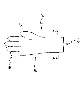
[0027] Figure 1 is a view of a glove in one embodiment of the disclosure.
[0028] Figure 2 is a cross-sectional view of a portion of the glove of
Figure 1 at line A-A
of Figure 1.
DETAILED DESCRIPTION
[0029] The invention relates to a hemostatic glove device. When describing
the present
invention, all terms not defined herein have their common art-recognized
meanings. To the
extent that the following description is of a specific embodiment or a
particular use of the
invention, it is intended to be illustrative only, and not limiting of the
claimed invention. The
following description is intended to cover all alternatives, modifications and
equivalents that
are included in the spirit and scope of the invention, as defined in the
appended claims.
[0030] The invention addresses critical aspects of field care, providing
point of injury care
that will have significant impact on acute events as well as improve outcome
late into the
6
CA 02894445 2015-06-09
WO 2014/091310 PCT/IB2013/003165
time course of treatment and recovery. It is therefore advantageous to provide
a means of
combining the advantages of applying a hemostatic dressing along with applying
a glove to a
wound to perform traditional manual pressure to introduce hemostatic agents to
reduce blood
loss.
[00311 To those ends are provided hemostatic products for combat and
civilian casualty
care. The invention exploits the tendencies of first responders or medics to
use manual
pressure for immediate care. Most medics quickly grab at an open wound as a
first response
to apply manual pressure. The present invention is a solution that integrates
a medical
hemostatic glove which may be adapted to tightly conform over a donned
surgical glove or
itself include an interior surgical glove.
[0032] Light and portable, the glove of the present disclosure is
immediately accessible
and can be applied over surgical gloves within seconds and subsequently acts
as a hemostatic
bandage. For example, the hemostatic glove may be used to pack into a wound
for contact
hemostasis, wherein an absorbent fabric layer of the glove having a hemostasis
agent can be
removed from the caregiver's hands and temporarily inserted into the wound.
[0033] As used herein, "user" or "wearer" refers to a human. As also used
herein, a "hand"
is the terminal part of the human arm located below the forearm consisting of
the wrist, palm,
four fingers, and an opposable thumb.
[0034] With reference to Figure 1, a hemostatic glove (2) of the present
invention is
illustrated. In at least one embodiment, the medical glove (2) is sized to fit
a wearer's hand.
The glove (2) has a closed end (4) and an open end (6). The open end (6) of
the glove (2)
provides an entry point for the wearer's hand. Thus an interior cavity that is
sized for the
wearer's hand is defined by the inner surface of the glove. The glove (2) can
be right handed,
left handed, or ambidextrous. The glove (2) can be considered to have two
"sides" with one
side being adjacent to a palm of the wearer's hand (hereinafter the palm side)
and the other
side being adjacent to the back of the wearer's hand (hereinafter the back
side).
[0035] The glove body (16) includes five fingers (18) at the closed end (4)
of the glove
(2). The five fingers (18) enclose the four fingers and thumb of the wearer's
hand. In some
embodiments, when the glove (2) is worn by the wearer, the glove body (16)
encloses the
wearer's hand. In other embodiments, when the glove (2) is worn by the wearer,
the glove
7
CA 02894445 2015-06-09
WO 2014/091310 PCT/IB2013/003165
body (16) encloses the wearer's hand and at least a portion of the wearer's
forearm adjacent to
the hand.
[0036] In general terms, one embodiment of the hemostatic glove is
configured with an
exterior absorbent fabric layer having a hemostatic agent impregnated or
disposed on the
fabric layer. In such an embodiment, the glove may be donned as an outer layer
over a user's
hand already having a surgical glove on it. Alternatively, the hemostatic
glove is configured
with an interior elastomeric layer which forms a fluid barrier to protect the
user from fluid.
[0037] As shown in Figure 2, the glove may include an outer absorbent
fabric layer (20)
having a hemostatic agent which is disposed over an interior elastomeric layer
(22). The
exterior fabric layer provides for absorption of fluid and hemostasis while
the interior
elastomeric layer provides a barrier to contact with body fluids thereby
preventing exposure
to pathogens or other biohazards to preventing transmission of disease or
contaminants.
[0038] In various embodiments, the elastomeric layer is composed of, but
not limited to,
latex, rubber, nitrile, neoprene, vinyl, and combinations thereof
[0039] In some embodiments, the absorbent fabric layer is woven, non-woven
or a
combination thereof. In general, the type of fabric, thickness of the fabric,
number of layers,
as well as the type of hemostatic agent used may be adjusted as desired for a
particular
clinical application. It is envisioned that a wide variety of fabrics may be
utilized, especially
those that are conventionally used to fabricate bandages. Fabric with the
following
characteristics are envisioned: those of sufficient absorbancy for use on
heavily bleeding
wounds; those capable of significantly slowing blood flow from the wound site
by applied
pressure at the wound vessel/dressing interface; those that do not shed fibers
nor leach out
hemostatic substances into the wound cavity or corresponding vessels; and
those that are not
chemically unsuitable for use as a first aid dressing, for example those that
leave a residue in
the wound that needs to be cleaned out after use.
[0040] In various embodiments, the fabric may be composed of one or more
different
types of fibers, including synthetic or naturally derived fibers. By way of
illustration, fibers
for use in the present invention may include, but are not limited to, glass,
such as fiberglass;
silk fibers; polyester fibers; nylon fibers; ceramic fibers; polysaccharide
fibers including plant
fibers such as raw or regenerated (e.g., chemically processed) bamboo, cotton,
rayon, linen,
ramie, jute, sisal, flax, soybean, corn, hemp, and lyocel; animal fibers such
as wool; lactide
8
CA 02894445 2015-06-09
WO 2014/091310 PCT/IB2013/003165
and/or glycolide polymers; lactide/glycolide copolymers; silicate fibers;
polyamide fibers;
feldspar fibers; zeolite fibers, zeolite-containing fibers; acetate fibers;
plant fibers that have
been genetically engineered to express mammalian coagulation proteins or
mammalian
vasoactive factors. Other fibers that are suitable for use in the present
invention are fibers that
have been covalently modified with polymers to promote water absorbancy (e.g.,
polyvinyl
alcohols) and polymers that contain molecular moieties that activate
hemostatic systems (e.g.,
linear or cyclized-arginine-glycine-aspartate-moieties such as those found in
eptifibatide). In
some embodiments, fibers include plant fibers such as raw or regenerated
(e.g., chemically
processed) bamboo fibers, cotton fibers, and the like, that have high moisture
absorbancy and
that are capable of activating the intrinsic coagulation cascade. The fibers
may be prepared
using conventional methods, including ring, open end (OE), rotor, or air jet
spinning, and
may have counts ranging from 1/1 to 100/1 Ne.
[0041] As will be appreciated by one of skill in the art, the fibers may be
used singly, or in
combinations of two, three, four, or more in a blended or plied state. In
addition, any type of
combination of fibers may be used. For example, in one embodiment, two or more
fibers may
be individually produced and then blended or plied together to form a
composite yarn. In
another embodiment, the fibers may be formed as a conjugate comprising blocks
of the
selected types of fibers, for example alternating blocks of polyesters and
polysaccharides. In
yet another embodiment, the fibers may be formed as a homogeneous combination
of
different threads.
[0042] As discussed herein, the absorbent fiber layer includes one or more
hemostatic
agents which may be impregnated or coat the fiber layer. The hemostatic agent
may be
applied to the entire layer such that it is disposed over the entire glove, or
alternatively be
disposed at discrete locations on the fiber layer, for example, on the palm or
finger regions.
By way of illustration, hemostatic agents that may be used include biological
and chemical
agents, without limitation, procoagulant enzymes, proteins and peptides,
either naturally
occurring, recombinant, or synthetic. Some hemostatic agents include,
rehydrated lyophilized
(RL) platelets, RL blood cells, prothrombin, thrombin, fibrinogen, fibrin,
fibronectin, Factor
X/Xa, Factor VIINIIa, Factor IX/IXa, Factor XI/XIa, Factor XII/XIIa, tissue
factor, von
Willebrand Factor, collagen, elastin, gelatin, synthetic peptides having
hemostatic activity,
clays, chitosan, polyacrylamides, chemically modified cellulose, derivatives
of the above and
9
any combination thereof, other coagulation cofactors such as components of
animal venom,
such as reptilase, or vasoactive agents such as endothelins, thromboxanes,
nitrous oxide (NO)
scavengers, or combinations thereof. These factors, or any of the factors
listed above, may be
in a dry or liquid form when incorporated into the fabric layer of the
invention.
[0043] The preferred amount of hemostatic agent in the fabric layer of the
invention
ranges from about 0.01% by weight to about 10% by weight, based on the total
weight of the
dry fabric layer. For example, amounts of hemostatic agent included in the
fabric layer of the
invention range from about 0.05% by weight to about 7% by weight, or from
about 0.1% by
weight to about 5% by weight, all based on the total weight of the dry fabric
layer.
[0044] As a complement to the hemostasis function of the glove, additional
therapeutic
agents may be included in the fiber layer of the glove. Such agents include,
for example,
anti-fibrinolytics, wound healing agents, antibacterial agents, antimicrobial
agents, growth
factors, analgesic and anesthetic agents for treatment. Therapeutic agents
that may be
included in the fabric layer of the invention include skin conditioners such
as aloe vera,
vitamin E, coenzyme Q, collagen, and the like; anti-inflammatory agents such
as aspiri17
ibuprofen, acetominophen, vitamin C, COX-2 inhibitors, steroids, and the like;
analgesics
such as lidocaine, tetrocaine, opiates, cocaine, antihistamines, and the like;
antimicrobial or
antifungal agents such as bacitracin, silver salts, iodide, and the like;
vasoconstrictors such as
epinepherine, norepinephrine, vasopressin, hemoglobin, endothelins, thrombox a
nes, NO
scavengers, and the like; growth factors such as MMP inhibitors, PDGF, and the
like; anti-
scar agents such as IL-11, anti-kheloid compounds, and the like; cauterizing
agents that
undergo an exothermic reaction upon rehydration such as zeolites; dehydrating
agents that are
hydroscopic such dextran; prothrombotic agents, such as zeolite, dextran
sulfate,
polyphosphate, mineral interfaces, phosphatidyl senile, calcium, and the like.
[0045] In use, the glove may be rapidly applied, taking only a few seconds
to properly
position for donning the gloves over the user's hands. Additionally, the
fabric layer of the
glove must be rapidly separable from the elastomeric layer for insertion into
a wound after
removal from the user's hand. In some embodiments, the glove includes one or
more pull
tabs integrated into the fabric layer to assist with one-handed removal of the
fabric layer. In
some embodiments, the fabric layer and elastomeric layer are releasably
coupled to one
another via an adhesive, velcro, heat bonding, stitching, or combination
thereof.
Date Recue/Date Received 2020-04-17
CA 02894445 2015-06-09
WO 2014/091310 PCT/IB2013/003165
[00461 As discussed herein, the hemostatic glove further provides a method
for promoting
hemostasis in a wound of a patient. After the caregiver puts a hemostatic
glove onto his
hand, the wound is contacted with the glove. The caregiver simultaneously
applies manual
pressure on or within the wound to limit egress of fluid from the wound,
thereby promoting
hemostasis in the wound. The method may further include separating the
absorbent layer
from the elastomeric layer and removing the absorbent layer from the user's
hand and using
the absorbent layer to pack the wound. Additionally, the user may apply a
wound clamp to
the wound.
[00471 To assist in applying manual pressure on the wound, the glove may
include grips
positioned in the finger and palm region. Such grips may be composed of latex,
rubber,
plastic or similar material to reduce slippage while applying manual pressure,
especially
when the device is covered with fluid such as blood.
[00481 The following is an illustrative use of the hemostatic glove in
which manual
pressure and bandage contact is provided at the same time. The user's finger
or palm is
applied to the external wound surface, the internal wound surface, or the skin
edges to seal
the affected vessel with manual pressure. When such pressure is applied to
stem the blood
flow and create a blood clot, there would be a reduction in the speed and
amount of fluid
egress. It also creates the conditions for a static blood being present in the
fabric layer of the
glove present in/near the wound, increasing the chance of clotting and
strengthening the
wound site. If an increase in pressure is required due to an amount of leakage
from the
wound, an increase in manual pressure from the finger or palm will further
reduce flow from
the affected vessel and further increase the chance of clotting. Each finger
can generate
adjustable pressure to keep the vessel closed.
[00491 Contemplated herein is application of hemostatic agent to the fiber
layer in the
finger and palm regions of the glove, by a chemical means so as to keep the
hemostatic agent
from going into the body of the patient and causing clotting complications.
For example,
adhered hemostatic agents that do not cause thrombotic complications in the
body may be
used. Alternatively, a hemostatic agent that is not adhered to the surface of
the fabric layer
and that does not cause thrombotic complications in the body may be utilized.
[00501 Further, it is envisioned that the fiber layer be disposed
completely over the hand
of the user, or alternatively only over one or more portions of the user's
hand, such as a single
11
CA 02894445 2015-06-09
WO 2014/091310 PCT/IB2013/003165
finger. By way of illustration, a nose bleed may be treated using an
embodiment of the
device wherein the fabric layer is only disposed over a single finger.
[0051] In another aspect, the present disclosure provides a kit. The kit
may include a
hemostatic glove of the present disclosure and instructions for promoting
hemostasis in a
wound of a patient using the glove. The kit may further include additional
medical devices,
such as wound clamps, needles and the like, as well as reagents commonly
utilized in medical
procedures.
[0052] The following examples are provided to further illustrate the
advantages and
features of the present invention, but are not intended to limit the scope of
the invention.
While they are typical of those that might be used, other procedures,
methodologies, or
techniques known to those skilled in the art may alternatively be used.
EXAMPLE 1
Glove Absorption
[0053] Absorption capability was shown by introduction of a worn glove
device to an
amount of fluid in the proximity of a porcine wound and the volume was
absorbed. The
whole volume was absorbed by the gloved index finger and gloved thumb, showing
substantial equivalence to standard gauze pads, and taking the same length of
time to absorb
an amount of fluid. Evidence of effect was the presence of blood transferred
from the skin to
the glove. Absorbing capability shown through weight of absorption in the
fingers of the
glove. The amount collected relative to a similar set up with 4 inch x 4 inch
gauze pads or
rolled gauze pads was equivalent.
[0054] Secondly, applying a finger cot with the same wound to a small
crevice was
convenient and easy. The glove was too large to fit into the wound pocket upon
removal, but
the finger cot could be removed and packed into the wound before sealing with
a wound
clamp.
[0055] Absorption capability was also shown by introduction of a worn glove
device to an
amount of fluid in the proximity of a cadaver wound and the volume was
absorbed. Sterile
water was introduced to the cadaver and pumped (as described by Mottet et al.
(Mottet K,
Filips D, Logsetty S, Atkinson I. Evaluation of the iTClamp 50 in a Human
Cadaver Model
of Severe Compressible Bleeding. The Journal of Trauma, Accepted 2013)) . The
whole
volume of sterile water in the wound cavity was absorbed by the gloved index
finger,
12
CA 02894445 2015-06-09
WO 2014/091310 PCT/IB2013/003165
showing substantial equivalence to standard gauze pads, and taking
approximately a similar
length of time to absorb an amount of fluid. Evidence of effect was the
presence of water
transferred from the skin and wound to the glove, as well as the weight of the
clear fluid to
the surface of the glove after finger contact surrounding and into the wound.
EXAMPLE 2
Method Using Wet Gloved Fingers as Retractors
[00561 A gauze-gloved finger dampened so as not to stick to the tissue was
determined to
help to stabilize slippery tissue, such as the tongue. Sterile water was used
to moisten the
index finger and thumb of the gauze glove. The tongue of a porcine model was
grasped with
index finger and thumb and manipulated in multiple dimensions. No damage was
done to the
tissue, and the glove surface did not stick to or tear the tongue tissue.
EXAMPLE 3
Method Using Agent Disposed on Fabric Layer
[00571 Where a wound needs to have a fluid solution added to the skin or
the wound site,
a dry gauze glove or a gauze finger cot is typically used to add and/or remove
fluid agent to
cut skin or another wound site. Wet active agent was absorbed to the fabric
layer of the glove.
[00581 Upon drying, a bloody wound site on an anaesthetized porcine model
was treated
with two fingers of the glove of the invention and the skin wound was
irrigated with several
milliliters of a 0.15% Chlorhexidine solution via a direct pour of ¨10m1 from
the bottle. A
dry finger of gauze on the prototype glove was used to collect expressed
overflow fluid on
the skin below the wound. One finger was used to spread the fluid onto a wider
area of the
skin than was originally covered by pouring. Instead of leaving the solution
in the wound for
a long period of time as is typical, the solution could be removed after only
a few seconds.
The palm and back of the glove were used to dry the skin surface volume; the
dry fingers (not
used to dry the wound of original fluids, nor to collect the overflow
solution) were used to
absorb the newly introduced solution from the internal wound area. Due to the
speed of
processing ¨ including not having to take the glove off to get access to dry
surfaces for drying
the irrigated skin or wound pocket¨the process was very timely and efficient.
[00591 After treatment as described, fluid spreading across the wound and
surrounding
skin was clear (blood-free) and excess fluid readily absorbed. If additional
fluid volume was
13
CA 02894445 2015-06-09
WO 2014/091310 PCT/IB2013/003165
present upon saturation of the existing gloved palm or fingers, the thumb
could also have
been used to aid in its absorption.
[0060] A 10% Povidone-iodine solution was used to disinfect the skin of a
cadaver model
at the location of a surgical incision. The index finger of the woven glove
was introduced into
the povidone-iodine solution for a few seconds, and then transferred to the
skin. With mild
pressure the antiseptic solution was squeezed from the finger onto the skin.
The finger was
moved around onto the skin area to spread the solution across the surface of
the skin where
the incision was performed. Upon completion, the palm was used to dry the skin
and absorb
the remaining solution. The absorptive capability shown through the transfer
of solution and
color to the skin surface combined with the minimal presence of iodine
staining of the human
skin evidenced substantial equivalence to the function of standard gauze pads
for this
purpose, but achieved more quickly and conveniently than possible through
pressure
application of gauze.
[0061] An active agent was dried onto the glove's surface and tested for
transfer to a
wound. Where a wound needs to have a dry agent applied to the skin or the
wound site, an
active therapeutic agent (e.g., an antibiotic) can be impregnated into the
outer surface of the
glove of the invention by application as a fluid followed by drying, covalent
bonding of the
agent with the glove material, application as a dry coating, or by similar
means. The active
agent is released onto the skin or wound site when contacted with fluid (e.g.,
blood).
[0062] To that end, a saturated solution of aluminum sulfate was created in
a 100m1
beaker. A hemostatic glove (gauze/woven fabric layer) was placed "fingers
first" into the
beaker, and the wrist was wrapped around the opening. Upon allowing time for
crystallization to occur, the fingers of the glove developed crystals of
several sizes on the
fibers. The glove was allowed to dry completely prior to use. Individual
fingers of the glove
were cut to form finger cots for use. Upon separation of the fabric layers to
open the finger
cots, the finger cots were ready for insertion onto the hand of the caregiver
already wearing a
blue nitrile surgical glove.
[0063] A bleeding wound was created on an anaesthetized porcine model. Upon
allowing
the wound to free-bleed for only a few seconds, a finger cot was placed onto
the index finger
of the surgical glove wearing caregiver, and was inserted into the wound
cavity to slow the
bleeding. Direct pressure was applied directly to the wounded vessel for 1
minute with the
14
CA 02894445 2015-06-09
WO 2014/091310 PCT/IB2013/003165
index finger. The finger cot was stripped off the finger, and pressed into the
wound cavity,
followed by skin closure over the finger cot with a wound closure clamp. No
further external
blood loss from the wound was observed. After 2 minutes, upon removal of the
clamp and
the finger cot from the wound, bleeding from the wound site was observed to be
slowed.
[0064] A wound was also created on the thigh of a cadaver model. 2 finger
cots with dried
aluminum sulfate were prepared for use. Upon allowing the wound to express
sterile water
for only a few seconds, both finger cots were applied to the caregiver's hand
and the index
and third finger were inserted into the wound cavity to slow the fluid flow.
Direct pressure
was applied directly to the wounded vessel for 1 minute with the 2 fingers.
The cots were
stripped off the hand, and pressed into the wound cavity, followed by skin
closure over the
glove with a wound clamp. No further external fluid loss from the wound was
observed.
Absorptive capability was shown through a weight of fluid absorption into the
fabric.
[0065] Active agent adsorbed onto the non-woven fabric layer with activated
carbon fiber.
Where a wound needs to have a dry agent applied to the wound site, but the
agent needs to
remain with the glove to avoid migration of the active agent for safety
reasons, the use of a
fabric glove or finger cot with adsorbed agent, as part of the glove surface,
is required.
[0066] To further demonstrate release of an adsorbed agent from the glove
of the
invention, a fabric containing adsorbed activated carbon was sewn into the
form of a glove to
form the hemostatic glove. A small bleeding wound was created on an
anaesthetized porcine
model. Upon allowing the wound to free-bleed for only a few seconds, the glove
was placed
onto the hand of the surgical glove wearing caregiver, and was inserted onto
the skin at the
wound to slow the bleeding. Direct pressure was applied directly to the
wounded vessel for 1
minute with the index finger. No further external blood loss from the wound
was observed.
Absorptive capability was shown through a weight of blood absorption into the
fabric.
[0067] A similar bleeding wound was created on an anaesthetized porcine
model in
parallel. The whole glove was prepared for use. Upon allowing the wound to
free-bleed for
only a few seconds, a glove was applied to the caregiver's hand and two
fingers were inserted
into the wound cavity to slow the bleeding. Direct pressure was applied
directly to the
wounded vessel for 1 minute with the index and third finger. The glove was
stripped off the
hand, and pressed into the wound cavity, followed by skin closure over the
glove with a
wound clamp. No further external blood loss from the wound was observed. Upon
removal
CA 02894445 2015-06-09
WO 2014/091310 PCT/IB2013/003165
of the glove, bleeding from the wound site was observed to be reduced.
Absorptive capability
was confirmed by measuring the weight of blood absorption into the fabric.
[0068] A wound was also created on the thigh of a cadaver model. Finger
cots were
prepared for use. Upon allowing the wound to express sterile water for only a
few seconds, a
finger cot was applied to the caregiver's hand and a single index finger was
inserted into the
wound cavity to slow the fluid flow. Direct pressure was applied directly to
the wounded
vessel for 1 minute with the index finger. The cot was stripped off the hand,
and pressed into
the wound cavity, followed by skin closure over the glove with a wound clamp.
No further
external fluid loss from the wound was observed. Absorptive capability was
confirmed by
measuring the weight of fluid absorption into the fabric.
[0069] Although the invention has been described with reference to the
above example, it
will be understood that modifications and variations are encompassed within
the spirit and
scope of the invention. Accordingly, the invention is limited only by the
following claims.
16