Note: Descriptions are shown in the official language in which they were submitted.
DOUBLE BALLOON CATHETER
TECHNICAL FIELD
This invention relates to an apparatus and method providing a seal between a
catheter
and a body orifice.
BACKGROUND
Certain diagnostic and therapeutic procedures require access to an internal
portion of a
patient's body using a catheter. Catheters have been used in cardiovascular,
urological,
-- gastrointestinal, neurovascular and ophthalmic applications. Example
gastrointestinal uses of
catheters include the treatment of intussusceptions, inversion of one portion
of the intestine
within another. Intussusception is the most common cause of intestinal
obstruction in children
and occurs when the bowel telescopes into itself. It is the main cause of
intestinal obstruction in
infants with 800/a to 90% of the events occurring in children ages three
months to three years.
While the cause of intussusception is unknown, there are theories of why it
occurs. Most
patients are diagnosed with intussusception after they have had a bout of
gastroenteritis, also
known as stomach flu. Gastrointestinal infections can cause swelling of the
lymph tissue that
lines the intestinal wall, which can pull one part of the intestine into the
other. Blood flow
through the intestine is decreased thereby causing swelling and inflammation.
The condition can
-- progress from intestinal obstruction to necrosis of a segment of the
intestine. The swelling can
lead to perforation and generalized abdominal infection. Shock and dehydration
can occur
rapidly.
While surgery is the main treatment method for intussusception, barium or air
enema
reduction can be used as a Less invasive treatment procedure. In general, the
air procedure tends
-- to be more efficient and quicker than using barium. During the air enema
procedure, a catheter
is placed in the patient's rectum. Once the catheter is in place, a seal must
be created to
substantially stop the leakage of air out of the rectum. The external portion
of the catheter is
attached to a pneumatic device which allows pressure regulation during the
procedure. During
treatment, as air and pressure within the bowel increase, the intussusception
is reduced. Once
-- reduced, the catheter is removed.
1
CA 2895167 2019-03-15
CA 02895167 2015-06-15
WO 2013/090778 PCT/US2012/069844
In order to achieve optimal results, a seal between the catheter and the
rectum must be
achieved to prevent leakage of airflow around the catheter. In most procedures
it is very
difficult, if not impossible, to achieve a complete seal. Current techniques
to achieve a seal
include holding or taping the buttocks together in an effort to compress the
rectum and the fleshy
buttocks to prevent air leakage. These methods are not only painful to the
patient, but they can
also be insufficient to achieve the desired seal. Failure to accomplish an
effective seal will not
only compromise the outcome of the procedure but will increase the procedure
time and result in
suboptimal outcome. In some cases, this suboptimal outcome requires surgery to
correct the
problem.
Accordingly, there remains a need in the art to provide safe and effective
apparatus that
maintains a seal between a catheter and a body orifice that can be used, for
example, during non-
surgical methods of treating intussusceptions using either air or barium enema
reduction
techniques.
SUMMARY
Presented are systems and methods to seal a catheter to a body orifice. An
aspect of the
present disclosure is directed a catheter comprising an elongated cannula, a
first balloon and a
second balloon. The elongated cannula can have a distal end, a proximal end
and a central
lumen extending there through. The first balloon can be configured to extend
from an outer
surface of the elongated cannula. The second balloon can also be configured to
extend from the
outer surface of the elongated cannula. The second balloon can be located on
the outer surface
of the elongated cannula between the first balloon and the distal end. The
second balloon, when
inflated, tapers from the outer surface of the elongated cannula towards the
distal end of the
elongated cannula. Inflation of the first balloon and the second balloon can
create a seal between
an internal portion of a person and an external portion of the person.
Another aspect of the present disclosure is directed to a colorectal sealing
device. The
sealing device can include a tubular body, a first balloon and a second
balloon. The tubular body
can be sized and configured to be inserted into a body of a person. The
tubular body can include
a distal end, a proximal end and a central lumen. The distal end can be
located outside the body
when at least a portion of the tubular body is inserted into the body. The
proximal end can be
located within the body of the person when at least a portion of the tubular
body is inserted into
the body. The central lumen can extend between the distal end and the proximal
end and
provide an access port for a surgical instrument. The first balloon can be
located on a surface of
the tubular body and have a first diameter at a deflated state and a second
diameter at an inflated
2
CA 02895167 2015-06-15
WO 2013/090778 PCT/US2012/069844
state. The second balloon can be located on the surface of the tubular body at
a location between
the first balloon and the distal end. The second balloon can have a first
diameter at a deflated
state and a second diameter at an inflated state. The second balloon, when at
the inflated state,
can taper from the surface of the tubular body towards the distal end of the
tubular body.
Inflation of the first balloon and the second balloon can create a seal
between an internal portion
of the body of the person and an external portion of the body of the person.
A further aspect of the present disclosure is directed to a method of creating
a seal
between an internal portion of a body of a person and an external portion of
the body of the
person. The method may include inserting an elongated cannula into the
internal portion of the
body of the person. The elongated cannula can include a first balloon
configured to extend from
a surface of the elongated cannula when inflated, the first balloon being
inserted into the body in
a deflated state. The elongated cannula can also include a second balloon
configured to extend
from the surface of the elongated cannula when inflated, the second balloon
being inserted into
the body in a deflated state. When inflated, the second balloon can taper from
the surface of the
elongated cannula towards the external portion of the body of the person. The
method may
further include inflating the first balloon and inflating the second balloon.
Another aspect of the present disclosure is directed to a catheter comprising
an elongated
cannula, a first balloon and a second balloon. The elongated cannula can have
a distal end, a
proximal end, and a central lumen extending there through. The first balloon
can be configured
to extend from an outer surface of the elongated cannula. The second balloon
can be configured
to extend from the outer surface of the elongated cannula. The second balloon
can be located
between the first balloon and the distal end. The second balloon can include a
tapering surface
that, when inflated, has a axial length greater than an axial length of a
sphincter of a person.
The details of one or more embodiments of the invention are set forth in the
accompa-
flying drawings and the description below. Other features, objects, and
advantages of the
invention will be apparent from the description and drawings, and from the
claims.
DESCRIPTION OF DRAWINGS
The device is explained in even greater detail in the following drawings. The
drawings
are merely examples to illustrate the structure of preferred devices and
certain features that may
be used singularly or in combination with other features. The invention should
not be limited to
the examples shown.
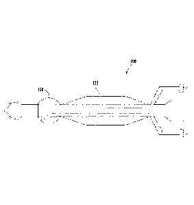
FIG. 1 is a top view of an example catheter;
FIG. 2A is a cross-section view of an example catheter;
FIG. 2B is a cross-section view of an example catheter;
3
CA 02895167 2015-06-15
WO 2013/090778 PCT/US2012/069844
FIG. 2C is a cross-section view of an example catheter;
FIG. 2D is a cross-section view of an example catheter;
FIG. 2E is a cross-section view of an example catheter;
FIG. 3 is a top view of an example catheter;
FIG. 4 is a partial top view of an example catheter;
FIG. 5A is a schematic diagram of an example catheter positioned within a
patient; and
FIG. 5B is a schematic diagram of a sphincter of a patient.
DETAILED DESCRIPTION
Certain examples of the invention will now be described with reference to the
drawings.
In general, such embodiments relate to systems and methods to seal a catheter
at a body orifice
in order to substantially reduce or eliminate leakage of fluids or air between
the orifice and the
catheter during the procedure. The catheter can include a plurality of
balloons extending from
the outer surface of the catheter that when inflated create a seal between the
catheter and the
body orifice. FIG. 1 is a perspective view of an example catheter 100. The
catheter 100 can
include a slender flexible or inflexible tube composed of biocompatible
material. For example,
the catheter 100 can be composed of latex, urethane, silicone, thermoplastic
elastomers, latex
free or silicone coated material. The catheter 100 can be disposable or
sterilized for repeated
use.
The catheter 100 can include an access port and/or fittings and connections
for
completing diagnostics, to provide treatment and/or perform surgical
procedures. For example,
the catheter 100 can be used to take measurements and/or tissue samples,
monitor an internal
body condition, administration of fluids or gases (therapies, irritants, and
gases, etc.), and allow
drainage or otherwise remove matter from the body of the patient.
As illustrated in FIG. 1, the catheter 100 can include an elongated cannula
102 having a
distal end 104 and a proximal end 106. The catheter 100 can include a central
lumen 108
extending within the elongated cannula 102 between the distal end 104 and the
proximal end
106. The central lumen 108 can extend between an opening 110 at the distal end
104 and an
opening 112 at the proximal end 106. In an example catheter 100, the opening
112 can be sealed
.. and/or blocked there by preventing access from central lumen 108 to the
exterior of the catheter
100 at the opening 112. FIG. 2B provides a cross-section view of the elongated
cannula 102 at
section line 2B-2B. The proximal end 106 of the catheter 100 can include a tip
114. The tip 114
can define any suitable shape including, for example, round, elliptical,
square, rectangular, or
any other regular or irregular shape. As illustrated n FIG. 1, an example tip
114 can define a
4
CA 02895167 2015-06-15
WO 2013/090778 PCT/US2012/069844
semi-circular profile. An example tip 114 can define a radius that extends
beyond the outer
surface 116 of the elongated cannula 102. For example, the tip 114 can define
a semi-circular
profile having radius of about 0.5 cm to 1 cm measured from the centerline of
the elongated
cannula 102. An example tip 114 can have a length of about 2 cm to 3 cm
between the end
surface of the elongated cannula 102 at the proximal end 106 and the location
where the profile
of the tip 114 joins the outer surface 116 of the elongated cannula 102. In an
alternate example
(not shown), the tip 114 can define a profile and/or contour corresponding to
the profile of the
outer surface 116.
In addition, or in the alternative to opening 112, the proximal end 106 can
include
secondary openings 118 located around on the surface of the tip 114. The size,
shape, number,
and location of the opening 112 and secondary openings 118 can vary depending
on intended use
of the catheter 100. As illustrated in FIG. 1, an example catheter 100 can
include one end
opening 112 and four secondary openings 118. In an example catheter 100, the
opening 112 and
the secondary openings 118 can include circular-shaped openings that vary in
size from about 2
mm to 3 mm.
The catheter 100 can include a first balloon 120 located along the outer
surface 116 of
the elongated cannula 102. FIG. 2A provides a cross-section view of the first
balloon 120 at
section line 2A-2A. The first balloon 120 can be sized and configured to
extend radially from
the outer surface 116 of the elongated cannula 102. The first balloon 120 can
be composed of
the same or different material as the elongated cannula 102. The first balloon
120 can also be
composed of a complaint or non-compliant material. The first balloon 120 can
be composed of a
flexible or semi-flexible material. Inflation of the first balloon 120 can
provide an anchor for the
catheter 100 within the body of the patient. Therefore, the size and shape of
the inflated first
balloon 120, with respect to physical dimensions and/or volume, can vary in
size depending on
the intended use of the catheter 100. The longitudinal and/or axial profile
defined by the outer
perimeter of the first balloon 120 can define any suitable shape including,
for example, circular,
elliptical, square, rectangular, or any other regular or irregular shape. For
example, as illustrated
in FIG. 1, the longitudinal profile defined by the outer perimeter of the
first balloon 120 can have
a partial circle or round shape. Likewise, as illustrated in FIG. 2A, the
axial profile defined by
the outer perimeter of the first balloon 120 can define a circular or round
shape. It is also
contemplated that the size of the first balloon 120 will vary depending on the
patient's anatomy.
For example, when the catheter 100 is a colorectal catheter used, for example,
to treat
intussusceptions, inflation of the first balloon 120 can anchor the catheter
100 within the
patient's rectum. That is, the diameter of the inflated first balloon 120 is
equal to or greater than
5
CA 02895167 2015-06-15
WO 2013/090778 PCT/US2012/069844
the diameter of the portion of the rectum where the catheter 100 is located.
An example, the
inflated first balloon 120 can have a diameter of about 2 cm to 4 cm and a
length of about 1.5 cm
to 2.5 cm. The first balloon 120 can have a capacity of 20 cc to 50 cc
depending on the
compliance of the material of the first balloon 120.
Similarly, the location of the first balloon 120 with respect to the proximal
end 106 along
the body of the elongated cannula 102 can vary depending on the intended use
of the catheter
100. As illustrated in FIG. 1, in an example catheter 100, the first balloon
120 is spaced apart
from the proximal end 106 and/or tip 114. For example, the first balloon 120
can be located 2
cm to 4 cm from tip 114 (measured from where the tip 114 joins the outer
surface 116). In
another example, the first balloon 120 can be located adjacent to the tip 114.
In another
example, the first balloon 120 can be located adjacent to the most distal side
hole 118. In a
further example, the first balloon 120 can be located on the proximal end ##
of the catheter 100,
eliminating the tip 114 and any of the secondary openings 118, such that the
opening 112 and the
central lumen 108 provide access to the internal portion of the patient's
body.
The cannula can also include a second balloon 122 located along the outer
surface 116 of
the elongated cannula 102. FIG. 2C provides a cross-section view of the second
balloon 122 at
section line 2C-2C. The second balloon 122 can be located between the first
balloon 120 and the
distal end 104. As illustrated in FIG. 1, the second balloon 122 can be
located at a distance from
the first balloon 120. In an alternate embodiment, illustrated in FIG. 3, the
second balloon 122
can abut or otherwise be adjacent to the first balloon 120. The distance
between the second
balloon 122 and the distal end 104 can vary in relation to the total catheter
length. The second
balloon 122 can be sized and configured to extend radially from the outer
surface 116 of the
elongated cannula 102. The second balloon 122 can be composed of the same or
different
material as the elongated cannula 102 and/or the first balloon 120. The second
balloon 122 can
be composed of a flexible or semi-flexible, compliant or non-compliant
material.
'file size and shape of the inflated second balloon 122 (dimensions and/or
volume) can
vary depending on the intended use and size of the catheter 100. The
longitudinal and/or axial
profile defined by the outer perimeter of the second balloon 122 can define
any suitable shape
including, for example, circular, elliptical, square, rectangular, or any
other regular or irregular
shape. As illustrated in FIG. 1, the longitudinal profile defined by the outer
perimeter of the
second balloon 122 includes a first taper 124, an inflated outer surface 126,
and a optional
second taper 128. The longitudinal profile defined by the first taper 128 can
extend in a
direction along a longitudinal axis of the elongated cannula 102 from the
outer surface 116 to an
inflated outer surface 126 of the inflated second balloon 122. As illustrated
in FIG. 4, the first
6
CA 02895167 2015-06-15
WO 2013/090778 PCT/US2012/069844
taper 124 can optionally include a base or "run" designated by the reference
symbol "A." The
run extends in the direction of the longitudinal axis of the elongated cannula
102 from the outer
surface 116 and defines the axial length of the first taper 124. The first
taper 124 can include a
height or "rise" designated by the reference symbol "B". The rise extends at a
90 angle from
the run and terminates at the inflated outer surface 126. The taper also
includes a hypotenuse-
length designated by the reference symbol "C" and defined by the hypotenuse of
the triangle
created by rise and the run.
The first taper 124 can be gradual over the length of the elongated cannula
102 or it may
be aggressive (i.e., have a steeper slope) as determined by the patient's
anatomy. For example, a
thicker and longer sphincter muscle (e.g., anal sphincter muscle) may require
a longer less
aggressive taper than a thinner shorter sphincter muscle (e.g., biliary
sphincter). In the case of
the biliary sphincter, for example, the sphincter muscle is axially short and
thinner (i.e., does not
extend deeply into the surrounding tissues), therefore, a more aggressive
(steep) taper can be
used. As illustrated in Table 1 below, the slope of the first taper 124 when
the catheter 100 is
used with the biliary sphincter can be about 33%. In another example, an obese
patient may
have a thicker and longer sphincter muscle when compared to a similar non-
obese patient.
Therefore, when used on an obese patient, the first taper 124 may be more
aggressive (steep)
than the first taper 124 used on a non-obese patient for the same procedure.
In some cases, including the rectum, the sphincter and surround tissues (e.g.,
buttocks)
can be considered when designing the second balloon 122 and the first taper
124. For example,
in a patient having excess body tissue (e.g.. an obese patient), the buttocks
can be considered as
part of the tissue compressed between the first balloon 120 and the second
balloon 122. The
second balloon 122 can also include a second taper 128 similar in form and
structure to the first
taper 124. The second taper 128 can extend from the inflated outer surface 126
to the outer
surface 116 in a direction towards the distal end 104. In an alternate
embodiment (not shown),
the second balloon 122 does not include the second taper 128. Instead, the
longitudinal profile
defined by the outer perimeter of the distal end of the second balloon 122 can
include various
other shapes and configurations such as, for example, rounded, square, or
include any other edge
formation known in the art.
As illustrated in FIG. 2C, the axial profile defined by the outer perimeter of
the inflated
outer surface 126 can define a circular or round shape. In an example catheter
100, the axial
profile of the outer perimeter of the inflated outer surface 126 can define
the maximum diameter
of the second balloon 122, when inflated. In the example catheter 100 shown in
FIG. 1, the
maximum axial diameter defined by the second balloon 122 is greater than the
maximum axial
7
CA 02895167 2015-06-15
WO 2013/090778 PCT/US2012/069844
diameter defined by the first balloon 120. In an alternative embodiment (not
shown), the
maximum axial diameter defined by the second balloon 122 is equal to or less
than the
maximum axial diameter defined by the first balloon 120.
Inflation of the first balloon 120 and the second balloon 122 creates a seal
at the catheter
100 between an internal portion of the patient and the body orifice
(external). This seal is
created by compressing internal body tissue between the first balloon 120 and
the second balloon
122. The seal can also be created by the "sliding" motion of the internal body
tissue towards the
first balloon 120 created by the inflation of the second balloon 122.
The catheter 100 can be sized and configured to be inserted into a body
cavity, duct, or
vessel of the patient. ln particular, the catheter 100 can be sized and
configured to be inserted
into internal body tissue including a sphincter muscle. An example catheter
100 can be
configured for use at sphincter muscles associated with a natural or created
valved lumen or
cavity within the patient including, for example, cardiovascular, urological,
gastrointestinal and
various percutaneous applications. The human body includes over 50 sphincter
muscles. The
catheter 100 can be used, for example, with the following sphincter muscles:
sphincter pupillae
or pupillary sphincter, belonging to the iris in the eye; orbicularis oris
muscle around the mouth;
cardiallower esophageal sphincter or cardiac sphincter at the upper portion of
the stomach which
prevents the acidic contents of the stomach from moving into the esophagus;
pyloric sphincter at
the lower end of the stomach; sphincter of Oddi, or Glisson's sphincter,
controlling secretions
from the liver, pancreas and gall bladder into the duodenum; urethral
sphincter (internal and
external), controlling the exit of urine from the body; and anus (internal and
external). An
appropriate catheter 100 length and size can be selected from those included
in the following
ranges, or from other sizes based on a variety of medical/surgical
considerations. For example, a
surgeon or other medical professional can determine an appropriate catheter
100 length and size
by evaluating the medical situation in which the device will be used. For
example, the catheter
100 can be used for gastrointestinal procedures, such as, identification and
treatment of colon
polyps, diverticulitis, diverticulosis, Crohn's disease, inflammatory bowel
disease (IBD),
ulcerative colitis (UC), and colon carcinoma. The catheter 100 optimally
ranges ranging in
length from 30 cm to 60 cm. The catheter 100 can also range in size from 8F to
28F, with sizes
14F to 24F being used on adults and 8F to 12F being used on children.
FIG. 5 provides a schematic diagram of the catheter 100 positioned within an
example
patient. The example catheter 100 includes a second balloon 122 positioned
adjacent to the first
balloon 120. This seal can be created by compressing the sphincter muscle
between the first
balloon 120 and the second balloon 122. For example, as the second balloon 122
is inflated, the
8
CA 02895167 2015-06-15
WO 2013/090778 PCT/US2012/069844
sphincter muscle is compressed/pushed in the direction towards the first
balloon 120 and a seal is
created. The axial diameter of the elongated cannula 102 is less than the
diameter of the
sphincter. When inserted, the first balloon 120 is located inside the rectum.
The second balloon
122 is positioned along the sphincter muscle. As the second balloon 122 is
inflated, the
sphincter is compressed between the balloons and a seal is created.
In an example catheter 100, the length and thickness of the sphincter muscle
correlate to
the run and rise of the first taper 124. Table 1 provides example correlations
between the length
and thickness of various sphincter muscles contemplated for use with the
present system and
example run and rise values for the first taper 124.
Axial Rise of Slope of
First
Length of Thickness of First Taper
Sphincter Sphincter Run of First Taper
Sphincter (mm) (mm) Taper (mm) (mm)
Anal 80-100 2-6 92 10 10/92 =
10%
Pyloric 80 10 100 20 20/100 =
20%
Urinary 20-100 14 120 30 30/120 =
25%
Biliary 20 5 30 10 10/30 =
33%
Table 1. Sphincter and Taper Comparison
As demonstrated in Table 1, the longer the sphincter the longer the run of
taper 124 is
required. In an example catheter 100, the run of the first taper 124 is
greater than the length of
the sphincter muscle along the longitudinal axis of the catheter 100. Example
sphincter muscle
lengths can range between about 2 cm and 10 cm, including, for example, 2 cm,
7.2 cm, 8 cm,
and 10 cm. Example taper run lengths can be greater than about 2 cm,
including, for example, 2
cm, 3 cm, 5, cm, 9 cm, 10 cm, and 12 cm. For example, when the catheter 100 is
a colorectal
catheter and the length of the anal sphincter is about 7.2 cm, the run of the
corresponding first
taper 124 can be about 9.2 cm. In a further example, the catheter 100 is a
pyloric catheter. The
length of the pyloric sphincter is about 8 cm, the run of the corresponding
first taper 124 is about
10 cm.
As similarly demonstrated in Table 1, the thicker the sphincter muscle the
greater the rise
of taper 124 is required. In an example catheter 100, the rise of the first
taper 124 is greater than
the thickness of the sphincter muscle in a direction perpendicular to the
longitudinal axis of the
catheter 100. For example, when the catheter 100 is a colorectal catheter and
the thickness of the
anal sphincter is about 2 cm to 6 cm, the rise of the corresponding first
taper 124 can be about 10
cm. It is also contemplated that an effective seal can be achieved when the
rise of the first taper
124 is equal to or less than the thickness of the sphincter muscle.
9
CA 02895167 2015-06-15
WO 2013/090778
PCT/US2012/069844
In a further example catheter 100, the distance between the first balloon 120
and the
second balloon 122 is less than the length of the sphincter muscle such that
at least a portion of
the first taper 124 is located adjacent to the sphincter muscle. For example,
when the catheter
100 is a colorectal catheter and the length of the anal sphincter is about 7.2
cm, the distance
between the first balloon 120 and the second balloon 122 can be about 2 cm to
8 cm.
As outlined above, the central lumen 108 of the elongated cannula 102
terminates at the
distal end 104 at opening 110. The opening 110 can have a diameter larger than
the diameter of
the elongated cannula 102. In an alternate embodiment (not shown), the opening
110 can have a
diameter less than or equal to the diameter of the elongated cannula 102. In
an example catheter
100, opening 110 can have a diameter of about 0.8 cm. The opening can include
a taper
extending from the outer surface 116 to the diameter of the opening 110. An
example opening
110 can include a syringe-type opening configured for the introduction of
fluid or air into the
patient's body.
The catheter 100 can also include a first lumen 130 and a second lumen 132
extending
within the central lumen 108. FIG. 2D provides a cross-section view of the
elongated cannula
102 including the first lumen 130 and the second lumen 132 at section line 2D-
2D. The first
lumen 130 can be in fluid communication with the first balloon 120. Using the
first lumen 130,
the user can provide a fluid (gas and/or liquid) to inflate/deflate the first
balloon 120. Likewise,
the second lumen 132 can be in fluid communication with the second balloon
122. Using the
second lumen 132, the user can provide a fluid (gas and/or liquid) to
inflate/deflate the second
balloon 122. In an alternate embodiment, the first lumen 130 and/or the second
lumen 132 are
located outside the central lumen 108 of the catheter 100. For example, the
first lumen 130
and/or the second lumen 132 can extend along the outer surface 116 of the
elongated cannula
102.
The first lumen 130 can extend through the outer surface 116 of the elongated
cannula
102 to an input 134 at the distal end 104 of the catheter 100. Similarly, the
second lumen 132
can extend through the outer surface 116 of the elongated cannula 102 to an
input 136 at the
distal end of the catheter 100. FIG. 2E provides a cross-section view of the
elongated cannula
102/opening 110, input 134 and input 136 at section line 2E-2E. The inputs 134
and 136 can
include a syringe-type opening similar to opening 110. In another example, the
inputs 134 and
136 can include a luer-lock syringe connection. In a further example, the
inputs 134 and 136
and/or first lumen 130 and second lumen 132 can include a one-way valve
member. In an
example catheter 100, the length of the catheter between the distal end 104
and where the first
lumen 130 and the second lumen 132 extend through the outer surface 116 can
vary in relation to
CA 02895167 2015-06-15
WO 2013/090778 PCT/US2012/069844
the total length of the catheter 100. In an example catheter 100, the distance
can be about 4 cm
to 6 cm. Likewise, the length of the first lumen 130 and the second lumen 132
extending outside
the outer surface 116 can vary. In an example catheter 100, the distance can
be about 4.5 cm to
6.5 cm. The shape of the first lumen 130 and the second lumen 132 extending
outside the outer
surface 116 can be curved, angle, straight, or take any other direction/shape.
In operation, the catheter 100 can optionally be used for intussusception
reduction,
barium or water soluble enema, fistulogram, virtual colonoscopy and other
procedures requiring
an effective seal between the catheter 100 and the patient. The catheter 100
can be used by
inserting the proximal end 106 of the elongated cannula 102 into the body of
the patient. The
catheter 100 can be inserted at either a natural or created orifice in the
patient's body. Natural
orifices include, for example, the anus, pylorus, urethra and biliary tract.
The user can also
create an orifice in the patient's body, for example, a surgical opening. A
portion of the
elongated cannula 102 can be inserted such that the proximal end 106 is
located within the
patient and the distal end 104 is located outside the patient's body. In an
example, the inserted
portion includes the first balloon 120 and a portion of the second balloon
122. For example, as
illustrated in FIG. 5, the first balloon 120 can be inserted within the
patient past the sphincter
muscle and at least a portion of the second balloon 122 can be inserted within
the patent.
At insertion, the first balloon 120 and the second balloon 122 are in a
deflated state. In
the deflated state, the first balloon 120 and the second balloon 122 may have
a diameter
corresponding to the diameter of the elongated cannula 102. In a further
example, in the deflated
state, the first balloon 120 and the second balloon 122 may have a diameter
greater than the
diameter of the elongated cannula 102 but less than the diameter of the
orifice in the body of the
patient. For example, the deflated balloons and the elongated cannula 102 may
have a diameter
less than the diameter defined by the sphincter muscle corresponding to the
orifice.
Upon insertion, internal tissue (i.e., the sphincter muscle) is located along
the second
balloon 122. For example, upon insertion, the sphincter muscle associated with
the orifice is
located along at least a portion of the second balloon 122. In an example
method, all or a
significant portion of the length sphincter muscle is located along first
taper 124 of the second
balloon 122.
Once the catheter 100 is properly located, the first balloon 120 is inflated.
The first
balloon 120 can be inflated at least partially or to capacity. In an example
method, the first
balloon 120 is inflated to its maximum intra-body capacity. Once inflated, the
first balloon 120
anchors the elongated cannula 102 within the patient's body.
11
CA 02895167 2015-06-15
WO 2013/090778
PCT/US2012/069844
The second balloon 122 is then inflated. The second balloon 122 can be
inflated at least
partially or to capacity. In an example method, the second balloon 122 is
inflated to its
maximum intra-body capacity. The second balloon 122 can be rapidly inflated.
In another
example, the second balloon 122 is slowly/steadily inflated to until an
effective seal is achieved.
By inflating the second balloon 122 slowly/steadily, the second balloon 122
can align with the
first balloon 120 to prevent motion of the catheter 100 in the longitudinal
direction.
The first balloon 120 can be inflated first, independently of the second
balloon 122. In
an alternate method, when inflation of the second balloon 122 is initiated,
the first balloon 120 is
partially inflated. Inflation of the second balloon 122 can compress the
internal body tissue (e.g.,
-- sphincter muscle) between the first balloon 120 and the second balloon 122.
Inflation of the
second balloon 122 can also result in the internal body tissue moving,
sliding, or rolling from the
area of the first taper 124 towards the first balloon 120. By inflating the
second balloon 122, and
pressing the internal body tissue between the first balloon 120 and the second
balloon 122, a seal
is created around the catheter 100 at the orifice thereby preventing leakage
from around catheter
-- 100.
In operation, the catheter 100 can also be used to provide improvement for CT
(computed
tomography) colonoscopy procedures. During the procedure, a catheter can be
inserted into the
rectum of the patient. Carbon dioxide is then insufflated into the colon. If
the colon is not
completely distended, the study is limited, that is not all areas of the colon
are visible thus not
-- giving complete information. Catheter 100 can be used to create the seal,
thus reducing the
possibility of carbon dioxide leak and improving the likelihood of a fully
distended colon.
The catheter 100 may also be used to provide an enema to a patient, in
particular, barium
enemas. When performing an enema, liquids such as barium, are introduced into
the colon. If
the patient does not have good rectal tone, the barium will leak out. This
prevents barium from
reaching the necessary location along the colon and into the cecum. Use of the
catheter 100 to
create seal significantly reduces or prevents liquid barium from leaking out
of the rectum.
During the procedure, the catheter 100 is connected to a tubing set with a
stopcock. The
stopcock is adjusted to close access to the tubing. The patient is placed on
the treatment table in
a left lateral position. The catheter tip 114 is lubricated using, for
example, petroleum jelly. The
-- catheter 100 is then inserted into the rectum until it is within the rectum
and the patient's
buttocks are taped together. The patient is placed in the supine position.
Under fluoroscopy, the
first balloon 120 is inflated by connecting a syringe filled with saline to
the input 134. An
insufflator with a gauge is connected to the catheter 100. The second balloon
122 is minimally
inflated with a syringe filled with saline attached at input 136. The second
balloon 122 is then
12
CA 02895167 2015-06-15
WO 2013/090778 PCT/US2012/069844
filled to accomplish the desired seal. The stopcock is then adjusted to allow
the flow of air into
the rectum and under fluoroscopy guidance, colonic insufflations is initiated.
During insufflation, the user can continue to observe the pressure gauge to
ensure the
pressure remains in the allotted range for the procedure. A pressure release
valve can be placed
between the insufflator and the stopcock. The pressure valve will release
pressure when the
pressure reaches 120 mmHg. The intussusceptum is followed until it is reduced
and air reflux
into the small bowel through the ileo-cecal valve is achieved. The escape of
air leaking out of
the rectum around the catheter 100 can be observed by the user. A leak will
decrease intra-
colonic pressure, thereby minimizing the chance of a successful reduction. If
a leak is found, the
second balloon 122 can be further inflated until the leak is eliminated. The
catheter 100 can be
used to eliminate the pen-rectal air leakage, which in turn, will provide
optimal intra-colonic
pressure and the success rate for intussusception reduction increases
exponentially when the
optimal pressure is maintained. This, in turn, will decrease the length of the
procedure and
thereby decreasing the radiation dose to the patients and the medical
personnel.
After the removal of the catheter 100 and the remaining intussusception tools,
the patient
may have a bowel movement. The patient is then cleaned up and removed from the
fluoroscopy
table. If there are questions/concerns with respect to the reduction of the
intussusception, an
ultrasound can be completed. In some cases, the radiologist may recommend the
patient to have
surgery for reduction secondary to the findings on the air enema procedure. In
other cases, the
reduction procedure can be repeated at a later time. When the patient returns
for a subsequent
procedure, all steps of the preparing and performing the procedures are the
same.
While the foregoing description and drawings represent examples of the present
invention, it will be understood that various additions, modifications,
combinations and/or
substitutions may be made therein without departing from the spirit and scope
of the present
invention as defined in the accompanying claims. In particular, it will be
clear to those skilled in
the art that the present invention may be embodied in other specific forms,
structures,
arrangements, proportions, and with other elements, materials, and components,
without
departing from the spirit or essential characteristics thereof. One skilled in
the art will appreciate
that the invention may be used with many modifications of structure,
arrangement, proportions,
materials, and components and otherwise, used in the practice of the
invention, which are
particularly adapted to specific environments and operative requirements
without departing from
the principles of the present invention. In addition, features described
herein may be used
singularly or in combination with other features. The presently disclosed
examples are, therefore,
13
CA 02895167 2015-06-15
WO 2013/090778 PCT/US2012/069844
to be considered in all respects as illustrative and not restrictive, the
scope of the invention being
indicated by the appended claims and not limited to the foregoing description.
In addition, the various examples disclosed herein may be adapted for use in
virtually any
interior body region where a seal around a catheter is required for a
therapeutic or diagnostic
purpose. It is also anticipated that certain examples could be used for
purposes other than
medical, such as construction, manufacturing, and excavation, among others;
accordingly,
nothing herein is intended to limit application of the various examples to
purely medical uses.
It will be appreciated by those skilled in the art that changes could be made
to the
examples described above without departing from the broad inventive concept
thereof. It is
understood, therefore, that this invention is not limited to the particular
examples disclosed, but
it is intended to cover modifications within the spirit and scope of the
present invention, as
defined by the following claims.
14