Note: Descriptions are shown in the official language in which they were submitted.
CA 02896442 2015-06-25
WO 2014/047540 PCT/US2013/061144
SPINAL CORD PULSATION-CANCELATION INJECTION SYSTEM
CROSS REFERENCE TO RELATED APPLICATION(S)
[0001] This application claims the benefit of priority under 35 U.S.C.
119(e) of U.S.
Serial No. 61/704,959, filed September 24, 2013, the entire content of which
is incorporated
herein by reference.
GRANT INFORMATION
[0002] This invention was made with government support under Grant No.
N5051644-
02A2 awarded by the National Institutes of Health. The United States
government has
certain rights in the invention.
BACKGROUND OF THE INVENTION
FIELD OF THE INVENTION
[0003] The invention relates generally to a drug or cell delivery system
and more
specifically, to a drug or cell delivery system that eliminates spinal cord
pulsation effects
during spinal cord injections in large animal species and humans.
BACKGROUND INFORMATION
[0004] The spinal cord is a delicate structure that rests within the spinal
canal and is
surrounded by a tough outer covering, called the dura. Normally, the spinal
cord ends at
about the first or second lumbar vertebrea in the adult. The spinal canal is
surrounded and
protected by the bony structure of the spinal column (or vertebrea).
Cerebrospinal fluid
(CSF) surrounds the spinal cord and flows from the brain, down the spinal
canal and back
up to the brain. Many nerves originate from the spinal cord, and are
responsible for
movement and sensation of the arms, legs and torso.
[0005] Intraspinal injections have been used for spinal anaesthesia,
chemotherapy, pain
management applications, and for taking samples of cerebral spinal fluid.
Administering a
substance to the spaces or potential spaces surrounding the spinal cord is
often performed in
order to avoid the blood-brain barrier. Intraspinal grafting of human neural
stem cells
represents a promising approach to promote recovery of function after spinal
trauma.
CA 02896442 2015-06-25
WO 2014/047540 PCT/US2013/061144
2
[0006] Dorso-ventral spinal cord pulsation resulting from respiration
and/or
cerebrospinal fluid movement represents a serious risk factor during the
procedure of direct
spinal parenchymal injections. Currently existing devises use internally
mounted frames
that are places over laminectomy sites and use freely floating cannulas that
are advanced
into the spinal cord parenchyma while being firmly attached to the
manipulator. The
cannulas are then released from the holder to create a "free-floating" effect.
However, such
devices are complicated to use due to the need for repetitive immobilization
and release of
the floating cannula in the Z-arm of the injector between individual
injections.
SUMMARY OF THE INVENTION
[0007] The present invention is based on the utilization of repulsive
forces produced by
micromagnets to create a spring effect that compensates for spinal cord
pulsation during
intraspinal injections.
[0008] Accordingly, in one aspect, there is provided a spinal cord
pulsation-cancelation
injection device. The device includes a frame having an elongated body and a
plurality of
holders extending therefrom; a plurality of first magnets, each being fixedly
attached to a
holder; a tube having a first end and a second end, the tube being slidingly
disposed within
through-holes disposed in each holder and in each first magnet; a plurality of
second
magnets fixedly attached to an exterior surface of the tube; and a needle
fixedly attached to
the first end of the tube. The frame may be made from any non-corrosive metal,
such as
stainless steel. The needle may range from about 27 to about 32 gauge. Each of
the first
magnets and the each of the second magnets may be disposed such that a north
pole of one
first magnet faces a north pole of one second magnet or a south pole of one
first magnet
faces a south pole of one second magnet, thereby providing a magnetic
repulsive force upon
which the tube floats.
[0009] In various embodiments, the frame comprises two holders, each having
attached
thereto a first magnet, and a single second magnet is fixedly attached to the
tube. In other
embodiments, the frame comprises two holders, each having attached thereto a
first magnet,
and two second magnets are fixedly attached to the tube.
CA 02896442 2015-06-25
WO 2014/047540 PCT/US2013/061144
3
[0010] The device may further include a stop ring fixedly attached to the
first end of the
tube or an area near the first end of the tube. The device may further include
a stop ring
fixedly attached to the second end of the tube or an area near the second end
of the tube.
The device may further include tubing removably attached to the second end of
the tube and
configured for supplying a substrate to the needle.
[0011] In another aspect, there is provided a spinal cord pulsation-
cancelation injection
system. The system includes the spinal cord pulsation-cancelation injection
device
described herein and a reservoir in fluid communication with the needle, the
reservoir
containing a substrate to be administered to a subject. The system may further
include a
digital microinjector configured to control flow of the substrate through the
needle. In
various embodiments, the substrate is selected from the group consisting of
cells, drugs,
viruses, plasmids, and growth factors.
[0012] In various embodiments, the frame comprises two holders, each having
attached
thereto a first magnet, and a single second magnet is fixedly attached to the
tube. In other
embodiments, the frame comprises two holders, each having attached thereto a
first magnet,
and two second magnets are fixedly attached to the tube.
[0013] In yet another aspect, there is provided a method of compensating
for spinal cord
pulsation during administration of a substrate to a spinal cord of a subject.
The method
includes positioning the spinal cord pulsation-cancelation injection system
described herein
over the spinal cord of the subject; lowering the needle into the spinal cord;
and delivering a
dose of the substrate to the spinal cord, wherein the needle and tube of the
device float due
to magnetic repulsive forces within the device, thereby compensating for
spinal cord
pulsation. In various embodiments, the method further includes repeating each
of the steps
at multiple sites along the spinal cord. The step of delivering may include
activating a
digital microinjector configured to control flow of the substrate through the
needle. The
step of lowering the needle may include inserting the needle into the spinal
parenchyma
until a needle stop ring that is fixedly attached to the needle contacts the
subject.
Accordingly, the present invention also provides use of the spinal cord
pulsation-
CA 02896442 2015-06-25
WO 2014/047540 PCT/US2013/061144
4
cancelation injection system described herein to compensate for spinal cord
pulsation during
administration of a substrate.
BRIEF DESCRIPTION OF THE DRAWINGS
[0014] Figure 1 is pictorial diagram showing a first exemplary embodiment
of the spinal
cord pulsation-cancelation injection system.
[0015] Figure 2 is pictorial diagram showing a second exemplary embodiment
of the
spinal cord pulsation-cancelation injection system.
[0016] Figure 3 is pictorial diagram showing use of the spinal cord
pulsation-cancelation
injection system for delivering a cell suspension to the spinal cord of a
subject.
[0017] Figure 4 is a flow chart describing steps for delivering a cell
suspension to the
spinal cord of a subject using the spinal cord pulsation-cancelation injection
system.
DETAILED DESCRIPTION OF THE INVENTION
[0018] The present invention is based on the utilization of magnetic
repulsive forces to
create a spring effect that compensates for spinal cord pulsation during
intraspinal
injections. Once the injection needle is advanced in the spinal parenchyma, it
can then
fluctuate with any pulsation of the spinal cord in the dorso-ventral direction
due to the
magnetic repulsive forces acting on the needle holder. As such, the present
invention
provides a spinal cord pulsation-cancelation injection system that may be used
for
spinal cord cell and vector delivery in large animals and humans.
[0019] Before the present compositions and methods are described, it is to
be understood
that this invention is not limited to particular compositions, methods, and
experimental
conditions described, as such compositions, methods, and conditions may vary.
It is also to
be understood that the terminology used herein is for purposes of describing
particular
embodiments only, and is not intended to be limiting, since the scope of the
present
invention will be limited only in the appended claims.
CA 02896442 2015-06-25
WO 2014/047540
PCT/US2013/061144
[0020] As used in this specification and the appended claims, the singular
forms "a",
"an", and "the" include plural references unless the context clearly dictates
otherwise.
Thus, for example, references to "the method" includes one or more methods,
and/or steps
of the type described herein which will become apparent to those persons
skilled in the art
upon reading this disclosure and so forth.
[0021] The term "comprising," which is used interchangeably with
"including,"
"containing," or "characterized by," is inclusive or open-ended language and
does not
exclude additional, unrecited elements or method steps. The phrase "consisting
of"
excludes any element, step, or ingredient not specified in the claim. The
phrase "consisting
essentially of" limits the scope of a claim to the specified materials or
steps and those that
do not materially affect the basic and novel characteristics of the claimed
invention. The
present disclosure contemplates embodiments of the invention compositions and
methods
corresponding to the scope of each of these phrases. Thus, a composition or
method
comprising recited elements or steps contemplates particular embodiments in
which the
composition or method consists essentially of or consists of those elements or
steps.
[0022] Unless defined otherwise, all technical and scientific terms used
herein have the
same meaning as commonly understood by one of ordinary skill in the art to
which this
invention belongs. Although any methods and materials similar or equivalent to
those
described herein can be used in the practice or testing of the invention, the
preferred
methods and materials are now described.
[0023] As used herein, the term "dorsoventral" or "dorso-ventral" is an
adjective that
refers to extending along or denoting an axis joining the dorsal and ventral
surfaces of a
primate. Included in the term is extending from the back to the belly of the
animal.
[0024] As used herein, the term "pulsation" when used in the context of the
spinal cord
of a human or animal, refers to involuntary movement of the spinal cord as a
result of
cardiac pulsation and/or respiration. Recent studies have indicated that
spinal cord
pulsation is derived mainly from the radicular arteries, rather than from the
change in the
brain volume or the influence of central and peripheral circulation. However,
included in
CA 02896442 2015-06-25
WO 2014/047540
PCT/US2013/061144
6
the term is pulsation of the spinal cord as a result of cerebrospinal fluid
moving back and
forth within the cervical canal.
[0025] As used herein, the terms "stereotaxis," "stereotaxic," and
"stereotactic" are used
interchangeably to refer to methods in neurosurgery and neurological research
for locating
points within the brain or spinal cord using an external, three-dimensional
frame of
reference usually based on the Cartesian coordinate system. Methods of
steretactic surgery
are known in the art.
[0026] Referring to FIG. 1, an exemplary embodiment of the spinal cord
pulsation-
cancelation injection system 100 is shown. The system includes a frame 110
having an
elongated body and a plurality of holders (190, 200) extending therefrom. The
body of the
frame may be a circular or square bar having a first end and a second end. The
first end is
fixedly connected to an XYZ manipulator (not shown), which is mounted directly
onto a
stereotaxic frame (not shown). While the body may be made from any rigid
material, in
certain embodiments, the bar is made from any non-corrosive metal, such as
stainless steel.
Extending from the second end of the bar is a first holder 200 of the
plurality of holders.
One or more additional holders may extend in the same direction, and parallel
to, the first
holder 200. In certain embodiments, the frame will include a first holder 200
extending
from the second end of the bar and a second holder 190 extending from the bar
at a
predetermined distance from the first holder 200. The predetermined distance
between the
first and second holders may range from about 1 cm to about 5 cm (i.e., 1 cm,
2 cm, 3 cm, 4
cm, 5 cm, or any fraction there between). Disposed within each holder is a
through-hole
195 such that the through-hole of one holder is aligned with the through-hole
of each
successive holder.
[0027] Slidingly disposed within the through-holes 195 of each holder is a
tube 120
having a top end 122 and a bottom end 124. The tube 120 may be formed from the
same
metal material from which the frame is formed. Thus, in an exemplary
embodiment, the
tube is made from stainless steel. Fixedly mounted to the bottom end 124 of
the tube 120 is
an injection needle 150, which may be from about 27 to about 32 gauge, and may
be made
from stainless steel or other non-corrosive materials. Plastic or Teflon
tubing may be
CA 02896442 2015-06-25
WO 2014/047540 PCT/US2013/061144
7
removably attached to the top end 122 of the tube for connecting the tube 120
to a reservoir
230 and/or syringe 220 for supplying a substrate to the spinal cord of a
subject. In certain
embodiments, injections may be performed by using a digital microinjector 240.
Optionally
disposed between the syringe 220 and the tube 120 of the system may be a cell
suspension
reservoir 230 to minimize sedimentation of cells in the tubing when mounted
vertically to
the XYZ manipulator (not shown).
[0028] As used herein, the term "substrate" refers to any injectable
substance, including
but not limited to cells, drugs, viruses, plasmids, growth factors and the
like. The substrate
may take any suitable form of matter, including a liquid, a suspension, a gel,
an
encapsulated solid, a nanoparticle suspension, a slow- or extended-release
polymer
composition and the like.
[0029] The term "subject" as used herein refers to any individual or
patient to which the
subject methods are performed. Generally the subject is human, although as
will be
appreciated by those in the art, the subject may be an animal. Thus other
animals, including
mammals such as rodents (including mice, rats, hamsters and guinea pigs),
cats, dogs,
rabbits, farm animals including cows, horses, goats, sheep, pigs, etc., and
primates
(including monkeys, chimpanzees, orangutans and gorillas) are included within
the
definition of subject.
[0030] Disposed in a surface of the first holder 200 and second holder 190
is a ring
magnet having a north-south polarity and a through-hole that corresponds to,
and is aligned
with, the through-hole 195 of each holder. As shown in FIG. 1, a first magnet
140 is
disposed in the first holder 200 and a second magnet 130 is disposed in the
second holder
190. Fixedly attached to an exterior surface of the tube 120 is one or more
ring magnets
160, which may be disposed between a pair of the holders. In the exemplary
embodiment
shown in FIG. 1, a single ring magnet 160 is fixedly attached to the exterior
surface of tube
120 such that the ring magnet 160 is located between each of the first magnet
140 and
second magnet 130. The ring magnet 160 also has a north-south polarity, and is
disposed
on the tube 120 such that the north pole of the ring magnet 160 faces the
north pole of the
first magnet 140 and the south pole of the ring magnet 160 faces the south
pole of the
CA 02896442 2015-06-25
WO 2014/047540 PCT/US2013/061144
8
second magnet 130. Of course, the ring magnet 160 may be disposed on the tube
120 such
that the north pole of the ring magnet 160 faces the north pole of the second
magnet 130 and
the south pole of the ring magnet 160 faces the south pole of the first magnet
140. As such,
a repulsive magnetic force Fl is created between the ring magnet 160 and the
magnet 130 of
the second holder 190. Likewise, a similar repulsive magnetic force F2 is
created between
the ring magnet 160 and the magnet 140 of the first holder 200. With repulsive
force Fl
being equal to repulsive force F2, tube 120 floats within the through-holes
195, thereby
creating a spring effect. Optionally disposed at the top end 122 and the
bottom end 124 of
the tube 120 is one or more plastic stop rings (not shown) to prevent the tube
from
exceeding a maximum allowable range of movement.
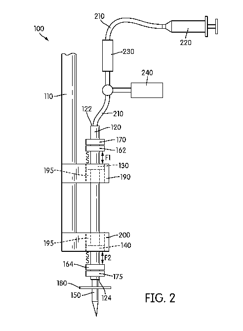
[0031] With reference now to FIG. 2, an alternative exemplary embodiment of
the spinal
cord pulsation-cancelation injection system 100 is shown, wherein like
reference numerals
refer to like elements throughout. Thus, only the differences between the
first exemplary
embodiment shown in FIG. 1 and the second exemplary embodiment shown in FIG. 2
will
be discussed herein.
[0032] As shown in FIG. 2, the system includes a first magnet 140 is
disposed in the first
holder 200 and a second magnet 130 is disposed in the second holder 190.
Fixedly attached
to an exterior surface of the tube 120 is one or more ring magnets 160. In the
exemplary
embodiment shown in FIG. 2, two ring magnets are fixedly attached to the
exterior surface
of tube 120 at a position that places the ring magnets in close proximity to
the magnets of
the first holder 200 and second holder 190. Thus, as shown, an upper ring
magnet 162 is
disposed at the top end 122 or in an area adjacent to the top end 122 of the
tube 120.
Likewise, a lower ring magnet 164 is disposed at the bottom end 124 or in an
area adjacent
to the bottom end 124 of the tube 120. Each of the upper magnet 162 and lower
magnet 164
has a north-south polarity, and are disposed on the tube 120 such that the
south pole of the
upper magnet 162 faces the south pole of the second magnet 130 and the south
pole of the
lower magnet 164 faces the south pole of the first magnet 140. Of course, the
upper and
lower magnets may be disposed such that the north pole of the upper magnet 162
faces the
north pole of the second magnet 130 and the north pole of the lower magnet 164
faces the
CA 02896442 2015-06-25
WO 2014/047540
PCT/US2013/061144
9
north pole of the first magnet 140. As such, a repulsive magnetic force F1 is
created
between the upper magnet 162 and the magnet 130 of the second holder 190.
Likewise, a
similar repulsive magnetic force F2 is created between the lower magnet 164
and the
magnet 140 of the first holder 200. With repulsive force Fl being equal to
repulsive force
F2, tube 120 floats within the through-holes 195, thereby creating a spring
effect.
Optionally disposed at the top end 122 and the bottom end 124 of the tube 120
is one or
more plastic stop rings (not shown) to prevent the tube from exceeding a
maximum
allowable range of movement.
[0033] In both exemplary embodiments, a sterile needle stop ring 180 may be
fixedly
attached to the needle 150 to serve a guide to a surgeon as to the maximum
distance that the
needle 150 will be lowered into the spinal cord during the procedure. When
present, the
stop ring 180 may be positioned along the length of the needle 150 such that
when the
needle reaches a predetermined depth, the stop ring 180 contacts the
subject/patient.
[0034] Referring now to FIGS. 3 and 4, use of the system 100 is as follows.
The
surgical table with stereotactic frame is prepared for the procedure and the
subject is
positioned on their prone surface. A standard posterior approach is performed,
targeting T1
to T10 of the spinal cord, followed by an "open door" laminoplasty, leaving
the dura matter
intact. Sterile saline may be used to clean/flush the operatory wound, and
sterile fields may
be applied to protect the subject.
[0035] The XYZ manipulator (not shown) is then attached to the stereotactic
frame
above the operatory wound of the subject. The spinal cord pulsation-
cancelation injection
system 100 is then attached to the XYZ manipulator (not shown) and connected
to a cell
suspension reservoir 230 and syringe containing the suspension to be
administered via
sterile tubing 210. The contents of the syringe are then loaded into the cell
suspension
reservoir 230 (S310). The sterile tubing is run through a digital
microinjector 240, which is
operated to remove any air gaps in the lines.
[0036] Continue the surgical procedure by opening the dura mater, perform a
longitudinal incision in the dura mater, avoiding damage to blood vessels.
Position the
CA 02896442 2015-06-25
WO 2014/047540 PCT/US2013/061144
needle 150 of the spinal cord pulsation-cancelation injection system 100 above
the spinal
cord at the point of injection (S320).
[0037] The needle is then lowered into the spinal cord parenchyma, avoiding
damage to
blood vessels under visual guidance (S330). The digital microinjector 240 is
then activated
to deliver the dose to the spinal cord (S340). After retracting the needle,
the needle may be
repositioned for repeated injection steps, as necessary (S350). Following
completion of all
injections, the needle is retracted and the XYZ manipulator (not shown) is
removed from
the stereotactic frame (not shown).
[0038] A dural closure is performed, followed by a laminary closure. The
anatomical
layers (muscle, subcuticle, skin) are then closed using resorbable materials
(S360). Thus,
the spinal pulsation-cancelation injection system 100 may be used in a human
patient
receiving direct spinal parenchymal injections of cells vectors, or drugs. As
discussed
above, the system 100 may eliminate any spinal cord pulsation effects that
occur during a
procedure of spinal cord injections in large animal species and in humans.
[0039] The following examples are intended to illustrate but not limit the
invention.
EXAMPLE 1
[0040] A 30G non-coring needle built into the magnetic spinal-pulsation-
cancellation
system will be used to deliver test/therapeutic materials to the spinal cord
of a subject. The
spinal-pulsation cancellation system will be attached to a solid circular
stainless steel bar
with a needle holder (collectively called the catheter holder), which is
connected to a
sterilized XYZ manipulator (Stoelting; Cat.No: 51600). The XYZ manipulator
will be
mounted directly onto a stereotaxic frame. The injection needle will be
interconnected with
a horizontally oriented cell suspension coil constructed from Teflon Medical
Micro Tubing
(ID:0.01"; OD:0.02"; Scientific Commodities, INC; AZ), and positioned just
above the
injection needle. The diameter of the coil is 12 mm. Cell suspension is loaded
into the coil
manually by the surgeon using previously autoclaved cell-loaded Hamilton
syringe (250 1).
The cell-loaded coil is then connected with CTS-filled Hamilton syringe (250
1) mounted
on a digital microinjector (Tritech Research; Model-MINJ-PD).
CA 02896442 2015-06-25
WO 2014/047540 PCT/US2013/061144
11
[0041] Set-up of Stereotactic Frame for Surgical Table ¨ Fit the surgical
table with the
stereotactic frame, which supports the entire body of the patient. Attach the
temperature-
controlling pad to the stereotactic frame and cover with clean sheets.
[0042] Positioning the Patient ¨ Position the patient on their prone
surface with Thoracic
Level 1 (T1) vertebra approximately at the first pair of stabilization bars.
Follow standard
of care to avoid compression points at the hip, abdomen, and chest.
[0043] Exposing Spinal Cord ¨ Perform a standard posterior approach to
target T1 to
T10 followed by an "open door" laminoplasty, leaving the dura mater intact.
Apply sterile
saline to the operatory wound and apply sterile fields for protection during
the next steps.
[0044] Installation of Stereotactic Arm, Stabilizing Bars, and Catheter Kit
¨ Create
openings in the sterile field to allow for the stereotactic arm to be attached
to the stereotactic
frame. Attach the stereotactic arm (XYZ manipulator) to the stereotactic
frame. Attach the
stabilizer bars. Aseptically isolate the sterile field openings. Install the
catheter holder.
Attach the catheter kit to the catheter holder by anchoring the upper and
lower magnets of
the pulse cancelling system into the holder.
[0045] Loading Catheter ¨ Fill a 1 cc syringe with vehicle and fill the
catheter tubing,
checking for leakage at tubing unions. Using a 250 [t.L gas tight syringe
(transfer syringe)
with 18G needle, aspirate 240 [t.L of cell-suspension product. Retract the
transfer syringe
plunger 5 [t.L and attach the syringe to the catheter end within 5-15 seconds.
Slowly load
the contents of the transfer syringe into the catheter tubing, observing the
air gap between
the vehicle and cellular product. Remove the syringe after complete transfer
of the cellular
product. Close the catheter end with a sterile Luer cap provided in the
catheter kit.
[0046] Micro-Injector Setup ¨ The following describes the use of sterile
methylene blue
to act as a visual aid in the progression of the end of the cellular product
throughout the
catheter. The digital micro-injector is located on a clean table next to the
patient and
separated with sterile sheets. The injector plunge is completely inserted in
the injector body
at the end of run. A 3-way stop is attached to the injector body. Attach a 1
cc syringe with
Methylene blue vial to the 3 way stop load port in the inverted position. Turn
the 3 way
CA 02896442 2015-06-25
WO 2014/047540 PCT/US2013/061144
12
stop to the load position. Set the micro-injector in reverse "R" to 0.2
p.L/sec for a total of 5
[t.L actuation steps. Actuate the micro-injector in steps to fill with
methylene blue until the
240 [1.1_, graduation. Do not actuate the injector to the end of run. Turn the
3-way stop to the
off position. Feed the sterile capped catheter end through an opening in the
sterile sheets.
Remove the cap and attach the catheter to the 3way stop on the micro-injector.
Turn the 3-
way stop to the "inject" position. Set the micro-injector in forward "F" to
0.2 [tUsec for a
total of 5 [t.L actuation steps. Actuate the pump to allow the vehicle in the
catheter to be
completely displaced by the cellular product, observing the location and
progression of the
air gap separating the cellular product. Actuate the pump for 2 more steps to
eliminate fluid
containing the cellular product. During the above steps, the fluid from the
catheter is
eliminated on a sterile gauze that will be disposed. Set the digital
microinjector settings to 1
p.L/60 sec, 50_, steps. Actuate the pump, observing the fluid at the needle
tip. Abort the
pump actuation and remove the liquid droplet from the needle by touching with
sterile
gauze. At this point the catheter is loaded with the cellular product and
ready for dose
delivery in the spinal cord parenchyma.
[0047] Administration of Cellular Product ¨ Continue the surgical procedure by
opening
the dura mater, perform a longitudinal incision in the dura mater, avoiding
damage to blood
vessels. Position the needle of the Delivery Device System above the spinal
cord median
sulcus at the T1 level, and then move the needle laterally approximately 2/3
of the distance
from the spinal cord median sulcus to the line of the emerging dorsal roots.
The exact
lateral needle displacement will be provided from the pre-operative MRI
calculations.
Lower the needle into the spinal cord parenchyma the entire distance to the
plastic stop ring,
avoiding damage to blood vessels under visual guidance. The length of the
needle on the
Delivery Device System will be selected from a stock of custom-length 30G non-
coring
needles based on the pre-operative MRI calculations for the depth of needle
insertion to
target the ventral horn of each patient. Confirm the placement of the
injection needle and
confirm normal electrophysiological responses with intraoperative
(electrophysiological or
neurophysiological) monitoring (IOM). Activate the digital micro-injector,
which is set at 1
p.L/60 seconds for a total of 5 p.L. Following injection of 5 [t.L of
MotorGraft Dose,
deactivate the digital microinjector and leave the needle in place for 2
minutes before
CA 02896442 2015-06-25
WO 2014/047540 PCT/US2013/061144
13
retracting and positioning to the next injection site. After retracting the
needle, reposition
the needle at the spinal cord median sulcus and move the needle laterally to
the other side
using the same distance as prior, and repeat the injection steps. Following
completion of
injections (bilateral) at Tl, repeat for T2 ¨ T8 with the positioning specific
to the respective
level. Monitor the progression of the methylene blue solution throughout the
surgical
procedure. If the solution reaches the lower 1/3 portion of the catheter coil,
the catheter
assembly must be replaced from a new kit and re-filled with test article as
described above.
Following completion of all injections (bilateral from T1-T8), retract the
Delivery Device
needle and remove the stereotactic arm from the stereotactic frame.
[0048] Surgical Wound Closure ¨ Perform dural closure and augment the
closure with
the fibrin glue Tisseel. Perform a laminary closure. Suture and wound close
the anatomical
layers (muscle, subcuticle, skin) using resorbable material.
[0049] Cleaning and Sterilization ¨ Discard the single use components
(catheter kit).
Aseptically clean all other parts and sterilize.
[0050] Although the invention has been described with reference to the
above example,
it will be understood that modifications and variations are encompassed within
the spirit and
scope of the invention. Accordingly, the invention is limited only by the
following claims.