Note: Descriptions are shown in the official language in which they were submitted.
CA 02896511 2015-06-25
WO 2014/105439
PCT/US2013/074620
-1 -
TITLE OF INVENTION
ANCHORED WORKING CHANNEL
FIELD OF THE INVENTION
[0001] The present invention relates to systems and methods for
anchoring a working channel in a patient's body for deployment and/or use of
surgical instruments and devices. More specifically, the present invention
relates to a working channel with an expansion apparatus for securing the
working channel at a desired location and orientation for precise and
minimally traumatic insertion and positioning of catheters, surgical
instruments, devices and implants in bodily cavities.
BACKGROUND OF THE INVENTION
[0002] In modern medical practice, there is extensive use of various
types of catheters, instruments, devices and implants for various medical
procedures. Medical science is increasingly adopting minimally invasive
technologies to address and remedy various pathologies and disease states
affecting the human body. One of the advantages of such minimally invasive
technologies is that they can be done through smaller keyhole incisions, stab
punctures and/or through natural orifices of the body into cavities and
vessels
in the body. Such methods are intended to mitigate trauma to the body and to
expedite patient recovery.
CA 02896511 2015-06-25
WO 2014/105439
PCT/US2013/074620
- 2 -
[0003] Various medical instruments, devices and implants that are
transported into and out of the body through these minimally invasive
incisions are typically small in diameter, linear and, consequently, can be
difficult to guide and navigate into, through, and out of the body. There are
several methods for introducing such instruments and devices into a patient's
body.
[0004] One of the methods utilizes a flexible guidewire over which the
desired medical or surgical instrument is introduced. The medical community
has long used guide wires to address the difficulties of exacting the location
and placement of medical instruments, devices and implants. Coring,
reaming, cutting and dilation devices, such as drills, reamers, dilators,
taps,
shears, energy delivery tools and similar instruments, are often guided into a
desired position over a guide wire to open or create new passages into the
body. Imaging devices such as cameras, scopes, probes and illumination
fibers have been known to be placed over guide wires. Implants, such as
stents, bone screws, intra-medullar rods, soft tissue anchors, valves and
various other implants are commonly placed over guide wires. Commonly,
the tubular structures of the body are intervened with devices known as
catheters that are placed and delivered over guide wires.
[0005] While guidewires are very useful in minimally invasive medical
procedures, they present a number of disadvantages. One of the
disadvantages is that the guidewires are only utilized during the insertion of
CA 02896511 2015-06-25
WO 2014/105439
PCT/US2013/074620
- 3 -
various instruments into bodily cavities, but have to be withdrawn once the
instrument is inserted and thus, are not useful during performance of surgical
procedures. In order to be able to introduce other devices necessary to carry
out a procedure, such as irrigation and suction channels, imaging and
illumination devices, etc., a catheter or endoscope with a working channel has
to be introduced into the patient's body. Further limitations of the guide
wires
include difficulty of precise positioning of the medical devices in a desired
location, as the guide wire will often move away from the target site during
the
insertion of the devices. Yet another limitation is that many of the guide
wires
do not provide imaging capabilities, thereby making the insertion and
positioning of the guide wire and other devices very difficult for a surgeon.
[0006] Another method of introducing various medical devices into a
patient's body is through a working channel of a catheter or an endoscope.
Most endoscopes and catheters currently include at least one of a plurality of
working channels which extend along the length of the endoscope or catheter
to provide access to body tissue within the body cavity. These working
channels typically include a rigid non-bendable section and a flexible
bendable section. The working channels allow for air insufflation, water flow,
suction, and introduction of other medical devices.
[0007] Although conventional catheters and endoscopes utilize a wide
variety of materials for the working channels, all of them typically require
the
working channel to be an integral part of the device. Because catheters and
CA 02896511 2015-06-25
WO 2014/105439
PCT/US2013/074620
- 4 -
endoscopes are subjected to repeated use and are required to follow tortuous
pathways within the body, a frequent cause of failure of the working channel
is
the bending, kinking or fracture of a section of the working channel. This
renders the catheter or endoscope useless until it is repaired, which requires
disassembly of the device and replacement of the working channel.
[0008] Another limitation in the utility of the catheters and endoscopes
is that their outer diameters are often too large, thereby making them
inadequate for use in the far reaches of the body's organs, vessels and
spaces, and furthermore, their inner working channels' diameters are often
too small. Optimizing the external and internal diameters of the catheter or
endoscope is limited by the size and requirements of the mechanical
structures required for the articulation and/or operation of the
catheter/endoscope, such as wires, optics, channels, etc.
[0009] Yet another disadvantage of known catheter and endoscope
devices with working channels is that, once the catheter/endoscope is
introduced in a patient's body and a surgical procedure is commenced, the
catheter/endoscope will often migrate away from the surgical site, thereby
making it difficult for a surgeon to carry out the procedure and requiring
further
repositioning of the device.
[0010] There have been some attempts to overcome the problems of
known working channel devices. For example, U.S. Patent No. 5,938,585 to
Donofrio describes an endoscope with an anchoring and positioning device, in
CA 02896511 2015-06-25
WO 2014/105439
PCT/US2013/074620
- 5 -
the form of an inflatable balloon, at its distal end. The endoscope includes
an
illumination source and an imaging device at its distal end. The inflatable
balloon includes a window portion therein for accommodating an imaging
device and is shaped such that it provides space between the imaging device
and the cavity wall when inflated so that the cavity wall may be viewed by the
imaging device.
[0011] U.S. Patent Publication No. 2011/0004058 to Oneda et al.
describes an imaging endoscope having an outer shaft and an inner shaft
movable therein. The endoscope further includes an imaging capsule
mounted on a distal end of the inner shaft. The outer shaft or the imaging
capsule may include an inflatable balloon at the distal end to anchor the
imaging unit in a bodily cavity.
[0012] U.S. Patent Publication No. 2004/0230219 to Roucher Jr.
describes an anchoring, supporting and centering catheter system for
treatment of coronary artery disease. The system includes a balloon sheath
apparatus having an inflatable balloon at its distal end, a guidewire lumen
and
an inflation lumen. The balloon sheath is used to facilitate the centering of
the
guidewire into an occlusion in the blood vessel. The system also includes a
hydraulic guidewire that is inserted through the guidewire lumen of the
balloon
= sheath, and an exchange sheath that is extended over the guidewire to
further
dilate the occlusion.
CA 02896511 2015-06-25
WO 2014/105439
PCT/US2013/074620
- 6 -
[0013] U.S. Patent No. 5,484,412 to Pierpont describes an angioplasty
catheter including a balloon dilatation catheter positioned inside an
anchoring
catheter, which in turn is positioned inside a guiding catheter. The guiding
catheter is inserted into an artery, then the anchoring catheter is extended
out
of the guiding catheter and anchored to the artery wall by inflating the
external
balloons, and then the dilatation catheter is extended out of the anchoring
catheter to perform an angioplasty procedure.
[0014] U.S. Patent Publication No. 2009/0076447 to Casas et al.
described a flexible catheter with an inflatable balloon at its distal end,
the
catheter including a wire lumen and a balloon inflation lumen. The flexible
catheter with a guide wire is inserted to a target site, the guide wire is
advanced through the catheter to an anchor location, and the flexible catheter
is withdrawn, leaving the guide wire in place. Then, an anchor catheter is
inserted over the guide wire, the guide wire is withdrawn, and the balloon is
inflated to anchor the catheter at the site. Another guide wire can then be
inserted through the anchor catheter, the balloon is deflated, and the anchor
catheter is withdrawn from the bodily cavity.
[0015] While these known devices provide some improvements over
the older systems, they still suffer from significant disadvantages. One of
the
major problems with the prior art systems described above is that is that they
'are rather bulky and complex in structure, which makes them unsuitable for
use in bodily cavities having a very small diameter, such as lungs.
CA 02896511 2015-06-25
WO 2014/105439
PCT/US2013/074620
- 7 -
Additionally, these known systems are typically constructed with expensive
materials and require multiple working components, and therefore have to be
reused multiple times, which requires complex sterilization procedures.
[0016] Another problem is that the devices described above often
migrate from the desired location during the insertion and operation of the
devices. This is because the only securing mechanism holding the devices in
place is the contact between the inflated balloon and surrounding cavity
walls.
The prior art devices have balloons with a smooth surface, thereby making
them prone to slippage during the operation of the devices due to linear
and/or rotational forces exerted upon the devices.
[0017] A further deficiency of the prior art working channel devices is
that they are not capable of being positioned as optimally and precisely as
may be desired. The known devices do not provide a direct visual feedback
of the area ahead, behind, and around the working channel to optimize
positioning and operation of the device.
[0018] Yet another shortcoming of the known working channel devices
is that they lack the capability to precisely gauge the size of the
environment
in which they are being used to provide physiological measurements and
feedback that could aid precise and secure positioning and operation of the
device. For example, the prior art devices do not enable the surgeon to
measure the intra-lumen diameter of the bodily cavity in which the working
channel is to be secured operated, and provide no way to accurately adjust
CA 02896511 2015-06-25
WO 2014/105439
PCT/US2013/074620
- 8 -
for changes in this diameter during the procedure. Because the known
devices have no mechanism for measuring the intra-lumen diameter at
different points within the cavity, the surgeon is not able to properly adjust
the
amount of pressure supplied to the anchoring balloon and thereby prevent
slippage or migration of the balloon.
[0019] What is desired, therefore, is an improved anchored working
channel that addresses the disadvantages and shortcoming of the prior art
systems described above.
SUMMARY OF THE INVENTION
[0020] It is, therefore, an object of the invention to provide a new and
improved anchored working channel that overcomes the problems of known
devices.
[0021] It is also an object of the invention to provide a new and
improved anchored working channel that addresses the dislocation, migration
and instability problems of the prior art devices.
[0022] It is another object of the invention to provide a new and
improved anchored working channel that provides improved imaging
capabilities to enable more precise positioning and operation of the device.
[0023] It is further an object of the invention to provide a new and
improved anchored working channel that may be used with existing catheter
and endoscope devices.
CA 02896511 2015-06-25
WO 2014/105439
PCT/US2013/074620
- 9 -
[0024] It is yet another object of the invention to provide a new and
improved anchored working channel that is simple in structure and is capable
of being used in bodily cavities having smaller diameters.
[0025] In order to achieve at least the above-mentioned objects of the
present invention, an anchored working channel is provided, comprising an
elongated shaft with a proximal end and a distal end, and at least one
inflatable balloon positioned at the distal end of the elongated shaft and
having an outer wall, said outer wall comprising an outer surface for
contacting surrounding tissue, wherein the elongated shaft has a first lumen
through which fluid is supplied to inflate said at least one inflatable
balloon
such that said at least one balloon anchors the shaft to surrounding tissue,
wherein the elongated shaft has a second lumen that accommodates at least
one medical instrument and/or device inserted therein, and wherein said outer
surface of said at least one inflatable balloon comprises a textured surface
for
preventing slippage of the outer surface on surrounding tissue.
[0026] In certain embodiments, the textured surface of the at least one
inflatable balloon comprises a mesh disposed on the outer wall of the balloon.
In some of these embodiments, the mesh is a weft knit mesh. In additional of
these embodiments, the mesh comprises polyethylene. In further of these
embodiments, the mesh comprises elastane.
[0027] In some embodiments, the anchored working channel further
includes an imaging device disposed in one of the first lumen and the second
CA 02896511 2015-06-25
WO 2014/105439
PCT/US2013/074620
- 10 -
lumen. In certain of these embodiments, a distal end of said imaging device
extends out from the distal end of said elongated shaft for viewing tissue in
front of the anchored working channel. In further of these embodiments, the
imaging device comprises a fiber optic bundle.
[0028] In certain embodiments, the imaging device comprises a
steerable distal section. In some of these embodiments, the imaging device
further includes a control unit for actuation of the steerable distal section
by a
user. In further of these embodiments, the imaging device comprises an inner
lumen and a plurality of steering lumens. In certain of these embodiments, the
imaging device further comprises at least one pull wire disposed in at least
one of the plurality of steering lumens for actuation of the distal section of
said
imaging device.
[0029] In certain embodiments, the fluid is a gas.
[0030] In some advantageous embodiments, the fluid is supplied to the
at least one balloon by a pump. In certain of these embodiments, the pump is
an electro-pneumatic pump. In additional of these embodiments, the pump
further comprises a vacuum source that evacuates the fluid from said at least
one inflatable balloon. In further of these embodiments, the pump includes at
least one sensor for measuring at least one parameter and a processor that
controls the supply of the fluid to said at least one inflatable balloon based
on
the at least one measured parameter. In yet further of these embodiments, a
CA 02896511 2015-06-25
WO 2014/105439
PCT/US2013/074620
- 11 -
data device is provided from which the pump identifies a particular type of
the
working channel connected thereto.
[0031] In certain embodiments, the at least one inflatable balloon
comprises at least one imaging marker. In some of these embodiments, the
at least one imaging marker comprises a radio-opaque ring.
[0032] In some cases, the proximal end of said elongated shaft
comprises a first port in communication with the first lumen and at least one
second port in communication with the second lumen.
[0033] In certain embodiments, the elongated shaft further comprises a
bypass lumen in fluid communication with an opening in the elongated shaft
positioned proximally from said inflatable balloon for passing bodily fluids
therethrough.
[0034] In certain advantageous embodiments, the at least one
inflatable balloon comprises a plurality of inflatable balloons positioned at
different locations along said elongated shaft. In some of these embodiments,
each of the plurality of inflatable balloons is inflatable separately from the
other balloons.
[0035] In some embodiments, the medical instrument and/or device is a
resecting balloon catheter. In other embodiments, the medical instrument
and/or device is a steerable catheter. In yet further embodiments, the medical
instrument and/or device is a fiberscope.
CA 02896511 2015-06-25
WO 2014/105439
PCT/US2013/074620
- 12 -
[0036] In certain embodiments, the working channel further includes at
least one opening in the outer wall of the elongated shaft for delivering a
therapeutic and/or diagnostic agent to surrounding tissue.
[0037] A method of performing a medical procedure via an anchored
working channel is also provided, including the steps of inserting a working
channel into a bodily cavity, wherein said working channel comprises an
elongated shaft having at least a first lumen and a second lumen therein, and
an inflatable balloon positioned at a distal end of the elongated shaft and
having an outer wall with a textured surface for preventing slippage of the
outer wall on surrounding tissue, advancing said working channel through the
bodily cavity until the inflatable balloon reaches an anchoring position,
supplying fluid to the first lumen with a pump until the balloon is inflated
such
that the textured surface exerts sufficient pressure on the wall of the bodily
cavity to retain the balloon in the anchoring position, inserting at least one
medical instrument and/or device through the second lumen and out of the
distal end of said elongated shaft for performing the medical procedure,
withdrawing the at least one medical instrument and/or device from the
second lumen, deflating the inflatable balloon, and withdrawing the working
channel from the bodily cavity.
[0038] In some embodiments, the pump includes at least one sensor
for measuring at least one parameter and a processor for controlling the
CA 02896511 2015-06-25
WO 2014/105439
PCT/US2013/074620
- 13 -
supply of fluid to the inflatable balloon based on at the least one measured
parameter.
[0039] In certain embodiments, the method further includes the step of
using an imaging device disposed in one of the first lumen and the second
lumen to visualize tissue in the bodily cavity.
[0040] In some cases, the step of using the imaging device comprises
extending a distal tip of said imaging device out of the distal end of said
elongated shaft to visualize tissue in front of said anchored working channel.
In certain of these cases, the imaging device comprises a steerable distal
section and the step of using the imaging device comprises actuating said
distal section via a control unit to maneuver said imaging device in the
bodily
cavity.
[0041] In certain embodiments, the method further includes the step of
using at least one imaging marker to position the inflatable balloon within
the
bodily cavity.
[0042] In some embodiments, the elongated shaft comprises a bypass
lumen in fluid communication with an opening in the elongated shaft
positioned proximally from the inflatable balloon, and the method further
includes the step of passing bodily fluids through the bypass lumen and out of
the opening in the elongated shaft. In certain of these embodiments, the
method further includes the step of measuring airflow through the bypass
lumen.
CA 02896511 2015-06-25
WO 2014/105439
PCT/US2013/074620
- 14 -
[0043] In certain embodiments, the textured surface of the inflatable
balloon comprises a mesh disposed on the outer wall of the balloon. In certain
of these embodiments, the mesh is a weft knit mesh. In additional of these
embodiments, the mesh comprises elastane.
[0044] In some embodin tents, the step of advancing the working
channel through the bodily cavity comprises the steps of inserting a guide
wire
into the bodily cavity and advancing the working channel over the guide wire
until it reaches the anchoring position.
[0045] In certain embodiments, the method further includes the step of
delivering a therapeutic and/or diagnostic agent to tissue via at least one
opening in the outer wall of the elongated shaft. In some of these
embodiments, the step of delivering the therapeutic and/or diagnostic agent to
tissue includes at least partially deflating the inflatable balloon and moving
the
elongated shaft in a proximal direction to facilitate extravasation of the
agent
into tissue.
[0046] These and other objects, advantages and features of this
invention will become apparent upon review of the following specification in
conjunction with the drawings
BRIEF DESCRIPTION OF THE DRAWINGS
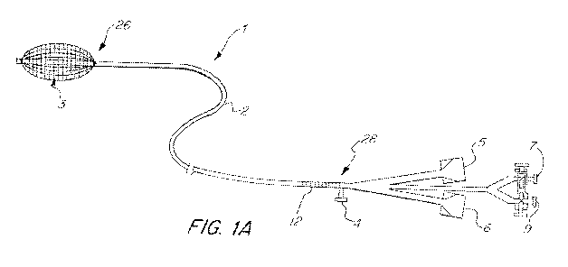
[0047] FIG. 1A is schematic view of an anchored working channel in
accordance with the invention.
CA 02896511 2015-06-25
WO 2014/105439
PCT/US2013/074620
- 15 -
[0048] FIG. 1B is a schematic view of the anchored working channel of
Figure 1A with a plurality of balloons.
[0049] FIG. 1C is a schematic view of the anchored working channel of
Figure 1A, showing various medical instruments inserted therethrough.
[0050] FIG. 1D is a schematic view of the anchored working channel of
Figure 1A, showing a proximal end of the working channel in more detail.
[0051] Figure 2 is an end view of the inflated balloon of the anchored
working channel of Figure 1A.
[0052] Figure 3A is a perspective cross-sectional view of a distal end of
the anchored working channel of Figure 1A.
[0053] Figure 3B is a plan cross-sectional view of a distal end of the
anchored working channel of Figure 1A.
[0054] Figure 4 is a partially schematic view of the working channel of
Figure 1A, showing connection to a pump.
[0055] Figure 5 is a perspective view of a distal end of the anchored
working channel of Figure 1A, showing an imaging device disposed therein.
[0056] Figure 6 is a cross-sectional view of a distal end of the imaging
device of Figure 5.
[0057] Figures 7-9 are views of the anchored working channel of Figure
1A being operated in a bodily cavity.
CA 02896511 2015-06-25
WO 2014/105439
PCT/US2013/074620
- 16 -
DETAILED DESCRIPTION OF THE INVENTION
[0058] The basic components of one embodiment of an anchored
working channel in accordance with the invention are illustrated in FIG. 1A.
As used in the description, the terms "top," "bottom," "above," "below,"
"over,"
"under," "above," "beneath," "on top," "underneath," "up," "down," "upper,"
"lower," "front," "rear," "back," "forward" and "backward" refer to the
objects
referenced when in the orientation illustrated in the drawings, which
orientation is not necessary for achieving the objects of the invention.
[0059] The anchored working channel of the present invention may be
used with various catheter or endoscope devices, various types of surgical
instruments, tools, and operative devices, implants and related medical
diagnostic and treatment systems that need to be inserted into bodily cavities
and operated therein via a suitable working channel. In an advantageous
embodiment, the anchored working channel is used with a resector balloon
system described in U.S. Patent No. 8,226,601, the disclosure of which is
incorporated by reference herein in its entirety. In another advantageous
embodiment, the working channel of the present invention is used with a
steerable catheter system described in U.S. Patent Application No.
13/037,874, the disclosure of which is also incorporated by reference herein
in
its entirety. In yet another adv9ntageous embodiment, the working chanel is
used with an anchored guidewire described in U.S. Patent Application No.
12/906,736 the disclosure of which is also incorporated by reference herein in
its entirety.
CA 02896511 2015-06-25
WO 2014/105439
PCT/US2013/074620
- 17 -
[0060] As shown in Figure 1A, the anchored working channel (1)
includes an elongated shaft (2) having a distal end (26) and a proximal end
(28). The shaft (2), which can be rigid or flexible, may have any suitable
diameter and length depending on a particular application and/or dimensions
of target bodily cavity, and may be flexible, rigid or semi rigid. In one
advantageous embodiment of the present invention, the elongated shaft has a
length of about 90 mm, an inner diameter of about 4 mm and an outer
diameter of about 4.5 mm.
[0061] The elongated shaft (2) may be made with any commercially
available material that is flexible enough to allow the shaft to be safely
inserted through the available opening of a bodily cavity such that it will
deflect from the walls of the cavity instead of puncturing them. In
particular, a
distal end section of the elongated shaft (2) is made flexible to ensure safe
insertion of the working channel into bodily cavities.
[0062] In some embodiments, the shaft (2) may include a coating made
of suitably smooth material to facilitate the movement of the working channel
through the bodily cavities. In one advantageous embodiment shown in
Figure 3, the elongated shaft (2) consists of a coil wire (30) made of any
suitable material, such as stainless steel, and a coating (32) made of
suitable
materials, such as polyethylene, polyurethane, Pebax and the like. A
braided sheath may also be used instead of the coil wire. ln some
CA 02896511 2015-06-25
WO 2014/105439
PCT/US2013/074620
- 18 -
advantageous embodiments, the coil wire or the braided sheath may be made
with a memory shape material, such as nitinol.
[0063] In further advantageous embodiments, the elongated shaft may
include a combination of braided sheath and coil wire materials to provide for
optimal flexibility and maneuverability of the shaft. For example, a distal
portion of the elongated shaft may be made with coiled wire material and thus,
have more flexibility, and the rest of the elongated shaft is made with the
braided sheath material and less flexible.
[0064] The coil wire (30) or braid can be molded over during the shaft
extrusion process and can run the entire length of the elongated shaft (2).
Alternatively, the elongated shaft (2) may be molded or extruded in a first
step
and the coil wire (30) may be disposed within an inner lumen of the shaft.
Such design improves torque, maneuverability, and kick resistance of the
elongated shaft (2), and also prevents reduction of the working channel
diameter.
[0065] The elongated shaft (2) may, as shown in Figure 1A, further
include calibrated markings (12) to gauge extent of insertion of the shaft (2)
into a bodily cavity. In some embodiments, the elongated shaft (2) may
further include imaging markers positioned at the distal end (26) of the shaft
or at any other location along the shaft to facilitate external imaging
thereof
and thereby allow for better vi:Flialization during insertion and positioning
of
the working channel (1) in bodily cavities.
CA 02896511 2015-06-25
WO 2014/105439
PCT/US2013/074620
- 19 -
[0066] The distal end of the elongated shaft (2) includes at least one
inflatable balloon (3) located at or near the tip of the distal end. The
inflatable
balloon (3) has an outer wall with a textured surface, which, when inflated,
grips the surrounding tissue in a bodily cavity. The inflatable balloon (3)
may
be made of latex, Yulex, polyethylene, nylon or other suitable material, and
may come in a variety of sizes and diameters, which allow the working
channel (1) to be used in bodily cavities of various diameters and dimensions,
such as large and small bronchial branches, sinuses, vessels, etc. In some
advantageous embodiments, the inflatable balloon (3) has a length of about
mm and a diameter of about 10 mm. In certain embodiments, a compliant
balloon is employed. In further advantageous embodiments, the inflatable
balloon (3) may comprise a plurality of balloons/bladders, which may be
controlled, inflated and deflated independently of each other.
(0067] Figure 2 illustrates an end view of the inflated balloon (3) of the
anchored working channel (1). The outer surface (8) of the balloon (3)
includes a woven mesh (10) disposed on the outer surface of the balloon.
The mesh may be made of elastane, latex, polyurethane, composite springs,
metallic fibers, elastic, steel fibers, or other appropriate material, or a
composite or coating thereof. In some advantageous embodiments, the mesh
is made with elastane material. In particularly advantageous embodiments,
the mesh is weft knit. In is understood, however, that the mesh sleeve may
be made using any suitable mesh manufacturing techniques.
CA 02896511 2015-06-25
WO 2014/105439
PCT/US2013/074620
- 20 -
[0068] The woven mesh sleeve (10) may be disposed on the outer
surface of the balloon (3) by using any suitable manufacturing method.
Alternatively, woven sleeve (10) may be knitted or woven from thread directly
onto the balloon (3). In some advantageous embodiments, the woven mesh
(10) may be affixed to the surface of the balloon (3) during the molding
process, which produces outwardly-facing protrusions on the outer surface of
the balloon (3) that assist in gripping of the balloon to the surrounding
tissue.
In other advantageous embodiments, dimensional surface structures, such as
bumps or inflatable sinuses, that are encapsulated in the surface substrate of
the balloon (3) may be used to produce the surface protrusions forming the
textured surface.
[0069] The protrusions forming the textured surface of the balloon (3)
can have various shapes and configurations, depending on a particular
application. In some embodiments, the outer surface of the balloon (3) may
have outwardly extending protrusions forming a lattice-like structure or a
spiral-like pattern extending circumferentially on the outer surface of the
balloon (3). In other embodiments, the protrusions may be in a form of
dimples that extend outwardly from the outer surface of the balloon (3). It
should be noted that any other shapes and configurations of the surface
protrusions can be used in accordance with the present invention, including
combinations of any of the aforementioned or other textures.
CA 02896511 2015-06-25
WO 2014/105439
PCT/US2013/074620
- 21 -
[0070] In certain advantageous embodiments, the balloon (3) includes
imaging markers, such as radio opaque rings, located at or near the ends
thereof. Such markers can be selected and appropriately positioned in order
to reflect or block the relevant waves of various imaging modalities (e.g., x-
ray) in order to allow the use of such modalities to assist with the precise
positioning of the balloon (3) within a bodily cavity. Similarly, the balloon
or
balloon mesh may include a radiopaque material, such as a mesh made of
yam having radiopaque iron fibers.
[0071] In some embodiments, the distal end of the elongated shaft (2)
includes a safety tip (70), such as shown in FIG. 1D. The safety tip has a
smooth convex shape designed to deflect from bodily tissues and cavity walls
during the insertion of the working channel (1) into a patient's body to
prevent
injuries to the bodily tissues during the insertion. The safety tip may be
made
with the same materials as the elongated shaft and has an opening
therethrough for introduction of various instruments/devices through the
working channel (1).
[0072] When in use, the working channel (1) is first introduced into a
bodily cavity and positioned adjacent the target tissue site. Then, the
balloon
(3) is inflated such that the woven mesh sleeve (10) covers at least a portion
of the balloon outer surface in an expanded state and adds texture, friction,
and surface area to the outer surface of the balloon. The crossover points of
the fiber threads forming the mesh produce outwardly-facing, small knots or
CA 02896511 2015-06-25
WO 2014/105439
PCT/US2013/074620
- 22 -
dimples, which grip the surrounding tissue, thereby anchoring the working
channel (1) at the target site.
[0073] It is understood that the working channel (1) may also include a
plurality of anchoring devices positioned at different locations along the
elongated shaft (2). The plurality of anchoring devices allow for more precise
and secure anchoring of the working channel (1) within the bodily cavity. As
shown in Figure 1 B, multiple balloons (61, 62, 63), each with textured
surface,
such as a mesh, may be positioned along the distal portion of the shaft (2).
[0074] In addition to serving as an anchoring device to secure the
working channel within the bodily cavity, the inflatable balloon (3) or a
plurality
of inflatable balloons can also be used to block or prevent fluids from
flowing
around the balloon in the target bodily lumen, vessel, airway or space.
[0075] It should be noted that in certain applications, such as when the
working channel device is used in very small bodily cavities or passages, it
may not be necessary to utilize an inflatable balloon to anchor the working
channel. For example, when the working channel (1) is used in small lung
airway passages, the outer diameter of the elongated shaft itself may be
sufficient to fixate the working channel inside the passage.
[0076] As shown in Figure 3A, the elongated shaft (2) of the working
channel (1) includes at least two inner lumens. An inflation lumen (13) is
connected to the fluid source provided at the proximal end of the elongated
shaft (2) and is in fluid communication with the interior of the inflatable
balloon
CA 02896511 2015-06-25
WO 2014/105439
PCT/US2013/074620
- 23 -
(3) via a plurality of openings (14) in the shaft wall positioned inside the
balloon (3). The fluid source supplies fluid to the inflation lumen (13) and
via
the openings (14) to inflate the balloon (3).
[0077] The elongated shaft (2) further includes a working channel
lumen (15). In the embodiment shown in Figure 3A, the working channel
lumen (15) is an inner lumen is positioned inside the outer inflation lumen
(13). However, it is understood that any other configuration of the lumens
may be used in accordance with the present invention. For example, as
shown in Figure 3B, the elongated shaft (2) may consist of a coating material
(32), such as polyethylene or polyurethane, with an embedded coil or braid
(30). The elongated shaft (2) includes an inner working channel lumen (15)
and one or more inflation lumens (13) provided in the coating material (32)
adjacent the working channel lumen (15).
[0078] In additional embodiments, the elongated shaft (2) may also be
divided into equal or unequal sections representing the inflation lumen and
the
working channel lumen. Furthermore, it is understood than the elongated
shaft (2) may include more than two inner lumens for performing different
functions.
[0079] The working channel lumen (15) may be used to deploy various
medical instruments or devices into the desired part of the airway, vessel,
lumen, pleural cavity or other bodily cavity. The working channel lumen (15)
may further be divided into a plurality of lumens (not shown), through which
CA 02896511 2015-06-25
WO 2014/105439
PCT/US2013/074620
- 24 -
an imaging device, an instrument, a device, or a fluid may be placed. The
working channel lumen(s) can be used to deliver any number of things to
assist a surgeon with performing a surgical or diagnostic medical procedure,
such as cutting or resecting tissue, aspiration, respiration, imaging,
delivering
various therapeutic and/or diagnostic agents, delivering stents, scaffolds or
implants, and such.
[0080] Referring back to Figure 1A, the proximal end (28) of the
elongated shaft (2) includes an inflation port (4) for connection of the
working
channel (1) to a fluid source, such as a pump, through which the balloon (3)
is
inflated. The inflation port (4) is provided with any suitable connector, such
as
a luer connector, for connection to the pump. The inflation port (4) is in
fluid
communication with the inflatable balloon (3) via the inflation lumen (13) of
the
elongated shaft (2).
[0081] As shown in Figure 1C, the proximal end of the elongated shaft
(2) further includes one or more ports through which various medical
instruments or devices are inserted into the working channel lumen. For
example, the proximal end (28) of the elongated shaft (2) includes an imaging
device port (5), an instrument port (6), a suction port (7) and an irrigation
port
(9). The imaging device port (5) is used for insertion of an imaging device
(30), as discussed in more detail below. The instrument port (6) provides an
access point for insertion of catheters, endoscopes, various surgical or
diagnostic medical devices, and the like. The camera port (5) and the
CA 02896511 2015-06-25
WO 2014/105439
PCT/US2013/074620
- 25 -
instrument port (6) may connect to the same working channel lumen or may
be each connected to a separate inner lumen provided in the elongated shaft
(2).
[0082] In the embodiment illustrated in Figure 1C, a resecting balloon
catheter system described in U.S. Patent No. 8,266,601 is inserted through
the instrument port (6) to perform a desired procedure within the bodily
cavity.
Preferably, a length of the catheter is sufficiently greater than the length
of the
elongated shaft (2) and the outer diameter of the catheter is sufficiently
smaller than the inner diameter of the working channel lumen such that the
catheter may be easily inserted into the lumen and extended out of the distal
end (26) of the shaft. In some advantageous embodiments, the length of the
catheter operated through the working channel (1) is about 120 mm. It is
noted that any other catheter system, such as a balloon catheter, a drug
delivery catheter, a steerable catheter, etc., may be used with the working
channel of the present invention.
[0083] The suction and irrigation ports (7 and 9) function to
deliver/suction irrigation fluid to the surgical site. The ports (7, 9) are
provided
with trumpet valves or any other suitable valve type and are connected to an
irrigation fluid/vacuum source positioned outside of the patient's body. The
irrigation fluid may be accommodated in the working channel lumen(s) (15),
or, alternatively, may be provided via a separate lumen of the elongated shaft
(2). In some advantageous embodiments, the suction/irrigation valves are
CA 02896511 2015-06-25
WO 2014/105439
PCT/US2013/074620
- 26 -
provided in an in-line arrangement to facilitate passage of debris out of the
working channel (1).
[0084] The proximal section (55) of the elongated shaft (2) may be
provided as a separate structure removably attachable to the proximal end of
the elongated shaft, as shown in FIG. 1D. This way, the same proximal end
section (55) may be used with various working channel devices, and may be
easily removed for sterilization or replaced with another attachment (60),
e.g.
bronch adapter shown in this figure, desired for a specific medical procedure.
In the exemplary embodiment shown in FIG. 1D, a threaded connector is
used to attach the proximal section (55) to the proximal end of the elongated
shaft. An external thread (61) is provided on the outer surface of the
elongated shaft and a corresponding internal thread (62) is provided on the
inner surface of the proximal section (55). It is understood, however, that
any
other suitable connection mechanism may be used to connect the proximal
section (55) to the elongated shaft (2).
[0085] The proximal section (55) includes various ports, e.g. an
imaging device port (63), an instrument port (64), a suction/irrigation port
(65),
etc., for connection to or insertion of various instruments and/or devices
needed to perform a particular procedure. The ports may be provided with any
suitable connectors and/or ad4ters, such as seal lip connector, luer
connector, Tuohy Borst type adapter, and the like.
CA 02896511 2015-06-25
WO 2014/105439
PCT/US2013/074620
- 27 -
[0086] In additional advantageous embodiments, the elongated shaft
(2) may further include a bypass lumen to allow bodily fluids, such as air or
blood, to flow through the working channel (1), which is necessary in certain
medical applications, e.g. pulmonology or cardiology. In the case of air
bypass, the air may flow through one of the shaft lumens and in/out of the
proximal end of the working channel (1) positioned outside of the patient's
body. In some cases, an external device, such as a respiration device, is in
communication with the shaft lumen in order to help facilitate this flow. If a
blood bypass is desired, an additional port/opening may be provided in the
elongated shaft (2) towards the distal end of the shaft to allow for blood to
flow
through one of the shaft lumens and out of the opening. It is understood that
a separate bypass lumen is not required and that the working channel
lumen(s) (15) may function as a bypass lumen.
[0087] The anchored working channel (1) with the fluid source (20) is
further shown in Figure 4. Any suitable fluid source may be used in
accordance with the present invention. In one advantageous embodiment,
the fluid source (20) is an electro-pneumatic pump having controls on the
front
thereof, from which a physician or assistant can control the system (as well
as
a remote control unit), such as that disclosed in U.S. Patent No. 8,266,601 to
Gunday et al. The pump (20) supplies a fluid, such as a gas, liquid, or
mixture
thereof, to the inflation lumen µ13) of the working channel via the inflation
port
(4). The pump (20) also includes a variety of capabilities for balloon
identification, proper inflation/deflation of the balloon, and feedback
CA 02896511 2015-06-25
WO 2014/105439
PCT/US2013/074620
- 28 -
measurements, many details of which are described in Gunday et al. In
certain advantageous embodiments, the pump (20) further includes a vacuum
source to evacuate fluid from the balloon (3). In other embodiments, a
handheld pump is used as a fluid source.
[0088] In some embodiments, the working channel (1) includes a data
device, such as optical, RFID, flash memory, etc. This way, the pump (20) is
able to identify the type of working channel device that is connected and read
the characterization data of the balloon, e.g. maxim pressure, volume,
dimensions, etc., and/or working channel included thereon, and then adjust its
control accordingly based on user input.
[0089] The pump (20) further includes a processor that controls the
supply of fluid to the inflatable balloon (3) based on at least one
predetermined parameter. In some embodiments, such predetermined
parameters may be manually entered by the user. Alternatively, the control of
the fluid is based on default parameters selected by the pump (20), which are
based on the characteristics of the particular balloon and/or the diameter
measurements of a particular bodily cavity made by the pump. Furthermore,
the pump may control and regulate the pressure by monitoring and taking into
account one or more vital signs and physiological parameters of the patient,
such as body temperature, heart rate, blood pressure, and respiratory rate.
[0090] In some advantageous embodiments, the working channel (1) of
the present invention is capable of measuring airflow through the bypass
CA 02896511 2015-06-25
WO 2014/105439
PCT/US2013/074620
- 29 -
lumen of the elongated shaft (2). The airflow may be measured by the pump
(20) or by a separate sensor coupled to the bypass lumen of the working
channel. This is particularly advantageous in pulmonary applications, where it
is important to measure the amount of airflow to and from a patient's lungs.
[0091] Referring to Figure 5, the working channel (1) is further provided
with an imaging device (30) disposed in the elongated shaft (2). The imaging
device is used to facilitate the insertion and positioning of the working
channel
in the bodily cavity, and may further assist the surgeon in performing a
medical procedure. The imaging device (30) is inserted into the working
channel lumen (15) through the imaging device port (5) and is extended out of
the distal end of the elongated shaft (2) such that the tissue in front of the
working channel can be viewed by the imaging device during the insertion of
the working channel (1) into a bodily cavity.
[0092] The imaging device (30) includes a camera head (31) disposed
at a distal end of a sheath (32). The sheath has a length that is sufficiently
greater than the length of the elongated shaft (2), such that the imaging
device (30) can be extended out of the distal end of the elongated shaft. In
some advantageous embodiments, the length of the imaging device sheath
(32) is about 105 mm. Additionally, an outer diameter of the imaging device
sheath is smaller than the inner diameter of the working channel lumen (15) to
facilitate the insertion of the imaging device through the lumen. In one
advantageous embodiment, tha outer diameter of the sheath (32) is less than
CA 02896511 2015-06-25
WO 2014/105439
PCT/US2013/074620
- 30 -
about 1 mm. The sheath (32) is preferably made with a flexible material that
allows for rotational or linear movement of the distal end of the sheath.
[0093] It is understood that the imaging device (30) may also be
introduced into a bodily cavity through the inflation lumen (13) of the
working
channel. This way, the inflation lumen (13) serves a dual purpose ¨ it is used
both for supply of fluid to inflate/deflate the balloon (3) and for
visualization via
the imaging device (30). In these embodiments, an imaging device aperture
may be positioned inside the balloon (3), and the outer wall of the balloon is
made transparent when inflated, such that imaging is made possible from
inside the balloon (3). The imaging device aperture can also serve as an
inflation/deflation opening through which the fluid is supplied to/from the
balloon (3). Additionally, the elongated shaft (2) may have one or more
imaging device apertures positioned at different locations along the shaft for
better visualization of the surrounding area during the introduction of the
working channel (1) into the patient's body.
[0094] In one advantageous embodiment shown in Figure 5, the
imaging device (30) includes a steerable flexible distal tip that can be
translated linearly or rotationally inside the bodily cavity. This allows for
enhanced visualization of the surrounding area during the insertion and
operation of the working channel (1). As shown in Figure 6, the imaging
device sheath (32) includes four steering lumens (33, 35, 37, 39) extending
through the entire length of the sheath. It is understood that a lesser or
great
CA 02896511 2015-06-25
WO 2014/105439
PCT/US2013/074620
- 31 -
number of steering lumens may also be provided, depending on the desired
level of maneuverability of the imaging device (30). A center lumen (34) is
also provided in the sheath (32) for accommodating components of the
imaging device (30). The steering lumens (33, 35, 37, 39) are shown
integrally formed as part of the sheath (32) and are radially offset from the
longitudinal axis of the sheath (32) and the center lumen (34). However, it is
understood that any other suitable configuration and/or construction of the
sheath and the steerable lumens may be used in accordance with the
invention.
[0095] In some advantageous embodiments, the distal end of the
imaging device (30) is actuated by engaging pull wire(s) disposed in each of
the steering lumens (33, 35, 37, 39). In other advantageous embodiments,
any one or more of the steering lumens (33, 35, 37, 39) may be filled with
pressured air in various amounts. In yet further embodiments, the opposite
steering lumen(s) (33, 37) or (35, 39) may be deflated with vacuum to
facilitate the movement of the distal tip of the imaging device (30).
[0096] There is a control unit positioned outside of a patient's body and
connected to the imaging device (30) via the imaging device port (5) to allow
for manipulation of the imaging device by a surgeon. The imaging device (30)
is further coupled to any suitable type of a processor and a display device
for
processing the imaging data received from the imaging device and displaying
the data to the surgeon. It is noted that the imaging device (30) may also be
CA 02896511 2015-06-25
WO 2014/105439
PCT/US2013/074620
- 32 -
wirelessly connected to the control unit, the processor and/or the display
device.
[0097] The distal end of the imaging device sheath (32) has a camera
head (31) disposed thereon. In an advantageous embodiment, the imaging
device (30) is a fiber optic image bundle. Two separate fiber optic bundles ¨
an incoherent fiber bundle for illumination and a coherent fiber bundle for
imaging ¨ can also be used in accordance with the present invention. It
should be noted that a suitable image sensor (e.g. CCD or CMOS) can be
positioned at the tip of the imaging device (30), eliminating the need for a
coherent imaging fiber bundle, thus increasing the image quality and reducing
cost. It should also be noted that other sources of illumination, such as
light
emitting diodes, can be employed.
[0098] In some embodiments, a fiberscope device may be used in
addition to the imaging device (30) for providing enhanced visualization of
the
target site. The fiberscope is inserted into the working channel lumen (15) of
the elongated shaft (2) through the instrument port (6) and is extended out of
the distal end (26) of the shaft. The fiberscope may be pushed through tumor
tissue to provide visualization from the inside and in front of the tumor.
[0099] In one advantageous embodiment, the fiberscope may be
inserted through one of the inner lumens of the steerable catheter or the
balloon catheter described above. Preferably, a length of the fiberscope is
sufficiently longer than the length of both the working channel (1) and the
CA 02896511 2015-06-25
WO 2014/105439
PCT/US2013/074620
- 33 -
catheter disposed in the working channel such that the fiberscope extends
past the distal end of the catheter. The distal end of the catheter may
include
a lens cleaning device for cleaning the fiberscope lens. The cleaning device
is made with any suitable type of material, for example, a textile bundle,
that is
affixed to the distal end of the catheter. The fiberscope is cleaned by moving
it
back and forth through the cleaning device, thus wiping the lens of the
fiberscope.
(00100j Figures 7-9 illustrate a method of insertion and operation of the
working channel (1) in a bodily cavity in accordance with the present
invention.
[ooioi]As shown in Figure 7, the working channel (1) is introduced into
a desired location within a patient's body. In order to assist the surgeon in
insertion and positioning of the working channel (1), the imaging device (30)
is
inserted into one of the working channel's lumens and is extended out of the
distal end of the working channel for visualizing the tissue adjacent the
distal
end of the working channel. As described above, the distal end of the
imaging device may be manipulated by the surgeon to steer the imaging
device (30) through the bodily passages to the target site. Additionally, the
elongated shaft (2) of the working channel may have imaging markers to
assist the surgeon in visualizing the exact position of the working channel
within the bodily cavity.
CA 02896511 2015-06-25
WO 2014/105439
PCT/US2013/074620
- 34 -
[00102] It should be noted that a guide wire may be first inserted into the
bodily cavity and anchored at the target site. Then, the working channel (1)
is
advanced over the guide wire and anchored at the target site, and the guide
wire is removed from the bodily cavity.
[00103] Once the working channel (1) is positioned at the target site
(40), the balloon (3) provided at the distal end of the elongated shaft (2) is
inflated by supplying fluid thereto from the pump or any other fluid source
via
the inflation port, as shown in Figure 8. The balloon (3) is inflated until
the
outer wall of the balloon contacts the surrounding tissue such that the
textured
outer surface of the balloon (3) grips the tissue, thereby anchoring the
working
channel at the target site.
[00104] Next, the imaging device (30) is removed from the working
channel lumen and a desired medical instrument or device is inserted therein
for performing a medical procedure. For example, as shown in Figure 9, a
resector balloon system (50) described in U.S. Patent No. 8,226,601 may be
inserted through the working channel lumen of the working channel (1) to
resect the tumor tissue (40). In some embodiments, the imaging device (30)
is not removed from the working channel (1) and is used to visualize the
surgical site during the procedure. Furthermore, as discussed above, a
fiberscope may be first pushed through the tumor tissue to provide an image
of the inside and in front of the tumor (40) prior to the resecting procedure
to
allow the surgeon to more precisely gauge the size, location and morphology
CA 02896511 2015-06-25
WO 2014/105439
PCT/US2013/074620
- 35 -
of the tumor. Additional instruments and/or devices may also be introduced
into the bodily cavity through the working channel (1) during the procedure to
perform various functions, such as, for example, delivering
therapeutic/diagnostic agents, providing irrigation fluid/suction, taking
tissue
samples, etc.
[00105] Once the procedure is completed, the instruments and/or
devices are removed out of the working channel (1). Then, the balloon (3) is
deflated and the working channel (1) is removed from the patient's body.
[00106] It would be appreciated by those skilled in the art that various
changes and modifications can be made to the illustrated embodiment without
departing from the spirit of the present invention. All such modifications and
changes are intended to be covered hereby.