Note: Descriptions are shown in the official language in which they were submitted.
CA 02896800 2015-06-26
WO 2014/113813
PCT/US2014/012388
DEVICES AND METHODS FOR CONTROLLING TREMOR
CROSS REFERENCE TO RELATED APPLICATIONS
[0001] This application claims priority to U.S. Provisional Application
No. 61/754,945,
filed January 21, 2013, U.S. Provisional Application No. 61/786,549, filed
March 15, 2013, U.S.
Provisional Application No. 61/815,919, filed April 25, 2013, U.S. Provisional
Application No.
61/822,215, filed May 10, 2013, and U.S. Provisional Application No.
61/857,248, filed July 23,
2013, each of which is herein incorporated by reference in its entirety.
INCORPORATION BY REFERENCE
[0002] All publications and patent applications mentioned in this
specification are herein
incorporated by reference to the same extent as if each individual publication
or patent
application was specifically and individually indicated to be incorporated by
reference.
FIELD
[0003] Embodiments of the invention relate generally to systems,
devices, and methods for
treating tremor, and more specifically relate to system, devices, and methods
for treating tremor
by stimulation of a peripheral nerve.
BACKGROUND
[0004] Essential tremor (ET) is the most common movement disorder,
affecting an estimated
10 million patients in the U.S., with growing numbers due to the aging
population. The
prevalence of ET rises with age, increasing from 6.3% of the population over
65, to above 20%
in the population over 95. ET is characterized by an involuntary oscillatory
movement, typically
between 4-12Hz. It can produce oscillations in the voice and unwanted
movements of the head
and limbs. Tremor in the hands and forearm is especially prevalent and
problematic because it
makes it difficult to write, type, eat, and drink. Unlike Parkinson's tremor,
which exists at rest,
essential tremor is postural and kinetic, meaning tremor is induced by holding
a limb against
gravity or during movement, respectively.
[0005] Disability with ET is variable, and ranges from embarrassment to the
inability to live
independently when tasks such as writing and self-feeding are not possible due
to the
uncontrolled movements of the hand and arm. Despite the high prevalence and
high disability in
many patients with ET, there are insufficient treatment options to address
tremor.
[0006] The drugs used to treat tremor (e.g., Propanolol and Primidone)
have been found to
be effective in reducing tremor amplitude by only 50% in only 60% of patients.
These drugs
- 1 -
CA 02896800 2015-06-26
WO 2014/113813
PCT/US2014/012388
have side effects that can be severe and are not tolerated by many patients
with ET. An
alternative treatment is surgical implantation of a stimulator within the
brain using deep brain
stimulation (DBS), which can be effective in reducing tremor amplitude by 90%,
but is a highly
invasive surgical procedure that carries significant risks and cannot be
tolerated by many ET
patients. Thus, there is a great need for alternative treatments for ET
patients that reduce tremors
without the side effects of drugs and without the risks of brain surgery.
[0007] Tremor is also a significant problem for patients with
orthostatic tremor, multiple
sclerosis and Parkinson's Disease. A variety of neurological disorders include
tremor such as
stroke, alcoholism, alcohol withdrawal, peripheral neuropathy, Wilson's
disease, Creutzfeldt-
Jacob disease, Guillain¨Barre syndrome and fragile X syndrome, as well as
brain tumors, low
blood sugar, hyperthyroidism, hypoparathyroidism, insulinoma, normal aging,
and traumatic
brain injury. Stuttering or stammering may also be a form of tremor. The
underlying etiology of
tremor in these conditions may differ from ET; however, treatment options for
some of these
conditions are also limited and alternative treatments are needed.
[0008] ET is thought to be caused by abnormalities in the circuit dynamics
associated with
movement production and control. Previous work has shown that these circuit
dynamics may be
temporarily altered by cooling, topical analgesics and vibration. Previous
work reported that
electrical stimulation using transcutaneous electrical nerve stimulation
(TENS) did not improve
tremor (Munhoz 2003). It was therefore surprising to discover in our clinical
study that circuit
dynamics associated with ET can be altered by peripheral nerve simulation
resulting in a
substantial reduction in the tremor of individuals with ET.
[0009] The present invention is a novel peripheral stimulation device to
send signals along
the sensory nerves to the central nervous system in order to modify the
abnormal network
dynamics. Over time, this stimulation normalizes the neural firing in the
abnormal network and
reduces tremor. While DBS stimulates the brain directly, our peripheral
stimulation influences
the abnormal brain circuit dynamics by sending signals along the sensory
nerves that connect the
periphery to the brain. This approach is non-invasive and expected to avoid
DBS's surgical risks
and associated problems with cognitive, declarative and spatial memory
dysarthria, ataxia or gait
disturbances. The peripheral nerve stimulation may effectively treat tremors
by dephasing,
overriding or obscuring the abnormal brain circuit dynamics. Overriding,
obscuring or training
the brain to ignore the abnormal brain circuit dynamics follows on hypotheses
for the
mechanisms of traditional DBS.
[00010] Perhaps the technology most closely related to our approach is
transcutaneous
electrical nerve stimulation (TENS). High-frequency TENS (50 to 250Hz) is
commonly used to
treat pain, with the hypothesis that excitation of large, myelinated
peripheral proprioceptive
- 2 -
CA 02896800 2015-06-26
WO 2014/113813
PCT/US2014/012388
fibers (A-beta) blocks incoming pain signals. While the inconsistent clinical
results achieved
using TENS for pain control have led many to question its use for treatment of
pain, it is well
documented that surface electrical stimulation excites A-beta neurons. A-beta
neurons
communicate proprioceptive sensory information into the same brain circuits
that are abnormal
in diseases including ET and Parkinson's disease. Without being limited by any
proposed
mechanism of action, this has led us to propose that neurostimulation could be
used to excite A-
beta nerves and thereby improve tremor. This proposal is particularly
surprising because a
previous study by Munhoz et al. failed to find any significant improvement in
any of the tremor
parameters tested after application of TENS. See Munhoz et al., Acute Effect
of Transcutaneous
Electrical Nerve Stimulation on Tremor, Movement Disorders, 18(2), 191-194
(2003).
SUMMARY OF THE DISCLOSURE
[00011] The present invention relates systems, devices, and methods for
treating tremor, and
more specifically relate to system, devices, and methods for treating tremor
by stimulation of a
peripheral nerve.
[00012] In some embodiments, a method of reducing tremor in a patient is
provided. The
method includes placing a first peripheral nerve effector at a first location
relative to a first
peripheral nerve; delivering a first stimulus to the first peripheral nerve
through the first
peripheral nerve effector; and reducing the tremor amplitude by modifying the
patient's neural
network dynamics.
[00013] In some embodiments, the placing step comprises placing the first
peripheral nerve
effector on the patient's skin and the first stimulus is an electrical
stimulus applied to a skin
surface.
[00014] In some embodiments, the first stimulus has an amplitude from about
0.1 mA to 10
mA and a frequency from about 10 to 5000 Hz. In some embodiments, the first
stimulus has an
amplitude that is less than about 15, 14, 13, 12, 11, 10, 9, 8, 7, 6, 5, 4, 3,
2 or 1 mA.
[00015] In some embodiments, the placing step comprises implanting the first
peripheral
nerve effector in the patient and the first stimulus is an electrical
stimulus.
[00016] In some embodiments, the implanting step comprises injecting the first
peripheral
nerve effector in the patient. In some embodiments, the first stimulus has an
amplitude less than
about 3 mA and a frequency from about 10 to 5000 Hz. In some embodiments, the
first stimulus
has an amplitude that is less than about 5, 4, 3, 2 or 1 mA.
[00017] In some embodiments, the peripheral nerve effector includes a power
source.
[00018] In some embodiments, the method further includes powering the first
peripheral
nerve effector wirelessly through an externally located power source.
- 3 -
CA 02896800 2015-06-26
WO 2014/113813
PCT/US2014/012388
[00019] In some embodiments, the first stimulus is vibrotactile.
[00020] In some embodiments, the first stimulus is chemical.
[00021] In some embodiments, the method further includes sensing motion of the
patient's
extremity using a measurement unit to generate motion data; and determining
tremor information
from the motion data.
[00022] In some embodiments, the delivery step comprises delivering the first
stimulus based
on the tremor information.
[00023] In some embodiments, the tremor information comprises a maximum
deviation from
a resting position for the patient's extremity.
[00024] In some embodiments, the tremor information comprises a resting
position for the
patient's extremity.
[00025] In some embodiments, the tremor information comprises tremor
frequency, phase,
and amplitude.
[00026] In some embodiments, the step of delivering the first stimulus
comprises delivering a
plurality of bursts of stimulation having a variable temporal delay between
the bursts of
stimulation.
[00027] In some embodiments, the method further includes placing a second
peripheral nerve
effector at a second location relative to a second peripheral nerve; and
delivering a second
stimulus to the second peripheral nerve through the second peripheral nerve
effector.
[00028] In some embodiments, the method further includes determining a period
of the
patient's tremor, wherein the step of delivering the second stimulus comprises
offsetting delivery
of the second stimulus from the delivery of the first stimulus by a
predetermined fraction or
multiple of a period of the tremor.
[00029] In some embodiments, the method further includes dephasing the
synchronicity of a
neural network in the patient's brain.
[00030] In some embodiments, the first location and second location are
located on adjacent
fingers.
[00031] In some embodiments, the first peripheral nerve and the second
peripheral nerve are
adjacent nerves.
[00032] In some embodiments, the first peripheral nerve is the median nerve
and the second
peripheral nerve is the ulnar or radial nerve.
[00033] In some embodiments, the first peripheral nerve and the second
peripheral nerve are
somatotopically adjacent.
[00034] In some embodiments, the first stimulus has an amplitude that is below
a sensory
threshold.
- 4 -
CA 02896800 2015-06-26
WO 2014/113813
PCT/US2014/012388
[00035] In some embodiments, the first stimulus is greater than 15 Hz.
[00036] In some embodiments, the first peripheral nerve carries proprioceptive
information
from the patient's extremity.
[00037] In some embodiments, the method further includes determining a
duration of efficacy
of the first stimulus on reducing the tremor amplitude; and delivering a
second stimulus before
the expiration of the duration of efficacy.
[00038] In some embodiments, the step of determining the duration of effect
comprises
analyzing multiple stimuli applications applied over a predetermined period of
time.
[00039] In some embodiments, the step of determining the duration of efficacy
further
comprises determining an activity profile for the patient.
[00040] In some embodiments, the step of determining the duration of efficacy
further
comprises determining a profile of the tremor.
[00041] In some embodiments, the activity profile includes data regarding
caffeine and
alcohol consumption.
[00042] In some embodiments, the method further includes placing a conduction
pathway
enhancer over the first peripheral nerve.
[00043] In some embodiments, the conduction pathway enhancer is a conductive
tattoo.
[00044] In some embodiments, the conduction pathway enhancer comprises one or
more
conductive strips.
[00045] In some embodiments, the first location is selected from the group
consisting of a
wrist, a forearm, a carpel tunnel, a finger, and an upper arm.
100046] In some embodiments, a system for treating tremor in a patient is
provided. The
device can include a decision unit; and an interface unit adapted to deliver
electrical stimuli to a
peripheral nerve, the interface unit comprising a first peripheral nerve
effector in communication
with the decision unit, the first peripheral nerve effector comprising at
least one electrode;
wherein the decision unit comprises a processor and a memory storing
instructions that, when
executed by the processor, cause the decision unit to: deliver a first
electrical stimulus to a first
peripheral nerve through the first peripheral nerve effector, the electrical
stimulus configured by
the controller to reduce tremor in the patient's extremity by modifying the
patient's neural
network dynamics.
[00047] In some embodiments, the first electrical stimulus has an amplitude
less than about 10
mA and a frequency from about 10 to 5000 Hz. In some embodiments, the
amplitude is less than
about 15, 14, 13, 12, 11, 10, 9, 8, 7, 6, 5, 4, 3, 2, or 1 mA.
[00048] In some embodiments, the interface unit further comprises a second
peripheral nerve
effector in communication with the decision unit, the second peripheral nerve
effector
- 5 -
CA 02896800 2015-06-26
WO 2014/113813
PCT/US2014/012388
comprising at least one electrode, wherein the memory storing instructions
that, when executed
by the processor, further cause the decision unit to deliver a second
electrical stimulus to a
second peripheral nerve in the patient's extremity through the second
peripheral nerve effector.
[00049] In some embodiments, the instructions, when executed by the processor,
cause the
decision unit to deliver the second electrical stimulus offset in time from
the first electrical
stimulus by a predetermined fraction or multiple a period of the tremor.
[00050] In some embodiments, the first peripheral nerve effector is adapted to
be placed on a
first finger and the second peripheral nerve effector is adapted to be placed
on a second finger.
[00051] In some embodiments, the first peripheral nerve effector comprises a
plurality of
electrodes arranged in linear array. In some embodiments, the plurality of
electrodes are spaced
about 1 to 100 mm apart.
[00052] In some embodiments, the first peripheral nerve effector comprises a
plurality of
electrodes arranged in a two dimensional array.
[00053] In some embodiments, the memory storing instructions that, when
executed by the
processor, further cause the decision unit to select a subset of the plurality
of electrodes based on
a position of first peripheral nerve effector on the patient's extremity,
wherein the selection of
the subset of the plurality of electrodes occurs each time the first
peripheral nerve effector is
positioned or repositioned on the extremity.
[00054] In some embodiments, the plurality of electrodes are spaced about 1 to
100 mm apart
along a first axis and about 1 to 100 mm apart along a second axis
perpendicular to the first axis.
In some embodiments, some of the electrodes are adjacent to each other to form
a strip. In some
embodiments, the spacing can be less than about 100, 90, 80, 70, 60, 50, 40,
30, 20, 10, 5, 4, 3, 2,
or 1 mm.
[00055] In some embodiments, the system further includes a measurement unit,
wherein the
memory storing instructions that, when executed by the processor, further
cause the decision unit
to: measure the movement of the patient's extremity using the measurement unit
to generate
motion data; and determine a tremor frequency and magnitude based on an
analysis of the
motion data.
[00056] In some embodiments, the analysis of the motion data comprises a
frequency analysis
of the spectral power of the movement data.
[00057] In some embodiments, the frequency analysis is restricted to between
about 4 to 12
Hz. In some embodiments, the frequency analysis is restricted to approximately
the expected
frequency range of the tremor or tremors of concern.
[00058] In some embodiments, the analysis of the motion data is done on a
predetermined
length of time of the motion data.
- 6 -
CA 02896800 2015-06-26
WO 2014/113813
PCT/US2014/012388
[00059] In some embodiments, the decision unit is further adapted to determine
tremor phase
information based on the motion data and to deliver the first electrical
stimulus based on the
tremor phase information.
[00060] In some embodiments, the tremor phase information comprises peak
tremor
deviation, the decision unit being further adapted to deliver the first
electrical stimulus at a time
corresponding to the peak tremor deviation.
[00061] In some embodiments, the memory storing instructions that, when
executed by the
processor, further cause the decision unit to deliver the first electrical
stimulus as a plurality of
bursts of electrical stimulation having a variable temporal delay between the
bursts of electrical
stimulation.
[00062] In some embodiments, the memory storing instructions that, when
executed by the
processor, further cause the decision unit to set parameters of the first
electrical stimulus based
on the determined tremor frequency.
[00063] In some embodiments, the memory storing instructions that, when
executed by the
processor, further cause the decision unit to set parameters of the first
electrical stimulus based
on the determined tremor magnitude.
[00064] In some embodiments, the memory storing instructions that, when
executed by the
processor, further cause the decision unit to compare the determined tremor
magnitude with a
predetermined threshold; and wherein the first electrical stimulus is
delivered when the
determined tremor magnitude exceeds a predetermined threshold.
[00065] In
some embodiments, the electrode is adapted to deliver the first electrical
stimulus
through the patient's skin.
[00066] In some embodiments, the electrode is adapted to be implanted and
deliver the
electrical In some embodiments, the decision unit comprises a user interface
adapted to accept
input from a user to adjust a parameter of the first electrical stimulus.
[00067] In some embodiments, the memory further stores a library of one or
more
predetermined stimulation protocols.
[00068] In some embodiments, the interface unit is integrated with the
decision unit.
[00069] In some embodiments, the interface unit and the decision unit are
separate from each
other and have separate housings.
[00070] In some embodiments, the decision unit is configured to wirelessly
provide power to,
or communicate with, the interface unit.
[00071] In some embodiments, the system further includes a measurement unit
located in the
decision unit.
- 7 -
CA 02896800 2015-06-26
WO 2014/113813
PCT/US2014/012388
[00072] In some embodiments, the system further includes a measurement unit
located in the
interface unit.
[00073] In some embodiments, the decision unit is a computing device selected
from the
group consisting of a smartphone, tablet and laptop.
[00074] In some embodiments, the system further includes a server in
communication with
the computing device, the server configured to receive from the computing
device motion data
along with a history of the electrical stimuli delivered to the patient.
[00075] In some embodiments, the server is programmed to: add the received
motion data and
the history of the electrical stimuli delivered to the patient to a database
storing data from a
plurality of patients.
[00076] In some embodiments, the server is programmed to: compare the received
motion
data and the history of the electrical stimuli delivered to the patient to the
data stored in the
database; determine a modified electrical stimulus protocol based on the
comparison of the
received motion data and the history of the electrical stimuli delivered to
the patient to the data
stored in the database; and transmit the modified electrical stimulus protocol
to the computing
device.
[00077] In some embodiments, the electronics are flexible and are disposed on
a flexible
substrate, which can be a sleeve, pad, band, or other housing.
[00078] In some embodiments, a system for monitoring tremor in a patient's
extremity is
provided. The system can include an interface unit having an inertial motion
unit for capturing
motion data, a power source and a wireless transmitter and receiver, the
interface unit adapted to
be worn on the patient's extremity; and a processing unit in communication
with the interface
unit, the processing unit configured to receive the motion data from the
interface unit, wherein
the processing unit is programmed to: determine a tremor signature and profile
over a
predetermined period of time based on an analysis of the motion data.
[00079] In some embodiments, the processing unit is a mobile phone.
[00080] In some embodiments, the system further includes a server in
communication with
the mobile phone, the server configured to receive motion data from the mobile
phone.
[00081] In some embodiments, the processing unit is further programmed to
compare the
tremor magnitude with a predetermined threshold.
[00082] In some embodiments, the processing unit is further programmed to
generate an alert
when the tremor magnitude exceeds the predetermined threshold.
[00083] In some embodiments, the predetermined threshold is adjustable by the
patient.
- 8 -
CA 02896800 2015-06-26
WO 2014/113813
PCT/US2014/012388
[00084] In some embodiments, the processing unit is programmed to prompt the
patient to
enter activity data, the activity data including a description of the activity
and a time the activity
occurred.
[00085] In some embodiments, the processing unit is programmed to correlate
the activity
data with the determined tremor frequency and magnitude.
[00086] In some embodiments, the activity data comprises consumption of
caffeine or
alochol.
[00087] In some embodiments, the activity data comprises consumption of a
drug.
[00088] We have invented a peripheral nerve stimulation device and method that
effectively
reduces tremors without the side effects of drugs and without the risks of
brain surgery. Our
approach is safe, and in some embodiments non-invasive, and effective in
reducing tremor. In
some embodiments, the device may work by altering the neural circuit dynamics
associated with
essential tremor, Parkinson's tremor, and other tremors. The device is simple
to use,
comfortable, and adjustable to achieve the best therapy for each patient.
BRIEF DESCRIPTION OF THE DRAWINGS
[00089] The novel features of the invention are set forth with particularity
in the claims that
follow. A better understanding of the features and advantages of the present
invention will be
obtained by reference to the following detailed description that sets forth
illustrative
embodiments, in which the principles of the invention are utilized, and the
accompanying
drawings of which:
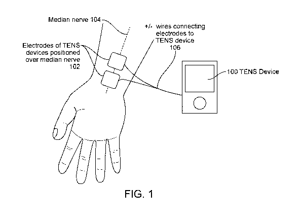
[00090] FIG. 1 illustrates one embodiment of delivering stimulation to the
median nerve
found to reduce tremor.
[00091] FIG. 2 illustrates treatment effect of an embodiment of peripheral
nerve stimulation in
a (A) mild, (B) moderate and (C) severe ET patient. It presents results of a
clinical study in
which a patient with essential tremor reduced tremor amplitude by the
configuration of
stimulation at 150Hz frequency, 300us, and for 40 minutes of stimulation on-
time. The tremor
reduction, shown by comparing the ET patient's ability to draw a spiral, was
observed
immediately after the stimulation was turned off.
[00092] FIGS. 3A-3C illustrate wrist flexion-extension calculated from
gyroscopic data in
subject B from FIG. 2. FIG. 3A shows the tremor before treatment; FIG. 3B
shows the reduction
in tremor immediately after treatment; FIG. 3C shows that the tremor reduction
is maintained
twenty minutes after the treatment.
[00093] FIG. 4 illustrates an example of ineffective treatment in a moderate
ET patient.
- 9 -
CA 02896800 2015-06-26
WO 2014/113813
PCT/US2014/012388
[00094] FIG. 5 illustrates various positions on a patient where the tremor
altering system can
be located.
[00095] FIG. 6 illustrates the major nerves innervating the hand and
their distal branches.
[00096] FIGS. 7A-7D are block diagrams illustrating various embodiments of a
tremor
altering system.
[00097] FIG. 8A illustrates an embodiment of an electrode pair used to excite
nerves in
different fingers, in which both electrodes are positioned on the finger. FIG.
8B illustrates an
alternative means of exciting nerves in different fingers, in which the second
electrode is
positioned at the wrist. FIG. 8C illustrates an embodiment of the placement of
electrodes on the
wrist to target different underlying nerves. FIG. 8D illustrates various
stimulation sites.
[00098] FIG. 9A is a diagram showing an embodiment of an excitation scheme to
dephase the
brain regions receiving sensory input from two fingers. FIG. 9B is a diagram
showing an
embodiment of an excitation scheme to dephase the brain regions receiving
sensory input from
four fingers.
[00099] FIGS. 10A-10C illustrate an embodiment where the position of the hand
may
determine the optimal stimulation duty cycle and timing.
[000100] FIG. 11 illustrates an embodiment of variable stimulation that
changes frequency
over time.
[000101] FIG. 12 is a drawing showing an embodiment where the stimulator is
chemical and
two neuromodulating chemicals can be mixed to provide tailored chemical
stimulation.
[000102] FIG. 13 illustrates various forms of user controls.
[000103] FIGS. 14A-14L illustrate various non-invasive or invasive embodiments
of the tremor
altering system. FIG. 14E is a drawing showing an embodiment in which the
stimulator is
mechanical. FIG. 14H illustrates an embodiment of a device having a form
factor of a wrist
watch. FIG. 141 illustrates the back of the device shown in FIG. 1414, showing
the electrodes
which are the interface with the user. FIG. 141 illustrates an embodiment of a
disposable
electrode interface that snaps into place of the wrist watch form factor of
the device housing.
FIG. 14K illustrates an embodiment of a self aligning snap feature that allows
the disposable
electrode interface to snap into the housing of the device in a wrist watch
form factor. FIG. 15L
is a drawing showing the potential placement of electrodes along the spine in
an embodiment of
the device where the effector is electrical.
[000104] FIGS. 15A-15C illustrate various embodiments of an array of
electrodes.
[000105] FIG. 16A-16D illustrate various embodiments of conductive ink
tattoos.
[000106] FIG. 17 is a diagram showing an embodiment of the positioning of an
accelerometer
on the hand or wrist for measuring the patient's activity and tremor.
- 10 -
CA 02896800 2015-06-26
WO 2014/113813
PCT/US2014/012388
[000107] FIG. 18 illustrates an example of spectral analysis of gyroscopic
motion data for a
patient with a tremor centered at 6.5Hz.
[000108] FIG. 19 illustrates the correlation of postural tremor with kinetic
tremor.
[000109] FIG. 20 illustrates an embodiment of a stimulation device that can
record and
transmit data, such as the tremor characteristics and stimulation history, to
a data portal device,
such as a smartphone, that transmits the data to a cloud-based server.
[000110] FIG. 21 is a flowchart showing the monitoring, integration, analysis
and display of
data used to inform the users or improve the stimulation.
[000111] FIG. 22 is a flowchart showing the feedback logic.
[000112] FIG. 23 is a drawing showing an embodiment where the stimulator is an
electrode
implanted at least partially subdermally.
[000113] FIGS. 24A-24D illustrate various embodiments of implantable devices
and skin
surface devices allowing wireless power and control.
[000114] FIGS. 25A-25F illustrate various geometries of electrodes for
implanted electrical
stimulation.
[000115] FIGS. 26A-26B illustrate two preferred embodiments of the controls
module that is
used to interact with the device. A control system for the tremor device
utilizes feedback to
modify the stimulation. It is a closed loop in which the stimulation is
adjusted based on
measurement of the activity and tremor.
DETAILED DESCRIPTION
[000116] DEFINITION OF TERMS
[000117] As used herein, the terms "stimulating" and "stimulator" generally
refer to delivery of
a signal, stimulus, or impulse to neural tissue of the targeted region. The
effect of such
stimulation on neuronal activity is termed "modulation;" however, for
simplicity, the terms
"stimulating" and "modulating," and variants thereof, are sometimes used
interchangeably
herein. The effect of delivery of the signal to the neural tissue may be
excitatory or inhibitory
and may potentiate acute and/or long-term changes in neuronal activity. For
example, the effect
of "stimulating" or "modulating" a neural tissue may comprise one or more of
the following
effects: (a) depolarizing the neurons such that the neurons fire action
potentials, (b)
hyperpolarizing the neurons to inhibit action potentials, (c) depleting
neurons ion stores to inhibit
firing action potentials (d) altering with proprioceptive input, (e)
influencing muscle
contractions, (f) affecting changes in neurotransmitter release or uptake, or
(g) inhibiting firing.
"Proprioception" refers to one's sensation of the relative position of one's
own body parts or the
effort being employed to move one's body part. Proprioception may otherwise be
referred to as
-11-
CA 02896800 2015-06-26
WO 2014/113813
PCT/US2014/012388
somatosensory, kinesthetic or haptic sensation. A "proprioceptor" is a
receptor providing
proprioceptive information to the nervous system and includes stretch
receptors in muscles,
joints, ligaments, and tendons as well as receptors for pressure, temperature,
light and sound. An
"effector" is the mechanism by which the device modulates the target nerve.
For example, the
"effector" may be electrical stimulation of the nerve or mechanical
stimulation of proprioceptors.
[000118] "Electrical stimulation" refers to the application of electrical
signals to the soft-tissue
and nerves of the targeted area. "Vibrotactile stimulation" refers to
excitation of the
proprioceptors, as by application of a biomechanical load to the soft-tissue
and nerves of the
targeted area. Applying "thermal stimulation" refers to induced cooling or
heating of the targeted
area. Applying "chemical stimulation" refers to delivery of either chemical,
drug or
pharmaceutical agents capable of stimulating neuronal activity in a nerve or
in neural tissue
exposed to such agent. This includes local anesthetic agents that affect
neurotransmitter release
or uptake in neurons, electrically excitable cells that process and transmit
information through
electrical and chemical signals. The "cloud" refers to a network of computers
communication
using real-time protocols such as the internet to analyze, display and
interact with data across
distributed devices.
[000119] CLINICAL STUDY
[000120] We evaluated the method of using peripheral nerve stimulation to
alter the circuit
dynamics associated with ET in a clinical study. A device 100 that delivers
transcutaneous
electrical nerve simulation (TENS) using surface electrodes 102 positioned on
the palmar side of
the wrist was used to stimulate the median nerve 104 with square waves at a
frequency of 150
Hz with a pulse width of 300 microseconds for 40 minutes, as illustrated in
FIG. 1. Wires 106
were used in this embodiment to connect the device 100 to the electrodes 102.
It was surprising
to discover that the tremor was reduced because previous work reported that
peripheral nerve
stimulation using TENS did not improve tremor (Munhoz 2003, referenced above).
[000121] This electrical stimulation effectively reduced the tremor in
subjects with tremors
ranging in severity from mild to severe. Kinetic tremors were evaluated using
a widely used
measure of kinetic tremor: the Archimedes Spiral drawing task of the Fahn
Tolosa Mann test.
Postural tremors were evaluated by measuring the angular velocity of
gyroscopes worn on the
back on the hand.
[000122] Three patients, represented as subject A, B and C in FIG. 2, show
spirals drawn by
subjects with mild, moderate and severe ET before and after stimulation. The
postural tremor
reductions were 70%, 78% and 92%, respectively, in the subjects with mild,
moderate and severe
tremor. Postural tremor could also be reduced with electrical stimulation, and
this effect was
maintained up to 45 minutes after the end of treatment. FIGS. 3A-3C shows the
effect on wrist
- 12 -
CA 02896800 2015-06-26
WO 2014/113813
PCT/US2014/012388
flexion-extension as determined from gyroscopic data in subject B from FIG. 2
as a
representative example. Fifteen minutes of treatment reduced the tremor
amplitude from 0.9
degrees (FIG. 3A) to 0.2 degrees (FIG. 3B). This reduction in tremor amplitude
was maintained
through 40 minutes of treatment. A measurement taken 20 minutes after
treatment showed the
tremor amplitude continued to be reduced and was maintained at 0.2 degrees
(FIG. 3C). The
tremor reduction was variable between subjects. Some subjects did not respond
to therapy, as
shown in FIG. 4.
[000123] Great therapeutic results were achieved by reducing the tremor in
subjects with ET
through the application of electrical stimulation. The stimulation was able to
reduce tremor
during the treatment, immediately after the treatment, and up to twenty
minutes after treatment.
To enable chronic use and allow patients with ET to integrate the treatment
into their lives, it is
important to make the system convenient to use and effective over a long
duration. The
following innovations and devices achieve this goal.
[000124] DEVICE LOCATION
[000125] The device stimulates the sensory nerves in order to modify the
abnormal network
dynamics. Over time, this stimulation normalizes the neural firing in the
abnormal network and
reduces tremor. Preferentially, the stimulated nerve is a nerve that carries
sensory proprioceptive
information from the limb affected by the tremor. The nerve may be modulated
directly, such as
by electrical stimulation anywhere along or adjacent to a nerve carrying
proprioceptive
information. Alternatively, the target nerve may be modulated indirectly, such
as by excitation of
the proprioceptors that stimulate the target nerve. FIG.5 shows access points
to nerves carrying
proprioceptive information from a limb or vocal cords or larynx. These access
points can
include, but are not limited to, the fingers (510), the hand (520), the wrist
(530), the lower arm
(540), the elbow (550), the upper arm (560), the shoulder (570), the spine
(580) or the neck
(590), foot, ankle, lower leg, knee, or upper leg. Nerves affecting
proprioception can include, for
example, the median, ulnar, radial, or other nerves in the hand, arm, and
spinal area, or along
muscle or within joints. These regions target to the nerves may include the
brachial plexus,
medial nerves, radial nerves, and ulnar, dermal, or joint space nerves. These
regions may also
target the musculature including muscles of the shoulder, muscles of the arm,
and muscles of the
forearm, hand, or fingers. Muscles of the shoulder may include, by non-
limiting example, the
deltoid, teres major and supraspinatus. Muscles of the arm may include the
coracobrachialis and
triceps brachii. Muscles of the forearm may include the extensor carpi
radialis longus, abductor
pollicis longus, extensor carpi unlarnis, and flexor carpi ulnaris.
[000126] In a preferred location, the device interfaces with the dermal
surface of the tremulous
upper extremities of the user and applies neuromodulatory signals to the nerve
bundles selected
- 13 -
CA 02896800 2015-06-26
WO 2014/113813
PCT/US2014/012388
from the group consisting of the brachial plexus, medial nerves, radial
nerves, and ulnar nerves
or the excitable structures in the musculature of the upper extremities on the
skin or within a
joint.
[000127] Proprioceptors can be found for example in muscles, tendons, joints,
skin, and the
inner ear. Criteria defining candidate nerves for direct modulation include
the location of the
tremor to be reduced and the proximity of the nerve to the skin's surface,
high density of
proprioceptive fibers, and distance from excitable pain receptors or muscles.
The median nerve
targeted at the wrist and the ulnar nerve targeted at the elbow rank high by
these criteria. Criteria
defining candidate location for indirect proprioceptive modulation include the
density and type
of proprioceptors. Pacinian corpuscles provide information about touch; Muscle
spindles provide
information about changes in muscle length by triggering action potentials in
the muscle spindle
afferent nerve when mechanically-gated ion channels open due to muscle
stretching; Golgi
tendon organs provide information about muscle tension. These structures may
also be
stimulated to alter circuit dynamics and reduce tremor.
[000128] The device targets the specific nerves that synapse on the abnormal
brain circuit. This
synapse may be either direct, or through multiple relay synapses. FIG. 6 shows
a set of
representative nerves that transmit proprioceptive information into the olivo-
cerebello network, a
network that is abnormal in ET. These nerves include the (610) distal branches
and main
branches of the (620) median nerve and (630) ulnar nerve, as well as the (640)
distal branches
and main branches of the (650) radial nerve. In preferred embodiments, this
device targets the
nerves inputting proprioceptive information from the hand, wrist and forearm.
1000129] In another embodiment, the combination of any parts described here
within, may be
used to affect the nerves associated with voice tremor, including but not
limited to branches of
the vagus nerve such as the superior laryngeal nerve or the recurrent
laryngeal nerve.
[000130] DEVICE COMPONENTS: VARIOUS EMBODIMENTS
10001311 FIGS. 7A-7D are conceptual diagrams illustrating some embodiments of
a tremor
altering system 700. System 700 includes a housing 720, one or more effectors
730, one or more
controls 740 in electrical communication with the effector 730, and one or
more power sources
750. The housing 720 can, in some embodiments, include an interface 760. The
interface
facilitates the coupling of the effector to the patient. For example, the
interface can provide a
physical, electrical, chemical, thermal or magnetic connection between the
device and the
patient's nerve. The housing 720 can also, in some embodiments, include a
sensor 780 to detect
the tremor, memory 770, display 790, and processor 797. The device in this
embodiment may
include a processor 797 coupled to the effector which could perform
computations and control of
other components. The device may also include a digital library stored on the
processor 797 or
- 14 -
CA 02896800 2015-06-26
WO 2014/113813
PCT/US2014/012388
memory 770 which could contain preloaded modulation protocols. The device
could include a
controls module 740 that communicates with the processor 797 and could be used
by the user to
control stimulation parameters. The controls allow the user to adjust the
operation of the device.
For example, the controls can be configured to turn the device on, turn the
device off, adjust a
parameter of the effector, such as the intensity. The device may include a
sensor 780 connected
to the processor 797 which may detect information of predefined parameters and
transmits said
parameter information to the processor 797. The device may include a data
storage unit 770
connected to the sensor 780 and processor 797; and a power supply 750 may be
connected to the
processor.
[000132] The device may further contain a display or indicators 790 to
communicate with the
user and report on the status of the device. Indicators are preferably a light-
emitting diode (LED)
or some visual indicator but can alternatively be an audio indicator. The
information may include
the battery power or the stimulation status.
[000133] The device might not have an Effector 730. It may be a diagnostic non-
therapeutic
device. In a preferred embodiment, the Interface Unit 704 would be worn on the
tremoring limb
to track the tremor over time. Providing feedback to the user of the device
can make them aware
of their tremor and allow monitoring over time. Even without therapeutic
stimulation this
biofeedback can help some individuals reduce their tremor. Alternatively, the
device might not
have a Sensor 780. It may be a therapeutic non-diagnostic device.
[000134] In order to make the device small and simple, many of these
components could be
housed in a separate unit. Processing, controlling and possibly sensing may be
done remotely in
a Decision Unit 702, making the Interface Unit 704 that provides the
therapeutic contact with the
patient compact, simple, and flexible for a variety of applications (FIGS. 7B-
7D). This Decision
Unit 702 may be a new device designed for this application, or it may be
integrated into an
existing technology such as a smartphone. This would allow the system to be
robust handheld
form-factor with a reduced cost and size.
[000135] In a preferred embodiment shown in FIG. 7B, the Interface Unit 704 is
an implant;
the Effector 730 provides electrical stimulation of the nerves; the
instruction set and power are
transmitted wirelessly from an external device. Alternatively, the implanted
Interface Unit 704
may be powered with an on-board battery. Alternatively, the implanted
Interface Unit 704 may
contain a sensor 780 for direct detection of the tremor or neuromuscular
activity detected by
electroneurography (ENG) or electromyography (EMG).
[000136] In the preferred embodiment shown in FIG. 7C, the Interface Unit 704
is worn on the
surface of the body; the Effector 730 provides electrical stimulation of the
underlying nerves or
- 15 -
CA 02896800 2015-06-26
WO 2014/113813
PCT/US2014/012388
vibrotactile stimulation of nearby proprioceptors. The sensor 780 could
include motion sensors
including accelerometers, gyroscopes and magnetometers.
[000137] In the preferred embodiment shown in Fig. 7D, one or more sensor
units 780, sensing
motion, temperature, etc. may be worn at different locations in the body. The
effector 730 and
decision unit 702 are a separate entity worn at a different location on the
body than the sensors
780. This is useful if stimulation of a nerve occurs in a location where
tremor is not as easily or
accurately measured. For instance, a stimulation device 700 placed on the
underside of the wrist
for reducing hand tremor is highly effective. However, measuring tremor of the
hand from the
wrist using accelerometer or gyroscopes could prove more difficult; a sensor
unit placed
separately on the palm or backside of the hand in a glove or worn as a ring on
one of the digits
would show greater sensitivity towards hand tremor since it is located beyond
wrist joint.
[000138] EFFECTORS: GENERAL
[000139] The effector may function to modulate the neural tissue in the upper
extremity region
at which stimulation is directed. For example, the effector can modify
neuronal signals in the
nerves and/or modify the flow or content of proprioceptive information. The
effectors may be
delivered transcutaneously or subcutaneously. One or more effectors can be
used to influence the
nerves. In some embodiments, the effector can be excitatory to the nerve. In
other embodiments,
the effector can be inhibitory to the nerve. In some embodiments, the system
can be used to
excite the nerve during some portions of the treatment and inhibit the nerve
during other portions
of the treatment.
[000140] EFFECTOR: ELECTRICAL STIMULATION
[000141] In some embodiments, the effector may be an electrical stimulator.
Electrical
effectors can include an electrode, an electrode pair, an array of electrodes
or any device capable
of delivering an electrical stimulation to a desired location. Electrical
stimulation may be
transcutaneous or subcutaneous. For example, transcutaneous electrical
stimulation may be
achieved with electrodes placed on the surface of the skin while subcutaneous
electrical
stimulation may be achieved with an implanted electrode positioned close to a
nerve.
[000142] The stimulation parameters may be adjusted automatically, or
controlled by the user.
The stimulation parameters may include on/off, time duration, intensity, pulse
rate, pulse width,
waveform shape, and the ramp of pulse on and off. In one preferred embodiment
the pulse rate
may be approximately 50 to 5000 Hz, and a preferred frequency of about 50Hz to
300Hz, or
150Hz. A preferred pulse width may range from 50 to 5001.ts (micro-seconds),
and a preferred
pulse width may be approximately 300 pts. The intensity of the electrical
stimulation may vary
from OmA to 500mA, and a preferred current may be approximately 1 to 6mA.
These preferred
settings are derived from the clinical study described above that provided a
valuable reduction in
- 16 -
CA 02896800 2015-06-26
WO 2014/113813
PCT/US2014/012388
tremor sustained for a time period. We note that the electrical stimulation
can be adjusted in
different patients and with different methods of electrical stimulation; thus,
these preferred
settings are non-limiting examples. The increment of intensity adjustment may
be 0.1mA to
1.0mA. In one preferred embodiment the stimulation may last for approximately
10 minutes to 1
hour.
[000143] In one preferred embodiment, the electrodes may be in contact with
the user at the
surface of the skin above one or more nerve(s) that may include the medial,
radial, and ulnar
nerves. The electrode may be in the configuration where there is an electrode
pair, in which one
electrode is proximal (closer to the elbow) and another is distal (closer to
the hand). The
electrodes may be in communication with the opposing electrode. The electrode
pair may have a
polarity of positive or negative charge in which electrical current passes.
[000144] The effector may include two electrodes, each with positive or
negative polarity, or
an electrode array may include multiple electrode pairs, where each pair is
independently
programmed or programmed dependently in relation to the other pairs of
electrodes. As an
example, the program can allow cyclic stimulation of different nerves at
different times, such as
ulnar, then median, then radial, or any combination thereof
[000145] Electrical stimulation may be designed to suppress tremors by
interfering with
proprioceptive input, inducing compensatory muscle contractions, or by a
combination of both
methods. The electrodes may be substituted by any equivalent material capable
of conducting
electrical signals through the stimulator interface with the dermal surface of
the upper extremity.
The electrodes may be attached to a control unit 740 which could apply
electrical stimulation via
the electrodes to the soft tissue and nerves in the region where the electrode
are placed and the
region immediately surrounding. In another variation of the embodiment,
several electrodes can
be placed to a combination of targeted regions.
[000146] A function generator connected to and controlled by the processor may
function to
modulate electrical stimulation parameters. The function generator is
preferably an arbitrary
waveform generator that uses direct digital synthesis techniques to generate
any waveform that
can be described by a table of amplitudes. The parameters are selected from a
group including
but not limited to frequency, intensity, pulse width or pulse duration, and
overall duration. The
outputs preferably have a power limit set by the maximum output voltage. In a
preferred
embodiment, the digitally stored protocols cycle through various stimulation
parameters to
prevent patient acclimation. Variation of electrical stimulation is achieved
by the function
generator.
-17-
CA 02896800 2015-06-26
WO 2014/113813
PCT/US2014/012388
[000147] OPTIMIZING STIMULATION: DEPHASING
[000148] In a preferred embodiment, the stimulation is designed to dephase
synchronicity in
the brain. The concept of dephasing the abnormal circuit follows on recent
work showing neural
retraining reduces the network's propensity to fall into an abnormal rhythm.
Interestingly,
movement disorders are often associated with abnormal periodic synchronous
firing in brain
circuits. In Parkinson's disease, this circuit is in the basal ganglia. In ET,
it is the olivo-cerebellar
circuit. These anomalous oscillations are thought to drive the tremor, as
supported by numerous
studies showing that the tremor observed in the hand and forearm muscles is
synched with
pathological rhythmic discharges in the brain. Recent DBS studies have shown
that low-voltage
phase-shifted bursts on adjacent pairs of electrodes (called Coordinated
Reset) can reduce
synchronization in abnormal brain networks and that this reduces Parkinsonian
tremors. The
application of Coordinated Reset theory to treat tinnitus supports the concept
of using synaptic
excitation to retrain neural networks.
[000149] The device disclosed herein offers several advantages over high-
frequency TENS
stimulation, including using lower power (leading to extended battery life,
less discomfort from
motor recruitment and contraction, less discomfort from sensory excitation),
less suppression of
firing in activity in adjacent nerves (by depletion or other mechanisms), and
maintaining longer-
lasting effects such that the device only need be used intermittently to train
or maintain training
of the neural circuit dynamics. The device stimulates sets of nerves in such a
way that it targets
neural subpopulations to reduce synchronization of the population. For
example, this may be
achieved by stimulating different fingers on the hand. FIG. 8A is a diagram
showing a preferred
embodiment of the device, in which (810) anode and (820) cathode electrode
pairs on the fingers
are used to excite the branches of the proprioceptive nerves (the median,
radial and ulnar nerves)
in each finger. This arrangement of anode (distal) and cathode (proximal) is
designed to induce a
nerve pulse traveling towards the brain. The unique stimulation pattern on
each finger will send a
unique signal to a specific subpopulation of neurons in the brain because of
the somatotopic
organization of the brain, in which signals from different adjacent or nearby
body parts synapse
at nearby locations in the brain. In an alternative embodiment, the anode and
cathode position
may be reversed to inhibit the passage of sensory impulses towards the brain
(antidromic
collision). FIG. 8B shows an alternate arrangement, in which there is only a
(830) single
electrode on the finger and the (840) second electrode is positioned on the
wrist. It will be
appreciated by one skilled in the art that the fingers represent only one
possible set of targets and
different locations may similarly be used target adjacent subpopulations of
neurons. In the
alternative embodiment shown in FIG. 8C, the electrodes are positioned on
different locations on
the wrist to target the (850) median, (860) ulnar and (870) radian nerves. It
will be appreciated
- 18-
CA 02896800 2015-06-26
WO 2014/113813
PCT/US2014/012388
by one skilled in the art that the input may also be positioned on other
locations or branches of
the nerves that input into the abnormal brain circuit. The location may be on
the same or
opposite side of the limb with tremors. The location may be on the surface of
the skin, crossing
the skin, or implanted. FIG. 8D illustrates various stimulation sites which
can be subjected to
stimulation that is delayed or offset by a predetermined fraction or multiple
of the tremor period,
T, as shown for example in FIG. 9.
[000150] The device uses stimulation schemes designed to dephase, override or
obscure the
abnormal network. FIG. 9A is a conceptual diagram showing a sample excitation
scheme to
dephase brain regions receiving sensory input from two sites. For example, the
two sites could be
two of the fingers shown in FIGS. 8A-8D. The stimulation at site 2 is delayed
after site 1 by time
T/2, where T is the period of the native tremor. For example, if the tremor is
at 8Hz the period is
125ms and the stimulation of site 2 would be delayed by 62.5ms. The
stimulation is designed to
reset the phase of the neuron, which may be implemented using high frequency
stimulation
(above 100Hz) or a DC pulse. FIG. 9B is a conceptual diagram showing a sample
excitation
scheme to dephase brain regions receiving sensory input from four sites, with
subsequent sites
delayed by T/4. In another embodiment, the stimulation at different locations
is variable in
parameters other than timing such as frequency or pulse width, or a
combination of these. These
variations are similarly designed to retrain the brain by dephasing,
overriding or obscuring the
abnormal network dynamics. In yet another embodiment, the stimulation may
occur at a single
location but vary in parameters over time. For example, it may vary in
frequency every few
seconds or turn on and off. In yet another embodiment, the stimulation is
constant and at a single
location. In preferred embodiments of these, the location is at the median
nerve close to the
wrist.
[000151] OPTIMIZING STIMULATION: SUB-SENSORY
[000152] Stimulating at intensities below the sensory threshold will avoid the
discomfort
(tingling, numbness, pain) that can be associated with peripheral nerve
stimulation. Because the
exact electrode position, size and surface contact have a large effect on the
stimulation level and
the anatomical structures that receive the stimulation, the sensory threshold
may needed to be
calibrated for each patient and even for each session. This calibration may be
done by the user
manually setting the stimulation parameters or otherwise indicating their
sensory threshold.
Another possible mechanism is for the device to automatically sweep through a
range of
stimulation parameters and the patient chooses the most comfortable set of
parameter values.
Another possible mechanism is for the patient to choose from among a set of
previously chosen
parameter values that provided effective and comfortable stimulation. In some
embodiments, the
electrode pad can include a topical analgesic, such as Lidocaine, to reduce
the discomfort from
- 19 -
CA 02896800 2015-06-26
WO 2014/113813
PCT/US2014/012388
stimulation, thereby increasing the sensory threshold tolerated by the
patient. In some
embodiments, the topical analgesic can be delivered using a controlled release
formation to
provide pain relief for the duration the electrode pad is to be worn, which
can be days, weeks or
months. Such a method may provide more comfort or greater therapeutic effect,
due to greater
stimulation intensity and/or synergistic effects with the topical analgesic,
which can reduce
tremor in some patients.
[000153] OPTIMIZING STIMULATION: HIGH FREQUENCY
[000154] Alternatively or additionally, the stimulation waveform may be very
high frequency,
typically in the kHz and above, such that the stimulation is not felt by the
user, or it is felt very
little. Very high frequency stimulation is thought to make a conduction
blockade. However, prior
to the blockade there is an onset response including a strong depolarization
of the nerve. To
effectively implement very high frequency stimulation without causing
discomfort for the
patient, it would be preferable to eliminate this onset response. This can be
done by cooling the
nerve during the initial stimulation. Motor nerves are generally excited by
stimulation at about
15 Hz and below, while sensory nerves are generally excited by stimulation at
about 50 Hz and
above. In some embodiments, it may be desirable to specifically stimulate
above the 15 Hz
threshold of motor neuron stimulation to avoid causing muscle contraction.
[000155] OPTIMIZING STIMULATION: TRIGGERED
[000156] Alternatively or additionally, triggering the stimulation to the
phase of the tremor can
improve effectiveness. The goal of such stimulation is to break the rhythmic
entrainment of
motor units. More effective treatment may permit stimulating at lower levels
to achieve similar
therapeutic benefits with less discomfort. Essential tremor is essentially a
problem of feedback in
a resonant circuit. Stimulation timed off-phase from the tremor may reduce the
tremor by
altering the circuit dynamics, for example by shifting the gains on the
feedback loop.
[000157] As shown in FIG. 10B, bursts of high-frequency stimulation may be
timed to occur
when the wrist is at its maximum flexion or extension (FIG 10A). In example
(FIG 10C), the
bursts have been shifted to a random phase. The position of the hand (FIG 10A)
may determine
the optimal stimulation duty cycle and timing, such as (FIG 10B) stimulating
off-resonance with
the maximum tremor deviation or (FIG 10C) using bursts of variable temporal
delays to avoid
resonance with the tremor.
[000158] Alternatively or additionally, the stimulation may be chaotic or
variable. The goal of
chaotic, random or variable stimulation is to prevent habituation and reduce
resonance in the
circuit. For example, this may be implemented by varying the stimulation
frequency over time
and/or by superimposing higher and lower frequency components, as illustrated
in FIG. 11.
- 20 -
CA 02896800 2015-06-26
WO 2014/113813
PCT/US2014/012388
[000159] Alternatively or additionally, the stimulation may be high frequency
alternating
current. This has been shown to block action potentials as they transmit along
axons and could
adjust circuit dynamics.
[000160] In some embodiments, the stimulation parameters as described above
can be cycled
according to a predetermined order to determine the optimal stimulation
parameter. In some
embodiments, the effectiveness of the stimulation parameters can be monitored
over time to
determine whether a particular set of stimulation parameters is losing
effectiveness. In some
embodiments, when the effectiveness of a particular set of stimulation
parameters has been
reduced by a predetermined amount, the stimulation parameters can be altered
or cycled
according to a predetermined order. For example, if stimulation is being
triggered to the phase
of the tremor, the stimulation can be delivered with random or variable
temporal delays, or if the
stimulation was using a set amplitude and/or frequency, the stimulation can be
changed to a
chaotic, random or variable modality to prevent or disrupt habituation. In
some embodiments,
the random or variable type stimulation parameters can be utilized according
to a predetermined
routine, such as daily for a predetermined number of hours, or weekly for a
predetermined
number of days, or at some other predetermined interval including time of day.
[000161] EFFECTOR: VIBROTACTILE STIMULATION
[000162] The effector may be mechanical excitation of the proprioceptors by
means including
vibrotactile or haptic sensation. The mechanical stimulation might include
force, vibration and/or
motion. The effector induces action potentials in the target nerves by
exciting the Golgi tendon
organs (GT0s) or Pacinian corpuscles. Mechanical effectors can include, for
example, small
motors; piezoelectrics; one or more vibrotactile units comprised of a mass and
an effector to
move the mass such that a vibratory stimulus is applied to the body; an
eccentric mass mounted
on a shaft such that a vibratory stimulus is produced when the shaft is
rotated; or an ultrasonic
motor but can alternatively be a magnetorheological fluid (MRF) effector or
electroactive
polymer (EAP) effector.
[000163] The vibratory stimulus is optimally 250Hz, corresponding to the
optimal sensitivity
of the Pacinian corpuscles (also known as lamellar corpuscles). The Pacinian
corpuscles are the
nerve endings in the skin that sense touch and vibration. Deformation of the
corpuscle opens
pressure-sensitive sodium ion channels to cause action potentials.
Alternatively, the vibration
may be below 50 Hz to excite the Meissner's corpuscles (also called tactile
corpuscles) in the
fingers that are sensitive to light touch.
[000164] This mechanical-type stimulator may function to reduce tremor through
several
methods. One method may be to transmit proprioceptive signals to the brain
that obscure or
modify the driving proprioceptive signal transmitted from the tremulous
muscles. Another
-21-
CA 02896800 2015-06-26
WO 2014/113813
PCT/US2014/012388
method may be impedance control. Joint impedance may altering co-contracting
muscles
through transcutaneous neurostimulation, affecting the stiffness of muscles
and consequently
muscle contractions. Another method may be the generation of compensatory
muscle
contractions, through neurostimulation, that oppose the tremulous
contractions. The stimulator is
preferably affixed firmly against the dermal surface, for example through an
elastic or Velcro
band.
[000165] EFFECTORS: CHEMICAL, THERMAL & OTHER
[000166] The examples herein have primarily described the stimulation as
electrical or
vibrotactile. However, stimulation may alternately be achieved using other
effectors that may
offer significant benefit in terms of patient comfort, portability, safety or
cost.
[000167] In another variation of the embodiment, the effector may be a
neuromodulating
chemical that either raises or lowers neurons firing thresholds. The chemical
used in the
invention may be a topical anesthetics including, but not limited to the
"caine" family. The
"caine" family of anesthetics may include but are not limited to benzocaine,
bupivacaine,
butacaine, carbisocaine, chloroprocaine, ciprocaine, dibucaine, etidocaine,
heptacaine,
levobupivacaine, lidocaine, lidocaine hydrochloride, mepivacaine, mesocaine,
prilocaine,
procaine, propanocaine, ropivacaine, and tetracaine. Other chemical families
may include those
of menthol family, or alpha-hydroxy sanshool from Szechuan peppercorn, or
capsaicin, all of
which are known to influence peripheral sensory nerves.
[000168] FIG. 12 shows a chemical stimulator that may deliver chemical
stimulus
transdermally through a patch or could be delivered by microinjection. The
preloaded protocols
may preferably be predetermined compositions of the one or more chemicals. The
topical
anesthetics in this invention may be known for other indications and the
recommended doses for
simulation have been tested and approved for treatment of other indications.
For example, the
topical anesthetic lidocaine may be administered at 2-10% by weight.
Alternatively, lidocaine
may be administered in conjunction with other anesthetics. As seen in FIG. 12,
the two
neuromodulating chemicals are mixed to provide a tailored composition. The
chemical
stimulator may be administered as a composition comprising lidocaine 2.5% and
prilocaine 2.5%
by weight. Alternatively, the chemical stimulator could be administered as a
composition
comprising lidocaine 0.1-5% and prilocaine 0.1-5% by weight.
[000169] The chemical stimulator may be alpha hydroxy sanshool from Szechuan
peppercorn.
The alpha hydroxy sanshool may be contained in an excipient or carrier. The
excipient may
include gels, creams, oils, or other liquid. If the method of delivery is a
transdermal patch, the
formulation of the chemical agent may preferably be a cream or gel. The
composition can be
- 22 -
CA 02896800 2015-06-26
WO 2014/113813
PCT/US2014/012388
selected by the user through the control module 740 (of FIGURE 7). If the
method of delivery is
microinjection, the formulation may preferably be a solution.
[000170] In some embodiments, the effector can be a temperature effector 732
(of FIGURE 7)
that induces cooling or heating. The effector may modulate neuronal firing by
directly cooling
the nerve or indirectly by cooling adjacent muscle, skin or other component of
the arm. A
temperature effector can include, for example, piezoelectrics (e.g. Peltier
cooling tiles),
circulating fluid, compressed expandable gas, cooled or warmed solid material
or evaporative
material. One example of a cooling effector can be as disclosed in U.S.
Publication No.
2010/0107657, which is incorporated herein by reference. Heating or cooling
may be applied as
a patch that adheres to the dermal surface, by attachment to affix the
stimulator to the dermal
surface, such as an armband, or by an implant.
[000171] In an embodiment with a thermal stimulator, the preloaded protocols
may preferably
be predetermined temperatures of stimulation and associated durations of
stimulation.
Preferably, a preloaded protocol may call for thermal cooling for the duration
of 15 minutes and
cooling temperatures in the range of 15-25 C. The duration of stimulation may
be
preprogrammed to (but is not limited to) approximately 5 minutes to 30
minutes. The maximum
length of stimulation should be well tolerated by the user and not cause any
muscular or
neurological damage. Temperature sensors may function to detect the effective
cooling
temperature in an embodiment where the stimulator is a thermal stimulator.
Effective cooling or
heating temperature may be the temperature felt by the user, and this is not
necessarily the same
as the applied temperature. If the temperature sensors determine that the
effective temperature
reaches a threshold, which may range from 5 degrees C greater or less than the
applied
temperature for a particular protocol, the processor 797 (from FIG 7) may
modify said protocol
to cool or heat more than originally programmed to compensate for the
discrepancy between
effective and intended cooling.
[000172] The invention may alternatively apply other effectors including
acoustic (using
ultrasonic excitation for exciting sensory nerves at the fingertips),
vibratory, tactile, luminescent
(e.g. light exposure in optogenetically modified nerves), magneticically (e.g.
by rapidly
switching RF fields) or a combination of mechanisms.
[000173] FORM FACTORS: GENERAL WEARABLE STIMULATOR
[000174] Referring to FIG.14A-E, the system 700 from FIG. 7 can be non-
invasive, fully
implantable, or partially implantable. For example, a non-invasive embodiment
can include a
non-invasive housing such as a sleeve 1400, or a patch 1410, or a glove. In
such non-invasive
embodiments, the interface of the housing is in communication with an external
part of the
patient. In some embodiments, one or more of the system components can be
implanted 1420.
- 23 -
CA 02896800 2015-06-26
WO 2014/113813
PCT/US2014/012388
For example, an effector and/or at least a portion of the housing interface
can be implanted in the
patient at a point of contact while the power source is external to the
patient.
[000175] A non-invasive system housing can facilitate in maintaining the
interface and/or
effector in close proximity to the patient. The sleeve can cover a long
stretch of arm or be a
narrow band. The sleeve can cover at least a portion of the circumference of
any part of a limb
or the sleeve may cover the full circumference of any part of the limb. The
function of the sleeve
may be to maintain the position of the external device relative to the
implant. The purpose of
maintaining the position may include achieving good power transfer, reliable
communication or
other purposes.
[000176] The housing may be made of any material suitable to achieve the
desired properties.
For example, the housing material may be flexible and/or stretchable material,
polymer or fabric.
The housing can include fasteners such as Velcro, laces, toggles and/or ties
to secure the device
to the patient. The housing can include multiple layers and/or pockets
configured to hold various
components of the system as disclosed herein.
[000177] The system may be positioned by the patient with or without the
assistance of a
caregiver. In some embodiments, the system may have assistive mechanisms to
position it on
the arm, such as pressure-responsive snaps and/or self-aligning magnets. In
some embodiments,
such as sleeve 1400, the system may be slipped on (similar to a sports sleeve)
over the end of a
limb or wrapped around the arm or self-wrapped around the arm (similar to a
snap-band).
In some embodiments, the housing may be in the form of a patch 1410. For
example, a housing
patch 1410 can be secured to the patient's skin using a removable or
degradable adhesive. The
patch may be worn for a variety of times, including but not limited to patches
worn only during
the period of stimulation and patches left in place for several days, weeks,
or months. The patch
may also be attached mechanically, chemically, or electrically. Such
embodiments include but
are not limited to staples, strings, or magnets that secure the patch in a
desired place.
[000178] In some embodiments, the non-invasive system can include an
interface, which is in
communication with the patient, but where the housing is not attached to the
patient. For
example, the system can be an external device with which the patient
interacts. For example, the
housing might be an open or closed tube-like structure in which the patient
can place a limb. As
illustrated in FIG. 14D, another example includes an external device that
resembles a pad 1430
or support structure, such as a wrist pad or support, over which a patient can
place at least a
portion of a limb.
[000179] In one embodiment, the housing 1450 may have the configuration of a
wristwatch as
shown in FIG. 14H-K worn on the wrist or arm of the user. The housing 1450 may
contain an
interface 1452 separated, partially separated, or connected to the housing,
and which may
- 24 -
CA 02896800 2015-06-26
WO 2014/113813
PCT/US2014/012388
interact with the user. The interface 1452 may connect to the housing 1450 and
be disposable
after use for a period of time. The electrodes 1454 of the interface may be
arranged in strips and
may be arranged in anode/cathode pairs. Other electrode configurations as
described herein may
also be used. The period of time may be after a single use, or after multiple
uses over the period
-- of minutes, hours, days, weeks, or months. The interface itself may be the
entire portion that is
the wristband or may be a portion of the wristband or be attached to the
wristband. The
wristband itself can be part of the interface or be part of the housing, or
both. In one example, the
wristband with or without the interface may snap around the wrist, by
including a feature of
elastic material that is slightly curved so that when moved, the wristband
wraps into a circular
-- shape around the wrist. In another example, there is a temperature
sensitive material, like nitinol,
that has shape memory, so that when the device comes into contact with skin,
the wristband with
or without the interface may change shape to wrap around the patient's wrist.
In another
example, the wristband with or without the interface has one or more metal
wires inside or
outside the wristband that retains a new shape when moved to allow the user to
place the device
-- on the wrist and add force to shape the wristband onto the user's unique
anatomy. In another
example, the wristband with or without the interface wraps partially or
completely around the
wrist. This wrap may be in the same axis, or may be a spiral wrap.
[0001801 The disposable or non-disposable interface may be connected to the
housing in a
number of different ways, including but not limited to snapping features,
velco, pressfit,
-- magnets, temperature, adhesive, that may or may not include self aligning
features. The
connection may be in one or more multiple dimensions or axes. As an example,
FIG. 14J and
FIG. 14K show one potential embodiment where there is a self aligning piece,
that can be a
magnet, that connects the interface to the body in 3 dimensions. The circular
shape of the
aligning piece may allow the first dimensional alignment in one plane. The bar
shape portion of
-- the aligning piece, which can be offset from the circular feature of the
aligning piece, may align
the interface in the proper axis. The overall shape of the aligning piece can
align the interface in
the final dimension, which in this particular example of embodiment is the
depth. The housing
can have a matching feature of this shape for which the connection can connect
to. It is possible
that the connection feature can be reversed and the aligning piece be placed
on the housing, and
-- the matching feature of shape be placed on the interface. These connections
of the aligning piece
can possibly have or not have magnets on one, both or none of the housing or
interface
components.
[000181] Alternatively, the external device may be an object not worn on the
body. For
example, it may have the form factor of a cellphone and the patient would
carry the device
-- around in their pocket, bag, hand or other ways that cellphones are
transported and supported,
- 25 -
CA 02896800 2015-06-26
WO 2014/113813
PCT/US2014/012388
such as on a tabletop. It may be designed to sit on a furniture surface in the
location where the
patient wants their tremor controlled, such as at the dining room table, in
the kitchen, or in their
dressing room.
[000182] As shown in FIG. 14L, another preferred embodiment of the invention
may comprise
a stimulation device with one or more electrodes 1460 applied along the spine.
The stimulation
device may function to stimulate the release of neurotransmitters and reduce
tremor through
neuromodulation of the nerves located along the spine. Stimulation may affect
the release and
uptake of neurotransmitters, thereby affecting the nerves innervating the
tremulous regions. The
electrodes are preferably placed on the dermal surface at the cervical spine
roots, preferably from
Cl to C8 but most preferably between C5 and C8. The electrodes are preferably
patch electrodes.
The operating unit is preferably affixable to the user and the leads
connecting the electrodes to
the operating unit are preferably magnetized for easy connection. The
operating unit may be
connected to and controlled by the processor. Since the electrodes are
preferably placed along
the spine (back side of the user), a detached and portable controls module may
be more
convenient for a user to operate.
1000183] In one embodiment the electrodes may be placed on either side of the
spine around
C2 to C8 region of the neck and shoulders. The electrodes may be placed
approximately 100 cm
to lcm away from the spine, and may be placed 200 cm to 5 cm apart from each
other. The
stimulation parameters may include a phase duration of between 500 and 30
seconds, which
may preferably be 300-60 seconds (micro-seconds). The pulse rate may range
from 10 Hz to
5000 Hz, and the preferable range may be 50 Hz to 200 Hz, or 150 Hz. The cycle
time may be
continuous, or may range from 5 seconds to 1 hour. The preferable cycle time
may be
approximately 5 seconds to 20 seconds, or 10 seconds. The duration of
electrical stimulation
may range from 5 minutes to 24 hours per day. The preferable range may include
30 minutes to
60 minutes repeated approximately 10 times per day, or the preferable range
may be
approximately 40 minutes to 1 hours per day and repeated once per week to once
every day. The
amplitude (which may be used interchangeably with intensity) may range from
0.1 mA to 200
mA, and a preferable range may include 1 mA to 10 mA. The length of time the
user may use
the device before having an effect on the user's tremor may be one day to one
month, or may
preferably range from 2 days to 4 days.
[000184] FORM FACTORS: FOR ELECTRICAL STIMULATION
[000185] Conventional TENS devices are often difficult to position, bulky and
uncomfortable.
The innovations below are solutions to make it easy to quickly apply, adjust a
simulator to
control ET and to enable patients to use it discretely and comfortably.
-26-
CA 02896800 2015-06-26
WO 2014/113813 PCT/US2014/012388
[000186] With a conventional TENS device, it is difficult to properly size and
position the
sticker electrodes to optimally target the desired nerve. Smaller electrodes
increase the current
density at the target nerve, but with smaller pads it is more likely they will
miss the nerve, and
higher current density from smaller electrodes can cause discomfort. Larger
pads are easier to
position, but need more power and are more likely to unintentionally stimulate
adjacent tissues.
The following innovations resolve these challenges and achieve consistent,
effective,
comfortable, and safe stimulation.
[000187] Instead of using only a single electrode as the cathode and a single
electrode as the
anode, the device may contain an array of electrodes 1500, as illustrated in
FIG. 15A-15C.
Although the electrodes are shown individually on the patient's skin for the
sake of clarity, in
practice the array of electrodes can be integrated into a sleeve, flexible pad
or substrate, or other
form factor as described herein. An appropriate combination of electrodes
would be selected
each time the device is repositioned or based off the detected stimulation
needs. The stimulation
may use single electrodes as the anode and cathode, or may use a combination
of electrodes to
shape the simulation field. The electrode selection may be automatic based on
feedback from
sensors in the device (see below). Alternatively, the electrode selection may
be done manually by
the user. For example, the user may cycle through the electrode combinations
until they find the
combination that provides optimal tremor reduction or achieves a surrogate for
the correct
placement such as tingling in the 1st (index) and 2nd finger as occurs with
median nerve sensory
stimulation. FIG. 15A illustrates a two dimensional array of discrete
electrodes 1500.
Alternatively, some of the electrodes can be combined into linear rows, such
that the two
dimensional array is formed from a plurality of rows of electrodes. FIG. 15B
illustrates a linear
array of electrodes 1500 which can be worn as bands, as shown, or patches,
pads, sleeves, and
the like. FIG. 15C illustrates a housing 1502 that can be used to hold the
array of electrodes
1500.
[000188] Alternatively, electrical stimulation from a poorly positioned
electrode may be
redirected to the target nerve by modifying the conduction pathway between the
electrode and
the target nerve. For example, a conduction pathway enhancer 1600, which can
be made from a
conductive material, can be placed on the patient's skin, embedded into the
skin, implanted, or a
combination of the above, in order to enhance the conduction of the electrical
stimulus from the
electrode 1602 to the target nerve 1604, as illustrated in FIGS. 16A-16D. The
conduction
pathway enhancer may be placed over the nerve and/or across the nerve. For
example, in one
embodiments, a tattoo of conductive ink may direct off-target stimulation
towards the median
nerve. A tattoo more conductive than adjacent structures (i.e. blood vessels,
nerves) will provide
the path of least resistance and redirect the current. To place or position
the conductive tattoo,
- 27 -
CA 02896800 2015-06-26
WO 2014/113813
PCT/US2014/012388
the target nerve is first positively identified. Then the conductive tattoo is
placed over the target
nerve. As illustrated in FIGS. 16A-16D, the conductive tattoo may include a
plurality of
conductive stripes that cross the nerve. In some embodiments, the stripes can
be parallel to each
other and cross the nerve transversely. In other embodiments, the stripes can
be formed into a
star or cross hatch pattern with a center located over the nerve. In other
embodiments, a stripe
can also be placed over and parallel to the nerve (not shown).
[000189] For user adoption, a wearable device should be discrete and
comfortable. In the
preferred embodiment shown in FIGS. 14B and 14F, for example, the effector is
electrical and
the skin patch has a single electrode or plurality of electrodes electronics
printed onto a flexible
substrate in a predetermined pattern to make a "second-skin", similar to a
bandaid. For optimal
comfort and surface adhesion, the mechanical characteristics such as the
elasticity and stiffness
should be matched to the skin. The circuitry and wiring for surface electrical
stimulation may be
printed or etched into a flexible material such that the device conforms to
the body or to tissue
within the body. For example, it may be copper printed on a flexible substrate
such as plastic.
[000190] In another embodiment as illustrated in FIG. 14G, the device may be
positioned on
the surface of the body but containing a transcutaneous penetrating elements
1470 to improve
influence on the nerves. These elements may be microneedles, used for
improvement of
stimulation and/or drug delivery. In some embodiments, the transcutaneous
penetrating elements
can form a microelectrode array that is placed on the skin surface and
penetrates through the
skin. The microelectrode array can function like microneedles, and can both
improve signal
transmission from the electrode to the nerve and to improve the permeability
of the skin to
improve topical drug delivery.
[000191] SENSORS: TYPES OF SENSORS
[000192] The device or system may include sensors. Sensors for monitoring the
tremor may
include a combination of single or multi-axis accelerometers, gyroscopes,
inclinometers (to
measure and correct for changes in the gravity field resulting from slow
changes in the device's
orientation), magnetometers; fiber optic electrogoniometers, optical tracking
or electromagnetic
tracking; electromyography (EMG) to detect firing of tremoring muscle;
electroneurogram
(ENG) signals; cortical recordings by techniques such as
electroencephalography (EEG) or direct
nerve recordings on an implant in close proximity to the nerve. FIG. 17 shows
representative
positions of motion sensors on the (1710) hand or (1720) wrist. Other tracking
locations may
include the fingers or other body parts.
[000193] The data from these tremor sensors is used measure the patient's
current and
historical tremor characteristics such as the amplitude, frequency and phase.
These sensors may
also be used to determine activities, such as to distinguish involuntary
movements (e.g. tremor)
- 28 -
CA 02896800 2015-06-26
WO 2014/113813
PCT/US2014/012388
from voluntary movements (e.g. drinking, writing) or the presence and absence
of the tremor
relative to the time of day or other detected activities such as sleep/wake
cycles.
[000194] The device may also include sensors to provide performance and usage
data,
including when the device was worn (e.g. from temperature sensors), the
device's location (e.g.
from GPS), battery level, or video recording. In another embodiment, the
sensor is a temperature
sensor to measure the temperature of a cooled limb. In another embodiment, the
sensor includes
video recording. In another embodiment, sensors from existing hardware such as
a smartphone
are used. For example, the tremor may be measured using the accelerometers on
a smartphone or
engaging the patient in a tremor-inducing writing task by analyzing a line
traced on a smartphone
screen.
[000195] SENSORS: ALGORITHMS TO EXTRACT TREMORS
[000196] Algorithms will be used to extract information about tremors from the
stream of data
provided by the sensors. The tremor may be identified based off its time-
domain signal,
frequency-domain signal, amplitude, or firing pattern (e.g. bursts, spikes).
For example, in FIG.
18, the frequency analysis of the spectral power of gyroscopic motion data
indicates that the
tremor is centered at approximately 6.5 Hz (see the maximum power in the lower
plot).
[000197] Motion data can be taken as each raw sensor channel or by fusing the
raw signals of
multiple sensors. As one example, multi-axis accelerometer data can be
combined into a single
numerical value for analysis. The algorithm will extract motion data in the 4
to 12Hz range to
remove motions that are not attributable to the tremor. This may be done using
any combination
of notch filters, low pass filters, weighted-frequency Fourier linear
combiners, or wavelet filters.
As each patient has a dominant tremor frequency, this range may be narrowed
based on specific
knowledge of the patient's tremor or tremor history. For example, for a
patient with a 6 Hz
tremor an analysis algorithm may extract only motion data in the 5 to 7Hz
range. Alternatively,
if a patient is known to have a tremor that flexes and extends the wrist by a
maximum of 5-
degrees then an analysis algorithm would determine that a measured motion of
45-degree wrist
flexion is likely due to intentional gross movement rather than tremor.
Alternatively, the
algorithm will sample the motion data by identifying time periods likely to
correspond to
postural holds or kinetic fine motor tasks.
[000198] Once the appropriate motion data has been extracted, the algorithm
will analyze key
characteristics of the tremor including the amplitude, center frequency,
frequency spread,
amplitude, phase, and spectral power.
[000199] Sensor fusion techniques can also be used to analyze different
aspects of the tremor.
For example, a multi-axis accelerometer and gyroscope attached to the backside
of the hand
could be combined to reduce noise and drift and determine an accurate
orientation of the hand in
-29-
CA 02896800 2015-06-26
WO 2014/113813
PCT/US2014/012388
space. If a second pair of multi-axis accelerometer and gyroscope were also
used on the wrist,
the joint angle and position of the wrist could be determined during the
tremor. This could
isolate what excitations of which nerves are causing damping of the different
muscle groups
controlling the tremor.
[000200] ET patients have two components of their tremor. Kinetic tremors are
present during
intentional movement and have a major impact on quality of life because they
impact a person's
ability to accomplish daily tasks like drinking, eating writing and dressing.
Postural tremors are
present during static positions held against gravity. They can be
embarrassing, though are less
impactful on quality of life. Postural tremors typically present earlier in
the disease course and
are thought to drive kinetic tremors. Both components are typically in the
range of 4 to 12 Hz,
with older patients experiencing lower frequency tremors.
[000201] Detecting postural and kinetic tremors is more challenging than
detecting resting
tremors. Resting tremors are present in other movement disorders including
Parkinson's disease
and can be easily identified by analyzing tremors present only while the limb
is at rest.
Extracting kinetic tremors from motion data is challenging because it is
necessary to separate the
motion due to tremor from the motion due to the task.
[000202] Identifying postural tremors may be easier than kinetic tremors since
accelerometer /
gyroscopic data during kinetic tasks are corrupted by the motion involved in
the task. It is
thought that postural tremors may drive the kinetic tremors because people
often have postural
tremors earlier in life than kinetic tremors and they are about the same
frequency. The correlation
of postural and kinetic tremors we discovered in our clinical study, as
illustrated in FIG. 19,
supports this theory of using postural tremor data to analyze or treat kinetic
tremors.
[000203] SENSORS: DATA STORAGE & USAGE
[000204] As shown in FIG. 20, the stimulation device 2000 can contain
hardware, software and
firmware to record and transmit data such as the tremor characteristics,
stimulation history,
performance, usage and/or control of the device to a data portal device 2002,
such as a
smartphone, cell phone, tablet computer, laptop computer, desktop computer or
other electronic
device using a wireless communication protocol, such as Bluetooth.
[000205] Data recorded using the device used the ET patients can be stored on
a smartphone
that transmits it to a cloud-based database / server 2004, or the device used
by the ET patients
may directly transmit data to a cloud-based database / server 2004, enabling
many activities
including tracking tremors, optimizing stimulation, sharing with caregivers
and physicians, and
building community. The data may provide information to the controller, real-
time feedback to
the patient, caregivers and/or clinicians, or may store the data to provide
historical data to the
patient, caregivers and clinicians. The data stored on the cloud 2004 can be
viewed on multiple
- 30 -
CA 02896800 2015-06-26
WO 2014/113813
PCT/US2014/012388
platforms 2006 by multiple users 2008. In addition, the data on the cloud 2004
can be pooled
and analyzed by a computing device 2010.
[000206] Patients are generally monitored for tremor every few months, or
perhaps annually,
when they visit their physician. This monitoring is typically highly
subjective. Further, tremor
severity can be dramatically affected by many factors, including sleep
patterns, emotional status,
previous physical activity, caffeine intake, food, medications etc.
[000207] Such infrequent and inaccurate monitoring limits the ability of
patients, their
caregivers and physicians to understand the severity and progression of a
patient's ET and the
effects of various treatments and behaviors. These factors can interact with
the effects of the
stimulation provided by the device, and it can be difficult to detect these
interactions. These
interactions could be identified to optimize the therapy and help patients
better understand how
their behavior affects their tremor.
[000208] In one embodiment shown in FIG. 21A, the tremor is 2100 monitored
using sensors
that may be IMUs, electrodes, or any of the other sensors previously
discussed. The monitoring
may be continuous or during discrete time periods. The data from these sensors
is 2110 analyzed
to identify changes in the tremor characteristics (amplitude, frequency etc.)
over time. The
results are recorded and 2120 displayed to the user. The 2110 analysis and/or
2120 display may
be done on the stimulation device itself or by communicating either the raw or
analyzed data to a
secondary device such as a smartphone or computer.
[000209] In another embodiment, 2101 behavioral data may also be collected
such that the
analysis may examine the relationship between the tremor history and the
user's behaviors.
Behavioral data may include consumption of caffeine, alcohol, medications and
anxiety levels.
The system can then alert the patient of the interactions between the
behaviors and the tremor.
[000210] In another embodiment in which the device is therapeutic (i.e. if it
has an effector),
the 2102 stimulation history may be collected such that the analysis may
examine the
relationship between the stimulation history and the tremor characteristics.
[000211] The embodiment shown in FIG. 21B adds a 2140 upload to the cloud. The
order of
2140 upload and 2110 analysis may be swapped such that the analysis is done on-
board prior to
upload (not shown). Use of the cloud enables the results to be 2120 displayed
to the user on a
variety of networked devices including smartphone, tablets, laptops and
desktop computers; to
other users such as 2150 physicians or caregivers; or for 2160 pooled analysis
across multiple
patients.
[000212] FIG. 21C shows some of the potential uses of the pooled data,
including 2170
connecting patients to similar patients based on features such as their tremor
characteristics,
geography, age and gender or 2180 improving the stimulation algorithms.
-31 -
CA 02896800 2015-06-26
WO 2014/113813
PCT/US2014/012388
[000213] FIG. 21D shows how the data monitoring and analysis shown in FIGS.
21A-C may
be used in a closed loop to adjust the stimulation parameters. In this way,
the algorithms detect
interactions between the variables to optimize the therapy.
[000214] The device may contain closed-loop control of the stimulation to
adaptively respond
to detected tremor or activity levels. The device enables sensation of tremor
through an activity
sensor, data logging and systematic adjustment of the stimulation parameters
to achieve an
optimal tremor reduction. FIG. 26A is a control diagram showing the basic
components of this
detection and response system. The (2650) target defines the intended profile.
For example, in
ET patient this profile may be absence of tremor and in a PD patient this
profile may be absence
of tremor or rigidity. The (2670) error between the (2650) target and (2660)
detection is fed into
the (2680) controller, which modifies the (2690) output. The (2680) controller
may include a
processor and memory. In addition to the error and measurements, the (2680)
controller
algorithms may also input the history of measurements, stimulation and
activity into its
algorithms. The output (2690) modifies the stimulation. If the effector is
electrical, this may
include modifying the waveform, frequency, phase, location and/or amplitude of
the stimulation.
In the preferred embodiment (FIG. 15), the device contains an array of small
electrodes and the
output modifies the selection of which electrodes to use as the anode and
cathode. The effect of
the modifications are then (2660) detected by the measurement device and the
process repeats.
The (2660) detection and/or (2690) output modification may occur continuously
in real-time,
with periodic delays between predefined times (e.g. hourly or daily), or in
response to a user-
generated signal such as a pre-defined sequence of movements or a button
press. Alternatively,
the controller may alert the patient to manually modify the stimulation
parameters. This closed
loop may be used for automatic self-calibration.
[000215] FIG. 26B illustrates a control diagram showing the basic components
of this detection
and response system, which is similar to the description shown in FIG. 26A,
but now with
internally and externally located components.
[000216] The control could also take into account other patterns in behavior,
more like a feed-
forward controller 2640. For example, typical patterns in eating times could
cause the effector to
fire more actively at particular times to reduce tremor for those activities.
Also, the person could
indicate in a schedule, based upon their activities for the day if they would
like increased
treatment at certain periods of time, for example if they had aspeech or other
anxiety causing
events. This type of information could also be obtained and learned over time
by the control
unit. Other data such as sleep, food intake, particularly alcohol and caffeine
consumption,
exercise history, emotional status, particularly levels of anxiety, and
medication usage collected
- 32 -
CA 02896800 2015-06-26
WO 2014/113813
PCT/US2014/012388
through other mobile technologies and applications, such as Azumio, Jawbone,
Fitbit, etc., which
may be integrated into the cloud-based patient database, as illustrated in
FIGS. 20 and 21. The
user can be prompted to enter such data, such as taking a photo of a meal to
determine food
uptake using an imaging processing application. The database will combine
discrete events (e.g.,
time and amount of caffeine intake) with time series data (e.g., tremor
measurements).
Algorithms will examine the relationship between patient behaviors,
stimulation, and tremor.
These will optimize stimulation and alert the patient of the behaviors that
influence tremor. This
will allow for individually optimized treatment for tremor and feed forward
into the system.
[000217] In some embodiments, the user may be prompted at predetermined times
by the
device or cell phone to perform a specific task, which may be tailored to the
type of tremor
afflicting the patient, such as holding the arm out in a specific posture for
ET, or placing the arm
in a rest position for Parkinson's. During this time, the sensors can record
the tremors. In some
embodiments, the patient may additionally or alternatively be instructed to
consume caffeine or
to record the time period that has elapsed since they last consumed caffeine.
This data may be
used to determine how caffeine affects tremor, the efficacy of the treatment
protocol and
stimulation parameters, the duration of the effectiveness, and the like. In
some embodiments, the
patient can be prompted at a predetermined amount of time after stimulation,
such as 10, 20, 30,
and/or 60 minutes after stimulation. The time can be adjusted depending on
measured duration
of the tremor reduction following stimulation.
[000218] The device will have on-board data logging and may transmit this
information to an
external data portal device, such as a smartphone or intemet-enabled charge &
sync station. This
transmission may be wireless or direct. The external device will have greater
storage capacity
and allow transmission to a database in the cloud. The external device may
analyze this data on-
board and present information on a screen or using indicators such as LED
lights, or the data
may be shown on the stimulation device itself
[000219] The data in the cloud will be viewable on multiple platforms
including smartphones,
tablets and computers. The data will be viewable by multiple people including
the user, his or
her physicians, caregivers or family members. This will provide a much more
comprehensive
picture of a patient's tremor and enable optimization of treatment. In some
embodiments, users
viewing the data can also add comments and notes to the data, which can be
tagged with the
identity of the user making the comment or note, and the time the comment or
note was made.
In some embodiments, the ability to make annotations can be restricted to the
health care
providers, such as the patient's physician, and the patient.
[000220] In some embodiments, access to the data is restricted to the health
care providers and
the patient. Access can be limited by requiring users to set up a secure
username and password
- 33 -
CA 02896800 2015-06-26
WO 2014/113813
PCT/US2014/012388
to access the data. In some embodiments, the patient can also provide others,
such as family and
friends, with access to the data.
[000221] ALGORITHMS FOR OPTIMIZATION:
[000222] Our data indicate that stimulation using a TENS device is highly
effective in some
patients, somewhat effective in other patients, and ineffective in others.
However, optimization
of the simulation parameters (simulation intensity, frequency, waveform, duty
cycle, phasing
etc.) enables the device to achieve the greatest tremor reduction with the
most comfort in each
patient and allows the device to adjust over time in response to changes in
the circuit dynamics,
device positioning, patient state, etc. FIG. 22 shows a decision algorithm /
controller for device.
[000223] In one embodiment, the optimization algorithm starts by initializing
one or more
parameters 2200, which may include stimulus amplitude, expected frequency, on-
time duration,
off-time duration, and expected stimulation effect delay time. Next, a sensor
detects 2202 and
records tremor characteristics, including tremor amplitude, frequency, phase,
and other
characteristics described herein. The detected tremor characteristics 2202 are
compared with
desired target tremor characteristics 2204, which may be no tremor or a
reduced tremor. The
comparison step 2206 can determine the error or difference between the
detected tremor
characteristics and the target tremor characteristics, and determine whether
tremor or reduced
tremor is present 2208, or in other words, whether the detected tremor meets
or exceeds the
target conditions. If no tremor is detected, or more generally, if a
predetermined target tremor
condition is not exceeded, then the algorithm loops back to the detection step
2202. If a tremor
is detected, or more generally, if a predetermined target tremor condition is
exceeded, then
stimulation can be turned on 2210. Once the stimulation has exceeded the set
on-time duration
2212, then the stimulation is turned off 2214, and the algorithm proceeds back
to the detection
step 2202. While stimulation is on, the device can upload the recorded data
2218 to the cloud or
another device for further processing. Once the stimulation has been turned
off 2214, the
algorithm can monitor the off-time duration 2216, and can continue to upload
data 2218 once the
off-time duration has elapsed. Alternatively, data can be uploaded even before
the off-time has
elapsed. User-reported events 2220, which can include caffeine or alcohol
intake, feelings of
anxiety, and other events that may affect tremor, can also be entered into the
system and sent to
the cloud. The data can be processed by a controller 2222 which can optimize
the stimulation
parameters using various algorithms, including machine learning algorithms.
Once the
parameters are optimized, the new stimulation parameters are set 2224. A
report 2226 can also
be sent to the patient that can highlight or correlate various behaviors
identified in the user-
reported events with measured tremors.
- 34 -
CA 02896800 2015-06-26
WO 2014/113813
PCT/US2014/012388
[000224] In one embodiment, the stimulation algorithm is designed to optimize
the therapeutic
"on"-time. The optimization algorithm may find the best solution for outputs
including but not
limited to controlling tremor during specific tasks, at specific times of day,
in specific location or
simply to optimize the overall daily minimization of tremor. The algorithm may
be self-
calibrating to adjust stimulation parameters including but not limited to the
frequency,
amplitude, pulse width, electrode selection for cathode and anode and/or
timing of turning the
stimulation on and off. The algorithm may respond user input or may be
entirely pre-
programmed. The algorithm may be a learning algorithm to tailor the
stimulation over time to
adjust in real-time to a patient's tremor or patient-defined needs. The
stimulation may be
triggered on or off in response to inputs including but not limited to user
input (e.g. turning the
device on or and off), timing since previous use, time of day, detection of
tremor (e.g. by
accelerometers), electrical recordings, or algorithms based on the previously
described or other
inputs. As an example, the user can use voice activation to turn the device
off to utilize the
therapeutic window (i.e., the time of tremor reduction after stimulation is
turned off) to provide a
time interval of steadiness needed for intentional movements. In another
example, the user bites
down or uses the tongue muscle detected by an external device placed inside or
outside the oral
cavity, which will signal to turn off the stimulation and allow the user
steadiness of the arm to
enable execution of intention actions with steadiness. In some embodiments,
the system and
algorithm can detect the type of tremor, such as differentiating between a
postural tremor and
kinetic tremor, based on an analysis of the tremor parameters and the measured
activity of the
patient. In some embodiments, the stimulation parameters may be determined in
part based on
the type of tremor detected.
[000225] In some embodiments, the system can be controlled by an event
trigger. Event
triggers can include defined movements, temperature, voice activation, GPS
location, or based
on data received by a sensor or any combination thereof. For example, the
device can be turned
on or off during an intentional movement, such as, before a tremor has started
or ended
respectively. In another example, the device is turned on or off when a
specified temperature is
reached. The system may act to achieve a desired tremor suppression profile.
For example, the
control may activate the device during a period of desired tremor suppression;
prior to a period
of desired tremor suppression, with effects lasting beyond the use of the
device; and/or in
response to detection of the tremor,
[000226] OPTIMIZATION BASED ON COMMUNITY DATA
[000227] At present, the time course of tremors is poorly understood. While
creating a database
for a single patient will improve our ability to reduce tremor in that
patient, combining individual
patient data into a database that includes recordings from many patients
enables more powerful
- 35 -
CA 02896800 2015-06-26
WO 2014/113813
PCT/US2014/012388
statistical methods to be applied to identify optimal stimulation parameters.
In some
embodiments, data from patients suffering from the same type of tremor can be
combined. In
some embodiments, the tremor data from each patient can include searchable and
sortable
metadata that allow the collection of data in the database to be sorted,
searched and/or
reorganized on demand. The metadata can include type of tremor (tremor
amplitude, tremor
frequency, temporal presence of tremor etc.), name, age, race, sex, location,
time, food and drink
consumption (particularly for caffeine and alcohol), activity history
(exercise, sleep etc.),
medications, past treatments, and current treatments.
[000228] The systems described above with respect to FIGS. 20 and 21 can be
adapted to data
from many patients going into a database, and the algorithms can operate on
the massive set of
data.
[000229] COMMUNITY BUILDING
[000230] Individuals with ET feel isolated by the disability associated with
their tremor. As a
result, they are highly motivated to meet other people with ET. There is an
active and growing
set of support groups that organize meetings and enable patients with ET talk
about their issues
and discuss possible solutions. Attending these meetings can be challenging
because some
patients with ET have difficulty driving. Also, the individuals within a
particular physical
location who attend a support group may have symptoms that are different from
each other, and
they lack the ability to identify other patients that are most like each
other.
[000231] Algorithms can help individuals find members of the ET community that
have similar
profiles. For example, algorithms can characterize patients based their age,
tremor severity,
tremor features, success with treatment, treatment type, medication type,
location (based on
address or GPS), and other characteristics. This will help them communicate
with each other and
to share information from the central community website that is customized to
a particular
individual with ET or a caregiver. For example, system can identify patients
within a
geographical location or identify other patients within a predetermined
distance from a particular
patient. Patients may have the option of joining an online ET community and
making their
location searchable on the system. The system may identify to a patient
existing ET community
support groups within a predetermined distance.
[000232] OTHER PROCESSOR, LIBRARY, DATA STORAGE:
[000233] The processor 797, as illustrated in FIGS. 7A-7D for example, may
function to
operate on data, perform computations, and control other components of the
tremor reduction
device. It may preferably be a microprocessor with peripherals or a
microcontroller. For
example, the processor could receive input from the user via the controls
module 740 and could
control the execution of stimulation as selected by the user. In another
embodiment, the
- 36 -
CA 02896800 2015-06-26
WO 2014/113813
PCT/US2014/012388
processor 797 could execute predefined stimulation protocols selected by the
user. These
stimulation protocols could be found in the digital library of stimulation
protocols 798, which
may be loaded in the processor 797 or stored in external memory, like an
EEPROM, SD card,
etc. The processor 797 can also receive information from the sensors 780 and
process that
information on board and adjust the stimulation accordingly. The selection of
the processor is
determined by the the degree of signal processing it needs to do and the
number and type of
peripherals it needs to control. Communication with peripherals can be
executed by any of the
well-known standards such as USB, UART, SPI, I2C/TWI, for example. The
processor may
also communicate wirelessly with other device components using Bluetooth,
Wifi, etc. The
processor may be on board the device, or the tremor data be transmitted via a
wireless link
between the processing unit and the stimulation unit.
[000234] In an embodiment with an electrical stimulator 730, the preloaded
protocols 798 may
be electrical stimulation or a sequence of electrical stimulations. Electrical
stimulation or
electrical signal refers to an electrical pulse or pattern of electrical
pulses. The electrical
stimulation may include parameters such as pulse frequency, amplitude, phase,
pulse width, or
time duration of electrical stimulation. These parameters may be predefined or
controlled by the
user.
[000235] The data storage unit 770 may function to store operational
statistics about the device
and usage statistics about the device, preferably in NAND flash memory. NAND
flash memory
is a data storage device that is non-volatile, which does not require power to
retain the stored
information, and can be electrically erased and rewritten to. In some cases,
it may be beneficial
to have this memory be removeable in the form of a micro-SD card.
10002361 POWER:
[000237] The effector may be electrically coupled to one or more power
sources, as illustrated
in FIGS. 7A-7D for example. The power source 750 functions to power the
device. The power
source 750 may be connected to the processor 797 and provide energy for the
processor to run.
The power source may preferably be rechargeable and detachable as this allows
the device to be
reused. The power source may preferably be a battery. Several different
combinations of
chemicals are commonly used, including lead¨acid, nickel cadmium (NiCd),
nickel metal
hydride (NiMH), lithium ion (Li-ion), and lithium ion polymer (Li-ion
polymer). Methods of
recharging the battery are preferably attaching to a wall socket or other
powered device, solar
power, radio frequency, and electrochemical. An alternative source of power is
ultracapacitors.
Ultracapacitors may be divided into three different families ¨ double-layer
capacitors,
pseudocapacitors, and hybrid capacitors. Ultracapacitors may preferably be
made with
nanoporous material including activated charcoal, graphene, carbon nanotubes,
carbide-derived
-37-
CA 02896800 2015-06-26
WO 2014/113813
PCT/US2014/012388
carbons, carbon aerogel, solid activated carbon, tunable nanoporous carbon,
and mineral-based
carbon. Ultracapacitors provide an advantage of faster charging than batteries
as well as
tolerance of more charge and discharge cycles. Batteries and ultracapacitors
could alternatively
be used in conjunction as the tolerance of ultracapacitors to a large number
of charge-discharge
cycles makes them well suited for parallel connections with batteries and may
improve battery
performance in terms of power density. Alternatively, the power source may
harness energy
from the body. In some embodiments the power can be harnessed by kinetic
motion, by thermal
energy, and/or by sound. The power source may alternatively include a plug to
an external
source, such as a general appliance.
[000238] In one embodiment, a special charging station or dongle could be used
to recharge the
device. The benefit of the special charging station is that it could also
facilitate the upload of
data from the device to the web via Wifi or another communication protocol.
[000239] IMPLANTS:
[000240] In some embodiments, at least a portion of the system is implantable.
An implanted
stimulator may offer greater control and comfort than surface stimulation
because it is located
closer to the nerve and avoids exciting cutaneous afferents.
[000241] The method of stimulating peripheral nerves to control hand tremors
introduces
specific requirements for an appropriate implanted stimulator. First, the
implant should be small
to minimize the invasiveness of the procedure used to position the implant and
make it
appropriate for implantation. Second, because the stimulation may be
responsive to the detected
tremor or user input, the implant should be capable of receiving communication
from an external
device. Third, the device should tolerate variability in the positioning of
the external device.
[000242] Any number of the system components disclosed herein can be
implanted. In some
embodiments, the housing, interface, effector and power source are implanted
and the controller
is external to the patient. In such embodiments, the controller, may be, for
example, in wireless
communication with the effector. In other embodiments, the power source is
external to the
patient.
[000243] The device may be implanted subcutaneously, partially implanted, or
may be
transcutaneous (passing through the skin), may be on the surface of the skin
or may not be in
contact with the body. It may be an assembly of these devices, such as a
surface component that
communicates with or powers an implanted component. If it is implanted, the
device may be
implanted in or around nerves, muscle, bone, ligaments or other tissues.
[000244] In one embodiment, the implant is positioned in or near the carpal
tunnel to influence
the nerves passing through the carpal tunnel. In another embodiment, the
implant is positioned
on or near the median nerve in the upper arm between the biceps. In another
embodiment, the
-38-
CA 02896800 2015-06-26
WO 2014/113813
PCT/US2014/012388
implant is positioned on or near the median, radial or ulnar nerve in the
forearm or wrist. In
another embodiment, the implant is positioned on or near the brachial plexus
to influence the
proprioceptive nerves passing from the arm toward the central nervous system.
[000245] The implanted portions may be placed or delivered intravascularly to
affect nerves in
the area within range of the implant's effect. In one example, a device is
placed in or through the
subclavian artery or vein to affect the nerves of the brachial plexus.
[000246] As shown in FIG. 23, a preferred embodiment of a controllable device
for a user to
reduce essential tremor comprises electrodes 2310 made from biocompatible
materials implanted
at least partially sub-dermally to stimulate targeted nerves; an external
operating unit 2320,
which contains a user controls interface, connected by leads to the implanted
electrode 2310.
The device may contain further elements which may include a processor 797 that
performs
computations and controls other components; a processor controlled function
generator; a digital
library 799 stored on the processor or memory which contains preloaded
modulation protocols; a
sensor 780 connected to or in communication with the processor 797 which
detects predefined
parameters and transmits that parameter information to the processor; a data
storage unit 770
connected to the sensor and processor; and a power supply 750.
[000247] In this embodiment, the implanted electrodes 2310 may function to
provide direct
electrical stimulation to the targeted nerves. Since the electrodes are
implanted at least partially
into the body and will remain an extended period of time (preferably several
years), the
electrodes may be made of material that has suitable electrical properties and
is biocompatible.
The electrode 2310 material is preferably selected from a group including
silicones, PTFE,
parylene, polyimide, polyesterimide, platinum, ceramic, and gold, or of
natural materials such as
collagen or hyaluronic acid. The electrodes 2310 can be of varying shape and
size but
importantly contact the nerves of interest. Electrode shapes include planar
shanks, simple
uniform microwires, and probes that taper to a thin tip from a wider base. The
electrode may
have a proximal end and a distal end. The distal end, may contact the nerves,
and be adapted to
deliver neural stimulation pulses to the selected nerves. The proximal end of
the lead may be
adapted to connect to the external operating unit run by a processor 797.
[000248] In a variation of the embodiment, there may be multiple leads
connected to different
nerve bundles. In another variation, there may be wireless communication with
the implant as
shown in FIGS. 24A-24D. The implant 2400, which can be a microelectrode or
microstimulator,
can be inserted proximate the nerve using needle insertion. The needle 2402
can be inserted into
the patient next to or near the target nerve 2404, and then the implant can be
ejected from the
needle. The implant 2400 can be in communication with, transfer and receive
data with, and be
powered by an externally located device 2406, such as a decision unit
described herein.
- 39 -
CA 02896800 2015-06-26
WO 2014/113813 PCT/US2014/012388
[000249] In one embodiment, the Interface may be an implanted nerve cuff. The
cuff may
either fully or partially encircle the nerve. The cuff may be attached to the
nerve by means of
closing butterfly arm electrodes. In another embodiment, the Interface may be
a nerve abutment.
The abutment may be in close proximity to the nerve or may lie along the
nerve. The function of
the cuff may be to provide good contact or close proximity between the device
and the nerve. In
another embodiment, the Interface may be anchored on the nerve or sheath
around the nerve. For
example, the device may be wrapped around, tied to, clamped to, tethered with
small barbs to or
chemically fused to the nerve or nerve sheath. The function of the cuff, coil,
abutment or anchor
is to provide good contact or close proximity between the device and the
nerve. Some of these
embodiments are depicted in FIGS. 25A-25F.
[000250] For example, FIGS. 25A-25C illustrate an embodiment of a coil
electrode interface,
which can be a multi-coil electrode, as shown, or a single coil electrode. In
some embodiments,
the coil electrode 2500 can be made of a shape memory material, such as
nitinol, and can have a
relaxed, straight configuration before insertion and implantation, and a
coiled configuration after
exposure to body temperature. FIGS. 25D and 25E illustrate embodiments of
butterfly cuff type
electrodes 2510, which may at least partially encircle the nerve. As in other
embodiments, the
interface can include single or multiple electrodes, and can be fabricated
from a shape memory
material to have an open configuration during delivery and a closed
configuration wrapped
around the nerve after implantation. FIG. 25F illustrates an embodiment of an
interface having a
linear array of electrodes 2520 that can abut against and lie along the nerve.
[000251] The method of inserting the implant may involve local or general
anesthesia. The
implant may be delivered through one or more punctures in the skin, such as a
needle or suture,
or it may be an open incision made in the skin to access the target area, or
it could include both
methods. In one embodiment, the device may be implanted by threading all or
part of the device
around the nerve and or surrounding tissue, such as blood vessels or tendons.
[000252] In one embodiment, the implant may include two electrodes positioned
along a
vascular pathway. The pathway may be along the palmar arch and the electrodes
may be
positioned in the brachial and axillary arteries. The fluid column between the
electrodes may
carry electricity and stimulate adjacent nerves. The electrodes may be either
internal to the
vascular pathway, like a stent, or external to the vascular pathway similar to
a vascular wrap. In
one embodiment, the device may be an implant capable of two-way communication
with an
external device. The embodiment may contain memory. The external "listener"
device may also
be a power source. The implant could communicate information such as its power
reserves or
usage history to the "listener". In another embodiment, the device is an
implant capable of
sensing activity on the nerve or adjacent nerves and reporting this
information to the listener.
- 40 -
CA 02896800 2015-06-26
WO 2014/113813
PCT/US2014/012388
[000253] In another embodiment, the device or devices used to place the device
may use
ultrasound for guidance. Ultrasound may be used to measure proximity to blood
vessels, nerves
or other tissues, or to characterize the type and location of adjacent
tissues.
[000254] In another embodiment, the electrodes for stimulation may be injected
as a liquid. In
another embodiment, the electrodes may be flexible and delivered in a viscous
medium like
hyaluronic acid. In another embodiment, the electrodes may be made of nitinol
that takes its
shape at 37 degrees Celsius. This would permit injecting or inserting the
electrodes in one
configuration, such as an elongated configuration to fit in a needle, and then
would take their
form when warmed to body temperature. Some of these examples are depicted in
FIG. 25.
[000255] The implant may contain the necessary components for uni- directional
or bi-
directional communication between the implant, an external power transmission,
a
communication system, and/or electronics to store programmable stimulation
parameters. The
device may contain a wireless micromodule that receives command and power
signals by
radiofrequency inductive coupling from an external antenna. If the effector is
electrical, the
incoming communication channel may include information including the
stimulation frequency,
delay, pulsewidth and on/off intervals.
[000256] Transcutaneous charging or powering reduces the implant size by
eliminating the
need for a large power source (e.g. battery) and eliminates the need to
replace the power source
with repeat surgeries. An external component may be used to wirelessly power
the internal
component, such as by radiofrequency (RF) power transfer. For example, the
external device
may emit RF power that the internal component receives with a resonant coil.
The power may be
transmitted at a variety of wavelengths, including but not limited the
radiofrequency and
microwave spectrums, which range from 3 kHz to 300 GHz. Alternatively, the
internal device
may contain a battery. The external device may be worn or carried on the body,
or it may be in
the nearby surroundings such as on a nearby table or wall. It may be portable
or fixed. The
device may contain a capacitative energy storage module electrode that
stimulates when it
discharges. The electronics may be significantly simplified if the powering
itself drives the
stimulation profile. The capacitor blocks direct current while allowing
alternating current to pass.
When the capacitor reaches its dielectric breakdown, it discharges and
releases a stimulation
pulse.
[000257] The implant may also sense the tremor directly, such as by using
electroneurography
(ENG) or electromyography (EMG) signals or an accelerometer or a combination
of the above.
In this case, the implant may include multiple electrodes since
microelectrodes and
macroelectrodes are preferable for sensing and stimulating, respectively. The
device may also
include an outgoing communication channel to communicate the detected events.
-41-
CA 02896800 2015-06-26
WO 2014/113813
PCT/US2014/012388
[000258] Various embodiments of a tremor altering device and methods for using
it have been
disclosed above. These various embodiments may be used alone or in
combination, and various
changes to individual features of the embodiments may be altered, without
departing from the
scope of the invention. For example, the order of various method steps may in
some instances be
changed, and/or one or more optional features may be added to or eliminated
from a described
device. Therefore, the description of the embodiments provided above should
not be interpreted
as unduly limiting the scope of the invention as it is set forth in the
claims.
[0002591 Certain features that are described in this specification in the
context of separate
embodiments also can be implemented in combination in a single embodiment.
Conversely,
various features that are described in the context of a single embodiment also
can be
implemented in multiple embodiments separately or in any suitable
subcombination. Moreover,
although features may be described above as acting in certain combinations and
even initially
claimed as such, one or more features from a claimed combination can in some
cases be excised
from the combination, and the claimed combination may be directed to a
subcombination or
variation of a subcombination.
- 42 -