Note: Descriptions are shown in the official language in which they were submitted.
CA 02897005 2015-06-30
WO 2014/110409
PCT/US2014/011098
T-Type Calcium Channel Inhibitors for Treatment of Cancer
CROSS-REFERENCE TO RELATED APPLICATIONS
This application claims the benefit of U.S. Provisional Application No.
61/751,038,
filed January 10, 2013, the entire disclosure of which is incorporated herein
by reference.
TECHNICAL FIELD
This disclosure relates to therapeutically useful compounds, methods of
treatment,
and methods to identify therapeutically useful compounds.
BACKGROUND
Ca2' influx at various points of the cell cycle is critical to progression,
although the
precise roles and pathways through which Ca- acts remain mostly elusive.
Recently it has
been possible to piece together one such pathway,42 namely the role of Ca2-
influx in
enabling passage through the GUS transition or restriction point; a growth
factor driven,
unidirectional step in cell cycle progression. The GU'S transition serves to
integrate
information from a number of essential cellular inputs including growth factor
signaling and
nutrient availability. This restriction point is central to the cancer
phenotype as genetic or
epigenetic changes in a number of the key proteins in the G1 to S transition
may allow cells
to proliferate independently of mitogenic stimuli.' Considerable effort has
focused on
targeting the cell cycle kinases to inhibit dysregulation of the Gl/S and
other transition points
in the cell cycle.3 However, Ca2 influx is a central element of the pathway
for growth factor
driven transition past the GUS restriction point and no studies have been able
to identify an
acquired independence from this event possibly because of the number of
Ca2. dependent
processes that are integral to release from the restriction.
Calcium is a critical regulator of many cellular processes and, consequently,
its influx
is tightly controlled. In very general terms, this regulation can be either
electrical or
biochemical. Electrical control was the first of these regulatory mechanisms
to be described
and was outlined in the pioneering work of Hodgkin and Huxley (Huxley and
Hodgkin, J.
Physiol. 1:424-544 (1952). In this form of regulation, Ca2- channels are
opened to admit Ca2-
and subsequently closed in response to changes in the membrane potential. The
details of this
"gating" can be modified by biochemical events such as activation of protein
kinase A4 or
1
CA 02897005 2015-06-30
WO 2014/110409
PCT/US2014/011098
calmodulin,' but the predominant regulatory event is alteration in the
membrane potential,
most notably in action potentials.
Intracellular calcium regulation is an important element of multiple signaling
pathways regulating cell cycle transition and apoptosis. Cancer cells are able
to progress
through the cell cycle and bypass normal calcium-mediated checkpoints,
indicating that
cancer cells have developed alternative mechanisms to regulate intracellular
calcium. New
evidence that cancer cells express T-type calcium channels suggests that these
channels play
a role in checkpoint-independent cell cycle progression and cellular
proliferation (Taylor JT
et al., World J. Gastroenterol. 14:4984-4991,2008).
The membrane potential is created by the presence of positively-charged ions
in the
intracellular space, such as sodium, potassium and calcium ions, at a
concentration higher
than the cell exterior. Membrane potentials in cells are typically in the
range of -40 mV
to -80 mV. In electrically excitable cells such as neurons, there are
essentially two levels of
membrane potential: the resting (non-excited) potential, and a higher,
threshold potential. In a
neuron, the resting potential is around -70 millivolts (mV) and the threshold
potential is
around -55 mV. Synaptic stimulation of a neuron causes the membrane potential
to
depolarize (rise) or hyperpolarize (fall). An action potential is a transitory
"spike" in the
electrical membrane potential of a cell. Action potentials are triggered when
enough
depolarization accumulates to bring the membrane potential up to threshold.
Although all cells have a membrane potential, most cells do not possess the
molecular
machinery or cellular geometry to generate action potentials. Nonetheless, all
cells use
increases in cytosolic Ca2- to regulate processes such as secretion or cell
division. These cells
are thought to initiate Ca2- influx by depletion of an internal Ca2- storage
depot in what is
called capacitive Ca2- entry.6 However, this mechanism may not be operative in
the process
of cell division and, if so, it would not be relevant to cancer biology or
therapy: Complex
models for the participation of components of the capacitive pathway have been
introduced to
implicate them in regulating the Ca' influx critically necessary for cell
division,' but a role
for this pathway in cell division remains unclear. A number of ion channels
have been
suggested as the molecular pathway through which Ca2 passes to enable the Gl/S
transition,s
although no consensus that a single pathway is predominant in a cell lineage,
not just a cell
line, has been achieved.
Evidence has accumulated that describes the regulation of Ca2- channels in
electrically excitable cells. There is also evidence that outlines the
regulation of Ca2- entry in
2
CA 02897005 2015-06-30
WO 201-1/110409
PCT/US2014/011098
electrically non-excitable cells, but this is unlikely to account for the
entry of Ca2+ that is
needed for cell division and transit past the Gl/S boundary. Then, there are T-
type Ca2'
channels that are expressed in cancer and stem cells, but which are voltage
gated. Because
most types of cancer cells and stem cells don't have action potentials that
are thought
necessary to regulate such voltage gated channels, there is little
understanding of the function
or regulation of these channels.
SUMMARY
This disclosure provides compounds that inhibit T-type Ca2' channel activity
in a cell
when the cell membrane potential is about -90 mV. Preferred compounds inhibit
T-type Ca2H-
channel activity with an IC50 of 10 I\A or less at a membrane potential of
about -90 mV.
Preferred compounds are also selective for inhibition of T-type Ca2- channel
activity at a
membrane potential of about -90 mV, and show selectivity for inhibiting T-type
Ca2- channel
activity at about -90 mV, relative to inhibition of T-type Ca2- channel
activity at about -30 to
-60 mV, of 10:1 or less. Such compounds are useful for preventing cellular
proliferation, and
can prevent proliferation of cancer and other neoplastic cells while
exhibiting little or no
inhibition of neuronal activity.
This disclosure further provides methods for inhibiting the proliferation of
cancer
cells by administering an effective amount of a compound that inhibits T-type
Ca2-- channel
activity in a cell when the cell membrane potential is about -90 mV, as
described above. The
cancer cells can be any cancer cells, such as epithelial cancer cells or
cancer stem cells. In
certain embodiments, the compound administered is mibefradil or TH-1177.
This disclosure also provides methods for treating cancer in a subject by
administering to a subject in need of cancer treatment an effective amount of
a compound
that inhibits T-typc Ca2- channel activity in a cell when the cell membrane
potential is
about -90 mV, as described above. The cancer can bc any cancer, such as
epithelial cancer. In
certain embodiments, the compound administered is mibefradil or TH-1177. In a
further
embodiment, the subject is human.
Further disclosed herein are pharmaceutical compositions for the treatment of
cancer,
which contain at least the compounds disclosed herein.
This disclosure further provides methods of identifying compounds that inhibit
T-type
Ca2- channel activity in a cell when the cell membrane potential is about -90
mV. These
methods include determining the ability of a compound to inhibit T-type Ca2--
channel
3
CA 02897005 2015-06-30
WO 2014/110409
PCT/US2014/011098
activity in a cell when the cell membrane potential is held at about -90 mV.
The membrane
potential can be held at about -90 mV by techniques known in the art, such as
the patch-
clamp technique. The ability of a compound to inhibit T-type Ca'- channel
activity in a cell
when the cell membrane potential is about -90 mV can be determined, for
example, by
determining the ability of the compound to prevent growth factor-stimulated
calcium entry
into the cell. Calcium entry into the cell can be determined by measuring
increases in levels
of intracellular calcium, such as by use of a calcium sensitive fluorescent
dye.
The present disclosure also provides a method for identifying a compound for
utility
in inhibiting cell cycle progression through the GU'S check point, inhibiting
proliferation of
cells in a cellular proliferative disorder, and/or enhancing the efficacy of
radiation and/or a
chemotherapeutic agent in treating a cellular proliferative disorder. The
method includes
determining that the compound inhibits T-type Ca- channel activity in a cell
when a first cell
membrane potential of the cell is held at a potential in the range from about -
70 mV to
about -110 mV; and, based on the determination, identifying a compound for
utility in
inhibiting cell cycle progression through the Gl/S check point, inhibiting
proliferation of
cells in treating a cellular proliferative disorder, and/or enhancing the
efficacy of radiation
and/or a chemotherapeutic agent in treating a cellular proliferative disorder.
The details of one or more embodiments of the invention are set forth in the
accompanying drawings and the description below. Other features, objects, and
advantages of
the invention will be apparent from the description and drawings, and from the
claims.
BRIEF DESCRIPTION OF THE DRAWINGS
Figure 1 is a schematic representation of one of the pathways linking growth
factor
receptor activated Ca2 with the biochemical cascade leading to transit past
the GI'S
restriction point.
Figurc 2 is a diagrammatic representation of the steps for growth factor-
regulated
activation of T-type Ca2' channels. [Ca2]1 is the intracellular Ca2-
concentration and is the
membrane potential.
DETAILED DESCRIPTION
In the present disclosure it will be appreciated that that certain features of
the
invention, which are, for clarity, described in the context of separate
embodiments, can also
be provided in combination in a single embodiment. Conversely, various
features of the
4
CA 02897005 2015-06-30
WO 2014/110409
PCT/US2014/011098
invention which are, for brevity, described in the context of a single
embodiment, can also be
provided separately or in any suitable sub-combination.
This disclosure provides treatments for cancer and neoplastic or proliferative
diseases,
involving inhibition of T-type Ca2+ channels. The inventors have determined
that inhibition
of T-type Ca2- channel activity, specifically by inhibiting T-type Ca2-
channel activity in a
cell when the cell membrane potential is about -90 mV, can prevent the
progression of
neoplastic disorders, and treat cancer.
The present invention is related to the discovery that inhibition of voltage-
gated T-
type Ca2- channels by inhibition of responsiveness at specific membrane
potentials is useful
in the treatment of neoplastic or cancer cell proliferation. Unlike typical
chemotherapeutic
agents, antagonists that selectively inhibit T-type Ca2' channel activity at
membrane
potentials about -90 mV can prevent proliferation of cancer cells, with
limited or no effect on
immune system function. Accordingly, administration of such antagonists is
herein presented
as a treatment for cancer.
Compounds that block T type calcium channels can exhibit either neuronal-like
activity (which can be used in the treatment of pain, epilepsy, etc.),
antiproliferative activity
(which can be used in the treatment of cancer, etc.), or occasionally both
activities. There are
several possibilities to rationalize the differences in the behaviors of
compounds that block T
type calcium channels, such as potential differences in the channels (e.g.,
post-translational
modifications) between the T-type channels in neurons and proliferating cells.
Others have
suggested that the activity of anti-proliferative compounds at T-type calcium
channels is
incidental and unrelated to the mechanism of anti-proliferation; that the anti-
proliferative
mechanism is a different target altogether.
The inventors have discovered that effective anti-proliferative compounds
block T-
type channels with 1050 values less than about 10 mM when the cellular
potential is held
at -90 mV. Compounds that block calcium entry through T type calcium channels
with high
potency when the potential is -40 mV are effective in neuronal disorders. A
compound
demonstrating selectivity for anti-proliferative activity is preferably a
compound with an IC50
value at the -90 mV state, relative to the -40 mV state, of <10 (i.e., the
ICS() value at
about -90 mV is 10 times or less the IC50 value at -40 mV).
Mibefradil preferentially blocks the -90 mV state and is antiproliferative.
TTL-1170
and chlopimozide, other anti-proliferative compounds with different scaffolds,
arc identified
herein as showing similar selectivity. Other compounds that exhibit potent
neuronal activity
5
CA 02897005 2015-06-30
WO 2014/110409
PCT/US2014/011098
without anti-proliferative activity (e.g., TTA-A2 and MK-8998) show decreased
selectivity at
-90 mV relative to at -40 mV. Accordingly, this disclosure encompasses methods
to identify
compounds that inhibit T-type Ca2- channel activity in a cell when the cell
membrane
potential is about -90 mV, as well as any compound identified through the use
of this
experimental protocol or its obvious extensions for anti-proliferative
activity.
A "T-type calcium channel" or "T-type Ca2* channel" is a low voltage activated
ion
channel with Ca 2- selective al subunits of the type of, or having similar
activity and/or amino
acid sequence identity to, Cav3.1 encoded by the CACNA1G gene, Cav3.2 encoded
by the
CACNA I H gene, or Cav3.3 encoded by the CACNAll gene. In onc embodiment, thc
T-typc
Ca2- channel has the al subunit Cav3.2 encoded by the CACNA1H gene.
"Inhibition" as used herein refers to reduction or prevention of activity.
An "antagonist" or "inhibitor" inhibits activity or function. For example, a
compound
can act as an antagonist or inhibitor by inhibiting, reducing or eliminating
protein expression,
or preventing protein activity, or preventing interaction of protein with
other proteins,
resulting in an inhibition of a protein-mediated function or signaling.
Examples of
antagonist/inhibitor compounds include peptides, polypeptides, proteins,
antibodies, antisense
oligonucleotides, RNAL, siRNA, small molecules, chemotherapeutic agents, and
fragments,
derivatives and analogs thereof, that inhibit T-type Ca2- channel activity. In
one example, the
compound inhibits T-type Ca2- channel activity with a half maximal inhibitory
concentration
(1050) of less than about 10 IAM when the cell membrane potential is about -90
mV. In
another example, the selectivity of a compound for inhibiting T-type Ca2-
channel activity
when the cell membrane potential is about -90 mV, relative to the selectivity
of the
compound for inhibiting T-type Ca- channel activity when the cell membrane
potential is
about -30 to -60 m V, is 1:10 or less.
Exemplary compounds of the invention inhibit T-type Ca2 channel activity with
a
half maximal inhibitory concentration (IC50) of less than about 10 [IM when
the cell
membrane potential is about -90 mV. The IC50 is a measure of the effectiveness
of a
compound in inhibiting biological activity. Methods to determine the 1050 of a
compound are
known in the art and include functional antagonist assays, for example using a
dose response
curve, or competition binding assays that measure, for example, the ability of
a compound to
displace a known binding partner from a target molecule.
Activities of a T-type Ca2- channel which can bc inhibited by thc present
invention
include, but are not limited to: cellular calcium uptakc; regulation and/or
mediation of
6
CA 02897005 2015-06-30
WO 2014/110409
PCT/US201-1/011098
intracellular calcium levels; regulation and/or mediation of intracellular
window currents;
calcium-mediated signaling and/or regulation of calcium signaling pathways;
enabling
passage through the GUS transition or restriction point; enabling cell cycle
progression;
initiating and/or maintaining cellular growth and proliferation, particularly
excessive or
unwanted proliferation; initiating and/or maintaining neoplasia and/or tumor
growth; and
initiating and/or maintaining angiogenesis and/or metastasis.
The inventors have discovered that inhibition of T-typc Ca2- channel activity
in a cell
vv-hen the cell membrane potential is about -90 mV can preferentially- inhibit
unwanted
cellular proliferation, such as cancer cell proliferation.
As used herein, the terms "about" and "approximately" indicate that a value
includes
the inherent variation based for example on the method being employed to
determine the
value, or naturally occurring variation, such as variation in resting or
membrane potential
found in a single cell, or variation in resting or membrane potential found
between different
cells. In one non-limiting embodiment the terms are defined to be within 10%,
within 5%,
within l %, or within 0.5%. Similarly, a membrane potential of "about -90 mV"
can include
membrane potentials within a measured range of-8O mV to -100 mV, or within a
range
of -85 mV to -95 mV, or within a range of -89 mV to -91 mV. In another
example, a
membrane potential of "about -30 to -60 mV" can includes membrane potentials
within a
range of -20 mV to -70 mV, or within a range of -25 mV to -65 mV, and also
encompasses
membrane potential ranges such as about -30 mV to -40 mV, about -30 mV to -50
mV, about
-30 mV to -70 mV, about -40 mV to -50 mV, about -40 mV to -60 mV, about -40 mV
to -70 mV, about -50 mV to -60 mV, and about -50 to -70 mV, as well as about -
30 mV,
about -40 mV, about -50 mV, and about -60 mV.
The terms "selectivity" and "specificity" are used interchangeably herein to
refer to
the preference for inhibition at one state or condition over another state or
condition.
Selectivity or specificity can be absolute, indicating inhibition only at one
state or condition
and no inhibition at a different state or condition. Selectivity or
specificity can also be
relative, indicating some inhibition at one state or condition (i.e., for a
cell or cell type at one
membrane potential) and also some inhibition at another state or condition
(i.e., for the same
cell or cell type at a different membrane potential).
A compound demonstrating selectivity for anti-proliferative activity is
exemplified as
a compound with an IC50 value at the -90 mV state, relative to about the -40
mV state, of 10:1
or less, i.e., the IC50 value of a compound at a membrane potential of about -
90 mV is no
7
CA 02897005 2015-06-30
WO 2014/110409
PCT/US2014/011098
more than ten times the IC50 value of the same compound at a membrane
potential of -30 mV
to -60 mV, or at about -40 mV. For example, the IC50 of a compound such as
mibefradil for
inhibiting T-type Ca2 channel activity at a cell membrane potential of -80 mV
to -90 mV can
be approximately 1 [iM, while the IC50 of a compound such as mibefradil for
inhibiting T-
type Ca2- channel activity when the cell membrane potential is about -30 mV to
-60 mV, can
be about 0.1 M or geater, such as 0.15 M, 0.2 M, 0.25 M, 0.3 IV, up to
1.0 IV or
greater.
Although the membrane potential of cells is about -30 mV in early GI it falls
to about
-60 mV in late G1 thcn drops quickly to about -90 mV as the cell exits G1 and
enters the S
phase.' It is at this point that the T type calcium channel opens to allow the
GI/S transition.
Thus, T-type calcium channel blockers with high potency at inhibiting channels
when they
are at about -30 mV to -60 mV will have little effect on entry into S phase.
Examples of such
compounds are TTA-A2 and MK-8998 (see Kraus et al., J. Pharmacol. Exp. Ther.
335: 409-
17 (2010) and U.S. Patent No. 7,875,636). These compounds have high potency
for inhibition
of the T-type calcium channel, but have little or no effect on the
proliferation of cancer cells.
Thus, high potency blockade of T-type calcium channels per se does not predict
clinical
utility in the treatment of cancer.
The situation with TTA-A2 and MK-8998 is distinct from that of another T type
calcium channel blocker, mibefradil. While mibefradil preferentially blocks
channels at about
-30 mV to -60 mV over -90 mV, this preference is about 10 to 1 [Gomora et al.,
1
Pharmacol. Exp. Ther. 292:96-103 (2000)] rather than about 1000 to 1 for other
compounds
[Kraus et al., J Pharmacy!. Exp. Ther. 335: 409-17 (2010)]. This marked
difference is
reflected in the ability of mibefradil to inhibit cancer cell proliferation as
shown in the
Figures. This inhibitory action of mibefradil gives it the potential to have
clinical utility in the
cancer unlike the more potent blocker MK-8998.
Thus, the potency of a pharmaceutical agent to block T type channels per se
does not
confer clinical utility in the treatment of cancer. Rather, the ability to
block T type calcium
channels at about -90 mV is a critical attribute. Further, high potency
binding at
about -30 mV to -60 mV is irrelevant and may contribute to undesired effects
of the
pharmaceutical agent.
Accordingly, compounds that selectively inhibit T-typc Ca2- channel activity
in a cell
when thc cell membrane potential is about -90 mV can inhibit unwanted cellular
proliferation, while having little or no effect on neuronal activity relative
to compounds such
CA 02897005 2015-06-30
WO 2014/110409
PCT/US2014/011098
as TTA-A2 and MK-8998. In addition, compounds that selectively inhibit T-type
Ca2
channelactivity in a cell when the cell membrane potential is about -90 mV can
treat cancer
cell proliferation, while having minimal effect on immune cell function
relative to other
chemotherapeutic compounds.
T-type Ca2- channels are activated and inactivated by small membrane
depolarizations, and display slow deactivation rates. Thus, these channels can
carry
depolarizing current at low membrane potentials and mediate cellular "window"
currents,
which occur within the voltage overlap between activation and steady state
inactivation at
low or resting membrane potentials (Tsicn RW, et al. in Low-voltage-activated
T-type Ca2
channels, Chester: Adis International Ltd, pp. 1-394, 1998; Cnutelli V, et
al., J. PhysioL
562:121-129,2005). T-type Ca2- channels can maintain window current at non-
stimulated or
resting membrane potentials, thereby allowing a sustained inward calcium
current can-ied by
a portion of channels that are not inactivated (Bean BP, McDonough SI, Neuron
20:825-828,
1998). Mediation of window current allows T-type Ca2' channels to regulate
intracellular
calcium levels, both in electrically firing cells such as neurons, and in non-
excitable tissues,
under non-stimulated or resting cellular conditions.
Like all voltage gated ion channels, T-type Ca2- channels have three primary
states,
which are closed, opened and inactivated.25 In simple terms, voltage gated
channels cycle in a
particular sequence: closed, open, inactivated; closed, open, inactivated;
etc. As might be
expected in voltage gated channels, these various states can be induced by
experimentally
imposed changes in membrane potential. In these experimental systems, T-type
Ca2- channels
are mostly inactivated at the resting membrane potential of cancer cells (-60
mV) and are
mostly closed, and available for opening, at the hyperpolarized potentials
(about -90 mV)
caused by activation of Ca2` activated K- channels.
The strongest evidence to date for a universal Ca2= entry pathway enabling the
G
transition has been presented for the voltage gated T-type Ca2 in cells not
derived from the
marrow.2 10 Since the first description of T-type Ca2 channels in cancer cells
in 1992,"
evidence for the physical and functional expression in cancer cells of T-type
Ca2+ channels
has mounted.12 But the suggestion of a central role for voltage gated Ca2
channels in cells
that do not generate action potentials, such as cancer cells, has been met
with skepticism.
The evidence for T-typc Ca2- channel involvement is derived from several lines
of
research. First, manipulation of T-type Ca2- channels in cell lines by
incorporation of
interfering RNA targeting T-type Ca2- channels blocks or slows proliferation
of these cells by
9
CA 02897005 2015-06-30
WO 2014/110409
PCT/US201-1/011098
inhibiting transit past the Gl/S boundary.13'14 Conversely, up regulation of T-
type Ca2'
channel expression increases the rate of proliferation.15 In addition,
pharmacologic inhibitors
from disparate chemical classes inhibit T-type Ca2-i channels and concordantly
block
proliferation of cancer cells by inhibiting transit past the G I/S boundary.I6
In addition,
mRNA for the T-type Ca2+ channel isoforrn Cav3.2 (calcium channel, voltage-
dependent, T-
type, alpha 1H subunit) and/or its 625 splice variant has been found in a
variety of cancer cell
types.16'17 Moreover there is a 1: 1 concordance of the presence or absence of
Cav3.2
message and drug sensitivity.17
T-type Ca2' channels have "electrically-regulated" or "action potential-
regulated"
to activity in that the channels open to admit calcium and close in
response to changes in the
membrane potential, particularly in response to alterations in action
potentials across the
membrane. For example, T-type Ca2+ channels are mostly inactivated at resting
membrane
potentials of about -30 mV to -60 mV, but become closed, and available for
opening, either
by calcium-activated calmodulin (CaM), or by a calmodulin activated protein
such as
CaMKII, at hyperpolarized potentials of about -90 mV.
T-type Ca2 channels have "growth factor-regulated" activity in that the
channels open
to admit calcium following growth factor signaling. For example, activation of
growth factor
receptors by growth factors such as, but not limited to, insulin-like growth
factor, epidermal
growth factor, nerve growth factor, transforming growth factors and platelet
derived growth
factor, initiates a signaling cascade that changes T-type Ca2- channels from
inactivated to
closed and available for opening. This mechanism can also be initiated by any
agent, such as
thapsigargin, that releases Ca2- from an intracellular Ca2- storage pool, such
as the
endoplasmic reticulum.
Accordingly, T-type Ca2- channels are regulated by both electrically-regulated
and
growth factor-regulated mechanisms. For example, growth factor binding leads
to changes in
membrane potential that change T-type Ca2' channels from inactivated to closed
and
available for opening, as in ER. The unique low voltage sensitivity of T-type
Ca2 channel
states - clearly distinct from the high voltage activated L, N, P, R and Q
type Ca2 channels -
is profiled exactly by the voltage regulation induced during growth factor
induced
proliferation. Thus, the resting state membrane potential and growth factor-
mediated,
activation-induced hypopolarized potential during the G I/S transition of
cancer and stem
cells aligns precisely with the voltage-dependent states of T-type Ca2-
channels.
CA 02897005 2015-06-30
WO 2014/110409
PCT/US2014/011098
Exemplary compounds inhibiting T-type Ca2 channel activity are disclosed in WO
00/059882, the contents of which are hereby incorporated by reference in their
entirety.
In a particular embodiment, an inhibitor of T-type Ca2- channel activity is TH-
1177,
with the formula as disclosed in WO 00/59882.
OCH3
111101 CI
=5
Examples of additional T-type Ca2' channel activity inhibitors include, but
are not
limited to, mibefradil, bepridil, clentiazem, diltiazem, fendiline,
gallopamil, prenylamine,
semotiadil, terodiline, verapamil, amlodipine, aranidipine, barnidipine,
benidipine,
cilnidipinc, cfonidipinc, clgodipinc, fclodipinc, isradipinc, lacidipinc,
lercanidipine,
manidipinc, nicardipinc, nifedipine, nilvadipinc, nimodipine, nisoldipinc,
nitrendipine,
cinnarizine, flunarizine, lidoflazine, lomerizine, bencyclane, etafenone,
fantofarone, and
perhexyline. In a preferred example, the growth factor-regulated T-type Ca2-
channel activity
inhibitor is mibefradil or TH-1177.
Compounds such as mibefradil or TH-1177 inhibit T-type Ca2 channel activity
when
the cell membrane potential is about -90 mV. Similarly, agents that bind to
the site occupied
by mibefradil or TH-1177 can inhibit T-type Ca2 channel activity in a cell
when the cell
membrane potential is about -90 mV.
This disclosure further provides methods of identifying compounds that inhibit
T-type
Ca2. channel activity in a cell when the cell membrane potential is about -90
mV. Such
compounds can be identified by measuring inhibition of T-type Ca2 channel
activity in a cell
using standard electrophysiological methods such as patch clamp or by
measuring the ability
of a pharmaceutical agent to block calcium entry into a cell, such as a cancer
cell, when that
cell is stimulated by a mitogen, such as a growth factor. Such methods are
disclosed, for
example, in Densmore, et al., FEBS Lett. 312:161-164 (1992); Haverstick, et
aL,íol. BioL
Cell 4:173-184 (1993); and Gomora et al., J. PhartnacoL Exp. Ther. 292:96-103
(2000), the
11
CA 02897005 2015-06-30
WO 20 14/1 10409
PCT/US2014/011098
contents of which are incorporated by reference. Calcium entry can be
determined by
methods such as intracellular entrapment of a Ca2' sensitive fluorescent dye.
Accordingly, this disclosure encompasses methods to identify compounds with
antiproliferative activity and/or ability to treat cancer, by determining the
ability of a
compound to inhibit T-type Ca2- channel activity in a cell when the cell
membrane potential
is about -90 m V. This disclosure further encompasses compounds identified by
the methods
disclosed herein.
As used herein, a "ncoplastic" cell or "cancer" cell means an abnormal cell
exhibiting
uncontrolled proliferation and potential to invade surrounding tissues.
to As used herein, the term "cancer stem cell" refers to a cell that can be
a progenitor of,
or give rise to a progenitor of, a highly proliferative cancer cell. A cancer
stem cell has the
ability to re-grow a tumor as demonstrated by its ability to form tumors in
immuno-
compromised mammals such as mice, and to form tumors upon subsequent serial
transplantation in immuno-compromised mammals such as mice.
The compounds disclosed herein can inhibit proliferation, differentiation or
development of neoplastic or cancer cells. Cancer or a neoplastic disease,
including, but not
limited to, neoplasms, tumors, metastases, leukemias or any disease or
disorder characterized
by uncontrolled cell growth, can be prevented, treated, andior managed by
administering to a
subject in need thereof a therapeutically effective amount of an inhibitor of
T-type Ca2
channel activity as disclosed herein.
Any type of cancer can be prevented, treated and/or managed in accordance with
the
invention. Non-limiting examples of cancers that can be prevented, treated
and/or managed in
accordance with the invention include cancers of epithelial origin such as
breast cancer, basal
cell carcinoma, adenocarcinoma, gastrointestinal cancer, lip cancer, mouth
cancer,
esophageal cancer, small bowel cancer and stomach cancer, colon cancer, liver
cancer,
bladder cancer, pancreas cancer, ovary cancer, cervical cancer, lung cancer,
breast cancer and
skin cancer, such as squamous cell and basal cell cancers, prostate cancer,
renal cell
carcinoma, and other known cancers that effect epithelial cells throughout the
body.
The methods of treatment and compositions provided herein are further useful
for
inhibiting proliferation of stem cells such as cancer stem cells.
The vital role of T-type Ca channels in the G PS transition is not limited to
cancer
cell proliferation. Embryonic stem cells also contain message for Cav3.2 that
increases at the
Gl/S transition, pharmacologic inhibitors of Cav3.2 block proliferation of
them and
12
CA 02897005 2015-06-30
WO 2014/110409
PCT/US2014/011098
interfering RNA directed at Cav3.2 decreases alkaline phosphatase and Oct 3/4
expression,
which characterize early stem cells." Taken at face value, these data show
that the expression
of Cav3.2 is critical for cell cycle progression in stem cells. The data for
embryonic stem
cells additionally suggest that T-type Ca2- channel levels are involved in
maintaining their
undifferentiated state.I7 However it has also been shown that homozygous
Cav3.2 knockout
mice develop normally displaying only abnormal coronary artery function and
significantly
lower birthweight."
Takcn together, it is apparent that the function of Cav3.2, normally necessary
for cell
cycle progression and embryonic cell self-renewal, can be taken over by
another Ca2- influx
to mechanism in its absence. Given the regulatory and biophysical
similarities among the three
T-type Ca2- isoforms (Cav3.1, 3.2 and 3.3), it is reasonable to speculate that
the normal
function of Cav3.2 can be subserved by one of the two other isoforms. Known
pharmacologic
T-type Ca2 antagonists do not sigtificantly differentiate among the three
isoforms19 and this
could explain the inability of cancer cells grown in the continuous presence
of a T-type Ca2
blocker (at its 1C30) for as long as year to develop resistance to the same
drug (D.M.
Haverstick, University of Virginia, unpublished observations).
As used herein, the terms "subject" and "patient" are used interchangeably and
refer to
an animal, preferably a mammal such as a non-primate (e.g., cows, pigs,
horses, cats, dogs,
rats etc.) and a primate (e.g., monkey and human), and most preferably a
human.
As used herein, "treatment" refers to clinical intervention in an attempt to
alter thc
disease course of the individual or cell being treated, and can be performed
either for
prophylaxis or during the course of clinical pathology. Therapeutic effects of
treatment
include without limitation, preventing occurrence or recurrence of disease,
alleviation of
symptoms, diminishment of any direct or indirect pathological consequences of
the disease,
decreasing the rate of disease progression, amelioration or palliation of the
disease state, and
remission or improved prognosis. For example, treatment of a cancer patient
may be
reduction of tumor size, elimination or reduction of neoplastic or malignant
cells, prevention
of metastasis, or the prevention of relapse in a patient whose tumor has
regressed.
As used herein, the terms "therapeutically effective amount" and "effective
amount"
are used interchangeably to refer to an amount of a composition of the
invention that is
sufficient to result in the prevention of the development, recurrence, or
onset of cancer stem
cells or cancer and one or more symptoms thereof, to enhance or improve the
prophylactic
effect(s) of another therapy, reduce the severity and duration of cancer,
ameliorate one or
13
CA 02897005 2015-06-30
WO 2014/110409
PCT/US2014/011098
more symptoms of cancer, prevent the advancement of cancer, cause regression
of cancer,
and/or enhance or improve the therapeutic effect(s) of additional anticancer
treatment(s).
A therapeutically effective amount can be administered to a patient in one or
more
doses sufficient to palliate, ameliorate, stabilize, reverse or slow the
progression of the
disease, or otherwise reduce the pathological consequences of the disease, or
reduce the
symptoms of the disease. The amelioration or reduction need not be permanent,
but may be
for a period of time ranging from at least one hour, at least one day, or at
least one week or
more. The effective amount is generally determined by the physician on a casc-
by-case basis
and is within the skill of onc in the art. Several factors arc typically taken
into account when
determining an appropriate dosage to achieve an effective amount. These
factors include age,
sex and weight of the patient, the condition being treated, the severity of
the condition, as
well as the route of administration, dosage form and regimen and the desired
result.
For example, an effective amount of an inhibitor of T-type Ca- channel
activity, may
be between 0.0001 to 10 mg/kg of body weight daily. The dosage range will
generally be
about 0.5 mg to 1.0 g. per patient per day which may be administered in single
or multiple
doses. In one embodiment, the dosage range will be about 0.5 mg to 200 mg per
patient per
day; in another embodiment about 1 mg to 100 mg per patient per day; and in
another
embodiment about 1 mg to 50 mg per patient per day; in yet another embodiment
about
10 mg to 20 mg per patient-per day. Pharmaceutical compositions of the present
invention
may be provided in a solid dosage formulation such as comprising about 0.5 mg
to 500 mg
active ingredient, or comprising about 1 mg to 250 mg active ingredient. The
pharmaceutical
composition may be provided in a solid dosage formulation comprising about 1
mg, 2 mg,
3 mg, 4 mg, 10 mg, 100 mg, 200 mg or 250 mg active ingredient. The compounds
may be
administered on a regimen of 1 to 4 times per day, such as once or twice per
day.
In certain embodiments of the invention, the therapeutically effective amount
is an
amount that is effective to achieve one, two or three or more of the following
results once it is
administered: (1) a reduction or elimination of the neoplastic cell
population; (2) a reduction
or elimination in the cancer cell population; (3) a reduction in the growth or
proliferation of a
tumor or neoplasm; (4) an impairment in the formation of a tumor; (5)
eradication, removal,
or control of primary, regional and/or metastatic cancer; (6) a reduction in
mortality; (7) an
increase in disease-free, relapse-free, progression-free, and/or overall
survival, duration, or
rate; (8) an increase in the response ratc, the durability of response, or
numbcr of patients
who respond or arc in remission; (9) the size of the tumor is maintained and
does not increase
14
CA 02897005 2015-06-30
WO 2014/110409
PCT/US2014/011098
or increases by less than 10%, or less than 5%, or less than 4%, or less than
2%, (10) an
increase in the number of patients in remission, (11) an increase in the
length or duration of
remission, (12) a decrease in the recurrence rate of cancer, (13) an increase
in the time to
recurrence of cancer, (14) an amelioration of cancer-related symptoms and/or
quality of life
and (15) a reduction in drug resistance of the cancer cells.
In some embodiments, the amount or regimen of an inhibitor of electrically
regulated
T-type Ca2-- channel activity results in a reduction in the bulk tumor size as
well as a
reduction in the cancer stem cell population. In certain embodiments, the
reduction in the
bulk tumor size; the reduction in the bulk tumor size and the reduction in the
cancer stem cell
population, including drug resistant cancer stem cells; or the reduction in
the bulk tumor size,
the reduction in the cancer stem cell population and the reduction in the
cancer cell
population are monitored periodically. Accordingly, in one example, the
invention provides a
method of preventing, treating and/or managing cancer in a subject, the method
comprising:
(a) administering to a subject in need thereof one or more doses of an
effective amount of an
inhibitor of electrically-regulated T-type Ca2 channel activity. In a
particular example, the
inhibitor inhibits CACNA 1H.
The terms "proliferation" and "growth" as used interchangeably herein with
reference
to cells, refer to an increase in the number of cells of the same type by cell
division, rapid and
repeated cellular reproduction, cell cycling, and cell growth, particularly
uncontrolled cellular
growth. "Development" refers to the progression from a smaller, less complex,
or benign
form to a larger, more complex, or neoplastic form. For example, a tumor may
develop from
a small mass to a larger mass. Cancer stem cell development can refer to the
progression
from a non-cancerous cell state to a cancerous cell state, or the progression
from non-
neoplastic tissue formation to neoplastic or tumor formation.
A "cellular proliferative disorder" means a disorder wherein cells are made by
the
body at an atypically accelerated rate. A cellular proliferative disorder can
include cancer.
Non-limiting examples of cancers include bladder cancer, brain cancer, breast
cancer,
colorectal cancer, cervical cancer, gastrointestinal cancer, genitourinary
cancer, head and
neck cancer, lung cancer, ovarian cancer, prostate cancer, renal cancer, skin
cancer and
testicular cancer.
More particularly, cancers that may be treated by the compound, compositions
and
methods described herein include, but arc not limited to, the following: (1)
Breast cancers,
including, e.g., ER breast cancer, ER- breast cancer, HER2- breast cancer,
HER2' breast
CA 02897005 2015-06-30
WO 2014/110409
PCT/US2014/011098
cancer, stromal tumors such as fibroadenomas, phyllodes tumors and sarcomas
and epithelial
tumors such as large duct papillomas; carcinomas of the breast including in
situ (noninvasive)
carcinoma that includes ductal carcinoma in situ (including Paget's disease)
and lobular
carcinoma in situ, and invasive (infiltrating) carcinoma including, but not
limited to, invasive
ductal carcinoma, invasive lobular carcinoma, medullary carcinoma, colloid
(mucinous)
carcinoma, tubular carcinoma, and invasive papillary carcinoma; and
miscellaneous
malignant neoplasms. Further examples of breast cancers can include luminal A,
luminal B,
basal A, basal B, and triple negative breast cancer, which is estrogen
receptor negative (ER-),
progesterone receptor negative, and HER2 negative (HER2-). In some
embodiments, the
breast cancer may have a high risk Oncotype score; (2) cardiac cancers,
including, e.g.,
sarcoma, e.g., angiosarcoma, fibrosarcoma, rhabdomyosarcoma, and liposarcoma;
myxoma;
rhabdomyoma; fibroma; lipoma and teratoma; (3) Lung cancers, including, e.g.,
bronchogenic carcinoma, e.g., squamous cell, undifferentiated small cell,
undifferentiated
large cell, and adenocarcinoma; alveolar and bronchiolar carcinoma; bronchial
adenoma;
sarcoma; lymphoma; chondromatous hamartoma; and mesothelioma; (4)
Gastrointestinal
cancer, including, e.g., cancers of the esophagus, e.g., squamous cell
carcinoma,
adenocarcinoma, leiomyosarcoma, and lymphoma; cancers of the stomach, e.g.,
carcinoma,
lymphoma, and leiomyosarcoma; cancers of the pancreas, e.g, ductal
adenocarcinoma,
insulinoma, glucagonoma, gastrinoma, carcinoid tumors, and vipoma; cancers of
the small
bowel, e.g., adenocarcinoma, lymphoma, carcinoid tumors, Kaposi's sarcoma,
Iciomyoma,
hemangioma, lipoma, neurofibroma, and fibroma; cancers of the large bowel,
e.g.,
adenocarcinoma, tubular adenoma, villous adenoma, hamartoma, and leiomyoma;
(5)
Genitourinary tract cancers, including, e.g., cancers of the kidney, e.g.,
adenocarcinoma,
Wilm's tumor (nephroblastoma), lymphoma, and leukemia; cancers of the bladder
and
urethra, e.g., squamous cell carcinoma, transitional cell carcinoma, and
adenocarcinoma;
cancers of the prostate, e.g., adenocarcinoma, and sarcoma; cancer of the
testis, e.g.,
seminoma, teratoma, embryonal carcinoma, teratocarcinoma, choriocarcinoma,
sarcoma,
interstitial cell carcinoma, fibroma, fibroadenoma, adenomatoid tumors, and
lipoma; (6)
Liver cancers, including, e.g., hepatoma, e.g., hepatocellular carcinoma;
cholangiocarcinoma;
hepatoblastoma; angiosarcoma; hepatocellular adenoma; and hemangioma; (7) Bone
cancers,
including, e.g., ostcogcnic sarcoma (osteosarcoma), fibrosarcoma, malignant
fibrous
histiocytoma, chondrosarcoma, Ewing's sarcoma, malignant lymphoma (rcticulum
cell
sarcoma), multiple mycloma, malignant giant cell tumor chordoma,
ostcochrondroma
16
CA 02897005 2015-06-30
WO 2014/110409
PCT/US2014/011098
(osteocartilaginous exostoses), benign chondroma, chondroblastoma,
chondromyxofibroma,
osteoid osteoma and giant cell tumors; (8) Nervous system cancers, including,
e.g., cancers of
the skull, e.g., osteoma, hemangioma, granuloma, xanthoma, and osteitis
deformans; cancers
of the meninges, e.g., meningioma, meningiosarcoma, and gliomatosis; cancers
of the brain,
e.g., astrocytoma, medulloblastoma, glioma, ependymoma, germinoma (pinealoma),
glioblastoma multiform, oligodendroglioma, schwannoma, retinoblastoma, and
congenital
tumors; and cancers of thc spinal cord, e.g., ncurofibroma, meningioma,
glioma, and
sarcoma; (9) Gynecological cancers, including, e.g., cancers of the uterus,
e.g., endometrial
carcinoma; cancers of the cervix, e.g., cervical carcinoma, and pre tumor
cervical dysplasia;
cancers of the ovaries, e.g., ovarian carcinoma, including serous
cystadenocarcinoma,
mucinous cystadenocarcinoma, unclassified carcinoma, granulosa thecal cell
tumors, Sertoli
Leydig cell tumors, dysgerminoma, and malignant teratoma; cancers of the
vulva, e.g.,
squamous cell carcinoma, intraepithelial carcinoma, adenocarcinoma,
fibrosarcoma, and
melanoma; cancers of the vagina, e.g., clear cell carcinoma, squamous cell
carcinoma,
botryoid sarcoma, and embryonal rhabdomyosarcoma; and cancers of the fallopian
tubes,
e.g., carcinoma; (10) Hematologic cancers, including, e.g., cancers of the
blood, e.g., acute
myeloid leukemia, chronic myeloid leukemia, acute lymphoblastic leukemia,
chronic
lymphocytic leukemia, myeloproliferative diseases, multiple myeloma, and
myelodysplastic
syndrome, Hodgkin's lymphoma, non-Hodgkin's lymphoma (malignant lymphoma) and
Waldenstrom's macroglobulinemia; (11) Skin cancers, including, e.g., malignant
melanoma,
basal cell carcinoma, squamous cell carcinoma, Kaposi's sarcoma, moles
dysplastic nevi,
lipoma, angioma, dermatofibroma, keloids, and psoriasis; (12) Adrenal gland
cancers,
including, e.g., neuroblastoma; (13) Pancreatic cancers, including, e.g.,
exocrine pancreatic
cancers such as adenocarcinomas (M8140/3), adenosquamous carcinomas, signet
ring cell
carcinomas, hepatoid carcinomas, colloid carcinomas, undifferentiated
carcinomas, and
undifferentiated carcinomas with osteoclast-like giant cells; and exocrine
pancreatic tumors.
Cancers may be solid tumors that may or may not be metastatic. Cancers may
also
occur, as in leukemia, as a diffuse tissue. Thus, the term "tumor cell," as
provided herein,
includes a cell afflicted by any one of the above identified disorders.
A cellular proliferative disorder can also include non-cancerous proliferative
disorders
including, but not limited to, hemangiomatosis in newborns, secondary
progressive multiple
sclerosis, chronic progressive myclodegenerative disease, neurofibromatosis,
17
CA 02897005 2015-06-30
WO 2014/110409
PCT/US2014/011098
ganglioneuromatosis, keloid formation, Paget's disease of the bone,
fibrocystic disease of the
breast, uterine fibroids, Peyronie's disease, Dupuytren's disease,
restenoisis, and cirrhosis.
The term "chemotherapeutic agent" as used herein refers to an agent that can
be used
to kill or inhibit the growth or proliferation of cells in the treatment of a
cellular proliferative
disorder. Examples of suitable chemotherapeutic agents include any of:
abarelix, aldesleukin,
alemtuzumab, alitretinoin, allopurinol, altretamine, anastrozole, arsenic
trioxide,
asparaginase, azacitidine, bcvacizumab, bexarotene, bleomycin, bortczombi,
bortezomib,
busulfan intravenous, busulfan oral, calusterone, capccitabine, carboplatin,
carmustinc,
cetuximab, chlorambucil, cisplatin, cladribinc, clofarabinc, cyclophosphamide,
cytarabinc,
dacarbazine, dactinomycin, dalteparin sodium, dasatinib, datinorubicin,
decitabine,
denileukin, denileukin diftitox, dexrazoxane, docetaxel, doxorubicin,
dromostanolone
propionate, eculizumab, epirubicin, erlotinib, estramustine, etoposide
phosphate, etoposide,
exemestane, filgrastim, floxuridine, fludarabine, fluorouracil, fulvestrant,
gefitinib,
gemcitabine, gemtuzumab ozogamicin, goserelin acetate, histrelin acetate,
ibritumomab
tiuxetan, idarubicin, ifosfamide, imaiinib mesylate, interferon alfa 2a,
irinotecan, lapatinib
ditosylate, lenalidomide, letrozole, leucovorin, leuprolide acetate,
levamisole, lomustine,
meclorethamine, megestrol acetate, melphalan, mercaptopurine, methotrexate,
methoxsalen,
mitomycin C, mitotane, mitoxantrone, nandrolone phenpropionate, nelarabine,
nofetumomab,
oxaliplatin, paclitaxel, pamidronate, panitumumab, pegaspargase,
pegfilgrastim, pemetrexed
disodium, pentostatin, pipobroman, plicamycin, procarbazinc, quinacrine,
rasburicase,
rituximab, ruxolitinib, sorafenib, streptozocin, sunitinib, sunitinib maleate,
tamoxifen,
temozolomide, teniposide, testolactone, thalidomide, thioguanine, thiotepa,
topotecan,
toremifene, tositumomab, trastuzumab, tretinoin, uracil mustard, valrubicin,
vinblastine,
vincristine, vinorelbine, vorinostat, and zoledronate.
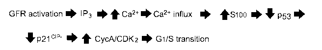
Biochemical activation of T-type Ca2 channels driving Gl/S transition. This
disclosure proposes the following sequence of steps from initial growth factor
activation to
release of the G 1 iS restriction, as illustrated in Figure 1. Growth factor
receptor (GFR)
activation increases the cytosolic inositol trisphosphate (IP3) concentration
through activation
of phospholipase C. IP3 then releases Ca2 from the internal storage pool
through interaction
with the IP3 receptor on the endoplasmic reticulum. The resulting small
increase in the
cytosolic Ca2+ concentration triggers a much larger increase resulting from
Ca2' influx
through T-type Ca 2- channels, as outlined in Figure 1. A necessary event in
thc pathway
involves Ca2- binding S100, which in tum binds to and inactivates p53, thus
relieving
18
CA 02897005 2015-06-30
WO 2014/110409
PCT/US2014/011098
activation of p21. Because activated p2I inactivates CD1(2, reduction in p21
activity allows
CDK2 to drive the Gl/S transition.
Events leading to cell division in electrically non-excitable cells. A model
has been
presented for the events that follow growth factor receptor activation leading
to cell division.
In this model, the Ca2" released from its internal depot activates Ca2 entry
by clearly Ca2'
dependent process rather than Ca entry being triggered secondarily by the
"emptiness" of
the internal depot.17 Simply, Ca2- released from the storage depot activates
calmodulin, which
in turn activates thc Ca2' influx leading to cell division.
The membrane potential of cancer cells has been reported to be between -30 mV
to -60 mV. However when membrane potential was measured as a function of
position in the
cell cycle in a human breast cancer line, it was shown to be about -30 mV in
early GI falling
to about -60 mV in late GI and S (Ouadid-Ahidouch et ed., Physiol. Cell.
Physiol.,
287:C125-34 (2004)), which may account for the variability of measured
membrane potential
in cancer cells reported in the literature. Growth factor activation produces
inositol
triphosphate, which releases Ca2 from an internal storage depot.2 One of the
first actions of
this increase in intracellular Ca2 can be the activation, and opening, of Ca2'
activated K-F
channels.21 The resulting efflux of 1(' will naturally result in a transient
decrease in the
membrane potential from the value of about -60 mV in late GI to a
hyperpolarized value of
about -90 mV, the equilibrium potential for potassium.
Interestingly, K channel blockers have been shown to inhibit growth factor
stimulated increases in cytosolic Ca2- and to block cellular proliferation by
inhibiting transit
past the Gl/S boundary in cancer cell lines and mesenchymal stem cells,)2_)4.
¨ an action
functionally identical to T-type Ca2' channel inhibitors. While the K- channel
blockers used
in such studies are promiscuous, it is unexpected that a I(' channel, or the
hyperpolarization
associated with K' channel activity, would have an effect on Ca2 channel
function or would
increase cytosolic Ca2 , leading to cell division. A widely cited belief is
that the
hyperpolarization mediated by K channel function serves to increase the
electrochemical
driving force for Ca2- entry. On the face of it, this is clearly true.
However, there is a 10,000-
fold concentration gradient for Ca2- entry at a membrane potential of 0 and it
is difficult to
reconcile the metabolic burden required to hyperpolarize the plasma membrane
potential and
the need to have tightly controlled Ca2' entry with the generally hypothesized
role of
hyperpolarization in increasing the driving force for Ca2- entry. Accordingly,
activation of K'
19
CA 02897005 2015-06-30
WO 2014/110409
PCT/US2014/011098
channels and the attendant drop in membrane potential toward potassium's
equilibrium
potential is herein disclosed as functioning to increase the driving force for
Ca2-.
According to a controversial but nonetheless popular hypothesis, a malignant
tumor is
comprised of a variable proportion of so-called cancer stem cells (Lathia JD
et al., Stem Cell
Rev. 7:227-37 (2011)). These cells are reported to be relatively resistant to
radiation and
chemotherapy and could account for cancer recurrence. Cancer stem cells are
thought to be
similar to embryonic stem cells and knowledge of the biology of both types of
stem cells may
reveal novel therapeutic strategies. Interestingly, Cav3.2 (Unigene cluster
Hs.459642) and the
type 2 small conductance calcium activated potassium channel (Unigene Cluster
Hs.98280)
have strikingly similar early gestational co-expression patterns as determined
by the National
Center for Biotechnology Information with the highest expression in the
embryoid body
falling off thereafter. This early gestational expression pattern is not seen
with Cav3.1 or
Cav3.3 nor is it seen with other calcium activated potassium channels. This co-
expression
pattern is consistent with the functional expression of Cav3.2 in embryonic
stem cells is as
well as the model described below, and may help to reveal new medical
approaches to cancer
treatment.
A model for growth factor regulated Ca2 influx enabling proliferation. These
observations can be synthesized into a coherent and simple model (Figure 2):
1. At the resting membrane potential, T-type channels are inactivated and
unable to be
opened.
2. Growth factor receptor is activated.
3. This causes the production of inositol trisphosphate.
4. Inositol triphosphate releases Ca2' from an internal storage pool.
5. This released Ca2' opens Ca2- activated K-( channels via constitutively
bound
calmodulin.
6. The resulting hyperpolarization relieves inactivation of T-type
channels.
7. T-type channels are now closed and, thus, available to be opened.
8. Ca2- activated calmodulin diffuses to and opens T-type channel perhaps via
T-type
channel phosphorylation by a calmodulin kinase.
9. A Ca2- activated S100 isoform inactivates p53 removing activation of p21,
which
releases CDK2 to propel progression into S phase.
These steps are further described as follows. In thc first arm of thc pathway,
thc
constitutive association of CaM with Ca2- activated K+ channels5.25 allows for
rapid opening
CA 02897005 2015-06-30
WO 2014/110409
PCT/US2014/011098
of them in response to an increase in cytosolic Ca2-. The need for diffusion
of the Ca2 7CaM
complex and the possible requirement for the participation of CaMKII will slow
the second
arm of the pathway, possibly providing the temporal sequencing of
hyperpolarization
followed by CaM dependent activation of Ca2- entry via T-type Ca2- channels.
Among the various points at which this pathway can be attacked for therapeutic
gain,
a vulnerable target is the T-type Ca2-- channel itself. One reason for this
vulnerability is the
limited number of T-type Ca2' channel isoforms. Growth factors, for example,
consist of a
large number of related proteins that can be recruited to bypass one that has
been blocked.
There arc only three T-type Ca2- channel proteins and all arc about equally
sensitive to
available pharmacologic inhibitors so that recruitment of an alternative
member would be
futile.
Another point of vulnerability results from the restricted distribution of
this protein,
which is normally expressed in embryonic stem cells, and not expressed in
cells that do not
normally divide in adults, but that is re-expressed in response to injury or
carcinogenic
stimulus. This re-emergent proliferation can result from something as
relatively simple as re-
expression in fibroblasts dividing in response to wound healing,26 which is a
standard
response to a pathological stimulus, or as complex as in solid cancers, which
may well be a
pathologic response to a normal stimulus. In addition, bone marrow derived
cells appear to
utilize a different Ca2- entry pathway, as T-type channel antagonists have no
effects on
proliferation or differentiation of these cells and no expression of Cav3.2 is
observed in cell
lines derived from bone marrow. The molecular basis for this is not
understood, but is the
source of active research. These attributes makes inhibitors of T-type Ca2-
channels very
appealing candidates for a new and unique category of cancer chemotherapeutic
agents that
inhibit proliferation of cancer cells while having reduced or no effect on
immune cell
proliferation.
As monotherapy, T-type calcium channel blockers slow cancer cell proliferation
and
reduce tumor growth in vivo as observed in a number of animal models of human
disease!' 28
Mibefradil is a T-type Ca2- channel blocker that was marketed by Roche for the
treatment of
hypertension and angina (Clozel et al., Cardiovasc. Drug Rev. 9:4-17 ( 1991)).
It was
withdrawn from the market after being used by almost a million patients when
it was
discovered to have undesirable drug-drug interactions caused by mibefradil' s
inhibition of
CYP 450 3A4 (Po and Zhang, Lancet. 351:1829-30 (1998)). Aside from this,
mibefradil was
remarkably well tolerated and devoid of side effect even for a member of its
therapeutic class
21
CA 02897005 2015-06-30
WO 2014/110409
PCT/US2014/011098
(Kobrin et al., Am. Cardiol. 80:40C-46C (1997)). This suggests that side
effects of T-type
Ca2' channel blockers will be modest at most and significantly better than
those generally
caused by many cancer chemotherapy drugs. In part because of this, use of T-
type Ca2-
channel blockade - as a cell cycle and cancer stem cell targeted cytostatic
agent - is actively
being pursued.
However, there is another possibility for the potential clinical utility of
such agents.
Most conventional cytotoxic agents act at a particular stage of the cell
cycle, usually during
DNA synthesis. If cancer cells could be "lined up" at thc Gl/S restriction
point and then
released into S phase, conventional cytotoxins might bc made more efficient at
killing cancer
cells. This appears to be the case in a murine model of human glioblastoma
(Keir et al., J.
Neurooncol. 111(2):97-102 (2013)). In this model, mice were treated with a
seven day course
of mibefradil to block Ca2- influx and halt progression through the cell cycle
at the GUS
restriction point, then 30 minutes after the last dose of mibefradil a five
day course of
temozolomide was started. This regimen significantly increased the cytotoxic
effect of
temozolomide and restored the sensitivity to temozolomide of drug resistant
cancer cell lines.
An [ND using this strategy in glioblastome multiforme opened in early 2012, a
phase 1 study
of escalating doses of mibefradil in normal, healthy volunteers is underway,
and a trial in
patients was initiated by the National Cancer Institute (NCI)'s Adult Brain
Tumor Consortium
in the Spring of 2012. Further details of the method are provided in WO
2010/141842, which
is incorporated herein by reference.
In some embodiments, the present disclosure provides a method for identifying
a
compound for utility in inhibiting cell cycle progression through the GUS
check point,
inhibiting proliferation of cells in a cellular proliferative disorder, and/or
enhancing the
efficacy of radiation and/or a chemotherapeutic agent in treating a cellular
proliferative
disorder. The method includes determining that the compound inhibits T-type
Ca2 channel
activity in a cell when a first cell membrane potential of the cell is held at
a potential in the
range about -70 mV to about -110 mV; and, based on the determination,
identifying a
compound for utility in inhibiting cell cycle progression through the Gl/S
check point,
inhibiting proliferation of cells in treating a cellular proliferative
disorder, andior enhancing
the efficacy of radiation and/or a chemotherapeutic agent in treating a
cellular proliferative
disorder. In some embodiments, the membrane potential can include can include
membrane
potentials within a measured range of about -80 mV to about -100 mV, or within
a range of
about 85 mV to about -95 mV, or within a range of about -89 mV to about -91
mV. In some
22
CA 02897005 2015-06-30
WO 2014/110409
PCT/US2014/011098
embodiments, the membrane potential is about 90 mV. In some embodiments, the
cells can
express one or more of the T-type calcium channel sub-types described herein.
In some
embodiments, the cells can be engineered to recombinantly express one or more
of the type
calcium channel sub-types described herein.
In some embodiments, the method can include determining a first IC50 that is
the IC,50
of the compound in inhibiting the T-type calcium channel activity when a cell
is held at the
first cell membrane potential. The compound can bc identified as useful for
the utility based
on a determination that the first IC50 is about 10000 p.M or less, about 1000
p.M or less, about
10001.IM or less, about 100 p.IVI or less, about 10 pM or less, about 111M or
less, or about
to 100 nM or less.
In some embodiments, the method can include determining a second ICS() of the
compound, wherein the second IC50 is the IC50 of the compound in inhibiting
the T-type
calcium channel activity in a cell when the cell is held at a second cell
membrane potential in
the range from about -30 mV to about -60 mV. The second membrane potential is
greater
(i.e., less negative) than the first membrane potential. In various
embodiments, the second
membrane potential can be within a range from about -20 mV to about -70 mV,
from
about -25 mV to about -65 mV, from about -30 mV to about -40 mV, from about -
30 mV to
about -50 mV, from about -30 mV to about -70 mV, from about -40 mV to about -
50 mV,
from about -40 mV to about -60 mV, from about -40 mV to about -70 mV, from
about -50 mV to about -60 mV, from about -50 to about -70 mV, as well as about
-30 mV,
about -40 mV, about -50 mV, or about -60 mV.
In some embodiments, the measurements at different membrane potentials are
performed using the same cell or group of cells. In some embodiments, the
measurements at
different membrane potentials are performed using the different cells or group
of cells. The
cells used are preferably of the same cell type. For example the cells can be
clones, cells from
the same cell line or proliferating cells from a single subject in need of
treatment for a
cellular proliferative disorder.
In some embodiments, the method can include identifying a compound as being
useful for the utility based on the determination that the ratio of the first
IC50 to the second
IC50 is about 20:1 or less, about 10:1 or less, about 5:1 or less, about 2:1
or less, about 1:1 or
less, about 1:2 or less, about 1:5 or less, about 1:10 or less, or about 1:100
or less. The
method can also include identifying a compound as having reduced or low
liability for
neuronally-mediated side-effects base on the determination that the ratio of
the first IC50 to
23
CA 02897005 2015-06-30
WO 2014/110409 PCT/US2014/011098
the second IC50 is about 20:1 or less, about 10:1 or less, about 5:1 or less,
about 2:1 or less,
about 1:1 or less, about 1:2 or less, about 1:5 or less, about 1:10 or less,
or about 1:100 or
less. Examples of neuronally based side-effects can include anxiety, attentive
deficits,
cognitive deficits, confusion, convulsions, depression, dizziness,
hallucinations, psychosis,
sedation, stimulation, etc.
In some embodiments, the cell membrane potential can be controlled using a
patch-
clamp technique. In some embodiments cell membrane potential can be controlled
using any
other technique described herein or known in the art.
In some embodiments, the ability of a compound to inhibit T-typc Ca2- channel
o activity is determined by determining the ability of the compound to
inhibit growth factor-
stimulated calcium entry into the cell. In some embodiments, the ability of a
compound to
inhibit T-type Ca2- channel activity is determined using any other technique
described herein
or known in the art.
In some embodiments, calcium entry into the cell is determined by measuring
increases in the levels of intracellular calcium using a calcium sensitive
marker such as a
calcium-sensitive fluorescent dye. In some embodiments calcium entry into the
cell is
determined by using any other technique described herein or known in the art.
In some embodiments, the method includes identifying the compound for utility
in
inhibiting cell cycle progression through the GUS check point.
In some embodiments, the method includes identifying thc compound for utility
in
inhibiting proliferation of cells in a cellular proliferative disorder. The
cellular proliferative
disorder can be a cancerous or non-cancerous proliferative disorder, including
any one or
more of the cancerous or non-cancerous proliferative disorders identified
herein. The cellular
proliferative disorder can be a disorder, the proliferating cells of which
express T-type
calcium channels. The cellular proliferative disorder can be a disorder, the
proliferating cells
of which express any isoform of a T-type calcium channels as described herein.
in some embodiments, the method includes identifying the compound for
enhancing
the efficacy of radiation and/or a chemotherapeutic agent in treating a
cellular proliferative
disorder when the compound is administered prior to administration of the
radiation andlor
chemotherapeutic agent. The cellular proliferative disorder can be a cancerous
or non-
cancerous proliferative disorder, including any one or more of the cancerous
or non-
cancerous proliferative disorders identified herein. The cellular
proliferative disorder can be a
disorder, thc proliferating cells of which express T-type calcium channels.
The
24
CA 02897005 2015-06-30
WO 20 14/1 10409 PCT/US2014/011098
chemotherapeutic agent can be any of the chemotherapeutic agents identified
herein, or any
combination thereof.
In some embodiments, the method can be performed wherein the cells used
comprise
one or more proliferating cells of a subject in need of treatment for the
proliferative disorder
and can identify the compound as being useful for the treatment of the
cellular proliferative
disorder and/or for use in enhancing the efficacy of radiation and/or a
chemotherapeutic agent
in treating a cellular proliferative disorder. In some embodiments, the
compound is
administered prior to administration of thc radiation and/or chemotherapeutic
agent. The
method can be used to identify the compound as being useful for treatment of
the subject.
The cellular proliferative disorder can be a cancerous or non-cancerous
proliferative disorder,
including any one or more of the cancerous or non-cancerous proliferative
disorders
identified herein. The cellular proliferative disorder can be a disorder, the
proliferating cells
of which express T-type calcium channels. The chemotherapeutic agent can be
any of the
chemotherapeutic agents identified herein, or any combination thereof.
In some embodiments, the method includes administering to the subject an
effective
amount of the compound to the subject to treat the cellular proliferative
disorder. In some
embodiments, the method includes administering to the subject an effective
amount of the
compound in combination with an effective amount of radiation and/or the
chemotherapeutic
agent to the subject to treat the cellular proliferative disorder. In some
embodiments, the
compound is administered to thc subject prior to administration of the
radiation and/or
chemotherapeutic agent. The cellular proliferative disorder can be a cancerous
or non-
cancerous proliferative disorder, including any one or more of the cancerous
or non-
cancerous proliferative disorders identified herein. The cellular
proliferative disorder can be a
disorder, the proliferating cells of which express T-type calcium channels.
The
chemotherapeutic agent can be any of the chemotherapeutic agents identified
herein, or any
combination thereof.
In some embodiments, the chemotherapeutic agent is selected from the group
consisting of consisting of temozolomide, 5-fluorouracil, 6-mercaptopurine,
bleomycin,
carboplatin, cisplatin, dacarbazine, doxonibicin, epirubicin, etoposide,
gemcitabine,
hydroxyurea, ifosfamide, irinotecan, topotecan, methotrexate, mitoxantrone,
oxaliplatin,
paclitaxel, docctaxel, vinblastine, vincristinc, vinorelbinc; vindesinc and
mitomycin C. In
some embodiments, the chemotherapeutic agent is temozolomide. In some
embodiments, the
CA 02897005 2015-06-30
WO 2014/110409
PCT/US2014/011098
chemotherapeutic agent is carboplatin. In some embodiments, the
chemotherapeutic agent is
gemcitabine.
In some embodiments, the cancer is selected from the group consisting of
selected
from the group consisting of brain cancer, breast cancer, colon cancer,
glioma, glioblastoma,
melanoma, ovarian cancer and pancreatic cancer. In some embodiments, the
cancer is brain
cancer. In some embodiments, the cancer is glioma. In some embodiments, the
cancer is
ovarian cancer. In some embodiments, thc cancer is pancreatic cancer.
The invention has been described with reference to various embodiments and
techniques. However, it should be understood that many variations and
modifications can be
to made while remaining within the spirit and scope of the invention. It
will be apparent to one
of ordinary skill in the art that compositions, methods, devices, device
elements, materials,
procedures and techniques other than those specifically described herein can
be applied to the
practice of the invention as broadly disclosed herein without resort to undue
experimentation.
All art-known functional equivalents of compositions, methods, devices, device
elements,
materials, procedures and techniques described herein are intended to be
encompassed by this
invention. Whenever a range is disclosed, all sub-ranges and individual values
are
encompassed. This invention is not to be limited by the embodiments disclosed,
including
any exemplified in the specification, which are given by way of example or
illustration and
not of limitation. The scope of the invention shall be limited only by the
claims.
All references cited herein are hereby incorporated by reference in their
entirety.
References
1. Rustandi RR, Baldisscri DM, Weber DJ. Structure of thc negative
regulatory domain of
p53 bound to S1OOB (betabeta). Nat. Struct. Biol., 2000; 7: 570-4.
2. Lu F, Chen H, Zhou C, et al. T-typc Ca2- channel expression in human
esophageal
carcinomas: a functional role in proliferation. Cell Calcium, 2008; 43: 49-58.
3. Lapenna S, Giordano A. Cell cycle kinases as therapeutic targets for
cancer. Nat. Rev.
Drug Discov. 2009;8:547-66.
4. Trautwein W, Hescheler J. Regulation of cardiac L-type calcium channels
by
phosphorylation and G proteins. Annu. Rev. Physiol., 1990; 52: 257-74.
5. Saimi Y, Kung C. Ion channel regulation by calmodulin binding, FEBS Lett.,
1994; 350:
155-8.
26
CA 02897005 2015-06-30
WO 2014/110409
PCT/US2014/011098
6. Putney JW, Jr. A model for receptor-regulated calcium entry. Cell Calcium,
1986; 7: 1-
12.
7. Smyth JT, Putney JW. Regulation of store-operated calcium entry during
cell division.
Biochem. Soc. Trans., 2012; 40: 119-23.
8. Cahalan MD. STIMulating store-operated Ca(2+) entry. Nat. Cell. Biol.,
2009; 11: 669-
77.
9. Harper JV, McLatchic L, Perez-Reyes E, Cribbs LL, Shattock MJ, Brooks G. T-
typc
calcium channel expression is necessary for G I-S progression in vascular
smooth muscle.
Circulation 2000; 102: 11-48.
10. Li W, Zhang SL, Wang N, Zhang BB, Li M. Blockade of T-Type Ca(2+) channels
inhibits human ovarian cancer cell proliferation. Cancer Invest., 2011, 29(5):
339-46.
11. Densmore JJ, Szabo G, Gray LS. A voltage-gated calcium channel is linked
to the antigen
receptor in Jurkat T lymphocytes. FEBS Lett. 1992; 312: 161-4.
12. Santoni G, Santoni M, Nabissi M. Functional role of T-type calcium
channels in tumour
growth and progression: Prospective in cancer therapy. Br. J. Pharmacol.,
2012, 166(4):
1244-6.
13. Mulgrew CJ, Cove-Smith A, McLatchie LM, Brooks G, Shattock MJ, Hendry BM.
Inhibition of human mesangial cell proliferation by targeting T-type calcium
channels.
Nephron Exp. Nephrol., 2009; 113: e77-88.
14. Rodman DM, Reese K, Harral J, et al. Low-voltage-activated (T-typc)
calcium channels
control proliferation of human pulmonary artery myocytes. Circ. Res., 2005;
96: 864-72.
15. Brooks G, Harper JV, Bates SE, et al. Over expression of the voltage-gated
T-type
calcium channel induces vascular smooth muscle cell proliferation.
Circulation, 1999;
100: 1-209.
16. Taylor JT, Zeng XB, Pottle JE, et al. Calcium signaling and T-type calcium
channels in
cancer cell cycling. World Gastroenterol., 2008; 14: 4984-91.
17. Gray LS, Perez-Reyes E, GamoiTa JC, et al. The role of voltage gated T-
type Ca2
channel isoforms in mediating "capacitative" C;+ entry in cancer cells. Cell
Calcium,
2004; 36: 489-97.
18. Rodriguez-Gomez JA, Levitsky KL, Lopez-Bameo J. T-type Ca2- channels in
mouse
embryonic stem cells: modulation during cell cycle and contribution to self
renewal. Am.
J. Physiol. Cell. Physiol., 2012; 302: C494-504.
27
CA 02897005 2015-06-30
WO 2014/110409
PCT/US2014/011098
19. Heady TN, Gomora JC, Macdonald TL, Perez-Reyes E. Molecular pharmacology
of T-
type Ca2- channels../pn. Pharmacol., 2001; 85: 339-50.
20. Exton JH. Regulation of phosphoinositide phospholipases by hormones,
neurotransmitters, and other agonists linked to G proteins. Annu. Rev.
Pharmacol.
Toxicol., 1996; 36: 481-509.
21. Wonderlin WF, Strobl JS. Potassium channels, proliferation and G1
progression. J.
Membr. Biol., 1996; 154: 91-107.
22. Chandy, KC, Wulff, H, Becton C, Pennington M, Gutman GA, Cahalan MD. K(+)
channels as targets for specific immunomodulation. Trends Pharmacol. Sci.,
2004; 25:
280-9.
23. Strobl JS, Wonderlin WF, Flynn DC. Mitogenic signal transduction in human
breast
cancer cells. Gen. Pharmacol., 1995; 26: 1643-9.
24. Tao R, Lau CP, Tse HF, Li GR. Regulation of cell proliferation by
intermediate
conductance Ca2' -activated potassium and volume-sensitive chloride channels
in mouse
mesenchymal stem cells. Am. J. Physiol. Cell. Physiol., 2008295:CI409-16.
25. Levitan IB. It is calmodulin after all! Mediator of the calcium modulation
of multiple ion
channels. Neuron 1999; 22: 645-8.
26. Estacion M, Mordan LJ. Expression of voltage-gated calcium channels
correlates with
PDGF-stimulated calcium influx and depends upon cell density in C3H 10T112
mouse
fibroblasts. Cell Calcium, 1993; 14: 161-71.
27. Haverstick DM, Heady TN, Macdonald TL, Gray LS. Inhibition of human
prostate cancer
proliferation in vitro and in a mouse model by a compound synthesized to block
Ca2'
entry. Cancer Res. 2000; 60: 1002-8.
28. Panner A, Wurster RD. T-type calcium channels and tumor proliferation.
Cell Calcium
2006; 40: 253-9.
29. Giles TD. Hypertension and pathologic cardiovascular remodeling: a
potential therapeutic
role for T-type calcium antagonists. Clin. Ther., 1997; 19 Suppl. A: 27-38.
28