Note: Descriptions are shown in the official language in which they were submitted.
CA 02899007 2015-07-22
WO 2014/116705 PCT/US2014/012553
PROBE WITH OPTOACOUSTIC ISOLATOR
CROSS-REFERENCE
[0001] This application claims priority to U.S. Patent Application Serial No.
13/746,905 filed
January 22, 2013. This application includes material which is subject to
copyright protection.
The copyright owner has no objection to the facsimile reproduction by anyone
of the patent
disclosure, as it appears in the Patent and Trademark Office files or records,
but otherwise
reserves all copyright rights whatsoever.
FIELD
[0002] The present invention relates in general to the field of medical
imaging, and in particular
to a probe with an optoacoustic isolator for use in medical imaging.
BRIEF DESCRIPTION OF THE DRAWINGS
[0003] The foregoing and other objects, features, and advantages of the
invention will be
apparent from the following more particular description of preferred
embodiments as illustrated
in the accompanying drawings, in which reference characters refer to the same
parts throughout
the various views. The drawings are not necessarily to scale, emphasis instead
being placed upon
illustrating principles of the invention.
[0004] FIG. 1 shows a schematic block diagram illustrating an embodiment of a
combined
optoacoustic and ultrasound system that may be used as a platform for the
methods and devices
disclosed herein.
[0005] FIG. 2 shows a schematic orthogonal view of an embodiment of a probe
that may be used
in connection with the methods and other devices disclosed herein.
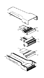
[0006] FIG. 3 shows an exploded view of an embodiment of the probe shown in
FIG. 2.
[0007] FIG. 4 shows a cutaway view taken along the centerline of the wider
side of the probe
shown in FIG. 2.
[0008] FIG. 5A is a side-view not-to-scale diagrammatic two dimensional
representation of light
exiting an optical fiber.
[0009] FIG. 5B shows an end view of a light pattern that may result on a
surface from placement
of optical fibers directly on to that surface.
[0010] FIG. 6A shows an end view of a desirable light pattern for use in
connection with the
optoacoustic techniques discussed herein.
- 1 -
CA 02899007 2015-07-22
WO 2014/116705 PCT/US2014/012553
[0011] FIG. 6B shows a side view diagrammatic representation of an effect of a
ground glass
beam expander on the light emitting from a fiber shown in FIG. 5A.
[0012] FIG. 6C shows a side view diagrammatic representation of an effect of a
concave lens
beam expander on the light emitting from a fiber shown in FIG. 5A.
[0013] FIG. 7 shows a schematic orthogonal view of another embodiment of a
probe that may be
used in connection with the methods and other devices disclosed herein.
[0014] FIG. 8 shows an exploded orthogonal view of the embodiment of the probe
shown in
FIG. 7.
[0015] FIG. 9 shows a lengthwise cutaway view taken along line A-A of the
probe shown in
FIG. 7.
[0016] FIG. 10 shows a lengthwise cutaway view taken along line B-B of the
probe shown in
FIG. 7.
[0017] FIG. 11 shows a lengthwise cutaway view of another embodiment of a
probe that may be
used in connection with the methods and other devices disclosed herein.
DETAILED DESCRIPTION
[0018] Reference will now be made in detail to various embodiments of the
present invention,
examples of which are illustrated in the accompanying drawings.
[0019] Generally, device 100 provides an optoacoustic system that may also be
employed as a
multi-modality, combined optoacoustic and ultrasound system. In an embodiment,
the device
100 includes a probe 102 connected via a light path 132 and an electrical path
108 to a system
chassis 101. Within the system chassis 101 is housed a light subsystem 129 and
a computing
subsystem 128. The computing subsystem 128 includes one or more computing
components for
ultrasound control and analysis and optoacoustic control and analysis; these
components may be
separate, or integrated. In an embodiment, the computing subsystem comprises a
relay system
110, an optoacoustic processing and overlay system 140 and an ultrasound
instrument 150.
[0020] The light system 129 is capable of producing pulses of light of at
least two different
wavelengths. In an embodiment, the light system 129 output should be capable
of producing
short pulses of light in each of those wavelengths, e.g., a pulse lasting less
than about 100 ns, and
more preferably around 5 ns. As will be apparent to one of ordinary skill in
the art from this
disclosure, the inventions disclosed herein may also be practiced using pulsed
light comprising
pulses lasting greater than 100 ns. In an embodiment, the light source 129
includes two separate
lights 130, 131. The output of the light system 129 is delivered to the probe
102 via the optical
- 2 -
CA 02899007 2015-07-22
WO 2014/116705 PCT/US2014/012553
path 132. In an embodiment, the lights 130, 131 are lasers producing light in
the infrared, near-
infrared, and/or visible spectrum. In an embodiment, light 130 and light 131
each produce light
at a different wavelength in the infrared or near-infrared spectrum. In an
embodiment, the optical
path 132 used to deliver light from the light source 129 to the probe 102 is a
fiber optic bundle
comprising multiple strands of optical fiber. In an embodiment, the optical
path 132 comprises
sufficient optical fibers of sufficient size (diameter) to carry a short, high
powered pulse of light
to the distal end of the optical path 132. In an embodiment, the total pulse
energy carried over
the optical path 132 may be on the order of one or more millijoules. In an
embodiment, the total
energy per light pulse carried over the optical path 132 is less than about
100 millijoules. In an
embodiment, the total energy per light pulse carried over the optical path 132
is in the range of
about 10-30 millijoules, and the optical path 132 comprises around 1,000
optical fibers of about
150 microns each. In an embodiment, a single fiber can be used as the optical
path. In such
embodiment, the fiber may be 400-1500 microns in diameter. Of course, the
diameter of such
single fiber may be smaller, e.g., 400 microns. Given the required total pulse
energy carried over
the fiber, one skilled in the art can calculate the diameter required of the
fiber accordingly.
[0021] In an illustrative embodiment, the light system 129 may use Nd-YAG and
Alexandrite
lasers as its two lights 130, 131, although other types, and additional
lights, may also be used.
Lights 130, 131 should be capable of producing a short pulse of light, e.g., a
pulse lasting less
than about 100 ns, and more preferably around 5 ns. In an embodiment, the two
lights 130, 131
can be separately triggered. In an embodiment, the light output by the lights
130, 131 may be
projected onto the same light path 132 through the use of an optical element
133 that generally
permits one light 130 to pass through from a first side to a second side,
while reflecting one light
131 that strikes the second side. The use of optical element 133 or a similar
element permits the
alignment of the output of two lights 130, 131 such as lasers onto proximal
end of the light path
132. In an embodiment, optical elements 133 can align the light output from
more than two
lasers, for example, through the use of multiple optical elements 133. In an
embodiment,
multiple light systems and light paths may be employed, with the light of each
light system being
carried on separate fibers that are intermingled at their distal ends.
[0022] Although the total energy per light pulse carried over the optical path
is in the order of
tens of millijoules, because the pulse of lights 130, 131 is so short, the
peak power output over
the optical path 132 is frequently approaching or in the megawatt range.
Accordingly, the output
of lights 130, 131 has the capacity to cause the optical fibers and/or the
cladding on the optical
- 3 -
CA 02899007 2015-07-22
WO 2014/116705 PCT/US2014/012553
fibers to burn. Burnt optical fibers and burnt cladding can exacerbate the
problem as they begin
to transmit less light power and cause more heating. Accordingly, in an
embodiment, sufficient
number and size optical fibers are present in the optical path 132 to permit
handling of the peak
power loads and avoid fiber burnout. To accommodate higher peak power, a
larger fiber bundle
can be used. It will be apparent to a person of skill in the art that the peak
power capacity of a
fiber bundle can be increased by increasing the number of optical fibers, or
the diameter of
optical fibers, or both. Notably, however, as the dimension of the fiber
bundle increases, the
weight and flexibility of the optical path 132 decreases. Moreover, when using
more optical
fibers, or optical fibers of a larger diameter, the output of light source 129
must be delivered to
the optical path 132 across the wider diameter of the larger bundle. In an
embodiment, regardless
of the ultimate size of the proximal end of light path 132, the output of
light source 129 should be
distributed sufficiently across its cross section to prevent burn-out failures
when operating in
expected peak power ranges.
[0023] In an embodiment, the fibers of the proximal end of the light path 132
may be fused to
form a fused entry point to the optical path 132 for the output of light
source 129. In an
embodiment, the fiber ends can be fused by applying heat. Once the proximal
end of optical path
132 has been fused, it will resist burnout at substantially higher peak power.
For example, using
a fused end light path 132 may permit carriage of three, four or even five
times as much peak
power. The ability to carry substantially higher peak power in a given optical
path 132 permits
use of a more flexible and lighter fiber optic bundle to carry the same peak
power as an un-fused
optical path 132. Thus, in an embodiment, where a 'A" fiber optic bundle may
have been
required in an un-fused bundle of optical fibers forming an optical path, a
'A" fiber optic bundle
with a fused proximal end may be used to carry the same peak power. A 1/4"
fiber optic bundle
with a fused proximal end is approximately 1/4 of the weight and much more
flexible than a 'A"
fiber optic bundle. Moreover, fusing of the proximal end of light path 132 may
produce an even
smaller fused area to illuminate using light source 132 as the fusing removes
the inter-fiber
spaces that would have existed in the bundled end of the round-cross-section
optical fibers.
Accordingly, one or more of the following advantages may be attained by fusing
the proximal
end of the optical fibers comprising the light path 132: reduced weight of the
light path; increased
flexibility of the light path; reduced failure; increased reliability; higher
peak power capacity.
[0024] In an embodiment, the light output by the lights 130, 131 is sent
towards a fused optical
fiber bundle at the proximal end of light path 132 via an optical path, which
may include optical
- 4 -
CA 02899007 2015-07-22
WO 2014/116705 PCT/US2014/012553
element 133, internal to the light source 129. In an embodiment, light source
129 is a laser
system capable of outputting laser light pulses, at one or a more wavelengths,
onto light path 132.
In an embodiment, light path 132 is a fiber optic bundle having a fused end
proximal to the light
source 129.
[0025] In an embodiment, the device 100 also comprises an electrical path 108
running to and/or
from the probe 102 to a relay system 110 within the system chassis 101. The
electrical path 108
may run near, alongside or coaxially with the optical path 132 from the probe
102 toward their
respective connections on the system chassis 101. In an embodiment, the
electrical path 108
comprises a plurality of separate coaxial wires. In an embodiment, the
electrical path 108 is run
in a common jacket with at least a portion of the optical path 132. Running
electrical path 108 in
a common jacket with at least a portion of the optical path 132 reduces the
number of cables
running from the system chassis 101 to the probe 102. Running electrical path
108 in a common
jacket with at least a portion of the optical path 132 may minimize the
diameter and weight of,
and increase the durability of, the combined cables (i.e., optical path 132
and electrical path 108)
running from the system chassis 101 to the probe 102.
[0026] In an embodiment, the plurality of coaxial wires is woven around at
least a portion of the
optical path 132. As discussed above, many considerations go into the number
of separate
optical fibers used in optical path 132. As discussed further below, numerous
design
considerations go into the number of separate electrical leads or traces
forming the electrical path
108. In an embodiment, there are about 256 leads (corresponding to 256
transducers) forming
the electrical path 108 and approximately 1,000 separate optical fibers
forming the optical path
132, making the fiber:lead ratio about 4:1. As will be apparent, it is
possible to comingle the
optical fibers and leads or traces in the electrical path in a variety of
ways, including, for
example, bundling a group of individual fibers with a single electrical lead
or trace, or bundling
proportionally larger groupings of fibers and leads together. In an
embodiment, the bundling of
fibers and leads or traces would be done generally in the proportion of
fibers:leads in the system.
[0027] One or more displays 112, 114, which may be touch screen displays, are
provided for
displaying images and all or portions of the device 100 user interface. One or
more other user
input devices (not shown) such as a keyboard, mouse and various other input
devices (e.g., dials
and switches) may be provided for receiving input from an operator. As an
option, power and
control signal lines 109 carry power to the probe 102 and control signals
between the probe 102
and the computing subsystem 128.
- 5 -
CA 02899007 2015-07-22
WO 2014/116705 PCT/US2014/012553
[0028] Turning now to FIG. 2, the probe 102 includes an array of ultrasound
transducer elements
forming an ultrasound transducer (not shown) covered by an acoustic lens 205.
In an
embodiment the ultrasound transducer comprises an array of piezoelectric
elements that can both
transmit and receive acoustic energy. In an embodiment, at least some of the
ultrasound
transducer elements are capable of detecting ultrasound frequencies over a
wide range. For
example, ultrasound transducer elements may be capable of detecting ultrasound
in the range
from about 50 Khz to 20 Mhz. This range can be achieved by applying a high
impedance load
(e.g., in the range of 5,000 to 50,000 ohms) to achieve a lower frequency
response. The
ultrasound transducer elements are capable of generating electrical energy in
response to
receiving ultrasound acoustic energy. The electrical energy generated by the
ultrasound
transducer elements receiving ultrasound is transmitted to the computing
subsystem 128 via
electrical path 108.
[0029] The probe 102 also includes one or more optical windows 203 through
which the light
carried on optical path 132 can be transmitted to the surface of a three-
dimensional volume 160.
In an embodiment, it is desirable to locate one side of the optical window 203
as close as
practical to the acoustic lens 205. The total area of an optical window 203 is
important to
maximize energy for a given fluence incident on the surface of the volume 160.
[0030] In an embodiment, the multiple strands of optical fiber making up the
optical path 132 are
terminated in two light bars (not shown). In an embodiment, the ultrasound
transducer elements
(not shown) are arranged in an array that runs along a geometric plane and are
generally spaced
equidistant from each other. In an embodiment, the light bars (not shown) are
oriented
longitudinally, on each side of the planar array of ultrasound transducer
elements. Preferably the
ultrasound transducer elements generate electrical energy in response to both
ultrasound acoustic
energy received in response to stimulation caused by the pulsed light sources
130, 131 and to
ultrasound acoustic energy received in response to acoustic output of the
ultrasound transducer
elements.
[0031] Referring back to FIG. 1, in use, the probe 102 may be placed in close
proximity with
organic tissue, phantom or other three-dimensional volume 160 that may have
one or more
localized inhomogeneities 161, 162, such as e.g., a tumor, within. An
ultrasound gel (not shown)
or other material may be used to improve acoustic coupling between the probe
102 and the
surface of the volume 160. The probe 102, when in proximity with the surface
of the volume
160, can emit a pulse of a light through the optical windows 203 or an
ultrasound through
- 6 -
CA 02899007 2015-07-22
WO 2014/116705 PCT/US2014/012553
acoustic lens 205, and then generate electrical energy corresponding to
ultrasound detected in
response to the emitted light or sound.
[0032] In an embodiment, the computing subsystem 128 can trigger activity from
light system
129 over control signal line 106. In an alternative embodiment, the light
system 129 can create
the trigger signal and inform the computing subsystem 128 of its activity over
control signal line
106. Such information can be used to by the computing subsystem 128 to begin
the data
acquisition process. In this respect, it is noted that communication over
control signal line 106
can flow both ways between the computing subsystem 128 (and/or the
optoacoustic processing
and overlay system 140 therein) and the light system 129.
[0033] In an embodiment, computing subsystem 128 can utilize control signal
line 106 to control
the start time and duration of light pulses from each light source 130, 131.
The computing
subsystem 128 can also trigger the probe 102 to emit ultrasound acoustic
energy via the
ultrasound transducer elements behind the acoustic lens 205.
[0034] In an embodiment, the computing subsystem 128 receives electrical
signals representative
of the ultrasound detected by the ultrasound transducer elements, in response
to an ultrasound
transmitted signal or an optically generated ultrasound signal, behind the
acoustic lens 205 via
electrical path 108. In an embodiment, the electrical signal representative of
the ultrasound
detected by the ultrasound transducer elements behind the acoustic lens 205 is
the analog
electrical signal created by the elements themselves. In such embodiment, the
electrical signals
representative of the ultrasound detected by the ultrasound transducer
elements behind the
acoustic lens 205 is transmitted to the computing subsystem via electrical
path 108, and electrical
path 108 is selectively directed by relay system 110 to the optoacoustic
processing and overlay
system 140 or the ultrasound instrument 150 for processing of the detected
ultrasound. In such
embodiment, the ultrasound instrument 150 can receive the same input (over the
same connector)
as it would receive from an ultrasound probe.
[0035] In another embodiment, the electrical signal representative of the
ultrasound detected by
the ultrasound transducer elements behind the acoustic lens 205 is digitized
by an analog-to-
digital converter which can be housed in the probe 102. In such embodiment,
time-resolved
electrical signal representative of the ultrasound detected by the ultrasound
transducer elements
behind the acoustic lens 205 is transmitted across the electrical path 108.
Where the electrical
signal is digitized at the probe 102, as will be apparent to one of skill in
the art, the relay system
- 7 -
CA 02899007 2015-07-22
WO 2014/116705 PCT/US2014/012553
110 may be implemented to deliver digital data to the optoacoustic processing
and overlay
system 140 or the ultrasound instrument 150, or may not be needed at all.
[0036] The signal representative of the ultrasound detected by each of the
plurality of ultrasound
transducer elements behind the acoustic lens 205 may be carried on a separate
wire over the
electrical path 108. Alternatively, the signal representative of the
ultrasound detected by a
plurality of ultrasound transducer elements behind the acoustic lens 205, or
even all of the
ultrasound transducer elements behind the acoustic lens 205, may be
multiplexed (e.g., time
division or frequency division) utilizing a multiplexer in the probe and a
demultiplexer in the
computing subsystem 128.
[0037] In an embodiment, the ultrasound instrument 150 processes ultrasound-
induced acoustic
signals to produce ultrasound images and the optoacoustic processing and
overlay system 140
processes light-induced acoustic signals to produce optoacoustic images. In an
embodiment, the
ultrasound instrument 150 and optoacoustic processing and overlay system 140
can be combined
into an integrated system performing the combined functions of both. As
discussed above, in an
embodiment, electrical signals representative of ultrasound detected by the
probe 102 and
delivered to the computing subsystem 128 via electrical path 108 is switched
between the
ultrasound instrument 150 and the optoacoustic instrument 140 via relay system
110 in
accordance with whether the signal results from ultrasound stimulation or
light stimulation.
[0038] In an embodiment, tomographic images reflecting the ultrasound-
stimulated data may be
generated by the ultrasound instrument 150 and tomographic images reflecting
the light-
stimulated data may be generated by the optoacoustic processing and overlay
system 140.
[0039] Images, including tomographic images, produced by the optoacoustic
processing and
overlay system 140 can be stored in a computer memory in that system, along
with data
associated with sequence or time and date of the image data that was captured.
Images, including
tomographic images, produced by the ultrasound instrument 150 may be
transmitted to the
optoacoustic processing and overlay system 140 via a suitable interface 170,
where they can be
stored, along with images generated from the light-stimulated data, in a time-
synchronized
manner. In an embodiment, images stored in the memory of the optoacoustic
processing and
overlay system 140 can be recorded to another memory, e.g., a non-volatile
memory internal to,
or external to, the device.
[0040] In an embodiment, the optoacoustic processing and overlay system 140
can overlay
images produced by the ultrasound instrument with images produced by
optoacoustic instrument
- 8 -
CA 02899007 2015-07-22
WO 2014/116705 PCT/US2014/012553
140 for storage in the memory and/or display on one or more monitors 112, 114.
In an
embodiment, the overlayed optoacoustic image may be shown in a distinct color
to distinguish it
from the ultrasound image. In an embodiment, the overlaid optoacoustic image
may contain
colors that correspond to details discernable through optoacoustic imaging,
such as, for example,
blood oxygenation. In an embodiment, oxygenated blood is shown more in red
than blue, while
deoxygenated blood is shown in more blue than red. As used herein, the
expression overlaid
includes merging of the image by mixing as well as traditional overlaying of
the image.
[0041] In an embodiment, the device 100 may be configured to operate in a
cycle comprising a
sequence of successively generating and acquiring data relating to one of the
device's modalities,
i.e., ultrasound or optoacoustic. The minimum time spacing between operation
of the device's
modalities depends on the device 100 components and their ability to fully
execute and recycle
for use. In an embodiment, a user can select between a variety of
preprogrammed cycles such as:
ultrasound only; wavelength one only; wavelength two only; wavelength one and
two; and
multiple iterations of wavelength one and two followed by ultrasound. Other
combinations will
be apparent to one of skill in the art. In an embodiment, additional cycles
can be added by the
machine operator. In an embodiment, the data collection of an entire cycle is
generally intended
to be directed to substantially the same portion of volume 160 and to be
accomplished in rapid
succession. In an embodiment, the device 100 cycles are normally in the range
of 1 to 50 per
second, and more typically in the range of 2 to 20 per second, as discussed
above. The maximum
cycle frequency is limited only by the capabilities of the cycle and
modalities.
[0042] In an embodiment, the displays 112, 114 of device 100 can be configured
to show various
information depending upon the selected operating cycles. In an embodiment,
any display 112,
144 or portion of the display can show at least one of the following: an
ultrasound only image; a
first wavelength response only image; a second wavelength response only image;
a combined
first and second wavelength response image; and/or an overlay ultrasound image
and a
wavelength response or combined wavelength response image. The combined first
and second
wavelength image may comprise a differential or other combinatorial means to
provide the
image. In an embodiment, an image can be displayed corresponding to each of
the separate data
collections in a cycle, or corresponding to the sum or difference between any
or all of them.
[0043] In an embodiment, the device can be operated using a three-phase data
collection
operation, one phase generating and collecting data in response to ultrasound
stimulus, one phase
- 9 -
CA 02899007 2015-07-22
WO 2014/116705 PCT/US2014/012553
generating and collecting data in response to a first wavelength of light, and
one phase generating
and collecting data in response to a second wavelength of light.
[0044] Using proper wavelength(s), optoacoustics is effective in identifying
blood within a
volume 160, and using multiple wavelengths can be used to readily distinguish
between
oxygenated and deoxygenated blood. Similarly, using proper wavelengths,
optoacoustics is
effective for measuring localized hemoglobin content within a volume 160.
Thus, for example, a
malignant tumor, which is characterized by increased blood concentration and
decreased
oxygenation, will appear very differently in an optoacoustic image than a
benign growth, which
is not characterized by such an increased blood concentration and has more
normal oxygenation.
Moreover, specific wavelengths of light can be selected to better distinguish
between various
biological tissues and organs. While a large spectrum of infrared, near-
infrared and visible
wavelengths can produce optoacoustic response in biological entities,
oxygenated blood is more
optoacoustically responsive than deoxygenated blood to a light source having a
wavelength of
about 1064 nm, while deoxygenated blood is more optoacoustically responsive
than oxygenated
blood to a light source having a wavelength of 757 nm. The number and specific
wavelength(s)
of light used in the device 100 are selected in accordance with the makeup of
the volume and the
type of target that is of interest.
[0045] FIG. 3 shows an exploded view of an embodiment of the probe 102 shown
in FIG. 2.
Shells 302, 304 are separated to show the components within the probe 102. The
shells 302, 304
may be made from plastic or any other suitable material. The surfaces of the
shells 302, 304 that
may be exposed to light, and especially light generated by the light subsystem
129, are preferably
both reflective (i.e., light colored) material and light scattering (i.e.,
having a scattering
coefficient between 1 and 10). In an embodiment, the surfaces of the shells
302, 304 are highly
reflective, i.e., more than 75% reflective. In an embodiment, the surfaces of
the shells 302, 304
are very highly reflective, i.e., more than about 90% reflective. In an
embodiment, the surfaces
of the shells 302, 304 have low optical absorption, i.e., less than 25%
absorptive. In an
embodiment, the surfaces of the shells 302, 304 have very low optical
absorption, i.e., less than
about 10% absorptive. In addition, the material forming the shells 302, 304
should be
acoustically absorbent to absorb, rather than reflect or transmit acoustic
energy. In an
embodiment, white plastic shells 302, 304 are used.
[0046] In an embodiment, flex circuit 312 comprises a plurality of electrical
traces (not shown)
connecting cable connectors 314 to an array of piezoelectric ultrasound
transducer elements (not
- 10 -
CA 02899007 2015-07-22
WO 2014/116705 PCT/US2014/012553
shown) forming ultrasound transducer 310. In an embodiment, flex circuit 312
is folded and
wrapped around a backing 311, and may be secured thereto using a bonding agent
such as
silicon. In an embodiment, a block 313 is affixed to the backing 311 opposite
the array of
piezoelectric ultrasound transducer elements. In an embodiment, the ultrasound
transducer 310
comprises at least 128 transducer elements, although it may be desirable to
have a greater
numbers of transducer elements, as additional elements may reduce distortion,
and/or increase
resolution, accuracy and/or depth of imaging of the device 100. The cable
connectors 314
operatively connect the electrical traces, and thus, the ultrasound transducer
310, to the electrical
path 108. In an embodiment, the electrical path 108 includes a coaxial wire
for each ultrasound
transducer element in the ultrasound transducer array 310.
[0047] The ultrasound transducer 310 fits within housing 316 so that the
transducer elements are
in close proximity to, or in contact with an acoustic lens 205. The acoustic
lens 205 may
comprise a silicon rubber, such as a room temperature vulcanization (RTV)
silicon rubber. In an
embodiment, the housing 316 and the acoustic lens 205 are formed as a single
unit, from the
same RTV silicon rubber material. In an embodiment, the ultrasound transducer
310, portions of
the flex circuit 312, backing 311 and block 313 are secured within the housing
316 including an
acoustic lens 205 using a suitable adhesive such as silicon to form a
transducer assembly 315.
The block 313 can be used to affix or secure the transducer assembly 315 to
other components.
[0048] To whiten, and reduce the optoacoustic effect of light generated by the
light subsystem
129 on an RTV silicon rubber acoustic lens 205 and/or the transducer assembly
315, in an
embodiment, the RTV silicon rubber forming the acoustic lens 205 and/or the
transducer
assembly 315 may be doped with Ti02. In an embodiment, the RTV silicon rubber
forming the
acoustic lens 205 and/or the transducer assembly 315 may be doped with
approximately 4%
Ti02. In an embodiment, the outer surface of the acoustic lens 205 and/or the
outer surface of the
transducer assembly 315 may additionally be, or alternatively be, coated with
a thin layer of
metal such as brass, aluminum, copper or gold. Gold, however, has been found
to have a
tendency to flake or crack off of RTV silicon rubber. It has been found that
the RTV silicon may
be first coated with perylene, then coated with nickel, then coated with gold,
and finally, again,
coated with perylene. The multiple layering provides a durable gold coating
without any
substantial adverse effect to the acoustic properties of the acoustic lens
205, and without any
substantial adverse effect to the transducer assembly 315 to detect
ultrasound. In practice, it has
been found that the perylene coatings beneath the nickel and over the gold
layers, may curl at the
-11-
CA 02899007 2015-07-22
WO 2014/116705 PCT/US2014/012553
edges rather than adhering well to the metals or rubber upon which it is
deposited. Thus, as
discussed in more detail below, in an embodiment, the portions of the acoustic
lens 205 and/or
transducer assembly 315 having a perylene coating edge are adapted to be
mechanically secured
against other components to prevent curling or peeling. In an embodiment,
substantially the
entire outer surface of the transducer assembly 315, including the acoustic
lens 205, are coated
with continuous layers of perylene, then nickel, then gold and then perylene
again.
[0049] In an embodiment, a reflective material surrounds the transducer
assembly 315 from the
rear edge of the housing 316 to the end of the flex circuit 312 to reflect any
light from the light
path 132 that may be incident upon its surfaces. In an embodiment, an
electromagnetic shield for
RF energy surrounds the transducer assembly 315 from the rear edge of the
housing 316 to the
end of the flex circuit 312. In an embodiment, the lights 130, 131, may draw
substantial energy
(e.g., more than 1,000 volts for a few nanoseconds) creating substantial
electromagnetic RF
energy in the area of the probe 102. In an embodiment, the transducer assembly
315 from the
rear edge of the housing 316 to the end of the flex circuit 312 is surrounded
by a foil, which may
act as a reflective material and an RF energy shield. In an embodiment, the
foil is selected from
the group: copper, gold, silver. In an embodiment, the foil is tied into the
device's 100 electrical
ground.
[0050] Spacers 320 space and position the light bar guide 322 with respect to
the transducer
assembly 315. Spacers are preferably made from materials that reduce its
optoacoustic response
to light generated by the light subsystem 129. In an embodiment, the spacers
320 are made from
a material similar to the light contacting portions of the shells 302, 304. In
an embodiment, the
light bar guide 322 encases optical fibers that are part of the light path
132. In an embodiment,
the optical fibers making up the light path 132 may be randomly (or pseudo-
randomly)
distributed throughout the light bar guide 322, thus making specific locations
on the light
receiving end of the fiber optic bundle at least pseudo-random with respect to
corresponding
specific locations on the light emitting end of the optical fibers retained by
the light bar guide
322. As used herein the term randomly (or pseudo-randomly) distributed optical
fibers making
up the light path 132 means that the mapping of fibers from the proximal end
to the distal end is
done such that a localized interference in the light path 132 (e.g., burnout
of a group of adjacent
optical fibers) or a localized phenomenon (e.g., non-uniform light at the
entry point to the optical
path 132) will have an effect on the overall power transmitted, but will not
have an operationally
significant effect on any specific part of the distal end of the light path
132. Thus, two optical
- 12 -
CA 02899007 2015-07-22
WO 2014/116705 PCT/US2014/012553
fibers adjacent at the proximal end are unlikely to be adjacent at the distal
end of the optical path
132. Where optical fiber bundles are fused at the proximal and distal ends,
the randomization
must be done before at least one end is fused. As used herein the term
randomly (or pseudo-
randomly) distributed optical fibers does not mean that two different optical
paths 132 ¨ i.e., for
different devices 100 ¨ must differ from each other. In other words, a single
"random" mapping
may be reproduced in the light path of different devices 100 while still
meeting the criteria of
being a randomized. Because light generally behaves in a Gaussian manner, the
entry point to
the light path 132 is typically less than perfectly uniform. Randomization, as
discussed above,
may accommodate for the non-uniform entry of light into the light path 132.
Randomization may
also provide homogenization of light fluence over area illuminated, as it may
aid in more evenly
distributing the light fluence.
[0051] In an embodiment, the optical fibers encased by a light bar guide 322
all end on
substantially the same geometric surface, e.g., a curved or flat plane. In one
embodiment, after
the fibers have been attached to the light bar guide 322, the fiber ends may
be lapped and
polished to provide for a more uniform angle of light emission. In an
embodiment, the light bar
guide 322, as installed in the assembled probe 102, directs the light emitting
there-from at an
angle slightly less than normal to the distal face of the probe 102, and
specifically, at small angle
inwards, towards the plane normal to and intersecting the center of the
acoustic transducer array
310. In an embodiment, the distal end(s) of the optical path 132 should match
¨ or closely
approximate the shape of the acoustic transducer array 132.
[0052] The term bar, as used in "light bar guide" herein is not intended to
import a specific
shape. For example, the light bar guide 322 may guide the distal ends of
optical fibers into
substantially any shape such as, without limitation, a whole or part of a
circle, oval, triangle,
square, rectangle or any irregular shape.
[0053] In an embodiment, one or more light bar guides 322 and optical windows
203 are external
to the shells 302, 304 housing the acoustic transducer assembly 315, and are
adapted to be
attached to the outer sides of one or more of the shells 302, 304.
[0054] In an embodiment, the angle of light emitting from the optical window
203 may be
adjustable. In an embodiment, the light emitting from the optical window 203
may be adjustable
across a range. At one end of the range, light may emit from the optical
window 203 in a
direction normal to the distal face of the probe 102, and at the other end of
the range light may
emit from the optical window 203 at an inward angle of up to 45 degrees or
more towards the
- 13 -
CA 02899007 2015-07-22
WO 2014/116705 PCT/US2014/012553
plane normal to and intersecting the center of the acoustic transducer array
310. The range can
be smaller or larger.
[0055] In an embodiment wherein a probe has two optical windows 203, the angle
of light
emitting from both optical windows 203 can be adjustable, individually, or
together. Where
adjusting the angle of light emitting from both optical windows 203 together,
the light direction
would, in each case increase or decrease the angle of inward projection, that
is, projection
towards the plane normal to and intersecting the center of the acoustic
transducer array 310. In
this manner, a larger light fluence can be directed deeper into the volume 160
(by angling toward
normal), or shallower (by angling more inwardly).
[0056] Controlling the direction of the light angle can be done by moving the
light guide 322, or
it can be accomplished optically through the use of post-light path 132
optics. Optical solutions
may include the use of one or more lenses and/or prisms to re-direct the light
that has been
transmitted through the light path 132. Re-directed light can be directed to
illuminate a desired
area, such as an area directly beneath the transducer elements 310.
Controlling the direction of
light transmitted by the probe 102 is useful to maintain safe and optimize the
direction of the
light with respect to the skin and the transducers.
[0057] Control line 109 may be used to send commands redirecting light and/or
to report the
actual direction of light at the time a light pulse is emitted from the light
path 132. The angle of
the light emitting from the optical window 203 may be important data to
consider when
interpreting acoustic information resulting from the light pulse.
[0058] In an embodiment, the device 100 can adjust the angle of incident laser
light emitting
from the probe 102. Adjustment of the angle of incident laser light emitting
from the probe 102
may be carried out under the control of commands which may be sent via control
line 109, or
may be manually carried out. In an embodiment, a standoff may be used, e.g.,
to help direct
incident laser light to the desired depth, or closer to the surface than can
be achieved without a
standoff. In an embodiment, the standoff is relatively transparent to both
acoustic and light, and
preferably to acoustics in the ultrasound range and light one or more of the
wavelengths utilized
by the light source 129. While the use of standoffs is known in ultrasound
applications to aid in
imaging of objects close to the surface of the volume 160 because ultrasound
resolution lacks the
capability to detect objects at a nominal distance from its transducers, the
use of a standoff in the
present application is for a different purpose, namely, to allow the light
sources to be aimed
directly under the transducer elements 310. In an embodiment, the standoff is
separate from the
- 14 -
CA 02899007 2015-07-22
WO 2014/116705 PCT/US2014/012553
probe 102, and placed between the volume 160, and the distal end of the probe
102 comprising
the acoustic lens 205 and one or more optical windows 203. In an embodiment,
the standoff
may be integral to the probe, and may be move into place and withdrawn as
desired.
[0059] Optical windows 203 may also be part of the probe 102 assembly. In an
embodiment, the
optical windows 203 is spaced from the end of the light bar guide 322, and
thus, from the ends of
the optical fibers making up the light path 132. The term optical window, as
used here, is not
limited to mechanically or optically flat optical matter, nor solely to
transparent optical matter.
Instead, the term is used to refer to an optical element that may or may not
effect light passing
there-through, but will permit at least a substantial portion of the light
incident on the side of the
window proximal to the light path 132 to exit the probe assembly 102 in a
manner that is
dependent on the properties of the optical element. In an embodiment, the
optical window 203
may be transparent, which permits transmission of light, and specifically
light emitted from the
end of the light path 132, to volume 160 when the distal end of the probe 102
is in contact with or
close proximity to that volume 160. In an embodiment, the optical window 203
may be
translucent, permitting diffusion and transmission of light, and specifically
light emitted from the
end of the light path 132, to volume 160 when the distal end of the probe 102
is in contact with or
close proximity to that volume 160. In an embodiment, the optical window 203
may be a lens,
permitting the shaping and directing of light, and specifically light emitted
from the end of the
light path 132, to volume 160 when the distal end of the probe 102 is in
contact with or close
proximity to that volume 160.
[0060] In the assembled probe 102, one edge of the optical window 203 is in
close proximity to,
or in contact with, the transducer assembly 315. The proximity of the optical
window 203 to the
transducer assembly 315 allows light emitted from the optical window 203 to be
emitted from a
location close to the acoustic lens 205, and thus close to the plane of the
transducer array 310.
[0061] In use, a coupling agent (e.g., gel) may be used to improve the
acoustic contact between
the distal end of probe 102 and the volume 160. If the coupling agent makes
contact with the
distal end of the optical fibers forming the light path 132, extraneous
acoustic signal may be
generated in response to light transmission over the light path 132. In an
embodiment, the distal
end of the probe 102, including optical window 203, mitigates the potential
acoustic effect of a
coupling agent in response to light emitting from the light path 132 by
creating a gap between the
coupling agent and the distal end of the optical fibers.
- 15 -
CA 02899007 2015-07-22
WO 2014/116705 PCT/US2014/012553
[0062] FIG. 4 shows a cutaway view taken along the centerline of the wider
face of one
embodiment of an assembled probe 102 such as the probe shown in FIG. 2. Shells
302, 304
support optical windows 203 and transducer assembly 315 at the distal end of
the probe 102.
Spacers 320 supported by transducer assembly 315 and shells 302, 304 aid in
the positioning of
optical widows 203 and light bar guides 322, and in maintaining gap 402
between light bar
guides 322 and the optical windows 203.
[0063] The distal ends of the optical fibers making up the light path 132 may
be positioned such
that they do not create a physical sound conduction path to the volume 160 or
to the acoustic
transducers 310. In an embodiment, the gap 402 serves the purpose of
preventing high frequency
sound conduction path between the distal ends of the optical fibers making up
the light path 132
and the volume 160 or the acoustic transducers 310. Specially selected
materials, as discussed
below, can be used to ensure that the light bar guide 322 reduces and/or
minimizes the physical
sound conduction path between the distal end of the light path 132 and the
volume 160 or the
acoustic transducers 310.
[0064] Flex circuit 312, with piezoelectric transducer elements (not shown)
thereon, wraps
around backing 311, and electrically connects the piezoelectric transducer
elements with the
cable connectors 314 at each end of the flex circuit.
[0065] Opening 404 in the shells 302, 304 provides an opening for optical path
132 (Fig. 1),
electrical path 108 (Fig. 1) and optional power and control lines 109 (Fig. 1)
to enter the inside of
the probe 102. In an embodiment, a rubber grommet (not shown) may be used to
provide
stability and strain relief to the paths or lines passing into the probe 102
through opening 404.
[0066] Turning to FIG. 5A, a typical pattern of light striking a surface in
close proximity to the
ends of ten optical fibers is shown. Today, typical, reasonably flexible
optical fibers have a
diameter in the range of about 50 to 200 microns. Light exiting an optical
fiber tends to expand
slowly, see, for example, an illustrative example of light expanding after
leaving the end of an
optical fiber in FIG. 5B. The rate of expansion of the light beam leaving an
optical fiber is a
function of the diameter of the optical fiber and the refraction index of the
optical fiber material.
When a group of optical fibers are placed in close proximity to a surface to
be illuminated, a light
pattern like that seen in FIG. 5A results.
[0067] In an embodiment, optical fibers having smaller diameters are employed
to broaden the
illuminated area and minimize weight and increase flexibility of the light
path 132. Light
diverges as it exits a fiber optic, and its divergence as it exits is
inversely related to the diameter
- 16-
CA 02899007 2015-07-22
WO 2014/116705 PCT/US2014/012553
of the fiber ¨ in other words, light diverges faster out of smaller diameter
fiber optics. Thus, for
example, optical fibers in the range of under 50 microns, and potentially less
than 30 microns
may be desirable to broaden the illuminated area, thus reducing, or
potentially eliminating the
need for a beam expander. In an embodiment, the distal end of one or more
groups of the
optical fibers comprising the light path 132 may be fused to avoid the
characteristic pattern of
light shown in FIG. 5A.
[0068] In an embodiment, an optoacoustic probe should produce a relatively
uniform light
distribution incident upon the surface of the illuminated volume. It may also
be desirable for an
optoacoustic probe to produce a relatively large area of light distribution.
Providing a relatively
large and uniform light distribution permits an optoacoustic probe to deliver
a maximum amount
of energy without exceeding a specific light fluence on any given area of the
illuminated surface,
which can maximize patient safety and/or improve the signal-to-noise ratio.
For these reasons, it
is not desirable to locate the optical fiber ends in too close proximity with
the surface of the
illuminated volume, and thus, obtain a small or uneven light distribution such
as the one seen in
FIG. 5A.
[0069] In an embodiment, the optical fibers may be moved away from the surface
of a volume to
be illuminated. Moving the end of the optical fibers away from the surface of
the volume to be
illuminated will cause the beams emitted from each optical fiber to expand,
and produce a more
uniform area of light distribution. One potential issue associated with moving
the optical fibers
away from the surface of the volume to be illuminated, is the optoacoustic
effects caused by stray
portions of the expanding beam. Another potential issue is the effect of
enlarging the distance
(between the end of the optical fibers and the surface to be illuminated) on
the shape or size of a
probe. Further, increasing the number of optical fibers (and thus enlarging
the area of the fiber
bundle emitting light) will increase the cost, weight and flexibility of the
optical path 132 (FIG.
1), and may also affect the size of the probe.
[0070] In an embodiment where the probe 102 is designed to be handheld, it is
desirable to keep
the probe head (the wider, distal portion of the probe 102) short so that the
probe stem (the
narrower, proximal portion of the probe 102) is relatively close to the
surface of volume 160.
Additionally, where a probe 102 is designed to be handheld, its total
thickness is also a
consideration for comfort, convenience and operational effectiveness.
Accordingly, locating the
distal ends of the fibers forming light path 132 at a sufficient distance from
the optical window
203 to permit expansion to fill the optical windows 203 with uniform light
fluence is not
- 17 -
CA 02899007 2015-07-22
WO 2014/116705 PCT/US2014/012553
preferred. Similarly, using a very large number of fibers to enlarge the area
of the fiber bundle
held by the light bar guide 322 at the distal end of the light path 132 and
thereby attempting to
permit expansion to fill the optical windows 203 with uniform light fluence is
also not preferred
as it would, among other things cause undue weight, inflexibility, size and
cost. Moreover,
reducing the size of the optical window 203 would reduce the total potential
safe energy output
of the device, and thus, is not preferred.
[0071] Turning to FIGs. 6B and 6C, in an embodiment, a beam expander 601b,
601c may be
used to expand the beam of light, causing it to become more uniform over a
shorter distance.
FIG. 6B shows the use of a ground or frosted glass beam expander 601b, while
FIG. 6C shows
the use of a lens beam expander 601c. In an embodiment, where the light bar
guide 322 is
generally rectangular, a lens beam expander 601c may be a cylindrical convex
lens or a
cylindrical concave lens. In an embodiment, a convex lens (not shown) may be
used as a beam
expander. It will be apparent to one of skill in the art that other lenses,
lens systems or other
optical systems or combinations thereof, can be used to spread and more evenly
distribute the
light.
[0072] Referring back to FIG. 4, in an embodiment, the light bar guides 322
are angled inward
toward the ultrasonic imaging plane on the end retaining the distal ends of
the fibers. The inward
angling of the distal end of the light bar guide 322 permits the light
emitting there-from to better
fill, and thus, evenly illuminate the optical window 203. Gap 402, which may
include a beam
expander, may provide space for the light transmitted across the light path
132 to expand to fill
the optical window 203. The inward angling tends to cause the direction of the
light incident on
the surface of the volume 160 to strike the surface at an angle less than
normal, and thus,
potentially, to better propagate into the volume beneath the acoustic lens 205
covering the
ultrasound transducers 310.
[0073] Turning back to FIG. 1, because the probe 102 may be intended for
handheld use, the
weight and flexibility of the light path 132, the electrical path 108 and the
optional power and
control lines 109 is of consideration. In an embodiment, to make the light
path 132 lighter and
more flexible, the light path 132 is constructed from as few fibers as
possible. A limiting factor
to how few a number of fibers that can be used, is the amount of light carried
across the optical
path 132. The transmission of too much light over a fiber will damage the
fiber. The light path
132 must carry the total amount of light that will be fluent on the surface of
the volume 160, plus
any light lost (e.g., absorbed or scattered) between the light source 129 and
the surface of the
- 18 -
CA 02899007 2015-07-22
WO 2014/116705 PCT/US2014/012553
volume 160 illuminated. Since the maximum area of illumination is known not to
exceed the
size of the optical window 203, and because the area of illumination is
subject to fluence limits
per unit area, a total light energy carried by the light path 132 can be
approximated by
multiplying the fluence limit by the size of the optical windows 203. The FDA
provides numbers
for the human safe level of fluence.
[0074] The volume 160 illuminated generally has its own optoacoustic response,
which is
especially apparent where light fluence is greatest, namely, at the surface of
the volume 160.
Increasing the area of illumination onto the surface of the volume 160 (e.g.,
by increasing the
size of the optical window 203 and beam) reduces the optoacoustic affect
generated by the
surface of the volume 160 itself, and thus may reduce the undesirable
optoacoustic signal
generated by the surface of the volume 160 itself as compared to a desired
signal representing the
inhomogenities 161, 162.
[0075] In addition to unwanted optoacoustic signal generated by the surface of
the volume 160
itself, there may be other sources of unwanted optoacoustic signals that can
be detected by the
ultrasound transducer, such as the side walls surrounding the space between
the optical windows
205 and the respective light bar guides 322, the acoustic lens 205 and
portions of the transducer
housing 316. The optical windows 203 and any optional beam expander 601b, 601c
may also be
sources of unwanted optoacoustic signals that can be detected by the
ultrasound transducer.
[0076] In an embodiment, the walls surrounding the space between the optical
windows 205 and
the respective light bar guides 322 may be made from a material that has high
acoustic absorption
properties and/or that is white and/or has high light scattering and/or
reflecting properties. Using
materials having these characteristics may reduce unwanted optoacoustic
signals that can be
detected by the ultrasound transducer. In an embodiment, the spacers 322 can
be made from a
resin material such as Micro-Mark CR-600, a two part high performance casting
resin that dries
to a white color.
[0077] In an embodiment, a layer (not shown) of material that has high
acoustic absorption
properties and/or that is white and/or has high light scattering properties is
placed between the
transducer assembly 315 and the light bar guides 322 in the assembled probe
102. Alternatively,
the layer may be applied directly to the transducer assembly 315 or the light
bar guide 322 where
the two parts contact in the assembled probe 102. This layer may reduce
unwanted optoacoustic
signals that can be detected by the ultrasound transducer. In an embodiment,
the layer can be
made from a resin material such as Micro-Mark CR-600, a two part high
performance casting
- 19 -
CA 02899007 2015-07-22
WO 2014/116705 PCT/US2014/012553
resin that dries to a white color. In an embodiment, the layer (not shown) may
also comprise a
reflective coating. In an embodiment a reflective coating of gold is applied
to the layer to reflect
light that might otherwise strike the layer.
[0078] In an embodiment, anti-reflective coatings may be used to reduce the
optoacoustic
signature of the optical window 203 and/or the beam expander 601b, 601c. In an
embodiment,
magnesium fluoride may be used as an anti-reflective coating on the optical
window 203 and/or
the beam expander 601b, 601c. Anti-reflective coatings may be used to reduce
and/or minimize
energy absorbed or reflected by the optical window 203.
[0079] In an embodiment, the optoacoustic signature of the transducer assembly
315 and/or
acoustic lens 205 can be reduced by whitening. In an embodiment, an acoustic
lens 205
comprising RTV silicon rubber may be whitened and have its optoacoustic
signature reduced by
being doped with about 4% Ti02. It is believed that the TiO2 doping increases
the reflectivity of
the acoustic lens and therefore the absorption, and also has a scattering
effect that tends to diffuse
the optoacoustic response of the RTV silicon rubber, bringing the response
down to a lower
frequency which can be more easily filtered. As discussed above, the outer
surface of the
transducer assembly 315 and/or acoustic lens 205 may be given a metal coating,
such as gold,
copper, aluminum or brass. In an embodiment, the metal coating, and in
particular, gold, reduces
the optoacoustic signature of the transducer assembly 315 and/or acoustic lens
205. It is believed
that gold reduces the optoacoustic signature of the acoustic lens 205 because
of its high
reflectivity in the light spectrum.
[0080] As discussed above, the optical fibers at the end of the optical path
132 are retained by the
light bar guide 322 with all of the fiber ends retained by the light bar guide
322 located on
substantially the same plane. In an embodiment, the fiber ends may be fixed in
place using
mechanical force, an adhesive, or a combination of mechanical force and an
adhesive. The fibers
may be glued near their distal end to keep them in the desired location and
pattern, and/or to
reduce output of mechanical energy due to laser firing. In an embodiment, the
spaces between
optical fibers fixed within the light bar guide 322 may be filled with a
material having one or
more of the following characteristics: sound absorbing, light scattering,
white and/or light
reflecting. In an embodiment, the optical fibers, which may be encased by a
light bar guide 322
at the distal end of the light path 132 are fused. Fusing fibers at the distal
end of the light path
132 may permit the light emitting from the light path to be more uniform.
- 20 -
CA 02899007 2015-07-22
WO 2014/116705 PCT/US2014/012553
[0081] In an embodiment, a reflective coating is placed on areas of the shells
302, 304 where
laser light emanating from the optical path 132 may strike it, including with
the assembled probe,
and in the areas designed to make skin contact, e.g., near the optical window
203 and other
portions of the distal end of the probe 102. In an embodiment, the shells 302,
304 are coated in
gold where laser light emanating from the optical path 132 may, or is likely
to strike it. In an
embodiment, portions of the shell 302, 304 may be made from gold, although at
present this may
be cost prohibitive.
[0082] In an embodiment, a proximity detector system (not shown) is used to
determine that the
distal end of the probe 102 is on or very near the surface of a volume. Among
the reasons such a
proximity detector system is desirable is that it can be used to prevent
pulsing of the light source
129 when the probe 102 is not in close proximity to a volume 160 under
inspection, or to be
inspected. This may be a safety issue as the light source 129 may produce
light at levels that can
be harmful, e.g., to the eyes. The proximity detector system may be
implemented in the form of:
a mechanical contact switch at the distal end of the probe; an optical switch
looking at reflections
of a non-harmful beam from the surface of the volume 160; a conductive switch
that is closed by
contact with the volume 160 and/or any acoustic gel or other materials between
the volume 160
and the distal end of the probe; a conductive switch and a standoff comprising
a conductive
surface for contact with the distal end of the probe 102; a conductive switch
and a thin, optically
and acoustically transparent, conductive surface applied to the surface of the
volume 160 of
interest; an acoustic transducer switch that can detect close proximity of the
volume 160 by
transmitting and looking for the reflection of a sound within a specific time;
an acoustic
transducer switch that can detect close proximity of the volume 160 by using a
narrow shape
sound transmitter and receiver and using the reflection to detect proximity;
using one or more of
the transducers in the transducer array as a proximity detector by looking for
a signal return; or
by operating the device 100 in an ultrasound mode and looking for an
ultrasound image.
[0083] In an embodiment, an optical detector (not shown) may be located in the
probe 102 to
take a measurement from which output energy can be estimated or deduced. In an
embodiment,
the optical detector will measure reflected energy such as energy reflected by
the beam expander
or optical window. In an embodiment, the optical detector will measure
scattered energy such as
energy scattered by the materials surrounding the gap 402. The measurement of
the optical
detector can be transmitted to the system chassis 101 via control signal line
109, where it can be
analyzed to deduce or estimate the light output of the probe 102. In an
embodiment, control
-21 -
CA 02899007 2015-07-22
WO 2014/116705 PCT/US2014/012553
functionality in the system chassis 101 can control or regulate the light
output of the light system
129, and thus the light output of the probe 102 based on a measurement made by
the optical
detector. In an embodiment, control functionality in the system chassis 101
can control or
regulate the gain in the transducer receivers to compensate for variation of
the light output of the
probe 102 based on a measurement made by the optical detector. In an
embodiment, the
computing subsystem 128 can trigger differing activity from light system 129
over control signal
line 106 based on a measurement made by the optical detector. In an
embodiment, a
measurement made by the optical detector can be used to control for variations
in the electrical
system or the power to the device 101. Similarly, in an embodiment, a
measurement made by the
optical detector can be used to control for variations in the optical path 132
or other optical
elements of the device 100. In an embodiment, the optical detector can be used
to cause the
fluence of light output by the probe 102 to remain close to, but below, safe
limits by
accommodating for variations in electrical or optical characteristics that
might otherwise cause
the fluence of light output by the probe 102 to exceed or fall far below the
safe limit.
[0084] FIG. 7 shows a schematic orthogonal view of another embodiment of a
probe 700. FIG. 8
shows an exploded view of the probe 700, with the shells 702, 704 and other
components
separated to show the components of the probe 700 in more detail. FIGs. 9 and
10 show cutaway
views of the probe 700 in its assembled state. FIGs. 11 shows a lengthwise
cutaway view of
another embodiment of a probe that may be used in connection with the methods
and other
devices disclosed herein. As discussed below, several important differences
exist between the
probe 700 illustrated in FIGs. 7-10, and probe 1100 illustrated in FIG. 11 on
the one hand, and
the probe 102 shown in FIGs. 3 and 4 on the other, including, without
limitation, physical
separation of the window from the transducer assembly, shortening of the
support housing for the
acoustic lens, and importantly, use of an isolator instead of spacers.
[0085] As in the case of the probe shown in FIGs. 3 and 4, the shells 702, 704
may be made from
plastic or any other suitable material. The surfaces of the shells 702, 704
that may be exposed to
light, and especially light generated by the light subsystem 129, are
preferably both reflective
(i.e., light colored) material and light scattering (i.e., having a scattering
coefficient between 1
and 10). In an embodiment, the surfaces of the shells 702, 704 are highly
reflective, i.e., more
than 75% reflective. In an embodiment, the surfaces of the shells 702, 704 are
very highly
reflective, i.e., more than about 90% reflective. In an embodiment, the
surfaces of the shells 702,
704 have low optical absorption, i.e., less than 25% absorptive. In an
embodiment, the surfaces
- 22 -
CA 02899007 2015-07-22
WO 2014/116705 PCT/US2014/012553
of the shells 702, 704 have very low optical absorption, i.e., less than about
10% absorptive. In
addition, the material forming the shells 702, 704 should be acoustically
absorbent to absorb,
rather than reflect or transmit acoustic energy. In an embodiment, white
plastic shells 702, 704
are used.
[0086] As with flex circuit 312, in an embodiment, flex circuit 712 comprises
a plurality of
electrical traces (not shown) connecting cable connectors 714 to an array of
piezoelectric
ultrasound transducer elements (not shown) forming ultrasound transducer 710.
In an
embodiment, flex circuit 712 is folded and wrapped around a backing 711, and
may be secured
thereto using a bonding agent such as silicone. In an embodiment, a block 713
is affixed to the
backing 711 opposite the array of piezoelectric ultrasound transducer
elements. In an
embodiment, the ultrasound transducer 710 comprises at least 128 transducer
elements, although
it may be desirable to have a greater numbers of transducer elements, as
additional elements may
reduce distortion, and/or increase resolution, accuracy and/or depth of
imaging of the device 100.
The cable connectors 714 operatively connect the electrical traces, and thus,
the ultrasound
transducer 710, to the electrical path 108. In an embodiment, the electrical
path 108 may include
a coaxial wire for each ultrasound transducer element in the ultrasound
transducer array 710.
[0087] A surround 716 surrounds an acoustic lens 705, which is located in
close proximity to, or
in contact with the ultrasound transducer 710. As discussed above with respect
to acoustic lens
205 and housing 216, the acoustic lens 705 and surround 716 may comprise a
silicon rubber,
such as a room temperature vulcanization (RTV) silicon rubber. In an
embodiment, the surround
716 and the acoustic lens 205 may be formed as a single unit, from the same
RTV silicon rubber
material. In an embodiment, the ultrasound transducer 710 is secured behind
the acoustic lens
705 using a suitable adhesive such as silicone. The transducer assembly 715,
thus, may comprise
the surround 716, acoustic lens 705, ultrasound transducer 710, the flex
circuit 712 and its cable
connectors 714, the backing 711, and block 713. In an embodiment, the backing
711 or block
713 can be used to affix or secure the transducer assembly 715 to other
components.
[0088] Similar to the embodiment shown in FIGs. 3 and 4, to whiten, and reduce
the
optoacoustic effect of light generated by the light subsystem 129 on an RTV
silicon rubber
acoustic lens 705 and/or the surround 716, in an embodiment, the RTV silicon
rubber forming
the acoustic lens 705 and/or the surround 716 may be doped with Ti02. And,
similar to the
embodiment shown in FIGs. 3 and 4, in an embodiment, the RTV silicon rubber
forming the
acoustic lens 705 and/or the surround 716 may be doped with approximately 4%
Ti02. In an
- 23 -
CA 02899007 2015-07-22
WO 2014/116705 PCT/US2014/012553
embodiment, the outer surface of the acoustic lens 705 and/or the outer
surface of the surround
716 may additionally be, or alternatively be, coated with a thin layer of
metal such as brass,
aluminum, copper or gold. In an embodiment, the outer surface of the acoustic
lens 705 and/or
the outer surface of the surround 716 may first coated with perylene, then
coated with nickel,
then coated with gold, and finally, again, coated with perylene. In an
embodiment, the portions
of the acoustic lens 705 and/or surround 716 having a perylene coating edge
are adapted to be
mechanically secured against other components to prevent curling or peeling.
In an embodiment,
substantially the entire outer surface of the surround 716, including the
acoustic lens 705, are
coated with continuous layers of perylene, then nickel, then gold and then
perylene again. In an
embodiment, substantially the entire outer surface of the surround 716
(including the acoustic
lens 705), as well as the sides and underside of the surround 716, (but not
the underside of the
acoustic lens 705) may be coated with a continuous layer as described.
[0089] As with the embodiment shown in FIGs. 3 and 4, portions of the
transducer assembly 715
behind the surround 716 may be surrounded, at least in part, by a reflective
material, which may
also serve as an electromagnetic shield.
[0090] In a substantial departure from the design of probe 102, however,
isolators 720 in the
probe 700 assembly physically separate the transducer assembly 715 from other
probe
components, including optical windows 703 and light bar guides 722, and in an
embodiment,
diffusers 750. Moreover, in an embodiment, the acoustic lens 705 and surround
716 are arranged
in such a manner as to be the distal-most component of the probe 700, with the
isolator 720 being
next-distal-most, and the window 703 (if any) being proximal thereto. In an
embodiment, the
isolator 720 is arranged in such a manner as to be the distal-most component
of the probe 700,
with the outermost convex portion of the acoustic lens 705 being next-distal-
most. In an
embodiment (as shown in FIG. 9), the outermost convex portion of the acoustic
lens 705 is
arranged in such a manner as to be the distal-most component of the probe 700,
with the isolator
720 being next-distal-most, and the window 703 (if any) and surround 716,
being proximal to
both the outermost portion of the acoustic lens 705 and the isolator 720. This
latter orientation
may better mitigate the propagation of acoustic / mechanical energy between
the acoustic lens
and the optical window 703 or other location where the light exits the probe
toward the tissue of
interest.
[0091] In an embodiment, isolators 720 are formed in a manner to aid in
location and/or securing
of optical windows 703, diffusers 750 and/or the surround 716. In an
embodiment, isolators 720
- 24 -
CA 02899007 2015-07-22
WO 2014/116705 PCT/US2014/012553
comprise ridges or detents for to aid in location and/or securing of optical
windows 703, diffusers
750 and/or the surround 716. In an embodiment, diffusers 750 may be
holographic diffusers
rather than a lens or ground or frosted glass beam expanders as discussed
above.
[0092] As with spacers 320, the isolators 720 are made from materials that
reduce the
optoacoustic response to light generated by the light subsystem 129 which is
ultimately
transmitted to the transducer 710 during sampling. In an embodiment such as
shown in FIGs. 3
and 4, the spacers 320 are whitened to reflect light generated by the light
subsystem 129, thereby
reducing the optoacoustic response of the spacers 320, thus mitigating the
potentially interfering
mechanical energy from transmission to the transducer during sampling. In a
dramatic and non-
obvious departure from that approach, in an embodiment, the isolators 720 are
designed to absorb
light generated by the light subsystem 129 rather than reflect it. In an
embodiment, the isolators
720 are fabricated from a material that absorbs light and substantially
prevents light from
reaching the transducer assembly 715, but also dampens transmission of
acoustic (e.g.,
mechanical) response to the light it has absorbed as well as the acoustic
energy of surrounding
components. In an embodiment, the isolators 720 are positioned so as to be
substantially in the
path of mechanical energy ¨ such as any optoacoustic response, that originates
with other
components (e.g., the windows 703, or the diffusers 750) ¨ that may reach the
transducers 710
during an acoustic sampling process. In an embodiment, when assembled, the
isolator 720
surrounds at least a substantial portion of the acoustic transducer assembly
715. In an
embodiment, when assembled, the isolator 720 completely surrounds the acoustic
transducer
assembly 715. By surrounding the transducer assembly 715 with the isolators
720 and
fabricating the isolators 720 from materials having the foregoing
characteristics, the amount of
mechanical or acoustic energy reaching the transducer 710 during sampling is
mitigated.
[0093] The space between the isolator 720 on the one hand, and the flex
circuit 712 and backing
711, on the other hand, is for illustrative purposes. In an embodiment, the
isolator 720 is
fabricated to fit snugly against the flex circuit 712 when it is assembled,
for example, from two
component parts. In such an embodiment, a thin layer of glue or other adhesive
may be used to
secure the isolator 720 in relation to the flex circuit 712, and thus, in
relation to the transducer
assembly 715. In an embodiment, the fit is not snug, and a gap between the
isolator 720 and the
flex circuit 712, and/or the backing 711 is filled, at least partially, with a
glue or adhesive.
- 25 -
CA 02899007 2015-07-22
WO 2014/116705 PCT/US2014/012553
[0094] In an embodiment, the isolators 720 are fabricated from materials that
will absorb that
energy. In an embodiment, the material used to fabricate the isolator 720 is a
compound made
from silicone rubber, carbon black and microspheres.
[0095] FIG. 11 shows a lengthwise cutaway view of another embodiment of a
probe 1100. The
shells 1102, 1104 may be made from plastic or any other suitable material. The
surfaces of the
shells 1102, 1104 that may be exposed to light may be reflective or highly
reflective and have
low or very low optical and acoustic absorption. In an embodiment, flex
circuit 1112 comprises
a plurality of electrical traces (not shown) connecting cable connectors 1114
to an array of
piezoelectric ultrasound transducer elements (not shown) forming ultrasound
transducer 1110. In
an embodiment, flex circuit 1112 is folded and wrapped around a backing 1111,
and may be
secured thereto using a bonding agent such as silicone. In an embodiment, a
block 1113 is
affixed to the backing 1111 opposite the array of piezoelectric ultrasound
transducer elements.
The cable connectors 1114 operatively connect the electrical traces, and thus,
the ultrasound
transducer 1110, to the electrical path 108. In an embodiment, the light path
132 and electrical
path 108 are be run through strain relief 1101.
[0096] An acoustic lens 1105 is located in close proximity to, or in contact
with the ultrasound
transducer 1110. The acoustic lens 1105 may comprise a silicon rubber, such as
a room
temperature vulcanization (RTV) silicon rubber. In an embodiment, the
ultrasound transducer
1110 is secured behind the acoustic lens 1105 using a suitable adhesive such
as silicone. The
transducer assembly 1115, thus, may comprise the acoustic lens 1105,
ultrasound transducer
1110, the flex circuit 1112 and its cable connectors 1114, the backing 1111,
and block 1113. In
an embodiment, the backing 1111 or block 1113 can be used to affix or secure
the transducer
assembly 1115 to other components.
[0097] In an embodiment, the RTV silicon rubber forming the acoustic lens 1105
may be doped
with Ti02. In an embodiment, the RTV silicon rubber forming the acoustic lens
1105 may be
doped with approximately 4% Ti02. In an embodiment, the outer surface of the
acoustic lens
1105 may additionally be, or alternatively be, coated with a thin layer of
metal such as brass,
aluminum, copper or gold. In an embodiment, the outer surface of the acoustic
lens 1105 may
first coated with perylene, then coated with nickel, then coated with gold,
and finally, again,
coated with perylene. In an embodiment, the portions of the acoustic lens 1105
having a
perylene coating edge are adapted to be mechanically secured against other
components to
prevent curling or peeling. In an embodiment, substantially the entire outer
surface of the
- 26 -
CA 02899007 2015-07-22
WO 2014/116705 PCT/US2014/012553
acoustic lens 1105, is coated with continuous layers of perylene, then nickel,
then gold and then
perylene again. In an embodiment, substantially the entire outer surface of
the acoustic lens 1105
(but not its underside) may be coated with a continuous layer as described.
Portions of the
transducer assembly 1115 behind the acoustic lens 1105 may be surrounded, at
least in part, by a
reflective material, which may also serve as an electromagnetic shield.
[0098] Isolators 1120 physically separate the transducer assembly 1115 from
other probe
components, including optical windows 1103 and light bar guides 1122, and in
an embodiment,
diffusers 1150, which may be, among other choices, holographic diffusers or
ground or frosted
glass beam expanders. In an embodiment, isolators 1120 are formed in a manner
to aid in
location and/or securing of optical windows 1103, diffusers 1150 and/or the
acoustic lens 1105.
In an embodiment, isolators 1120 comprise ridges or detents for to aid in
location and/or securing
of optical windows 1103, diffusers 1150 and/or the lens 1105.
[0099] The isolators 1120 are made from materials that reduce the optoacoustic
response to light
generated by the light subsystem 129 which is ultimately transmitted to the
transducer 1110
during sampling. In an embodiment, the isolators 1120 are fabricated from a
material that
absorbs light and substantially prevents light from reaching the transducer
assembly 1115, but
also dampens transmission of acoustic (e.g., mechanical) response to the light
it has absorbed as
well as the acoustic energy of surrounding components. In an embodiment, the
isolators 1120
are positioned so as to be substantially in the path of mechanical energy ¨
such as any
optoacoustic response, that originates with other components (e.g., the
windows 1103, or the
diffusers 1150) ¨ that may reach the transducers 1110 during an acoustic
sampling process. In an
embodiment, when assembled, the isolator 1120 surrounds at least a substantial
portion of the
acoustic transducer assembly 1115. In an embodiment, when assembled, the
isolator 1120
completely surrounds the acoustic transducer assembly 1115. By surrounding the
transducer
assembly 1115 with the isolators 1120 and fabricating the isolators 1120 from
materials having
the foregoing characteristics, the amount of mechanical or acoustic energy
reaching the
transducer 1110 during sampling is mitigated.
[00100] In an embodiment, the isolator 1120 is fabricated to fit snugly
against the flex
circuit 1112 when it is assembled. In an embodiment, a thin layer of glue or
other adhesive may
be used to secure the isolator 1120 in relation to the flex circuit 1112, and
thus, in relation to the
transducer assembly 1115. In an embodiment, the fit is not snug, and a gap
between the isolator
1120 and the flex circuit 1112, and/or the backing 1111 is filled, at least
partially, with a glue or
-27 -
CA 02899007 2015-07-22
WO 2014/116705 PCT/US2014/012553
adhesive. In an embodiment, the isolators 1120 are fabricated from materials
that will absorb
that energy. In an embodiment, the material used to fabricate the isolator
1120 is a compound
made from silicone rubber, carbon black and microspheres.
Formulation
[00101] In an embodiment, an isolator 720 or 1120 is fabricated from three
principal
components, a flexible carrier, a coloring and microbubbles. As used herein,
the term
microbubbles includes microspheres, low density particles or air bubbles. In
an embodiment, an
isolator 720 or 1120 may be fabricated from components in the following
proportions: 22 g
flexible material as a carrier; at least a small amount of coloring, but not
so much that it thickens
past mix-ability; and from about 10% to 80% microspheres by volume. In an
embodiment, an
isolator 720 or 1120 may be fabricated from components in the following
proportions: 22 g
flexible material as a carrier; at least a small amount of coloring, but not
so much that it thickens
past mix-ability; and about 10% to 80% air by volume, the air occurring in
small bubbles. In an
embodiment, an isolator 720 or 1120 may be fabricated from components in the
following
proportions: 22 g flexible material as a carrier; at least a small amount of
coloring, but not so
much that it thickens past mix-ability; and about 10% to 80% low density
material particles ¨ as
compared to the flexible carrier.
[00102] In an embodiment, an isolator 720 or 1120 is fabricated from the
following
components: 22 g flexible material; between about 1/16 tsp and 1 tsp of
coloring; and from about
25% to 70% by volume microbubbles. In an embodiment, the isolator 720 or 1120
is fabricated
from the following components: 22 g flexible material; about 1/4 tsp of
coloring; and around 50%
by volume microbubbles. Although several of the foregoing proportions are
given using 22 g of
flexible carrier, that number is only given as an illustration. What is
important are the
proportional ranges of the materials used, not that it is made in batches of a
specific size.
[00103] In an embodiment, the microspheres may have shells made from
phenolic, acrylic,
glass, or any other material that will create gaseous bubbles in the mixture.
In an embodiment,
the microspheres are small individual hollow spheres. As used herein the term
sphere (e.g.,
microsphere), is not intended to define a particular shape, e.g., a round
shape, but rather, is used
to describe a void or bubble ¨ thus, a phenolic microsphere defines a phenolic
shell surrounding a
gaseous void which could be cubic, spherical or other shapes. In an
embodiment, air bubbles or a
low density particles may be used instead of, or in addition to, the
microspheres as microbubbles.
In an embodiment, the microspheres, low density particles or air bubbles may
range in size from
-28-
CA 02899007 2015-07-22
WO 2014/116705 PCT/US2014/012553
about 10 to about 250 microns. In an embodiment, the microspheres, low density
particles or air
bubbles may range in size from about 50 to about 100 microns. In an
embodiment, the isolator
720 or 1120 is formed from two or more parts. In an embodiment, the isolator
720 or 1120 is
formed in two substantially identical halves.
[00104] In an embodiment, the silicon rubber compound may be a two part
silicon rubber
compound that can cure at room temperature. The flexible carrier may be a
silicone rubber
compound, or other rubber compound such as a high temperature cured rubber
compound. In an
embodiment, the flexible material may be any plastic material that can be
molded or otherwise
formed into the desired shape after being compounded with microspheres, low
density particles
and/or air bubbles and color ingredients. The coloring may be carbon black, or
any other
suitable coloring, including ink or dye, that will impart a dark, light-
absorbing characteristic to
the mixed compound.
[00105] In an embodiment, the following steps can be used to fabricate the
isolators 720 or
1120. A mold may be prepared by applying thereto a thin release layer, such as
a petroleum
jelly. The ingredients are carefully measured and mixed until a uniform
consistency is reached.
Note care should be exercised in mixing because excessive mixing speed may
entrap air in the
mixture. The mixture is then placed into a mold appropriately shaped to form
the isolator 720 or
1120 (or parts thereof). In an embodiment, an instrument is used to work the
mixture into the
corners of the mold. The mold is closed and pressed, with excess permitted to
exit through vent
holes. The mixture is then permitted to cure. Once cured, the casted part may
be removed from
the mold and cleaned to remove excess material, as is common, with a razor
blade or other
instrument(s). The cleaned parts may be washed with soap and water and wiped
with alcohol to
remove grease and/or dirt.
[00106] In an embodiment, portions of the fabricated part are coated with
a reflective or
highly reflective material such as gold or brass powder. In an embodiment,
reflective gold
coating may be used. In an embodiment, to coat the part, acrylic can be added
drop-wise to a
small amount of gold, brass or other reflective material until a suitable gold
paint is achieved. In
an embodiment, any reflective paint, e.g., gold colored paint, may be used. In
an embodiment,
care should be taken to avoid coating the distal end of the isolators 720 or
1120 which may come
in contact with human tissue. To avoid such coating, the end of the isolators
720 or 1120 may be
taped, such as with Teflon tape. In an embodiment, gold paint is painted on
the front and side of
the isolators 720 or 1120, i.e., the sides that will contact the glass 703 or
1103, diffuser 750 or
- 29 -
CA 02899007 2015-07-22
WO 2014/116705 PCT/US2014/012553
1150 and other components, excluding the transducer assembly 715, 1115. In an
embodiment, a
portion of the outer surface of the isolator 720 or 1120 may be coated with a
layer of gold paint.
[00107] In an embodiment, a pair of halves of isolators 720 or 1120 may be
made using
the following amounts of the following components:
= 20 g of Momentive RTV 630A silicone rubber base (P/N: 038141)
= 2 g of Momentive RTV 630B silicone rubber curing agent (P/N: 038141)
= 1/4 tsp of carbon black (Leco P/N: 502-196)
= 5 tsp. of 70 micron phenolic microspheres (Eastech P/N: PHENOSET BJ0-
0840)
Clean tools should be used to thoroughly mix the ingredients. Use of accurate
proportions of the
Momentive RTV is important to producing a good result. The mixture may be
sufficiently cured
for handling after leaving it overnight, or within 24 hours, but it may take
as much as a week for
the mixture to fully cure at or around room temperature. Elevating the
temperatures will speed
the curing process, and thus, for example, heating the mixture to between 40-
50 C may permit
handling within several hours.
[00108] The present system and methods are described above with reference
to block
diagrams and operational illustrations of methods and devices comprising an
optoacoustic probe.
It is understood that each block of the block diagrams or operational
illustrations, and
combinations of blocks in the block diagrams or operational illustrations, may
be implemented
by means of analog or digital hardware and computer program instructions.
These computer
program instructions may be provided to a processor of a general purpose
computer, special
purpose computer, ASIC, or other programmable data processing apparatus, such
that the
instructions, which execute via the processor of the computer or other
programmable data
processing apparatus, implements the functions/acts specified in the block
diagrams or
operational block or blocks. In some alternate implementations, the
functions/acts noted in the
blocks may occur out of the order noted in the operational illustrations. For
example, two blocks
shown in succession may in fact be executed substantially concurrently or the
blocks may
sometimes be executed in the reverse order, depending upon the
functionality/acts involved.
[00109] While the invention has been particularly shown and described with
reference to a
preferred embodiment thereof, it will be understood by those skilled in the
art that various
changes in form and details may be made therein without departing from the
spirit and scope of
the invention.
- 30 -