Note: Descriptions are shown in the official language in which they were submitted.
CA 02899889 2015-07-30
WO 2014/121221
PCT/US2014/014490
TITLE
[0001] Administration of an Anti-Activin-A Compound to a Subject
CROSS REFERENCE TO RELATED APPLICATIONS
[0002] This application claims the benefit of U.S. Provisional Application
No.
61/759,961, filed February 1, 2013, and U.S. Provisional Application No.
61/815,220, filed
April 23, 2013. The entire teachings of these applications are incorporated
herein by
reference for all purposes.
[0003] This application is further related to: U.S. Application No.
12/626,375, filed
November 25, 2009; U.S. Application No. 13/080,515, filed April 5, 2011; U.S.
Application
No. 13/329,897, filed December 19, 2011; U.S. Application No. 13/550,447,
filed July 16,
2012; U.S. Patent No. 8,309,082, filed September 7, 2007; U.S. Patent No.
7,947, 646, filed
March 5,2008; PCT Aplication No. WO 2008/031061, filed September 7,2007; PCT
Application No. WO 2008/109167, filed March 6, 2008; and PCT Application No.
WO
2010/062383, filed November 25, 2009, all herein incorporated in their
entirety by reference.
REFERENCE TO A SEQUENCE LISTING
[0004] This application includes a Sequence Listing submitted
electronically as an ASCII
text file. The sequence listing is incorporated by reference. The SEQ ID NO
identifiers
shown in the sequence listing should be disregarded. Said ASCII copy, created
on January
31, 2014, is named 25885PCT_CRF_sequencelisting.txt and is 218,613 bytes in
size.
BACKGROUND
[0005] Activin-A is a member of the TGF-13 family that was originally
identified in
gonadal fluids. It plays an important role in regulating the menstrual cycle
by controlling
Follicle Stimulating Hormone (FSH) release from the pituitary gland. Activin-A
is also
known to serve diverse other functions such as in cell growth and
differentiation, immune
responses, and wound healing.
[0006] Ovarian cancer is the deadliest of all gynecologic cancers. In the
United States,
approximately one in every 60 women develops ovarian cancer, and more than
25,000 new
cases are diagnosed each year. Less than 25% of ovarian cancer cases are
diagnosed before
cancer has spread beyond the ovary, and the chance of five-year survival for
late stage
ovarian cancer is less than 30%.
1
CA 02899889 2015-07-30
WO 2014/121221
PCT/US2014/014490
SUMMARY
[0007] Disclosed herein are methods for treating ovarian cancer in a
subject by
administering anti-activin-A compounds to the subject, including anti-activin-
A antibodies
and/or activin receptors. Also disclosed are methods of identifying subjects
for treatment of
ovarian cancer by evaluating levels of specific proteins in a subject.
[0008] In one embodiment, the method comprises administering a
therapeutically
effective amount of an anti-activin-A compound to a subject. In another
embodiment, the
anti-activin-A compound is formulated with a pharmaceutically-acceptable
carrier.
[0009] In a further embodiment, the anti-activin-A compound comprises:
(a) a light chain CDR3 comprising a sequence selected from the group
consisting of:
i. a light chain CDR3 sequence that differs by no more than a total of two
amino
acid additions, substitutions, and/or deletions from a CDR3 sequence selected
from the group consisting of the light chain CDR3 sequences disclosed herein,
and;
ii. X73QX74)(75X76X77X78X79X80 (SEQ ID NO: 132);
iii. LQHNX81YX82X83T (SEQ ID NO: 131); and
iv. QAWDX84STX85X86 (SEQ ID NO: 248);
wherein X73 is a methionine residue, a glutamine residue, or an arginine
residue,
X74 is an alanine residue, a tyrosine residue, a glutamine residue, or a
serine
residue, X75 is a leucine residue, a tyrosine residue, or an asparagine
residue, X76
is a glutamine residue, a serine residue, or a threonine residue, X77 is a
threonine
residue, a tyrosine residue, or an isoleucine residue, X78 is a proline
residue or a
serine residue, X76 is a cysteine residue, a tryptophan residue, a leucine
residue, or
a proline residue, X80 is a serine residue or a threonine residue, X81 is a
threonine
residue or a serine residue, X82 is a proline residue or a threonine residue,
X83 is a
phenylalanine residue or a tryptophan residue, X84 is an arginine residue or a
serine residue, X85 is a valine residue or an alanine residue, and X86 is a
valine
residue or no residue, and said anti-activin-A compound binds specifically to
human activin-A; or
(b) a heavy chain CDR3 comprising a sequence selected from the group
consisting of:
2
CA 02899889 2015-07-30
WO 2014/121221
PCT/US2014/014490
i. a heavy chain CDR3 sequence that differs by no more than a total of three
amino acid additions, substitutions, and/or deletions from a CDR3 sequence
selected from the group consisting of the heavy chain CDR3 sequences disclosed
herein;
ii. X87X88X89X90X91X92X93X94FDY (SEQ ID NO: 187);
iii. X95X96X97Y X98 D X99 X100 GWX101X102X103 (SEQ ID NO: 188); and
iv. Xio4X105Xio6Xio7Xio8X109YX110X111X112X113X114X115X116X117Xii8 (SEQ ID
NO: 249);
wherein X87 is a valine residue or no residue, X88 is a glutamine residue or
no
residue, X89 is an aspartate residue, a tryptophan residue, or no residue, X90
is a
serine residue, a leucine residue, or no residue, X91 is an isoleucine
residue, a
glutamate residue, or a glutamine residue, X92 is an alanine residue, a
leucine
residue, or a glycine residue, X93 is an alanine residue or a leucine residue,
X94 is
a proline residue, a tyrosine residue, or a glycine residue, X95 is an
aspartate
residue or no residue, X96 is a glutamine residue or no residue, X97 is an
aspartate
residue or an alanine residue, X98 is a tyrosine residue or a glycine residue,
X99 is
a serine residue or a tyrosine residue, X100 is a serine residue or an
arginine
residue, X101 is a phenylalanine residue or no residue, X102 is a glycine
residue or
an aspartate residue, X103 is a histidine residue or a proline residue, X104
is a
glycine residue or no residue X105 is a serine residue, a glutamate residue,
or no
residue X106 is an arginine residue, a serine residue, or no residue, X107 is
an
aspartate residue, an asparagine residue, a serine residue, or a glutamine
residue
X108 is a serine residue, an arginine residue, or a tryptophan residue, X109
is a
glycine residue, an aspartate residue, an asparagine residue, a tyrosine
residue, or
a leucine residue, Xi io is a serine residue, a glycine residue, an aspartate
residue,
or no residue, Xiii is a serine residue, a valine residue, an asparagine
residue, or a
tyrosine residue, X112 is a serine residue, an asparagine residue, a tyrosine
residue,
or a histidine residue X113 is a tryptophan residue, a tyrosine residue, or a
glutamine residue, X114 is a histidine residue, an aspartate residue, a
tyrosine
residue, or no residue, X115 is a phenylalanine residue, an alanine residue,
or a
glycine residue, X116 is an aspartate residue, a phenylalanine residue, a
leucine
residue, or a methionine residue, X117 is a tyrosine residue, or an aspartate
residue,
3
CA 02899889 2015-07-30
WO 2014/121221
PCT/US2014/014490
X118 is an isoleucine residue, a valine residue, or no residue, and said anti-
activin-
A compound binds specifically to human activin-A; or
(c) the light chain CDR3 sequence of (a) and the heavy chain CDR3 sequence of
(b),
and said anti-activin-A compound binds specifically to human activin-A.
[0010] In another embodiment, the anti-activin-A compound comprises:
(a) a light chain variable domain comprising: i. a light chain CDR1 sequence
disclosed
herein; ii. a light chain CDR2 sequence disclosed herein; and iii. a light
chain CDR3
sequence disclosed herein;or
(b) a heavy chain variable domain comprising: i. a heavy chain CDR1 sequence
disclosed
herein; ii. a heavy chain CDR2 sequence disclosed herein; and iii. a heavy
chain
CDR3 sequence disclosed herein; or
(c) the light chain variable domain of (a) and the heavy chain variable domain
of (b).
[0011] In another embodiment, the anti-activin-A compound comprises:
(a) a light chain variable domain sequence selected from the group consisting
of: i. a
sequence of amino acids at least 80% identical to a light chain variable
domain
sequence of L1-L14 of a light chain variable domain sequence disclosed herein;
ii. a
sequence of amino acids encoded by a polynucleotide sequence that is at least
80%
identical to a polynucleotide sequence encoding a light chain variable domain
sequence of Li-L14 of a light chain variable domain sequence disclosed herein;
and
iii. a sequence of amino acids encoded by a polynucleotide sequence that
hybridizes
under moderately stringent conditions to the complement of a polynucleotide
consisting of a light chain variable domain sequence of Li-Li 4 of a light
chain
variable domain sequence disclosed herein; or
(b) a heavy chain variable domain sequence selected from the group consisting
of: i. a
sequence of amino acids at least 80% identical to a heavy chain variable
domain
sequence of Hl-H14 of a heavy chain variable domain sequence disclosed herein;
ii. a
sequence of amino acids encoded by a polynucleotide sequence that is at least
80%
identical to a polynucleotide sequence encoding a heavy chain variable domain
sequence of Hl-H14 of a heavy chain variable domain sequence disclosed herein;
and
iii. a sequence of amino acids encoded by a polynucleotide sequence that
hybridizes
under moderately stringent conditions to the complement of a polynucleotide
4
CA 02899889 2015-07-30
WO 2014/121221
PCT/US2014/014490
consisting of a heavy chain variable domain sequence of H1-H14 of a heavy
chain
variable domain sequence disclosed herein; or
(c) the light chain variable domain of (a) and the heavy chain variable domain
of (b);
wherein said antigen binding protein binds to human activin-A.
[0012] In another embodiment, the anti-activin-A compound comprises:
(a) a light chain variable domain sequence selected from the group consisting
of Li-L14
of a light chain variable domain sequence disclosed herein; or
(b) a heavy chain variable domain sequence selected from the group consisting
of H1-
H14 of a heavy chain variable domain sequence disclosed herein; or
(c) the light chain variable domain of (a) and the heavy chain variable domain
of (b).
[0013] In another embodiment, the anti-activin-A compound comprises a
stabilized
activin JIB receptor polypeptide (svActRIIB), wherein said polypeptide is
selected from the
group consisting of:
(a) a polypeptide comprising a variant of the sequence set forth in SEQ ID NO:
2,
wherein said variant sequence comprises an amino acid substitution at position
28,
and an amino acid substitution at position 44, wherein the substitution at
position 28
is selected from the group consisting of W and Y, and the substitution at
position 44 is
T;
(b) a polypeptide comprising a variant of the sequence set forth in amino
acids 19
through 134 of SEQ ID NO: 2, wherein said variant sequence comprises an amino
acid substitution at position 28, and an amino acid substitution at position
44, wherein
the substitution at position 28 is selected from the group consisting of W and
Y, and
the substitution at position 44 is T;
(c) a polypeptide comprising a variant of the sequence set forth in amino
acids 23
through 134 of SEQ ID NO: 2, wherein said variant sequence comprises an amino
acid substitution at position 28, and an amino acid substitution at position
44, wherein
the substitution at position 28 is selected from the group consisting of W and
Y, and
the substitution at position 44 is T;
(d) a polypeptide comprising a variant of the sequence set forth in amino
acids 25
through 134 of SEQ ID NO: 2, wherein said variant sequence comprises an amino
acid substitution at position 28, and an amino acid substitution at position
44, wherein
CA 02899889 2015-07-30
WO 2014/121221
PCT/US2014/014490
the substitution at position 28 is selected from the group consisting of W and
Y, and
the substitution at position 44 is T; and
(e) a polypeptide having at least 80% sequence identity to any one of (a)
through (d),
wherein the sequence comprises an amino acid substitution at position 28, and
an
amino acid substitution at position 44, wherein the substitution at position
28 is
selected from the group consisting of W and Y, and the substitution at
position 44 is
T, wherein the polypeptide is capable of binding myostatin, activin-A, or GDF-
11.
[0014] In another embodiment, the anti-activin-A compound comprises a
stabilized
activin JIB receptor polypeptide (svActRIIB), wherein said polypeptide is
selected from the
group consisting of:
(a) a polypeptide consisting of the sequence set forth in the group consisting
of SEQ ID
NO: 4, 6, 12 and 14;
(b) a polypeptide having at least 90% sequence identity to (a), wherein the
polypeptide
has a W or a Y at position 28 and a T at position 44, wherein the polypeptide
is
capable of binding myostatin, activin-A, or GDF-11, and
(c) a polypeptide having at least 95% sequence identity to (a), wherein the
polypeptide
has a W or a Y at position 28 and a T at position 44, wherein the polypeptide
is
capable of binding myostatin, activin-A, or GDF-11.
[0015] In a further embodiment, the polypeptide is operably linked to at
least one
heterologous polypeptide. In another embodiment, the polypeptide comprises an
alanine
residue at position 64. In another embodiment, the heterologous polypeptide
comprises an
IgG Fc domain. In another embodiment, the heterologous polypeptide is operably
linked to
the anti-activin-A compound by a linker sequence. In a further embodiment, the
linker is
selected from the group consisting of: SEQ ID NO: 25, SEQ ID NO: 27, SEQ ID
NO: 38,
SEQ ID NO: 40, SEQ ID NO: 42, SEQ ID NO: 44, SEQ ID NO: 45, SEQ ID NO: 46, SEQ
ID NO: 48, SEQ ID NO: 49 and SEQ ID NO: 50.
[0016] In another embodiment, the anti-activin-A compound comprises a
polypeptide
selected from the group consisting of:
(a) a polypeptide consisting of the sequence set forth in the group consisting
of SEQ ID
NO: 8, 10, 16 and 18;
6
CA 02899889 2015-07-30
WO 2014/121221
PCT/US2014/014490
(b) a polypeptide having at least 90% sequence identity to (a), wherein the
polypeptide
has a W or a Y at position 28 and a T at position 44, wherein the polypeptide
is
capable of binding myostatin, activin-A, or GDF-11, and
(c) a polypeptide having at least 95% sequence identity to (a), wherein the
polypeptide
has a W or a Y at position 28 and a T at position 44, wherein the polypeptide
is
capable of binding myostatin, activin-A, or GDF-11.
[0017] In some embodiments, a method of treating ovarian cancer (including
serous
ovarian cancer and clear cell ovarian cancer) in a subject comprises
administering a
therapeutically effective amount of at least two compounds: a first compound
and a second
compound, wherein the first compound is an anti-activin-A compound, and
wherein the
second compound is a chemotherapeutic compound. For example, the
chemotherapeutic
compound can be capecitabine, or a doxorubicin lipid complex. In a further
embodiment, one
or more of the at least two compounds is formulated with a pharmaceutically-
acceptable
carrier. In another embodiment, the second compound is administered after the
first
compound is administered. In another embodiment, the first compound is
administered after
the second compound is administered. In another embodiment, the first compound
and the
second compound are administered simultaneously.
[0018] In a further embodiment, the subject is identified by detecting
elevated levels of
biomarker CA-125 and/or activin-A in the subject compared to a control. In
another
embodiment, the subject is identified by a method comprising: evaluating the
subject's
expression levels of biomarker CA-125 and/or activin-A; comparing the
subject's expression
levels of biomarker CA-125 and/or activin-A to expression levels of biomarker
CA-125
and/or activin-A in a negative control sample; and determining that the
expression levels of
biomarker CA-125 and/or activin-A factors in the subject exceed the expression
levels of
biomarker CA-125 and/or activin-A in the negative control sample. In another
embodiment,
the subject is identified by a method comprising: evaluating the subject's
expression levels of
biomarker CA-125 and/or activin-A; comparing the subject's expression levels
of biomarker
CA-125 and/or activin-A to expression levels of biomarker CA-125 and/or
activin-A in a
positive control sample; and determining that the expression levels of
biomarker CA-125
and/or activin-A factors in the subject meet or exceed the expression levels
of biomarker CA-
125 and/or activin-A in the positive control sample.
7
CA 02899889 2015-07-30
WO 2014/121221
PCT/US2014/014490
[0019] In another embodiment, the subject is identified by detecting
elevated levels of
activin-A, VEGF, and/or Ang-1 factors in the subject compared to a control. In
another
embodiment, the subject is identified by a method comprising: evaluating the
subject's
expression levels of activin-A, VEGF, and/or Ang-1 factors; comparing the
subject's
expression levels of activin-A, VEGF, and/or Ang-1 factors to expression
levels of activin-A,
VEGF, and/or Ang-1 factors in a negative control sample; and determining that
the
expression levels of activin-A, VEGF, and/or Ang-1 factors in the subject
exceed the
expression levels of activin-A, VEGF, and/or Ang-1 factors in the negative
control sample. In
another embodiment, the subject is identified by a method comprising:
evaluating the
subject's expression levels of activin-A, VEGF, and/or Ang-1 factors;
comparing the
subject's expression levels of activin-A, VEGF, and/or Ang-1 factors to
expression levels of
activin-A, VEGF, and/or Ang-1 factors in a positive control sample; and
determining that the
expression levels of activin-A, VEGF, and/or Ang-1 factors in the subject meet
or exceed the
expression levels of activin-A, VEGF, and/or Ang-1 factors in the positive
control sample.
[0020] In another embodiment, the anti-activin-A compound is administered
to a subject
subcutaneously, intravenously, or intraperitoneally. In a further embodiment,
the anti-activin-
A compound is administered to a subject once a week at a dosage of at least
0.5 mg/kg. In
another embodiment, the capecitabine is administered to a subject
subcutaneously,
intravenously, intraperitoneally. In a further embodiment, the capecitabine is
administered to
a subject orally. In a further embodiment, the capecitabine is administered to
a subject twice
daily for two weeks at a dosage of 1250 mg/m2. In some embodiments, there is a
one week
rest period after the capecitabine is administered for two weeks. In another
embodiment, the
doxorubicin lipid complex is administered to a subject subcutaneously,
intravenously, or
intraperitoneally. In another embodiment, the doxorubicin lipid complex is
administered to a
subject once every four weeks at a dosage of 40 mg/m2IV.
BRIEF DESCRIPTION OF THE SEVERAL VIEWS OF THE DRAWINGS
[0021] These and other features, aspects, and advantages of the present
invention will
become better understood with regard to the following description, and
accompanying
drawings, where:
[0022] FIG. 1 shows activin-A levels in ovarian cancer subjects (OC) and
normal control
subjects. ***p<0.001; student's t-test; n=20.
[0023] FIG. 2A is a graph showing the effects of sActRIIB treatment on
serum activin-A
levels in inh-KO mice over time. ***p<0.001 vs. WT; student's t-test; n=6-12.
8
CA 02899889 2015-07-30
WO 2014/121221
PCT/US2014/014490
[0024] FIG. 2B is a bar graph showing the effects of sActRIIB treatment on
serum
activin-A levels in inh-K0 mice over time. In each group of 3 bars, the left
bar is wild-type,
the middle bar is KO plus PBS, and the right bar is KO plus sActRIIB.
[0025] FIG. 3A is a graph showing the effects of sActRIIB treatment on the
ovarian
tumor mass in inh-K0 mice over time. ***p<0.001 vs. WT. Student's t-test; n=6-
12.
[0026] FIG. 3B is a representative gross morphology depicting advanced
ovarian tumors
in inh-K0 mice after sActRIIB treatment. Scale bar = 5 mm.
[0027] FIG. 4A is a graph showing the effects of sActRIIB treatment on the
testicular
tumor mass in inh-K0 mice over time. In each group of 3 bars, the left bar is
wild-type, the
middle bar is KO plus PBS, and the right bar is KO plus sActRIIB.
[0028] FIG. 4B is a representative gross morphology depicting advanced
testicular
tumors in inh-K0 mice after sActRIIB treatment. Scale bar = 10 mm.
[0029] FIG. 5A shows two Northern blot analyses of activin-A mRNA in the
ovarian
tumors of inh-K0 mice after sActRIIB treatment. n=10
[0030] FIG. 5B shows two Western blot analyses of p-Smad2 signaling in the
ovarian
tumors of inh-K0 mice after sActRIIB treatment. n=10
[0031] FIG. 6A shows a Western blot analysis of E-cadherin protein in the
ovarian
tumors of inh-K0 mice after sActRIIB treatment. n=10
[0032] FIG. 6B shows an immunohistochemical staining of E-cadherin in
ovarian
sections in inh-K0 mice after sActRIIB treatment, where E-cadherin is stained
in gray and
cell nuclei are counterstained in red. Scale bar = 50 [tm.
[0033] FIG. 7A shows representative H&E microscopic images of ovarian
sections in
inh-K0 mice after sActRIIB treatment. Scale bar = 500 [tm.
[0034] FIG. 7B shows representative H&E microscopic images of testicular
tissue
sections in inh-K0 mice after sActRIIB treatment. Scale bar = 500 [tm.
[0035] FIG. 8A shows two graphs depicting the effects of sActRIIB treatment
on serum
VEGF in inh-K0 mice. *p<0.05; student's t-test; n=10.
[0036] FIG. 8B shows representative images of immunostaining depicting the
effects of
sActRIIB treatment on VEGF and Ang-1 immunoreactivities in ovarian (top) and
testicular
(bottom) tumor sections in inh-K0 mice. Scale bar = 100 [tm. **p<0.01;
student's t-test. The
bar graphs show the quantitative analyses of the VEGF and Ang-1
immunoreactivities.
[0037] FIG. 8C shows Northern blot analyses of Ang-2 mRNA expression levels
in the
ovarian or testicular tumors of inh-K0 mice after sActRIIB treatment.
9
CA 02899889 2015-07-30
WO 2014/121221
PCT/US2014/014490
[0038] FIG. 8D shows Western blot analyses of endoglin, osteopontin, IGFBP-
1 and
IGFBP-2 proteins in the ovarian tumors of inh-KO mice after sActRIIB
treatment.
[0039] FIG. 9 shows representative images of immunostaining depicting the
effects of
sActRIIB treatment on caspase-3 activation in the ovarian (top) and testicular
(bottom)
tumors of inh-KO mice. Arrows point to active caspase-3. The bar graphs show
the
quantitative analyses of the active caspase-3. n=10. *p<0.05, **p<0.01;
student's t-test.
[0040] FIG. 10A is a graph showing serum activin-A levels in nude mice
after
subcutaneous TOV-21G implantation. **p<0.01; student's t-test; n=10.
[0041] FIG. 10B is a graph showing the changes in TOV-21G tumor volumes
after
treatment with sActRIIB or activin-A antibody; *p<0.05, ***p<0.001 vs. PBS;
n=12.
[0042] FIG. 11 shows two graphs depicting either the tumor weight or tumor
take rate
(defined by the percentage of mice with visually identifiable tumors in the
quadriceps on day
21 post-implantation) after activin-A antibody treatment of CD1 nude mice
implanted with
naïve or activin-A-transfected CHO cells. ***p<0.001; one-way ANOVA; n=12.
[0043] FIG. 12 shows two graphs depicting either tumor volume or tumor
weight after
sActRIIB treatment of CD1 nude mice implanted with activin-A-transfected OV-90
cells.
*p<0.05; n=12-13.
[0044] FIG. 13 is a graph showing tumor volumes after treatment with
sActRIIB and 5-
fluorouracil in nude mice implanted with TOV-21G cells. ***p<0.001 vs. PBS;
#p<0.05 vs.
5-FU; 1Jp<0.01 vs. sActRIIB; n=12.
[0045] FIG. 14 is a graph showing the effects of sActRIIB and activin-A
antibody on
TOV-21G cell growth.
[0046] FIG. 15A shows representative images of immunostaining depicting the
effects of
sActRIIB treatment on VEGF, Ang-1, and osteopontin in TOV-21G xenograft tumors
in
mice. The bar graphs show the quantitative analyses of the VEGF, Ang-1, and
osteopontin
immunoreactivities. *p<0.05; **p<0.01; ***p<0.001.
[0047] FIG. 15B shows representative images of immunostaining depicting the
effects of
sActRIIB treatment on CD31 in TOV-21G xenograft tumors in mice. The bar graph
shows
the quantitative analysis of CD31 immunoreactivity. *p<0.05; **p<0.01;
***p<0.001.
[0048] FIG. 15C shows representative images of immunostaining depicting the
effects of
sActRIIB treatment on caspase-3 activation and cell apoptosis in TOV-21G
xenograft tumors
in mice. The bar graphs show the quantitative analysis of caspase-3
immunoreactivity and
immunoreactivity changes due to apoptosis. *p<0.05; **p<0.01; ***p<0.001.
CA 02899889 2015-07-30
WO 2014/121221
PCT/US2014/014490
[0049] FIG. 16A shows graphs of VEGF-A mRNA expression levels in TOV-21G,
BAEC, MRC-5, CCD-Lu, and U937 cell cultures after treatment with recombinant
activin-A
and sActRIIB. *p<0.05; **p<0.01; ***p<0.001; student's t-test; n=3.
[0050] FIG. 16B shows graphs of Ang-1 mRNA expression levels in BAEC, MRC-
5, and
CCD-Lu cell cultures after treatment with recombinant activin-A and sActRIIB.
*p<0.05;
**p<0.01; ***p<0.001; student's t-test; n=3.
[0051] FIG. 17A shows graphs of VEGF levels in TOV-21G, MRC-5, CCD-Lu, and
THP-1 cell cultures after treatment with recombinant activin-A and sActRIIB.
*p<0.05;
**p<0.01; ***p<0.001; student's t-test; n=3.
[0052] FIG. 17B shows graphs of Ang-1 levels in MRC-5 and CCD-Lu cell
cultures after
treatment with recombinant activin-A and sActRIIB. *p<0.05; **p<0.01;
***p<0.001;
student's t-test; n=3.
[0053] FIG. 18 shows graphs of activin-A mRNA expression levels in TOV-21G,
BAEC,
MRC-5, CCD-Lu, U937, and THP-1 cell cultures after treatment with recombinant
activin-A
and sActRIIB.
[0054] FIG. 19 shows the effects of sActRIIB treatment on the growth of
human G361
melanoma xenografts in nude mice, and the effects of activin-A antibody on the
growth of
5637 bladder carcinoma xenografts in nude mice. *p<0.05; **p<0.01.
[0055] FIG. 20 shows levels of activin-A transcripts in various cell types,
based on
analysis of the Oncomine microarray databases.
[0056] FIG. 21 shows the effect of sActRIIB single dose, withdrawal, and re-
dose on
body weight in female Inha KO mice and wild-type littermate control mice. Body
weights for
female Inha KO mice were plotted as the mean SEM; *** p < 0.001 for Inha KO
groups
treated with sActRIIB vs. PBS. 8' p < 0.05 and 8'84 p <0.01 for sActRIIB
treated Inha KO
group vs. WT group. ### p <0.001 for PBS-treated Inha KO group vs. WT group.
[0057] FIG. 22 shows a graph of percent survival in female Inha KO mice and
wild-type
littermate control mice resulting from a single dose of sActRIIB, where chi
square = 23.72, P
value <0.0001, and the survival curves are significantly different. Survival
data were
analyzed by chi-square t-test using GraphPad Prism 5.0 Software. p < 0.0001:
sActRIIB vs.
PBS, n = 8 to 18.
[0058] FIG. 23 shows a bar graph depicting ovarian tumor weights in female
Inha KO
mice and wild-type littermate control mice at week 2 after a single dose of
sActRIIB. Data
11
CA 02899889 2015-07-30
WO 2014/121221
PCT/US2014/014490
were plotted as mean SEM; **p <0.01. Standard 2-tailed Student's t-test (MS
Excel 5.0),
n = 8.
[0059] FIG. 24 shows a bar graph depicting ovary tumor weights in female
Inha KO mice
and wild-type littermate control mice at week 8 after a single dose of
sActRIIB and at week
12 after re-administration (at week 8) of a single dose of sActRIIB. Data were
plotted as
mean SEM; ***p <0.001.
[0060] FIG. 25 shows a graph of the effect of sActRIIB in combination with
doxorubicin
on body weight in TOV-21G tumor-bearing mice. Body weight was recorded
longitudinally.
Data were plotted as mean SEM, n = 10 to 18/group. Statistical significance
is represented
by aaap <0.001: TOV-21G + sActRIIB vs. TOV-21G + PBS; bbp< 0.01, bbb p <0.001:
TOV-
21G + sActRIIB vs. Normal + PBS; cp<0.05 TOV-21G + DOX vs. Normal + PBS; ddp<
0.01:
TOV-21G + DOX + sActRIIB vs. Normal + PBS; eeep<0.001: TOV-21G + DOX +
sActRIIB
vs. TOV-21G + PBS; p<0.01, fffp<0.001 TOV-21G + DOX + sActRIIB vs. TOV-21G +
DOX + PBS.
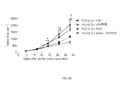
[0061] FIG. 26 shows the effects of sActRIIB in combination with
doxorubicin on tumor
size (top) and weight (bottom) in TOV-21G tumor-bearing mice. Tumor size
(upper panel)
and tumor weight (lower panel) were plotted as the mean standard error of
the mean
(SEM); n = 10 to 18/group. Statistical significance is represented by * p <
0.05, ** p <0.01:
TOV-21G + DOX + sActRIIB vs. TOV-21G + PBS; #p<0.05; #14p<0.01: TOV-21G + DOX
vs. TOV-21G + PBS; 8'p<0.05 TOV-21G + DOX + sActRIIB vs. TOV-21G + DOX.
[0062] FIG. 27 shows bar graphs illustrating the effect of sActRIIB in
combination with
doxorubicin on muscle mass in TOV-21G tumor-bearing mice. Lean carcass and
calf muscle
weights were determined at terminal necropsy. Data were plotted as mean + SEM;
n = 10 to
18/group. Statistical significance is represented by * p < 0.05; ** p <0.01;
*** p < 0.001.
[0063] FIG. 28 shows a bar graph illustrating the effect of activin-A
antibody on serum
activin A levels in female Inha KO mice and wild-type littermate control mice.
Measurements of free activin A level in female Inha KO mouse groups were
plotted as the
mean SEM; ***p < 0.001 and **p < 0.01
[0064] FIG. 29 shows the effect of activin-A antibody treatment on body
weight in
female Inha KO mice and wild-type littermate control mice. Measurements of
body weight in
female Inha KO mice were plotted as the mean SEM; ** p <0.01 and *** p
<0.001 for
Inha KO groups treated with activin-A antibody vs PBS. 8' p <0.05 and 8484p <
0.01 for
12
CA 02899889 2015-07-30
WO 2014/121221
PCT/US2014/014490
activin-A antibody treated Inha KO group vs PBS treated WT group. # p < 0.05
for PBS
treated Inha KO group vs PBS treated WT group.
[0065] FIG. 30 shows the effect of activin-A antibody treatment on lean
body mass and
fat mass in female Inha KO mice and wild-type littermate control mice.
Measurements of
lean mass (upper panel) and fat mass (lower panel) in female Inha KO mouse
were plotted as
the mean SEM; *** p < 0.001 and ** p <0.01 for Inha KO groups treated with
activin-A
antibody vs PBS. 84848'p <0.001 for activin-A antibody treated Inha KO group
vs PBS treated
WT group. #1414p <0.001 for PBS treated Inha KO group vs PBS treated WT group.
[0066] FIG. 31 shows a bar graph illustrating the effect of activin-A
antibody treatment
on calf muscle weight in female Inha KO mice and wild-type littermate control
mice. Calf
muscle weights were plotted as mean SEM; ***p <0.001, **p <0.01, *p <0.05.
[0067] FIG. 32A shows the effect of activin-A antibody treatment on ovary
weight in
female Inha KO mice and wild-type littermate control mice. Data was plotted as
mean
SEM; ***p <0.001, **p <0.01.
[0068] FIG. 32B shows the effect of activin-A antibody treatment on uterus
weight in
female Inha KO mice and wild-type littermate control mice. Data was plotted as
mean
SEM; ***p <0.001, **p <0.01.
[0069] FIG. 33 shows a bar graph illustrating the effect of activin-A
antibody treatment
on serum VEGF levels in female Inha KO mice and wild-type littermate control
mice.
Measurements of serum VEGF level were plotted as the mean SEM; ***p < 0.001,
*p < 0.05.
[0070] FIG. 34 shows the effect of activin-A antibody treatment on VEGF and
angiopoietin-1 protein expression levels in ovarian tumor tissues of Inha KO
mice and wild-
type littermate control mice. Upper panel: Representative images of VEGF and
Ang-
limmunostaining (grayish blue) on ovarian tissue sections. Nuclei were counter
stained with
Fast Red. Bar graphs: VEGF and Ang-1 immunoreactivities in ovarian sections
from 3
animals per group were quantified by imaging with Metamorph software and
plotted as the
mean SEM. ***p <0.001 and *p < 0.05
[0071] FIG. 35 shows the effect of activin-A antibody in combination with
doxorubicin
on body weight in TOV-21G tumor-bearing mice. Body weight was recorded
longitudinally.
Arrows point to timings of doxorubicin dosing. Data were plotted as mean
SEM; n = 8-14
per group. Standard 2-tailed Student's t-test (MS Excel 5.0) was used to
analyze the
13
CA 02899889 2015-07-30
WO 2014/121221
PCT/US2014/014490
differences between groups. Statistical significance is represented by *: p
<0.05: Normal vs.
TOV-21G + PBS; # p <0.05, #4 p <0.01, ### p <0.001: Normal vs. TOV-21G +
DOX;*: p <
0.05: TOV-21G + PBS vs. TOV-21G + Activin Ab.
[0072] FIG. 36 shows the effect of activin-A antibody in combination with
doxorubicin
on tumor size in TOV-21G tumor-bearing mice. Measurements of tumor size were
plotted as
the mean standard error of the mean (SEM); n = 8-14 per group. Standard 2-
tailed
Student's t-test (MS Excel 5.0) was used to analyze the differences between
groups.
Statistical significance is represented by * p < 0.05, ** p <0.01: TOV-21G +
Activin Ab vs.
TOV-21G + PBS, #4 p <0.01; ###: p <0.001: TOV-21G + DOX vs. TOV-21G + PBS;
8484p <
0.01; 848'84 p <0.001: TOV-21G + DOX + Activin Ab vs. TOV-21G + PBS; * p
<0.05; p <
0.01: TOV-21G + DOX + Activin Ab vs. TOV-21G + DOX.
[0073] FIG. 37 shows the effect of activin-A antibody in combination with
doxorubicin
on tumor weight in TOV-21G tumor-bearing mice. Measurements of tumor weight
were
plotted as the mean standard error of the mean (SEM); n = 8-14 per group.
Standard
2-tailed Student's t-test (MS Excel 5.0) was used to analyze the differences
between groups.
Statistical significance is represented by *: p < 0.05; **: p <0.01; ***: p
<0.001.
[0074] FIG. 38 shows the effect of activin-A antibody in combination with
doxorubicin
on muscle mass in TOV-21G tumor-bearing mice. Lean carcass and calf muscle
weights were
determined at terminal necropsy procedures. Data were plotted as mean + SEM; n
= 8-14 per
group. Standard 2-tailed Student's t-test (MS Excel 5.0) was used to analyze
the differences
between groups. Statistical significance is represented by *: p <0.05; **: p <
0.01; ***: p <
0.001.
DETAILED DESCRIPTION
[0075] The present invention relates to the effects of blocking activin-A.
Blocking
activin-A in vivo reduces several tumor angiogenesis factors and prevents
tumor
neovascularization, thereby inducing tumor apoptosis. In some aspects, the
invention
provides methods for identifying ovarian cancer in a subject by evaluating the
subject's
expression levels of various factors. In some aspects, the invention also
provides methods of
treating ovarian cancer, including serous ovarian cancer, by administering
anti-activin-A
compounds, including anti-activin-A antibodies and activin receptors, to a
subject. In some
aspects, the invention further provides methods of treating ovarian cancer,
including serous
ovarian cancer, clear cell ovarian cancer, Granulosa cell ovarian cancer,
Leydig cell tumors,
14
CA 02899889 2015-07-30
WO 2014/121221
PCT/US2014/014490
and sex cord stromal testicular tumors, by administering at least an anti-
activin-A compound
and a chemotherapeutic compound to a subject.
[0076] The details of one or more embodiments are set forth in the
description below.
Other features, objects, and advantages will be apparent from the description
and the
drawings, and from the claims.
[0077] Activin-A is the homodimer of the polypeptide chains PA (see GenBank
Accession No: NM 002192). Activins A, B, and AB are the homodimers and
heterodimer
respectively of two polypeptide chains, PA and PB. The term "activin" refers
to activin-A, -
B, and -AB, as well as variants and species homologs of that protein.
[0078] The present invention provides compositions, kits, and methods
relating to
molecules that bind to activin-A, including molecules and antigen-binding
proteins that
agonize or antagonize activin-A, such as activin JIB receptor polypeptides
(svActRIIB),
svActRIIB fragments, svActRIIB derivatives, anti-activin-A antibodies,
antibody fragments,
and antibody derivatives, e.g., antagonistic anti-activin-A antibodies,
antibody fragments, or
antibody derivatives. Also provided are compositions, kits, and methods
relating to
molecules that specifically bind to a portion of activin-A, such as amino
acids R13-Y39, or
amino acids V82-N107 of activin-A. Such molecules can include antibodies,
antibody
fragments, and antibody derivatives. Also provided are nucleic acids, and
derivatives and
fragments thereof, comprising a sequence of nucleotides that encodes all or a
portion of a
polypeptide that binds to activin-A, e.g., a nucleic acid encoding all or part
of an activin JIB
receptor, svActRIIB fragment, svActRIIB derivative, anti-activin-A antibody,
antibody
fragment, antibody variant, or antibody derivative, plasmids and vectors
comprising such
nucleic acids, and cells or cell lines comprising such nucleic acids and/or
vectors and
plasmids. The provided methods include, for example, methods of making,
identifying, or
isolating molecules that bind to activin-A, such as activin JIB receptors,
anti-activin-A
antibodies, methods of determining whether a molecule binds to activin-A,
methods of
making compositions, such as pharmaceutical compositions, comprising a
molecule that
binds to activin-A, and methods for administering a molecule that binds
activin-A to a
subject, for example, methods for treating a condition mediated by activin-A,
and for
modulating a biological activity of activin-A in vivo or in vitro.
[0079] The present invention relates to regions of the human activin-A that
contain
cysteine knot domains recognized by antibodies that also bind to full-length
activin-A, and/or
a region of activin-A that overlaps or encompasses a cysteine knot region of
activin-A, and
CA 02899889 2015-07-30
WO 2014/121221
PCT/US2014/014490
methods of making and using these cysteine knot domains. The invention also
provides
antigen binding agents, including antibodies, that specifically bind to
activin-A or portions of
activin-A, and methods for using such binding agents. The binding agents are
useful to block
or impair binding of human activin-A to one or more ligand.
[0080] Activins can interact with two structurally related classes of
serine/threonine
kinase receptors (type I and type II). Inhibin antagonizes activin by binding
to the
proteoglycan, betaglycan, and forming a stable complex with and thereby
sequestering type II
activin receptors while excluding type I receptors. Two major forms of activin
exist: activin-
A is a homodimer of PA-subunits and activin B is a homodimer of 3B-subunits.
(Vale, et al.,
Recent Frog Horm Res V. 44: 1-34, 1988). Heterodimers of an a-subunit that is
dissimilar to
either 3-subunit results in the functional antagonist inhibin.
[0081] The literature has shown that activin-A is overexpressed and/or
localized in
cancer tissues. For example, high levels of serum activin-A were found in
women with
endometrial and cervical carcinoma (Petraglia, F. et al., Jour. Clin.
Endocrin. Metab.
83:1194-1200, 1998). Activin-A was overexpressed in stage IV colorectal cancer
(Wildi, S.
et al., Gut 49:409-417, 2001). A role of activin-A in ovarian cancer was
reported (Steller,
M.D. et al., Mol. Cancer Res. 3:50-61, 2005).
[0082] The literature has also implicated activin-A in renal disease.
(Yamashita, S. et al.
1 Am. Soc. Nephrol. 15:91-101, 2004.) Serum immunoreactive activin-A levels in
normal
subjects and patients with disease were reported by Harada, K. et al. in J.
Clin. Endocrin. and
Metab. 81:2125-2130, 1996. Activin-A is a potent activator of renal
interstitial fibroblasts
(Harada, K. et al., J Am. Soc. Nephrol. 15:91-101, 2004). Glomerular activin-A
overexpression is linked to fibrosis in anti-Thy 1 glomerulonephritis
(Gaedeke, J. et al.,
Neph. Dial. Transpl. 20:319-328, 2005).
[0083] Serum activin-A levels in heart failure patients increased according
to disease
severity (Yndestal et al., Circulation 109:1379-1385, 2004). In a rat model of
heart failure,
serum activin-A elevated immediately after myocardial infarct and persisted
for six months,
and activin-A immunostaining was localized solely to cardiomyocytes (Yndestad
et al.,
2004). Elevated levels of activin-A were reported in heart failure (Yndestad,
A. et
al., Circulation 109:1379-1385, 2004).
[0084] Unless otherwise defined herein, scientific and technical terms used
in connection
with the present invention shall have the meanings that are commonly
understood by those of
ordinary skill in the art. Further, unless otherwise required by context,
singular terms shall
16
CA 02899889 2015-07-30
WO 2014/121221
PCT/US2014/014490
include pluralities and plural terms shall include the singular. Generally,
nomenclatures used
in connection with, and techniques of, cell and tissue culture, molecular
biology,
immunology, microbiology, genetics and protein and nucleic acid chemistry and
hybridization described herein are those well known and commonly used in the
art. The
methods and techniques of the present invention are generally performed
according to
conventional methods well known in the art and as described in various general
and more
specific references that are cited and discussed throughout the present
specification unless
otherwise indicated. See, e.g., Sambrook et al. Molecular Cloning: A
Laboratory Manual, 2d
ed., Cold Spring Harbor Laboratory Press, Cold Spring Harbor, N.Y. (1989) and
Ausubel et
al., Current Protocols in Molecular Biology, Greene Publishing Associates
(1992), and
Harlow and Lane Antibodies: A Laboratory Manual Cold Spring Harbor Laboratory
Press,
Cold Spring Harbor, N.Y. (1990), which are incorporated herein by reference.
Enzymatic
reactions and purification techniques are performed according to
manufacturer's
specifications, as commonly accomplished in the art or as described herein.
The terminology
used in connection with, and the laboratory procedures and techniques of,
analytical
chemistry, synthetic organic chemistry, and medicinal and pharmaceutical
chemistry
described herein are those well known and commonly used in the art. Standard
techniques
can be used for chemical syntheses, chemical analyses, pharmaceutical
preparation,
formulation, and delivery, and treatment of patients.
[0085] The following terms, unless otherwise indicated, shall be understood
to have the
following meanings:
[0086] The term "isolated molecule" (where the molecule is, for example, a
polypeptide,
a polynucleotide, or an antibody) is a molecule that by virtue of its origin
or source of
derivation (1) is not associated with at least one naturally associated
component that
accompany it in its native state, (2) is substantially free of other molecules
from the same
species (3) is expressed by a cell from a different species, or (4) does not
occur in nature.
Thus, a molecule that is chemically synthesized, or synthesized in a cellular
system different
from the cell from which it naturally originates, will be "isolated" from its
naturally
associated components. A molecule also may be rendered substantially free of
naturally
associated components by isolation, using purification techniques well known
in the art.
Molecule purity or homogeneity may be assayed by a number of means well known
in the
art. For example, the purity of a polypeptide sample may be assayed using
polyacrylamide
gel electrophoresis and staining of the gel to visualize the polypeptide using
techniques well
17
CA 02899889 2015-07-30
WO 2014/121221
PCT/US2014/014490
known in the art. For certain purposes, higher resolution may be provided by
using HPLC or
other means well known in the art for purification.
[0087] The terms "anti-activin-A compound", "activin-A inhibitor" and
"activin-A
antagonist" are used interchangeably. Each is a molecule that detectably
inhibits at least one
function of activin-A. Conversely, an "activin-A agonist" is a molecule that
detectably
increases at least one function of activin-A. The inhibition caused by an
activin-A inhibitor
need not be complete so long as it is detectable using an assay. Any assay of
a function of
activin-A can be used, examples of which are provided herein. Examples of
functions of
activin-A that can be inhibited by an activin-A inhibitor, or increased by an
activin-A agonist,
include binding to activin-A. Examples of types of activin-A inhibitors and
activin-A
agonists include, but are not limited to, activin-A binding polypeptides such
as antigen
binding proteins (e.g., activin-A inhibiting antigen binding proteins),
activin JIB receptors
(svActRIIB), svActRIIB fragments, svActRIIB derivatives, antibodies, antibody
fragments,
and antibody derivatives.
[0088] The terms "peptide," "polypeptide" and "protein" each refers to a
molecule
comprising two or more amino acid residues joined to each other by peptide
bonds. These
terms encompass, e.g., native and artificial proteins, protein fragments and
polypeptide
analogs (such as muteins, variants, and fusion proteins) of a protein sequence
as well as post-
translationally, or otherwise covalently or non-covalently, modified proteins.
A peptide,
polypeptide, or protein may be monomeric or polymeric. Polynucleotide and
polypeptide
sequences are indicated using standard one- or three-letter abbreviations.
Unless otherwise
indicated, polypeptide sequences have their amino termini at the left and
their carboxy
termini at the right, and single-stranded nucleic acid sequences, and the top
strand of double-
stranded nucleic acid sequences, have their 5' termini at the left and their
3' termini at the
right. A particular polypeptide or polynucleotide sequence also can be
described by
explaining how it differs from a reference sequence. Unless otherwise
indicated, it is
understood that polynucleotide and polypeptide sequences include each nucleic
acid or amino
acid listed, respectively, as well as the intervening nucleic acids or amino
acids. For
example, the polypeptide sequence R13-Y39 sets forth a polypeptide sequence
that includes
the amino acids R13, and Y39, as well as the amino acids found between R13 and
Y39 in the
polypeptide sequence. Correspondingly, the polynucleotide sequence C1¨T5 sets
forth a
polynucleotide sequence that includes nucleic acids Cl, and T5, as well as
nucleic acids at
positions 2, 3, and 4 of the sequence. Accordingly, designations of SEQ ID NO:
1-5 likewise
18
CA 02899889 2015-07-30
WO 2014/121221
PCT/US2014/014490
designates the inclusive group of SEQ ID NO: 1, SEQ ID NO: 2, SEQ ID NO: 3,
SEQ ID
NO: 4, and SEQ ID NO: 5. Finally, amino acid groupings are also intended to be
inclusive,
unless otherwise designated. For example, the phrase "amino acids 1-5 of SEQ
ID NO: 28"
includes amino acids at positions 1, 2, 3, 4, and 5 of SEQ ID NO: 28.
[0089]
Polypeptides of the invention include polypeptides that have been modified in
any
way and for any reason, for example, to: (1) reduce susceptibility to
proteolysis, (2) reduce
susceptibility to oxidation, (3) alter binding affinity for forming protein
complexes, (4) alter
binding affinities, and (4) confer or modify other physicochemical or
functional properties.
Analogs include muteins of a polypeptide. For example, single or multiple
amino acid
substitutions (e.g., conservative amino acid substitutions) may be made in the
naturally
occurring sequence (e.g., in the portion of the polypeptide outside the
domain(s) forming
intermolecular contacts). A "conservative amino acid substitution" is one that
does not
substantially change the structural characteristics of the parent sequence
(e.g., a replacement
amino acid should not tend to break a helix that occurs in the parent
sequence, or disrupt
other types of secondary structure that characterize the parent sequence or
are necessary for
its functionality). Examples of art-recognized polypeptide secondary and
tertiary structures
are described in Proteins, Structures and Molecular Principles (Creighton,
Ed., W. H.
Freeman and Company, New York (1984)); Introduction to Protein Structure (C.
Branden
and J. Tooze, eds., Garland Publishing, New York, N.Y. (1991)); and Thornton
et al. Nature
354:105 (1991), which are each incorporated herein by reference.
[0090] The term
"polypeptide fragment" as used herein refers to a polypeptide that has an
amino-terminal and/or carboxy-terminal deletion as compared to a corresponding
full-length
protein. Fragments can be, for example, at least 5, 6, 7, 8, 9, 10, 11, 12,
13, 14, 15, 20, 50,
70, 80, 90, 100, 150 or 200 amino acids in length. Fragments can also be, for
example, at
most 1,000, 750, 500, 250, 200, 175, 150, 125, 100, 90, 80, 70, 60, 50, 40,
30, 20, 15, 14, 13,
12, 11, or 10 amino acids in length. A fragment can further comprise, at
either or both of its
ends, one or more additional amino acids, for example, a sequence of amino
acids from a
different naturally-occurring protein (e.g., an Fc or leucine zipper domain)
or an artificial
amino acid sequence (e.g., an artificial linker sequence).
[0091] A
"variant" of a polypeptide (e.g., an antibody) comprises an amino acid
sequence
wherein one or more amino acid residues are inserted into, deleted from and/or
substituted
into the amino acid sequence relative to the native polypeptide sequence, and
retains
essentially the same biological activity as the native polypeptide. The
biological activity of
19
CA 02899889 2015-07-30
WO 2014/121221
PCT/US2014/014490
the polypeptide can be measured using standard techniques in the art (for
example, if the
variant is an antibody, its activity may be tested by binding assays, as
described herein).
Variants of the invention include fragments, analogs, recombinant
polypeptides, synthetic
polypeptides, and/or fusion proteins.A "derivative" of a polypeptide is a
polypeptide (e.g., an
antibody) that has been chemically modified, e.g., via conjugation to another
chemical
moiety such as, for example, polyethylene glycol, albumin (e.g., human serum
albumin),
phosphorylation, and glycosylation. Unless otherwise indicated, the term
"antibody"
includes, in addition to antibodies comprising two full-length heavy chains
and two full-
length light chains, derivatives, variants, fragments, and muteins thereof,
examples of which
are described below.
[0092] The terms "polynucleotide," "oligonucleotide" and "nucleic acid" are
used
interchangeably throughout and include DNA molecules (e.g., cDNA or genomic
DNA),
RNA molecules (e.g., mRNA), analogs of the DNA or RNA generated using
nucleotide
analogs (e.g., peptide nucleic acids and non-naturally occurring nucleotide
analogs), and
hybrids thereof The nucleic acid molecule can be single-stranded or double-
stranded. In
one embodiment, the nucleic acid molecules of the invention comprise a
contiguous open
reading frame encoding an antibody, or a fragment, derivative, mutein, or
variant thereof, of
the invention.
[0093] The term percent "identity," in the context of two or more nucleic
acid or
polypeptide sequences, refer to two or more sequences or subsequences that
have a specified
percentage of nucleotides or amino acid residues that are the same, when
compared and
aligned for maximum correspondence, determined by comparing the sequences
using the
GAP computer program (a part of the GCG Wisconsin Package, version 10.3
(Accelrys, San
Diego, CA)) using its default parameters. Depending on the application, the
percent
"identity" can exist over a region of the sequence being compared, e.g., over
a functional
domain, or, alternatively, exist over the full length of the two sequences to
be compared.
[0094] For sequence comparison, typically one sequence acts as a reference
sequence to
which test sequences are compared. When using a sequence comparison algorithm,
test and
reference sequences are input into a computer, subsequence coordinates are
designated, if
necessary, and sequence algorithm program parameters are designated. The
sequence
comparison algorithm then calculates the percent sequence identity for the
test sequence(s)
relative to the reference sequence, based on the designated program
parameters.
CA 02899889 2015-07-30
WO 2014/121221
PCT/US2014/014490
[0095] Two single-stranded polynucleotides are "the complement" of each
other if their
sequences can be aligned in an anti-parallel orientation such that every
nucleotide in one
polynucleotide is opposite its complementary nucleotide in the other
polynucleotide, without
the introduction of gaps, and without unpaired nucleotides at the 5' or the 3'
end of either
sequence. A polynucleotide is "complementary" to another polynucleotide if the
two
polynucleotides can hybridize to one another under moderately stringent
conditions. Thus, a
polynucleotide can be complementary to another polynucleotide without being
its
complement.
[0096] A "vector" is a nucleic acid that can be used to introduce another
nucleic acid
linked to it into a cell. One type of vector is a "plasmid," which refers to a
linear or circular
double stranded DNA molecule into which additional nucleic acid segments can
be ligated.
Another type of vector is a viral vector (e.g., replication defective
retroviruses, adenoviruses
and adeno-associated viruses), wherein additional DNA segments can be
introduced into the
viral genome. Certain vectors are capable of autonomous replication in a host
cell into which
they are introduced (e.g., bacterial vectors comprising a bacterial origin of
replication and
episomal mammalian vectors). Other vectors (e.g., non-episomal mammalian
vectors) are
integrated into the genome of a host cell upon introduction into the host
cell, and thereby are
replicated along with the host genome. An "expression vector" is a type of
vector that can
direct the expression of a chosen polynucleotide.
[0097] A nucleotide sequence is "operably linked" to a regulatory sequence
if the
regulatory sequence affects the expression (e.g., the level, timing, or
location of expression)
of the nucleotide sequence. A "regulatory sequence" is a nucleic acid that
affects the
expression (e.g., the level, timing, or location of expression) of a nucleic
acid to which it is
operably linked. The regulatory sequence can, for example, exert its effects
directly on the
regulated nucleic acid, or through the action of one or more other molecules
(e.g.,
polypeptides that bind to the regulatory sequence and/or the nucleic acid).
Examples of
regulatory sequences include promoters, enhancers and other expression control
elements
(e.g., polyadenylation signals). Further examples of regulatory sequences are
described in,
for example, Goeddel, 1990, Gene Expression Technology: Methods in Enzymology
185,
Academic Press, San Diego, CA and Baron et al., 1995, Nucleic Acids Res.
23:3605-06.
[0098] A "host cell" is a cell that can be used to express a nucleic acid,
e.g., a nucleic
acid of the invention. A host cell can be a prokaryote, for example, E. coli,
or it can be a
eukaryote, for example, a single-celled eukaryote (e.g., a yeast or other
fungus), a plant cell
21
CA 02899889 2015-07-30
WO 2014/121221
PCT/US2014/014490
(e.g., a tobacco or tomato plant cell), an animal cell (e.g., a human cell, a
monkey cell, a
hamster cell, a rat cell, a mouse cell, or an insect cell) or a hybridoma.
Examples of host cells
include CS-9 cells, the COS-7 line of monkey kidney cells (ATCC CRL 1651) (see
Gluzman
et al., 1981, Cell 23:175), L cells, C127 cells, 3T3 cells (ATCC CCL 163),
Chinese hamster
ovary (CHO) cells or their derivatives such as Veggie CHO and related cell
lines which grow
in serum-free media (see Rasmussen et al., 1998, Cytotechnology 28:31), HeLa
cells, BHK
(ATCC CRL 10) cell lines, the CV1/EBNA cell line derived from the African
green monkey
kidney cell line CV1 (ATCC CCL 70) (see McMahan et al., 1991, EMBO 1 10:2821),
human
embryonic kidney cells such as 293, 293 EBNA or MSR 293, human epidermal A431
cells,
human Colo205 cells, other transformed primate cell lines, normal diploid
cells, cell strains
derived from in vitro culture of primary tissue, primary explants, HL-60,
U937, HaK or
Jurkat cells. Typically, a host cell is a cultured cell that can be
transformed or transfected
with a polypeptide-encoding nucleic acid, which can then be expressed in the
host cell. The
phrase "recombinant host cell" can be used to denote a host cell that has been
transformed or
transfected with a nucleic acid to be expressed. A host cell also can be a
cell that comprises
the nucleic acid but does not express it at a desired level unless a
regulatory sequence is
introduced into the host cell such that it becomes operably linked with the
nucleic acid. It is
understood that the term host cell refers not only to the particular subject
cell but to the
progeny or potential progeny of such a cell. Because certain modifications may
occur in
succeeding generations due to, e.g., mutation or environmental influence, such
progeny may
not, in fact, be identical to the parent cell, but are still included within
the scope of the term
as used herein.
Nucleic acids
[0099] In one
aspect, the present invention provides isolated nucleic acid molecules. The
nucleic acids comprise, for example, polynucleotides that encode all or part
of an antigen
binding protein, for example, one or both chains of an antibody of the
invention, or a
fragment, derivative, mutein, or variant thereof, polynucleotides sufficient
for use as
hybridization probes, PCR primers or sequencing primers for identifying,
analyzing, mutating
or amplifying a polynucleotide encoding a polypeptide, anti-sense nucleic
acids for inhibiting
expression of a polynucleotide, and complementary sequences of the foregoing.
The nucleic
acids can be any length. They can be, for example, 5, 10, 15, 20, 25, 30, 35,
40, 45, 50, 75,
100, 125, 150, 175, 200, 250, 300, 350, 400, 450, 500, 750, 1,000, 1,500,
3,000, 5,000 or
more nucleotides in length, and/or can comprise one or more additional
sequences, for
22
CA 02899889 2015-07-30
WO 2014/121221
PCT/US2014/014490
example, regulatory sequences, and/or be part of a larger nucleic acid, for
example, a vector.
The nucleic acids can be single-stranded or double-stranded and can comprise
RNA and/or
DNA nucleotides, and artificial variants thereof (e.g., peptide nucleic
acids).
[00100] Nucleic acids encoding antibody polypeptides (e.g., heavy or light
chain, variable
domain only, or full length) can be isolated from B-cells of mice that have
been immunized
with activin-A. The nucleic acid can be isolated by conventional procedures
such as
polymerase chain reaction (PCR).
[00101] Nucleic acid sequences encoding the variable regions of the heavy and
light chain
variable regions are shown herein. The skilled artisan will appreciate that,
due to the
degeneracy of the genetic code, each of the polypeptide sequences disclosed
herein is
encoded by a large number of other nucleic acid sequences. The present
invention provides
each degenerate nucleotide sequence encoding each antigen binding protein of
the invention.
[00102] The invention further provides nucleic acids that hybridize to other
nucleic acids
(e.g., nucleic acids comprising a nucleotide sequence of any of A 1-A 14)
under particular
hybridization conditions. Methods for hybridizing nucleic acids are well-known
in the art.
See, e.g., Curr. Prot. in Mol. Biol., John Wiley & Sons, N.Y. (1989), 6.3.1-
6.3.6. As defined
herein, a moderately stringent hybridization condition uses a prewashing
solution containing
5X sodium chloride/sodium citrate (SSC), 0.5% SDS, 1.0 mM EDTA (pH 8.0),
hybridization
buffer of about 50% formamide, 6X SSC, and a hybridization temperature of 55
C (or other
similar hybridization solutions, such as one containing about 50% formamide,
with a
hybridization temperature of 42 C), and washing conditions of 60 C, in 0.5X
SSC, 0.1%
SDS. A stringent hybridization condition hybridizes in 6X SSC at 45 C,
followed by one or
more washes in 0.1X SSC, 0.2% SDS at 68 C. Furthermore, one of skill in the
art can
manipulate the hybridization and/or washing conditions to increase or decrease
the stringency
of hybridization such that nucleic acids comprising nucleotide sequences that
are at least 65,
70, 75, 80, 85, 90, 95, 98, or 99% identical to each other typically remain
hybridized to each
other. The basic parameters affecting the choice of hybridization conditions
and guidance for
devising suitable conditions are set forth by, for example, Sambrook, Fritsch,
and Maniatis
(1989, Molecular Cloning: A Laboratory Manual, Cold Spring Harbor Laboratory
Press,
Cold Spring Harbor, N.Y., chapters 9 and 11; and Curr. Prot. in Mol. Biol.
1995, Ausubel et
al., eds., John Wiley & Sons, Inc., sections 2.10 and 6.3-6.4), and can be
readily determined
by those having ordinary skill in the art based on, for example, the length
and/or base
composition of the DNA.
23
CA 02899889 2015-07-30
WO 2014/121221
PCT/US2014/014490
[00103] Changes can be introduced by mutation into a nucleic acid, thereby
leading to
changes in the amino acid sequence of a polypeptide (e.g., an antigen binding
protein) that it
encodes. Mutations can be introduced using any technique known in the art. In
one
embodiment, one or more particular amino acid residues are changed using, for
example, a
site-directed mutagenesis protocol. In another embodiment, one or more
randomly selected
residues are changed using, for example, a random mutagenesis protocol.
However it is
made, a mutant polypeptide can be expressed and screened for a desired
property (e.g.,
binding to activin-A).
[00104] Mutations can be introduced into a nucleic acid without significantly
altering the
biological activity of a polypeptide that it encodes. For example, one can
make nucleotide
substitutions leading to amino acid substitutions at non-essential amino acid
residues. In one
embodiment, a nucleotide sequence provided herein for Al-A14, or a desired
fragment,
variant, or derivative thereof, is mutated such that it encodes an amino acid
sequence
comprising one or more deletions or substitutions of amino acid residues that
are shown
herein for Al-A14 to be residues where two or more sequences differ. As
described herein
inter alia, Al-A14 refers to 14 sequences, Al, and A14, as well as the 12
intervening amino
acid residues. In another embodiment, the mutagenesis inserts an amino acid
adjacent to one
or more amino acid residues shown herein for Al-A14 to be residues where two
or more
sequences differ. Alternatively, one or more mutations can be introduced into
a nucleic acid
that selectively change the biological activity (e.g., binding of activin-A)
of a polypeptide
that it encodes. For example, the mutation can quantitatively or qualitatively
change the
biological activity. Examples of quantitative changes include increasing,
reducing or
eliminating the activity. Examples of qualitative changes include changing the
antigen
specificity of an antigen binding protein.
[00105] In another aspect, the present invention provides nucleic acid
molecules that are
suitable for use as primers or hybridization probes for the detection of
nucleic acid sequences
of the invention. A nucleic acid molecule of the invention can comprise only a
portion of a
nucleic acid sequence encoding a full-length polypeptide of the invention, for
example, a
fragment that can be used as a probe or primer or a fragment encoding an
active portion (e.g.,
an activin-A binding portion) of a polypeptide of the invention.
[00106] Probes based on the sequence of a nucleic acid of the invention can be
used to
detect the nucleic acid or similar nucleic acids, for example, transcripts
encoding a
polypeptide of the invention. The probe can comprise a label group, e.g., a
radioisotope, a
24
CA 02899889 2015-07-30
WO 2014/121221
PCT/US2014/014490
fluorescent compound, an enzyme, or an enzyme co-factor. Such probes can be
used to
identify a cell that expresses the polypeptide.
Expression Vectors
[00107] The present invention provides vectors comprising a nucleic acid
encoding a
polypeptide of the invention or a portion thereof Examples of vectors include,
but are not
limited to, plasmids, viral vectors, non-episomal mammalian vectors and
expression vectors,
for example, recombinant expression vectors.
[00108] In another aspect of the present invention, expression vectors
containing the
nucleic acid molecules and polynucleotides of the present invention are also
provided, and
host cells transformed with such vectors, and methods of producing the
polypeptides are also
provided. The term "expression vector" refers to a plasmid, phage, virus or
vector for
expressing a polypeptide from a polynucleotide sequence. Vectors for the
expression of the
polypeptides contain at a minimum sequences required for vector propagation
and for
expression of the cloned insert. An expression vector comprises a
transcriptional unit
comprising an assembly of (1) a genetic element or elements having a
regulatory role in gene
expression, for example, promoters or enhancers, (2) a sequence that encodes
polypeptides
and proteins to be transcribed into mRNA and translated into protein, and (3)
appropriate
transcription initiation and termination sequences. These sequences may
further include a
selection marker. Vectors suitable for expression in host cells are readily
available and the
nucleic acid molecules are inserted into the vectors using standard
recombinant DNA
techniques. Such vectors can include promoters which function in specific
tissues, and viral
vectors for the expression of polypeptides in targeted human or animal cells.
For example, an
expression vector suitable for expression of svActRIIB is the pDSRa,
(described in WO
90/14363, herein incorporated by reference) and its derivatives, containing
svActRIIB
polynucleotides, as well as any additional suitable vectors known in the art.
[00109] The recombinant expression vectors of the invention can comprise a
nucleic acid
of the invention in a form suitable for expression of the nucleic acid in a
host cell. The
recombinant expression vectors include one or more regulatory sequences,
selected on the
basis of the host cells to be used for expression, which is operably linked to
the nucleic acid
sequence to be expressed. Regulatory sequences include those that direct
constitutive
expression of a nucleotide sequence in many types of host cells (e.g., 5V40
early gene
enhancer, Rous sarcoma virus promoter and cytomegalovirus promoter), those
that direct
expression of the nucleotide sequence only in certain host cells (e.g., tissue-
specific
CA 02899889 2015-07-30
WO 2014/121221
PCT/US2014/014490
regulatory sequences, see Voss et al., 1986, Trends Biochem. Sci. 11:287,
Maniatis et al.,
1987, Science 236:1237, incorporated by reference herein in their entireties),
and those that
direct inducible expression of a nucleotide sequence in response to particular
treatment or
condition (e.g., the metallothionin promoter in mammalian cells and the tet-
responsive and/or
streptomycin responsive promoter in both prokaryotic and eukaryotic systems
(see id.). It
will be appreciated by those skilled in the art that the design of the
expression vector can
depend on such factors as the choice of the host cell to be transformed, the
level of
expression of protein desired, etc. The expression vectors of the invention
can be introduced
into host cells to thereby produce proteins or peptides, including fusion
proteins or peptides,
encoded by nucleic acids as described herein.
[00110] The invention further provides methods of making polypeptides. A
variety of
other expression/host systems may be utilized. Vector DNA can be introduced
into
prokaryotic or eukaryotic systems via conventional transformation or
transfection techniques.
These systems include but are not limited to microorganisms such as bacteria
(for example,
E. coli) transformed with recombinant bacteriophage, plasmid or cosmid DNA
expression
vectors; yeast transformed with yeast expression vectors; insect cell systems
infected with
virus expression vectors (e.g., baculovirus); plant cell systems transfected
with virus
expression vectors (e.g., cauliflower mosaic virus, CaMV; tobacco mosaic
virus, TMV) or
transformed with bacterial expression vectors (e.g., Ti or pBR322 plasmid); or
animal cell
systems. Mammalian cells useful in recombinant protein production include but
are not
limited to VERO cells, HeLa cells, Chinese hamster ovary (CHO) cell lines, or
their
derivatives such as Veggie CHO and related cell lines which grow in serum-free
media (see
Rasmussen et al., 1998, Cytotechnology 28:31) or CHO strain DX-B11, which is
deficient in
DHFR (see Urlaub et al., 1980, Proc. Natl. Acad. Sci. USA 77:4216-20) COS
cells such as
the COS-7 line of monkey kidney cells (ATCC CRL 1651) (see Gluzman et al.,
1981, Cell
23:175), W138, BHK, HepG2, 3T3 (ATCC CCL 163), RIN, MDCK, A549, PC12, K562, L
cells, C127 cells, BHK (ATCC CRL 10) cell lines, the CV1/EBNA cell line
derived from the
African green monkey kidney cell line CV1 (ATCC CCL 70) (see McMahan et al.,
1991,
EMBO J. 10:2821), human embryonic kidney cells such as 293, 293 EBNA or MSR
293,
human epidermal A431 cells, human Colo205 cells, other transformed primate
cell lines,
normal diploid cells, cell strains derived from in vitro culture of primary
tissue, primary
explants, HL-60, U937, HaK or Jurkat cells. Mammalian expression allows for
the
26
CA 02899889 2015-07-30
WO 2014/121221
PCT/US2014/014490
production of secreted or soluble polypeptides which may be recovered from the
growth
medium.
[00111] For stable transfection of mammalian cells, it is known that,
depending upon the
expression vector and transfection technique used, only a small fraction of
cells may integrate
the foreign DNA into their genome. In order to identify and select these
integrants, a gene
that encodes a selectable marker (e.g., for resistance to antibiotics) is
generally introduced
into the host cells along with the gene of interest. Once such cells are
transformed with
vectors that contain selectable markers as well as the desired expression
cassette, the cells
can be allowed to grow in an enriched media before they are switched to
selective media, for
example. The selectable marker is designed to allow growth and recovery of
cells that
successfully express the introduced sequences. Resistant clumps of stably
transformed cells
can be proliferated using tissue culture techniques appropriate to the cell
line employed. An
overview of expression of recombinant proteins is found in Methods of
Enzymology, v. 185,
Goeddell, D.V., ed., Academic Press (1990).Preferred selectable markers
include those which
confer resistance to drugs, such as G418, hygromycin and methotrexate. Cells
stably
transfected with the introduced nucleic acid can be identified by drug
selection (e.g., cells
that have incorporated the selectable marker gene will survive, while the
other cells die),
among other methods.
[00112] In some cases, such as in expression using procaryotic systems, the
expressed
polypeptides of this invention may need to be "refolded" and oxidized into a
proper tertiary
structure and disulfide linkages generated in order to be biologically active.
Refolding can be
accomplished using a number of procedures well known in the art. Such methods
include,
for example, exposing the solubilized polypeptide to a pH usually above 7 in
the presence of
a chaotropic agent. The selection of chaotrope is similar to the choices used
for inclusion
body solubilization; however a chaotrope is typically used at a lower
concentration.
Exemplary chaotropic agents are guanidine and urea. In most cases, the
refolding/oxidation
solution will also contain a reducing agent plus its oxidized form in a
specific ratio to
generate a particular redox potential which allows for disulfide shuffling to
occur for the
formation of cysteine bridges. Some commonly used redox couples include
cysteine/cystamine, glutathione/dithiobisGSH, cupric chloride, dithiothreitol
DTT/dithiane
DTT, and 2-mercaptoethanol (bME)/dithio-bME. In many instances, a co-solvent
may be
used to increase the efficiency of the refolding. Commonly used cosolvents
include glycerol,
polyethylene glycol of various molecular weights, and arginine.
27
CA 02899889 2015-07-30
WO 2014/121221
PCT/US2014/014490
[00113] In addition, the polypeptides can be synthesized in solution or on a
solid support
in accordance with conventional techniques. Various automatic synthesizers are
commercially available and can be used in accordance with known protocols.
See, for
example, Stewart and Young, Solid Phase Peptide Synthesis, 2d.Ed., Pierce
Chemical Co.
(1984); Tam et al., J Am Chem Soc, 105:6442, (1983); Merrifield, Science
232:341-347
(1986); Barany and Merrifield, The Peptides, Gross and Meienhofer, eds,
Academic Press,
New York, 1-284; Barany et al., Int J Pep Protein Res, 30:705-739 (1987).
[00114] The polypeptides and proteins of the present invention can be purified
according
to protein purification techniques are well known to those of skill in the
art. These
techniques involve, at one level, the crude fractionation of the proteinaceous
and non-
proteinaceous fractions. Having separated the peptide polypeptides from other
proteins, the
peptide or polypeptide of interest can be further purified using
chromatographic and
electrophoretic techniques to achieve partial or complete purification (or
purification to
homogeneity). The term "purified polypeptide" as used herein, is intended to
refer to a
composition, isolatable from other components, wherein the polypeptide is
purified to any
degree relative to its naturally-obtainable state. A purified polypeptide
therefore also refers
to a polypeptide that is free from the environment in which it may naturally
occur.
Generally, "purified" will refer to a polypeptide composition that has been
subjected to
fractionation to remove various other components, and which composition
substantially
retains its expressed biological activity. Where the term "substantially
purified" is used, this
designation will refer to a peptide or polypeptide composition in which the
polypeptide or
peptide forms the major component of the composition, such as constituting
about 50 %,
about 60 %, about 70 %, about 80 %, about 85 %, or about 90 % or more of the
proteins in
the composition.
[00115] Various techniques suitable for use in purification will be well known
to those of
skill in the art. These include, for example, precipitation with ammonium
sulphate, PEG,
antibodies (immunoprecipitation) and the like or by heat denaturation,
followed by
centrifugation; chromatography such as affinity chromatography (Protein-A
columns), ion
exchange, gel filtration, reverse phase, hydroxylapatite, hydrophobic
interaction
chromatography, isoelectric focusing, gel electrophoresis, and combinations of
these
techniques. As is generally known in the art, it is believed that the order of
conducting the
various purification steps may be changed, or that certain steps may be
omitted, and still
28
CA 02899889 2015-07-30
WO 2014/121221
PCT/US2014/014490
result in a suitable method for the preparation of a substantially purified
polypeptide.
Exemplary purification steps are provided in the Examples below.
[00116] Various methods for quantifying the degree of purification of
polypeptide will be
known to those of skill in the art in light of the present disclosure. These
include, for
example, determining the specific binding activity of an active fraction, or
assessing the
amount of peptide or polypeptide within a fraction by SDS/PAGE analysis. A
preferred
method for assessing the purity of a polypeptide fraction is to calculate the
binding activity of
the fraction, to compare it to the binding activity of the initial extract,
and to thus calculate
the degree of purification, herein assessed by a "-fold purification number."
The actual units
used to represent the amount of binding activity will, of course, be dependent
upon the
particular assay technique chosen to follow the purification and whether or
not the
polypeptide or peptide exhibits a detectable binding activity.
Anti-Activin-A Antibody
[00117] Activin-A can be purified from host cells that have been transfected
by a gene
encoding activin-A by elution of filtered supernatant of host cell culture
fluid using a Heparin
HP column, using a salt gradient.
[00118] The term "antibody" refers to an intact immunoglobulin, or a binding
fragment
thereof An antibody may comprise a complete antibody molecule (including
polyclonal,
monoclonal, chimeric, humanized, or human versions having full length heavy
and/or light
chains), or comprise an antigen binding fragment thereof Antibody fragments
include
F(ab')2, Fab, Fab', Fv, Fc, and Fd fragments, and can be incorporated into
single domain
antibodies, single-chain antibodies, maxibodies, minibodies, intrabodies,
diabodies,
triabodies, tetrabodies, v-NAR and bis-scFy (See e.g.,, Hollinger and Hudson,
2005, Nature
Biotech., 23, 9, 1126-1136).
[00119] A Fab fragment is a monovalent fragment having the VL, VH, CL and CH 1
domains; a F(ab')2 fragment is a bivalent fragment having two Fab fragments
linked by a
disulfide bridge at the hinge region; a Fd fragment has the VH and CH1
domains; an Fy
fragment has the VL and VH domains of a single arm of an antibody; and a dAb
fragment has
a VH domain, a VL domain, or an antigen-binding fragment of a VH or VL domain
(US Pat.
No. 6,846,634, 6,696,245, US App. Pub. No. 05/0202512, 04/0202995, 04/0038291,
04/0009507, 03/0039958, Ward et al., Nature 341:544-546, 1989).
[00120] Polynucleotide and polypeptide sequences of particular light and heavy
chain
variable domains are described below. Antibodies comprising a light chain and
heavy chain
29
CA 02899889 2015-07-30
WO 2014/121221
PCT/US2014/014490
are designated by combining the name of the light chain and the name of the
heavy chain
variable domains. For example, "L4H7," indicates an antibody comprising the
light chain
variable domain of L4 and the heavy chain variable domain of H7.
[00121] Kappa light chain constant sequences are shown in SEQ ID NO: 84, 100
and 108,
and heavy chain constant sequence are shown in SEQ ID NOs: 214, 215 and 221.
Polynucleotides encoding these sequences are shown in, for the light chains,
respectively,
SEQ ID NOs: 222, 223 and 239, and for the heavy chains, respectively, SEQ ID
NOs: 240,
241, and 242. Thus, in addition to the variable sequences as disclosed herein,
an antibody
can comprise one or both of SEQ ID NOs: 84 and 214; or SEQ ID NOs: 215 and
223; or SEQ
ID NOs: 108 and 221. These sequences are illustrated in the table below:
SEQ ID NO Sequence
SEQ ID NO: 84 Gly Gin Pro Lys Ala Ala Pro Ser Val Thr
Leu Phe Pro
Pro Ser Ser Glu Glu Leu Gin Ala Asn Lys Ala Thr
Leu Val Cys Leu Ile Ser Asp Phe Tyr Pro Gly Ala Val
Thr Val Ala Trp Lys Ala Asp Ser Ser Pro Val Lys Ala
Gly Val Glu Thr Thr Thr Pro Ser Lys Gin Ser Asn
Asn Lys Tyr Ala Ala Ser Ser Tyr Leu Ser Leu Thr Pro
Glu Gin Trp Lys Ser His Arg Ser Tyr Ser Cys Gin Val
Thr His Glu Gly Ser Thr Val Glu Lys Thr Val Ala Pro
Thr Glu Cys Ser
SEQ ID NO: 100 Arg Thr Val Ala Ala Pro Ser Val Phe Ile
Phe Pro Pro
Ser Asp Glu Gin Leu Lys Ser Gly Thr Ala Ser Val Val
Cys Leu Leu Asn Asn Phe Tyr Pro Arg Glu Ala Lys
Val Gin Trp Lys Val Asp Asn Ala Leu Gin Ser Gly
Asn Ser Gin Glu Ser Val Thr Glu Gin Asp Ser Lys
Asp Ser Thr Tyr Ser Leu Ser Ser Thr Leu Thr Leu Ser
Lys Ala Asp Tyr Glu Lys His Lys Val Tyr Ala Cys
Glu Val Thr His Gin Gly Leu Ser Ser Pro Val Thr Lys
Ser Phe Asn Arg Gly Glu Cys
SEQ ID NO: 108 Arg Thr Val Ala Ala Pro Ser Val Phe Ile
Phe Pro Pro
Ser Asp Glu Gin Leu Lys Ser Gly Thr Ala Ser Val Val
Cys Leu Leu Asn Asn Phe Tyr Pro Arg Glu Ala Lys
Val Gin Trp Lys Val Asp Asn Ala Leu Gin Ser Gly
Asn Ser Gin Glu Ser Val Thr Glu Gin Asp Ser Lys
Asp Ser Thr Tyr Ser Leu Ser Ser Thr Leu Thr Leu Ser
Lys Ala Asp Tyr Glu Lys His Lys Val Tyr Ala Cys
Glu Val Thr His Gin Gly Leu Ser Ser Pro Val Thr Lys
Ser Phe Asn Arg Gly Glu Cys
SEQ ID NO: 214 Ala Ser Thr Lys Gly Pro Ser Val Phe Pro
Leu Ala Pro
Cys Ser Arg Ser Thr Ser Glu Ser Thr Ala Ala Leu Gly
Cys Leu Val Lys Asp Tyr Phe Pro Glu Pro Val Thr
Val Ser Trp Asn Ser Gly Ala Leu Thr Ser Gly Val His
Thr Phe Pro Ala Val Leu Gin Ser Ser Gly Leu Tyr Ser
Leu Ser Ser Val Val Thr Val Pro Ser Ser Asn Phe Gly
Thr Gin Thr Tyr Thr Cys Asn Val Asp His Lys Pro
Ser Asn Thr Lys Val Asp Lys Thr Val Glu Arg Lys
Cys Cys Val Glu Cys Pro Pro Cys Pro Ala Pro Pro
Val Ala Gly Pro Ser Val Phe Leu Phe Pro Pro Lys Pro
Lys Asp Thr Leu Met Ile Ser Arg Thr Pro Glu Val Thr
Cys Val Val Val Asp Val Ser His Glu Asp Pro Glu
CA 02899889 2015-07-30
WO 2014/121221
PCT/US2014/014490
Val Gin Phe Asn Trp Tyr Val Asp Gly Val Glu Val
His Asn Ala Lys Thr Lys Pro Arg Glu Glu Gin Phe
Asn Ser Thr Phe Arg Val Val Ser Val Leu Thr Val
Val His Gin Asp Trp Leu Asn Gly Lys Glu Tyr Lys
Cys Lys Val Ser Asn Lys Gly Leu Pro Ala Pro Ile Glu
Lys Thr Ile Ser Lys Thr Lys Gly Gin Pro Arg Glu Pro
Gin Val Tyr Thr Leu Pro Pro Ser Arg Glu Glu Met
Thr Lys Asn Gin Val Ser Leu Thr Cys Leu Val Lys
Gly Phe Tyr Pro Ser Asp Ile Ala Val Glu Trp Glu Ser
Asn Gly Gin Pro Glu Asn Asn Tyr Lys Thr Thr Pro
Pro Met Leu Asp Ser Asp Gly Ser Phe Phe Leu Tyr
Ser Lys Leu Thr Val Asp Lys Ser Arg Trp Gin Gin
Gly Asn Val Phe Ser Cys Ser Val Met His Glu Ala
Leu His Asn His Tyr Thr Gin Lys Ser Leu Ser Leu Ser
Pro Gly Lys
SEQ ID NO: 215 Ala Ser Thr Lys Gly Pro Ser Val Phe Pro Leu Ala Pro
Cys Ser Arg Ser Thr Ser Glu Ser Thr Ala Ala Leu Gly
Cys Leu Val Lys Asp Tyr Phe Pro Glu Pro Val Thr
Val Ser Trp Asn Ser Gly Ala Leu Thr Ser Gly Val His
Thr Phe Pro Ala Val Leu Gin Ser Ser Gly Leu Tyr Ser
Leu Ser Ser Val Val Thr Val Pro Ser Ser Asn Phe Gly
Thr Gin Thr Tyr Thr Cys Asn Val Asp His Lys Pro
Ser Asn Thr Lys Val Asp Lys Thr Val Glu Arg Lys
Cys Cys Val Glu Cys Pro Pro Cys Pro Ala Pro Pro
Val Ala Gly Pro Ser Val Phe Leu Phe Pro Pro Lys Pro
Lys Asp Thr Leu Met Ile Ser Arg Thr Pro Glu Val Thr
Cys Val Val Val Asp Val Ser His Glu Asp Pro Glu
Val Gin Phe Asn Trp Tyr Val Asp Gly Val Glu Val
His Asn Ala Lys Thr Lys Pro Arg Glu Glu Gin Phe
Asn Ser Thr Phe Arg Val Val Ser Val Leu Thr Val
Val His Gin Asp Trp Leu Asn Gly Lys Glu Tyr Lys
Cys Lys Val Ser Asn Lys Gly Leu Pro Ala Pro Ile Glu
Lys Thr Ile Ser Lys Thr Lys Gly Gin Pro Arg Glu Pro
Gin Val Tyr Thr Leu Pro Pro Ser Arg Glu Glu Met
Thr Lys Asn Gin Val Ser Leu Thr Cys Leu Val Lys
Gly Phe Tyr Pro Ser Asp Ile Ala Val Glu Trp Glu Ser
Asn Gly Gin Pro Glu Asn Asn Tyr Lys Thr Thr Pro
Pro Met Leu Asp Ser Asp Gly Ser Phe Phe Leu Tyr
Ser Lys Leu Thr Val Asp Lys Ser Arg Trp Gin Gin
Gly Asn Val Phe Ser Cys Ser Val Met His Glu Ala
Leu His Asn His Tyr Thr Gin Lys Ser Leu Ser Leu Ser
Pro Gly Lys
SEQ ID NO: 221 Ala Ser Thr Lys Gly Pro Ser Val Phe Pro Leu Ala Pro
Cys Ser Arg Ser Thr Ser Glu Ser Thr Ala Ala Leu Gly
Cys Leu Val Lys Asp Tyr Phe Pro Glu Pro Val Thr
Val Ser Trp Asn Ser Gly Ala Leu Thr Ser Gly Val His
Thr Phe Pro Ala Val Leu Gin Ser Ser Gly Leu Tyr Ser
Leu Ser Ser Val Val Thr Val Pro Ser Ser Asn Phe Gly
Thr Gin Thr Tyr Thr Cys Asn Val Asp His Lys Pro
Ser Asn Thr Lys Val Asp Lys Thr Val Glu Arg Lys
Cys Cys Val Glu Cys Pro Pro Cys Pro Ala Pro Pro
Val Ala Gly Pro Ser Val Phe Leu Phe Pro Pro Lys Pro
Lys Asp Thr Leu Met Ile Ser Arg Thr Pro Glu Val Thr
Cys Val Val Val Asp Val Ser His Glu Asp Pro Glu
Val Gin Phe Asn Trp Tyr Val Asp Gly Val Glu Val
His Asn Ala Lys Thr Lys Pro Arg Glu Glu Gin Phe
Asn Ser Thr Phe Arg Val Val Ser Val Leu Thr Val
Val His Gin Asp Trp Leu Asn Gly Lys Glu Tyr Lys
Cys Lys Val Ser Asn Lys Gly Leu Pro Ala Pro Ile Glu
31
Z
EEET2220 0101210001
01.002E2EE2
E020E13E 33EE3E3213132201E32 1E21233132 1E31311312
3E0222E32 E322122E32 E2EE0E221.2 00E01.02EE0
2E0E1010011011.001022 0E20010E22 1021E00010 0E0E00E2EE
0E1.0EE0EE2 02332E322 2TEE3200 2212E22123
3231E3E232 E3333E1311 322EEE31.22 T332133E21
332E3122E3 3E0EE330 1E2E22E222 3331E33333
21333E3E12 122E3E33EE 2E233332E3 222EEE00EE
EE001.01.E00 EEEE2E201.E 000002E0001.0022EEE0E
E001.01.22EE 0212EE0E12 E22EE0220E E21.0221.0E2
2E00E021211200E01.001. 202E01221212001120E0
2E0EE01.12E 32E22E2223 E002EEE0E2 EE0021.EETE
32122E2212 3223E22123 ET2213EE3112E001.22E2
0000E2EE20 E002E21.20E 2212212212 02120E0122
E210000E22 0001.01.E21.E 01000E0E22 EE000EEEE0
0000011.010 01.1012E012 00E22E0221 2133E33E32
E33321233E 333212031 21.2112TEEE 32320112E
0E2EE0E221 22EE00E0EE 02E0002EE0 E01.E2E120E
E02100E0E1 00E2E000E0 223113EE32 E001.000212
00E2122120 2E02E0100010E101.0E22 E01.0012E0E 10012102E0
0011.00E0E0 21232232E3 3E21313232 2E313E021
23121223E2 12233E033 33113E13E2 2EE31.221.33
2132221333 2232E3E32E 2E233133E3 2E22E33132
1333232213 3333113122 3TE333222E E33E331332 017Z :ON ca OiiS
212E2E2222 EME01.1.02E
2EEE0E0120 3323132E2133222E31E3 33E312E03
210020E1012EEE0E0EEE 2E20E10E2E 02EEE02E21.
020E21000E 02E02E0100 2E0E100E02 E0E22EE02E
0E22E02E2E 0E01.212E2E 22E0001.0EE 1.22201.EE00
1.00020E1TE 221.22EE221. 2E0E1.2EEE0 022E2E2E00
01.E1.01.1.0EE 1.EE21.021.00 212121.121.01.0021.0EE221.01.EEE2112
E02E21.E21.01.E00200011. 01.E01.1.0121.01.E00E021.0 221.21.0EE20 6EZ :ON
CFI OIS
212E2E2222 EME01.1.02E
2EEE0E0120 3323132E2133222E31E3 33E312E03
210020E1012EEE0E0EEE 2E20E10E2E 02EEE02E21.
020E21000E 02E02E0100 2E0E100E02 E0E22EE02E
0E22E02E2E 0E01.212E2E 22E0001.0EE 1.22201.EE00
1.00020E1TE 221.22EE221. 2E0E1.2EEE0 022E2E2E00
01.E1.01.1.0EE 1.EE21.021.00 212121.121.01.0021.0EE221.01.EEE2112
E02E21.E21.01.E00200011. 31E31131.21. 31E33E3213 221213E03 EZZ :ON ca OIS
E01.121.EE 2E0E100002
212E0E2EE2 E221200E02 E222EE21.E0 20E0122E00
2102E0E102 EE2E0E0001. 2EE221.2E02 E21.0020E21.
002E21.01E102E02E0022 323E12EE0E E0EE02EEE0
EEE001.000E 0E00E00E2E 2212E22232 2EE3123333
2E32E1E2E3 22E021332 212E3E2123 32E222333E
13113012E ETE31.31.21.2 12213E3E33 22EE3EE332 EE3113202
013133133 323331121313E3122313 000021022E E0002E0122 ZZZ :ON ca Ois
ski AID oJd
.13s nol .13S nol Jos sAl uj9 .141 JAI sill usy sill nol
Ely nto sTH TNAi FA .13S SAD .13s 311.d PA usy AID
um um dJi, 2.ty .13s SA1 dsy TAJta flJ sAl .13S
JAI nol otid otid .13s AID dsy .13s dsy 1131 TIAI oJd
oJd Jta Jta ski JAI usy usy nto oJd um Am usy
-MS nID (LI nID lAETV 311 dsy .13S OM JAI otid AID
sAl PA nol SAD _nu nol -13S FA uID usy
Tow nm lDaty .13S OM OM 1131 JLII JAI FA um
oJd nto aly oJd um AID ski mu ski .13S 311 J11.1 SA1
06trioitIOZSI1LIDCI
IZZIZIitIOZ OM
0-LO-STOZ 688668Z0 YD
`aidurexa Joj `Aq pouItunlap aq AMU Trig `ltrenaion! w/o JO `snonosFuoJd aq
AMU UIBUO TO!'
JO /CAEN AIBUTOLUOIdLUO0 31.11 imp lions `u!'etio /CAEN. JO TO!' agIoads B JO
ABM Aq. (y-u!np,ae
s'e lions) 1.1&!lue agipads puIci
samocipIre apripu! upJati sluaumoquia u!'epao `xeinopsed
.palpoodsun SUIELUOJ UIELUOp OICIBIJEA UIBUO /CAEN JO TO!' kreluatuaiduloo
alltim
`u!'etio TO!' JO /CAEN 01y03dS OSIJdLUO0 AMU ApOCI9UE u `sluatuIpoquia JOUTO
UT IZZI001
ONO 0E0E00E2EE 0E1.0EE0EE2
02332E322 2TEE3200 221.2021.23 3231E3E232
E3333ET3TT 322EEE3T22 m2133E21332E3122E3
3E0EE00E2 1E2E22E222
0001E00000 21000E0E12 122E0E00EE 2E200002E0
222EEE00EE EE001.01.E00 EEEE2E201.E 000002E000
1.0022EEE0E E001.01.22EE 0212EE0E12 E22EE0220E
E21.0221.0E2 2E00E02121.1200E01.001.202E012212
12001120E0
2E0EE01.12E 32020223 E002EEE0E2 EE0021.EEIE
32122E2212 322302123 ET2213EE3T2E331.220
3333E2E03 E3320123E 2212212212 32123E3122
E21.333302 333131E2TE 31333E3E22 EE000EEEE0
0000011.010 01.1012E012 00E22E0221 2100E00E02
E00021200E 000212E201 2121.121.EEE 3232E2112E
3E2EE3E221. 22EE3OE31E 02E0002EE0 E01.E2E1.20E
E02100E0E1 00E2E000E0 2201.1.0EE02 E001.000212
00E2122120 2E02E0100010E101.0E22 E01.001.2E0E 10012102E0
0011.00E0E0 21232232E3 3E21313232 2EopEE221
23121223E2 T2233E033 33113E13E2 2EE31221.33
2132221.333 2232E3E32E 2E233133E3 2E22E33132
13332322p 3333113122 31E333222E 133E331.332 Zi7Z :om
EEET2220 0101210001
01.002E2EE2
E323E3E3E 33EE3E32131322E2TE32 1E21233132 1E31311312
3E0222E32 E3221.22E32 E2EE3E221.2 33E31.32EE3
2E3E131.331 Tou331322 3E23313E22 T321E33313 0E0E00E2EE
0E1.0EE0EE2 02332E322 2TEE3200 221202123
3231E3E232 E0000E1.011. 022EEE0122 1002100E21
002E0122E0 0EE2EE00E2 1E2E22E222 0001E00000
21333E3E12 1.22E3E33EE 2E200002E0 222EEE00EE
EE001.01.E00 EEEE2E201.E 000002E00013322EEE3E
E33131.22EE 3212EE3E12 E22EE3223E 0132213E2
2E33E321.211.233E3p3T 232E31221212331123E3
2E3EE3TT2E 32020223 E002EEE0E2 EE0021.EEIE
32122E2212 322302123 ET2213EE3T2E331.220
3333E2E03 E3320123E 2212212212 32123E3122
E21.333302 333131E2TE 31333E3E22 EE000EEEE0
0000011010 01.1012E012 00E22E0221 2100E00E02
E00021200E 000212E201 2121.121.EEE 3232E2112E
3E2EE3E221. 22EE3OE31E 02E0002EE0 E01.E2E1.20E
E02100E0E1 00E2E000E0 2201.1.0EE02 E001.000212
00E2122120 2E02E0100010E101.0E22 E01.001.2E0E 10012102E0
0011.00E0E0 21232232E3 3E21313232 2EopEE221
23121223E2 T2233E033 33113E13E2 2EE31221.33
2132221.333 2232E3E32E 2E233133E3 2E22E33132
13332322p 3333113122 NE333222E E33E331332 itZ :ON
CU OIS
06ttIO/tIOZSI1LID.:1
IZZIZI/tIOZ OM
0-LO-STOZ 688668Z0 YD
CA 02899889 2015-07-30
WO 2014/121221
PCT/US2014/014490
screening combinatorial libraries. Portolano et al., J. Immunol. V. 150 (3),
pp. 880-887
(1993); Clackson et al., Nature v. 352 pp. 624-628 (1991).
[00123] Naturally occurring immunoglobulin chains exhibit the same general
structure of
relatively conserved framework regions (FR) joined by three hypervariable
regions, also
called complementarity determining regions or CDRs. From N-terminus to C-
terminus, both
light and heavy chains comprise the domains FR1, CDR1, FR2, CDR2, FR3, CDR3
and FR4.
The assignment of amino acids to each domain is in accordance with the
definitions of Kabat
et al. in Sequences of Proteins of Immunological Interest, 5th Ed., US Dept.
of Health and
Human Services, PHS, NIH, NIH Publication no. 91-3242, 1991.
[00124] The term "human antibody," also referred to as "fully human antibody,"
includes
all antibodies that have one or more variable and constant regions derived
from human
immunoglobulin sequences. In one embodiment, all of the variable and constant
domains are
derived from human immunoglobulin sequences (a fully human antibody). These
antibodies
may be prepared in a variety of ways, examples of which are described below,
including
through the immunization with an antigen of interest of a mouse that is
genetically modified
to express antibodies derived from human heavy and/or light chain-encoding
genes.
[00125] A humanized antibody has a sequence that differs from the sequence of
an
antibody derived from a non-human species by one or more amino acid
substitutions,
deletions, and/or additions, such that the humanized antibody is less likely
to induce an
immune response, and/or induces a less severe immune response, as compared to
the non-
human species antibody, when it is administered to a human subject. In one
embodiment,
certain amino acids in the framework and constant domains of the heavy and/or
light chains
of the non-human species antibody are mutated to produce the humanized
antibody. In
another embodiment, the constant domain(s) from a human antibody are fused to
the variable
domain(s) of a non-human species. In another embodiment, one or more amino
acid residues
in one or more CDR sequences of a non-human antibody are changed to reduce the
likely
immunogenicity of the non-human antibody when it is administered to a human
subject,
wherein the changed amino acid residues either are not critical for
immunospecific binding of
the antibody to its antigen, or the changes to the amino acid sequence that
are made are
conservative changes, such that the binding of the humanized antibody to the
antigen is not
significantly worse than the binding of the non-human antibody to the antigen.
Examples of
how to make humanized antibodies may be found in U.S. Pat. Nos. 6,054,297,
5,886,152 and
5,877,293.
34
CA 02899889 2015-07-30
WO 2014/121221
PCT/US2014/014490
[00126] The term "chimeric antibody" refers to an antibody that contains one
or more
regions from one antibody and one or more regions from one or more other
antibodies. In
one embodiment, one or more of the CDRs are derived from a human anti-activin-
A
antibody. In another embodiment, all of the CDRs are derived from a human anti-
activin-A
antibody. In another embodiment, the CDRs from more than one human anti-
activin-A
antibodies are mixed and matched in a chimeric antibody. For instance, a
chimeric antibody
may comprise a CDR1 from the light chain of a first human anti-activin-A
antibody, a CDR2
and a CDR3 from the light chain of a second human anti-activin-A antibody, and
the CDRs
from the heavy chain from a third anti-activin-A antibody. Further, the
framework regions
may be derived from one of the same anti-activin-A antibodies, from one or
more different
antibodies, such as a human antibody, or from a humanized antibody. In one
example of a
chimeric antibody, a portion of the heavy and/or light chain is identical
with, homologous to,
or derived from an antibody from a particular species or belonging to a
particular antibody
class or subclass, while the remainder of the chain(s) is/are identical with,
homologous to, or
derived from an antibody (-ies) from another species or belonging to another
antibody class
or subclass. Also included are fragments of such antibodies that exhibit the
desired
biological activity (i.e., the ability to specifically bind activin-A).
[00127] Fragments or analogs of antibodies can be readily prepared by those of
ordinary
skill in the art following the teachings of this specification and using
techniques well-known
in the art. Preferred amino- and carboxy-termini of fragments or analogs occur
near
boundaries of functional domains. Structural and functional domains can be
identified by
comparison of the nucleotide and/or amino acid sequence data to public or
proprietary
sequence databases. Computerized comparison methods can be used to identify
sequence
motifs or predicted protein conformation domains that occur in other proteins
of known
structure and/or function. Methods to identify protein sequences that fold
into a known
three-dimensional structure are known. See, e.g., Bowie et al., 1991, Science
253:164.
[00128] Antigen binding fragments derived from an antibody can be obtained,
for
example, by proteolytic hydrolysis of the antibody, for example, pepsin or
papain digestion
of whole antibodies according to conventional methods. By way of example,
antibody
fragments can be produced by enzymatic cleavage of antibodies with pepsin to
provide a 5S
fragment termed F(ab')2. This fragment can be further cleaved using a thiol
reducing agent
to produce 3.5S Fab' monovalent fragments. Optionally, the cleavage reaction
can be
performed using a blocking group for the sulfhydryl groups that result from
cleavage of
CA 02899889 2015-07-30
WO 2014/121221
PCT/US2014/014490
disulfide linkages. As an alternative, an enzymatic cleavage using papain
produces two
monovalent Fab fragments and an Fc fragment directly. These methods are
described, for
example, by Goldenberg, U.S. Patent No. 4,331,647, Nisonoff et al., Arch.
Biochem. Biophys.
89:230, 1960; Porter, Biochem. J. 73:119, 1959; Edelman et al., in Methods in
Enzymology
1:422 (Academic Press 1967); and by Andrews, S.M. and Titus, J.A. in Current
Protocols in
Immunology (Coligan J.E., et al., eds), John Wiley & Sons, New York (2003),
pages
2.8.1-2.8.10 and 2.10A.1-2.10A.5. Other methods for cleaving antibodies, such
as separating
heavy chains to form monovalent light-heavy chain fragments (Fd), further
cleaving of
fragments, or other enzymatic, chemical, or genetic techniques may also be
used, so long as
the fragments bind to the antigen that is recognized by the intact antibody.
[00129] An antibody fragment may also be any synthetic or genetically
engineered
protein. For example, antibody fragments include isolated fragments consisting
of the light
chain variable region, "Fv" fragments consisting of the variable regions of
the heavy and
light chains, recombinant single chain polypeptide molecules in which light
and heavy
variable regions are connected by a peptide linker (scFy proteins).
[00130] Another form of an antibody fragment is a peptide comprising one or
more
complementarity determining regions (CDRs) of an antibody. CDRs (also termed
"minimal
recognition units", or "hypervariable region") can be incorporated into a
molecule either
covalently or noncovalently to make it an antigen binding protein. CDRs can be
obtained by
constructing polynucleotides that encode the CDR of interest. Such
polynucleotides are
prepared, for example, by using the polymerase chain reaction to synthesize
the variable
region using mRNA of antibody-producing cells as a template (see, for example,
Larrick et
al., Methods: A Companion to Methods in Enzymology 2:106, 1991; Courtenay-
Luck,
"Genetic Manipulation of Monoclonal Antibodies," in Monoclonal Antibodies:
Production,
Engineering and Clinical Application, Ritter et al. (eds.), page 166
(Cambridge University
Press 1995); and Ward et al., "Genetic Manipulation and Expression of
Antibodies," in
Monoclonal Antibodies: Principles and Applications, Birch et al., (eds.), page
137
(Wiley-Liss, Inc. 1995)).
[00131] Thus, in one embodiment, the binding agent comprises at least one CDR
as
described herein. The binding agent may comprise at least two, three, four,
five or six CDR's
as described herein. The binding agent further may comprise at least one
variable region
domain of an antibody described herein. The variable region domain may be of
any size or
amino acid composition and will generally comprise at least one CDR sequence
responsible
36
CA 02899889 2015-07-30
WO 2014/121221
PCT/US2014/014490
for binding to human activin-A, for example CDR-H1, CDR-H2, CDR-H3 and/or the
light
chain CDRs specifically described herein and which is adjacent to or in frame
with one or
more framework sequences. In general terms, the variable (V) region domain may
be any
suitable arrangement of immunoglobulin heavy (VH) and/or light (VL) chain
variable
domains. Thus, for example, the V region domain may be monomeric and be a VH
or VL
domain, which is capable of independently binding human activin-A with an
affinity at least
equal to 1 x 10⁻⁷M or less as described below. Alternatively, the V region
domain may be
dimeric and contain VH-VH, VH-VL, or VL-VL, dimers. The V region dimer
comprises at least
one VH and at least one VL chain that may be non-covalently associated
(hereinafter referred
to as Fv). If desired, the chains may be covalently coupled either directly,
for example via a
disulfide bond between the two variable domains, or through a linker, for
example a peptide
linker, to form a single chain Fy (scFv).
[00132] The variable region domain may be any naturally occurring variable
domain or an
engineered version thereof By engineered version is meant a variable region
domain that has
been created using recombinant DNA engineering techniques. Such engineered
versions
include those created, for example, from a specific antibody variable region
by insertions,
deletions, or changes in or to the amino acid sequences of the specific
antibody. Particular
examples include engineered variable region domains containing at least one
CDR and
optionally one or more framework amino acids from a first antibody and the
remainder of the
variable region domain from a second antibody.
[00133] The variable region domain may be covalently attached at a C-terminal
amino
acid to at least one other antibody domain or a fragment thereof Thus, for
example, a VH
domain that is present in the variable region domain may be linked to an
immunoglobulin
CH1 domain, or a fragment thereof Similarly a VL domain may be linked to a CK
domain or
a fragment thereof In this way, for example, the antibody may be a Fab
fragment wherein
the antigen binding domain contains associated VH and VL domains covalently
linked at their
C-termini to a CH1 and CK domain, respectively. The CH1 domain may be extended
with
further amino acids, for example to provide a hinge region or a portion of a
hinge region
domain as found in a Fab' fragment, or to provide further domains, such as
antibody CH2 and
CH3 domains.
[00134] As described herein, antibodies comprise at least one of these CDRs.
For
example, one or more CDR may be incorporated into known antibody framework
regions
(IgGl, IgG2, etc.), or conjugated to a suitable vehicle to enhance the half-
life thereof
37
CA 02899889 2015-07-30
WO 2014/121221
PCT/US2014/014490
Suitable vehicles include, but are not limited to Fc, polyethylene glycol
(PEG), albumin,
transferrin, and the like. These and other suitable vehicles are known in the
art. Such
conjugated CDR peptides may be in monomeric, dimeric, tetrameric, or other
form. In one
embodiment, one or more water-soluble polymer is bonded at one or more
specific position,
for example at the amino terminus, of a binding agent.
[00135] Antigen specific (i.e. activin-A specific) antibodies can be produced
by methods
known in the art by using a specific VL or VH domain to screen a library of
the
complementary variable domain. Such methods of producing antibodies are known
in the art.
For example, antibody fragments fused to another protein, such as a minor coat
protein, can
be used to enrich phage with antigen. Then, using a random combinatorial
library of
rearragned heavy (VH) and light (VL) chains from mice immune to the antigen
(e.g. activin-
A), diverse libraries of antibody fragments are displayed on the surface of
the phage. These
libraries can be screened for complementary variable domains, and the domains
purified by,
for example, affinity column. See Clackson et al., Nature, V. 352 pp. 624-628
(1991).
[00136] In another example, individual VL or VH chains from an antibody (i.e.
activin-A
antibody) can be used to search for other VH or VL chains that could form
antigen-binding
fragments (or Fab), with the same specificity. Thus, random combinations of VH
and VL
chain Ig genes can be expresses as antigen-binding fragments in a
bacteriophage library (such
as fd or lambda phage). For instance, a combinatorial library may be generated
by utilizing
the parent VL or VH chain library combined with antigen-binding specific VL or
VH chain
libraries, respectively. The combinatorial libraries may then be screened by
conventional
techniques, for example by using radioactively labeled probe (such as
radioactively labeled
activin-A). See, for example, Portolano et al., J. Immunol. V. 150 (3) pp. 880-
887 (1993).
[00137] A "CDR grafted antibody" is an antibody comprising one or more CDRs
derived
from an antibody of a particular species or isotype and the framework of
another antibody of
the same or different species or isotype.
[00138] A "multi-specific antibody" is an antibody that recognizes more than
one epitope
on one or more antigens. A subclass of this type of antibody is a "bi-specific
antibody"
which recognizes two distinct epitopes on the same or different antigens.
[00139] An "antigen binding domain," "antigen binding region," or "antigen
binding site"
is a portion of an antigen binding protein that contains amino acid residues
(or other
moieties) that interact with an antigen and contribute to the antigen binding
protein's
38
CA 02899889 2015-07-30
WO 2014/121221
PCT/US2014/014490
specificity and affinity for the antigen. For an antibody that specifically
binds to its antigen,
this will include at least part of at least one of its CDR domains.
[00140] An "epitope" is the portion of a molecule that is bound by an antigen
binding
protein (e.g., by an antibody). An epitope can comprise non-contiguous
portions of the
molecule (e.g., in a polypeptide, amino acid residues that are not contiguous
in the
polypeptide's primary sequence but that, in the context of the polypeptide's
tertiary and
quaternary structure, are near enough to each other to be bound by an antigen
binding
protein), and includes the end sequence amino acids listed. For example the
polypeptide
sequence R13¨Y39 includes amino acids R13, and Y39, as well as the amino acids
found
between R13 and Y39 in the sequence. In embodiments in which the epitope
comprises non-
contiguous portions of a molecule, the sequences will be noted accordingly
Antigen binding proteins
[00141] In one aspect, the present invention provides antigen binding
proteins (e.g.,
antibodies, antibody fragments, antibody derivatives, antibody muteins, and
antibody
variants), that bind to activin-A, e.g., human activin-A.
[00142] An "antigen binding protein" is a protein comprising a portion that
binds to an
antigen and, optionally, a scaffold or framework portion that allows the
antigen binding
portion to adopt a conformation that promotes binding of the antigen binding
protein to the
antigen. Examples of antigen binding proteins include antibodies, antibody
fragments (e.g.,
an antigen binding portion of an antibody), antibody derivatives, and antibody
analogs. The
antigen binding protein can comprise, for example, an alternative protein
scaffold or artificial
scaffold with grafted CDRs or CDR derivatives. Such scaffolds include, but are
not limited
to, antibody-derived scaffolds comprising mutations introduced to, for
example, stabilize the
three-dimensional structure of the antigen binding protein as well as wholly
synthetic
scaffolds comprising, for example, a biocompatible polymer. See, for example,
Korndorfer
et al., 2003, Proteins: Structure, Function, and Bioinformatics, Volume 53,
Issue 1:121-129;
Roque et al., 2004, Biotechnol. Frog. 20:639-654. In addition, peptide
antibody mimetics
("PAMs") can be used, as well as scaffolds based on antibody mimetics
utilizing fibronection
components as a scaffold.
[00143] An antigen binding protein can have, for example, the structure of a
naturally
occurring immunoglobulin. An "immunoglobulin" is a tetrameric molecule. In a
naturally
occurring immunoglobulin, each tetramer is composed of two identical pairs of
polypeptide
chains, each pair having one "light" (about 25 kDa) and one "heavy" chain
(about 50-70
39
CA 02899889 2015-07-30
WO 2014/121221
PCT/US2014/014490
kDa). The amino-terminal portion of each chain includes a variable region of
about 100 to
110 or more amino acids primarily responsible for antigen recognition. The
carboxy-terminal
portion of each chain defines a constant region primarily responsible for
effector function.
Human light chains are classified as kappa and lambda light chains. Heavy
chains are
classified as mu, delta, gamma, alpha, or epsilon, and define the antibody's
isotype as IgM,
IgD, IgG, IgA, and IgE, respectively. Within light and heavy chains, the
variable and
constant regions are joined by a "J" region of about 12 or more amino acids,
with the heavy
chain also including a "D" region of about 10 more amino acids. See generally,
Fundamental
Immunology Ch. 7 (Paul, W., ed., 2nd ed. Raven Press, N.Y. (1989))
(incorporated by
reference in its entirety for all purposes). The variable regions of each
light/heavy chain pair
form the antibody binding site such that an intact immunoglobulin has two
binding sites.
[00144] Antigen binding proteins in accordance with the present invention
include antigen
binding proteins that inhibit a biological activity of activin-A. For example,
antigen binding
proteins may attenuate cachexia, and this activity can be present when the
antigen binding
protein is fully human, such as a fully human antibody.
[00145] Different antigen binding proteins may bind to different domains or
cysteine knot
domains of activin-A or act by different mechanisms of action. Examples
include but are not
limited to antigen binding proteins that specifically bind one or more
particular cysteine knot
domains, or regions interspersed between disulfide bonds, including regions
spanning from
about amino acids 4-12, amino acids 11-81, amino acids 11-33, amino acids 13-
39, amino
acids 40-113, amino acids 44-115, amino acids 81-111, and/or amino acids 82-
107 of the
following sequence: tcctatgagg tgactcaggc accctcagtg tccgtgtccc caggacagac
agccagcatc
acctgctctg gagataaatt gggggataaa tatgcttgtt ggtatcagca gaagccaggc cagtcccctg
tgctggtcat
ctatcaagat agcaagcggc cctcagggat ccctgagcga ttctctggct ccaactctgg aaacacagcc
actctgacca
tcagcgggac ccaggctatg gatgaggctg actattactg tcaggcgtgg gacagcagca ctgcggtatt
cggcggaggg
accaagctga ccgtccta (SEQ ID NO: 267)). As indicated herein inter alia, the
domain region
are designated such as to be inclusive of the group, unless otherwise
indicated. For example,
amino acids 4-12 refers to nine amino acids: amino acids at positions 4, and
12, as well as
the seven intervening amino acids in the sequence. Other examples include
antigen binding
proteins that inhibit binding of activin-A to its receptor. An antigen binding
protein need not
completely inhibit an activin-A-induced activity to find use in the present
invention; rather,
antigen binding proteins that reduce a particular activity of activin-A are
contemplated for
use as well. (Discussions herein of particular mechanisms of action for
activin-A-binding
CA 02899889 2015-07-30
WO 2014/121221
PCT/US2014/014490
antigen binding proteins in treating particular diseases are illustrative
only, and the methods
presented herein are not bound thereby.)
[00146] In another aspect, the present invention provides antigen binding
proteins that
comprise a light chain variable region selected from the group consisting of
Al-A14 or a
heavy chain variable region selected from the group consisting of Al-A14, and
fragments,
derivatives, muteins, and variants thereof Such an antigen binding protein can
be denoted
using the nomenclature "LxHy", wherein "x" corresponds to the number of the
light chain
variable region and "y" corresponds to the number of the heavy chain variable
region as they
are labeled in the sequences below. That is to say, for example, that "A1HC"
denotes the
heavy chain variable region of antibody Al; "AlLC" denotes the light chain
variable region
of antibody Al, and so forth. More generally speaking, "L2H1" refers to an
antigen binding
protein with a light chain variable region comprising the amino acid sequence
of L2 and a
heavy chain variable region comprising the amino acid sequence of Hl. For
clarity, all
ranges denoted by at least two members of a group include all members of the
group between
and including the end range members. Thus, the group range Al-A14, includes
all members
between Al and A14, as well as members Al and A14 themselves. The group range
A4-A6
includes members A4, A5, and A6, etc.
[00147] Also shown below are the locations of the CDRs (underlined) that
create part of
the antigen-binding site, while the Framework Regions (FRs)are the intervening
segments of
these variable domain sequences. In both light chain variable regions and
heavy chain
variable regions there are three CDRs (CDR 1-3) and four FRs (FR 1-4). The CDR
regions
of each light and heavy chain also are grouped by antibody type (Al, A2, A3,
etc.). Antigen
binding proteins of the invention include, for example, antigen binding
proteins having a
combination of light chain and heavy chain variable domains selected from the
group of
combinations consisting of L1H1 (antibody Al), L2H2 (antibody A2), L3H3
(antibody A3),
L4H4 (antibody A4), L5H5 (antibody A5), L6H6 (antibody A6), L7H7 (antibody
A7), L8H8
(antibody A8), L9H9 (antibody A9), L10H10 (antibody A10), Ll1H11 (antibody
All),
L12H12 (antibody Al2), L13H13 (antibody A13), and L14H14 (antibody A14).
[00148] Antibodies Al-A14 heavy and light chain variable region
polynucleotides (also
referred to herein as Hl-H14 and Ll-L14).
Al HC
41
CA 02899889 2015-07-30
WO 2014/121221
PCT/US2014/014490
CAGGTTCAGCTGGTGCAGTCTGGAGCTGAGGTGAAGAAGCCTGGGGCCTCAGTG
AAGGTCTCCTGCAAGGCTTCTGGTTACACCTTTACCAGTTATGGTCTCAGCTGGGT
GCGACAGGCCCCTGGACAAGGGCTTGAGTGGATGGGATGGATCATCCCTTACAA
TGGTAACACAAACTCTGCACAGAAACTCCAGGGCAGAGTCACCATGACCACAGA
CACATCCACGAGCACAGCCTACATGGAGCTGAGGAGCCTGAGATCTGACGACAC
GGCCGTGTATTTCTGTGCGAGAGACAGGGACTACGGTGTCAATTATGATGCTTTT
GATATCTGGGGCCAAGGGACAATGGTCACCGTCTCTTCA (SEQ ID NO: 268)
Al LC
TCCTATGAGGTGACTCAGGCACCCTCAGTGTCCGTGTCCCCAGGACAGACAGCCA
GCATCACCTGCTCTGGAGATAAATTGGGGGATAAATATGCTTGTTGGTATCAGCA
GAAGCCAGGCCAGTCCCCTGTGCTGGTCATCTATCAAGATAGCAAGCGGCCCTCA
GGGATCCCTGAGCGATTCTCTGGCTCCAACTCTGGAAACACAGCCACTCTGACCA
TCAGCGGGACCCAGGCTATGGATGAGGCTGACTATTACTGTCAGGCGTGGGACA
GCAGCACTGCGGTATTCGGCGGAGGGACCAAGCTGACCGTCCTA (SEQ ID NO:
267)
A2 HC
CAGGTGCAGCTGGTGGAGTCTGGGGGAGGCGTGGTCCAGCCTGGGAGGTCCCTG
AGACTCTCCTGTGCAGCGTCTGGATTCACCTTCAGTAGTTACGGCATGCACTGGG
TCCGCCAGGCTCCAGGCAAGGGGCTGGAGTGGGTGGCAGTTATATGGTATGATG
GAAGTAATAAATACCATGCAGACTCCGTGAAGGGCCGATTCACCATCTCCAGAG
ACAATTCCAAGAACACGCTGTATCTGCAAGTGAACAGCCTGAGAGCCGAGGACA
CGGCTGTGTATTACTGTGTGAGAAGTCGGAACTGGAACTACGACAACTACTACTA
CGGTCTGGACGTCTGGGGCCAAGGGACCACGGTCACCGTCTCCTCAG (SEQ ID
NO: 269)
A2 LC
GACATCCAGATGACCCAGTCTCCATCCTCCCTGTCTGCATCTGTAGGAGACAGAG
TCACCATCACTTGCCGGGCAAGTCAGGGCATTAGAAATAATTTAGGCTGGTATCA
GCAGAAACCAGGGAAAGCCCCTAAGCGCCTGATTTATGCTGCATCCAGTTTGCAA
AGTGGGGTCCCATCAAGGTTCAGCGGCAGTGGATCTGGGACAGAATTCACTCTCA
CAATCAGCAGTCTGCAGCCTGAAGATTTTACAACTTATTACTGTCTACAGCATAA
42
CA 02899889 2015-07-30
WO 2014/121221
PCT/US2014/014490
TAGTTACCCGTGGACGTTCGGCCAAGGGACCAAGGTGGAAATCAAA (SEQ ID NO:
270)
A3 HC
GAGGTGCAGTTGGTGGAGTCTGGGGGAGGCTTGGTCCAGCCTGGGGGGTCCCTG
AGACTCTCCTGTGCAGCCTCTGGATTCACCTTTAGTAGTTATTGGATGAGCTGGGT
CCGCCAGGCTCCAGGGAAGGGGCTGGAGTGCGTGGCCAACATAAAGCAAGATGG
AAGTGAGGAATACTATGTGGACTCTGTGAAGGGCCGATTCACCATCTCCAGAGAC
AACGCCAAGAATTCACTGTATCTGCAAATGAACAGCCTGAGAGCCGAGGACACG
GCTGTGTATTACTGTGCGAGAGGTAGCAGCAGCTGGTACTACTACAACTACGGTA
TGGACGTCTGGGGCCAAGGGACCACGGTCACCGTCTCCTCA (SEQ ID NO: 271)
A3 LC
GACATCCAGATGACCCAGTCTCCATCCTCCCTGTCTGCATCTGTAGGAGACAGAG
TCACCATCACTTGCCGGGCAAGTCAGGGCATTAGAAATGATTTAGGCTGGTATCA
GCAGAAACCAGGGAAAGCCCCTAAGCGCCTGATCTATGCTGCATCCAGTTTGCAA
AGTGGGGTCCCATCAAGGTTCAGCGGCAGTGGATCTGGGACAGAATTCACTCTCA
CAATCAGCAGCCTGCAGCCTGAAGATTTTGCAACTTATTACTGTCGACAGCAAAA
TACTTACCCGCTCACTTTCGGCGGAGGGACCAAGGTGGAGATCAAA (SEQ ID NO:
272)
A4 HC
CAGGTGCAGCTGGTGCAGTCTGGGGCTGAGGTGAAGAAGCCTGGGGCCTCAGTG
AAGGTCTCCTGCAAGGCTTCTGGATACACCTTCACCGGCTACTATATCCACTGGG
TGCGACAGGCCCCTGGACAAGGGCTTGAGTGGATGGGATGGATCAACCCTAACA
GTGGTGGCACAAACTATGCACAGAAGTTTCAGGGCAGGGTCACCATGACCAGGG
ACACGTCCATCAGCACAGCCTACATGGAGCTGAGCAGGCTGAGATCTGACGACA
CGGCCGTGTATTTCTGTGCGAGAGATTCGGGGTATAGCAGCAGCTGGCACTTTGA
CTACTGGGGCCAGGGAACCCTGGTCACCGTCTCCTCA (SEQ ID NO: 273)
A4 LC
GATATTGTGATGACTCAGTCTCCACTCTCCCTGCCCGTCACCCCTGGAGAGCCGG
CCTCCATCTCCTGCAGGTCTAGTCAGAGCCTCCTGCATAGTACTGGATACAACTA
43
CA 02899889 2015-07-30
WO 2014/121221
PCT/US2014/014490
TTTGGATTGGTACCTGCAGAAGCCAGGGCAGTCTCCACAGCTCCTGATCTATTTG
GGTTCTTTTCGGGCCTCCGGGGTCCCTGACAGGTTCAGTGGCAGTGGGTCAGGCA
CAGATTTTACACTGAAAATCAGCAGAGTGGAGGCTGAGGATGTTGGGGTTTATTA
CTGCATGCAAGCTCTCCAAACTCCGTGCAGTTTTGGCCAGGGGACCAAGCTGGAG
ATCAAG (SEQ ID NO: 274)
A5 HC
CAGGTGCAGCTGCAGGAGTCGGGCCCAGGACTGGTGAAGCCTTCGGAGACCCTG
TCCCTCACCTGCACTGTCTCTGGTGGCTCCATCAATAGTTTCTACTGGAGCTGGAT
CCGGCAGCCCCCAGGGAAGGGACTGGAGTGGATTGGGTATATCTATTACAGTGG
GAGCACCAACTACAATCCCTCCCTCAAGAGTCGAGTCACCATATCAGTAGACACG
TCCAAGACCCAGTTCTCCCTGAAGCTGAGCTCTGTGACCGCTGCGGACACGGCCG
TGTATTACTGTGCGAGAGACAGTATAGCAGCCCCCTTTGACTACTGGGGCCAGGG
AACCCTGGTCACCGTCTCCTCAGCTTCCACCAAGGGCCCATCCGTCTTCCCCCTGG
CGCCCTGCTCCAGGAGCACCTCCGAGAGCACAGCCGCCCTGGGCTGCCTGGTCAA
GGACTACTTCCCCGAACCGGTGACGGTGTCGTGGAACTCATGCGCCCT (SEQ ID
NO: 66)
AS LC
GACATCGTGATGACCCAGTCTCCAGACTCCCTGGCTGTGTCTCTGGGCGAGAGGG
CCACCATCACCTGCAAGTCCAGCCAGAGTATTTTATACAGTTCCAACAATAAGAA
GTATCTAGTTTGGTACCAGCAGAAACCAGGACAGCCTCCTAAGCTGATCATTTAC
TGGACATCTATGCGGGAATCCGGGGTCCCTGACCGATTCAGTGGCAGCGGGTCTG
GGACAGATTTCACTCTCACCATCAACAGCCTGCAGGCTGAAGATGTGGCAGTTTA
TTACTGTCAGCAATATTATAGTACTCCGTGGACGTTCGGCCAAGGGACCAAGGTG
GAAATCAAA (SEQ ID NO: 65)
A6 HC
CAGGTGCAGCTACAGCAGTGGGGCGCAGGACTGTTGAAGCCTTCGGAGACCCTG
TCCCTCACCTGCGCTGTCTATGGTGGGTCCTTCAGTGCTTACTACTGGAGCTGGAT
CCGCCAGCCCCCAGGGAAGGGACTGGAGTGGATTGGGGAAATCAATCATAGTGG
AGGCACCAACTACAACCCGTCCCTCAAGAGTCGAGTCACCATATCAGTAGACAC
GTCCAAGAACCAGTTCTCCCTGAAGCTGAGCTCTGTGACCGCCGCGGACACGGCT
44
CA 02899889 2015-07-30
WO 2014/121221
PCT/US2014/014490
GTGTATTACTGTGCGAGAGTACAGTGGCTCGAACTGGCCTACTTTGACTACTGGG
GCCAGGGAACCCTGGTCACCGTCTCCTCA (SEQ ID NO: 82)
A6 LC
GACATCCAGATGACCCAGTCTCCATCCTCCCTGTCTGCATCTGTAGGAGACAGAG
TCACCATCACTTGCCGGGCAAGTCAGAGCATTAGCAACTATTTAAATTGGTATCA
GCAGAGACCAGGGAAAGCCCCTAAGCTCCTGATCTATGCTACATCCAGTTTGCAA
AGTGGGGTCCCATCAAGGTTCAGTGGCAGTGGATCTGGGACAGATTTCACTCTCA
CCATCAGCAGTCTGCAACCTGAAGATTTTGTAAGTTACTACTGTCAACAGAGTTA
CAGTATTTCGCCCACTTTCGGCGGCGGGACCAAGGTGGAGAACAAA (SEQ ID NO:
81)
A7 HC
CAGGTGCAGCTGGTGGACTCTGGGGGAGGCGTGGTCCAGCCTGGGAGGTCCCTG
AGACTCTCCTGTGCAGCGTCTGGATTCACCTTCATTAGCTATGGCATGCACTGGG
TCCGCCAGGCTCCAGGCAAGGGGCTGGAGTGGGTGGCAGTTATCTGGTATGATG
GAAGTACTGAATACTATGCAGACTCCGTGAAGGGCCGATTCACCATCTCCAGAGA
CAATTCCAAGAACACGCTGTATCTGCAAATGAACAGCCTGAGAGCCGAGGACAC
GGCTGTGTATTACTGTGCGAGAGAGAGGCAGTGGCTCTACCACTACGGTATGGAC
GTCTGGGGCCAAGGGACCACGGTCACCGTCTCCTCA (SEQ ID NO: 98)
A7 LC
GACATCCAGATGACCCAGTCTCCATCCTCCCTGTCTGCATCTGTAGGAGACAGAG
TCACCATCACTTGCCGGGCAGGTCAGGGCATTAGAAATGATTTAGTCTGGTATCA
GCAGAAACCAGGGAAAGCCCCTAAGCGCCTGATCTATGCTGCATCCAGTTTGCAA
AGTGGGGTCCCATCAAGGTTCAGCGGCAGTGGATCTGGGACAGAATTCACTCTCA
CAATCAGCAGCCTGCAGCCTGAAGATTTTGCAACTTATTACTGTCTACAACATAA
TACTTACCCATTCACTTTCGGCCCTGGGACCAAAGTGGATATCAAA (SEQ ID NO:
97)
A8 HC
CAGGTGCAGCTGCAGGAGTCGGGCCCAGGACTGGTGAAGCCCTCGGAGACCCTG
TCCCTCACCTGCACTGTCTCTGGTGGCTCCATCAATAGTTTCTACTGGAGCTGGAT
CA 02899889 2015-07-30
WO 2014/121221
PCT/US2014/014490
CCGGCAGCCCCCAGGGAAGGGACTGGAGTGGATTGGGTATATCTATTACAGTGG
GAGCACCAACTACAATCCCTCCCTCAAGAGGCGAGTCACCATATCAGTAGACAC
GTCCAAGACCCAGTTCTCCCTGAAGCTGAGCTCTGTGACCGCTGCGGACACGGCC
GTGTATTACTGTGCGAGAGACAGTATAGCAGCCCCCTTTGACTACTGGGGCCAGG
GAACCCTGGTCACCGTCTCCTCA (SEQ ID NO: 114)
A8 LC
GACATCGTGATGACCCAGTCTCCAGACTCCCTGGCTGTGTCTCTGGGCGAGAGGG
CCACCATCACCTGCAAGTCCAGCCAGAGTATTTTATACAGCTCCAACAATAAGAA
GTATCTAGTTTGGTACCAGCAGAAACCAGGACAGCCTCCTAAGTTGATCATTTAC
TGGACATCTATGCGGGAATCCGGGGTCCCTGACCGATTCAGTGGCAGCGGGTCTG
GGACAGATTTCACTCTCACCATCAGCAGCCTGCAGGCTGAAGATGTGGCAGTTTA
TTACTGTCAGCAATATTATAGTACTCCGTGGACGTTCGGCCAAGGGACCAAGGTG
GAAATCAAA (SEQ ID NO: 113)
A9 HC
CAGGTGCAGCTGGTGGAGTCTGGGGGAGGCGTGGTCCAGCCTGGGAGGTCCCTG
AGACTCTCCTGTGCAGCGTCTGGATTCACCTTCAGTAGTTACGGCATGCACTGGG
TCCGCCAGGCTCCAGGCAAGGGGCTGGAGTGGGTGGCAGTTATATGGTATGATG
GAAGTAATAAATACCATGCAGACTCCGTGAAGGGCCGATTCACCATCTCCAGAG
ACAATTCCAAGAACACGCTGTATCTGCAAGTGAACAGCCTGAGAGCCGAGGACA
CGGCTGTGTATTACTGTGTGAGAAGTCGGAACTGGAACTACGACAACTACTACTA
CGGTCTGGACGTCTGGGGCCAAGGGACCACGGTCACCGTCTCCTCA (SEQ ID NO:
130)
A9 LC
GACATCCAGATGACCCAGTCTCCATCCTCCCTGTCTGCATCTGTAGGAGACAGAG
TCACCATCACTTGCCGGGCAAGTCAGGGCATTAGAAATAATTTAGGCTGGTATCA
GCAGAAACCAGGGAAAGCCCCTAAGCGCCTGATTTATGCTGCATCCAGTTTGCAA
AGTGGGGTCCCATCAAGGTTCAGCGGCAGTGGATCTGGGACAGAATTCACTCTCA
CAATCAGCAGCCTGCAGCCTGAAGATTTTACAACTTATTACTGTCTACAGCATAA
TAGTTACCCGTGGACGTTCGGCCAAGGGACCAAGGTGGAAATCAAA (SEQ ID NO:
129)
46
CA 02899889 2015-07-30
WO 2014/121221
PCT/US2014/014490
A10 HC
GAGGTGCAGCTGGTGCAGTCTGGAGCAGAGGTGAAAAAGCCCGGGGAGTCTCTG
AAGATCTCCTGTCAGGGTTCTGGATACAGCTTTACCAGCTACTGGATCGGCTGGG
TGCGCCAGATGCCCGGGAAAGGCCTGGAGTGGATGGGGATCATCTATCCTGGTG
ACTCTGATACCAGATACAGCCCGTCCTTCCAAGGCCAGGTCACCATCTCAGCCGA
CAAGTCCATCAGCACCGCCTACCTGCAGTGGAGCAGCCTGAAGGCCTCGGACAC
CGCCATGTATT
ACTGTGCGAGACAAGGACTGGGGTTTGACTACTGGGGCCAGGGAACCCTGGTCA
CCGTCTCCTCA (SEQ ID NO: 146)
A10 LC
TCCTATGAGCTGACTCAGCCACCCTCAGTGTCCGTGTCCCCAGGACAGACAGCCA
GCATCACCTGCTCTGGAGAAAAATGGGGAGAGAAATATGCTTGTTGGTATCAGC
AGAAGCCAGGCCAGTCCCCTGTGCTGGTCATCTATCAAGATACCAAGCGGCCCTC
CGGGATCCCTGAGCGATTCTCTGGCTCCATTTCTGGGAACACAGCCACTCTGACC
ATCAGCGGGACCCAGGCTATGGATGAGGCTGACTATTATTGTCAGGCGTGGGAC
AGGAGCACTGTATTCGGCGGAGGGACCAAGCTGACCGTCCTA (SEQ ID NO: 145)
All HC
CAGGTGCAGCTGCAGGAGTCGGGCCCAGGACTGGTGAAGCCTTCACAGACCCTG
TCCCTCACCTGCACTGTCTCTGGTGGCTCCATCAGCAGTGGTGGTTACTACTGGA
GCTGGATCCGCCAGCACCCAGGGAAGGGCCTGGAGTGGATTGGGTACATCTCTTA
CAGTGGGAGCACCTACTACAACCCGTCCCTCAAGAGTCGAGTTACCATATCAGTT
GACACGTCTAAGAACCAGTTCTCCCTGAAGCTGAACTCTGTGACTGCCGCGGACA
CGGCCGTGTATTACTGTGCGCGCGCTTACGGTGACTATCGCGGCTGGTTCGACCC
CTGGGGCCAGGGAACCCTGGTCACCGTCTCCTCA (SEQ ID NO: 162)
All LC
TCCTATGAGCTGACTCAGCCACCCTCAGTGTCCGTGTCCCCAGGACAGACAGCCA
GCATCACCTGCTCTGGAGATAAATTGGGGGATAAATTTGCTTTCTGGTATCAGCT
GAAGCCAGGCCAGTCCCCTGTGCTGGTCATCTATCAAGATAACAAGCGGCCCTCA
GGGATCCCTGAGCGATTCTCTGGCTCCAACTCTGGGAACACAGCCACTCTGACCA
47
CA 02899889 2015-07-30
WO 2014/121221
PCT/US2014/014490
TCAGCGGGACCCAGGCTATGGATGCGGCTGACTTTTACTGTCAGGCGTGGGACAG
CAGCACTGTGGTATTCGGCGGAGGGACCAAGCTGACCGTCCTA (SEQ ID NO: 161)
Al2 HL
CAGGTGCAGCTGGTGGAGTCTGGGGGAGGCGTGGTCCAGCCTGGGAGGTCCCTG
AGACTCTCCTGTGTAGCGTCTGGATTCACCTTCAGTGCCTATGGCATGCACTGGG
TCCGCCAGGCTCCAGGCAAGGGGCTGGAGTGGGTGGCAGTTATATGGTATGATG
GAAGTAATAAATACTATGCAGACTCCGTGAAGGGCCGATTCATCATCTCCAGAGA
CAATTCCAAGAACACGCTGTATCTGCAAATGAACAGCCTGAGAGCCGAGGACAC
GGCTGTGTATTACTGTGCGAGAAGTCGGAACTGGAACTACGACTCCTACCAATAC
GGTTTGGACGTCTGGGGCCAAGGGACCACGGTCACCGTCTCCTCA (SEQ ID NO:
178)
Al2 LC
GACATCCAGATGACCCAGTCTCCATCCTCCCTGTCTGCATCTGTAGGAGACAGAG
TCACCATCACTTGCCGGGCAAGTCAGGGCATTAGAAATGATTTAGGCTGGTATCA
GCAGAAACCAGGGAAAGCCCCTAAGCGCCTGATCTATGCTGCATCCAGTTTGCAA
AGTGGGGTCCCATCAAGGTTCAGCGGCAGTGGATCTGGGACAGAATTCACTCTCA
CAATCAGCAGCCTGCAGCCTGAAGATTGTGCAACTTATTATTGTCTACAGCATAA
TAGTTATACGTGGACGTTCGGCCAAGGGACCAAGGTGGAAATCAAA (SEQ ID NO:
177)
A13 HC
CAGGTTCAGCTGGTGCAGTCTGGAGCTGAGGTGAAGAAGCCTGGGGCCTCAGTG
AAGGTCTCCTGCAAGGCTTCTGGTTACACCTTTACCAGCTATGGTATCAGCTGGG
TGCGACAGGCCCCTGGACAAGGGCTTGAGAGGATGGGATGGATCAGCGCTTACA
ATGGTAACACAAACTATGCACAGAAGTTCCAGGGCAGAGTCACCATGACCACAG
ACACATCAACGACCACAGCCTACATGGAGCTGAGGAGCCTGAGATCTGACGACA
CGGCCGTGTATTACTGTGCGAGAGATCAAGATTACTATGATAGTAGTGGTTGGGG
CCACTGGGGCCAGGGAACCCTGGTCACCGTCTCCTCA (SEQ ID NO: 194)
A13 LC
48
CA 02899889 2015-07-30
WO 2014/121221
PCT/US2014/014490
TCCTATGAGCTGACTCAGCCACCCTCAGTGTCCGTGTCCCCAGGACAGACAGCCA
GCATCACCTGCTCTGGAGATAAATTGGGGGATAAATATGTTTGTTGGTATCAGCA
GAAGCCAGGCCAGTCCCCTGAACTGGTCATCTATCTAGATAACAAGCGGCCCTCA
GGGATCCCTGAGCGATTCTCTGGCTCCAACTCTGGGAACACAGCCACTCTGACCA
TCAGCGGGACCCAGGCTATGGATGAGGCTGACTATTACTGTCAGGCGTGGGACA
GCAGCACGGTATTCGGCGGAGGGACCAAACTGACCGTCCTG (SEQ ID NO: 193)
A14 HC
CAGGTTCAGCTGGTGCAATCTGGAGCTGAGGTGAAGAAGCCTGGGGCCTCAGTG
AAGGTCTCCTGCAAGACTTCTGGTTACACCTTTACCAGCTATGGTATCAGCTGGG
TGCGACAGGCCCCTGGACAAGGGCTTGAGTGGATGGGATGGATCAGCCCTTACA
ATGGTAACACAAACTATGCACAGAAGTTCCAGGGCAGAGTCACCATGACCACAG
ACAAATCCACGAGCACAGCCTACATGGAGCTGAGGAGCCTGCGATCTGACGACA
CGGCCGTGTATTACTGTGCGAGAGATCAAGATTACTATGATAGTAGTGGTTGGGA
CCCCTGGGGCCAGGGAACCCTGGTCACCGTCTCCTCG (SEQ ID NO: 210)
A14 LC
TCCTATGAGCTGACTCAGCCACCCTCAGTGTCCGTGTCCCCAGGACAGACAGCCT
CCATCACCTGCTCTGGAGATAAATTGGGGGATAAATATGCTTTCTGGTATCAGCA
GAAGCCAGGCCAGTCCCCTGTGCTGGTCTTCTATCATGATACCAAGCGGCCCTCA
GGGATCCCTGAGCGATTCTCTGGCTCCAACTCTGGGAACACAGCCACTCTGACCA
TCAGCGGGACCCAGGCTATGGATGAGGCTGACTATCACTGTCAGGCGTGGGACA
GCAGCACGGTCTTCGGCGGAGGGACCAAGCTGACCGTCCTAC (SEQ ID NO: 209)
[00149] Antibodies A 1 -A14 amino acid sequences, light chain variable
regions. CDR
regions are underlined; the intervening segments or regions are referred to as
framework (FR)
herein.
Al
SYEVTQAP SVSVSPGQTASITCSGDKLGDKYACWYQQKPGQSPVLVIYQDSKRP SGIP
ERFSGSNSGNTATLTISGTQAMDEADYYCQAWDSSTAVFGGGTKLTVL (SEQ ID NO:
275)
A2
49
CA 02899889 2015-07-30
WO 2014/121221
PCT/US2014/014490
DIQMTQSPSSLSASVGDRVTITCRASQGIRNNLGWYQQKPGKAPKRLIYAASSLQSGV
PSRFSGSGSGTEFTLTISSLQPEDFTTYYCLQHNSYPWTFGQGTKVEIK (SEQ ID NO:
276)
A3
DIQMTQSPSSLSASVGDRVTITCRASQGIRNDLGWYQQKPGKAPKRLIYAASSLQSGV
PSRFSGSGSGTEFTLTISSLQPEDFATYYCRQQNTYPLTFGGGTKVEIK (SEQ ID NO:
277)
A4
DIVMTQSPLSLPVTPGEPASISCRSSQSLLHSTGYNYLDWYLQKPGQSPQLLIYLGSFR
ASGVPDRFSGSGSGTDFTLKISRVEAEDVGVYYCMQALQTPCSFGQGTKLEIK (SEQ
ID NO: 57)
AS
DIVMTQSPDSLAVSLGERATITCKSSQSILYSSNNKKYLVWYQQKPGQPPKLIIYWTS
MRESGVPDRFSGSGSGTDFTLTINSLQAEDVAVYYCQQYYSTPWTFGQGTKVEIK
(SEQ ID NO: 73)
A6
DIQMTQSPSSLSASVGDRVTITCRASQSISNYLNWYQQRPGKAPKLLIYATSSLQSGV
PSRFSGSGSGTDFTLTISSLQPEDFVSYYCQQSYSISPTFGGGTKVENK (SEQ ID NO:
89)
A7
DIQMTQSPSSLSASVGDRVTITCRAGQGIRNDLVWYQQKPGKAPKRLIYAASSLQSG
VPSRFSGSGSGTEFTLTISSLQPEDFATYYCLQHNTYPFTFGPGTKVDIK (SEQ ID NO:
105)
A8
DIVMTQSPDSLAVSLGERATITCKSSQSILYSSNNKKYLVWYQQKPGQPPKLIIYWTS
MRESGVPDRFSGSGSGTDFTLTISSLQAEDVAVYYCQQYYSTPWTFGQGTKVEIK
(SEQ ID NO: 121)
CA 02899889 2015-07-30
WO 2014/121221
PCT/US2014/014490
A9
DIQMTQSPSSLSASVGDRVTITCRASQGIRNNLGWYQQKPGKAPKRLIYAASSLQSGV
PSRFSGSGSGTEFTLTISSLQPEDFTTYYCLQHNSYPWTFGQGTKVEIK (SEQ ID NO:
137)
A 1 0
SYELTQPPSVSVSPGQTASITCSGEKWGEKYACWYQQKPGQSPVLVIYQDTKRPSGIP
ERFSGSISGNTATLTISGTQAMDEADYYCQAWDRSTVFGGGTKLTVL (SEQ ID NO:
153)
All
SYELTQPPSVSVSPGQTASITCSGDKLGDKFAFWYQLKPGQSPVLVIYQDNKRPSGIP
ERFSGSNSGNTATLTISGTQAMDAADFYCQAWDSSTVVFGGGTKLTVL (SEQ ID NO:
169)
Al2
DIQMTQSPSSLSASVGDRVTITCRASQGIRNDLGWYQQKPGKAPKRLIYAASSLQSGV
PSRFSGSGSGTEFTLTISSLQPEDCATYYCLQHNSYTWTFGQGTKVEIK (SEQ ID NO:
185)
Al3
SYELTQPPSVSVSPGQTASITCSGDKLGDKYVCWYQQKPGQSPELVIYLDNKRPSGIP
ERFSGSNSGNTATLTISGTQAMDEADYYCQAWDSSTVFGGGTKLTVL (SEQ ID NO:
201)
Al4
SYELTQPPSVSVSPGQTASITCSGDKLGDKYAFWYQQKPGQSPVLVFYHDTKRPSGIP
ERFSGSNSGNTATLTISGTQAMDEADYHCQAWDSSTVFGGGTKLTVL (SEQ ID NO:
217)
51
CA 02899889 2015-07-30
WO 2014/121221
PCT/US2014/014490
[00150] Antibodies Al-A14, amino acid sequences of heavy chain variable
regions. CDR
regions are shaded and underlined; the other regions are referred to as
framework (FR)
herein.
Al
QVQLVQ S GAEVKKP GA SVKV S CKA S GYTFT SYGLSWVRQAP GQGLEWMGWIIPYN
GNTNSAQKLQGRVTMTTDT ST STAYMELRSLRSDDTAVYFCARDRDYGVNYDAFDI
WGQGTMVTVSS (SEQ ID NO: 278)
A2
QVQLVE S GGGVVQP GRS LRLS CAA S GF TF SSYGMHWVRQAPGKGLEWVAVIWYDG
SNKYHAD SVKGRFTI SRDN SKNTLYLQVN SLRAEDTAVYYCVRSRNWNYDNYYYG
LDVWGQGTTVTVSS (SEQ ID NO: 279)
A3
EVQLVES GGGLVQP GG SLRL S CAA S GFTF SSYWMSWVRQAPGKGLECVANIKQDGS
EEYYVD SVKGRFTI SRDNAKN SLYLQMN SLRAEDTAVYYCARG S S SWYYYNYGMD
VWGQGTTVTVSS (SEQ ID NO: 280)
A4
QVQLVQ S GAEVKKP GA SVKVS CKA S GYTFT GYYIHWVRQAP GQ GLEWMGWINPN S
GGTNYAQKF Q GRVTMTRDT SI STAYMEL SRLRSDDTAVYF CARD S GYS SSWHFDYW
GQGTLVTVSS (SEQ ID NO: 58)
AS
QVQLQESGPGLVKP SETLSLTCTVSGGSINSFYWSWIRQPPGKGLEWIGYIYYSGSTN
YNP SLKSRVTISVDTSKTQF SLKLS SVTAADTAVYYCARDSIAAPFDYWGQGTLVTV
SS (SEQ ID NO: 74)
A6
QVQLQQWGAGLLKP SETL SLT CAVYGG SF SAYYWSWIRQPPGKGLEWIGEINHSGG
TNYNP SLKSRVTISVDT SKNQF SLKLSSVTAADTAVYYCARVQWLELAYFDYWGQG
TLVTVSS (SEQ ID NO: 90)
52
ES
E 1 V
(981 :ON CII Os) SSAIAIIDODMACI
IDAOASCIANY\ANNS21VDAAAVICIIVNISNV\IOIKIINNSNMISIIDIDNASCIVAANNS
DCIAMIAVAi\AH'IDNDdVO?IAi\AHV\IDAVSJIJDSVADS'DI'ISNDdOAADDDSHKIOAO
Z TV
(OL 1 :ON CII WS) SSAIKII
DOOMdCIDAMIACIDAV?IVDAAAVICIVVIASN'INISJONNSICIASIIANSNIS cINAAI
SOSASIADIMTIONDdHO-21IMSMAADDSSISDOSAI3EISIIOSdNAIDdDSHOIOAO
ITV
(I7SI :ON CII Os) SSAI
AlIDODMACIJOIDO-21V3AAV\IVICISVNISSMOIAVISISNCIVSIIAODOJSdSANICI
SCIDdAIIDV\IMTIONDH\10-21Ai\ADIMASJASADSDOOSINISIDdNNAIVDSONIOAH
OIV
(SEI :ON CII Os) SSAIAIIDODMACH
DAAANCIANYWNNSNADAAAVICIIVNISNAOIKIINNSNMISIIDIDNASCIVHANNS
DCIAMIAVAi\AH'IDNDdVO?IAi\AHV\IDASSJIJDSVVDS'Th'IS?IDdOAADDDSHA'IOAO
6V
(ZZI :ON cm Os) SS
AIKIIDODMACIJdVVISMIVOAAAVICIVVIASSINISJOINSICIASIIA=FISdNA
NISOSAAIADIAMONDdc10-21IMSMAJSNISDOSAI3EISIIISdNAIDdDSHOIOAO
8V
(901 :ON CII WS) SSAIAIIDODM
ACIV\IDAHAIMO-21MIVOAAAVICIIVNISNV\IOIKIINNSNMISIIDIDNASCIVAAHIS
DCIAMIAVAi\AH'IDNDdVO?IAi\AHV\IDASIJJADSVVDS'DfIS?IDdOAADDDSCIKIOAO
LV
06ttIO/tIOZSI1LID.:1
IZZIZI/tIOZ OM
0-LO-STOZ 688668Z0 VD
CA 02899889 2015-07-30
WO 2014/121221
PCT/US2014/014490
QVQLVQSGAEVKKPGASVKVSCKASGYTFTSYGISWVRQAPGQGLERMGWISAYN
GNTNYAQKFQGRVTMTTDTSTTTAYMELRSLRSDDTAVYYCARDQDYYDSSGWGH
WGQGTLVTVSS (SEQ ID NO: 202)
Al4
QVQLVQSGAEVKKPGASVKVSCKTSGYTFTSYGISWVRQAPGQGLEWMGWISPYN
GNTNYAQKFQGRVTMTTDKSTSTAYMELRSLRSDDTAVYYCARDQDYYDSSGWDP
WGQGTLVTVSS (SEQ ID NO: 218)
Table 1: Light chain CDR1 consensus sequences for Antibodies A1-A14.
Light Chain CDR1 Sequence
L4 RSSQSLLHSTGYN-YLD
L5, L8 KSSQSI LYSSNNKKYLV
CONSENSUS: X15 S Q S X2 L X3 S X4 X5 X6 X7 X8 Y L X9
(SEQ ID NOS 253, 75 and 115, respectively, in
order of appearance)
X1 is an arginine residue or a lysine residue,
X2 is a leucine residue or a isoleucine residue,
X3 is a histidine residue or a tyrosine residue,
X4 is a threonine residue or a serine residue,
X5 is a glycine residue or an asparagine residue,
X6 is a tyrosine residue or an asparagine residue,
X2 is an asparagine residue or a lysine residue,
X8 is a lysine residue or no residue,
X9 is an aspartate residue or a valine residue
L2, L9 RA SQGIRNNLG
L3, L12 RASQGIRNDLG
L6 RASQSISNYLN
L7 RAGQGIRNDLV
CONSENSUS: R A Xio Q XII I X12N X13 L X14
(SEQ ID NOS 281-282, 91, 107 and 116,
54
CA 02899889 2015-07-30
WO 2014/121221
PCT/US2014/014490
respectively, in order of appearance)
X10 is a serine residue or a glycine residue,
X11 is a serine residue or a glycine residue,
X12 is a serine residue or an arginine residue,
X13 is a tyrosine residue, an aspartate residue, or an asparagine residue
X14 is an aspartate residue, a valine residue, or a glycine residue
Li SGDKLGDKYAC
L10 SGEKWGEKYAC
L11 SGDKLGDKFAF
L13 SGDKLGDKYVC
L14 SGDKLGDKYAF
CONSENSUS: S G X15 K X16 G X17 I(X18X19X20
(SEQ ID NOS 59, 155, 171, 203, 219 and 123,
respectively, in order of appearance)
X15 is a glutamate residue or an aspartate residue,
X16 is a tryptophan residue or a leucine residue,
X17 is a glutamate residue or an aspartate residue,
X18 is a tyrosine residue or a phenylalanine residue,
X19 is an alanine residue or a valine residue,
X20 is a cysteine residue or a phenylalanine residue
Table 2: Light chain CDR2 consensus sequences for Antibodies Al-A14.
Light Chain CDR2 Sequence
L2 ATSSLQS
L3, L6, L7, L9, L12 AASSLQS
L5, L8 WTSMRES
L4 LGS FRAS
CONSENSUS: X40X415X42X43X445
(SEQ ID NOS 92, 283, 76, 254 and 124õ
respectively, in order of appearance)
X40 is an alanine residue, a tryptophan residue, or a leucine residue,
X41 is a threonine residue, an alanine residue, or a glycine residue,
CA 02899889 2015-07-30
WO 2014/121221
PCT/US2014/014490
X42 is a serine residue, a methionine residue, or a phenylalanine residue,
X43 is a leucine residue or an arginine residue,
X44 is a glutamine residue, a glutamate residue, or an alanine residue
L10 QDTKRPS
L11 QDNKRPS
Li QDSKRPS
L13 LDNKRPS
L14 HDTKRPS
CONSENSUS: X45 D X46 KRPS
(SEQ ID NOS 156, 172, 60, 204, 220 and 128,_
respectively, in order of appearance)
X45 is a glutamine residue, a leucine residue, or a histidine residue,
X46 is a threonine residue, an asparagine residue, or a serine residue
Table 3: Light chain CDR3 consensus sequences for Antibodies Al-A14.
Light Chain CDR3 Sequence
Li QAWDSSTAV
L10 QAWDRST-V
L11 QAWDSSTVV
L13, L14 QAWDSSTV-
L2 LQHNSYPWT
L7 LQHNTYPFT
L9 LQHNSYPWT
L12 LQHNSYTWT
CONSENSUS: LQHNX81YX82 X83 T
(SEQ ID NOS 61, 157, 173, 205, 141, 109, 141,
189 and 131, respectively, in order of appearance)
X81 is a threonine residue or a serine residue,
X82 is a proline residue or a threonine residue,
X83 is a phenylalanine residue or a tryptophan residue
L3 RQQNTYPLT
L4 MQALQTPCS
L5 QQYYSTPWT
56
CA 02899889 2015-07-30
WO 2014/121221
PCT/US2014/014490
L6 QQSYS I SP T
L8 QQYYSTPWT
CONSENSUS: X73QX74X75X76X77X78X79X80
(SEQ ID NOS 284, 255, 77, 93, 125 and 132,
respectively, in order of appearance)
X73 is a methionine residue, a glutamine residue, or an arginine residue,
X74 is an alanine residue, a tyrosine residue, a glutamine residue, or a
serine residue,
X75 is a leucine residue, a tyrosine residue, or an asparagine residue,
X76 is a glutamine residue, a serine residue, or a threonine residue,
X77 is a threonine residue, a tyrosine residue, or an isoleucine residue,
X78 is a proline residue or a serine residue,
X79 is a cysteine residue, a tryptophan residue, a leucine residue, or a
proline residue,
X80 is a serine residue or a threonine residue
Table 4: Heavy chain CDR1 consensus sequences for Antibodies A1-A14.
Heavy Chain CDR1 Sequence
H5 GGSINS-- FYWS
H6 GGSFSA--YYWS
H8 GGSINS-- FYWS
H11 GGSISSGGYYWS
CONSENSUS: G G 5X21X22X23X24)(25X26YW S
(SEQ ID NOS 126, 94, 126, 174 and 252õ
respectively, in order of appearance)
X21 is an isoleucine residue or a phenylalanine residue
X22 is an asparagine residue or a serine residue
X23 is a serine residue or an alanine residue
X24 is a glycine residue or no residue
X25 is a glycine residue or no residue
X26 is a phenylalanine residue or a tyrosine residue
H7 GFTFI SYGMH
H4 GYTFTGYYI H
H2, H9 GFTFS SYGMH
57
CA 02899889 2015-07-30
WO 2014/121221
PCT/US2014/014490
H10 GYSFT SYWIG
CONSENSUS: G X27X28FX29X30YX31X32X33
(SEQ ID NOS 110, 256, 285, 158 and 257,
respectively, in order of appearance)
X27 is a tyrosine residue or a phenylalanine residue,
X28 is a threonine residue or a serine residue,
X29 is a threonine residue,a serine residue, or an isoleucine residue,
X30 is a glycine residue or a serine residue,
X31 is a tyrosine residue, a glycine residue, or a tryptophan residue,
X32 is an isoleucine residue or a methionine residue,
X33 is a histidine residue or a glycine residue
H13 GYTFTSYGL S
H12 GFTFSAYGMH
H3 GFTFSSYWMS
H1, H14 GYTFTSYGI S
CONSENSUS: GX34TFX35X36YX37X38X39
(SEQ ID NOS 62, 190, 286, 206 and 140,
respectively, in order of appearance)
X34 is a tyrosine residue or a phenylalanine residue,
X35 is a threonine residue or a serine residue,
X36 is a serine residue or an alanine residue,
X37 is a glycine residue or a tryptophan residue,
X38 is a leucine residue, a methionine residue, or an isoleucine residue,
X39 is a serine residue or a histidine residue
Table 5: Heavy chain CDR2 consensus sequences for Antibodies A1-A14.
Heavy Chain CDR2 Sequence
H11 YISYSGSTYYNPSLKS
H5 YIYYSGSTNYNPSLKS
H6 EINHSGGTNYNPSLKS
H8 YIYYSGSTNYNPSLKR
CONSENSUS: X47 IX48 X49 SGX50 TX51 YNPSLKX52
(SEQ ID NOS 175, 79, 95, 127 and 142õ
58
CA 02899889 2015-07-30
WO 2014/121221
PCT/US2014/014490
respectively, in order of appearance)
X47 is a tyrosine residue or a glutamate residue,
X48 is a serine residue, a tyrosine residue, or an asparagine residue,
X49 is a tyrosine residue or a histidine residue
X5() is a serine residue or a glycine residue,
X51 is a tyrosine residue or an asparagine residue,
X52 is a serine residue or an arginine residue
H2, H9 VIWYDGSNKYHADSVKG
H12 VIWYDGSNKYYADSVKG
H3 NIKQDGSEEYYVDSVKG
H7 VIWYDGSTEYYADSVKG
CONSENSUS: X53IX54X55DGSX56 X57 Y X58 X59DSVKG
(SEQ ID NOS 143, 191, 287, 111 and 179,
respectively, in order of appearance)
X53 is an asparagine residue or a valine residue,
X54 is a tryptophan residue or a lysine residue,
X55 is a tyrosine residue or a glutamine residue,
X56 is an asparagine residue, a glutamate residue, or a serine residue,
X57 is a lysine residue or a glutamate residue,
X58 is a histidine residue or a tyrosine residue,
X59 is an alanine residue or a valine residue
H4 WINPNSGGTNYAQKFQG
H1 WII PYNGNTNSAQKLQG
H13 WISAYNGNTNYAQKFQG
H14 WISPYNGNTNYAQKFQG
H10 I IYPGDS DTRYS PS FQG
CONSENSUS: X60 I X61 X62 X63 X64 X65 X66 T X67 X68 X69 X70
X71 X72. Q G
(SEQ ID NOS 258, 63, 207, 259, 159 and 180,
respectively, in order of appearance)
X60 is a tryptophan residue or an isoleucine residue,
X61 is an asparagine residue, an isoleucine residue, a serine residue, or a
tyrosine residue,
X62 is a proline residue or an alanine residue,
59
CA 02899889 2015-07-30
WO 2014/121221
PCT/US2014/014490
X63 is an asparagine residue, a tyrosine residue, or a glycine residue,
X64 is a serine residue, an asparagine residue, or an aspartate residue,
X65 is a glycine residue or a serine residue,
X66 is a glycine residue, an asparagine residue, or an aspartate residue,
X67 is an asparagine residue or an arginine residue,
X68 is a tyrosine residue or a serine residue,
X69 is an alanine residue or a serine residue
X70 is a glutamine residue or a proline residue,
X71 is a lysine residue or a serine residue,
X72 is a phenylalanine residue or a leucine residue
Table 6: Heavy chain CDR3 consensus sequences for Antibodies A1-A14.
Heavy Chain CDR3 Sequence
H5, H8 - - D SIAAPFDY
H6 VQWLELAYFDY
H10 - - - -QGLGFDY
CONSENSUS: X87X88X89X90X91X92X93X94FDY
(SEQ ID NOS 80, 96, 160 and 187, respectively, in
order of appearance)
X87 is a valine residue or no residue,
X88 is a glutamine residue or no residue,
X89 is an aspartate residue, a tryptophan residue, or no residue,
X90 is a serine residue, a leucine residue, or no residue,
X91 is an isoleucine residue, a glutamate residue, or a glutamine residue,
X92 is an alanine residue, a leucine residue, or a glycine residue,
X93 is an alanine residue or a leucine residue,
X94 is a proline residue, a tyrosine residue, or a glycine residue
H13 DQDYYDSSGW- GH
H14 DQDYYDSSGW- DP
H11 - - AYGDYRGWFDP
CONSENSUS: X95 X96 X97 Y X98 D X99 X100 G W X101 X102 X103
(SEQ ID NOS 208, 224, 176 and 188, respectively,
in order of appearance)
CA 02899889 2015-07-30
WO 2014/121221
PCT/US2014/014490
X95 is an aspartate residue or no residue,
X96 is a glutamine residue or no residue,
X97 is an aspartate residue or an alanine residue,
X98 is a tyrosine residue or a glycine residue,
X99 is a serine residue or a tyrosine residue,
X100 is a serine residue or an arginine residue,
Xim is a phenylalanine residue or no residue,
X102 is a glycine residue or an aspartate residue,
X103 is a histidine residue or a proline residue
H4 - - -DS GYS S SWHFDY-
H1 - - -DR DYGVNYDAFD I
H2 -SRNWNYDNYYYGLDV
H12 -SRNWNYDSYQY GLDV
H9 -SRNWNYDNYYYGLDV
H3 GSSSWYY- YNGMDV-
H7 -E RQWL Y - -HYGMDV
CONSENSUS: Xio4X105X106X107X108X109YX110X111X112Xii3Xii4X1
15X116X117X118
(SEQ ID NOS 260, 64, 144, 192, 144, 261, 112 and
249, respectively, in order of appearance)
X104 is a glycine residue or no residue
X105 is a serine residue, a glutamate residue, or no residue
X106 is an arginine residue, a serine residue, or no residue,
X107 is an aspartate residue, an asparagine residue, a serine residue, or a
glutamine resiude
X108 is a serine residue, an arginine residue, or a tryptophan residue,
X109 is a glycine residue, an aspartate residue, an asparagine residue, a
tyrosine residue, or a
leucine residue,
X110 is a serine residue, a glycine residue, an aspartate residue, or no
residue,
X111 is a serine residue, a valine residue, an asparagine residue, or a
tyrosine residue,
X112 is a serine residue, an asparagine residue, a tyrosine residue, or a
histidine residue
X113 is a tryptophan residue, a tyrosine residue, or a glutamine residue,
X114 is a histidine residue, an aspartate residue, a tyrosine residue, or no
residue,
X115 is a phenylalanine residue, an alanine residue, or a glycine residue,
61
CA 02899889 2015-07-30
WO 2014/121221
PCT/US2014/014490
X116 an aspartate residue, a phenylalanine residue, a leucine residue, or a
methionine residue
X117 a tyrosine residue, or an aspartate residue,
X118 is an isoleucine residue, a valine residue, or no residue
[00151] In one embodiment, the present invention provides an antigen binding
protein
comprising a light chain variable domain comprising a sequence of amino acids
that differs
from the sequence of a light chain variable domain selected from the group
consisting of Li
through L14 only at 15, 14, 13, 12, 11, 10, 9, 8, 7, 6, 5, 4, 3, 2, or 1
residues, wherein each
such sequence difference is independently either a deletion, insertion, or
substitution of one
amino acid residue. In another embodiment, the light-chain variable domain
comprises a
sequence of amino acids that is at least 70%, 75%, 80%, 85%, 90%, 95%, 97%, or
99%
identical to the sequence of a light chain variable domain selected from the
group consisting
of Li-L14. In another embodiment, the light chain variable domain comprises a
sequence of
amino acids that is encoded by a nucleotide sequence that is at least 70%,
75%, 80%, 85%,
90%, 95%, 970,/0,
/ or 99%
identical to a nucleotide sequence that encodes a light chain variable
domain selected from the group consisting of Li-L14 (which includes Li, L2,
L3, L4, L5,
L6, L7, L8, L9, L10, L11, L12, L13, and L14). In another embodiment, the light
chain
variable domain comprises a sequence of amino acids that is encoded by a
polynucleotide
that hybridizes under moderately stringent conditions to the complement of a
polynucleotide
that encodes a light chain variable domain selected from the group consisting
of Li-L14. In
another embodiment, the light chain variable domain comprises a sequence of
amino acids
that is encoded by a polynucleotide that hybridizes under moderately stringent
conditions to
the complement of a polynucleotide that encodes a light chain variable domain
selected from
the group consisting of Li-L14. In another embodiment, the light chain
variable domain
comprises a sequence of amino acids that is encoded by a polynucleotide that
hybridizes
under moderately stringent conditions to a complement of a light chain
polynucleotide of Ll-
L14.
[00152] In another embodiment, the present invention provides an antigen
binding protein
comprising a heavy chain variable domain comprising a sequence of amino acids
that differs
from the sequence of a heavy chain variable domain selected from the group
consisting of
Hl-H14 only at 15, 14, 13, 12, 11, 10, 9, 8, 7, 6, 5, 4, 3, 2, or 1
residue(s), wherein each such
sequence difference is independently either a deletion, insertion, or
substitution of one amino
62
CA 02899889 2015-07-30
WO 2014/121221
PCT/US2014/014490
acid residue. In another embodiment, the heavy chain variable domain comprises
a sequence
of amino acids that is at least 70%, 75%, 80%, 85%, 90%, 95%, 97%,¨
or vv% identical to the
sequence of a heavy chain variable domain selected from the group consisting
of Hl-H14. In
another embodiment, the heavy chain variable domain comprises a sequence of
amino acids
that is encoded by a nucleotide sequence that is at least 70%, 75%, 80%, 85%,
90%, 95%,
97%, or 99% identical to a nucleotide sequence that encodes a heavy chain
variable domain
selected from the group consisting of Hl-H14. In another embodiment, the heavy
chain
variable domain comprises a sequence of amino acids that is encoded by a
polynucleotide
that hybridizes under moderately stringent conditions to the complement of a
polynucleotide
that encodes a heavy chain variable domain selected from the group consisting
of Hl-H14.
In another embodiment, the heavy chain variable domain comprises a sequence of
amino
acids that is encoded by a polynucleotide that hybridizes under moderately
stringent
conditions to the complement of a polynucleotide that encodes a heavy chain
variable domain
selected from the group consisting of Hl-H14. In another embodiment, the heavy
chain
variable domain comprises a sequence of amino acids that is encoded by a
polynucleotide
that hybridizes under moderately stringent conditions to a complement of a
heavy chain
polynucleotide disclosed herein.
[00153] Particular embodiments of antigen binding proteins of the present
invention
comprise one or more amino acid sequences that are identical to the amino acid
sequences of
one or more of the CDRs and/or FRs referenced herein. In one embodiment, the
antigen
binding protein comprises a light chain CDR1 sequence illustrated above. In
another
embodiment, the antigen binding protein comprises a light chain CDR2 sequence
illustrated
above. In another embodiment, the antigen binding protein comprises a light
chain CDR3
sequence illustrated above. In another embodiment, the antigen binding protein
comprises a
heavy chain CDR1 sequence illustrated above. In another embodiment, the
antigen binding
protein comprises a heavy chain CDR2 sequence illustrated above. In another
embodiment,
the antigen binding protein comprises a heavy chain CDR3 sequence illustrated
above. In
another embodiment, the antigen binding protein comprises a light chain FR1
sequence
illustrated above. In another embodiment, the antigen binding protein
comprises a light chain
FR2 sequence illustrated above. In another embodiment, the antigen binding
protein
comprises a light chain FR3 sequence illustrated above. In another embodiment,
the antigen
binding protein comprises a light chain FR4 sequence illustrated above. In
another
embodiment, the antigen binding protein comprises a heavy chain FR1 sequence
illustrated
63
CA 02899889 2015-07-30
WO 2014/121221 PCT/US2014/014490
above. In another embodiment, the antigen binding protein comprises a heavy
chain FR2
sequence illustrated above. In another embodiment, the antigen binding protein
comprises a
heavy chain FR3 sequence illustrated above. In another embodiment, the antigen
binding
protein comprises a heavy chain FR4 sequence illustrated above.
[00154] In one embodiment, the present invention provides an antigen binding
protein that
comprises one or more CDR sequences that differ from a CDR sequence shown
above by no
more than 5, 4, 3, 2, or 1 amino acid residues.
[00155] In another embodiment, at least one of the antigen binding protein's
CDR3
sequences is a CDR3 sequence from Al-A14, as shown in Table 7 or Table 8. In
another
embodiment, the antigen binding protein's light chain CDR3 sequence is a light
chain CDR3
sequence from Al-A14 as shown in Table 7 and the antigen binding protein's
heavy chain
CDR3 sequence is a heavy chain sequence from Al-A14 as shown in Table 8. In
another
embodiment, the antigen binding protein comprises 1, 2, 3, 4, or 5 CDR
sequence(s) that each
independently differs by 6, 5, 4, 3, 2, 1, or 0 single amino acid additions,
substitutions, and/or
deletions from a CDR sequence of Al-A14, and the antigen binding protein
further
comprises 1, 2, 3, 4, or 5 CDR sequence(s) that each independently differs by
6, 5, 4, 3, 2, 1,
or 0 single amino acid additions, substitutions, and/or deletions from a CDR
sequence.
[00156] The light chain CDRs of antibodies Al-A14 are shown below in Table 7,
and the
heavy chain CDRs of antibodies Al-A14 are shown below in Table 8.
Table 7: Light chain CDRs
Antibody CDR1 CDR2 CDR3
Al SGDKLGDKYAC (SEQ QDSKRPS (SEQ ID NO: QAWDSSTAV (SEQ
ID NO: 59) 60) ID NO: 61)
A2 RASQGIRNNLG (SEQ ID AASSLQS (SEQ ID NO: LQHNSYPWT (SEQ
NO: 281) 283) ID NO: 141)
A3 RASQGIRNDLG (SEQ ID AASSLQS (SEQ ID NO: RQQNTYPLT (SEQ
NO: 282) 283) ID NO: 284)
A4 RSSQSLLHSTGYNYLD LGSFRAS (SEQ ID NO: MQALQTPCS (SEQ
(SEQ ID NO: 253) 254) ID NO: 255)
AS KSSQSILYSSNNKKYLV WTSMRES (SEQ ID NO: QQYYSTPWT (SEQ
(SEQ ID NO: 75) 76) ID NO: 77)
A6 RASQSISNYLN (SEQ ID ATSSLQS (SEQ ID NO: QQSYSISPT (SEQ
NO: 91) 92) ID NO: 93)
64
S9
(ZIT :ON ca Oas) (iii :ON ca Oas) ON (OTT :ON cu
AGV\IDAHAIMOIll ASGVAAHISOGAMIA OM) HV\IDASIIIAD LV
(96 :ON GI OHS) (S6 :ON GI OHS) (176 :ON GI
AGJAVITIMOA SNISdNANLIDDSHNII Os) SMAAVSJSOD 9V
(08 :ON (6L :ON GI OHS) (8L :ON co
GI OHS) ACIddVVISCI SNISdNANISOSAAIA OHS) SMAJSNISOD SV
(09Z :ON GI OHS) (8SZ :ON GI OHS) DO (9SZ :ON GI
AGJIPASSSADSG dNOVANIDDSNdNIM OHS) HIAADIJIAD 17V
(88Z :ON GI OHS) A (Ls Z :ON co Os) o (98z :ON GI
GV\IDANAXAMSSSO NASCIAAAHISOCIONIN OM) SV\IMASSILJD V
(1717I :ON GI OHS) AG (171 :ON GI OHS) ON (S8Z :ON co
goxxxNaxtvANNS ASGVHANNSOGAMIA OHS) HV\IDASSILJD ZV
(179 :ON GI OHS) (9 :ON GI OHS) D (Z9 :ON GI
ICHVGANADAGIIG OINOVSNINDNAdIIM Os) SIDASIdIAD TV
114:1D MUD MUD /Cpocipmv
%co u!Etio AnBaH :8 ___________________________________________________ awl
(SOZ :ON cm (ozz (6Tz :ON GI
Os) Aissamvo :ON GI OHS) SMIIGH OHS) dVANCIDINGDS 171V
(SOZ :ON cm (toz (0Z :ON ca
Os) Aissamvo :ON GI OHS) SMING1 Os) DAANCIDINGDS IV
(681 :ON GI (8Z (Z8Z :ON
Oas) imixst\IHOg :ON ca Os) sogssvv ca Os) ogat\nuoOsvu zpvr
(ELI :ON cu (al (ILI :ON
Os) AAISSGMVO :ON GI OHS) SMINGO GI Os) dVdNCIDINGDS ITV
(LSI :ON cu (9s1 (ssI :ON ca
Os) AIM:MVO :ON GI OHS) SaINIGO OHS) DVANIDAMDS 01V
(I17I :ON cu (Ls (I8Z :(N
Os) IMcIASNHO'l :ON GI Os) sorissvv GI Os) DINNIIIDOSVII 6V
(LL :ON co (9L (SL :ON co Os)
Os) IMclISAAO0 :ON GI OHS) SM1V\ISIM A71)MINNSSKIISOSSN 8V
(601 :ON GI (8Z (LOT :ON
OHS) IddAINHOI :ON GI Os) sorissvv GI OHS) KICINNIDODV?1 LV
06ttIO/tIOZSI1LIDd IZZIZI/tIOZ OM
0-LO-STOZ 688668Z0 VD
CA 02899889 2015-07-30
WO 2014/121221 PCT/US2014/014490
A8 GGSINSFYWS (SEQ YIYYSGSTNYNPSLKR DSIAAPFDY (SEQ ID
ID NO: 126) (SEQ ID NO: 127) NO: 80)
A9 GFTFSSYGMH (SEQ VIWYDGSNKYHADSV SRNWNYDNYYYGL
ID NO: 285) KG (SEQ ID NO: 143) DV (SEQ ID NO: 144)
A 1 0 GYSFTSYWIG (SEQ IIYPGDSDTRYSPSFQG QGLGFDY (SEQ ID
ID NO: 158) (SEQ ID NO: 159) NO: 160)
All GGSISSGGYYWS YISYSGSTYYNPSLKS AYGDYRGWFDP
(SEQ ID NO: 174) (SEQ ID NO: 175) (SEQ ID NO: 176)
Al2 GFTFSAYGMH (SEQ VIWYDGSNKYYADSV SRNWNYDSYQYGL
ID NO: 190) KG (SEQ ID NO: 191) DV (SEQ ID NO: 192)
A13 GYTFTSYGIS (SEQ WISAYNGNTNYAQKF DQDYYDSSGWGH
ID NO: 206) QG (SEQ ID NO: 207) (SEQ ID NO: 208)
A14 GYTFTSYGIS (SEQ WISPYNGNTNYAQKF DQDYYDSSGWDP
ID NO: 206) QG (SEQ ID NO: 259) (SEQ ID NO: 224)
[00157] The nucleotide sequences of A 1-A14, or the amino acid sequences of A
1-A14,
can be altered, for example, by random mutagenesis or by site-directed
mutagenesis (e.g.,
oligonucleotide-directed site-specific mutagenesis) to create an altered
polynucleotide
comprising one or more particular nucleotide substitutions, deletions, or
insertions as
compared to the non-mutated polynucleotide. Examples of techniques for making
such
alterations are described in Walder et al., 1986, Gene 42:133; Bauer et al.
1985, Gene 37:73;
Craik, BioTechniques, January 1985, 12-19; Smith et al., 1981, Genetic
Engineering:
Principles and Methods, Plenum Press; and U.S. Patent Nos. 4,518,584 and
4,737,462.
These and other methods can be used to make, for example, derivatives of anti-
activin-A
antibodies that have a desired property, for example, increased affinity,
avidity, or specificity
for activin-A, increased activity or stability in vivo or in vitro, or reduced
in vivo side-effects
as compared to the underivatized antibody.
[00158] Other derivatives of anti-activin-A antibodies within the scope of
this invention
include covalent or aggregative conjugates of anti-activin-A antibodies, or
fragments thereof,
with other proteins or polypeptides, such as by expression of recombinant
fusion proteins
comprising heterologous polypeptides fused to the N-terminus or C-terminus of
an anti-
activin-A antibody polypeptide. For example, the conjugated peptide may be a
heterologous
signal (or leader) polypeptide, e.g., the yeast alpha-factor leader, or a
peptide such as an
66
CA 02899889 2015-07-30
WO 2014/121221
PCT/US2014/014490
epitope tag. Antigen binding protein-containing fusion proteins can comprise
peptides added
to facilitate purification or identification of antigen binding protein (e.g.,
poly-His). An
antigen binding protein also can be linked to the FLAG peptide Asp-Tyr-Lys-Asp-
Asp-Asp-
Asp-Lys (DYKDDDDK) (SEQ ID NO: 226) as described in Hopp et al.,
Bio/Technology
6:1204, 1988, and U.S. Patent 5,011,912. The FLAG peptide is highly antigenic
and
provides an epitope reversibly bound by a specific monoclonal antibody (mAb),
enabling
rapid assay and facile purification of expressed recombinant protein. Reagents
useful for
preparing fusion proteins in which the FLAG peptide is fused to a given
polypeptide are
commercially available (Sigma, St. Louis, MO).
[00159] The term "Fc polypeptide" as used herein includes native and mutein
forms of
polypeptides derived from the Fc region of an antibody. Truncated forms of
such
polypeptides containing the hinge region that promotes dimerization also are
included.
Fusion proteins comprising Fc moieties (and oligomers formed therefrom) offer
the
advantage of facile purification by affinity chromatography over Protein A or
Protein G
columns.
[00160] One suitable Fc polypeptide, described in PCT application WO 93/10151
(hereby
incorporated by reference), is a single chain polypeptide extending from the N-
terminal hinge
region to the native C-terminus of the Fc region of a human IgG1 antibody.
Another useful
Fc polypeptide is the Fc mutein described in U.S. Patent 5,457,035 and in Baum
et al., 1994,
EMBO J. 13:3992-4001. The amino acid sequence of this mutein is identical to
that of the
native Fc sequence presented in WO 93/10151, except that amino acid 19 has
been changed
from Leu to Ala, amino acid 20 has been changed from Leu to Glu, and amino
acid 22 has
been changed from Gly to Ala. The mutein exhibits reduced affinity for Fc
receptors.
[00161] In other embodiments, the variable portion of the heavy and/or light
chains of an
anti-activin-A antibody may be substituted for the variable portion of an
antibody heavy
and/or light chain.
[00162] Oligomers that contain one or more antigen binding proteins may be
employed as
activin-A antagonists. Oligomers may be in the form of covalently-linked or
non-covalently-
linked dimers, trimers, or higher oligomers. Oligomers comprising two or more
antigen
binding protein are contemplated for use, with one example being a homodimer.
Other
oligomers include heterodimers, homotrimers, heterotrimers, homotetramers,
heterotetramers,
etc.
67
CA 02899889 2015-07-30
WO 2014/121221
PCT/US2014/014490
[00163] One embodiment is directed to oligomers comprising multiple antigen
binding
proteins joined via covalent or non-covalent interactions between peptide
moieties fused to
the antigen binding proteins. Such peptides may be peptide linkers (spacers),
or peptides that
have the property of promoting oligomerization. Leucine zippers and certain
polypeptides
derived from antibodies are among the peptides that can promote
oligomerization of antigen
binding proteins attached thereto, as described in more detail below.
[00164] In particular embodiments, the oligomers comprise from two to four
antigen
binding proteins. The antigen binding proteins of the oligomer may be in any
form, such as
any of the forms described above, e.g., variants or fragments. Preferably, the
oligomers
comprise antigen binding proteins that have activin-A binding activity.
[00165] In one embodiment, an oligomer is prepared using polypeptides derived
from
immunoglobulins. Preparation of fusion proteins comprising certain
heterologous
polypeptides fused to various portions of antibody-derived polypeptides
(including the Fc
domain) has been described, e.g., by Ashkenazi et al., 1991, PNAS USA
88:10535; Byrn et
al., 1990, Nature 344:677; and Hollenbaugh et al., 1992 Curr. Prots in
Immunol., Suppl. 4,
pages 10.19.1 - 10.19.11.
[00166] One embodiment of the present invention is directed to a dimer
comprising two
fusion proteins created by fusing an activin-A binding fragment of an anti-
activin-A antibody
to the Fc region of an antibody. The dimer can be made by, for example,
inserting a gene
fusion encoding the fusion protein into an appropriate expression vector,
expressing the gene
fusion in host cells transformed with the recombinant expression vector, and
allowing the
expressed fusion protein to assemble much like antibody molecules, whereupon
interchain
disulfide bonds form between the Fc moieties to yield the dimer.
[00167] Alternatively, the oligomer is a fusion protein comprising multiple
antigen
binding proteins, with or without peptide linkers (spacer peptides). Among the
suitable
peptide linkers are those described in U.S. Patents 4,751,180 and 4,935,233.
[00168] Another method for preparing oligomeric antigen binding proteins
involves use of
a leucine zipper. Leucine zipper domains are peptides that promote
oligomerization of the
proteins in which they are found. Leucine zippers were originally identified
in several DNA-
binding proteins (Landschulz et al., 1988, Science 240:1759), and have since
been found in a
variety of different proteins. Among the known leucine zippers are naturally
occurring
peptides and derivatives thereof that dimerize or trimerize. Examples of
leucine zipper
domains suitable for producing soluble oligomeric proteins are described in
PCT application
68
CA 02899889 2015-07-30
WO 2014/121221
PCT/US2014/014490
WO 94/10308, and the leucine zipper derived from lung surfactant protein D
(SPD) described
in Hoppe et al., 1994, FEBS Letters 344:191, hereby incorporated by reference.
The use of a
modified leucine zipper that allows for stable trimerization of a heterologous
protein fused
thereto is described in Fanslow et al., 1994, Semin. Immunol. 6:267-78. In one
approach,
recombinant fusion proteins comprising an anti-activin-A antibody fragment or
derivative
fused to a leucine zipper peptide are expressed in suitable host cells, and
the soluble
oligomeric anti-activin-A antibody fragments or derivatives that form are
recovered from the
culture supernatant.
[00169] In one aspect, the present invention provides antigen binding proteins
that
interfere with the binding of activin-A to an activin-A receptor. Such antigen
binding
proteins can be made against activin-A, or a fragment, variant or derivative
thereof, and
screened in conventional assays for the ability to interfere with binding of
activin-A to
activin-A receptor. Examples of suitable assays are assays that test the
antigen binding
proteins for the ability to inhibit binding of activin-A to cells expressing
activin-A receptor,
or that test antigen binding proteins for the ability to reduce a biological
or cellular response
that results from the binding of activin-A to cell surface activin-A
receptors. For example,
antibodies can be screened according to their ability to bind to immobilized
antibody surfaces
(activin-A and/or activin B). Antigen binding proteins that block the binding
of activin-A to
an activin-A receptor can be employed in treating any activin-A-related
condition, including
but not limited to cachexia. In an embodiment, a human anti-activin-A
monoclonal antibody
generated by procedures involving immunization of transgenic mice is employed
in treating
such conditions.
[00170] Antigen-binding fragments of antigen binding proteins of the invention
can be
produced by conventional techniques. Examples of such fragments include, but
are not
limited to, Fab and F(ab')2 fragments. Antibody fragments and derivatives
produced by
genetic engineering techniques also are contemplated.
[00171] Additional embodiments include chimeric antibodies, e.g., humanized
versions of
non-human (e.g., murine) monoclonal antibodies. Such humanized antibodies may
be
prepared by known techniques, and offer the advantage of reduced
immunogenicity when the
antibodies are administered to humans. In one embodiment, a humanized
monoclonal
antibody comprises the variable domain of a murine antibody (or all or part of
the antigen
binding site thereof) and a constant domain derived from a human antibody.
Alternatively, a
humanized antibody fragment may comprise the antigen binding site of a murine
monoclonal
69
CA 02899889 2015-07-30
WO 2014/121221
PCT/US2014/014490
antibody and a variable domain fragment (lacking the antigen-binding site)
derived from a
human antibody. Procedures for the production of chimeric and further
engineered
monoclonal antibodies include those described in Riechmann et al., 1988,
Nature 332:323,
Liu et al., 1987, Proc. Nat. Acad. Sci. USA 84:3439, Larrick et al., 1989,
Bio/Technology
7:934, and Winter et al., 1993, TIPS 14:139. In one embodiment, the chimeric
antibody is a
CDR grafted antibody. Techniques for humanizing antibodies are discussed in,
e.g., U.S. Pat.
No.s 5,869,619, 5,225,539, 5,821,337, 5,859,205, 6,881,557, Padlan et al.,
1995, FASEB J.
9:133-39, and Tamura et al., 2000, J. Immunol. 164:1432-41.
[00172] Procedures have been developed for generating human or partially human
antibodies in non-human animals. For example, mice in which one or more
endogenous
immunoglobulin genes have been inactivated by various means have been
prepared. Human
immunoglobulin genes have been introduced into the mice to replace the
inactivated mouse
genes. Antibodies produced in the animal incorporate human immunoglobulin
polypeptide
chains encoded by the human genetic material introduced into the animal. In
one
embodiment, a non-human animal, such as a transgenic mouse, is immunized with
an activin-
A polypeptide, such that antibodies directed against the activin-A polypeptide
are generated
in the animal.
[00173] One example of a suitable immunogen is a soluble human activin-A, such
as a
polypeptide comprising the extracellular domain of the protein having the
following
sequence: Gly Leu Glu Cys Asp Gly Lys Val Asn Ile Cys Cys Lys Lys Gln Phe Phe
Val Ser
Phe Lys Asp Ile Gly Trp Asn Asp Trp Ile Ile Ala Pro Ser Gly Tyr His Ala Asn
Tyr Cys Glu
Gly Glu Cys Pro Ser His Ile Ala Gly Thr Ser Gly Ser Ser Leu Ser Phe His Ser
Thr Val Ile
Asn His Tyr Arg Met Arg Gly His Ser Pro Phe Ala Asn Leu Lys Ser Cys Cys Val
Pro Thr
Lys Leu Arg Pro Met Ser Met Leu Tyr Tyr Asp Asp Gly Gln Asn Ile Ile Lys Lys
Asp Ile Gln
Asn Met Ile Val Glu Glu Cys Gly Cys Ser (SEQ ID NO: 225)), or other
immunogenic
fragment of the protein. Examples of techniques for production and use of
transgenic
animals for the production of human or partially human antibodies are
described in U.S.
Patents 5,814,318, 5,569,825, and 5,545,806, Davis et al., 2003, Production of
human
antibodies from transgenic mice in Lo, ed. Antibody Engineering: Methods and
Protocols,
Humana Press, NJ:191-200, Kellermann et al., 2002, Curr Opin Biotechnol.
13:593-97,
Russel et al., 2000, Infect Immun. 68:1820-26, Gallo et al., 2000, Eur J
Immun. 30:534-40,
Davis et al., 1999, Cancer Metastasis Rev. 18:421-25, Green, 1999, J Immunol
Methods.
231:11-23, Jakobovits, 1998, Advanced Drug Delivery Reviews 31:33-42, Green et
al., 1998,
CA 02899889 2015-07-30
WO 2014/121221
PCT/US2014/014490
J Exp Med. 188:483-95, Jakobovits A, 1998, Exp. Opin. Invest. Drugs. 7:607-14,
Tsuda et al.,
1997, Genomics. 42:413-21, Mendez etal., 1997, Nat Genet. 15:146-56,
Jakobovits, 1994,
Curr Biol. 4:761-63, Arbones etal., 1994, Immunity. 1:247-60, Green etal.,
1994, Nat Genet.
7:13-21, Jakobovits etal., 1993, Nature. 362:255-58, Jakobovits etal., 1993,
Proc Natl Acad
Sci U S A. 90:2551-55. Chen, J., M. Trounstine, F. W. Alt, F. Young, C.
Kurahara, J. Loring,
D. Huszar. Inter'l Immunol. 5 (1993): 647-656, Choi etal., 1993, Nature
Genetics 4: 117-23,
Fishwild etal., 1996, Nature Biotech. 14: 845-51, Harding et al., 1995, Annals
of the New
York Academy of Sciences, Lonberg etal., 1994, Nature 368: 856-59, Lonberg,
1994,
Transgenic Approaches to Human Monoclonal Antibodies in Handbook of
Experimental
Pharmacology 113: 49-101, Lonberg etal., 1995, Internal Review of Immunology
13: 65-93,
Neuberger, 1996, Nature Biotechnology 14: 826, Taylor et al., 1992, Nucleic
Acids Res. 20:
6287-95, Taylor etal., 1994, Inter'l Immunol. 6: 579-91, Tomizuka et al.,
1997, Nature
Genetics 16: 133-43, Tomizuka et al., 2000, Pro. Nat'lAcad. Sci. USA 97: 722-
27, Tuaillon
et al., 1993, Pro.NarlAcad.Sci. USA 90: 3720-24, and Tuaillon et al., 1994,
J.Immunol. 152:
2912-20.
[00174] In another aspect, the present invention provides monoclonal
antibodies that bind
to activin-A. Monoclonal antibodies of the invention may be generated using a
variety of
known techniques. In general, monoclonal antibodies that bind to specific
antigens may be
obtained by methods known to those skilled in the art (see, for example,
Kohler et al., Nature
256:495, 1975; Coligan et al. (eds.), Current Protocols in Immunology,
1:2.5.12.6.7 (John
Wiley & Sons 1991); U.S. Patent Nos. RE 32,011, 4,902,614, 4,543,439, and
4,411,993;
Monoclonal Antibodies, Hybridomas: A New Dimension in Biological Analyses,
Plenum
Press, Kennett, McKearn, and Bechtol (eds.) (1980); and Antibodies: A
Laboratory Manual,
Harlow and Lane (eds.), Cold Spring Harbor Laboratory Press (1988); Picksley
et al.,
"Production of monoclonal antibodies against proteins expressed in E. coli,"
in DNA Cloning
2: Expression Systems, 2nd Edition, Glover et al. (eds.), page 93 (Oxford
University Press
1995)). Antibody fragments may be derived therefrom using any suitable
standard technique
such as proteolytic digestion, or optionally, by proteolytic digestion (for
example, using
papain or pepsin) followed by mild reduction of disulfide bonds and
alkylation.
Alternatively, such fragments may also be generated by recombinant genetic
engineering
techniques as described herein.
[00175] Monoclonal antibodies can be obtained by injecting an animal, for
example, a rat,
hamster, a rabbit, or preferably a mouse, including for example a transgenic
or a knock-out,
71
CA 02899889 2015-07-30
WO 2014/121221
PCT/US2014/014490
as known in the art, with an immunogen comprising human activin-A (caggtgcagc
tgcaggagtc
gggcccagga ctggtgaagc cttcggagac cctgtccctc acctgcactg tctctggtgg ctccatcaat
agtttctact
ggagctggat ccggcagccc ccagggaagg gactggagtg gattgggtat atctattaca gtgggagcac
caactacaat
ccctccctca agagtcgagt caccatatca gtagacacgt ccaagaccca gttctccctg aagctgagct
ctgtgaccgc
tgcggacacg gccgtgtatt actgtgcgag agacagtata gcagccccct ttgactactg gggccaggga
accctggtca
ccgtctcctc agcttccacc aagggcccat ccgtcttccc cctggcgccc tgctccagga gcacctccga
gagcacagccgccctgggct gcctggtcaa ggactacttc cccgaaccgg tgacggtgtc gtggaactca
tgcgccct
(SEQ ID NO: 66)), or a fragment thereof, according to methods known in the art
and
described herein. The presence of specific antibody production may be
monitored after the
initial injection and/or after a booster injection by obtaining a serum sample
and detecting the
presence of an antibody that binds to human activin-A or peptide using any one
of several
immunodetection methods known in the art and described herein. From animals
producing
the desired antibodies, lymphoid cells, most commonly cells from the spleen or
lymph node,
are removed to obtain B-lymphocytes. The B lymphocytes are then fused with a
drug-sensitized myeloma cell fusion partner, preferably one that is syngeneic
with the
immunized animal and that optionally has other desirable properties (e.g.,
inability to express
endogenous Ig gene products, e.g., P3X63 - Ag 8.653 (ATCC No. CRL 1580); NSO,
5P20)
to produce hybridomas, which are immortal eukaryotic cell lines.
[00176] The lymphoid (e.g., spleen) cells and the myeloma cells may be
combined for a
few minutes with a membrane fusion-promoting agent, such as polyethylene
glycol or a
nonionic detergent, and then plated at low density on a selective medium that
supports the
growth of hybridoma cells but not unfused myeloma cells. A preferred selection
media is
HAT (hypoxanthine, aminopterin, thymidine). After a sufficient time, usually
about one to
two weeks, colonies of cells are observed. Single colonies are isolated, and
antibodies
produced by the cells may be tested for binding activity to human activin-A,
using any one of
a variety of immunoassays known in the art and described herein. The
hybridomas are
cloned (e.g., by limited dilution cloning or by soft agar plaque isolation)
and positive clones
that produce an antibody specific to activin-A are selected and cultured. The
monoclonal
antibodies from the hybridoma cultures may be isolated from the supernatants
of hybridoma
cultures.
[00177] An alternative method for production of a murine monoclonal antibody
is to inject
the hybridoma cells into the peritoneal cavity of a syngeneic mouse, for
example, a mouse
that has been treated (e.g., pristane-primed) to promote formation of ascites
fluid containing
72
CA 02899889 2015-07-30
WO 2014/121221
PCT/US2014/014490
the monoclonal antibody. Monoclonal antibodies can be isolated and purified by
a variety of
well-established techniques. Such isolation techniques include affinity
chromatography with
Protein-A Sepharose, size-exclusion chromatography, and ion-exchange
chromatography
(see, for example, Coligan at pages 2.7.1-2.7.12 and pages 2.9.1-2.9.3; Baines
et al.,
"Purification of Immunoglobulin G (IgG)," in Methods in Molecular Biology,
Vol. 10, pages
79-104 (The Humana Press, Inc. 1992)). Monoclonal antibodies may be purified
by affinity
chromatography using an appropriate ligand selected based on particular
properties of the
antibody (e.g., heavy or light chain isotype, binding specificity, etc.).
Examples of a suitable
ligand, immobilized on a solid support, include Protein A, Protein G, an
anticonstant region
(light chain or heavy chain) antibody, an anti-idiotype antibody, and a TGF-
beta binding
protein, or fragment or variant thereof
[00178] Monoclonal antibodies may be produced using any technique known in the
art,
e.g., by immortalizing spleen cells harvested from the transgenic animal after
completion of
the immunization schedule. The spleen cells can be immortalized using any
technique
known in the art, e.g., by fusing them with myeloma cells to produce
hybridomas.
Hybridoma cell lines are identified that produce an antibody that binds an
activin-A
polypeptide. Such hybridoma cell lines, and anti-activin-A monoclonal
antibodies produced
by them, are encompassed by the present invention. Myeloma cells for use in
hybridoma-
producing fusion procedures preferably are non-antibody-producing, have high
fusion
efficiency, and enzyme deficiencies that render them incapable of growing in
certain
selective media which support the growth of only the desired fused cells
(hybridomas).
Examples of suitable cell lines for use in mouse fusions include Sp-20, P3-
X63/Ag8, P3-
X63-Ag8.653, NS1/1.Ag 4 1, 5p210-Ag14, FO, NSO/U, MPC-11, MPC11-X45-GTO 1.7
and
5194/5XXO Bul; examples of cell lines used in rat fusions include R210.RCY3,
Y3-Ag 1.2.3,
IR983F and 4B210. Other cell lines useful for cell fusions are U-266, GM1500-
GRG2,
LICR-LON-HMy2 and UC729-6. Hybridomas or mAbs may be further screened to
identify
mAbs with particular properties, such as the ability to block an activin-A-
induced activity.
[00179] An antibody of the present invention may also be a fully human
monoclonal
antibody. An isolated fully human antibody is provided that specifically binds
to the cysteine
knot region (amino acids C11-S33 and/or amino acids C81-E111) of activin-A,
wherein the
antigen binding protein possesses at least one in vivo biological activity of
a human anti-
activin-A antibody. The biological activity may be attenuation of cachexia,
for example
cachexia in colon cancer, such as in a mouse model of colon cancer described
herein. The
73
CA 02899889 2015-07-30
WO 2014/121221
PCT/US2014/014490
cachexia amenable to such treatment is associated with loss of body weight,
loss of muscle
mass, and/or loss of fat mass. The cachexia may be associated with rheumatoid
arthritis,
such as in a collagen-induced animal model of rheumatoid arthritis. Treatment
with a fully
human antibody described herein ameliorates the loss of body weight, the loss
of muscle
mass, and/or the loss of fat mass in vivo in a collagen-induced animal model
of rheumatoid
arthritis. A fully human antibody described herein ameliorates the loss of
body weight in a
AAV-activin-A transfected animal model. A fully human antibody described
herein, that
specifically binds to the cysteine knot region (amino acids C11-S33 and/or
amino acids C81-
E111) of activin-A, inhibits the binding of activin-A to activin-A receptor in
vitro. A fully
human isolated antibody that specifically binds to the cysteine knot region
(amino acids C11-
S33 and/or amino acids C81-E111) of activin-A, inhibits the binding of activin-
A to activin-
A receptor in vivo.
[00180] Fully human monoclonal antibodies may be generated by any number of
techniques with which those having ordinary skill in the art will be familiar.
Such methods
include, but are not limited to, Epstein Barr Virus (EBV) transformation of
human peripheral
blood cells (e.g., containing B lymphocytes), in vitro immunization of human B-
cells, fusion
of spleen cells from immunized transgenic mice carrying inserted human
immunoglobulin
genes, isolation from human immunoglobulin V region phage libraries, or other
procedures
as known in the art and based on the disclosure herein. For example, fully
human
monoclonal antibodies may be obtained from transgenic mice that have been
engineered to
produce specific human antibodies in response to antigenic challenge. Methods
for obtaining
fully human antibodies from transgenic mice are described, for example, by
Green et al.,
Nature Genet. 7:13, 1994; Lonberg et al., Nature 368:856, 1994; Taylor et al.,
Int. Immun.
6:579, 1994; U.S. Patent No. 5,877,397; Bruggemann et al., 1997 Curr. Opin.
Biotechnol.
8:455-58; Jakobovits et al., 1995 Ann. N. Y. Acad. Sci. 764:525-35. In this
technique,
elements of the human heavy and light chain locus are introduced into strains
of mice derived
from embryonic stem cell lines that contain targeted disruptions of the
endogenous heavy
chain and light chain loci (see also Bruggemann et al., Curr. Opin.
Biotechnol. 8:455-58
(1997)). For example, human immunoglobulin transgenes may be mini-gene
constructs, or
transloci on yeast artificial chromosomes, which undergo B-cell-specific DNA
rearrangement
and hypermutation in the mouse lymphoid tissue. Fully human monoclonal
antibodies may
be obtained by immunizing the transgenic mice, which may then produce human
antibodies
specific for activin-A. Lymphoid cells of the immunized transgenic mice can be
used to
74
CA 02899889 2015-07-30
WO 2014/121221
PCT/US2014/014490
produce human antibody-secreting hybridomas according to the methods described
herein.
Polyclonal sera containing fully human antibodies may also be obtained from
the blood of the
immunized animals.
[00181] Another method for generating human antibodies of the invention
includes
immortalizing human peripheral blood cells by EBV transformation. See, e.g.,
U.S. Patent
No. 4,464,456. Such an immortalized B-cell line (or lymphoblastoid cell line)
producing a
monoclonal antibody that specifically binds to activin-A can be identified by
immunodetection methods as provided herein, for example, an ELISA, and then
isolated by
standard cloning techniques. The stability of the lymphoblastoid cell line
producing an
anti-activin-A antibody may be improved by fusing the transformed cell line
with a murine
myeloma to produce a mouse-human hybrid cell line according to methods known
in the art
(see, e.g., Glasky et al., Hybridoma 8:377-89 (1989)). Still another method to
generate
human monoclonal antibodies is in vitro immunization, which includes priming
human
splenic B-cells with human activin-A, followed by fusion of primed B-cells
with a
heterohybrid fusion partner. See, e.g., Boerner et al., 1991 J. Immunol.
147:86-95.
[00182] In certain embodiments, a B-cell that is producing an anti-human
activin-A
antibody is selected and the light chain and heavy chain variable regions are
cloned from the
B-cell according to molecular biology techniques known in the art (WO
92/02551; U.S.
Patent 5,627,052; Babcook et al., Proc. Natl. Acad. Sci. USA 93:7843-48
(1996)) and
described herein. B-cells from an immunized animal may be isolated from the
spleen, lymph
node, or peripheral blood sample by selecting a cell that is producing an
antibody that
specifically binds to activin-A. B-cells may also be isolated from humans, for
example, from
a peripheral blood sample. Methods for detecting single B-cells that are
producing an
antibody with the desired specificity are well known in the art, for example,
by plaque
formation, fluorescence-activated cell sorting, in vitro stimulation followed
by detection of
specific antibody, and the like. Methods for selection of specific antibody-
producing B-cells
include, for example, preparing a single cell suspension of B-cells in soft
agar that contains
human activin-A. Binding of the specific antibody produced by the B-cell to
the antigen
results in the formation of a complex, which may be visible as an
immunoprecipitate. After
the B-cells producing the desired antibody are selected, the specific antibody
genes may be
cloned by isolating and amplifying DNA or mRNA according to methods known in
the art
and described herein.
CA 02899889 2015-07-30
WO 2014/121221
PCT/US2014/014490
[00183] An additional method for obtaining antibodies of the invention is by
phage
display. See, e.g., Winter et al., 1994 Annu. Rev. Immunol. 12:433-55; Burton
et al., 1994
Adv. Immunol. 57:191-280. Human or murine immunoglobulin variable region gene
combinatorial libraries may be created in phage vectors that can be screened
to select Ig
fragments (Fab, Fv, sFy, or multimers thereof) that bind specifically to TGF-
beta binding
protein or variant or fragment thereof See, e.g., U.S. Patent No. 5,223,409;
Huse et al., 1989
Science 246:1275-81; Sastry et al., Proc. Natl. Acad. Sci. USA 86:5728-32
(1989);
Alting-Mees et al., Strategies in Molecular Biology 3:1-9 (1990); Kang et al.,
1991 Proc.
Natl. Acad. Sci. USA 88:4363-66; Hoogenboom et al., 19921 Molec. Biol. 227:381-
388;
Schlebusch et al., 1997 Hybridoma 16:47-52 and references cited therein. For
example, a
library containing a plurality of polynucleotide sequences encoding Ig
variable region
fragments may be inserted into the genome of a filamentous bacteriophage, such
as M13 or a
variant thereof, in frame with the sequence encoding a phage coat protein. A
fusion protein
may be a fusion of the coat protein with the light chain variable region
domain and/or with
the heavy chain variable region domain. According to certain embodiments,
immunoglobulin
Fab fragments may also be displayed on a phage particle (see, e.g., U.S.
Patent No.
5,698,426).
[00184] Heavy and light chain immunoglobulin cDNA expression libraries may
also be
prepared in lambda phage, for example, using 2dmmunoZapTm(H) and
MmmunoZapim(L)
vectors (Stratagene, La Jolla, California). Briefly, mRNA is isolated from a B-
cell
population, and used to create heavy and light chain immunoglobulin cDNA
expression
libraries in the MmmunoZap(H) and MmmunoZap(L) vectors. These vectors may be
screened individually or co-expressed to form Fab fragments or antibodies (see
Huse et al.,
supra; see also Sastry et al., supra). Positive plaques may subsequently be
converted to a
non-lytic plasmid that allows high level expression of monoclonal antibody
fragments from
E. coli.
[00185] In one embodiment, in a hybridoma the variable regions of a gene
expressing a
monoclonal antibody of interest are amplified using nucleotide primers. These
primers may
be synthesized by one of ordinary skill in the art, or may be purchased from
commercially
available sources. (See, e.g., Stratagene (La Jolla, California), which sells
primers for mouse
and human variable regions including, among others, primers for VHa, VHb,
Vide, VHd, CH1, VL
and CL regions.) These primers may be used to amplify heavy or light chain
variable regions,
which may then be inserted into vectors such as ImmunoZAPTMH or ImmunoZAPTML
76
CA 02899889 2015-07-30
WO 2014/121221
PCT/US2014/014490
(Stratagene), respectively. These vectors may then be introduced into E. coli,
yeast, or
mammalian-based systems for expression. Large amounts of a single-chain
protein
containing a fusion of the VH and VL domains may be produced using these
methods (see
Bird et al., Science 242:423-426, 1988).
[00186] Once cells producing antibodies according to the invention have been
obtained
using any of the above-described immunization and other techniques, the
specific antibody
genes may be cloned by isolating and amplifying DNA or mRNA therefrom
according to
standard procedures as described herein. The antibodies produced therefrom may
be
sequenced and the CDRs identified and the DNA coding for the CDRs may be
manipulated
as described previously to generate other antibodies according to the
invention.
[00187] Activin-A binding agents of the present invention preferably modulate
activin-A
function in the cell-based assay described herein and/or the in vivo assay
described herein
and/or bind to one or more of the cysteine knot domains described herein
and/or cross-block
the binding of one of the antibodies described in this application and/or are
cross-blocked
from binding activin-A by one of the antibodies described in this application.
Accordingly
such binding agents can be identified using the assays described herein.
[00188] In certain embodiments, antibodies are generated by first identifying
antibodies
that bind to one more of the cysteine knot domains provided herein and/or
neutralize in the
cell-based and/or in vivo assays described herein and/or cross-block the
antibodies described
in this application and/or are cross-blocked from binding activin-A by one of
the antibodies
described in this application. The CDR regions from these antibodies are then
used to insert
into appropriate biocompatible frameworks to generate activin-A binding
agents. The non-
CDR portion of the binding agent may be composed of amino acids, or may be a
non-protein
molecule. The assays described herein allow the characterization of binding
agents.
Preferably the binding agents of the present invention are antibodies as
defined herein.
[00189] Other antibodies according to the invention may be obtained by
conventional
immunization and cell fusion procedures as described herein and known in the
art.
[00190] Molecular evolution of the complementarity determining regions (CDRs)
in the
center of the antibody binding site also has been used to isolate antibodies
with increased
affinity, for example, antibodies having increased affinity for c-erbB-2, as
described by
Schier et al., 1996, J. Mol. Biol. 263:551. Accordingly, such techniques are
useful in
preparing antibodies to activin-A. Antigen binding proteins directed against
an activin-A can
be used, for example, in assays to detect the presence of activin-A
polypeptides, either in
77
CA 02899889 2015-07-30
WO 2014/121221
PCT/US2014/014490
vitro or in vivo. The antigen binding proteins also may be employed in
purifying actiyin-A
proteins by immunoaffinity chromatography.
[00191] Antigen binding proteins (e.g., antibodies, antibody fragments, and
antibody
derivatives) of the invention can comprise any constant region known in the
art. The light
chain constant region can be, for example, a kappa- or lambda-type light chain
constant
region, e.g., a human kappa- or lambda-type light chain constant region. The
heavy chain
constant region can be, for example, an alpha-, delta-, epsilon-, gamma-, or
mu-type heavy
chain constant regions, e.g., a human alpha-, delta-, epsilon-, gamma-, or mu-
type heavy
chain constant region. In one embodiment, the light or heavy chain constant
region is a
fragment, derivative, variant, or mutein of a naturally occurring constant
region.
[00192] Techniques are known for deriving an antibody of a different subclass
or isotype
from an antibody of interest, i.e., subclass switching. Thus, IgG antibodies
may be derived
from an IgM antibody, for example, and vice versa. Such techniques allow the
preparation of
new antibodies that possess the antigen-binding properties of a given antibody
(the parent
antibody), but also exhibit biological properties associated with an antibody
isotype or
subclass different from that of the parent antibody. Recombinant DNA
techniques may be
employed. Cloned DNA encoding particular antibody polypeptides may be employed
in such
procedures, e.g., DNA encoding the constant domain of an antibody of the
desired isotype.
See also Lantto et al., 2002, Methods Mol. Biol. 178:303-16.
[00193] In one embodiment, an antigen binding protein of the invention
comprises the
IgG1 heavy chain domain of any of A 1-A 14 (H1-H14) or a fragment of the IgG1
heavy chain
domain of any of Al-A14 (H1-H14). In another embodiment, an antigen binding
protein of
the invention comprises the kappa light chain constant chain region of Al-A14
(Ll-L14), or a
fragment of the kappa light chain constant region of Al-A14 (Ll-L14). In
another
embodiment, an antigen binding protein of the invention comprises both the
IgG1 heavy
chain domain, or a fragment thereof, of Al-A14 (Ll-L14) and the kappa light
chain domain,
or a fragment thereof, of Al-A14 (Ll-L14).
[00194] Accordingly, the antigen binding proteins of the present invention
include those
comprising, for example, the variable domain combinations L1H1, L2H2, L3H3,
L4H4,
L5H5, L6H6, L7H7, L8H8, L9H9, L10H10, Ll1H11, L12H12, L13H13, and L14H14,
haying a desired isotype (for example, IgA, IgGl, IgG2, IgG3, IgG4, IgM, IgE,
and IgD) as
well as Fab or F(ab')2 fragments thereof Moreover, if an IgG4 is desired, it
may also be
desired to introduce a point mutation (CPSCP -> CPPCP) in the hinge region as
described in
78
CA 02899889 2015-07-30
WO 2014/121221
PCT/US2014/014490
Bloom et al., 1997, Protein Science 6:407, incorporated by reference herein)
to alleviate a
tendency to form intra-H chain disulfide bonds that can lead to heterogeneity
in the IgG4
antibodies.
[00195] In one embodiment, the antigen binding protein has a Koff of 1x10⁻⁴ s-
1 or lower.
In another embodiment, the Koff is 5x10⁻⁵ s-1 or lower. In another embodiment,
the Koff is
substantially the same as an antibody having a combination of light chain and
heavy chain
variable domain sequences selected from the group of combinations consisting
of L1H1,
L2H2, L3H3, L4H4, L5H5, L6H6, L7H7, L8H8, L9H9, L10H10, Ll1H11, L12H12,
L13H13, and L14H14. In another embodiment, the antigen binding protein binds
to activin-
A with substantially the same Koff as an antibody that comprises one or more
CDRs from an
antibody having a combination of light chain and heavy chain variable domain
sequences
selected from the group of combinations consisting of L1H1, L2H2, L3H3, L4H4,
L5H5,
L6H6, L7H7, L8H8, L9H9, L10H10, Ll1H11, L12H12, L13H13, and L14H14. In another
embodiment, the antigen binding protein binds to activin-A with substantially
the same Koff
as an antibody that comprises one of the amino acid sequences illustrated
above. In another
embodiment, the antigen binding protein binds to activin-A with substantially
the same Koff
as an antibody that comprises one or more CDRs from an antibody that comprises
one of the
amino acid sequences illustrated above.
[00196] Although human, partially human, or humanized antibodies will be
suitable for
many applications, particularly those involving administration of the antibody
to a human
subject, other types of antigen binding proteins will be suitable for certain
applications. The
non-human antibodies of the invention can be, for example, derived from any
antibody-
producing animal, such as mouse, rat, rabbit, goat, donkey, or non-human
primate (such as
monkey (e.g., cynomologous or rhesus monkey) or ape (e.g., chimpanzee)). Non-
human
antibodies of the invention can be used, for example, in in vitro and cell-
culture based
applications, or any other application where an immune response to the
antibody of the
invention does not occur, is insignificant, can be prevented, is not a
concern, or is desired. In
one embodiment, a non-human antibody of the invention is administered to a non-
human
subject. In another embodiment, the non-human antibody does not elicit an
immune response
in the non-human subject. In another embodiment, the non-human antibody is
from the same
species as the non-human subject, e.g., a mouse antibody of the invention is
administered to a
mouse. An antibody from a particular species can be made by, for example,
immunizing an
animal of that species with the desired immunogen (e.g., a soluble activin-A
polypeptide) or
79
CA 02899889 2015-07-30
WO 2014/121221
PCT/US2014/014490
using an artificial system for generating antibodies of that species (e.g., a
bacterial or phage
display-based system for generating antibodies of a particular species), or by
converting an
antibody from one species into an antibody from another species by replacing,
e.g., the
constant region of the antibody with a constant region from the other species,
or by replacing
one or more amino acid residues of the antibody so that it more closely
resembles the
sequence of an antibody from the other species. In one embodiment, the
antibody is a
chimeric antibody comprising amino acid sequences derived from antibodies from
two or
more different species.
[00197] Antigen binding proteins may be prepared, and screened for desired
properties, by
any of a number of conventional techniques. Certain of the techniques involve
isolating a
nucleic acid encoding a polypeptide chain (or portion thereof) of an antigen
binding protein
of interest (e.g., an anti-activin-A antibody), and manipulating the nucleic
acid through
recombinant DNA technology. The nucleic acid may be fused to another nucleic
acid of
interest, or altered (e.g., by mutagenesis or other conventional techniques)
to add, delete, or
substitute one or more amino acid residues, for example. Furthermore, the
antigen binding
proteins may be purified from cells that naturally express them (e.g., an
antibody can be
purified from a hybridoma that produces it), or produced in recombinant
expression systems,
using any technique known in the art. See, for example, Monoclonal Antibodies,
Hybridomas: A New Dimension in Biological Analyses, Kennet et al. (eds.),
Plenum Press,
New York (1980); and Antibodies: A Laboratory Manual, Harlow and Land (eds.),
Cold
Spring Harbor Laboratory Press, Cold Spring Harbor, NY, (1988).
[00198] Any expression system known in the art can be used to make the
recombinant
polypeptides of the invention. Expression systems are detailed comprehensively
above. In
general, host cells are transformed with a recombinant expression vector that
comprises DNA
encoding a desired polypeptide. Among the host cells that may be employed are
prokaryotes,
yeast or higher eukaryotic cells. Prokaryotes include gram negative or gram
positive
organisms, for example E. coli or Bacilli. Higher eukaryotic cells include
insect cells and
established cell lines of mammalian origin. Examples of suitable mammalian
host cell lines
include the COS-7 line of monkey kidney cells (ATCC CRL 1651) (Gluzman et al.,
1981,
Cell 23:175), L cells, 293 cells, C127 cells, 3T3 cells (ATCC CCL 163),
Chinese hamster
ovary (CHO) cells, HeLa cells, BHK (ATCC CRL 10) cell lines, and the CVI/EBNA
cell line
derived from the African green monkey kidney cell line CVI (ATCC CCL 70) as
described
by McMahan et al., 1991, EMBO J. 10: 2821. Appropriate cloning and expression
vectors
CA 02899889 2015-07-30
WO 2014/121221
PCT/US2014/014490
for use with bacterial, fungal, yeast, and mammalian cellular hosts are
described by Pouwels
et al. (Cloning Vectors: A Laboratory Manual, Elsevier, New York, 1985).
[00199] The transformed cells can be cultured under conditions that promote
expression of
the polypeptide, and the polypeptide recovered by conventional protein
purification
procedures (as defined above). One such purification procedure includes the
use of affinity
chromatography, e.g., over a matrix having all or a portion (e.g., the
extracellular domain) of
activin-A bound thereto. Polypeptides contemplated for use herein include
substantially
homogeneous recombinant mammalian anti-activin-A antibody polypeptides
substantially
free of contaminating endogenous materials.
[00200] In one aspect, the present invention provides antigen-binding
fragments of an anti-
activin-A antibody of the invention. Such fragments can consist entirely of
antibody-derived
sequences or can comprise additional sequences. Examples of antigen-binding
fragments
include Fab, F(ab')2, single chain antibodies, diabodies, triabodies,
tetrabodies, and domain
antibodies. Other examples are provided in Lunde et al., 2002, Biochem. Soc.
Trans. 30:500-
06.
[00201] Single chain antibodies (scFv) may be formed by linking heavy and
light chain
variable domain (Fy region) fragments via an amino acid bridge (short peptide
linker, e.g., a
synthetic sequence of amino acid residues), resulting in a single polypeptide
chain. Such
single-chain Fvs (scFvs) have been prepared by fusing DNA encoding a peptide
linker
between DNAs encoding the two variable domain polypeptides (VL and VH). The
resulting
polypeptides can fold back on themselves to form antigen-binding monomers, or
they can
form multimers (e.g., dimers, trimers, or tetramers), depending on the length
of a flexible
linker between the two variable domains (Kortt et al., 1997, Prot. Eng.
10:423; Kortt et al.,
2001, Biomol. Eng. 18:95-108, Bird et al., 1988, Science 242:423-26 and Huston
et al., 1988,
Proc. Natl. Acad. Sci. USA 85:5879-83). By combining different VL and VH-
comprising
polypeptides, one can form multimeric scFvs that bind to different epitopes
(Kriangkum et
al., 2001, Biomol. Eng. 18:31-40). Techniques developed for the production of
single chain
antibodies include those described in U.S. Patent No. 4,946,778; Bird, 1988,
Science
242:423; Huston et al., 1988, Proc. Natl. Acad. Sci. USA 85:5879; Ward et al.,
1989, Nature
334:544, de Graaf et al., 2002, Methods Mol Biol. 178:379-87. Single chain
antibodies
derived from antibodies provided herein include, but are not limited to, scFvs
comprising the
variable domain combinations L1H1, L2H2, L3H3, L4H4, L5H5, L6H6, L7H7, L8H8,
L9H9,
L10H10, Ll1H11, L12H12, L13H13, and L14H14 are encompassed by the present
invention.
81
CA 02899889 2015-07-30
WO 2014/121221
PCT/US2014/014490
[00202] Diabodies are bivalent antibodies comprising two polypeptide
chains, wherein
each polypeptide chain comprises VH and VL domains joined by a linker that is
too short to
allow for pairing between two domains on the same chain, thus allowing each
domain to pair
with a complementary domain on another polypeptide chain (see, e.g., Holliger
et al., 1993,
Proc. Natl. Acad. Sci. USA 90:6444-48, and Poljak et al., 1994, Structure
2:1121-23). If the
two polypeptide chains of a diabody are identical, then a diabody resulting
from their pairing
will have two identical antigen binding sites. Polypeptide chains having
different sequences
can be used to make a diabody with two different antigen binding sites.
Similarly, tribodies
and tetrabodies are antibodies comprising three and four polypeptide chains,
respectively,
and forming three and four antigen binding sites, respectively, which can be
the same or
different.
[00203] Antibody polypeptides are also disclosed in U. S. Patent No.
6,703,199, including
fibronectin polypeptide monobodies. Other antibody polypeptides are disclosed
in U.S.
Patent Publication 2005/0238646, which are single-chain polypeptides.
[00204] In certain preferred embodiments, an antibody comprises one or more
water
soluble polymer attachments, including, but not limited to, polyethylene
glycol,
polyoxyethylene glycol, or polypropylene glycol. See, e.g., U.S. Pat. Nos.
4,640,835,
4,496,689, 4,301,144, 4,670,417, 4,791,192 and 4,179,337. In certain
embodiments, a
derivative binding agent comprises one or more of monomethoxy-polyethylene
glycol,
dextran, cellulose, or other carbohydrate based polymers, poly-(N-vinyl
pyrrolidone)-
polyethylene glycol, propylene glycol homopolymers, a polypropylene
oxide/ethylene oxide
co-polymer, polyoxyethylated polyols (e.g., glycerol) and polyvinyl alcohol,
as well as
mixtures of such polymers. In certain embodiments, one or more water-soluble
polymer is
randomly attached to one or more side chains. In certain embodiments, PEG can
act to
improve the therapeutic capacity for a binding agent, such as an antibody.
Certain such
methods are discussed, for example, in U.S. Pat. No. 6,133,426, which is
hereby incorporated
by reference for any purpose.
[00205] It will be appreciated that an antibody of the present invention may
have at least
one amino acid substitution, providing that the antibody retains binding
specificity.
Therefore, modifications to the antibody structures are encompassed within the
scope of the
invention. These may include amino acid substitutions, which may be
conservative or non-
conservative, that do not destroy the activin-A binding capability of an
antibody.
Conservative amino acid substitutions may encompass non-naturally occurring
amino acid
82
CA 02899889 2015-07-30
WO 2014/121221
PCT/US2014/014490
residues, which are typically incorporated by chemical peptide synthesis
rather than by
synthesis in biological systems. These include peptidomimetics and other
reversed or
inverted forms of amino acid moieties. A conservative amino acid substitution
may also
involve a substitution of a native amino acid residue with a normative residue
such that there
is little or no effect on the polarity or charge of the amino acid residue at
that position.
[00206] Non-conservative substitutions may involve the exchange of a member of
one
class of amino acids or amino acid mimetics for a member from another class
with different
physical properties (e.g. size, polarity, hydrophobicity, charge). Such
substituted residues
may be introduced into regions of the human antibody that are homologous with
non-human
antibodies, or into the non-homologous regions of the molecule.
[00207] Moreover, one skilled in the art may generate test variants containing
a single
amino acid substitution at each desired amino acid residue. The variants can
then be
screened using activity assays known to those skilled in the art. Such
variants could be used
to gather information about suitable variants. For example, if one discovered
that a change to
a particular amino acid residue resulted in destroyed, undesirably reduced, or
unsuitable
activity, variants with such a change may be avoided. In other words, based on
information
gathered from such routine experiments, one skilled in the art can readily
determine the
amino acids where further substitutions should be avoided either alone or in
combination
with other mutations.
[00208] A skilled artisan will be able to determine suitable variants of the
polypeptide as
set forth herein using well-known techniques. In certain embodiments, one
skilled in the art
may identify suitable areas of the molecule that may be changed without
destroying activity
by targeting regions not believed to be important for activity. In certain
embodiments, one
can identify residues and portions of the molecules that are conserved among
similar
polypeptides. In certain embodiments, even areas that may be important for
biological
activity or for structure may be subject to conservative amino acid
substitutions without
destroying the biological activity or without adversely affecting the
polypeptide structure.
[00209] Additionally, one skilled in the art can review structure-function
studies
identifying residues in similar polypeptides that are important for activity
or structure. In
view of such a comparison, one can predict the importance of amino acid
residues in a
protein that correspond to amino acid residues which are important for
activity or structure in
similar proteins. One skilled in the art may opt for chemically similar amino
acid
substitutions for such predicted important amino acid residues.
83
CA 02899889 2015-07-30
WO 2014/121221
PCT/US2014/014490
[00210] One skilled in the art can also analyze the three-dimensional
structure and amino
acid sequence in relation to that structure in similar polypeptides. In view
of such
information, one skilled in the art may predict the alignment of amino acid
residues of an
antibody with respect to its three dimensional structure. In certain
embodiments, one skilled
in the art may choose not to make radical changes to amino acid residues
predicted to be on
the surface of the protein, since such residues may be involved in important
interactions with
other molecules.
[00211] A number of scientific publications have been devoted to the
prediction of
secondary structure. See Moult J., Curr. Op. in Biotech., 7(4):422-427 (1996),
Chou et al.,
Biochem., 13(2):222-245 (1974); Chou et al., Biochem., 113(2):211-222 (1974);
Chou et al.,
Adv. Enzymol. Re/at. Areas Mol. Biol., 47:45-148 (1978); Chou et al., Ann.
Rev. Biochem.,
47:251-276 and Chou et al., Biophys. J., 26:367-384 (1979). Moreover, computer
programs
are currently available to assist with predicting secondary structure. One
method of
predicting secondary structure is based upon homology modeling. For example,
two
polypeptides or proteins which have a sequence identity of greater than 30%,
or similarity
greater than 40% often have similar structural topologies. The recent growth
of the protein
structural database (PDB) has provided enhanced predictability of secondary
structure,
including the potential number of folds within a polypeptide's or protein's
structure. See
Holm et al., Nucl. Acid. Res., 27(1):244-247 (1999). It has been suggested
(Brenner et al.,
Curr. Op. Struct. Biol., 7(3):369-376 (1997)) that there are a limited number
of folds in a
given polypeptide or protein and that once a critical number of structures
have been resolved,
structural prediction will become dramatically more accurate.
[00212] Additional methods of predicting secondary structure include
"threading" (Jones,
D., Curr. Opin. Struct. Biol., 7(3):377-87 (1997); Sippl et al., Structure,
4(1):15-19 (1996)),
"profile analysis" (Bowie et al., Science, 253:164-170 (1991); Gribskov et
al., Meth. Enzym.,
183:146-159 (1990); Gribskov et al., Proc. Nat. Acad. Sci., 84(13):4355-4358
(1987)), and
"evolutionary linkage" (See Holm, supra (1999), and Brenner, supra (1997)).
[00213] In certain embodiments, variants of antibodies include glycosylation
variants
wherein the number and/or type of glycosylation site has been altered compared
to the amino
acid sequences of a parent polypeptide. In certain embodiments, variants
comprise a greater
or a lesser number of N-linked glycosylation sites than the native protein. An
N-linked
glycosylation site is characterized by the sequence: Asn-X-Ser or Asn-X-Thr,
wherein the
amino acid residue designated as X may be any amino acid residue except
proline. The
84
CA 02899889 2015-07-30
WO 2014/121221
PCT/US2014/014490
substitution of amino acid residues to create this sequence provides a
potential new site for
the addition of an N-linked carbohydrate chain. Alternatively, substitutions
which eliminate
this sequence will remove an existing N-linked carbohydrate chain. Also
provided is a
rearrangement of N-linked carbohydrate chains wherein one or more N-linked
glycosylation
sites (typically those that are naturally occurring) are eliminated and one or
more new N-
linked sites are created. Additional preferred antibody variants include
cysteine variants
wherein one or more cysteine residues are deleted from or substituted for
another amino acid
(e.g., serine) as compared to the parent amino acid sequence. Cysteine
variants may be
useful when antibodies must be refolded into a biologically active
conformation such as after
the isolation of insoluble inclusion bodies. Cysteine variants generally have
fewer cysteine
residues than the native protein, and typically have an even number to
minimize interactions
resulting from unpaired cysteines.
[00214] Desired amino acid substitutions (whether conservative or non-
conservative) can
be determined by those skilled in the art at the time such substitutions are
desired. In certain
embodiments, amino acid substitutions can be used to identify important
residues of
antibodies to activin-A, or to increase or decrease the affinity of the
antibodies to activin-A
described herein.
[00215] According to certain embodiments, preferred amino acid substitutions
are those
which: (1) reduce susceptibility to proteolysis, (2) reduce susceptibility to
oxidation, (3) alter
binding affinity for forming protein complexes, (4) alter binding affinities,
and/or (4) confer
or modify other physiochemical or functional properties on such polypeptides.
According to
certain embodiments, single or multiple amino acid substitutions (in certain
embodiments,
conservative amino acid substitutions) may be made in the naturally-occurring
sequence (in
certain embodiments, in the portion of the polypeptide outside the domain(s)
forming
intermolecular contacts). In certain embodiments, a conservative amino acid
substitution
typically may not substantially change the structural characteristics of the
parent sequence
(e.g., a replacement amino acid should not tend to break a helix that occurs
in the parent
sequence, or disrupt other types of secondary structure that characterizes the
parent
sequence). Examples of art-recognized polypeptide secondary and tertiary
structures are
described in Proteins, Structures and Molecular Principles (Creighton, Ed., W.
H. Freeman
and Company, New York (1984)); Introduction to Protein Structure (C. Branden
and J.
Tooze, eds., Garland Publishing, New York, N.Y. (1991)); and Thornton et al.
Nature
354:105 (1991), which are each incorporated herein by reference.
CA 02899889 2015-07-30
WO 2014/121221
PCT/US2014/014490
[00216] In certain embodiments, antibodies of the invention may be chemically
bonded
with polymers, lipids, or other moieties.
[00217] The binding agents may comprise at least one of the CDRs described
herein
incorporated into a biocompatible framework structure. In one example, the
biocompatible
framework structure comprises a polypeptide or portion thereof that is
sufficient to form a
conformationally stable structural support, or framework, or scaffold, which
is able to display
one or more sequences of amino acids that bind to an antigen (e.g., CDRs, a
variable region,
etc.) in a localized surface region. Such structures can be a naturally
occurring polypeptide
or polypeptide "fold" (a structural motif), or can have one or more
modifications, such as
additions, deletions or substitutions of amino acids, relative to a naturally
occurring
polypeptide or fold. These scaffolds can be derived from a polypeptide of any
species (or of
more than one species), such as a human, other mammal, other vertebrate,
invertebrate, plant,
bacteria or virus.
[00218] Typically the biocompatible framework structures are based on protein
scaffolds
or skeletons other than immunoglobulin domains. For example, those based on
fibronectin,
ankyrin, lipocalin, neocarzinostain, cytochrome b, CP1 zinc finger, PST1,
coiled coil, LAC-
Dl, Z domain and tendamistat domains may be used (See e.g., Nygren and Uhlen,
1997,
Curr. Opin. in Struct. Biol., 7, 463-469).
[00219] It will be appreciated that the antibodies of the invention include
the humanized
antibodies described herein. Humanized antibodies such as those described
herein can be
produced using techniques known to those skilled in the art (Zhang, W., et
al., Molecular
Immunology. 42(12):1445-1451, 2005; Hwang W. et al., Methods. 36(1):35-42,
2005;
Dall'Acqua WF, et al., Methods 36(1):43-60, 2005; and Clark, M., Immunology
Today.
21(8):397-402, 2000).
[00220] Additionally, one skilled in the art will recognize that suitable
binding agents
include portions of these antibodies, such as one or more of CDR-H1, CDR-H2,
CDR-H3,
CDR-L1, CDR-L2 and CDR-L3 as specifically disclosed herein. At least one of
the regions
of CDR-HI, CDR-H2, CDR-H3, CDR-L1, CDR-L2 and CDR-L3 may have at least one
amino acid substitution, provided that the antibody retains the binding
specificity of the non-
substituted CDR. The non-CDR portion of the antibody may be a non-protein
molecule,
wherein the binding agent cross-blocks the binding of an antibody disclosed
herein to activin-
A and/or neutralizes activin-A. The non-CDR portion of the antibody may be a
non-protein
molecule in which the antibody exhibits a similar binding pattern to human
activin-A
86
CA 02899889 2015-07-30
WO 2014/121221
PCT/US2014/014490
peptides in a competition binding assay as that exhibited by at least one of
antibodies Al-
A14, and/or neutralizes activin-A. The non-CDR portion of the antibody may be
composed
of amino acids, wherein the antibody is a recombinant binding protein or a
synthetic peptide,
and the recombinant binding protein cross-blocks the binding of an antibody
disclosed herein
to activin-A and/or neutralizes activin-A. The non-CDR portion of the antibody
may be
composed of amino acids, wherein the antibody is a recombinant antibody, and
the
recombinant antibody exhibits a similar binding pattern to human activin-A
peptides in the
human activin-A peptide epitope competition binding assay (described
hereinbelow) as that
exhibited by at least one of the antibodies A1-A14, and/or neutralizes activin-
A.
[00221] Where an antibody comprises one or more of CDR-H1, CDR-H2, CDR-H3,
CDR-L1, CDR-L2 and CDR-L3 as described above, it may be obtained by expression
from a
host cell containing DNA coding for these sequences. A DNA coding for each CDR
sequence may be determined on the basis of the amino acid sequence of the CDR
and
synthesized together with any desired antibody variable region framework and
constant
region DNA sequences using oligonucleotide synthesis techniques, site-directed
mutagenesis
and polymerase chain reaction (PCR) techniques as appropriate. DNA coding for
variable
region frameworks and constant regions is widely available to those skilled in
the art from
genetic sequences databases such as GenBank0.
[00222] Once synthesized, the DNA encoding an antibody of the invention or
fragment
thereof may be propagated and expressed according to any of a variety of well-
known
procedures for nucleic acid excision, ligation, transformation, and
transfection using any
number of known expression vectors. Thus, in certain embodiments expression of
an
antibody fragment may be preferred in a prokaryotic host, such as Escherichia
coli (see, e.g.,
Pluckthun et al., 1989 Methods Enzymol. 178:497-515). In certain other
embodiments,
expression of the antibody or a fragment thereof may be preferred in a
eukaryotic host cell,
including yeast (e.g., Saccharomyces cerevisiae, Schizosaccharomyces pombe,
and Pichia
pastoris), animal cells (including mammalian cells) or plant cells. Examples
of suitable
animal cells include, but are not limited to, myeloma (such as a mouse NSO
line), COS,
CHO, or hybridoma cells. Examples of plant cells include tobacco, corn,
soybean, and rice
cells.
[00223] One or more replicable expression vectors containing DNA encoding an
antibody
variable and/or constant region may be prepared and used to transform an
appropriate cell
line, for example, a non-producing myeloma cell line, such as a mouse NSO line
or a
87
CA 02899889 2015-07-30
WO 2014/121221
PCT/US2014/014490
bacteria, such as E. coli, in which production of the antibody will occur. In
order to obtain
efficient transcription and translation, the DNA sequence in each vector
should include
appropriate regulatory sequences, particularly a promoter and leader sequence
operatively
linked to the variable domain sequence. Particular methods for producing
antibodies in this
way are generally well-known and routinely used. For example, basic molecular
biology
procedures are described by Maniatis et al. (Molecular Cloning, A Laboratory
Manual, 2nd
ed., Cold Spring Harbor Laboratory, New York, 1989; see also Maniatis et al,
3rd ed., Cold
Spring Harbor Laboratory, New York, (2001)). DNA sequencing can be performed
as
described in Sanger et al. (PNAS 74:5463, (1977)) and the Amersham
International plc
sequencing handbook, and site directed mutagenesis can be carried out
according to methods
known in the art (Kramer et al., Nucleic Acids Res. 12:9441, (1984); Kunkel
Proc. Natl.
Acad. Sci. USA 82:488-92 (1985); Kunkel et al., Methods in Enzymol. 154:367-82
(1987); the
Anglian Biotechnology Ltd. handbook). Additionally, numerous publications
describe
techniques suitable for the preparation of antibodies by manipulation of DNA,
creation of
expression vectors, and transformation and culture of appropriate cells
(Mountain A and
Adair, J R in Biotechnology and Genetic Engineering Reviews (ed. Tombs, M P,
10, Chapter
1, 1992, Intercept, Andover, UK); "Current Protocols in Molecular Biology",
1999, F.M.
Ausubel (ed.), Wiley Interscience, New York).
[00224] Where it is desired to improve the affinity of antibodies according to
the invention
containing one or more of the above-mentioned CDRs can be obtained by a number
of
affinity maturation protocols including maintaining the CDRs (Yang et al., J.
Mol. Biol., 254,
392-403, 1995), chain shuffling (Marks et al., Bio/Technology, 10, 779-783,
1992), use of
mutation strains of E. coli. (Low et al., J. Mol. Biol., 250, 350-368, 1996),
DNA shuffling
(Patten et al., Curr. Opin. Biotechnol., 8, 724-733, 1997), phage display
(Thompson et al., J.
Mol. Biol., 256, 7-88, 1996) and sexual PCR (Crameri, et al., Nature, 391, 288-
291, 1998).
All of these methods of affinity maturation are discussed by Vaughan et al.
(Nature Biotech.,
16, 535-539, 1998).
[00225] It will be understood by one skilled in the art that some proteins,
such as
antibodies, may undergo a variety of posttranslational modifications. The type
and extent of
these modifications often depends on the host cell line used to express the
protein as well as
the culture conditions. Such modifications may include variations in
glycosylation,
methionine oxidation, diketopiperizine formation, aspartate isomerization and
asparagine
deamidation. A frequent modification is the loss of a carboxy-terminal basic
residue (such as
88
CA 02899889 2015-07-30
WO 2014/121221
PCT/US2014/014490
lysine or arginine) due to the action of carboxypeptidases (as described in
Harris, R.J.
Journal of Chromatography 705:129-134, 1995).
svActRIIB: Activin IIB Receptor
[00226] The present invention discloses an isolated protein comprising a
stabilized human
activin JIB receptor (svActRIIB) polypeptide. The protein and polypeptide of
the invention
are characterized by their ability to bind to at least one of three TGF-P
proteins, myostatin
(GDF-8), activin-A, or GDF-11, to inhibit the activities of at least one of
these proteins, and
to have improved manufacturability properties compared with other ActRIIB
soluble
receptors. The stabilized human activin JIB receptor polypeptide is
characterized by amino
acid substitutions at both positions E28 and S44 with reference to the
extracellular domain of
ActRIIB, as set forth in the following sequence: Met Thr Ala Pro Trp Val Ala
Leu Ala Leu
Leu Tip Gly Ser Leu Cys Ala Gly Ser Gly Arg Gly Glu Ala Glu Thr Arg Glu Cys
Ile Tyr Tyr
Asn Ala Asn Tip Glu Leu Glu Arg Thr Asn Gln Ser Gly Leu Glu Arg Cys Glu Gly
Glu Gln
Asp Lys Arg Leu His Cys Tyr Ala Ser Trp Arg Asn Ser Ser Gly Thr Ile Glu Leu
Val Lys Lys
Gly Cys Trp Leu Asp Asp Phe Asn Cys Tyr Asp Arg Gln Glu Cys Val Ala Thr Glu
Glu Asn
Pro Gln Val Tyr Phe Cys Cys Cys Glu Gly Asn Phe Cys Asn Glu Arg Phe Thr His
Leu Pro
Glu Ala Gly Gly Pro Glu Val Thr Tyr Glu Pro Pro Pro Thr Ala Pro Thr (SEQ ID
NO:2). In
one embodiment, a stabilized human activin JIB receptor polypeptide can have a
further
substitution of alanine at position 64 with respect to the above sequence.
[00227] "TGF-P family members" or "TGF-P proteins" refers to the
structurally related
growth factors of the transforming growth factor family including activins,
and growth and
differentiation factor (GDF) proteins (Kingsley et al. Genes Dev. 8: 133-146
(1994),
McPherron et al., Growth factors and cytokines in health and disease, Vol. 1B,
D. LeRoith
and C. Bondy. ed., JAI Press Inc., Greenwich, Conn, USA: pp 357-393).
[00228] GDF-8, also referred to as myostatin, is a negative regulator of
skeletal muscle
tissue (McPherron et al. PNAS USA 94:12457-12461 (1997)). Myostatin is
synthesized as
an inactive protein approximately 375 amino acids in length, having GenBank
Accession No:
AAB86694 for human. The precursor protein is activated by proteolytic cleavage
at a
tetrabasic processing site to produce an N-terminal inactive prodomain and an
approximately
109 amino acid C-terminal protein which dimerizes to form a homodimer of about
25 kDa.
This homodimer is the mature, biologically active protein (Zimmers et al.,
Science 296, 1486
(2002)).
89
CA 02899889 2015-07-30
WO 2014/121221
PCT/US2014/014490
[00229] A "prodomain" or "propeptide" is the inactive N-terminal protein which
is
cleaved off to release the active C-terminal protein. As used herein the term
"myostatin" or
"mature myostatin" refers to the mature, biologically active C-terminal
polypeptide, in
monomer, dimer or other form, as well as biologically active fragments or
related
polypeptides including allelic variants, splice variants, and fusion peptides
and polypeptides.
The mature myostatin has been reported to have 100% sequence identity among
many
species including human, mouse, chicken, porcine, turkey, and rat (Lee et al.,
PNAS 98, 9306
(2001)).
[00230] GDF-11 refers to the BMP (bone morphogenic protein) having Swissprot
accession number 095390, as well as variants and species homologs of that
protein. GDF-11
is involved in the regulation of anterior/posterior patterning of the axial
skeleton (McPhen-on
et al, Nature Genet. 22 (93): 260-264 (1999); Gamer et al, Dev. Biol. 208 (1),
222-232
(1999)) but postnatal functions are unknown.
Receptor Polypeptides
[00231] An activin type II B receptor (ActRIIB) can be a human activin
receptor having
accession number NP 001097 or a variant thereof, such as that having the
arginine at
position 64 substituted with alanine. The term soluble ActRIIB (wild type)
refers to the
extracellular domain of ActRIIB, amino acids 1 to 134 (with signal sequence),
or amino acids
19 through 134 of SEQ ID NO: 2 (without signal sequence).
[00232] The present invention provides an isolated protein comprising a
stabilized ActIIB
receptor polypeptide (referred herein as "svActRIIB polypeptide"). A
"svActRIIB protein" is
a protein comprising a stabilized ActRIIB polypeptide. The term "isolated"
refers to a
protein or polypeptide molecule purified to some degree from endogenous
material. These
polypeptides and proteins are characterized as having the ability to bind and
inhibit the
activity of any one of activin-A, myostatin, or GDF-11, in addition to having
improved
manufacturability characteristics.
[00233] The stabilized ActRIIB polypeptide is characterized by having an amino
acid
substitution at both position 28 and 44 with respect to SEQ ID NO: 2. For
consistency, the
amino acid positions on the stabilized ActRIIB polypeptides and proteins are
always referred
to with respect to the positions in SEQ ID NO: 2, regardless of whether the
polypeptide is
mature or truncated. As used herein, the term "mature" refers to a polypeptide
or peptide
without its signal sequence. As used herein, the term "truncated" refers to
polypeptides
having N terminal amino acids or C terminal amino acids removed.
CA 02899889 2015-07-30
WO 2014/121221
PCT/US2014/014490
[00234] In one embodiment, the isolated stabilized activin JIB receptor
polypeptide
(svActRIIB) has the polypeptide sequence set forth in SEQ ID NO: 2, except for
a single
amino acid substitution at position 28, and a single amino acid substitution
at position 44,
wherein the substitution at position 28 is selected from W or Y, and the
substitution at
position 44 is T. In another embodiment, the polypeptide has the sequence set
forth in amino
acids 19 through 134 of SEQ ID NO: 2, except for a single amino acid
substitution at position
28, and a single amino acid substitution at position 44, wherein the
substitution at position 28
is selected from W or Y, and the substitution at position 44 is T. In another
embodiment, the
polypeptide has the sequence set forth in amino acids 23 through 134 of SEQ ID
NO: 2,
except for a single amino acid substitution at position 28, and a single amino
acid substitution
at position 44, wherein the substitution at position 28 is selected from W or
Y, and the
substitution at position 44 is T. In another embodiment, the polypeptide has
the sequence set
forth in amino acids 25 through 134 of SEQ ID NO: 2, except for a single amino
acid
substitution at position 28, and a single amino acid substitution at position
44, wherein the
substitution at position 28 is selected from W or Y, and the substitution at
position 44 is T.
In another embodiment, the polypeptide has an amino acid sequence with at
least 80 %, 85
%, 90 %, 95 %, 96 %, 97 %, 98 % or ¨
vv % identity to any one of the polypeptides above,
wherein the polypeptide has single amino acid substitution at position 28, and
a single amino
acid substitution at position 44, wherein the substitution at position 28 is
selected from W or
Y, and the substitution at position 44 is T, and wherein the polypeptide is
capable of binding
myostatin, activin-A, or GDF-11. In one embodiment, the substitution of the
above
polypeptides at position 28 is W, and the substitution at position 44 is T,
wherein the
polypeptide is capable of binding myostatin, activin-A, or GDF-11.
[00235] In one embodiment, the svActRIIB polypeptide includes a signal
sequence, for
example, SEQ ID NO: 4, 8, 12, and 16 (see below for sequences). However,
various signal
peptides can be used in the preparation of the polypeptides of the instant
application. The
signal peptides can have the sequence set forth in amino acids 1 to 19 of SEQ
ID NO: 4, for
example, or the signal sequences set forth in SEQ ID NO: 31 and 32. Any other
signal
peptides useful for expressing svActRIIB polypeptides may be used. In other
embodiments,
the signal sequence is removed, leaving the mature peptide. Examples of
svActRIIB
polypeptides lacking a signal sequence includes, for example, SEQ ID NO: 6,
10, 14 and 18.
[00236] In one embodiment, the protein comprises a stabilized activin IIB
receptor
polypeptide, wherein the polypeptide is selected from the group consisting of
polypeptides
91
CA 02899889 2015-07-30
WO 2014/121221
PCT/US2014/014490
having the sequence set forth in the group consisting of SEQ ID NO: 4, 6, 12
and 14. These
polypeptides represent amino acids 25 to 134 of SEQ ID NO: 2, wherein the
polypeptide has
single amino acid substitution at position 28, and a single amino acid
substitution at position
44, wherein the substitution at position 28 is selected from W or Y, and the
substitution at
position 44 is T, and wherein the polypeptide is capable of binding myostatin,
activin-A, or
GDF-11, with and without a signal sequence different from that shown in SEQ ID
NO: 2. In
another embodiment the protein comprises a polypeptide having at least 80 %,
85 %, 90 %,
95 %, 96 %, 97 %, 98 %, or 99 % sequence identity to SEQ ID NO: 4, 6, 12 or
14, wherein
the polypeptide has a W or Y at position 28 and a T at position 44, and
wherein the
polypeptide is capable of binding myostatin, activin-A, or GDF-11. In one
embodiment, the
substitution at position 28 is W and the substitution at position 44 is T,
wherein the
polypeptide is capable of binding myostatin, activin-A or GDF-11.
[00237] In a further embodiment the svActRIIB protein further comprises a
heterologous
protein. In one embodiment, the heterologous protein is an Fc domain. In a
further
embodiment, the Fc domain is a human IgG Fc domain. In one embodiment, the
protein
comprises a polypeptide having the sequence set forth in the group consisting
of SEQ ID NO:
8, 10, 16 and 18. In another embodiment, the protein comprises a polypeptide
having at least
80 %, 85 %, 90 %, 95 %, 96 %, 97 %, 98 %, or 99 % sequence identity to SEQ ID
NO: 8, 10,
16 or 18, wherein the polypeptide has a W or Y at position 28 and a T at
position 44, and
wherein the polypeptide is capable of binding myostatin, activin-A, or GDF-11.
In one
embodiment, the substitution at position 28 is W and the substitution at
position 44 is T,
wherein the polypeptide is capable of binding myostatin, activin-A or GDF-11.
[00238] In a further embodiment, the protein comprises the any one of the
polypeptides
described above, wherein the amino acid residue at position 64 is alanine.
[00239] In another embodiment, the term svActRIIB polypeptide and protein
encompasses
proteins comprising fragments of SEQ ID NO: 2, 4, 6, 12 and 14, including N
and C terminal
truncations, wherein position 28 is W or Y, and position 44 is T, and wherein
the polypeptide
is capable of binding myostatin, activin-A or GDF-11.
[00240] The term "derivative" of the svActRIIB polypeptide refers to the
attachment of at
least one additional chemical moiety, or at least one additional polypeptide
to form covalent
or aggregate conjugates such as glycosyl groups, lipids, acetyl groups, or C-
terminal or N-
terminal fusion polypeptides, conjugation to PEG molecules, and other
modifications which
are described more fully below. Stabilized ActRIIB receptor polypeptides can
also include
92
CA 02899889 2015-07-30
WO 2014/121221
PCT/US2014/014490
additional modifications and derivatives, including modifications to the C and
N termini
which arise from processing due to expression in various cell types such as
mammalian cells,
E. coli, yeasts and other recombinant host cells.
[00241] The svActRIIB proteins of the present invention may further comprise
heterologous polypeptides attached to the svActRIIB polypeptide either
directly or through a
linker sequence to form a fusion protein. As used herein the term "fusion
protein" refers to a
protein having a heterologous polypeptide attached via recombinant DNA
techniques.
Heterologous polypeptides include but are not limited to Fc polypeptides, his
tags, and
leucine zipper domains to promote oligomerization and further stabilization of
the stabilized
ActRIIB polypeptides as described in, for example, WO 00/29581, which is
herein
incorporated by reference. In one embodiment, the heterologous polypeptide is
an Fc
polypeptide or domain. In one embodiment, the Fc domain is selected from a
human IgG1 Fc
(SEQ ID NO: 23), modified IgG1 Fc (SEQ ID NO: 47), IgG2 Fc (SEQ ID NO: 22),
and IgG4
Fc (SEQ ID NO: 24) domain. The svActRIIB protein can further comprise all or a
portion of
the hinge sequence of the IgG1 (SEQ ID NO: 29), IgG2 (SEQ ID NO: 28), or IgG4
(SEQ ID
NO: 30). Exemplary svActRIIB polypeptides are selected from polypeptides
consisting of
the sequences as set forth in SEQ ID NO: 8, 10, 16 and 18, as well as those
polypeptides
having substantial similarity to these sequences, wherein the substitutions at
positions 28 and
44 are retained. As used herein, "substantial similarity" refers to sequences
that are at least
80 % identical, 85 % identical, 90 % identical, 95 % identical, 96 %
identical, 97 % identical,
98 % identical, 99 % identical to any of SEQ ID NO: 8, 10, 16, and 18, wherein
the
polypeptides retain W or Y at position 28 and T at position 44, and wherein
the polypeptide
is capable of binding myostatin, activin-A or GDF-11. In one embodiment, the
substitution
at position 28 is W and the substitution at position 44 is T, wherein the
polypeptide is capable
of binding myostatin, activin-A or GDF-11.
[00242] The svActRIIB polypeptide can optionally further comprise a "linker"
sequence.
Linkers serve primarily as a spacer between a polypeptide and a second
heterologous
polypeptide or other type of fusion or between two or more stabilized ActRIIB
polypeptides.
In one embodiment, the linker is made up of amino acids linked together by
peptide bonds,
preferably from 1 to 20 amino acids linked by peptide bonds, wherein the amino
acids are
selected from the 20 naturally occurring amino acids. One or more of these
amino acids may
be glycosylated, as is understood by those of skill in the art. In one
embodiment, the 1 to 20
amino acids may be selected from glycine, alanine, proline, asparagine,
glutamine, and
93
CA 02899889 2015-07-30
WO 2014/121221
PCT/US2014/014490
lysine. In one embodiment, a linker is made up of a majority of amino acids
that are
sterically unhindered, such as glycine and alanine. Exemplary linkers are
polyglycines
(particularly (Gly)5 (SEQ ID NO: 289), (Gly)8 (SEQ ID NO: 290), poly(Gly-Ala),
and
polyalanines. One exemplary suitable linker as shown in the Examples below is
(Gly)4Ser
(SEQ ID NO: 25). In a further embodiment, svActRIIB can comprise a "hinge
linker", that is
a linker sequence provided adjacent to a hinge region or a partial hinge
region of an IgG, as
exemplified in SEQ ID NO: 27. Hinge sequences include IgG2Fc (SEQ ID NO: 28),
IgG 1Fc
(SEQ ID NO: 29), and IgG4Fc (SEQ ID NO: 30).
[00243] Hinge linker sequences may also be designed to improve
manufacturability and
stability of the svActRIIB-Fc proteins. In one embodiment, the hinge linkers
of SEQ ID NO:
27, 38, 40, 42, 44, 45, and 46 are designed to improve manufacturability with
the IgG2 Fc
(SEQ ID NO: 22) when attached to svActRIIB polypeptides. In one embodiment,
the hinge
linker sequences is designed to improve manufacturability when attaching
svActRIIB
polypeptides to a human IgG1 Fc (SEQ ID NO: 23) or a modified human IgG1 Fc
(SEQ ID
NO: 47), for example, the hinge linkers having SEQ ID NO: 48, SEQ ID NO: 49
and SEQ ID
NO: 50.
[00244] Linkers may also be non-peptide linkers. For example, alkyl linkers
such as -NH-
(CH2)s-C(0)-, wherein s = 2-20 can be used. These alkyl linkers may further be
substituted
by any non-sterically hindering group such as lower alkyl (e.g., C1-C6) lower
acyl, halogen
(e.g., Cl, Br), CN, NH2, phenyl, etc.
[00245] The svActRIIB polypeptides disclosed herein can also be attached to a
non-
polypeptide molecule for the purpose of conferring desired properties such as
reducing
degradation and/or increasing half-life, reducing toxicity, reducing
immunogenicity, and/or
increasing the biological activity of the svActRIIB polypeptides. Exemplary
molecules
include but are not limited to linear polymers such as polyethylene glycol
(PEG), polylysine,
a dextran; a lipid; a cholesterol group (such as a steroid); a carbohydrate,
or an
oligosaccharide molecule.
[00246] The svActRIIB proteins and polypeptides have improved
manufacturability
properties when compared to other ActRIIB soluble polypeptides. As used
herein, the term
"manufacturability" refers to the stability of a particular protein during
recombinant
expression and purification of that protein. Manufacturability is believed to
be due to the
intrinsic properties of the molecule under conditions of expression and
purification.
94
CA 02899889 2015-07-30
WO 2014/121221
PCT/US2014/014490
[00247] Activities of the svActRIIB polypeptides include, but are not limited
to, the ability
to bind to myostatin or activin-A or GDF-11, and the ability to inhibit or
neutralize an
activity of myostatin or activin-A or GDF-11. As used herein, the term
"capable of binding"
to myostatin, activin-A, or GDF-11 refers to binding measured by methods known
in the art.
In vitro inhibition of myostatin, activin-A, or GDF-11 can be measured using,
for example,
the pMARE C2C12 cell-based assay. In vivo activity, is demonstrated by
increased lean
muscle mass in mouse models. In vivo activities of the svActRIIB polypeptides
and proteins
include but are not limited to increasing body weight, increasing lean muscle
mass, and
increasing the ratio of lean muscle to fat mass. Therapeutic activities
further include
reducing or preventing cachexia caused by certain types of tumors, preventing
the growth of
certain types of tumors, and increasing survival of certain animal models.
Further discussion
of the svActRIIB protein and polypeptide activities is provided below.
[00248] In another aspect, the present invention provides an isolated nucleic
acid molecule
comprising a polynucleotide encoding an svActRIIB polypeptide of the present
invention.
As used herein the term "isolated" refers to nucleic acid molecules purified
to some degree
from endogenous material.
[00249] In one embodiment, the polynucleotide encodes a polypeptide having the
sequence set forth in SEQ ID NO: 2, except for a single amino acid
substitution at position
28, and a single amino acid substitution at position 44, wherein the
substitution at position 28
is selected from W or Y, and the substitution at position 44 is T. In another
embodiment, the
polynucleotide encodes a polypeptide having the sequence set forth in amino
acids 19
through 134 of SEQ ID NO: 2, except for a single amino acid substitution at
position 28, and
a single amino acid substitution at position 44, wherein the substitution at
position 28 is
selected from W or Y, and the substitution at position 44 is T. In another
embodiment, the
polynucleotide encodes a polypeptide having the sequence set forth in amino
acids 23
through 134 of SEQ ID NO: 2, except for a single amino acid substitution at
position 28, and
a single amino acid substitution at position 44, wherein the substitution at
position 28 is
selected from W or Y, and the substitution at position 44 is T. In another
embodiment, the
polynucleotide encodes a polypeptide having the sequence set forth in amino
acids 25
through 134 of SEQ ID NO: 2, except for a single amino acid substitution at
position 28, and
a single amino acid substitution at position 44, wherein the substitution at
position 28 is
selected from W or Y, and the substitution at position 44 is T. In another
embodiment, the
polynucleotide encodes the a polypeptide having an amino acid sequence at
least 80 %, 85 %,
CA 02899889 2015-07-30
WO 2014/121221
PCT/US2014/014490
90 %, 95 %, 98 % or 99 % identity to any one of the polypeptides above,
wherein the
polypeptide has single amino acid substitution at position 28, and a single
amino acid
substitution at position 44, wherein the substitution at position 28 is
selected from W or Y,
and the substitution at position 44 is T, and wherein the polypeptide is
capable of binding
myostatin, activin-A, or GDF-11. In one embodiment, the polynucleotide of the
above
embodiments encodes a polypeptide wherein the substitution at position 28 is W
and the
substitution at position 44 is T.
[00250] In one embodiment, the isolated nucleic acid molecule of the present
invention
comprises a polynucleotide encoding a polypeptide having the sequence set
forth in the group
consisting of SEQ ID NO: 4, 6, 12, and 14. In another embodiment, the nucleic
acid
comprises a polynucleotide encoding a polypeptide having at least 80 %, 90
0/0, 95 0/0, 96 0/0,
97 0/0, 98 /0 0,,
99 % sequence identity to SEQ ID NO: 4, 6, 12 or 14, wherein the polypeptide
has a W or Y at position 28 and a T at position 44, and wherein the
polypeptide is capable of
binding activin-A, GDF-11, or myostatin. In one embodiment, the polynucleotide
of the
above embodiments encodes a polypeptide wherein the substitution at position
28 is W and
the substitution at position 44 is T, and wherein the polypeptide is capable
of binding activin-
A, GDF-11 or myostatin.
[00251] In another embodiment, the isolated nucleic acid molecule further
comprises a
polynucleotide encoding at least one heterologous protein. In one embodiment,
the
heterologous protein is an Fc domain, in a further embodiment, the Fc domain
is a human
IgG Fc domain. In another embodiment, the nucleic acid molecule further
comprises
polynucleotides encoding the linkers and hinge linkers set forth in SEQ ID NO:
25, 27, 38,
40, 42, 44, 45, 46, 48, 49 or 50. In a further embodiment, such
polynucleotides have
sequences selected from the group consisting of SEQ ID NO: 26, 37, 39, 41, and
43.
[00252] In one embodiment, the nucleic acid molecule comprises a
polynucleotide
encoding a polypeptide consisting of the sequence set forth in the group
consisting of SEQ ID
NO: 8, 10, 16 and 18. In another embodiment, the nucleic acid comprises a
polynucleotide
encoding a polypeptide having at least 80 0/0, 90 0/0, 95 0/0, 96 0/0, 97 0/0,
98 0/0, -
vv % sequence
identity to the group consisting of SEQ ID NO: 8, 10, 16 and 18, wherein the
polypeptide has
a W or Y at position 28 and a T at position 44, and wherein the polypeptide is
capable of
binding activin-A, GDF-11, or myostatin. In one embodiment, the polynucleotide
of the
above embodiments encodes a polypeptide wherein the substitution at position
28 is W and
96
CA 02899889 2015-07-30
WO 2014/121221
PCT/US2014/014490
the substitution at position 44 is T, and wherein the polypeptide is capable
of binding
myostatin, activin-A or GDF-11.
[00253] In one embodiment, the isolated nucleic acid molecule comprises a
polynucleotide
having the sequence selected from the group consisting of SEQ ID NO: 3, 5, 11
or 13, or its
complement. In another embodiment, the isolated nucleic acid molecule
comprises a
polynucleotide having the sequence selected from the group consisting of the
sequence SEQ
ID NO: 7, 9, 15 and 17, or its complement. In a further embodiment the
isolated nucleic acid
molecule hybridizes under stringent or moderate conditions with SEQ ID NO: 3,
5, 7, 9, 11,
13, 15 or 17 wherein the encoded polypeptide is substantially similar to SEQ
ID NO: 4, 6, 8,
10, 12, 14, 16, or 18, wherein the polypeptide comprises an amino acid
sequence having W or
Y at position 28, and T at position 44, and wherein the encoded polypeptide is
capable of
binding or inhibiting activin-A, myostatin or GDF-11.
[00254] Nucleic acid molecules of the invention include DNA in both single-
stranded and
double-stranded form, as well as the RNA complement thereof DNA includes, for
example,
cDNA, genomic DNA, synthetic DNA, DNA amplified by PCR, and combinations
thereof
Genomic DNA may be isolated by conventional techniques, such as by using the
DNA of
SEQ ID NO: 3, 5, 11 or 13, or a suitable fragment thereof, as a probe. Genomic
DNA
encoding ActRIIB polypeptides is obtained from genomic libraries which are
available for a
number of species. Synthetic DNA is available from chemical synthesis of
overlapping
oligonucleotide fragments followed by assembly of the fragments to
reconstitute part or all of
the coding regions and flanking sequences. RNA may be obtained from
procaryotic
expression vectors which direct high-level synthesis of mRNA, such as vectors
using T7
promoters and RNA polymerase. cDNA is obtained from libraries prepared from
mRNA
isolated from various tissues that express ActRIIB. The DNA molecules of the
invention
include full length genes as well as polynucleotides and fragments thereof The
full length
gene may also include sequences encoding the N-terminal signal sequence.
[00255] The invention further provides the nucleic acid molecule described
above,
wherein the polynucleotide is operably linked to a transcriptional or
translational regulatory
sequence.
Exemplary polynucleotide and polypeptide sequences.
svActRIIB without signal sequence
Met Thr Ala Pro Trp Val Ala Leu Ala Leu Leu Trp Gly Ser Leu Cys Ala Gly Ser
Gly
Arg Gly Glu Ala Glu Thr Arg Glu Cys Ile Tyr Tyr Asn Ala Asn Trp Glu Leu Glu
Arg Thr
97
CA 02899889 2015-07-30
WO 2014/121221
PCT/US2014/014490
Asn Gin Ser Gly Leu Glu Arg Cys Glu Gly Glu Gin Asp Lys Arg Leu His Cys Tyr
Ala Ser
Trp Arg Asn Ser Ser Gly Thr Ile Glu Leu Val Lys Lys Gly Cys Tip Leu Asp Asp
Phe Asn
Cys Tyr Asp Arg Gin Glu Cys Val Ala Thr Glu Glu Asn Pro Gin Val Tyr Phe Cys
Cys Cys
Glu Gly Asn Phe Cys Asn Glu Arg Phe Thr His Leu Pro Glu Ala Gly Gly Pro Glu
Val Thr
Tyr Glu Pro Pro Pro Thr Ala Pro Thr (SEQ ID NO: 2)
svActRIIB (E28W, 544T) with signal sequence
atggagtttgggctgagctgggttttcctcgttgctcttttaagaggtgtccagtgtgagacacggtggtgcatctact
acaac
gccaactgggagctggagcgcaccaaccagaccggcctggagcgctgcgaaggcgagcaggacaagcggctgcactgct
acgc
ctcctggcgcaacagctctggcaccatcgagctcgtgaagaagggctgctggctagatgacttcaactgctacgatagg
caggagtg
tgtggccactgaggagaacccccaggtgtacttctgctgctgtgagggcaacttctgcaacgagcgcttcactcatttg
ccagaggctg
ggggcccggaagtcacgtacgagccacccccgacagcccccacc (SEQ ID NO: 3)
svActRIIB (E28W, 544T) with signal sequence
mefglswvflvallrgvqcetrwciyynanwelertnqtglercegeqdkrlhcyaswrns
sgtielvkkgcwlddfn
cydrqecvateenpqvyfcccegnfcnerfthlpeaggpevtyeppptapt (SEQ ID NO: 4)
svActRIIB (E28W, 544T) without signal sequence
gagacacggtggtgcatctactacaacgccaactgggagctggagcgcaccaaccagaccggcctggagcgctgcgaa
ggc gage aggac aagc ggctgc actgctac gcctcctggc gc aac agctctggc acc atc gagctc
gtgaagaagggctgctggc
tagatgacttcaactgctacgataggcaggagtgtgtggccactgaggagaacccccaggtgtacttctgctgctgtga
gggcaactt
ctgcaacgagcgcttcactcatttgccagaggctgggggcccggaagtcacgtacgagccacccccgacagcccccacc
(SEQ
ID NO: 5)
svActRIIB (E28W, 544T) without signal sequence
etrwc iyynanwelertnqtglerc egeqdkrlhcyaswrns s
gtielvkkgcwlddfncydrqecvateenpqvyfc
ccegnfcnerfthlpeaggpevtyeppptapt (SEQ ID NO: 6)
svActRIIB-Fc (E28W, 544T) polynucleotide sequence with signal sequence
atggagtttgggctgagctgggttttcctcgttgctcttttaagaggtgtccagtgtgagacacggtggtgcatctact
acaac
gccaactgggagctggagcgcaccaaccagaccggcctggagcgctgcgaaggcgagcaggacaagcggctgcactgct
acgc
ctcctggcgcaacagctctggcaccatcgagctcgtgaagaagggctgctggctagatgacttcaactgctacgatagg
caggagtg
tgtggccactgaggagaacccccaggtgtacttctgctgctgtgagggcaacttctgcaacgagcgcttcactcatttg
ccagaggctg
ggggcccggaagtcacgtacgagccacccccgacagcccccaccggagggggaggatctgtcgagtgcccaccgtgccc
agca
cc acctgtggc aggacc gtc agtcttcctcttcccccc aaaaccc aaggac accctc atgatctccc
ggacccctgaggtc ac gtgc g
tggtggtggacgtgagccacgaagaccccgaggtccagttcaactggtacgtggacggcgtggaggtgcataatgccaa
gacaaa
gccacgggaggagcagttcaacagcacgttccgtgtggtcagcgtcctcaccgttgtgcaccaggactggctgaacggc
aaggagt
ac aagtgc aaggtctcc aac aaaggcctccc agccccc atc gagaaaacc atctccaaaacc aaagggc
agcccc gagaacc ac a
ggtgtacaccctgcccccatcccgggaggagatgaccaagaaccaggtcagcctgacctgcctggtcaaaggcttctat
cccagcg
acatcgccgtggagtgggagagcaatgggcagccggagaacaactacaagaccacacctcccatgctggactccgacgg
ctccttc
ttcctctac agc aagctc acc gtggac aagagc aggtggc agc aggggaac gtcttctc atgctcc
gtgatgc atgaggctctgcac a
accactacacgcagaagagcctctccctgtctccgggtaaa (SEQ ID NO: 7)
svActRIIB-Fc (E28W, 544T) polypeptide sequence with signal sequence
mefglswvflvallrgvqcetrwciyynanwelertnqtglercegeqdkrlhcyaswrns
sgtielvkkgcwlddfn
cydrqecvateenpqvyfcccegnfcnerfthlpeaggpevtyeppptaptggggsvecppcpappvagpsvflfppkp
kdt1
misrtpevtcvvvdvshedpevqfnwyvdgvevhnaktkpreeqfnstfrvvsyltvvhqdwingkeykckvsnkglpa
pie
ktisktkgqprepqvytlppsreemtknqvsltclvkgfyps diavewesngqpennykttppmlds
dgsfflyskltvdksrwq
qgnvfscsvmhealhnhytqks1s1spgk (SEQ ID NO: 8)
98
66
TOOTOODETOTO'BOTOORBOa'BOaa0Ba0),00"a),0000"6"BOORBOO'BOO'6),0"E
TORBOODEBOBTOBTOTEOTOBTOBOaai2)2BOOTMaRETMOTOWOTOOTTUTOaTOTOBIE
aouanbas ire0Is twm aouanbas apipaionuAiod (Jtts `Agza) od-ginpoyns
(17T :ON ai Oas) ldelddda/Clnadeadp.ppouojOapo
opabduaaTenoabp/CoujppprowpOsstumsE/CotipvbaaamplbupaionAtreuXICipkila
aouanbas ire0Is flOqTM (IttS `A8 za) EannovAs
(ET :ON ai
Os) 00E00000E0a00000E00a0B)20BOTEa000,06BOOTTIBOTOBOTT0a0EBOTO
TIOREO6)2TOTOTOTTOBI2),E00000"BaaalOBOOTI2laa'BOBI6OBTOTORBOTTOalaB),
O),0),0"E'EaBa)20),0a0):BOO'BOTOTOBOBBOOTOOTOODETOTOBOTOORBOaBOaa0
"Ba0),00a),0000aBOORBOO'BOOal0aBTORBOODEBOBTOBTOTEOTOBTOBOaa
aouanbas ire0Is flOqTM (IttS `A8 za) EannovAs
(ZT :ON ca Oas) ldeldddoxiAodeadp.ppouojOapoopabduaaienoobviC
oujppprowpOsstumsE/CotimpboaampbupaionAtreu/CAIokiloobnagirentjAmspjau
aouanbas ire0Is qTM (I1717S`A8Z1) HIRIPVAs
(Ti :0Nui Os) 00E00000E0a00000E00a0B)20BOTEa000
I,Oaa'BOOTTIBOTOBOTTOO'60EBOTOTTORBOallOTOTOTTOBT.ME00000BEaaalOBOOT
4="a'BO'Bia0B),0),OBBOTTO'alaBTOTOTOBaBal.=0),0"60):BOO'BOTOTOBOBBOOTOOTO
O0'ETOTO'BOTOORBOa'BOa0"E'60),00'6),0000"6"BOORBOO'BOO'6),O'BTO'B'BOO
OBBOBTOBTOTBOTOBTOBOaai2)2E00)2MaRETMOTOWOTOOPTOaTOTOBIE
aouanbas ire0Is qTM (Jtts
`A8za) Eannoyns
(oT :ON ai Oas) VdsisisI
blAtimpatFunsosjnObbm.isIpAlivAlus4spituddWuuadNusamanth!psdAflIniousAbuTtuaa
..1Sddpiabd3.1dNIPISIPIOIdEdPICISA31031A3VilpApbtlAATIASAA_IpSUPOOKNIABULIA3A4A
/CAMIPAO
dpOLISApAAAOTA3dpSILLIIIVIthiddjHASaBAddEdOdd0OASTdElddd3/CIA3aBOditlijnilOja30
0
opabduaaienoabp/CoujppprowpOsstumsBANkrubaamapbupaionAtreuXICIonrula
aouanbas ire0Is nOqTM aouanbas app,claclAwd '(Jts `mgza) od-ginpoyns
(6
:ON om Os) BEEMooloi2l000lolooaRaBoaeaelaeopReaeololoaieoWlooloieolop,
Ol.=0"BaBO'BOMBO'6"E'BO'ai200BOTORBOBOBTOTOOTTOTTOOTO0a00),OaTOTBOOOTOOBOB
00aBBOBTORBOBaa00E0TEBOaala4000),E0a0BOODIETOTTOREBOMTOOTOOal
00"BOTBOOBaRBOOalaa'BOODIE00000),000BOBITEOBOOBaa0000E0ERBOOREBBOO
TOTBOOREBaa0TE00000BOOOTOOREBOBBOOTOMEBOBBOBIEBOORaTOTOBBOOBOT
)200BOT00)20BoMpoODEOBOBBOO'BOa0BOOREBOaRBOOTREIBOMai200a
4,o'eMloReo0BooMa0000aBa0B00aa)20al.=040BOTal0000a000TOTaIBOTO
00E0aBBOOORBEE000000TTOTOOTTOTE0)200aBOTTOOBOOBOB00000E000laa0)2TOTaa
'600E00000aB0a00000E00a0B)20BOTEa000,0aBOOTTIBOTOBOTTOaDEBOTO
TIOREO6)2TOTOTOTTOBI2),E00000"BaaalOBOOT.4)2aBOBI6OBTOTORBOTTOalaBi,
O),0),0"E'EaBa)20),0a0):BOO'BOTOTOBOBBOOTOOTOODETOTOBOTOORBOaBOa0
"Ba0),00a),0000aBOORBOO'BOOBTOBTORBOODEBOBTOBTOTEOMMOBOaa
aouanbas ire0Is nOqTM aouanbas app,oaionuAiod (Jtts `mgza) od-ginpoyns
06ttIO/tIOZSI1LID.:1
IZZIZI/tIOZ OM
0-LO-STOZ 688668Z0 YD
CA 02899889 2015-07-30
WO 2014/121221
PCT/US2014/014490
actgaggagaacccccaggtgtacttctgctgctgtgagggcaacttctgcaacgagcgcttcactcatttgccagagg
ctgggggcc
cggaagtcacgtacgagccacccccgacagcccccaccggagggggaggatctgtcgagtgcccaccgtgcccagcacc
acctg
tggcaggaccgtcagtettectettecceccaaaacccaaggacaccetcatgatacceggaccectgaggtcacgtgc
gtggtggt
ggacgtgagccacgaagaccccgaggtccagttcaactggtacgtggacggcgtggaggtgcataatgccaagacaaag
ccacgg
gaggagcagttcaacagcacgttccgtgtggtcagcgtcctcaccgttgtgcaccaggactggctgaacggcaaggagt
acaagtgc
aaggtctcc aac aaaggcctccc agccccc atc gagaaaacc atctccaaaacc aaagggc agcccc
gagaacc ac aggtgtac a
ccctgcccccatcccgggaggagatgaccaagaaccaggtcagcctgacctgcctggtcaaaggcttctatcccagcga
catcgcc
gtggagtgggagagcaatgggcagccggagaacaactacaagaccacacctcccatgctggactccgacggctccttct
tcctctac
agcaagctcaccgtggacaagagcaggtggcagcaggggaacgtcttctcatgctccgtgatgcatgaggctctgcaca
accactac
acgcagaagagcctctccctgtctccgggtaaa (SEQ ID NO: 15)
svActRIIB-Fc (E28Y, 544T) polypeptide sequence with signal sequence
mefglswvflvallrgyqcetryciyynanwelertnqtglercegeqdkrlheyaswrnssgtielykkgewlddfnc
ydrqecvateenpqvyfcccegnfcnerfthlpeaggpeytyeppptaptggggsvecppcpappyagpsvflfppkpk
dtlmi
srtpeytevyydvshedpevqfnwyvdgvevhnaktkpreeqfnstfrvysyltyvhqdwlngkeykckvsnkglpapi
ekti
sktkgqprepqvytlppsreemtknqvsltelvkgfypsdiavewesngqpennykttppmlds dgs
fflyskltvdksrwqqg
nyfscsvmhealhnhytqks1s1spgk (SEQ ID NO: 16)
svActRIIB-Fc (E28Y, 544T) polynucleotide sequence without signal sequence
gagacacggtactgcatctactacaacgccaactgggagctggagcgcaccaaccagaccggcctggagcgctgcgaa
ggc gage aggac aagc ggctgc actgctac gcctectggc gc aac agactggc acc atc gagctc
gtgaagaagggctgctggc
tagatgacttcaactgctacgataggcaggagtgtgtggccactgaggagaacccccaggtgtacttctgctgctgtga
gggcaactt
ctgcaacgagegettcactcatttgccagaggctgggggcccggaagtcacgtacgagccacceccgacagcceccacc
ggaggg
ggaggatctgtcgagtgcccaccgtgcccagcaccacctgtggcaggaccgtcagtcttcctcttccccccaaaaccca
aggacacc
ctcatgatctcccggacccctgaggtcacgtgcgtggtggtggacgtgagccacgaagaccccgaggtccagttcaact
ggtacgtg
gacggcgtggaggtgcataatgccaagacaaagccacgggaggagcagttcaacagcacgttccgtgtggtcagcgtec
tcaccgt
tgtgcaccaggactggctgaacggcaaggagtacaagtgcaaggtaccaacaaaggccteccagcceccatcgagaaaa
ccatct
cc aaaaccaaagggc agcccc gagaacc ac aggtgtac accctgccccc atccc gggaggagatgacc
aagaacc aggtc agcc
tgacctgcctggtcaaaggcttctatcccagcgacatcgccgtggagtgggagagcaatgggcagccggagaacaacta
caagacc
acacctcccatgctggactccgacggctccttcttcctctacagcaagctcaccgtggacaagagcaggtggcagcagg
ggaacgtc
ttctcatgctccgtgatgcatgaggctctgcacaaccactacacgcagaagagcctctccctgtctccgggtaaa
(SEQ ID NO:
17)
svActRIIB-Fc (E28Y, 544T) polypeptide sequence without signal sequence
etryciyynanwelertnqtglercegeqdkrlhcyaswrns
sgtielykkgewlddfncydrqecvateenpqvyfc
cc egnfcnerfthlp eaggp evtyeppptaptggggsvecppcp appvagpsvfl fppkpkdtlmisrtp
evtcyyvdvshedp
evqfnwyydgvevhnaktkpreeqfnstfryvsyltyvhqdwingkeykekvs nkglp api
ektisktkgqprepqvytlpp sr
eemtknqvsltelvkgfypsdiavewesngqpennykttppmldsdgsfflyskltvdksrwqqgnyfscsvmhealhn
hytq
ks1s1spgk (SEQ ID NO: 18)
Glu Thr Arg Trp Cys Ile Tyr Tyr Asn Ala Asn Trp Glu Leu Glu Arg Thr Asn Gln
Ser
Gly Leu Glu Arg Cys Glu Gly Glu Gln Asp Lys Arg Leu His Cys Tyr Ala Ser Trp
Arg Asn
Ser Ser Gly Thr Ile Glu Leu Val Lys Lys Gly Cys Trp Leu Asp Asp Phe Asn Cys
Tyr Asp
Arg Gln Glu Cys Val Ala Thr Glu Glu Asn Pro Gln Val Tyr Phe Cys Cys Cys Glu
Gly Asn
Phe Cys Asn Glu Arg Phe Thr His Leu Pro Glu Ala Gly Gly Pro Glu Val Thr Tyr
Glu Pro
Pro Pro Thr Ala Pro Thr (SEQ ID NO: 19)
Ala Pro Pro Val Ala Gly Pro Ser Val Phe Leu Phe Pro Pro Lys Pro Lys Asp Thr
Leu
Met Ile Ser Arg Thr Pro Glu Val Thr Cys Val Val Val Asp Val Ser His Glu Asp
Pro Glu Val
100
CA 02899889 2015-07-30
WO 2014/121221
PCT/US2014/014490
Gin Phe Asn Tip Tyr Val Asp Gly Val Glu Val His Asn Ala Lys Thr Lys Pro Arg
Glu Glu
Gin Phe Asn Ser Thr Phe Arg Val Val Ser Val Leu Thr Val Val His Gin Asp Trp
Leu Asn
Gly Lys Glu Tyr Lys Cys Lys Val Ser Asn Lys Gly Leu Pro Ala Pro Ile Glu Lys
Thr Ile Ser
Lys Thr Lys Gly Gin Pro Arg Glu Pro Gin Val Tyr Thr Leu Pro Pro Ser Arg Glu
Glu Met
Thr Lys Asn Gin Val Ser Leu Thr Cys Leu Val Lys Gly Phe Tyr Pro Ser Asp Ile
Ala Val Glu
Tip Glu Ser Asn Gly Gin Pro Glu Asn Asn Tyr Lys Thr Thr Pro Pro Met Leu Asp
Ser Asp
Gly Ser Phe Phe Leu Tyr Ser Lys Leu Thr Val Asp Lys Ser Arg Trp Gin Gin Gly
Asn Val
Phe Ser Cys Ser Val Met His Glu Ala Leu His Asn His Tyr Thr Gin Lys Ser Leu
Ser Leu Ser
Pro Gly Lys (SEQ ID NO: 22)
Ala Pro Glu Leu Leu Gly Gly Pro Ser Val Phe Leu Phe Pro Pro Lys Pro Lys Asp
Ile
Leu Met Ile Ser Arg Thr Pro Glu Val Thr Cys Val Val Val Asp Val Ser His Glu
Asp Pro Glu
Val Lys Phe Asn Trp Tyr Val Gly Gly Val Glu Val His Asn Ala Lys Thr Lys Pro
Arg Glu
Glu Gin Tyr Asn Ser Thr Tyr Arg Val Val Ser Val Leu Thr Val Leu His Gin Asp
Tip Leu
Asn Gly Lys Glu Tyr Lys Cys Lys Val Ser Asn Lys Ala Leu Pro Ala Pro Ile Glu
Lys Thr Ile
Ser Lys Ala Lys Gly Gin Pro Arg Glu Pro Gin Val Tyr Thr Leu Pro Pro Ser Arg
Asp Glu Leu
Thr Lys Asn Gin Val Ser Leu Thr Cys Leu Val Lys Gly Phe Tyr Pro Ser Asp Ile
Ala Val Glu
Tip Glu Ser Asn Gly Gin Pro Glu Asn Asn Tyr Lys Thr Thr Pro Pro Val Leu Asp
Ser Asp
Gly Ser Phe Phe Leu Tyr Ser Lys Leu Thr Val Asp Lys Ser Arg Trp Gin Gin Gly
Asn Val
Phe Ser Cys Ser Val Met His Glu Ala Leu His Asn His Tyr Thr Gin Lys Ser Leu
Ser Leu Ser
Pro Gly Lys (SEQ ID NO: 23)
Ala Pro Glu Phe Leu Gly Gly Pro Ser Val Phe Leu Phe Pro Pro Lys Pro Lys Asp
Thr
Leu Met Ile Ser Arg Thr Pro Glu Val Thr Cys Val Val Val Asp Val Ser Gin Glu
Asp Pro Glu
Val Gin Phe Asn Trp Tyr Val Asp Gly Val Glu Val His Asn Ala Lys Thr Lys Pro
Arg Glu
Glu Gin Phe Asn Ser Thr Tyr Arg Val Val Ser Val Leu Thr Val Leu His Gin Asp
Trp Leu
Asn Gly Lys Glu Tyr Lys Cys Lys Val Ser Asn Lys Gly Leu Pro Ser Ser Ile Glu
Lys Thr Ile
Ser Lys Ala Lys Gly Gin Pro Arg Glu Pro Gin Val Tyr Thr Leu Pro Pro Ser Gin
Glu Glu Met
Thr Lys Asn Gin Val Ser Leu Thr Cys Leu Val Lys Gly Phe Tyr Pro Ser Asp Ile
Ala Val Glu
Tip Glu Ser Asn Gly Gin Pro Glu Asn Asn Tyr Lys Thr Thr Pro Pro Val Leu Asp
Ser Asp
Gly Ser Phe Phe Leu Tyr Ser Arg Leu Thr Val Asp Lys Ser Arg Trp Gin Glu Gly
Asn Val
Phe Ser Cys Ser Val Met His Glu Ala Leu His Asn His Tyr Thr Gin Lys Ser Leu
Ser Leu Ser
Leu Gly Lys (SEQ ID NO: 24)
Linker
Gly Gly Gly Gly Ser (SEQ ID NO: 25)
Hinge Linker
gga ggg gga gga tct gtc gag tgc cca ccg tgc cca (SEQ ID NO: 26)
Hinge Linker
Gly Gly Gly Gly Ser Val Glu Cys Pro Pro Cys Pro (SEQ ID NO: 27)
Glu Arg Lys Cys Cys Val Glu Cys Pro Pro Cys Pro (SEQ ID NO: 28)
Glu Pro Lys Ser Cys Asp Lys Thr His Thr Cys Pro Pro Cys Pro (SEQ ID NO: 29)
Glu Ser Lys Thr Gly Pro Pro Cys Pro Ser Cys Pro (SEQ ID NO: 30)
101
CA 02899889 2015-07-30
WO 2014/121221
PCT/US2014/014490
Met Thr Ala Pro Trp Val Ala Leu Ala Leu Leu Trp Gly Ser Leu Trp Pro Gly
(SEQ ID NO: 31)
Met Thr Ala Pro Trp Val Ala Leu Ala Leu Leu Trp Gly Ser Leu Cys Ala Gly
(SEQ ID NO: 32)
Hinge Linker
gga ggg gga gga tct gag cgc aaa tgt tgt gtc gag tgc cca ccg tgc (SEQ ID NO:
37)
Hinge Linker
Gly Gly Gly Gly Ser Glu Arg Lys Cys Cys Val Glu Cys Pro Pro Cys (SEQ ID NO:
38)
Hinge Linker
gga ggg gga gga tct ggt gga ggt ggt tea ggt cca ccg tgc (SEQ ID NO: 39)
Hinge Linker
Gly Gly Gly Gly Ser Gly Gly Gly Gly Ser Gly Pro Pro Cys (SEQ ID NO: 40)
gga ggg gga gga tct ggt gga ggt ggt tea ggt cca ccg gga (SEQ ID NO: 41)
Gly Gly Gly Gly Ser Gly Gly Gly Gly Ser Gly Pro Pro Gly (SEQ ID NO: 42)
Hinge Linker
gga ggg gga gga tct gag cgc aaa tgt cca cct tgt gtc gag tgc cca ccg tgc (SEQ
ID
NO:43)
Hinge Linker
Gly Gly Gly Gly Ser Glu Arg Lys Cys Pro Pro Cys Val Glu Cys Pro Pro Cys (SEQ
ID NO: 44)
Hinge Linker
Gly Pro Ala Ser Gly Gly Pro Ala Ser Gly Pro Pro Cys Pro (SEQ ID NO: 45)
Hinge Linker
Gly Pro Ala Ser Gly Gly Pro Ala Ser Gly Cys Pro Pro Cys Val Glu Cys Pro Pro
Cys
Pro (SEQ ID NO: 46)
Ala Pro Glu Leu Leu Gly Gly Pro Ser Val Phe Leu Phe Pro Pro Lys Pro Lys Asp
Thr
Leu Met Ile Ser Arg Thr Pro Glu Val Thr Cys Val Val Val Asp Val Ser His Glu
Asp Pro Glu
Val Lys Phe Asn Trp Tyr Val Asp Gly Val Glu Val His Asn Ala Lys Thr Lys Pro
Arg Glu
Glu Gln Tyr Asn Ser Thr Tyr Arg Val Val Ser Val Leu Thr Val Leu His Gln Asp
Tip Leu
Asn Gly Lys Glu Tyr Lys Cys Lys Val Ser Asn Lys Ala Leu Pro Ala Pro Ile Glu
Lys Thr Ile
Ser Lys Ala Lys Gly Gln Pro Arg Glu Pro Gln Val Tyr Thr Leu Pro Pro Ser Arg
Glu Glu Met
Thr Lys Asn Gln Val Ser Leu Thr Cys Leu Val Lys Gly Phe Tyr Pro Ser Asp Ile
Ala Val Glu
Tip Glu Ser Asn Gly Gln Pro Glu Asn Asn Tyr Lys Thr Thr Pro Pro Val Leu Asp
Ser Asp
Gly Ser Phe Phe Leu Tyr Ser Lys Leu Thr Val Asp Lys Ser Arg Trp Gln Gln Gly
Asn Val
Phe Ser Cys Ser Val Met His Glu Ala Leu His Asn His Tyr Thr Gln Lys Ser Leu
Ser Leu Ser
Pro Gly Lys (SEQ ID NO: 47)
102
CA 02899889 2015-07-30
WO 2014/121221
PCT/US2014/014490
Hinge Linker
Gly Gly Gly Gly Ser Val Asp Lys Thr His Thr Cys Pro Pro Cys Pro (SEQ ID NO:
48)
Hinge Linker
Gly Gly Gly Gly Ser Val Asp Lys Thr His Thr Gly Pro Pro Cys Pro (SEQ ID NO:
49)
Gly Gly Gly Gly Ser Gly Gly Gly Gly Ser Val Asp Lys Thr His Thr Gly Pro Pro
Cys
Pro (SEQ ID NO: 50)
[00256] Stabilized activin type JIB polypeptides bind to ligands that
activate muscle-
degradation cascades. svActRIIB polypeptides capable of binding and inhibiting
the activity
of the ligands activin-A, myostatin, and/or GDF-11, and have the ability to
treat diseases that
involve muscle atrophy, as well as the treatment of certain cancers, and other
diseases.
Pharmaceutical Compositions and Methods for Treatment
Methods of Treatment
[00257] In one aspect, the present invention provides methods of treating a
subject. The
method can, for example, have a generally beneficial effect on the subject's
health, e.g., it can
increase the subject's expected longevity. Alternatively, the method can, for
example, treat,
prevent, cure, relieve, or ameliorate ("treat") a disease, disorder,
condition, or illness ("a
condition"). Among the conditions to be treated in accordance with the present
invention are
conditions characterized by inappropriate expression or activity of activin-A.
In some such
conditions, the expression or activity level is too high, and the treatment
comprises
administering an activin-A antagonist as described herein. As used herein the
term "subject"
refers to any animal, such as mammals including humans.
[00258] One example of a type of condition that can be treated using the
methods and
compositions of the present invention is a condition that involves cell
growth, for example, a
cancerous condition which is accompanied by cachexia. Thus, in one embodiment,
the
present invention provides compositions and methods for treating a cancerous
condition. In
particular, the cancerous condition is a gonadal cancer, including tumors of
the ovary and
testis. (Fujii, Y. et al., Am. J. Phys. Endocrin. Metab., 286:E927-E931, 2004;
Reis, F. M. et
al., J. Clin. Endocrin. 87:2277-2282, 2005.) Activin-A is known for its action
in stimulating
FSH biosynthesis and secretion in the pituitary gland, and has a physiological
role in the
regulation of gonadal function. Activin-A has been associated with many types
of human
cancers and in particular with tumors of the reproductive system.
Specifically,
overexpression or deregulation of activin-A has been implicated in ovarian
cancer, (Menon
103
CA 02899889 2015-07-30
WO 2014/121221
PCT/US2014/014490
U, et al., BJOG: An International Journal of Obstetrics & Gynaecology;
107(9):1069-74,
2000. Choi KC, et al., Molecular & Cellular Endocrinology. 174(1-2):99-110,
2001; Zheng
W, et al., American Journal of Reproductive Immunology. 44(2):104-13, 2000;
Lambert-
Messerlian GM, et al., Gynecologic Oncology. 74(1):93-7, 1999; Steller MD, et
al.,
Molecular Cancer Research: MCR. 3(1):50-61, 2005; Corbellis L., et al.,
Journal of the
Society for Gynecologic Investigation. 11(4):203-6, 2004; Welt CK, et al.,
Journal of
Clinical Endocrinology & Metabolism. 82(11):3720-7, 1997; and Harada K., et
al., Journal
of Clinical Endocrinology & Metabolism. 81(6):2125-30, 1996, endometrial
adenocarcinoma
Otani, T, et a., Gynecologic Oncology. 83(1):31-8, 2001; Tanaka T, et al.,
International
Journal of Oncology. 23(3):657-63, 2003 and prostate cancer (Thomas TZ, et
al., Journal of
Clinical Endocrinology & Metabolism. 82(11):3851-8, 1997; Zhang, Z, et al.,
Biochemical &
Biophysical Research Communications. 234(2):362-5, 1997; and Risbridger GP, et
al.,
Molecular & Cellular Endocrinology. 180(1-2):149-53, 2001
[00259] The cancerous condition can be any cancerous condition that can be
treated using
the compositions comprised herein, for example, anti-activin-A compounds such
as activin
JIB receptor polypeptides (svActRIIB), and activin-A antigen binding proteins
such as anti-
activin-A antibodies, antibody fragments, or antibody derivatives. Examples of
cancerous
conditions include, for example, acute lymphoblastic leukemia, adrenocortical
carcinoma,
AIDS-related cancers, AIDS-related lymphoma, anal cancer, childhood cerebellar
astrocytoma, childhood cerebral astrocytoma, basal cell carcinoma,
extrahepatic bile duct
cancer, bladder cancer, osteosarcoma/malignant fibrous histiocytoma bone
cancer, brain
tumors (e.g., brain stem glioma, cerebellar astrocytoma, cerebral
astrocytoma/malignant
glioma, ependymoma, medulloblastoma, supratentorial primitive neuroectodermal
tumors,
visual pathway and hypothalamic glioma), breast cancer, bronchial
adenomas/carcinoids,
Burkitt's Lymphoma, carcinoid tumor, gastrointestinal carcinoid tumor,
carcinoma of
unknown primary, primary central nervous system, cerebellar astrocytoma,
cerebral
astrocytoma/malignant glioma, cervical cancer, childhood cancers, chronic
lymphocytic
leukemia, chronic myelogenous leukemia, chronic myeloproliferative disorders,
colon
cancer, colorectal cancer, cutaneous t-cell lymphoma, endometrial cancer,
ependymoma,
esophageal cancer, ewing's family of tumors, extracranial germ cell tumor,
extragonadal
germ cell tumor, extrahepatic bile duct cancer, intraocular melanoma eye
cancer,
retinoblastoma eye cancer, gallbladder cancer, gastric (stomach) cancer,
gastrointestinal
carcinoid tumor, germ cell tumors (e.g., extracranial, extragonadal, and
ovarian), gestational
104
CA 02899889 2015-07-30
WO 2014/121221
PCT/US2014/014490
trophoblastic tumor, glioma (e.g., adult, childhood brain stem, childhood
cerebral
astrocytoma, childhood visual pathway and hypothalamic), hairy cell leukemia,
head and
neck cancer, hepatocellular (liver) cancer, Hodgkin's lymphoma, hypopharyngeal
cancer,
hypothalamic and visual pathway glioma, intraocular melanoma, islet cell
carcinoma
(endocrine pancreas), Kaposi's Sarcoma, kidney (renal cell) cancer, laryngeal
cancer,
leukemia (e.g., acute lymphoblastic, acute myeloid, chronic lymphocytic,
chronic
myelogenous, and hairy cell), lip and oral cavity cancer, liver cancer, non-
small cell lung
cancer, small cell lung cancer, lymphoma (e.g., AIDS-related, Burkitt's,
cutaneous t-cell,
Hodgkin's, non-Hodgkin's, and primary central nervous system), Waldenstrom's
Macroglobulinemia, malignant fibrous histiocytoma of bone/osteosarcoma,
medulloblastoma,
melanoma, intraocular (eye) melanoma, Merkel cell carcinoma, mesothelioma,
metastatic
squamous neck cancer with occult primary, multiple endocrine neoplasia
syndrome, multiple
myeloma/plasma cell neoplasm, mycosis fungoides, myelodysplastic syndromes,
myelodysplastic/myeloproliferative diseases, myelogenous leukemia, chronic
myeloid
leukemia, multiple myeloma, chronic myeloproliferative disorders, nasal cavity
and paranasal
sinus cancer, nasopharyngeal cancer, neuroblastoma, oral cancer, oropharyngeal
cancer,
osteosarcoma/malignant fibrous histiocytoma of bone, ovarian cancer, ovarian
epithelial
cancer, ovarian germ cell tumor, ovarian low malignant potential tumor,
pancreatic cancer,
islet cell pancreatic cancer, paranasal sinus and nasal cavity cancer,
parathyroid cancer,
penile cancer, pheochromocytoma, pineoblastoma, pituitary tumor, plasma cell
neoplasm/multiple myeloma, pleuropulmonary blastoma, primary central nervous
system
lymphoma, prostate cancer, rectal cancer, renal cell (kidney) cancer, renal
pelvis and ureter
transitional cell cancer, retinoblastoma, rhabdomyosarcoma, salivary gland
cancer, soft tissue
sarcoma, uterine sarcoma, Sezary syndrome, non-melanoma skin cancer, merkel
cell skin
carcinoma, small intestine cancer, soft tissue sarcoma, squamous cell
carcinoma, cutaneous t-
cell lymphoma, testicular cancer, thymoma, thymic carcinoma, thyroid cancer,
gestational
trophoblastic tumor, carcinoma of unknown primary site, cancer of unknown
primary site,
urethral cancer, endometrial uterine cancer, uterine sarcoma, vaginal cancer,
visual pathway
and hypothalamic glioma, yulvar cancer, Waldenstrom's Macroglobulinemia, and
Wilms'
Tumor.
[00260] Certain methods provided herein comprise administering an activin-A
binding
protein to a subject, thereby reducing an activin-A-induced biological
response that plays a
role in a particular condition. In particular embodiments, methods of the
invention involve
105
CA 02899889 2015-07-30
WO 2014/121221
PCT/US2014/014490
contacting endogenous activin-A with an activin-A binding protein, e.g., via
administration
to a subject or in an ex vivo procedure.
[00261] The term "treatment" encompasses alleviation or prevention of at least
one
symptom or other aspect of a disorder, or reduction of disease severity, and
the like. In
addition, "treatment" further relates to administering a therapeutic agent
described herein for
preventing or alleviating at least one symptom or other aspect of a disorder
in a subject in
need thereof An antigen binding protein need not affect a complete cure, or
eradicate every
symptom or manifestation of a disease, to constitute a viable therapeutic
agent. As is
recognized in the pertinent field, drugs employed as therapeutic agents may
reduce the
severity of a given disease state, but need not abolish every manifestation of
the disease to be
regarded as useful therapeutic agents. Similarly, a prophylactically
administered treatment
need not be completely effective in preventing the onset of a condition in
order to constitute a
viable prophylactic agent. Simply reducing the impact of a disease (for
example, by reducing
the number or severity of its symptoms, or by increasing the effectiveness of
another
treatment, or by producing another beneficial effect), or reducing the
likelihood that the
disease will occur or worsen in a subject, is sufficient. One embodiment of
the invention is
directed to a method comprising administering to a patient an activin-A
antagonist in an
amount and for a time sufficient to induce a sustained improvement over
baseline of an
indicator that reflects the severity of the particular disorder.
[00262] Use of antigen binding proteins in ex vivo procedures also is
contemplated. For
example, a patient's blood or other bodily fluid may be contacted with a
protein that binds
full-length activin-A, one or more activin-A isoform, or other partial length
activin-A ex vivo.
The antigen binding protein may be bound to a suitable insoluble matrix or
solid support
material.
Identifying a Subject for Treatment
[00263] A subject's levels of biomarker CA-125 and/or activin-A can be
monitored to
identify a subject in need of treatment for ovarian cancer, including serous
ovarian cancer
(ovarian neoplasms, including surface epithelial-stromal tumors). For example,
levels of
biomarker CA-125 and/or activin-A can be detected in the subject and compared
to a control.
First, the subject's expression levels of CA-125 and/or activin A are
evaluated. Next, the
subject's expression levels of CA-125 and/or activin-A are compared to
expression levels in
a negative control sample or a positive control sample. If the expression
levels of CA-125
and/or activin-A in the subject exceed the expression levels in the negative
control sample, or
106
CA 02899889 2015-07-30
WO 2014/121221
PCT/US2014/014490
if the expression levels meet or exceed the expression levels in the positive
control sample,
the subject is identified as one needing ovarian cancer treatment. In some
aspects, if the
expression levels exceed the expression levels of the subject taken at a
previous time, in
particular when the tumor was in its early stages, the subject can be
identified as one needing
ovarian cancer treatment. Known techniques can be employed for measuring CA-
125 and/or
activin-A levels, e.g., in a subject's serum. CA-125 and/or activin-A levels
in blood samples
can be measured using any suitable technique, for example, ELISA or RT-PCR.
[00264] A subject's levels of activin-A, VEGF, and/or Ang-1 factors can be
monitored to
identify a subject in need of treatment for ovarian cancer, including clear
cell ovarian cancer
(epithelial ovarian neoplasm arising from embryonic mesonephros), Granulosa
cell ovarian
cancer (neoplasms from sex-cord stromal cells), Leydig cell tumors (testicular
tumor derived
from Leydig cells), and sex cord stromal testicular tumors (derived from
testicular and
ovarian stroma). Levels of activin-A, VEGF, and/or Ang-1 factors can be
detected in the
subject and compared to a control. First, the subject's expression levels of
activin-A, VEGF,
and/or Ang-1 are evaluated. Next, the subject's expression levels of activin-
A, VEGF, and/or
Ang-1 are compared to expression levels in a negative control sample or a
positive control
sample. If the expression levels of activin-A, VEGF, and/or Ang-1 in the
subject exceed the
expression levels in the negative control sample, or if the expression levels
meet or exceed
the expression levels of the respective factors in the positive control
sample, the subject is
identified as one needing ovarian cancer treatment. In one embodiment, if
activin-A levels in
a subject are three times the activin-A levels in the average person of the
same age, or if
activin-A levels in a subject exceed 3200 pg/mL, it can predict that the
particular subject
should begin receiving treatment. Known techniques can be employed for
measuring activin-
A, VEGF, and/or Ang-1 levels, e.g., in a subject's serum. Activin-A, VEGF,
and/or Ang-1
levels in blood samples can be measured using any suitable technique, for
example, ELISA.
[00265] In some embodiments, the subject has a mutated activin gene or a
mutated activin
counteaegulator gene, such as inhibin. In further embodiments, the mutation is
an
Asn386Ser mutation in the Beta-A-subunit of inhibin or activin proteins
(GenBank Accession
Number: NM 002192.2; MIM #147290; www.loyd.nl/inhba), an Arg60Leu mutation of
the
alpha prodomain of inhibin or activin proteins, (GenBank Accession Number:
NM 002191.3; www.loyd.nl/inha), or a Gly280Glu mutation of the alpha prodomain
of
inhibin or activin proteins (GenBank Accession Number: NM_002192.2) (see
Tournier et al.,
Hum. Mutat. 0: 1-4, 2013).
107
CA 02899889 2015-07-30
WO 2014/121221
PCT/US2014/014490
Compositions
[00266] Pharmaceutical compositions containing the proteins and polypeptides
of the
present invention are also provided. Such compositions comprise a
therapeutically or
prophylactically effective amount of the polypeptide or protein in admixture
with
pharmaceutically acceptable materials, and physiologically acceptable
formulation materials.
The pharmaceutical composition may contain formulation materials for
modifying,
maintaining or preserving, for example, the pH, osmolarity, viscosity,
clarity, color,
isotonicity, odor, sterility, stability, rate of dissolution or release,
adsorption or penetration of
the composition.
[00267] Suitable
formulation materials include, but are not limited to, amino acids (such as
glycine, glutamine, asparagine, arginine or lysine); antimicrobials;
antioxidants (such as
ascorbic acid, sodium sulfite or sodium hydrogen-sulfite); buffers (such as
borate,
bicarbonate, Tris-HC1, citrates, phosphates, other organic acids); bulking
agents (such as
mannitol or glycine), chelating agents (such as ethylenediamine tetraacetic
acid (EDTA));
complexing agents (such as caffeine, polyvinylpyrrolidone, beta-cyclodextrin
or
hydroxypropyl-beta-cyclodextrin); fillers; monosaccharides; disaccharides and
other
carbohydrates (such as glucose, mannose, or dextrins); proteins (such as serum
albumin,
gelatin or immunoglobulins); coloring; flavoring and diluting agents;
emulsifying agents;
hydrophilic polymers (such as polyvinylpyrrolidone); low molecular weight
polypeptides;
salt-forming counterions (such as sodium); preservatives (such as benzalkonium
chloride,
benzoic acid, salicylic acid, thimerosal, phenethyl alcohol, methylparaben,
propylparaben,
chlorhexidine, sorbic acid or hydrogen peroxide); solvents (such as glycerin,
propylene
glycol or polyethylene glycol); sugar alcohols (such as mannitol or sorbitol);
suspending
agents; surfactants or wetting agents (such as pluronics, PEG, sorbitan
esters, polysorbates
such as polysorbate 20, polysorbate 80, triton, tromethamine, lecithin,
cholesterol, tyloxapal);
stability enhancing agents (sucrose or sorbitol); tonicity enhancing agents
(such as alkali
metal halides (preferably sodium or potassium chloride, mannitol sorbitol);
delivery vehicles;
diluents; excipients and/or pharmaceutical adjuvants. Neutral buffered saline
or saline mixed
with conspecific serum albumin are examples of appropriate diluents. In
accordance with
appropriate industry standards, preservatives such as benzyl alcohol may also
be added. The
composition may be formulated as a lyophilizate using appropriate excipient
solutions (e.g.,
sucrose) as diluents. Suitable components are nontoxic to recipients at the
dosages and
108
CA 02899889 2015-07-30
WO 2014/121221
PCT/US2014/014490
concentrations employed. Further examples of components that may be employed
in
pharmaceutical formulations are presented in Remington's Pharmaceutical
Sciences, 16th Ed.
(1980) and 20th Ed. (2000), Mack Publishing Company, Easton, PA.
[00268] Optionally, the composition additionally comprises one or more
physiologically
active agents, for example, a second activin-A receptor-inhibiting substance,
an anti-
angiogenic substance, a chemotherapeutic substance (such as capecitabine, 5-
fluorouracil, or
doxorubicin), an analgesic substance, etc., non-exclusive examples of which
are provided
herein. In various particular embodiments, the composition comprises one, two,
three, four,
five, or six physiologically active agents in addition to an activin-A-binding
protein.
[00269] In another embodiment of the invention, the compositions disclosed
herein may
be formulated in a neutral or salt form. Illustrative pharmaceutically-
acceptable salts include
the acid addition salts (formed with the free amino groups of the protein) and
which are
formed with inorganic acids such as, for example, hydrochloric or phosphoric
acids, or such
organic acids as acetic, oxalic, tartaric, mandelic, and the like. Salts
formed with the free
carboxyl groups can also be derived from inorganic bases such as, for example,
sodium,
potassium, ammonium, calcium, or ferric hydroxides, and such organic bases as
isopropylamine, trimethylamine, histidine, procaine and the like. Upon
formulation,
solutions will be administered in a manner compatible with the dosage
formulation and in
such amount as is therapeutically effective.
[00270] The carriers can further comprise any and all solvents, dispersion
media, vehicles,
coatings, diluents, antibacterial and antifungal agents, isotonic and
absorption delaying
agents, buffers, carrier solutions, suspensions, colloids, and the like. The
use of such media
and agents for pharmaceutical active substances is well known in the art.
Except insofar as
any conventional media or agent is incompatible with the active ingredient,
its use in the
therapeutic compositions is contemplated. Supplementary active ingredients can
also be
incorporated into the compositions. The phrase "pharmaceutically-acceptable"
refers to
molecular entities and compositions that do not produce an allergic or similar
untoward
reaction when administered to a human.
[00271] The optimal pharmaceutical composition will be determined by one
skilled in the
art depending upon, for example, the intended route of administration,
delivery format, and
desired dosage. See for example, Remington's Pharmaceutical Sciences, supra.
Such
compositions may influence the physical state, stability, rate of in vivo
release, and rate of in
109
CA 02899889 2015-07-30
WO 2014/121221
PCT/US2014/014490
vivo clearance of the polypeptide. For example, suitable compositions may be
water for
injection, physiological saline solution for parenteral administration.
Administration of Treatment
[00272] The formulations can be delivered in a variety of methods, for
example,
subcutaneously, intravenously, intraperitoneally, orally, or by inhalation
therapy. Such
approaches are well known to the skilled artisan, some of which are further
described, for
example, in U.S. Patent No. 5,543,158; U.S. Patent No. 5,641,515 and U.S.
Patent No.
5,399,363. When parenteral administration is contemplated, the therapeutic
compositions for
use in this invention may be in the form of a pyrogen-free, parenterally
acceptable aqueous
solution comprising the desired polypeptide in a pharmaceutically acceptable
vehicle. A
particularly suitable vehicle for parenteral injection is sterile distilled
water in which a
polypeptide is formulated as a sterile, isotonic solution, properly preserved.
Yet another
preparation can involve the formulation of the desired molecule with an agent,
such as
injectable microspheres, bio-erodible particles, polymeric compounds
(polylactic acid,
polyglycolic acid), beads, or liposomes, that provides for the controlled or
sustained release
of the product which may then be delivered via a depot injection. Hyaluronic
acid may also
be used, and this may have the effect of promoting sustained duration in the
circulation.
Other suitable means for the introduction of the desired molecule include
implantable drug
delivery devices.
[00273] In another aspect, pharmaceutical formulations suitable for injectable
administration may be formulated in aqueous solutions, preferably in
physiologically
compatible buffers such as Hanks' solution, Ringer's solution, or
physiologically buffered
saline. Aqueous injection suspensions may contain substances that increase the
viscosity of
the suspension, such as sodium carboxymethyl cellulose, sorbitol, or dextran.
Additionally,
suspensions of the active compounds may be prepared as appropriate oily
injection
suspensions. Suitable lipophilic solvents or vehicles include fatty oils, such
as sesame oil, or
synthetic fatty acid esters, such as ethyl oleate, triglycerides, or
liposomes. Non-lipid
polycationic amino polymers may also be used for delivery. Optionally, the
suspension may
also contain suitable stabilizers or agents to increase the solubility of the
compounds and
allow for the preparation of highly concentrated solutions. In another
embodiment, a
pharmaceutical composition may be formulated for inhalation. Inhalation
solutions may also
be formulated with a propellant for aerosol delivery. In yet another
embodiment, solutions
110
CA 02899889 2015-07-30
WO 2014/121221
PCT/US2014/014490
may be nebulized. Pulmonary administration is further described in PCT
Application No.
PCT/US94/001875, which describes pulmonary delivery of chemically modified
proteins.
[00274] In one embodiment, for parenteral administration in an aqueous
solution, the
solution should be suitably buffered if necessary and the liquid diluent first
rendered isotonic
with sufficient saline or glucose. These particular aqueous solutions are
especially suitable
for intravenous, intramuscular, subcutaneous and intraperitoneal
administration. In this
connection, a sterile aqueous medium that can be employed will be known to
those of skill in
the art in light of the present disclosure. For example, one dosage may be
dissolved in 1 ml
of isotonic NaC1 solution and either added to 1000 ml of hypodermoclysis fluid
or injected at
the proposed site of infusion, (see for example, Remington 's Pharmaceutical
Sciences, 15th
ed., pp. 1035-1038 and 1570-1580). Some variation in dosage will necessarily
occur
depending on the condition of the subject being treated. Moreover, for human
administration, preparations will of course preferably meet sterility,
pyrogenicity, and the
general safety and purity standards as required by FDA Office of Biologics
standards.
[00275] It is also contemplated that certain formulations may be administered
orally. In
one embodiment of the present invention, molecules that are administered in
this fashion can
be formulated with or without those carriers customarily used in the
compounding of solid
dosage forms such as tablets and capsules. For example, a capsule may be
designed to
release the active portion of the formulation at the point in the
gastrointestinal tract when
bioavailability is maximized and pre-systemic degradation is minimized.
Additional agents
can be included to facilitate absorption of the therapeutic molecule.
Diluents, flavorings, low
melting point waxes, vegetable oils, lubricants, suspending agents, tablet
disintegrating
agents, and binders may also be employed. Pharmaceutical compositions for oral
administration can also be formulated using pharmaceutically acceptable
carriers well known
in the art in dosages suitable for oral administration. Such carriers enable
the pharmaceutical
compositions to be formulated as tablets, pills, dragees, capsules, liquids,
gels, syrups,
slurries, suspensions, and the like, for ingestion by the patient.
[00276] Pharmaceutical preparations for oral use can be obtained through
combining
active compounds with solid excipient and processing the resultant mixture of
granules
(optionally, after grinding) to obtain tablets or dragee cores. Suitable
auxiliaries can be
added, if desired. Suitable excipients include carbohydrate or protein
fillers, such as sugars,
including lactose, sucrose, mannitol, and sorbitol; starch from corn, wheat,
rice, potato, or
other plants; cellulose, such as methyl cellulose, hydroxypropylmethyl-
cellulose, or sodium
111
CA 02899889 2015-07-30
WO 2014/121221
PCT/US2014/014490
carboxymethylcellulose; gums, including arabic and tragacanth; and proteins,
such as gelatin
and collagen. If desired, disintegrating or solubilizing agents may be added,
such as the
cross-linked polyvinyl pyrrolidone, agar, and alginic acid or a salt thereof,
such as sodium
alginate.
[00277] Dragee cores may be used in conjunction with suitable coatings, such
as
concentrated sugar solutions, which may also contain gum arabic, talc,
polyvinylpyrrolidone,
carbopol gel, polyethylene glycol, and/or titanium dioxide, lacquer solutions,
and suitable
organic solvents or solvent mixtures. Dyestuffs or pigments may be added to
the tablets or
dragee coatings for product identification or to characterize the quantity of
active compound,
i.e., dosage.
[00278] Pharmaceutical preparations that can be used orally also include push-
fit capsules
made of gelatin, as well as soft, sealed capsules made of gelatin and a
coating, such as
glycerol or sorbitol. Push-fit capsules can contain active ingredients mixed
with fillers or
binders, such as lactose or starches, lubricants, such as talc or magnesium
stearate, and,
optionally, stabilizers. In soft capsules, the active compounds may be
dissolved or suspended
in suitable liquids, such as fatty oils, liquid, or liquid polyethylene glycol
with or without
stabilizers.
[00279] Additional pharmaceutical compositions will be evident to those
skilled in the art,
including formulations involving polypeptides in sustained- or controlled-
delivery
formulations. Techniques for formulating a variety of other sustained- or
controlled-delivery
means, such as liposome carriers, bio-erodible microparticles or porous beads
and depot
injections, are also known to those skilled in the art. See for example,
PCT/U593/00829 that
describes controlled release of porous polymeric microparticles for the
delivery of
pharmaceutical compositions. Additional examples of sustained-release
preparations include
semipermeable polymer matrices in the form of shaped articles, e.g. films, or
microcapsules.
Sustained release matrices may include polyesters, hydrogels, polylactides
(U.S. 3,773,919,
EP 58,481), copolymers of L-glutamic acid and gamma ethyl-L-glutamate (Sidman
et al.,
Biopolymers, 22:547-556 (1983), poly (2-hydroxyethyl-methacrylate) (Langer et
al., J.
Biomed. Mater. Res., 15:167-277, (1981); Langer et al., Chem. Tech.,12:98-
105(1982)),
ethylene vinyl acetate (Langer et al., supra) or poly-D(+3-hydroxybutyric acid
(EP
133,988). Sustained-release compositions also include liposomes, which can be
prepared by
any of several methods known in the art. See e.g., Eppstein et al., PNAS
(USA), 82:3688
(1985); EP 36,676; EP 88,046; EP 143,949.
112
CA 02899889 2015-07-30
WO 2014/121221
PCT/US2014/014490
[00280] In certain embodiments, liposomes, nanocapsules, microparticles,
lipid particles,
vesicles, and the like, are used for the introduction of the compositions of
the present
invention into suitable host cells/organisms. In particular, the compositions
of the present
invention may be formulated for delivery either encapsulated in a lipid
particle, a liposome, a
vesicle, a nanosphere, or a nanoparticle or the like. Alternatively,
compositions of the
present invention can be bound, either covalently or non-covalently, to the
surface of such
carrier vehicles.
[00281] The formation and use of liposome and liposome-like preparations as
potential
drug carriers is generally known to those of skill in the art (see for
example, Lasic, Trends
Biotechnol. /6(7):307-21, 1998; Takakura, Nippon Rinsho 56(3):691-95, 1998;
Chandran et
al., Indian J. Exp. Biol. 35(8):801-09, 1997; Margalit, Grit. Rev. Ther. Drug
Carrier Syst.
/2(2-3):233-61, 1995; U.S. Patent No. 5,567,434; U.S. Patent No. 5,552,157;
U.S. Patent No.
5,565,213; U.S. Patent No. 5,738,868 and U.S. Patent No. 5,795,587, each
specifically
incorporated herein by reference in its entirety). The use of liposomes does
not appear to be
associated with autoimmune responses or unacceptable toxicity after systemic
delivery. In
certain embodiments, liposomes are formed from phospholipids that are
dispersed in an
aqueous medium and spontaneously form multilamellar concentric bilayer
vesicles (also
termed multilamellar vesicles (MLVs)).
[00282] Alternatively, in other embodiments, the invention provides for
pharmaceutically-
acceptable nanocapsule formulations of the compositions of the present
invention.
Nanocapsules can generally entrap compounds in a stable and reproducible way
(see, for
example, Quintanar-Guerrero et al., Drug Dev. Ind. Pharm. 24(12):1113-28,
1998). To avoid
side effects due to intracellular polymeric overloading, such ultrafine
particles (sized around
0.1 um) may be designed using polymers able to be degraded in vivo. Such
particles can be
made as described, for example, by Couvreur et al., Grit. Rev. Ther. Drug
Carrier Syst.
5(1):1-20, 1988; zur Muhlen et al., Eur. J. Pharm. Biopharm. 45(2):149-55,
1998; Zambaux
et al., J. Controlled Release 50(1-3):31-40, 1998; and U.S. Patent No.
5,145,684.
[00283] The pharmaceutical composition to be used for in vivo administration
typically
must be sterile. This may be accomplished by filtration through sterile
filtration membranes.
Where the composition is lyophilized, sterilization using this method may be
conducted
either prior to or following lyophilization and reconstitution. The
composition for parenteral
administration may be stored in lyophilized form or in solution. In addition,
parenteral
compositions generally are placed into a container having a sterile access
port, for example,
113
CA 02899889 2015-07-30
WO 2014/121221
PCT/US2014/014490
an intravenous solution bag or vial having a stopper pierceable by a
hypodermic injection
needle.
[00284] Once the pharmaceutical composition has been formulated, it may be
stored in
sterile vials as a solution, suspension, gel, emulsion, solid, or a dehydrated
or lyophilized
powder. Such formulations may be stored either in a ready-to-use form or in a
form (e.g.,
lyophilized) requiring reconstitution prior to administration.
[00285] In a specific embodiment, the present invention is directed to kits
for producing a
single-dose administration unit. The kits may each contain both a first
container having a
dried protein and a second container having an aqueous formulation. Also
included within
the scope of this invention are kits containing single and multi-chambered pre-
filled syringes
(e.g., liquid syringes and lyosyringes).
[00286] In addition, pharmaceutical compositions of the present invention may
be placed
within containers, along with packaging material that provides instructions
regarding the use
of such pharmaceutical compositions. Generally, such instructions will include
a tangible
expression describing the reagent concentration, as well as within certain
embodiments,
relative amounts of excipient ingredients or diluents (e.g., water, saline or
PBS) that may be
necessary to reconstitute the pharmaceutical composition.
[00287] The invention also provides a diagnostic kit comprising at least one
anti-activin-A
binding agent according to the present invention. The binding agent may be an
antibody. In
addition, such a kit may optionally comprise one or more of the following: (1)
instructions
for using the one or more binding agent(s) for screening, diagnosis,
prognosis, therapeutic
monitoring or any combination of these applications; (2) a labeled binding
partner to the anti-
activin-A binding agent(s); (3) a solid phase (such as a reagent strip) upon
which the anti-
activin-A binding agent(s) is immobilized; and (4) a label or insert
indicating regulatory
approval for screening, diagnostic, prognostic or therapeutic use or any
combination thereof
If no labeled binding partner to the binding agent(s) is provided, the binding
agent(s) itself
can be labeled with one or more of a detectable marker(s), e.g. a
chemiluminescent,
enzymatic, fluorescent, or radioactive moiety.
[00288] An effective amount of a pharmaceutical composition to be employed
therapeutically will depend, for example, upon the therapeutic context and
objectives. One
skilled in the art will appreciate that the appropriate dosage levels for
treatment will thus vary
depending, in part, upon the molecule delivered, the indication for which the
polypeptide is
being used, the route of administration, and the size (body weight, body
surface or organ
114
CA 02899889 2015-07-30
WO 2014/121221
PCT/US2014/014490
size) and condition (the age and general health) of the patient. Accordingly,
the clinician
may titer the dosage and modify the route of administration to obtain the
optimal therapeutic
effect. A typical dosage may range from about 0.1mg/kg to up to about 100
mg/kg or more,
depending on the factors mentioned above. Polypeptide compositions may be
preferably
injected or administered intravenously. Long-acting pharmaceutical
compositions may be
administered every three to four days, every week, or biweekly depending on
the half-life and
clearance rate of the particular formulation. The frequency of dosing will
depend upon the
pharmacokinetic parameters of the polypeptide in the formulation used.
Typically, a
composition is administered until a dosage is reached that achieves the
desired effect. The
composition may therefore be administered as a single dose, or as multiple
doses (at the same
or different concentrations/dosages) over time, or as a continuous infusion.
Further
refinement of the appropriate dosage is routinely made. Appropriate dosages
may be
ascertained through use of appropriate dose-response data.
[00289] Dosages and the frequency of administration may vary according to such
factors
as the route of administration, the particular proteins employed, the nature
and severity of the
disease to be treated, whether the condition is acute or chronic, and the size
and general
condition of the subject. Appropriate dosages can be determined by procedures
known in the
pertinent art, e.g. in clinical trials that may involve dose escalation
studies.
[00290] A polypeptide or protein of the invention may be administered, for
example, once
or more than once, e.g., at regular intervals over a period of time. In
particular embodiments,
a protein is administered over a period of at least a month or more, e.g., for
one, two, or three
months or even indefinitely. For treating chronic conditions, long-term
treatment is generally
most effective. However, for treating acute conditions, administration for
shorter periods,
e.g. from one to six weeks, may be sufficient. In general, the protein is
administered until the
patient manifests a medically relevant degree of improvement over baseline for
the chosen
indicator or indicators.
[00291] Particular embodiments of the present invention involve administering
a protein at
a dosage of from about 1 ng of protein per kg of subject's weight per day ("1
ng/kg/day") to
about 10 mg/kg/day, more preferably from about 500 ng/kg/day to about 5
mg/kg/day, and
most preferably from about 5 ng/kg/day to about 2 mg/kg/day, to a subject. In
additional
embodiments, a protein is administered to adults one time per week, two times
per week, or
three or more times per week, to treat an activin-A mediated disease,
condition or disorder,
e.g., a medical disorder disclosed herein. If injected, the effective amount
of protein per adult
115
CA 02899889 2015-07-30
WO 2014/121221
PCT/US2014/014490
dose may range from 1-20 mg/m2, and preferably is about 5-12 mg/m2.
Alternatively, a flat
dose may be administered; the amount may range from 5-100 mg/dose. One range
for a flat
dose is about 20-30 mg per dose. In one embodiment of the invention, a flat
dose of 25
mg/dose is repeatedly administered by injection. If a route of administration
other than
injection is used, the dose is appropriately adjusted in accordance with
standard medical
practices. One example of a therapeutic regimen involves injecting a dose of
about 20-30 mg
of protein one to three times per week over a period of at least three weeks,
though treatment
for longer periods may be necessary to induce the desired degree of
improvement. For
pediatric subjects (age 4-17), one exemplary suitable regimen involves the
subcutaneous
injection of 0.4 mg/kg, up to a maximum dose of 25 mg of protein administered
two or three
times per week.
[00292] Particular embodiments of the methods provided herein involve
subcutaneous
injection of from 0.5 mg to 10 mg, preferably from 3 to 5 mg, of a protein,
once or twice per
week. Another embodiment is directed to pulmonary administration (e.g., by
nebulizer) of 3
or more mg of protein once a week.
[00293] Examples of therapeutic regimens provided herein comprise subcutaneous
injection of a protein once a week, at a dose of 1.5 to 3 mg, to treat a
condition in which
activin-A signaling plays a role. Examples of such conditions are provided
herein and
include, for example, cachexia, cancer, rheumatoid arthritis, and all
conditions in which loss
of body weight, body mass, body fat, or inability to maintain body weight,
body mass, body
fat, play a role. Weekly administration of protein is continued until a
desired result is
achieved, e.g., the subject's symptoms subside. Treatment may resume as
needed, or,
alternatively, maintenance doses may be administered.
[00294] Other examples of therapeutic regimens provided herein comprise
subcutaneous
or intravenous administration of a dose of 0.5, 1, 3, 5, 6, 7, 8, 9, 10, 11,
12, 15, or 20
milligrams of an activin-A inhibitor of the present invention per kilogram
body mass of the
subject (mg/kg). The dose can be administered once to the subject, or more
than once at a
certain interval, for example, once a day, three times a week, twice a week,
once a week,
three times a month, twice a month, once a month, once every two months, once
every three
months, once every six months, or once a year. The duration of the treatment,
and any
changes to the dose and/or frequency of treatment, can be altered or varied
during the course
of treatment in order to meet the particular needs of the subject.
116
CA 02899889 2015-07-30
WO 2014/121221
PCT/US2014/014490
[00295] Other routes of administration of the pharmaceutical composition are
in accord
with known methods, e.g. orally, through injection by intraperitoneal,
intracerebral (intra-
parenchymal), intracerebroventricular, intramuscular, intra-ocular,
intraarterial, intraportal,
intralesional routes, intramedullary, intrathecal, intraventricular,
transdermal, or
intraperitoneal; as well as intranasal, enteral, topical, sublingual,
urethral, vaginal, or rectal
means, by sustained release systems or by implantation devices. Where desired,
the
compositions may be administered by bolus injection or continuously by
infusion, or by
implantation device. Alternatively or additionally, the composition may be
administered
locally via implantation of a membrane, sponge, or another appropriate
material on to which
the desired molecule has been absorbed or encapsulated. Where an implantation
device is
used, the device may be implanted into any suitable tissue or organ, and
delivery of the
desired molecule may be via diffusion, timed-release bolus, or continuous
administration.
[00296] In another embodiment, a protein is administered to the subject in an
amount and
for a time sufficient to induce an improvement, preferably a sustained
improvement, in at
least one indicator that reflects the severity of the disorder that is being
treated. Various
indicators that reflect the extent of the subject's illness, disease or
condition may be assessed
for determining whether the amount and time of the treatment is sufficient.
Such indicators
include, for example, clinically recognized indicators of disease severity,
symptoms, or
manifestations of the disorder in question. In one embodiment, an improvement
is
considered to be sustained if the subject exhibits the improvement on at least
two occasions
separated by two to four weeks. The degree of improvement generally is
determined by a
physician, who may make this determination based on signs, symptoms, biopsies,
or other
test results, and who may also employ questionnaires that are administered to
the subject,
such as quality-of-life questionnaires developed for a given disease.
[00297] A subject's levels of activin-A may be monitored before, during and/or
after
treatment with a protein, to detect changes, if any, in their levels. For some
disorders, the
incidence of elevated activin-A levels may vary according to such factors as
the stage of the
disease or the particular form of the disease. Known techniques may be
employed for
measuring activin-A levels, e.g., in a subject's serum. Activin-A levels in
blood samples may
be measured using any suitable technique, for example, ELISA. In one
embodiment, if
activin-A levels in a subject are three times the activin-A levels in the
average person of the
same age, or if activin-A levels in a subject exceed 3200 pg/mL, it indicates
that the
particular subject should begin receiving treatment. In a further embodiment,
activin-A levels
117
CA 02899889 2015-07-30
WO 2014/121221
PCT/US2014/014490
can be monitored to determine whether treatment should continue. For example,
if activin-A
levels in a subject have declined from a baseline level after a certain period
of treatment, it
indicates that the particular subject is benefitting from the treatment and
should continue to
receive treatment
[00298] In some cases, the polypeptides of the present invention can be
delivered by
implanting certain cells that have been genetically engineered, using methods
such as those
described herein, to express and secrete the polypeptide. Such cells may be
animal or human
cells, and may be autologous, heterologous, or xenogeneic. Optionally, the
cells may be
immortalized. In order to decrease the chance of an immunological response,
the cells may
be encapsulated to avoid infiltration of surrounding tissues. The
encapsulation materials are
typically biocompatible, semi-permeable polymeric enclosures or membranes that
allow the
release of the polypeptide product(s) but prevent the destruction of the cells
by the patient's
immune system or by other detrimental factors from the surrounding tissues.
[00299] Gene therapy in vivo is also envisioned wherein a nucleic acid
molecule encoding
a polypeptide of the present invention, or a derivative of a polypeptide of
the present
invention is introduced directly into the subject. For example, a nucleic acid
sequence
encoding a polypeptide of the present invention is introduced into target
cells via local
injection of a nucleic acid construct with or without an appropriate delivery
vector, such as an
adeno-associated virus vector. Alternative viral vectors include, but are not
limited to,
retroviruses, adenovirus, herpes simplex, virus and papilloma virus vectors.
Physical transfer
of the virus vector may be achieved in vivo by local injection of the desired
nucleic acid
construct or other appropriate delivery vector containing the desired nucleic
acid sequence,
liposome-mediated transfer, direct injection (naked DNA), or microparticle
bombardment
(gene-gun).
Combination therapy
[00300] The compositions of the present disclosure may be used alone or in
combination
with other therapeutic agents to enhance their therapeutic effects or decrease
potential side
effects. Particular embodiments of methods and compositions of the invention
involve the
use of an antigen binding protein and one or more additional activin-A
antagonists, for
example, two or more antigen binding proteins of the invention, or an antigen
binding protein
of the invention and one or more other activin-A antagonists. In further
embodiments,
antigen binding protein are administered alone or in combination with other
agents useful for
treating the condition with which the patient is afflicted. Examples of such
agents include
118
CA 02899889 2015-07-30
WO 2014/121221
PCT/US2014/014490
both proteinaceous and non-proteinaceous drugs. When multiple therapeutics are
co-
administered, dosages may be adjusted accordingly, as is recognized in the
pertinent art.
"Co-administration" and combination therapy are not limited to simultaneous
administration,
but also include treatment regimens in which a protein is administered at
least once during a
course of treatment that involves administering at least one other therapeutic
agent to the
patient.
[00301] Examples of other agents that may be co-administered with a protein
are other
proteins or therapeutic polypeptides that are chosen according to the
particular condition to
be treated. Alternatively, non-proteinaceous drugs that are useful in treating
one of the
particular conditions discussed above may be co-administered with an activin-A
antagonist.
[00302] In one embodiment, a combination therapy achieves synergy or an
additive effect
by, for example, attacking multiple sites or molecular targets in a tumor.
Types of
combination therapies that can be used in connection with the present
invention include
inhibiting or activating (as appropriate) multiple nodes in a single disease-
related pathway,
multiple pathways in a target cell, and multiple cell types within a target
tissue (e.g., within a
tumor). For example, an activin-A inhibitor of the present invention can be
combined with a
treatment that promotes apoptosis or inhibits angiogenesis. In another
embodiment, a
targeted agent, that, when used by itself, fails to elicit a therapeutically
desired effect, could
be used to, for example, sensitize cancer cells or augment treatment effect of
other agents. In
another embodiment, an activin-A inhibitor according to the invention is used
in combination
with a cytotoxic drug or other targeted agent that induces apoptosis. In
another embodiment,
an activin-A inhibitor is used in combination with one or more agents that
inhibit different
targets that are involved in cell survival (e.g., PKB, mTOR), different
receptor tyrosine
kinases (e.g., ErbB1, ErbB2, c-Met, c-kit), or different cell types (e.g., KDR
inhibitors, c-
fms). In another embodiment, an activin-A inhibitor of the invention is added
to the existing
standard of care for a particular condition. In another embodiment, the
combination therapy
comprises treating a subject with an activin-A inhibiting proteins and anti-
cancer treatments
(such as surgery, ultrasound, radiotherapy, chemotherapy, or treatment with
another anti-
cancer agent).
[00303] Where a method of combination therapy comprises administering more
than one
treatment to a subject, it is to be understood that the order, timing, number,
concentration,
and volume of the administrations is limited only by the medical requirements
and limitations
of the treatment, i.e., two treatments can be administered to the subject,
e.g., simultaneously,
119
CA 02899889 2015-07-30
WO 2014/121221
PCT/US2014/014490
consecutively, alternately, or according to any other regimen. Examples of
agents that can be
administered in combination with the activin-A antagonists described herein
include, but are
not limited to, capecitabine, 5-fluorouracil, doxorubicin, taxol, taxotere,
CPT-11, neutrophil-
boosting agents, irinothecan, SN-38, gemcitabine, herstatin, or an activin-A-
binding herstatin
derivative (as described, for example, in U.S. Pat. App. No. 05/0272637),
AVASTINO
(Genentech, South San Francisco, CA), HERCEPTINO (Genentech), RITUXANO
(Genentech), ARIMIDEXO (AstraZeneca, Wilmington, DE), IRESSAO (AstraZeneca),
BEXXARO (Corixa, Seattle, WA), ZEVALINO (Biogen Idec, Cambridge, MA),
ERBITUXO (Imclone Systems Inc., New York, NY), GEMZARO (Eli Lilly and Co.,
Indianapolis, IN), CAMPTOSARO (Pfizer, New York, NY), GLEEVECO (Novartis), SU-
11248 (Pfizer), BMS-354825 (Bristol-Myers Squibb), panitumumab (Abgenix,
Fremont,
CA/Amgen Inc., Thousand Oaks, CA), and denosumab (Amgen Inc., Thousand Oaks,
CA).
[00304] In one embodiment, both an anti-activin-A compound and capecitabine
are
administered to a subject. The capecitabine, or XELODARO (Roche) (which is
converted in
the body to 5-fluorouracil), can be administered orally to a subject at 1250
mg/m2 twice a day
for two weeks, followed by a one week rest period. The capecitabine can also
be
administered at a different dosage and schedule. In another embodiment, both
an anti-activin-
A compound and a doxorubicin lipid complex are administered to a subject. The
doxorubicin
lipid complex, or DOXILO (Janssen Biotech, Inc.), can be administered to a
subject at 40
mg/m2IV once every four weeks. The doxorubicin lipid complex can also be
administered as
a different dosage and schedule.
[00305] The development of suitable dosing and treatment regimens for using
the
particular compositions described herein in a variety of treatment regimens,
including e.g.,
subcutaneous, oral, parenteral, intravenous, intranasal, and intramuscular
administration and
formulation, is well known in the art, and is described above.
Antibody treatment
[00306] Therapeutic antibodies may be used that specifically bind to intact
activin-A, in
which sequences in the region of approximately C11-S33 (first loop) and
approximately C81-
E111 (second loop) retain the conformation of native activin-A.
[00307] An oligopeptide or polypeptide is within the scope of the invention if
it has an
amino acid sequence that is at least 75%, 76%, 77%, 78%, 79%, 80%, 81%, 82%,
83%, 84%,
85%, 86%, 87%, 88%, 89%, 90%, 91%, 92%, 93%, 94%, 95%, 96%, 97%, 98% or 99%
identical to least one of the CDR's of antibodies A1-A14; and/or to a CDR of a
activin-A
120
CA 02899889 2015-07-30
WO 2014/121221
PCT/US2014/014490
binding agent that cross-blocks the binding of at least one of antibodies Al-
A14 to activin-A,
and/or is cross-blocked from binding to activin-A by at least one of
antibodies Al-A14;
and/or to a CDR of a activin-A binding agent wherein the binding agent can
block the
binding of activin-A to activin-A receptor.
[00308] Activin-A binding agent polypeptides and antibodies are within the
scope of the
invention if they have amino acid sequences that are at least 85%, 86%, 87%,
88%, 89%,
90%, 91%, 92%, 93%, 94%, 95%, 96%, 97%, 98% or 99% identical to a variable
region of at
least one of antibodies A 1-A 14, and cross-block the binding of at least one
of antibodies Al-
A14 to activin-A, and/or are cross-blocked from binding to activin-A by at
least one of
antibodies Al-A14; and/or can block the inhibitory effect of activin-A on an
activin-A
receptor.
[00309] Antibodies according to the invention may have a binding affinity for
human
activin-A of less than or equal to 1 x 10⁻⁷M, less than or equal to 1 x 10⁻⁸M,
less than or equal
to 1 x 10⁻⁹M, less than or equal to 1 x 10⁻¹⁰M, less than or equal to 1 x 10-
11M, or less than or
equal to lx 10-12M.
[00310] The affinity of an antibody or binding partner, as well as the extent
to which an
antibody inhibits binding, can be determined by one of ordinary skill in the
art using
conventional techniques, for example those described by Scatchard et al. (Ann.
NY. Acad.
Sci. 51:660-672 (1949)) or by surface plasmon resonance (SPR; BIAcore,
Biosensor,
Piscataway, NJ). For surface plasmon resonance, target molecules are
immobilized on a
solid phase and exposed to ligands in a mobile phase running along a flow
cell. If ligand
binding to the immobilized target occurs, the local refractive index changes,
leading to a
change in SPR angle, which can be monitored in real time by detecting changes
in the
intensity of the reflected light. The rates of change of the SPR signal can be
analyzed to
yield apparent rate constants for the association and dissociation phases of
the binding
reaction. The ratio of these values gives the apparent equilibrium constant
(affinity) (see,
e.g., Wolff et al., Cancer Res. 53:2560-65 (1993)).
[00311] An antibody according to the present invention may belong to any
immunoglobin
class, for example IgG, IgE, IgM, IgD, or IgA. It may be obtained from or
derived from an
animal, for example, fowl (e.g., chicken) and mammals, which includes but is
not limited to a
mouse, rat, hamster, rabbit, or other rodent, cow, horse, sheep, goat, camel,
human, or other
primate. The antibody may be an internalizing antibody. Production of
antibodies is
disclosed generally in U.S. Patent Publication No. 2004/0146888 Al.
121
CA 02899889 2015-07-30
WO 2014/121221
PCT/US2014/014490
[00312] In the methods described above to generate antibodies according to the
invention,
including the manipulation of the specific A1-A14 CDRs into new frameworks
and/or
constant regions, appropriate assays are available to select the desired
antibodies (i.e. assays
for determining binding affinity to activin-A; cross-blocking assays; Biacore-
based
competition binding assay;" in vivo assays).
svActRIIB Treatment
[00313] The present invention provides methods and pharmaceutical compositions
for
reducing or neutralizing the amount or activity of myostatin, activin-A, or
GDF-11 in vivo
and in vitro. svActRIIB polypeptides have a high binding affinity for
myostatin, activin-A,
and GDF-11, and are capable of reducing and inhibiting the biological
activities of at least
one of myostatin, activin-A and GDF-11.
[00314] In one aspect, the present invention provides methods and reagents for
treating
myostatin-related and/or activin-A related disorders in a subject in need of
such a treatment
by administering an effective dosage of an svActRIIB composition to the
subject.
[00315] The compositions of the present invention are useful for increasing
lean muscle
mass in a subject. The compositions may also be useful to increase lean muscle
mass in
proportion to fat mass, and thus decrease fat mass as percentage of body
weight in a subject.
Example 3 demonstrates that the svActRIIB polypeptides and proteins of the
invention can
increase lean muscle mass in animals.
[00316] The disorders that can be treated by an svActRIIB composition include
but are not
limited to various forms of muscle wasting, as well as metabolic disorders
such as diabetes
and related disorders, and bone degenerative diseases such as osteoporosis.
[00317] Muscle wasting disorders also include dystrophies such as Duchenne's
muscular
dystrophy, progressive muscular dystrophy, Becker's type muscular dystrophy,
Dejerine-
Landouzy muscular dystrophy, Erb's muscular dystrophy, and infantile
neuroaxonal
muscular dystrophy. Additional muscle wasting disorders arise from chronic
diseases or
disorders such as amyotrophic lateral sclerosis, congestive obstructive
pulmonary disease,
cancer, AIDS, renal failure, organ atrophy, androgen deprivation, and
rheumatoid arthritis.
[00318] Over-expression of myostatin and/or activin may contribute to
cachexia, a severe
muscle wasting syndrome. Cachexia results from cancers, and also arises due to
rheumatoid
arthritis, diabetic nephropathy, renal failure, chemotherapy, injury due to
burns, as well as
other causes. In another example, serum and intramuscular concentrations of
myostatin-
immunoreactive protein was found to be increased in men exhibiting AIDS-
related muscle
122
CA 02899889 2015-07-30
WO 2014/121221
PCT/US2014/014490
wasting and was inversely related to fat-free mass (Gonzalez-Cadavid et al.,
PNAS USA 95:
14938-14943 (1998)). Myostatin levels have also been shown to increase in
response to
burns injuries, resulting in a catabolic muscle effect (Lang et al, FASEB J
15, 1807-1809
(2001)). Additional conditions resulting in muscle wasting may arise from
inactivity due to
disability such as confinement in a wheelchair, prolonged bed rest due to
stroke, illness,
spinal chord injury, bone fracture or trauma, and muscular atrophy in a
microgravity
environment (space flight). For example, plasma myostatin immunoreactive
protein was
found to increase after prolonged bed rest (Zachwieja et al. J Gravit Physiol.
6(2):11(1999).
It was also found that the muscles of rats exposed to a microgravity
environment during a
space shuttle flight expressed an increased amount of myostatin compared with
the muscles
of rats which were not exposed (Lalani et al., J.Endocrin 167 (3):417-28
(2000)).
[00319] In addition, age-related increases in fat to muscle ratios, and age-
related muscular
atrophy appear to be related to myostatin. For example, the average serum
myostatin-
immunoreactive protein increased with age in groups of young (19-35 yr. old),
middle-aged
(36-75 yr. old), and elderly (76-92 yr old) men and women, while the average
muscle mass
and fat-free mass declined with age in these groups (Yarasheski et al. J Nutr
Aging 6(5):343-
8 (2002)). In addition, myostatin has now been found to be expressed at low
levels in heart
muscle and expression is upregulated in cardiomyocytes after infarct (Sharma
et al., J Cell
Physiol. 180 (1):1-9 (1999)). Therefore, reducing myostatin levels in the
heart muscle may
improve recovery of heart muscle after infarct.
[00320] Myostatin also appears to influence metabolic disorders including type
2 diabetes,
noninsulin-dependent diabetes mellitus, hyperglycemia, and obesity. For
example, lack of
myostatin has been shown to improve the obese and diabetic phenotypes of two
mouse
models (Yen et al. FASEB J. 8:479 (1994). The svActRIIB polypeptides of the
present
disclosure are suitable for treating such metabolic disorders. Therefore,
administering the
compositions of the present invention will improve diabetes, obesity, and
hyperglycemic
conditions in suitable subjects. In addition, compositions containing the
svActRIIB
polypeptides may decrease food intake in obese individuals.
[00321] Administering the stabilized ActRIIB polypeptides of the present
invention may
improve bone strength and reduce osteoporosis and other degenerative bone
diseases. It has
been found, for example, that myostatin-deficient mice showed increased
mineral content and
density of the mouse humerus and increased mineral content of both trabecular
and cortical
bone at the regions where the muscles attach, as well as increased muscle mass
(Hamrick et
123
CA 02899889 2015-07-30
WO 2014/121221
PCT/US2014/014490
al. Calcif Tissue Int 71(1):63-8 (2002)). In addition, the svActRIIB
compositions of the
present invention can be used to treat the effects of androgen deprivation in
cases such as
androgen deprivation therapy used for the treatment of prostate cancer, for
example.
[00322] The present invention also provides methods and compositions for
increasing
muscle mass in food animals by administering an effective dosage of the
svActRIIB proteins
to the animal. Since the mature C-terminal myostatin polypeptide is similar or
identical in all
species tested, svActRIIB polypeptides would be expected to be effective for
increasing lean
muscle mass and reducing fat in any agriculturally important species including
cattle,
chicken, turkeys, and pigs.
[00323] The svActRIIB polypeptides and compositions of the present invention
also
antagonize the activity of activin-A, as shown in the in vitro assays below.
Activin-A is
known to be expressed in certain types of cancers, particularly gonadal tumors
such as
ovarian carcinomas, and to cause severe cachexia. (Ciprano et al. Endocrinol
141 (7):2319-27
(2000), Shou et al., Endocrinol 138 (11):5000-5 (1997); Coerver et al, Mol
Endocrinol
10(5):534-43 (1996); Ito et al. British J Cancer 82(8):1415-20 (2000), Lambert-
Messerlian, et
al, Gynecologic Oncology 74:93-7 (1999). Therefore, the compositions of the
present
disclosure may be used to treat conditions related to activin-A
overexpression, as well as
myostatin expression, such as cachexia from certain cancers and the treatment
of certain
gonadal type tumors.
[00324] In addition, the svActRIIB polypeptides of the present invention are
useful for
detecting and quantitating myostatin, activin-A, or GDF-11 in any number of
assays. In
general, the stabilized ActRIIB polypeptides of the present invention are
useful as capture
agents to bind and immobilize myostatin, activin-A, or GDF-11 in a variety of
assays, similar
to those described, for example, in Asai, ed., Methods in Cell Biology, 37,
Antibodies in Cell
Biology, Academic Press, Inc., New York (1993). The polypeptides may be
labeled in some
manner or may react with a third molecule such as an antibody which is labeled
to enable
myostatin to be detected and quantitated. For example, a polypeptide or a
third molecule can
be modified with a detectable moiety, such as biotin, which can then be bound
by a fourth
molecule, such as enzyme-labeled streptavidin, or other proteins. (Akerstrom,
J Immunol
135:2589 (1985); Chaubert, Mod Pathol 10:585 (1997)).
EXAMPLES
[00325] Below are examples of specific embodiments for carrying out the
present
invention. The examples are offered for illustrative purposes only, and are
not intended to
124
CA 02899889 2015-07-30
WO 2014/121221
PCT/US2014/014490
limit the scope of the present invention in any way. Efforts have been made to
ensure
accuracy with respect to numbers used (e.g., amounts, temperatures, etc.), but
some
experimental error and deviation should, of course, be allowed for.
[00326] The practice of the present invention will employ, unless otherwise
indicated,
conventional methods of protein chemistry, biochemistry, recombinant DNA
techniques and
pharmacology, within the skill of the art. Such techniques are explained fully
in the
literature. See, e.g., T.E. Creighton, Proteins: Structures and Molecular
Properties (W.H.
Freeman and Company, 1993); A.L. Lehninger, Biochemistry (Worth Publishers,
Inc., current
addition); Sambrook, et al., Molecular Cloning: A Laboratory Manual (2nd
Edition, 1989);
Methods In Enzymology (S. Colowick and N. Kaplan eds., Academic Press, Inc.);
Remington 's Pharmaceutical Sciences, 18th Edition (Easton, Pennsylvania: Mack
Publishing
Company, 1990); Carey and Sundberg Advanced Organic Chemistry 3rd Ed. (Plenum
Press)
Vols A and B(1992).
Methods
Materials
[00327] sActRIIB (soluble ActRIIB-Fc) expression construct was engineered by
subcloning a cDNA fragment corresponding to the extracellular domain of human
activin
type-2B receptor (aa7-100) into an IgG2 Fc fusion split vector. The construct
was transfected
into CHO cells and the recombinant sActRIIB was purified from culture medium
using a
mAb Select SuRe affinity column (GE) followed by Fractogel chromatography (EMD
Chemicals).
[00328] Activin-A antibody (fully human monoclonal antibody against activin-A)
was
generated using XenoMouse technology (Amgen Inc). Recombinant activin-A was
produced
using mammalian expression system (Amgen Inc).
[00329] The sequences of the sActRIIB peptide and the Activin-A antibody used
below
are shown in the tables below.
Table 9: sActRIIB sequences
ActRIIB Peptide Linker IgG2 Fc Domain Full Length
E TRWC I YYNANWE
sActRI LERTNQSGLERCE GGGGSV APPVAGPSVFLEPPK E TRWC I YYNANWE LERTNQ
SGLERCEG
I B GEQDKRTHCYASW ECPPCP PKDTLMI SRT PEVIC EQDKRLHCYASWRNSSGT I7LVKKGCW
RNSSGT I ELVKKG (SEQ VVVDVSHEDPEVQFN LDDFNCYDRQECVATEENPQVYFCCCE
CWLDDFNCYDRQE ID NO: WYVDGVEVHNAKTKP GNECNERFTHLPEAGGPEVTYEPPP TA
CVATEENPQVYFC 2 7 ) REEQFNS I FRVVSVL PTGGGGSVECPPCPAPPVAGPSVFL FP
CCEGNECNERFTH TVVHQDWLNGKEYKC PKPKDTLMI SRT PEVTCVVVDVSHE DP
LPEAGGPEVT YE P KVSNKCL PAP IEKT I EVQFNWYVDCVEVHNAKTKPREEQFNS
SKTKGQPREPQVYTL TERVVSVLIVVHQDWLNGKEYKCKVSN
125
CA 02899889 2015-07-30
WO 2014/121221
PCT/US2014/014490
ActRIIB Peptide Linker IgG2 Fc Domain Full Length
PPTAPT (SEQ
PPSREEMTKNQVSLT KGLPAPIEKTISKTKGQPREPQVYTLP
ID NO: 19)
CLVKGFYPSDIAVEW PSREEMTKNQVSLICLVKGEYESDIAV
ESNGQPENNYKTTPP EWESNGQPENNYKTTPPMLDSDGSFFL
MLDSDGSFFLYSKLT YSKLTVDKSRWQQGNVFSCSVMHEALH
VDKSRWQQGNVFSCS NHYTQKSLSLSPGK (SEQ ID NO:
VMHEALHNHYTQKSL 21)
SLSPGK (SEQ ID
NO: 22)
Table 10: Activin-A light and heavy chain variable domain sequences
Light Chain Variable Domain Heavy Chain Variable Domain
SYEVTQAPSVSVSPGQTASITCSGD
Activin A
KLGDKYACWYQQKPGQSPVLVIYQD QVQLVQSGAEVKKPGASVKVSCKASGYTF
Antibody
SKRPSGIPERFSGSNSGNTATLTIS TSYGLSWVRQAPGQGLEWMGWIIPYNGNT
GTQAMDEADYYCQAWDSSTAVFGGG NSAQKLQGRVTMTTDTSTSTAYMELRSLR
TKLTVL (SEQ ID NO: 275)
SDDTAVYFCARDRDYGVNYDAFDIWGQGT
MVTVSS (SEQ ID NO: 278)
Table 11: Activin-A light and heavy chain constant domain sequences
Light Chain Constant Domain Heavy Chain Constant Domain
Activin A Gly Gln Pro Lys Ala Ala Pro Ser Ala Ser Thr Lys Gly Pro Ser
Val Phe
Antibody Val Thr Leu Phe Pro Pro Ser Ser Pro Leu Ala Pro Cys Ser Arg Ser
Thr
Glu Glu Leu Gln Ala Asn Lys Ala Ser Glu Ser Thr Ala Ala Leu Gly
Thr Leu Val Cys Leu Ile Ser Asp Cys Leu Val Lys Asp Tyr Phe Pro
Phe Tyr Pro Gly Ala Val Thr Val Glu Pro Val Thr Val Ser Trp Asn Ser
Ala Trp Lys Ala Asp Ser Ser Pro Gly Ala Leu Thr Ser Gly Val His
Val Lys Ala Gly Val Glu Thr Thr Thr Phe Pro Ala Val Leu Gln Ser Ser
Thr Pro Ser Lys Gln Ser Asn Asn Gly Leu Tyr Ser Leu Ser Ser Val Val
Lys Tyr Ala Ala Ser Ser Tyr Leu Ser Thr Val Pro Ser Ser Asn Phe Gly Thr
Leu Thr Pro Glu Gln Trp Lys Ser Gln Thr Tyr Thr Cys Asn Val Asp
His Arg Ser Tyr Ser Cys Gln Val His Lys Pro Ser Asn Thr Lys Val
Thr His Glu Gly Ser Thr Val Glu Asp Lys Thr Val Glu Arg Lys Cys
Lys Thr Val Ala Pro Thr Glu Cys Cys Val Glu Cys Pro Pro Cys Pro
Ser (SEQ ID NO: 84) Ala Pro Pro Val Ala Gly Pro Ser Val
Phe Leu Phe Pro Pro Lys Pro Lys
Asp Thr Leu Met Ile Ser Arg Thr Pro
Glu Val Thr Cys Val Val Val Asp
Val Ser His Glu Asp Pro Glu Val
Gln Phe Asn Trp Tyr Val Asp Gly
Val Glu Val His Asn Ala Lys Thr
Lys Pro Arg Glu Glu Gln Phe Asn
Ser Thr Phe Arg Val Val Ser Val
Leu Thr Val Val His Gln Asp Trp
Leu Asn Gly Lys Glu Tyr Lys Cys
Lys Val Ser Asn Lys Gly Leu Pro
126
CA 02899889 2015-07-30
WO 2014/121221
PCT/US2014/014490
Ala Pro Ile Glu Lys Thr Ile Ser Lys
Thr Lys Gly Gln Pro Arg Glu Pro
Gln Val Tyr Thr Leu Pro Pro Ser
Arg Glu Glu Met Thr Lys Asn Gln
Val Ser Leu Thr Cys Leu Val Lys
Gly Phe Tyr Pro Ser Asp Ile Ala Val
Glu Trp Glu Ser Asn Gly Gln Pro
Glu Asn Asn Tyr Lys Thr Thr Pro
Pro Met Leu Asp Ser Asp Gly Ser
Phe Phe Leu Tyr Ser Lys Leu Thr
Val Asp Lys Ser Arg Trp Gln Gln
Gly Asn Val Phe Ser Cys Ser Val
Met His Glu Ala Leu His Asn His
Tyr Thr Gln Lys Ser Leu Ser Leu Ser
Pro Gly Lys (SEQ ID NO: 214)
Mouse Models
[00330] Ethics committee approval. All mouse experiments were performed with
the
approval of Institutional Animal Care and Use Committee and are in accordance
with the
NIH Guide for the Care and Use of Laboratory Animals.
[00331] Inh-KO mice. 12-week-old female and 8-week-old male inh-KO mice with
fully
established ovarian or testicular tumors received a single injection of PBS or
sActRIIB (30
mg/kg, SC). As a control, age-matched wild-type littermates received a single
injection of
PBS. Ovarian and testicular organ weights were determined by necropsy 14 days
after the
injection.
[00332] TOV-21G xenograft. 5x10⁶T0V-21G ovarian cancer cells were implanted
subcutaneously into individual female athymic nu/nu mice (Harlan). Treatment
was initiated
at day 12 after tumor implantation, when the average tumor volume reached
approximately
150 mm3. The mice received PBS, sActRIIB (30mg/kg, SC, 1X/week) or activin-A
antibody
(30mg/kg, SC, 2X/week). In a separate chemotherapy combination experiment, the
mice
were treated with PBS, sActRIIB (10 mg/kg, SC, 1X/week), 5-FU (50 mg/kg, IP, 3
cycles, 4
daily injections per cycle) or sActRIIB and 5-FU combination at the same doses
above.
[00333] CHO xenograft. 3x10⁶ naïve or activin-A-transfected CHO cells were
implanted
intramuscularly into the right quadriceps in individual female CD1 nude mice
(Harlan). The
mice received PBS or activin-A antibody (20mg/kg, 1X/week, SC) at the time of
implantation.
[00334] OV-90 xenograft. 3x10⁶ OV-90 ovarian cancer cells transfected with
activin-A
were implanted SC in individual female CD1 nude mice (CRL). The mice were
treated with
127
CA 02899889 2015-07-30
WO 2014/121221
PCT/US2014/014490
PBS or sActRIIB (20 mg/kg, SC, 1X/week) beginning at day 11 post tumor
implantation,
when the average tumor volume had reached approximately 150 mm3.
[00335] G361 and 5637 xenografts. 5x10⁶ G361 melanoma cells and 5637 bladder
carcinoma cells, respectively, were inoculated SC into individual athymic
nu/nu mice (Harlan
Inc). Treatment was initiated 4 days after 5637 implantation and 14 days after
G361
implantation, when the tumor volumes reached 130 mm3-150 mm3. 5637-implanted
mice
received PBS or activin-A antibody (10 mg/kg, SC, 2X/week). G361-implanted
mice
received PBS or sActRIIB (20 mg/kg, SC, 1X/week).
[00336] Tumor Size and Weight
[00337] For all xenograft experiments, the tumor sizes were measured
longitudinally by
using an electronic caliper. Immediately prior to the 1st dose, the tumor-
bearing mice were
randomized to ensure even distribution in tumor sizes across different groups.
Tumor
volumes (mm3) were calculated as tumor length (mm) x tumor width (mm) x tumor
height
(mm). Tumor weights were determined by necropsy.
Cell Cultures
[00338] Primary BAEC cultures (Lanza) were grown at 37 C in 5% CO2 in DMEM
with
10% fetal bovine serum (Invitrogen). TOV-21G cells (ATCC) were cultured in a
1:1 mixture
of MCDB 105 medium (Sigma, M6395) and Medium 199 (Invitrogen) containing 15%
fetal
bovine serum. MRC-5 and CCD-Lu cells (ATCC) were cultured in MEM (Invitrogen),
supplemented with 10% FBS. U937 and THP-1 cells (ATCC) were grown in RPMI 1640
medium (Invitrogen) containing 10% FBS and L-glutamine.
In Vitro Proliferation Assay
[00339] In vitro growth rates of TOV-21G cancer cells were analyzed by using a
real-time
live cell imaging system (IncuCyte) following the manufacturer's recommended
protocol.
Real-Time RT-PCR
[00340] Total RNA was isolated from cell cultures using the RNeasy mini RNA
kit
(QIAGEN). 25 ng of total RNA was subjected to one-step quantitative RT-PCR
analysis
using the TaqMan one-Step RT-PCR Master Mix Reagents and the Prism 7900HT
Detection
System (Applied Biosystems). GAPDH was used to normalize gene expression
levels. All
primers used for real-time PCR analyses except the human 13A primer set were
obtained from
Applied Biosystems. The catalog numbers for the specific primers used in the
current studies
are as follows:
128
CA 02899889 2015-07-30
WO 2014/121221
PCT/US2014/014490
[00341] Bovine primers: VEGF (Bt03213282), Ang-1 (Bt03249559); Activin (13A)
(Bt03259358), GAPDH (Bt03210913); Human Primers: VEGF (Hs00900054), Ang-1
(Hs00375822), GAPDH (Hs02758991). The human 13A primer sequences used are as
follows: 5'-GAA AAG GAG CAG TCG CAC AGA-3' (SEQ ID NO: 291), 5'-C TTC TGG
TGG GAG TAG CGG-3' (SEQ ID NO: 292), and TaqMan probe ATG CTG CAG GCC
CGG CAG TC (SEQ ID NO: 293).
Northern Blot
[00342] Total RNA was isolated from individual tissue samples after
homogenization in
Trizol (Invitrogen). A pool of 10 lig RNA for each group containing equal
amounts of total
RNA isolated from individual animals was subjected to Northern blot analysis.
The northern
probes used for 13A and Ang-2 were generated by using RT-PCR (Phusion,
Biolabs). 13A
primer set: 5'¨CCC TTG CTT TGG CTG AGA GGA-3' (SEQ ID NO: 294) and 5'¨TC
ACA GGT CGT CGT AGG TCG-3' (SEQ ID NO: 295); Ang-2 primer set: 5'¨TGT GCC
GGG GAG AAG AG (SEQ ID NO: 296) and 5'¨TAC AGT AGT GGG TTG AGG TTC-3'
(SEQ ID NO: 297).To normalize the expression, northern blot membranes were re-
probed
with 13-actin.
Western Blot
[00343] Protein extracts were prepared from cell cultures or tissues in T-PER
tissue
protein extraction reagent (Pierce) containing a mixture of protease
inhibitors (Roche). A
pool of 50 pg total protein for each group containing equal amounts of protein
extract
isolated from individual animals was separated by NuPAGE 4-12% Bis-Tris gel
(Invitrogen)
and transferred to PVDF. The membranes were probed with primary antibodies
against total
Smad2, p-Smad2 or E-cadherin (1:1000; Cell Signaling), endoglin, osteopontin
(1:500; R&D
Systems), IGFBP-1, IGFBP-2 (1:500; Abcam) followed by HRP-conjugated secondary
antibody (1:2000; Cell Signaling). The membranes were stripped and re-probed
with
antibody against a-tubulin (1:1000; Cell Signaling).
Activin-A ELISA
[00344] All serum samples from ovarian cancer patients and healthy subjects
were
collected under informed consent and were purchased from Bioreclamation, Inc.
The serum
samples were diluted in buffer (DY995, R&D Systems) and pretreated overnight
at 4 C with
4 M urea (Sigma) to dissociate any protein bound to activin-A. The samples
were then
transferred to 96 well plates pre-coated with an activin-A monoclonal
antibody. After 2 hr
incubation at room temperature and a washing step (0.05% Tween 20 in DPBS), a
biotin-
129
CA 02899889 2015-07-30
WO 2014/121221
PCT/US2014/014490
labeled activin-A monoclonal antibody was added for 1 hr at room temperature.
The plates
were then washed and incubated with Streptavidin-Horseradish Peroxidase
(Amersham) for
lh at room temperature. Following a washing step, tetramethylbenzidine (KPL)
substrate was
added to the wells for 10 minutes at room temperature. An acidic stopping
solution (KPL)
was added and the degree of enzymatic turnover of the substrate was determined
by
wavelength absorbance measurement at 450 nm. The absorbance measured is
directly
proportional to the concentration of activin-A present. A standard curve of
absorbance versus
activin-A concentration was used to determine the amount of Activin-A in the
test sample.
Serum activin-A levels in inh-KO mice were measured by using ELISA.
VEGF and Ang-1 ELISA
[00345] The serum VEGF levels in inh-KO mice were measured by using
immunoassay
kit (R&D Systems), and the levels of human VEGF and Ang-1 in cell line culture
medium
were quantified using ELISA kits purchased from Invitrogen (VEGF) and R&D
Systems
(Ang-1), by following the manufacturers' recommended protocols.
Histology and Light Microscopy
[00346] Testes and ovaries from inh-KO mice were fixed with Zinc-formalin.
Tissue
sections were subjected to H&E staining and then examined with a Nikon Eclipse
90i
microscope.
Immunohistochemistry
[00347] Zinc-formalin fixed paraffin tumor tissue sections of 4 p.m in
thickness were
prepared. The sections were subjected to antigen retrieval by microwave 3 min
in Unmask
solution (Vector H-3300) followed by incubation in 10 g/m1Proteinase-K for 20
min and in
1% H202 in dH20 for 10 min at room temperature. The sections were further
incubated in
0.1% Tween-20 in PBS for 3 min to permeabilize the cell membrane and in goat
serum for 30
min to block non-specific binding. The sections were then incubated at room
temperature
with specific primary antibody for 3 hours followed by incubation in
biotinylated or
fluorescently labeled secondary antibody. Substrate developed in Vector SG kit
(SK-4700) or
DAB and nuclear-counterstained in Vector Fast Red (H-3403) or in hematoxylin.
The
immunostained tissue sections were analyzed and photographed using a Nikon
Eclipse 90i
microscope equipped with DS-Ril camera. The primary antibodies used and their
dilutions
are as follows: VEGF (BD Pharmingen 550549) 1:20 or VEGF (Abcam ab46154)
1:100,
active caspase-3 (Abcam ab32042) 1:50, Ang-1 (Abcam ab8451) 1:500, osteopontin
(Abcam
ab8448) 1:200, CD-31 (Abcam ab56299) 1:100, E-cadherin (Abcam ab76319) 1:80.
For
130
CA 02899889 2015-07-30
WO 2014/121221
PCT/US2014/014490
immunofluorescence staining, FITC-conjugated secondary antibody (Invitrogen)
was added
at 1:50 dilution in PBS and incubated for 30 min. Cell nuclei were
counterstained with
Vectashield PE (Vector).
TUNEL Assay
[00348] Cell apoptosis in TOV-21G tumors was analyzed by measuring the amounts
of
fragmented DNA in the tumor sections using the DeadEnd Fluorometric TUNEL
System
following the manufacturer's recommended protocols (Promega, G3250). The
fluorescein-
12-dUTP-labeled DNA was visualized by Nikon fluorescence microscopy.
Statistics
[00349] Groups of tissue samples were compared using Student's t-test.
Longitudinal
measurements were analyzed by repeat measures ANOVA. P values <0.05 were
considered
significant.
Example 1: Activin Blockade Causes Regression of Advanced Ovarian and
Testicular Tumors in Inhibin-Deficient Mice
Activin-A measurements in inh-KO mice
[00350] Serum activin-A levels were measured in patients with ovarian cancer
and in
healthy controls. As shown in Figure 1, circulating activin-A levels were
significantly higher
in ovarian cancer patients.
[00351] Next, to understand the mechanism by which activin-A influences tumor
growth,
effects were analyzed of activin blockade on further growth of gonadal tumors
that had been
fully established in the inhibin-a KO mice (a model of activin deregulation,
spontaneous
tumor formation and cancer cachexia) (referred to below as inh-KO mice).
Activin signaling
was interrupted after the gonadal tumors had developed to an advanced stage to
better
evaluate the therapeutic potential of activin-Antagonism.
[00352] Measurements of tumor weights as a function of age in inh-KO mice
indicated
that by 12 weeks in females and 8 weeks in males, the ovarian and testicular
tumors had been
fully established. A single dose of the activin-Antagonist sActRIIB was
administered to 12-
week-old-female and 8-week-old male inh-KO mice and the resulting alterations
in activin-A
levels and ovarian and testicular tumor sizes were examined. As expected,
there was a
marked increase in serum activin-A levels in these inh-KO mice with
established gonadal
tumors (Figure 2A and Figure 2B). However, within one day after
administration, sActRIIB
reduced the elevated activin-A in the inh-KO mice to normal control levels
seen in the wild-
type (WT) mice, and this activin-A-neutralizing effect persisted throughout
the 14-day study
131
CA 02899889 2015-07-30
WO 2014/121221
PCT/US2014/014490
period. Unexpectedly, necropsy analysis revealed that upon activin
neutralization by the
sActRIIB treatment, the very large ovarian tumor masses in the inh-KO mice
regressed
rapidly to the sizes seen in the WT control mice (Figure 3A and Figure 3B).
Similarly, in the
male inh-KO mice treated with sActRIIB, there was a dramatic regression of
testicular tumor
masses to the WT control levels (Figure 4A and Figure 4B). Thus, sActRIIB
rapidly and
completely eradicated the ovarian and testicular tumor masses that had been
fully established
in the inh-KO mice.
Northern blot analysis
[00353] Next, activin-A (13A) mRNA expression in the tumors was examined by
Northern
blot analysis. The levels of flA transcripts in the tumors were much greater
than in WT
controls, but this increase was completely blocked by the sActRIIB treatment
(Figure 5A).
This finding suggests the existence of a novel feed-forward loop within the
tumors by which
activin-A upregulates its own expression (see below). Activin-A-induced Smad2
signaling
was also markedly increased in the tumors above levels in the WT controls, as
shown by
Western blot assay of the amounts of phospho-Smad2. Furthermore, sActRIIB
treatment
eliminated this increase in phospho-Smad2 in the tumor tissues (Figure 5B).
Thus, sActRIIB
prevented both the upregulation of activin-A mRNA and the activation of Smad2
signaling in
the ovarian and testicular tumors.
Western blot analysis
[00354] To verify that the marked decreases in ovarian tumor size in response
to sActRIIB
treatment indeed reflected tumor regression, Western blot analysis was used to
examine the
expression in the tumors of E-cadherin, a cell adhesion protein that is
critical in maintaining
normal differentiation of the ovary. Remarkably, no E-cadherin protein could
be detected in
the ovarian tumors from the untreated inh-KO mice, but the single injection of
sActRIIB
dramatically restored the lost E-cadherin (Figure 6A). These observations were
corroborated
by immunostaining. Although no immunoreactivity for E-cadherin was detected in
the
sections of the ovarian tumors in untreated inh-KO mice, the treatment with
sActRIIB led to
the reappearance of distinctive E-cadherin immunoreactivity in the ovarian
sections (Figure
6B). Thus, the increased activin signaling down-regulates E-cadherin in the
ovary. The
reversal of this down-regulation is noteworthy because the loss of E-cadherin
has been
implicated in ovarian cancer progression.
Light microscopy analysis
132
CA 02899889 2015-07-30
WO 2014/121221
PCT/US2014/014490
[00355] The morphological changes in the ovarian and testicular tumors were
examined by
light microscopy. In the untreated female inh-KO mice, the greatly enlarged
ovaries were
predominantly filled with solid tumor mass and many hemorrhagic lesions with
virtually no
recognizable follicles remaining. By contrast, in the sActRIIB-treated female
inh-KO mice,
the ovaries were normal in size and contained many recognizable follicles,
minimal tumor
cell invasion and few hemorrhagic lesions (Figure 7A). In the untreated male
inh-KO mice,
the normal structures in the testes were displaced by massive,
undifferentiated solid tumor
mass, and no seminiferous tubules were evident. By contrast, in the sActRIIB-
treated male
inh-KO mice, the testes were normal in size and filled with seminiferous
tubules, although
the number of spermatogonia was less than normal and a few small areas still
contained
tumor cells (Figure 7B). These histological findings imply that sActRIIB
treatment not only
caused regression of the gonadal tumors, but also promoted normal tissue
differentiation.
Thus, the shrinkage of tumors upon sActRIIB treatment (Figure 3A, Figure 3B,
Figure 4A,
and Figure 4B) is not simply an involution of mass, but represents a reversal
to a
differentiated phenotype.
Example 2: Activin Blockade Abolishes Angiogenesis Factor Induction and
Causes Caspase-3 Activation in Gonadal Tumors
[00356] The profound tumor suppression seen upon activin neutralization makes
it likely
that tumor-derived activin-A stimulates tumor progression by inducing known
tumorigenesis-
related factors. To test this possibility, angiogenic factors VEGF and
angiopoietins that play
well-established roles in tumor angiogenesis and tumorigenesis were analyzed.
ELISA
revealed that the inh-KO mice with advanced ovarian and testicular tumors had
greatly
increased levels of VEGF in their circulation. A single dose of sActRIIB
rapidly lowered the
elevated VEGF to WT control levels (Figure 8A). Furthermore, both VEGF and Ang-
1
immunoreactivities were dramatically increased in sections of the ovarian and
testicular
tumors; however, sActRIIB treatment completely abolished the VEGF and Ang-1
inductions
in the tumors (Figure 8B, top and bottom respectively). In addition, Northern
blot analysis
revealed that Ang-2 mRNA was expressed at high levels in the ovarian and
testicular tumors,
while sActRIIB treatment inhibited its overexpression (Figure 8C).
Furthermore, Western
blot analyses revealed that several other factors known to be involved in
ovarian tumor
angiogenesis and growth, including endoglin, osteopontin, IGFBP-1, and IGFPB-
2, were
markedly upregulated in the ovarian tumors, but the inductions of these
tumorigenesis-related
proteins were abolished completely by sActRIIB administration (Figure 8D).
133
CA 02899889 2015-07-30
WO 2014/121221
PCT/US2014/014490
[00357] Next, immunostaining was used to analyze the activity of apoptotic
enzyme
caspase-3 in tumor tissue sections. No active caspase-3 was detected in the
ovarian or
testicular tumor sections from the untreated inh-KO mice; however, in sActRIIB-
treated inh-
KO mice, strong immunostaining of active caspase-3 was found in the ovarian
and testicular
tissue sections at the regions where residual tumor cells were clustered
(Figure 9), indicating
activation of tumor apoptosis. These results show that elevated activin-A in
the tumors drives
the overproduction of multiple tumor angiogenesis- and tumorigenesis-related
factors and
accordingly, blocking tumor-derived activin-A causes the deprivation of these
factors, which
in turn induces caspase-3 activation and apoptosis in the tumor cells, leading
to tumor
suppression.
Example 3: Activin-Antnonist Inhibits In Vivo Growth of Human Ovarian
Cancer Xeno2rafts with Additive Effects with Chemotherapy
[00358] To further determine whether activin-antagonism can suppress growth of
tumors
that secrete activin-A, the in vivo growth of multiple xenograft tumors in
nude mice was
analyzed. The analysis heavily focused on the growth in vivo of TOV-21G
xenograft, a
human epithelial ovarian cancer model, because in cultures, these cancer cells
secrete a high
amount of activin-A. Subcutaneous implantation of TOV-21G in nude mice
resulted in a
sharp rise in serum activin-A (Figure 10A). We administered sActRIIB or
activin-A antibody
to TOV-21G-implanted mice after the tumors had established. Both activin-A
antagonists
significantly inhibited the growth of the TOV-21G ovarian cancer xenografts
(Figure 10B).
[00359] To further evaluate the functional relevance of elevated activin-A to
ovarian
tumor growth, two additional ovarian tumor xenografts were analyzed, including
the Chinese
hamster ovary (CHO) and the human ovarian cancer OV-90 xenografts. After
implantation
into the quadriceps, naïve CHO cells failed to form detectable tumors.
However, when the
CHO cells were transfected with activin-A, they became highly capable of
forming tumors in
the nude mice. Moreover, activin-Antagonist treatment greatly reduced the rate
of tumor
formation by the activin-A transfected CHO cells (Figure 11). Furthermore,
activin blockade
markedly inhibited the growth of activin-A overexpressing OV-90 xenografts in
nude mice
(Figure 12). These observations provide additional evidence that the elevated
activin-A is an
important stimulus of tumor growth.
[00360] These findings suggested that activin-Antagonism might be a valuable
therapy in
ovarian cancer treatment. The effects of sActRIIB on the growth of TOV-21G
xenografts
receiving 5-Fluorouracil (5-FU) chemotherapy was examined. When sActRIIB
treatment or
134
CA 02899889 2015-07-30
WO 2014/121221
PCT/US2014/014490
5-FU was administered alone to TOV-21G xenograft-bearing mice, each decreased
the rate of
tumor growth significantly (Figure 13), but when sActRIIB and 5-FU were
injected together,
an even greater effect on tumor growth inhibition was observed (Figure 13).
Thus, sActRIIB
and 5-FU clearly show additive effects in tumor suppression.
[00361] In another experiment, athymic nude mice received TOV-21G xenografts
in the
abdominal flank. After 14 days, subcutaneous hu-sActRIIB-Fc was administered
weekly
alone or in combination with 5-FU.
[00362] 52 days after tumor cell injection, hu-sActRIIB-Fc treatment resulted
in 43%
(p<0.0001) tumor growth reduction, versus the vehicle-treated tumor-bearing
group tested
using ANOVA. 5-FU monotherapy resulted in 47% (p<0.0001) tumor growth
reduction, and
the combination of hu-sActRIIB-Fc and 5-FU together resulted in 73% (p<0.0001)
tumor
growth reduction. During the course of this experiment, the body weight of the
mice
receiving hu-sActRIIB-Fc increased by 26%, while the body weight of the mice
receiving hu-
sActRIIB-Fc and 5-FU increased by 22%, while control tumor-bearing mice
receiving
vehicle exhibited a 10% body weight loss.
[00363] Next, the effects of activin-A antagonists on the growth of TOV-21G in
cell
cultures was examined. Surprisingly, increasing concentrations of sActRIIB or
activin-A
antibody were found to have no direct effect on TOV-21G cell proliferation in
vitro (Figure
14). Thus, the tumor-suppressive effect of the activin-Antagonists in TOV-21G
xenograft
mice must have been achieved through an indirect mechanism in vivo.
Example 4: Blocking Activin-A Prevents Angiogenesis and Induces Apoptosis in
Human Ovarian Cancer Xenografts
[00364] Because activin-A induced overexpression of several angiogenic factors
in the
tumors in inh-KO mice, the influence of blockade of activin-A on angiogenesis
in TOV-21G
tumor xenografts in vivo was analyzed. Examination of the TOV-21G tumor
sections
revealed strong immunostaining for VEGF and Ang-1 in the untreated sections,
but virtually
none in the sActRIIB-treated sections (Figure 15A). Similar results were found
for
immunostaining of osteopontin, a secreted protein involved in tumor
angiogenesis and cancer
progression, in the tumor sections (Figure 15A). Immunostaining of CD31, a
marker for
newly formed microvessels, further demonstrated the existence of neo-
microvasculature in
the untreated tumor sections and the lack of such new microvessels in sections
of the
sActRIIB-treated tumors (Figure 15B). These results indicate that sActRIIB
treatment
suppressed multiple angiogenesis factors and prevented neovascularization in
the TOV-21G
135
CA 02899889 2015-07-30
WO 2014/121221
PCT/US2014/014490
tumors. To assess the possible impact of this angiogenesis deprivation on
tumor apoptosis,
active caspase-3 immunostaining and TUNEL assays were performed on the tumor
sections.
As shown in Figure 15C, sActRIIB treatment led to profound increases in active
caspase-3
and DNA fragmentation in the treated tumors. Therefore, consistent with those
on gonadal
tumors in the inh-KO mice, these findings from the TOV-21G ovarian cancer
xenografts
further demonstrate a major role of activin-A in tumor angiogenesis and
growth.
Example 5: Activin-A Stimulates An2io2enic Factor Overproduction in Cancer
and Stromal Cells
[00365] In addition to cancer cells, the tumor microenvironment contains the
neighboring
stromal, endothelial and infiltrating immune cells. There is growing evidence
that the
complex interplay between the cancer and non-cancer cells in the tumor is
critical in
determining the tumor's malignant state and progression. To understand the
cellular
mechanisms by which activin-A regulates tumor growth, the effect of activin-A
on the
expression of angiogenesis factors was examined in four different cell types
found in tumors
¨ cancer cells, fibroblasts, endothelial cells, and monocytes. Specially,
cultures of TOV-21G
cancer cells, BAEC endothelial cells, MRC-5 or CCD-Lu fibroblasts, and U937
monocytic
cells were each treated with recombinant activin-A and the expression of VEGF
and Ang-1
were analyzed by real-time PCR. Activin-A treatment caused marked increases in
the levels
of VEGF transcripts in all these cultures (Figure 16A) and also of Ang-1 mRNA
in BAEC,
MRC-5 and CCD-Lu cultures (Figure 16B). Accordingly, the activin-Antagonist
sActRIIB
prevented this induction of VEGF and Ang-1 by recombinant activin-A (Figure
16A and
Figure 16B). Moreover, ELISA revealed that activin-A treatment increased the
release of
VEGF by the TOV21G, MRC-5, CCD-Lu and TPH-1 cells (Figure 17A) and of Ang-1 by
MRC-5 and CCD-Lu cells (Figure 17B) into the culture medium, while sActRIIB
blocked
completely this activin-A-induced release of angiogenic factors (Figure 17A
and Figure
17B). Thus, activin-A is able to upregulate the transcription and secretion of
angiogenesis
factors in various cell types that reside in the tumor microenvironment. In
addition, the
effects of exposure to activin-A were examined, particularly to determine
whether the
exposure could induce endogenous expression of activin-A (13A) mRNA in these
cell lines.
Remarkably, addition of recombinant activin-A to the TOV21G, BAEC, MRC-5, CCD-
Lu,
U937 and THP-1 cultures markedly upregulated 13A expression in all these cells
(Figure 18),
and this induction could be blocked completely by sActRIIB. Thus, activin-A
production can
amplify its own expression in cancer cells and also in endothelial cells,
fibroblasts and
136
CA 02899889 2015-07-30
WO 2014/121221
PCT/US2014/014490
monocytes. These findings demonstrate a novel feed-forward angiogenic
mechanism, in
which cancer cell-derived activin-A via autocrine and paracrine actions
triggers increasingly
higher activin-A overexpression in multiple cell types, leading to enhanced
production of
VEGF and Ang-1 in the tumor microenvironment.
Example 6: Activin Blockade Inhibits Growth of Human Melanoma and Bladder
Carcinoma Xenografts
[00366] To learn whether activin-A may also contribute to pathogenesis of non-
ovarian
cancers, the in vivo growth of two other cancer types, the G361 human melanoma
and 5637
human bladder carcinoma were examined, because they were shown to release
activin-A
when cultured in vitro. Nude mice were implanted with G361 and 5637 xenografts
and after
the tumors were established, the implanted mice were treated with sActRIIB or
activin-A
antibody. As shown in Figure 19, activin-A blockade significantly decreased
the growth
rates and sizes of both these non-ovarian xenografts. This inhibition raises
the possibility that
activin-A may influence the progression of various malignancies.
Example 7: Activin-A Transcripts Are Highly Elevated in Many Human Cancers
[00367] There is increasing evidence for elevated activin-A in multiple kinds
of cancer. To
further validate activin-A overexpression in human cancers, the Oncomine
microarray
databases were used to search for activin-A (13A) expression levels. As shown
in Figure 20, in
a wide variety of human cancer types examined, including breast, gastric,
pancreatic,
colorectal, and head and neck cancers, the levels of flA transcript were
elevated in the
cancerous tissues compared to the respective control tissues.
Example 8: Effects of Withdrawal and Re-administration of sActRIIB on
Ovarian Tumor Growth and Cachexia in Female Inhibin-a Knockout Mice
[00368] The objectives of this study were to examine the long term
pharmacological
effects of sActRIIB withdrawal and re-administration on body weight, tumor
mass and
survival in Inha KO (inhibin-a-deficient knock-out) mice with established
ovarian tumors.
Inha KO mice (Matzuk et al, 1992) were licensed from Dr. Martin M. Matzuk
(Baylor
College of Medicine, Houston, TX). Mice were maintained on a mixed
C57BL6/129S6/SvEv genetic background and the colonies were bred at Charles
River
Laboratories, Inc. (Wilmington, MA). Genotyping of Inha KO mice was conducted
by PCR
using genomic tail DNA and performed by Genetically Engineered Models and
Services
(Charles River Laboratories, Inc. Wilmington, MA).
137
CA 02899889 2015-07-30
WO 2014/121221
PCT/US2014/014490
[00369] Eleven-to-fourteen-week-old female Inha KO mice with body weight from
19.41
to 26.82 grams were subcutaneously (SC) injected with either 30 mg/kg sActRIIB
or PBS.
Age-matched WT littermate control mice were injected with PBS and served as
baseline
controls. The study ended by necropsy 2 weeks after injections. Two cohorts of
mice were
used in the study. Eleven-to-thirteen-week-old female Inha KO mice with body
weight from
17.7 to 27.4 grams were SC injected with either 30 mg/kg sActRIIB or PBS. Age-
matched
WT littermate control mice were injected with PBS and served as baseline
controls. The
withdrawal lasted for 8 weeks. At the end of the withdrawal, the sActRIIB
treated mice were
divided by balanced body weight into 2 groups (Group 3 and Group 4).
[00370] Group 2 and Group 3 were euthanized to examine the ovarian tumors.
Group 4
received another dose of 30 mg/kg sActRIIB and the mice were euthanized
together with the
WT mice 4 weeks later (total 12 weeks). Mouse body weights were recorded once
per week
up to 12 weeks. For necropsy, mice were euthanized in a CO2 chamber. Normal
ovaries in
WT mice and ovarian tumors in Inha KO mice were collected and weighed. All
results were
expressed as the mean standard error of the mean (SEM). Statistical
significance of
difference between groups was analyzed using Student's 2-tailed t-test on MS
Excel 5.0
software. The Chi-Square test (GraphPad Software Inc, San Diego, CA) was used
to examine
the differences in animal survival time. Statistical significance between
groups is represented
by p < 0.05 values.
Table 12: sActRIIB Single Dosing for Two Weeks, Study Schedule
Dose Conc. Volume
Group No. Treatment n Route (mg/kg) (mg/mL)
(mL) Dosing Schedule
Single Dose
1 (WT) PBS 8 SC - 0 0.1 Week 0
2 (Inha KO) PBS 8 SC - 0 0.1 Single Dose
Week 0
3 (Inha KO-wk2) sActRIIB 8 SC 30 9 0.1 Single Dose
Week 0
Table 13: sActRIIB Withdrawal and Re-administration Schedule
Dose Conc. Volume
Group No. Treatment n Route (mg/kg) (mg/mL)
(mL) Dosing Schedule
1 (WT) PBS 11 SC - 0 0.1 Single Dose
Week 0 and Week
138
CA 02899889 2015-07-30
WO 2014/121221
PCT/US2014/014490
8
Single Dose
2 (Inha KO) PBS 8 SC - 0 0.1
Week 0
3 (Inha KO-wk8) sActRIIB 7 SC 30 9 0.1 Single Dose
Week 0
Single Dose
4 (Inha KO-wk12) sActRIIB 11 SC 30 9 0.1 Week 0 and Week
8
[00371] A single dose of sActRIIB (30 mg/kg, SC) resulted in sustained weight
gain in
Inha KO mice that exceeded the level of WT littermate control during the first
4 weeks.
Thereafter and up to 8 weeks after the initial dose, the body weight of the
sActRIIB -treated
Inha KO mice was similar to the WT control, while the average body weight of
PBS-treated
KO mice was significantly below WT control throughout the study period. At
week 8, eleven
of the sActRIIB -treated Inha KO mice were given another single dose of
sActRIIB. The re-
administration of sActRIIB stimulated further weight gain in Inha KO mice
during the
proceeding 4-week period (up to week 12) (Figure 21).
[00372] The Inha KO mice treated with PBS developed ovarian tumors and
displayed
dramatic muscle and organ wasting around 15 weeks of age. When the lethal
conditions
occurred in the mice, they were euthanized by CO2 inhalation. There was one WT
mouse
found dead in cage (DIC) with no clear explanation as to the cause of death.
There was also
one Inha KO mouse treated with sActRIIB found DIC at week 7 post the initial
dose. The
Inha KO mice were analyzed for survival rates during the 8-week period after a
single dose
of sActRIIB. At week 8, survival rate of sActRIIB -treated Inha KO mice was
94% (17 in 18
survival) compared to 12.5% (1 in 8 survival) in the PBS-treated Inha KO group
(Figure 22).
[00373] Two weeks after a single dose of sActRIIB, the average tumor weight of
KO mice
was reduced to the WT control level. During the same period the PBS-treated
Inha KO mice
developed large ovarian tumors (Figure 23). At 8 weeks after withdrawal from
the initial
single dose, the ovarian tumor weight in sActRIIB -treated Inha KO mice had
grown in size
similar to that of the PBS-treated Inha KO mice, suggesting a regrowth of the
ovarian tumors
during the compound withdrawal period. Re-administration of sActRIIB was given
to 11 of
the withdrawal Inha KO mice at week 8. Data on ovarian tumor mass analyzed at
week 12 (4
weeks after re-administration) indicate that the re-administration of sActRIIB
after 8 weeks
withdrawal effectively reduced the tumor mass (Figure 24).
139
CA 02899889 2015-07-30
WO 2014/121221
PCT/US2014/014490
[00374] The present study demonstrates that sActRIIB is effective in reversing
cancer
cachexia and suppressing ovarian tumor growth in female Inhcc KO mice. A
single dose of
sActRIIB has a long-lasting effect on body weight gain in the Inhcc KO mice.
The data
indicates that sActRIIB treatment significantly suppressed ovarian tumor
growth in the
Inhcc KO mice. The ovarian tumor mass in the Inhcc KO mice regressed to WT
control level
after 2 weeks of a single dose treatment with sActRIIB. After 8 weeks of
withdrawal from the
initial dose of sActRIIB, the weight of the ovarian tumors in Inhcc KO mice
was nearly the
same as that seen in the PBS-treated group. However, re-administration of
sActRIIB
effectively regressed the ovarian tumor mass to the size of the WT control
group. These data
indicate that intermittent administration of sActRIIB given at a prolonged
interval of 8 weeks
is highly effective in preventing weight loss, suppressing ovarian tumor
growth, and
prolonging survival in female Inhcc KO mice.
Example 9: Effects of sActRIIB in Combination with Doxorubicin on Tumor
Growth, Body Weight and Muscle Mass in TOV-21G Ovarian Carcinoma-
Implanted Nude Female Mice
1003751 The objective of the present study was to examine the effect of
pharmacological
administration of sActRIIB, doxorubicin (dox), and sActRIIB plus doxorubicin,
respectively,
on body weight, tumor growth and muscle mass in nude mice implanted with TOV-
21G
ovarian xenograft tumors. Eight-week-old female Athymic nude mice were SC
injected with
0.2 mL of 5 x 10⁶ TOV-21G cells into the left site of the lower flank of the
mice. After 10
days of tumor implantation, the mice were divided into 4 groups by body weight
and tumor
size and then treated with vehicle, sActRIIB, doxorubicin or the combination
of sActRIIB
and doxorubicin. In addition, a group of non-tumor bearing mice was used as
normal control
and received PBS. The dosing and treatment schedule are indicated in the table
below:
Table 14: Dosing and treatment schedule
Group n Test Dose Conc. Volume Route Regimen
Article mg/kg mg/mL mL/20g
Normal 10 PBS NA NA NA SC 1 x / week
TOV-21O 18 PBS NA NA NA SC 1 x /week
PBS
TOV-21O 14 sActRIIB 10 mg/kg 1 0.2 SC 1 x / week
sActRIIB
TOV-21G 14 DOX 2 mg/kg 0.2 0.2 IP 1 x /week
140
CA 02899889 2015-07-30
WO 2014/121221
PCT/US2014/014490
+ DOX 4
consecutiv
e IP
injections
TOV-21G sActRIIB 10 mg/kg 1 0.2 SC 1 x / week
sActRIIB
+ DOX
TOV-21G 14 DOX 2 mg/kg 0.2 0.2 IP
1 x / week
4
sActRIIB consecutiv
+ DOX e IP
injections
[00376] Mice were weighed weekly. Body weight data were recorded
longitudinally.
Tumor size was measured longitudinally by using an electronic caliper. The
following
formula was used to calculate actual tumor volume (Tomayko M and Reynolds C,
1989):
(Volume of a rectangular solid tumor: Tumor volume (mm3) = length (mm) x width
(mm) x
height (mm) of tumor). At the end of the study, mice in all groups were
subjected to terminal
necropsy and lean carcass weight (excluding skin, adipose tissue, internal
organs, and head)
was determined by using standard anatomical dissection procedures. The calf
muscles from
left and right sides of each mouse were excised and weighed. All results were
expressed as
the mean SEM (standard error of the mean). For statistical analysis, a
standard 2-tailed t-
test was used in conjunction with the MS Excel 5.0 software to determine the
statistical
differences. Any p value less than 0.05 was considered to be statistically
significant.
[00377] As shown in Figure 25, TOV-21G tumor-bearing mice showed a loss in
body
weight compared with non-tumor-bearing normal control mice. sActRIIB
administration in
TOV-21G tumor-bearing mice effectively prevented the weight loss. Doxorubicin
treatment
resulted in a further decline (non-statistically significant) in body weight
in the TOV-21G
implanted mice; however, the decrease was significant when compared to normal
control
mice at day 38. sActRIIB administered in combination with doxorubicin
effectively mitigated
the weight loss as seen in doxorubicin treated TOV-21G implanted mice.
[00378] Tumor size of each individual mouse was measured every week throughout
the
4-week study period. Tumor weights were recorded via terminal necropsy
procedures at week
4. As shown in Figure 26 DOX significantly reduced tumor size and tumor weight
in
TOV-21G tumor bearing mice compared with vehicle-treated TOV-21G group.
sActRIIB in
combination with DOX treatment further inhibited the tumor growth and reduced
the tumor
size compared with vehicle-treated TOV-21G tumor bearing mice. At day 38,
sActRIIB in
141
CA 02899889 2015-07-30
WO 2014/121221
PCT/US2014/014490
combination with DOX treatment significantly reduced the tumor size compared
to
DOX-treated TOV-21G tumor bearing mice, the tumor weight was reduced 25%. In
addition,
sActRIIB and doxorubicin had additive effects on tumor suppression. Thus,
sActRIIB and
doxorubicin were each capable of inhibiting TOV-21G tumor growth in nude mice
and when
combined, they led to greater inhibition of TOV-21G tumor growth.
[00379] Mouse lean carcass weight and calf muscle weight were determined via
necropsy
procedures at the end of the 4-week experiment. As shown in Figure 27, TOV-21G
tumor-bearing mice showed significant decreases in lean carcass weight and
calf muscle
mass compared with normal controls; however, administration of sActRIIB
prevented the
loss in lean carcass weight and calf muscle mass in TOV-21G-implanted nude
mice.
Doxorubicin had no effect on the loss of lean carcass weight and calf muscle
mass in TOV-
21G xenograft mice; however, combination treatment with sActRIIB significantly
prevented
the loss in lean carcass weight and calf muscle mass in TOV-21G-implanted nude
mice.
Thus, sActRIIB administered alone or in combination with doxorubicin was
capable of
preventing muscle loss in TOV-21G tumor-bearing mice.
[00380] sActRIIB administered alone or in combination with doxorubicin
inhibited TOV-
21G xenograft tumor growth and attenuated muscle wasting in the tumor-bearing
nude mice.
Moreover, sActRIIB and doxorubicin appeared to have additive effects on
suppression of
TOV-21G xengraft tumor growth in nude mice.
Example 10: Effects of Activin A Blockade with Activin-A Antibody on Body
Weight, Muscle Mass, Lean Body and Fat Mass, Organ Weights, Ovarian
Tumor Growth and Tumor Angiogenesis Factor Expression in Female Inhibin-a
Knockout Mice
[00381] The objectives of this study were to examine the pharmacological
effects of
activin-A antibody on circulating activin A level, body weight, lean body and
fat mass,
muscle and organ weights, and ovarian tumor weight, as well as tumor
angiogenic factor
(VEGF and Ang-1) expression levels in ovarian tissues, in Inha KO mice with
established
ovarian tumors and cachexia.
[00382] Inha KO mice were licensed from Dr. Martin M. Matzuk (Baylor College
of
Medicine, Houston, TX). Mice were maintained on a mixed C57BL6/129S6/SvEy
genetic
background and the colonies were bred at Charles River Laboratories, Inc.
(Wilmington,
MA). Genotyping of Inha KO mice was conducted by PCR using genomic tail DNA
and
performed by Genetically Engineered Models and Services (Charles River
Laboratories, Inc.
Wilmington, MA).
142
CA 02899889 2015-07-30
WO 2014/121221
PCT/US2014/014490
Weekly injection for 4 week experiment
Treatmen Rout Dose Conc. Volume Dosing
Group No. t n e (mg/kg) (mg/mL) (mL) Schedule
1 (WT) PBS 6 SC- 0 0.1 1x/week
Activin A
2 (WT) 5 SC 20 6 0.1 1x/week
Ab
3 (Inha KO) PBS 9 SC- 0 0.1 1x/week
4 (Inha KO) Activin A 9 SC 20 6 0.1 1x/week
Ab
[00383] Eleven-week-old female Inhcc KO mice were subcutaneously (SC) injected
with
either 20 mg/kg activin-A antibody or PBS (vehicle control). The activin-A
antibody used
was the same as described in the Materials section above. Age-matched WT
littermate
control mice were injected with 20 mg/kg activin-A antibody or PBS (served as
baseline
controls). The weekly injections lasted for 4 weeks. At the end of the 4-week
study, terminal
blood samples were drawn by cardiac puncture and serum was stored at -80 C for
activin A
analysis. MSD Standard plates were used to detect free activin A levels
according to the
protocol provided by the manufacturer. Serum collected at necropsy was used in
the assay.
VEGF Immunoassay kit was used to detect VEGF (vascular endothelial growth
factor) levels
by following the protocol provided.
[00384] Mouse body weights were recorded once per week for 4 weeks. Body
composition
(lean mass and fat mass) was analyzed by nuclear magnetic resonance (NMR)
imaging on
week 0 and week 4 using the Mini Spec NMR imaging instrument (Bruker BioSpin
GmbH,
Rheinstetten, Germany) according to the protocol provided by the manufacturer.
At the end
of the 4-week study, all animals were euthanized in a CO2 chamber and were
subjected to
terminal necropsy procedures. Immediately following euthanization, the calf
muscle and
ovary, as well as uterus in Inha KO mice were excised and weighed.
[00385] Mouse ovaries and ovarian tumors were fixed in Zinc-formalin for
paraffin
blocks. Paraffin sections of 4 !um in thickness were used for IHC. Antigen
retriever was by
microwaving 3 min in Unmask Solution (Vector H-3300). None specific staining
blocking
was in CAS (Zymed Lab 00-8120) for 30 minutes at room temperature. Primary
antibodies
diluted in CAS are: rabbit anti Angiopoietin 1 (Abcam ab8451) 1:500; rabbit
anti VEGF
(Abcam ab46154) 1:150. Incubation was at room temperature for 3 hours. The
secondary
143
CA 02899889 2015-07-30
WO 2014/121221
PCT/US2014/014490
antibody was linked by Vector Elite rabbit IgG ABC kit (pk-6101). Vector SG
kit (SK-4700)
was used for the blue/gray stain with nuclear counterstained in Fast Red
(Vector H-3403). All
results were expressed as the mean standard error of the mean (SEM).
Statistical
significance of difference between activin-A antibody -treated groups and PBS-
treated
groups was analyzed for all data, using Student's 2-tailed t-test. Statistical
significance
between groups is represented by p <0.05 values.
[00386] The serum activin A levels were significantly elevated in Inha KO mice
compared
to the WT control groups. The injections of activin-A antibody completely
eliminated the
increase in serum activin A in Inha KO mice after 4-weeks of treatment (Figure
28).
Administration of activin-A antibody in Inha KO mice increased body weight
significantly
compared to the PBS-treated Inha KO and WT littermates within 1 week of
treatment. The
significant body weight increase continued through week 4. During this 4-week
period, the
body weight of the Inha KO mice treated with PBS remained constant. In the WT
littermate
group, activin-A antibody had no effect on the body weight (Figure 29).
[00387] As revealed by NMR imaging, administration of 20 mg/kg activin-A
antibody in
female Inha KO mice led to the significant increase of lean body mass beyond
that of the
PBS-treated Inha KO mice and WT littermates by week 4. Conversely, Inha KO
mice treated
with PBS had significantly lower lean body mass compared to the WT mice and
activin-A
antibody -treated Inha KO mice. In the WT littermate control groups, activin-A
antibody had
no effect on lean body mass. Activin-A antibody in Inha KO mice increased fat
mass to the
levels of WT littermate control group by the end of the 4-week treatment
period, and it was
significantly higher than that of Inha KO mice treated with PBS. In the WT
littermate
control groups, activin-A antibody had no significant effect on fat mass
(Figure 30).
[00388] The calf muscle mass was measured at the end of the study via terminal
necropsy
procedures. Activin-A antibody administration of Inha KO mice resulted in
significantly
increased muscle mass compared to the PBS-treated Inha KO mice and WT
littermate control
groups. Activin-A antibody had no significant effect on calf muscle mass in
the WT
littermate control groups (Figure 31).
[00389] Ovaries and uterus (Inha KO mice only) were examined at the end of the
study
via necropsy procedures. The data revealed that all the female Inha KO mice
developed
large hemorrhagic ovarian tumors. Gross weights of the ovaries of Inha KO mice
were
significantly higher than that of the WT littermate control group.
Administration of activin-A
antibody in Inha KO mice led to a significantly reduced tumor sizes in
comparison to the
144
CA 02899889 2015-07-30
WO 2014/121221
PCT/US2014/014490
tumors in the Inha KO mice treated with PBS. Furthermore, most of the uterus
in the Inha
KO group with PBS were enlarged full of fluid. The activin-A antibody
treatment
significantly reduced the uterus weight by 90%. Activin-A antibody had no
effect on ovaries
weights in the WT littermate control groups (Figure 32).
[00390] Serum VEGF ELISA revealed that the Inha KO mice with advanced ovarian
tumors had greatly increased levels of VEGF in their circulation (Figure 33).
Both VEGF
and angiopoietins-1 (Ang-1) immunoreactivities were significantly increased in
the sections
of ovarian tumors from PBS-treated Inha KO mice. Activin-A antibody treatment
abolished
the VEGF and Ang-1 inductions in the ovaries (Figure 34).
[00391] The results from the present study indicate that weekly dose of 20
mg/kg activin-
A antibody for 4 weeks reduced circulating activin levels, ameliorated
cachexia, suppressed
ovarian tumor growth and decreased the expression of tumor angiogenesis
factors in female
Inha KO mice. Activin-A antibody administration significantly increased body
weights and
skeletal muscle mass, decreased ovarian tumor size, and abolished VEGF and Ang-
1
overexpression in the ovaries in Inha KO mice.
Example 11: Effects of Activin-A Antibody in Combination with Doxorubicin on
Tumor Growth, Body Weight and Muscle Mass in Nude Female Mice Implanted
with TOV-21G Ovarian Carcinoma
[00392] The objective of this study was to examine the effects of
pharmacological
administration of activin-A antibody, doxorubicin, and activin-A antibody plus
doxorubicin,
on body weight, tumor growth and muscle mass in nude mice implanted with TOV-
21G
ovarian xenograft tumors. The activin-A antibody used was the same as
described in the
Materials section above. Eight-week-old female Athymic nude mice were each
injected with
2.2 x 10⁶ TOV-21G cells subcutaneously (SC) into the left site of the lower
flank of the mice.
On day 12 post tumor implantation, the mice were divided into 4 groups by body
weight and
tumor size and then treated with vehicle, activin-A antibody, doxorubicin, or
the combination
of activin-A antibody and doxorubicin. In addition, a group of non-tumor
bearing mice was
used as normal control and received PBS. The dosing and treatment schedule are
indicated in
the table below:
1
Test Dose Conc. Volume
Group n Article mg/kg mg/mL mL/20g Route Regimen
145
CA 02899889 2015-07-30
WO 2014/121221
PCT/US2014/014490
Normal 8 PBS NA NA NA SC 1 x /
week
TOV-21G
14 PBS NA NA NA SC 1 x /
week
+ PBS
TOV-21G
Activin- 20
+ Activin- 14 2 0.2 SC 1 x /
week
A Ab A Ab mg/kg
1 x / week
TOV-21G 4 4
14 DOX 0.4 0.2 IP
+ DOX mg/kg
consecutive
IP injections
TOV-21G Activin- 20
2 0.2 SC 1 x /
week
A Ab mg/kg
Activin-A 14*
4 1 x /
week
Ab + DOX 0.4 0.2 IP
mg/kg 4
consecutive
DOX IP
injections
*Two mice in Group 5 were killed by other mice in the cage, they were
multicaged
(4 mice/cage).
[00393] Mice were weighed weekly. Body weight data were recorded
longitudinally.
Tumor size was measured longitudinally by using an electronic caliper (Fred V.
Fowler
Company, Inc.). The following formula was used to calculate actual tumor
volume (Tomayko
and Reynolds, 1989): Volume of a rectangular solid tumor: Tumor volume (mm3) =
length
(mm) x width (mm) x height (mm) of tumor. At the end of the study, mice in all
groups were
subjected to terminal necropsy and lean carcass weight (excluding skin,
adipose tissue,
internal organs, and head) was determined by using standard anatomical
dissection
procedures. The calf muscles from left and right sides of each mouse were
excised and
weighed. All results were expressed as the mean SEM. For statistical
analysis, a standard
2-tailed t-test was used to determine the statistical differences. Any p value
less than 0.05
was considered to be statistically significant.
[00394] As shown in Figure 35, TOV-21G tumor-bearing mice showed a significant
loss
in body weight compared with non-tumor-bearing normal control mice. Activin-A
antibody
administration prevented the weight loss in TOV-21G tumor-bearing mice.
Doxorubicin
treatment led to further decline (non-statistically significant) in body
weight in TOV-21G
implanted mice. Combination treatment with activin-A antibody and doxorubicin
appeared to
cause less weight loss (non-statistically significant) than doxorubicin
treatment alone.
146
CA 02899889 2015-07-30
WO 2014/121221
PCT/US2014/014490
[00395] Tumor size of each individual mouse was measured every week throughout
the
5-week study period. Tumor weights were recorded via terminal necropsy
procedures at week
5. As shown in Figure 36 and Figure 37, statistically significant decreases in
the tumor size
and tumor weight at day 47 were observed in activin-A antibody -treated TOV-
21G-bearing
mice versus the vehicle-treated TOV-21G-bearing mice. In addition, combination
treatment
with activin-A antibody and doxorubicin had an additive effect on tumor
suppression. Thus,
activin-A antibody or doxorubicin was each capable of inhibiting TOV-21G tumor
growth in
nude mice and when these two agents were combined, they led to greater
inhibition of TOV-
21G tumor growth.
[00396] Mouse lean carcass weight and calf muscle weight were determined via
necropsy
procedures at the end of the 5-week experiment. As shown in Figure 38, TOV-21G
tumor-
bearing mice showed significant decreases in lean carcass weight and calf
muscle mass
compared with normal controls; however, administration of activin-A antibody
prevented the
loss in lean carcass weight and calf muscle mass in TOV-21G-implanted nude
mice.
Doxorubicin treatment had no effect on the loss of lean carcass weight and
calf muscle mass
in TOV-21G xenograft mice; however, combination treatment with activin-A
antibody
attenuated the loss in lean carcass weight and calf muscle mass in TOV-21G-
implanted nude
mice. Thus, activin-A antibody administered alone or in combination with
doxorubicin was
capable of preventing muscle loss in TOV-21G tumor-bearing mice.
[00397] Activin-A antibody administered alone or in combination with
doxorubicin
inhibited TOV-21G xenograft tumor growth and also attenuated muscle wasting in
the tumor-
bearing nude mice. Moreover, activin-A antibody treatment appeared to have an
additive
effect with doxorubicin chemotherapy on suppression of in vivo growth of TOV-
21G
xengraft tumors in nude mice.
[00398] While the invention has been particularly shown and described with
reference to a
preferred embodiment and various alternate embodiments, it will be understood
by persons
skilled in the relevant art that various changes in form and details can be
made therein
without departing from the spirit and scope of the invention.
[00399] All references, issued patents and patent applications cited within
the body of the
instant specification are hereby incorporated by reference in their entirety,
for all purposes.
147
CA 02899889 2015-07-30
WO 2014/121221
PCT/US2014/014490
REFERENCES
Alibhai, SMH, Gogov S, Allibhai Z. Long-term side effects of androgen
deprivation therapy
in men with non-metastatic prostate cancer: a systematic literature review.
Crit Rev
Oncol/Hematol. 2006;60:201-15.
Chang KP, Kao HK, Liang Y, et al. Overexpression of activin A in oral squamous
cell
carcinoma: association with poor prognosis and tumor progression. Ann Surg
Oncol.
2010;17:1945-1956.
Cobellis L, Reis FM, Luisi S, et al. High concentrations of activin A in the
peritoneal fluid of
women with epithelial ovarian cancer. J Soc Gynecol Investig. 2004;11:203-206.
de Kretser DM, Hedger MP, and Phillips DJ. Activin A and follistatin: their
role in the acute
phase reaction and inflammation. Journal of Endocrinology. 1999:161:195-198.
Doherty TJ. Aging and sarcopenia. J Appl Physiol. 2003;95:1717-27.
Do TV, Kubba LA, Antenos M, Rademaker AW, Sturgis CD, and Woodruff TK. The
role of
activin A and Akt/GSK signaling in ovarian tumor biology. Endocrinology.
2008;149:3809-16.
Gabizon A, Martin F. Polyethylene glycol-coated (pegylated) liposomal
doxorubicin:
rationale for use in solid tumours. Drugs. 1997;54(suppl 4):15-21.
Harada K, Shintani Y, Sakamoto Y, Wakatsuki M, Shitsukawa K, and Saito S.
Serum
immunoreactive activin A levels in normal subjects and patients with various
diseases. J Clin Endocrinol Metab. 1996;81:2125-2130.
Hubner G, Alzheimer C, and Werner S. Activin: a novel player in tissue repair
processes.
Histology & Histopathology. 1999;14:295-304.
Jones KL, de Kretser DM, Patella S, and Phillips,DJ. Activin A and follistatin
in systemic
inflammation. Molecular & Cellular Endocrinology. 2004;225:119-125.
Lambert-Messerlian GM, DePasquale SE, Maybruck WM, Steinhoff MM, and Gajewski
WH. Secretion of activin A in recurrent epithelial ovarian carcinoma. Gynecol
Oncol.
1999;74:93-97.
Lee SJ, Reed LA, Davies MV, et al. Regulation of muscle growth by multiple
ligands
signaling through activin type II receptors. Proc Natl Acad Sci USA.
2005;102:18117-22.
Lee SJ, McPherron AC. Regulation of myostatin activity and muscle growth. Proc
Natl.
Acad. Sci., USA. 2001;98:9306-9311.
Luisi S, Florio P, Reis FM, and Petraglia F. Expression and secretion of
activin A: possible
physiological and clinical implications. European Journal of Endocrinology.
2001;145:225-236.
MacDonald N, Easson AM, Mazurak VC, Dunn, GP, Baracos VE. Understanding and
managing cancer cachexia. J Am Coll Surg. 2003;197:143-61.
148
CA 02899889 2015-07-30
WO 2014/121221
PCT/US2014/014490
Matzuk MM, Finegold MJ, Su JGJ, Hsueh AJW, Bradley A. a-inhibin is a tumour-
suppressor
gene with gonadal specificity in mice. Nature. 1992;360:313-19.
Matzuk MM, Finegold MJ, Mather JP, Krummen L, Lu H, Bradley A. Development of
cancer cachexia-like syndrome and adrenal tumors in inhibin-deficient mice.
Proe
Nail Acad Sci USA. 1994;91:8817-21.
Morley JE, Thomas DR, Wilson WI-MG. Cachexia: pathophysiology and clinical
relevance.
Am Jain Mar. 2006;83 :735-43.
Muscaritoli M, Bossola M, Aversa Z, Bellantone R, Fanelli FR. Prevention and
treatment of
cancer cachexia: new insights into an old problem. Eur. J Cancer. 200642:31-
41..
Provencher DM, Lounis H, Champoux L, et al. Characterization of four novel
epithelial
ovarian cancer cell lines. In Vitro Cellular & Developmental Biology Animal.
2000.36:357-361.
Roth SM, Walsh S. Myostatin: A therapeutic target for skeletal muscle wasting.
Curr Opin
Clin Nutr Metab Care. 2004;7:259-63.
Roubenoff R. Origins and clinical relevance of sarcopenia. Can J Appl Phys.
2001;26:78-89.
Roubenoff R, Heymsfield SB, Kehayias JJ, Cannon JG, Rosenberg IH.
Standardization of
nomenclature of body composition in weight loss. Am J Clin Nutr. 1997;66:192-
96.
Tomayko M and Reynolds CP. Determination of subcutaneous tumor size in athymic
(nude)
mice. Cancer Chemother Pharmacol. 1989;24:148-154
Wildi S, Kleeff J, Maruyama H, Maurer CA, Buchler MW, and Korc M.
Overexpression of
activin A in stage IV colorectal cancer. Gut. 2001;49:409-417.
Yoshinaga K, Mimori K, Yamashita K, Utsunomiya T, Inoue H, and Mori M.
Clinical
significance of the expression of activin A in esophageal carcinoma. Int J
Oncol
2003;22:75-80.
Zhou X, Wang JL, Lu J, et al. Reversal of cancer cachexia and muscle wasting
by ActRIIB
antagonism leads to prolonged survival. Cell. 2010;142: 531-543.
Zimmers TA, Davies MV, Koniaris LG, et al. Induction of cachexia in mice by
systemically
administered myostatin. Science. 2002;296:1486-88.
149