Note: Descriptions are shown in the official language in which they were submitted.
MINIMALLY INVASIVE LAPAROSCOPIC TISSUE
REMOVAL DEVICE
BACKGROUND OF THE INVENTION
Field of the Invention
This invention relates, generally, to laparoscopic operations. More
particularly, it relates to a
morcellator for laparoscopic hysterectomies
Brief Description of the Prior Art
Laparoscopic surgery, a type of minimally invasive surgery, has increased over
the past 10
years due to a dramatic decrease in post-operative patient recovery time,
minimized risk of
infections, less pain, and reduced scarring. Compared to traditional open cut
surgery,
laparoscopic surgery uses several small incisions from five (5) millimeters to
fifteen (15)
millimeters. These incisions are known as ports that hold hollow tubular
trochars that are
designed for the passing of instruments and devices. Typically, these ports
are inserted
through the subject or patient's abdominal wall into the peritoneal cavity to
provide the
surgeon with access to necessary organs. This complex system allows surgeons
to perform
surgeries with smaller incisions than traditional operations (ASCRS, 2008).
According to the U.S. Department of Health & Human Services, a hysterectomy
(surgical
removal of the uterus) is the second-most performed surgery (after cesarean
section) on
women in the United States with more than 600,000 of these operations
performed each year.
Of these, 42% were performed laparoscopically. It is estimated that one in
three women in the
U.S. has undergone a hysterectomy by the age of 60.
During hysterectomy laparoscopic surgeries, surgeons need clear visibility and
range of
device control at all times to avoid damaging nearby vital organs and blood
vessels while
reducing scars and pain for the patient. Depending on the severity and depth
of the
CA 2 90 0017 2 01 9-1 0-03
CA 02900017 2015-07-31
WO 2014/123571
PCT/US2013/050085
hysterectomy surgery, the operation can last from an hour to a few hours. This
prolonged
period of time, combined with repetitive hand actions, may cause strain and
fatigue (Gale
Encyclopedia of Medicine, 2008). These problems lead to concerns in designing
more
ergonomic devices that may reduce the overall safety of the operation for
physicians and
patients.
Several devices that may be considered to be internal tissue removal devices
exist for use in
laparoscopic surgery. Oftentimes, surgeons use a device called a morcellator
to extract large
tissue masses through these incisions by cutting the tissue into smaller
segments. The
GYNECARE MORCELLEX tissue morcellator is one of the oldest single-patient-use
devices.
The device is inserted into the patient and allows tissue to be grasped with a
standard
grasping instrument extended through the device's central lumen. The tissue
can be drawn up
manually inside the device's central lumen into the inner stationary sheath as
the exposed
blade cuts the tissue. The greatest shortcoming of the GYNECARE MORCELLEX
tissue
morcellator is the uncertainty of the exposed blade that can cause damage to
vital organs that
surround the abdomen (Zullo, 2012).
Another possible prior art device, LINA XCISE disposable laparoscopic
morcellator, consists
of similar features, and therefore has the same flaws, as the GYNECARE
MORCELLEX, but
provides a cordless disposable morcellator, intended for tissue morcellation
during
laparoscopic gynecological procedures.
Another commercial device known as the MYOSURE Tissue Removal System is a
device
having a distal end with a side window that rotates and reciprocates so as to
cut tissue.
Simultaneously, the cut tissue is sucked out of the body through the
body/sheath of the
device. The device has a diameter of about 6.5 mm, so although a smaller
incision site is
necessary, the targeted tissue must be broken down into significantly smaller
portions for
suction out of the body. Additionally, this device runs across similar flaws
as previously
described, such as remnants remaining in the body (particularly here where
tissue must be
broken down so much).
U.S. Patent No. 7,510,563 to Cassidy et al. and U.S. Patent No. 7,226,459 to
Cassidy et al.
have a similar cutting window with a cutting blade exposed when the window is
opened. The
blade is rotated, thereby cutting tissue and bringing the cut tissue into the
cutting window for
aspiration. However, to be cut, the tissue within the cutting window must
catch on a sharp
hook disposed on the opposite side of the cutting window as the blade. Then
the blade slices
the tissue and rotates to advance the tissue out of the body through a suction
mechanism.
Thus, the sharp hook and blade are exposed to peripheral tissue, and the
cutting window
significantly limits the amount of tissue that can be cut in an efficient
manner.
2
CA 02900017 2015-07-31
WO 2014/123571
PCT/US2013/050085
Another example is U.S. Patent No. 6,039,748 to Savage et al. This device
utilizes a circular
rotating blade to cut tissue while a grasping device is inserted through the
proximal end of a
sheath to the distal end of the sheath to manually grasp and pull tissue while
the cutting blade
cuts tissue. Savage is provides a device designed to fit inside the lumen of
the morcellator,
allowing the grasper to be inserted and removed while maintaining positive
pressure within
the abdomen of the patient. As the tissue is pulled proximally back through
the lumen of the
morcellator, the cutting blade cuts the tissue. However, this device requires
excessive manual
effort from the surgical team, it would be easy for a portion of the tissue to
become entirely cut
off from the remaining tissue and be improperly left within the subject.
U.S. Patent No. 5,562,694 to Sauer et al. teaches a device designed with
cutting teeth that
reciprocate back and forth in a sawing motion to cut masses. The top of this
device has
cooperative jaw members that open and close in a jaw-like manner in order to
grasp tissue.
When the tissue is grasped, the saw blade is activated and cuts through the
tissue in an
effectuating remote reciprocal movement, as the jaw closes to encapsulate the
mass. The
device is designed to operate with a manual grasper inserted through the
proximal end that
extends from the sheath when the jaw is open to grasp and pull tissue within
the device for
cutting. However, the device requires manual manipulation of the grasper to
remove tissue
from the body. Additionally, the methodology of cutting, using a sawing
motion, is inefficient
and exposes surrounding tissue to being improperly cut.
U.S. Patent No. 5,443,472 to Li discloses a morcellation system primarily
consisting of two
main mechanisms. One mechanism acts as a capture device to capture a tissue
mass while
the other mechanism acts as a morcellator to cut and remove tissue from the
body. The mass
is captured in a net-like, tissue containment structure that can articulate at
an angle while the
cutting device is positioned inside. The cutting device has a blade opening,
and the net-like
structure is used to squeeze the mass into the opening created by the blade
opening. From
this point, the surgeon squeezes the handle to close the blade, which cuts the
tissue, and the
action of opening the blade causes the piece of tissue that was cut to move
proximally
through the device. Within the device, barbs exist that allow only one way
proximal
movement through the tube of the device. As each additional piece of tissue is
cut by the
device, the newest piece of tissue within the tube pushes the last piece of
tissue up towards
the proximal end. However, the entirety of the mechanism of this device is
manual and in
particular relies on the grip of the surgeon's hand to actuate the cutting
blade. The device
itself provides several points of inefficiency and an excess of energy
consumption required
from the surgical team.
3
CA 02900017 2015-07-31
WO 2014/123571
PCT/US2013/050085
U.S. Patent No. 5,520,634 to Fox et al. relates to a morcellator that is
structured with a
rotatable cutting head (e.g., a blade) and a motor that communicates the
rotation of the
cutting head, along with suction through the cutting head to aspirate the
masses that are cut.
The cutting head is "relatively retractable' but extends out of the sheath,
thereby exposing the
blade to other non-targeted tissue within the body. Further, the device is
designed to go
.. through an abdominal port and thus is limited to the size of the port.
U.S. Patent No. 5,569,284 to Young et al. teaches a morcellator that includes
a tubular
portion with an elongated auger rotatably positioned within its bore. An
aperture near the
distal end of the tubular portion permits access for body tissue to contact
the auger. However,
the device is limited to fifteen (15) mm and is inserted in an abdominal port
to come in contact
with the tissue. Additionally, the auger has a cover flap over the aperture,
where the cover
flap that opens to allow the auger to contact the tissue for cutting and
transporting out of the
body. The cover flap opens at an angle and thus the amount of tissue that can
be efficiently
cut is quite limited.
U.S. Patent No. 5,215,521 to Cochran et al. discloses a device that is
designed to contain a
bag within a sheath until the bag is deployed within a body to enclose and
hold a mass within
the body. The bag holds the mass in place to allow the surgeon to use a
morcellator to break
down the mass until the bag containing the morcellized mass can be removed
through the
device sheath and port. This device is focused on holding the material and
attempting to
reduce the amount of tissue that escapes when being morcellated. However, it
appears that
the organ must physically be placed into the bag prior to morcellation. This
creates a larger
burden on the surgical team. Additionally, there are many moving parts and
complexities that
may result in a malfunction of the device, particularly when deploying the bag
within a body.
U.S. Patent No. 6,468,228 to Topel et al. relates to a morcellator that
includes a helical coil
inserted through the hollow sheath and extends from the distal cutting end of
sheath to
embed into a tissue mass and affix itself in the mass. The helical coil then
is pulled back with
the tissue mass, and the cutting end of the sheath cores or morcellates the
tissue mass until
completion. This device suffers many of the drawbacks previously explained
through
traditional morcellation techniques. In particular, the tissue must be
manually removed, and
remnant tissue may remain within the body improperly.
U.S. Patent No. 8,282,572 to Bilsbury teaches a bag deployment device that
uses a sheath to
guide and insert the bag into the body. The device uses a flexible metal to
push and expand
the bag/funnel once outside of the sheath. The bag is intended to capture the
tissue within the
body and fit within the sheath for removal outside of the body. The drawbacks
of this device
are that the tissue masses must be small enough for removal from the body, as
the device
4
does not morcellate the tissue, rather simply attempting to transport whole
tissue outside of
the body.
U.S. Patent No. 5,591,187 to Dekel discloses a device that includes a hollow
cylindrical
sheath with a rotatable auger disposed therein for rotational cutting. The
distal end of the
auger includes a cutting blade outside of the sheath. Additionally, the distal
end of the auger
has an opening that receives a corkscrew-like structure that extends furthest
distally from the
auger and is used to engage the tissue mass to be cut. Thus, the corkscrew-
like structure
engages the tissue mass and positions the tissue mass for cutting by the
serrated blade.
Then the tissue is drawn into the sheath by the auger, which is being manually
rotated by the
user. Major drawbacks of this device include peripheral or extraneous tissue
being exposed to
both the cutting blade and the corkscrew-like structure. This may be
particularly true when
there are smaller pieces of tissue that need to be removed, and the corkscrew-
like structure is
incapable of engaging it. Additionally, the tissue must be extracted manually,
and as such,
small pieces of tissue may be left in the body. The device provides an
inefficient and
dangerous manner of removing tissue from a body.
The devices currently used for laparoscopic surgeries lead to problems that
are evident
during an operation. These problems include lack of proper cutting efficiency,
safety
concerns, and excessive scarring. As indicated, a wide variety of different
morcellation
devices have been used in attempts to facilitate tissue removal, but there has
been no growth
for an effective cutting design.
Accordingly, what is needed is an improved streamlined morcellation cutting
device for
laparoscopic surgeries that is safe and effective during and after surgery.
However, in view of
the art considered as a whole at the time the present invention was made, it
was not obvious
to those of ordinary skill in the field of this invention how the shortcomings
of the prior art
could be overcome.
While certain aspects of conventional technologies have been discussed to
facilitate
disclosure of the invention, Applicants in no way disclaim these technical
aspects, and it is
contemplated that the claimed invention may encompass one or more of the
conventional
technical aspects discussed herein.
5
CA 2900017 2019-10-03
CA 02900017 2015-07-31
WO 2014/123571
PCT/US2013/050085
.. The present invention may address one or more of the problems and
deficiencies of the prior
art discussed above. However, it is contemplated that the invention may prove
useful in
addressing other problems and deficiencies in a number of technical areas.
Therefore, the
claimed invention should not necessarily be construed as limited to addressing
any of the
particular problems or deficiencies discussed herein.
In this specification, where a document, act or item of knowledge is referred
to or discussed,
this reference or discussion is not an admission that the document, act or
item of knowledge
or any combination thereof was at the priority date, publicly available, known
to the public,
part of common general knowledge, or otherwise constitutes prior art under the
applicable
statutory provisions; or is known to be relevant to an attempt to solve any
problem with which
this specification is concerned.
BRIEF SUMMARY OF THE INVENTION
The long-standing but heretofore unfulfilled need for an automated, efficient,
accurate, and
safe morcellator with all-in-one design is now met by a new, useful, and
nonobvious invention.
In an embodiment, the current invention is a morcellator for removing a
targeted tissue mass
inside the body of a subject or patient through a laparoscopic port or vagina
of the subject or
patient. The morcellator includes a gripping apparatus for stabilizing the
morcellator when it is
inserted into the patient's body. An elongate sheath is included and has
proximal and distal
ends. The proximal end of the sheath is coupled to the gripping apparatus. The
distal end of
the sheath is coupled to a mouth, which has a substantially hollow interior.
The mouth has a
window that allows open communication between the interior of the mouth and
the external
environment. Internal to the morcellator, a pair of elongate jaws extends
within the interior of
both the sheath and the mouth. The jaws are positioned in fixed, spaced
relation to each
other. The distal portion of each jaw has blades to cut the tissue to be
morcellated. The
blades can take on a variety of configurations including blades positioned
essentially in
alignment with the jaw, perpendicular to the jaw, diagonally aligned down the
length of the
jaw, or any combination thereof. These blades can be aligned along each jaw,
similar to teeth
that oppose the teeth on the other jaw. The blades cut or shred the tissue and
draw it inwardly
toward the inside of the mouth via rotation of the jaws. The morcellator
further includes an
auger that extends within the interior of both the sheath and the mouth. The
auger is
positioned in fixed, spaced relation underneath the jaws. The auger transports
the cut or
shredded tissue proximally toward the gripping apparatus and out of the body
via rotation of
the auger. The morcellator further includes a control apparatus that control
the rotation of the
jaws and the auger.
6
CA 02900017 2015-07-31
WO 2014/123571
PCT/US2013/050085
The gripping apparatus may be a handle conformed to a user's hand. In a
further
embodiment, this handle may be a pistol grip positioned in perpendicular
relation to the
longitudinal axis of the sheath.
The sheath and mouth may be formed from a single piece.
There may be three (3) elongate channels along the interior of the sheath. The
channels are
structured to snugly fit the two jaws and auger, respectively.
The morcellator may further include a cup assembly positioned in overlying
relation to the
window of the mouth. The cup assembly would have an open top with top edge and
open
bottom with bottom edge. The bottom edge would be attached to the outer edges
of the
window. The top edge would be formed of a wire frame that stabilizes the cup
assembly in an
upright position. In a further embodiment, the interior of the sheath can
include a
supplementary channel, such that the cup assembly can be partially folded into
the
supplementary channel, while remaining attached to the outer edges of the
window, thus
covering the window in the compressed position. In yet a further embodiment, a
deploy
apparatus can be included that fits in the supplementary channel, such that it
can deploy the
cup assembly from its compressed position into an upright/expanded position.
The morcellator may further include a safety tip at the distal end of the
morcellator. The safety
tip can manipulate or push extraneous tissue to better reach or target the
tissue mass.
The morcellator may further include a cover apparatus for covering or filling
the window of the
mouth. The cover apparatus typically would have a closed position that
protects extraneous
tissue from the teeth or blades and an open position that exposes the teeth to
the tissue.
In a further embodiment, the cover apparatus can be a bay rotating outer
sheath disposed in
outer relation to the mouth. This outer sheath would have a cutout that is at
least as big as the
window. When this outer sheath is rotated, exposure of the window can be
controlled.
Maximum exposure would occur with the cutout is maximally aligned with the
window. In
another embodiment, the cover apparatus can be a bay sliding outer sheath
disposed in outer
relation to the elongate sheath and mouth. This outer sheath is slidable in a
proximal-distal
direction to cover and uncover the mouth.
The control apparatus may include a motor unit and gear assembly in
communication with
each jaw and the auger. In a further embodiment, the gear assembly can be a
plurality of spur
.. gears with a specific configuration as follows. A driving gear is securely
coupled to the auger.
A first driven gear is meshably engaged to the driving gear. A conjointly-
rotating driving gear
is positioned in fixed spaced relation to the first driven gear and is
concentric with the first
7
CA 02900017 2015-07-31
WO 2014/123571
PCT/US2013/050085
driven gear. The conjointly-rotating driving gear is securely coupled to one
of the jaws. A
second driven gear is meshably engaged to the conjointly-rotating driving
gear. The second
driven gear is securely coupled to the other jaw. Thus, rotation of the
initial driving gear
rotates the plurality of gears, the jaws, and the auger.
A set of teeth associated with one jaw and another set of teeth associated
with the other jaw
can have a staggered, interlocking relationship with each other.
The morcellator may further include hook-like protrusions disposed on the
teeth to facilitate
drawing the tissue mass inwardly between the jaws toward the auger.
Activation of the morcellator by the control apparatus can rotate the jaws and
auger
simultaneously.
The elongate sheath may have a diameter greater than about twenty (20)
millimeters.
The targeted tissue mass may be a uterus, such that removal of the uterus
would be a
hysterectomy.
In a separate embodiment, the current invention is a morcellator for removing
a uterus of a
subject or patient through a vagina of the subject or patient. The morcellator
includes an
elongate sheath having three (3) channels therewithin along the length of the
sheath. The
sheath has a maximum diameter of about thirty (30) millimeters. A pistol grip
is coupled to the
proximal end of the sheath in perpendicular relation to the sheath. A mouth is
coupled to the
distal end of the sheath and has a substantially hollow interior. A window in
the mouth
provides open communication between the interior of the mouth and the external
environment. Internal to the morcellator, a pair of elongate jaws is disposed
within two (2) of
the three (3) channels within the sheath. The jaws extend into the interior of
the mouth. The
jaws have opposing teeth that have a staggered, interlocking relation with
each other and
draw the uterus inwardly between the jaws. The morcellator further includes an
auger in the
third channel of the three (3) channels in the sheath. Each of the three (3)
channels have
spaced relation to each other. While the jaws lie on the same horizontal plane
as each other,
the auger lies beneath the jaws and is substantially centered between the
jaws. The jaws cut
or shred the uterus, whereas the auger transports the cut or shredded uterus
proximally out of
the patient's body. A safety tip is positioned at a distal end of the
morcellator to push or
manipulate extraneous tissue to reach or target the uterus. The morcellator
further includes a
cover apparatus for covering or filling the window of the mouth. The cover
apparatus has a
closed position for protecting extraneous tissue from the teeth and an open
position for
exposing the teeth to the tissue. A motor unit and gear assembly are also
coupled to the jaws
8
CA 02900017 2015-07-31
WO 2014/123571
PCT/US2013/050085
and auger for controlling the simultaneous rotation of the jaws and auger. The
gear assembly
is configured as follows. A driving gear is securely coupled to the auger. A
first driven gear is
meshably engaged to the driving gear. A conjointly-rotating driving gear is
positioned in fixed
spaced relation to the first driven gear and is concentric with the first
driven gear. The
conjointly-rotating driving gear is securely coupled to one of the jaws. A
second driven gear is
meshably engaged to the conjointly-rotating driving gear. The second driven
gear is securely
coupled to the other jaw. Thus, rotation of the initial driving gear rotates
the plurality of gears,
the jaws, and the auger.
In a separate embodiment, the current invention is a method of morcellating a
targeted tissue
mass inside the body of a female subject or patient. A morcellator is inserted
into the vagina
of the patient to reach the targeted tissue mass. The morcellator comprises a
gripping
apparatus for stabilizing the morcellator when inserted into the patient, an
elongate sheath
coupled to the gripping apparatus on its proximal end, and a mouth coupled to
the distal end
of the sheath. The morcellator further comprises a pair of elongate jaws
extending along the
interiors of the sheath and mouth, where the jaws have opposing teeth. The
morcellator also
.. includes an auger extending along the interiors of the sheath and mouth
underneath the jaws.
The morcellator further includes a control apparatus for controlling rotation
of the jaws and
auger. Once inserted into the body, the morcellator is actuated or activated
to initiate rotation
of the jaws and auger. The teeth of the morcellator contact the targeted
tissue mass and
draws the tissue inwardly toward the auger. The auger transports the tissue
proximally toward
.. the gripping apparatus and out of the patient's body.
The targeted tissue may be a uterus, whereby removal of the uterus is a
hysterectomy.
The elongate sheath may have a diameter greater than about twenty (20)
millimeters, since
the morcellator is to be inserted into a vagina and not a conventional
laparoscopic port in the
abdomen.
These and other important objects, advantages, and features of the invention
will become
clear as this disclosure proceeds.
The invention accordingly comprises the features of construction, combination
of elements,
and arrangement of parts that will be exemplified in the disclosure set forth
hereinafter and
the scope of the invention will be indicated in the claims.
BRIEF DESCRIPTION OF THE DRAWINGS
For a fuller understanding of the invention, reference should be made to the
following detailed
description, taken in connection with the accompanying drawings, in which:
9
CA 02900017 2015-07-31
WO 2014/123571
PCT/US2013/050085
Fig. 1 is a perspective view of an embodiment of the invention.
Fig. 2 is a top view of the embodiment of Fig. 1.
Fig. 3 is a perspective view of the embodiment of Fig. 1.
Fig. 4 is a side view of the embodiment of Fig. 1.
Fig. 5 is a back view of the embodiment of Fig. 1.
Fig. 6 is a front view of the embodiment of Fig. 1.
Fig. 7 is a perspective wire-frame view of the embodiment of Fig. 1.
Fig. 8 is a perspective exploded view of the embodiment of Fig. 1.
Fig. 9 is a top exploded view of the embodiment of Fig. 1.
Fig. 10 is a top exploded view of the distal end, in particular the mouth, of
the embodiment
Fig. 1.
Fig. 11 is a side exploded view of the distal end, in particular the mouth, of
the embodiment
Fig. 1.
Fig. 12 is a perspective view of the sheath of the embodiment of Fig. 1.
Fig. 13 is a perspective view of a handle as used in an embodiment of the
current invention.
Fig. 14 is a perspective cross-sectional view of the handle of Fig. 13.
Fig. 15 is a top view of the mouth of an embodiment of the current invention,
in particular
depicting the teeth, drive shafts, and auger contained therein.
Fig. 16 is a perspective view of an embodiment of the current invention that
utilizes a deploy
apparatus for deploying a cup assembly into an expanded position.
Fig. 17 is a side view of an auger transporter utilized in an embodiment of
the current
invention.
Fig. 18 is a schematic drawing of a cup assembly utilized in an embodiment of
the current
invention.
CA 02900017 2015-07-31
WO 2014/123571
PCT/US2013/050085
Fig. 19 is a schematic drawing of the blades guarded by a safety tip without
the cup assembly
of Fig. 18.
Fig. 20 is an exploded perspective view of an alternate embodiment of the
current invention
as may be utilized in traditional laparoscopic ports.
Fig. 21 is s perspective view of an alternate embodiment of the current
invention utilizing a
safety sheath.
Fig. 22 is a perspective view of the distal end, in particular the mouth, of
the embodiment Fig.
21.
Fig. 23a is a perspective view of an alternative embodiment of the current
invention.
Fig. 23b is an upper perspective wireframe view of the embodiment of Fig. 23a.
Fig. 23c is a rear perspective wireframe view of the embodiment of Fig. 23a.
Fig. 23d is a side wireframe view of the embodiment of Fig. 23a.
Fig. 24 depicts all parts used in the embodiment Fig. 23a as detached from one
another.
Fig. 25a is a front perspective view of the mouth of an embodiment of the
current invention.
Fig. 25b is a wireframe view of Fig. 25a.
Fig. 25c is a rear perspective wireframe view of the mouth depicted in Fig.
25a.
Fig. 26 is an exploded view of the mouth of Fig. 25a.
Fig. 27 is a rear exploded view of an embodiment of the current invention.
Fig. 28a is a rear perspective, close-up wireframe view of a gear assembly
utilized in an
embodiment of the current invention.
Fig. 28b is front perspective, close-up wireframe view of the gear assembly of
Fig. 28a.
Fig. 29a is a side perspective view of a cup assembly utilized in an
embodiment of the current
invention.
Fig. 29b is a front perspective view of the cup assembly of Fig. 29a.
11
CA 02900017 2015-07-31
WO 2014/123571
PCT/US2013/050085
Fig. 30a is a perspective view of a bay door assembly utilized in an
embodiment of the current
invention for covering the mouth of the morcellator, where the bay door
assembly is in a
closed position.
Fig. 30b is a perspective view of the bay door assembly of Fig. 30a in an open
position.
Fig. 30c is a rear perspective view of the bay door assembly of Fig. 30a in an
open position.
Fig. 31a is a perspective view of a bay rotating sheath assembly utilized in
an embodiment of
the current invention for covering the mouth of the morcellator, where the bay
rotating sheath
assembly is in a closed position.
Fig. 31b is a perspective view of the bay rotating sheath assembly of Fig. 31a
in a half-open
position.
Fig. 31c is a perspective view of the bay rotating sheath assembly of Fig. 31a
in an open
position.
Fig. 32a is a perspective view of a bay sliding sheath assembly utilized in an
embodiment of
the current invention for covering the mouth of the morcellator, where the bay
sliding sheath
assembly is in a closed position.
Fig. 32b is a perspective view of the bay sliding sheath assembly of Fig. 32a
in a half-open
position.
Fig. 32c is a perspective view of the bay sliding sheath assembly of Fig. 32a
in an open
position.
DETAILED DESCRIPTION OF THE PREFERRED EMBODIMENT
In the following detailed description of the preferred embodiments, reference
is made to the
accompanying drawings, which form a part thereof, and within which are shown
by way of
illustration specific embodiments by which the invention may be practiced. It
is to be
understood that other embodiments may be utilized and structural changes may
be made
without departing from the scope of the invention.
During hysterectomy laparoscopic surgeries and laparoscopic surgeries
generally, surgeons
need to have clear visibility and device control at all times to avoid
damaging nearby vital
organs and blood vessels while reducing scars and pain for the patient all in
a timely and
efficient manner. The objective of this medical device design was to design a
safe,
ergonomic, and time efficient appropriate laparoscopic tissue removal device
for use during
12
CA 02900017 2015-07-31
WO 2014/123571
PCT/US2013/050085
traditional and complex hysterectomy surgeries, among other procedures in the
pelvic region.
Surgeons also face the problem of tissue being left behind within the body
cavity during
surgery; this invention aims to reduce the possibility of this problem. The
primary advantage
of the design reported is its ability to cut and transport tissue in an all in
one design. This
advantage allows the device to have a retractable sheath that safely houses
the cutting
.. blades. The sheath can then be used as an adjustable blade barrier to allow
an increase or
decrease in surface area of the blade exposed. This design will reduce surgery
time, while
reducing surgical fatigue.
The device is utilized for cutting, coring, and extracting tissue during
laparoscopy operations.
In an embodiment, the device consists of a handle, hand guard, hollow
retractable sheath,
barrel, auger for tissue transportation, cup assembly, and two spiral-cutting
rotating blades
housed at the end of the barrel. The interaction of the device with the tissue
is a direct touch
interaction. The sheath of the morcellator is inserted in the vaginal port.
Once the distal end of
the sheath is inserted, the cutting blades are engaged in a rotational motion,
and tissue
contact may begin on the opening side of the sheath. Once cut, the cutting
auger transports
the tissue down the sheath to the proximal end where it can be removed.
The device has two blades that are counter rotating at the face of the device.
In an
embodiment, these blades interlock with each other or are otherwise staggered
and can have
small hook like protrusions, which are designed to grasp and pull tissue
inwards. While the
tissue is being pulled in by these hooks, the sides of the blades are designed
to cut and pull
the tissue in the device towards the auger. The inside of the morcellator has
an auger feed
screw which rotates to motivate the cut tissue transport down the barrel and
out of the body.
While the blades and auger are in motion, the surgeon can adjust how much
tissue needs to
be cut by moving the retractable sheath to allow for an increase or decrease
in surface area
on the exposed blades. In order to help guide the tissue to the morcellator
during a
.. procedure, the end of the device can expose a cup shaped material. This
material would
allow the tissue to be placed in the cup and to be guided to the cutting
blades while keeping
smaller tissue pieces in one area.
The present invention aims to make laparoscopic surgeries safer, more
efficient, and less
stressful for the physician and surgical team by providing a device and
methodology that can
remove tissue faster while protecting or housing the blade for safety of the
subject or patient's
internal bodies and extraneous tissues. In an embodiment, the device is a
surgical
morcellation device that can be used during hysterectomies to remove various
degrees of
uterine tissue through a vaginal port.
13
CA 02900017 2015-07-31
WO 2014/123571
PCT/US2013/050085
The invention comprises a device that provides a safe solution to the problems
referenced in
the prior art by increasing the size of the barrel shaft and using a larger
port that is inserted
into the vagina to reduce surgery time. The vagina serves as a natural portal
into the
abdominal cavity, is easily distensible to accommodate larger incisions for
wider diameter
instruments, and leaves no visible abdominal scar.
In an embodiment, the structure of the current invention includes spiral feed
blades and an
auger, both of which are used to transport the tissue down the device. The
structure further
includes a retractable sheath that safely houses the blades, thereby providing
an adjustable
blade surface area. The current invention makes morcellation surgeries safer
and more
effective for women by reducing scarring and recovery time and not requiring
additional
incisions to be made into their abdominal cavities.
One objective of the invention is to provide a safe, ergonomic, and time-
efficient laparoscopic
tissue removal device for use during traditional and complex hysterectomy
surgeries.
Surgeons are often confronted with the problem of tissue being left behind
within the body
cavity during surgery; the current invention aims to reduce the possibility of
this problem.
The invention is a significant advancement over the prior art because it has
both a larger
barrel and an auger feed screw for increased flow along with a cup assembly
and tip for
superior safety in certain embodiments. One of the advantages of an embodiment
of the
present invention is a closed safety tip at the end of the device that
significantly reduces the
risk of an exposed blade during a procedure. The improved design enhances the
user
experience for physicians, hospitals, and medical institutions.
A significant advantage of the apparatus is its ability to both cut tissue and
transport tissue in
a single apparatus. The device can include a variety of cover mechanisms that
safely houses
the cutting blades, for example a retractable sheath. A retractable sheath can
be used as an
adjustable blade barrier to allow an increase or decrease in surface area of
the blade
exposed. This design, among others with certain cover mechanisms reduce
surgery time,
while reducing surgical fatigue of the surgical team.
The device can be used for cutting, coring, and extracting tissue during
laparoscopy
operations. The device includes a handle, a hand guard, a hollow retractable
sheath, a barrel,
an auger for tissue transportation, a cup assembly, and two spiral-cutting
rotating blades
housed at the end of the barrel. The interaction of the device with the tissue
is a direct touch
interaction.
14
CA 02900017 2015-07-31
WO 2014/123571
PCT/US2013/050085
Methodologically, the sheath of the morcellator is inserted into the vaginal
port. Once the
distal end of the sheath is inserted, the cutting blades are engaged in a
rotational motion, and
tissue contact may begin on the opening side of the sheath. Once cut, the
cutting auger
transports the tissue down the sheath to the proximal end where it can be
removed. The
terms "proximal" and "distal" refer to spatial positions relative to the
subject or patient; thus,
the proximal end of the sheath is the end of the sheath closer to the subject
or patient than
the distal end of the sheath. In other words, the distal end of the device is
the cutting end
inside of the body, and the proximal end of the device is the gripping end
outside of the body.
In an embodiment, the present invention is a minimally invasive laparoscope
surgical
morcellation device used during hysterectomies to remove various degrees of
uterine tissue.
Compared to the traditional procedures using a morcellator from a top-down
approach into
the abdomen by moving the device to the tissue, the present invention reverses
the process
to a bottom-up approach through a new vaginal port/platform. This embodiment
of the present
invention implements this new platform, which is structured specifically for
entry through the
vagina. The vagina serves as a natural portal into the abdominal cavity, is
easily distensible to
accommodate larger incisions for wider diameter instruments, and leaves no
visible
abdominal scars. Thus, having the port relocated to the vagina dramatically
decreases the
risk of herniation, provides surgeons with a platform for a wide variety of
tools and devices,
and decreases strain on the physician's hands.
Some of the main advantages of the device are its ability to cut and transport
tissue in an all-
in-one design and a barrel diameter that is double the size of a regular
morcellator. The
present invention is used for cutting, shredding, and extracting tissue. The
interaction of the
device with the tissue is a direct touch interaction. The sheath of the
present invention is
inserted in the vaginal port and mounted in place. Once the distal end of the
sheath is
inserted, a cup assembly can be pushed out manually to uncover the cutting
blades and
provide a funnel like flow for the tissue to be excised by the cutting blades.
The cutting blades
are then engaged in a rotational motion and tissue contact is initiated by
surgical team
bringing the tissue to the device using abdominal ports. Once the tissue is
cut, the auger
transports the tissue particles down the sheath to the proximal end where it
can be removed.
The device has two (2) blades that are counter-rotating at the face of the
device. These
blades interlock with each other or are otherwise staggered and have small
hook-like
protrusions, which are designed to grab and pull tissue inwards. While the
tissue is being
pulled in by these hooks the sides of the blades are designed to cut and pull
the tissue in the
device towards the auger. The inside of the morcellator has an auger feed
screw that rotates
to help transport the cut tissue down the barrel and out of the patient's
body. While the blades
CA 02900017 2015-07-31
WO 2014/123571
PCT/US2013/050085
and auger are in motion, the surgeon can adjust how much tissue that needs to
be cut by
moving the retractable sheath, thereby permitting an increase or decrease in
the surface area
on the exposed blades. In order to help guide the tissue to the morcellator
during a
procedure, the distal end of the device can expose a cup-shaped material. This
allows the
tissue to be placed in the cup and guided to the cutting blades, while keeping
smaller tissue
pieces in one area.
In an embodiment, the present invention includes a handle, hand guard, barrel,
auger for
tissue transportation, cup assembly, closed safety tip, and two spiral-cutting
rotating blades
housed at the end of the barrel. The device is preferably made from medical
grade stainless
steel and thermoplastic for the handle and hand guard.
Increased Barrel Size
The present invention can have a barrel diameter of about thirty (30) mm,
which is double
size of a traditional morcellator with a standard diameter of only fifteen
(15) mm. The present
invention size increase is due to the vaginal platform having a 35 mm opening
to
accommodate larger devices. Having a larger barrel size provides a greater
removal rate for
the unwanted tissue, thus causing a decrease in surgery time. The increased
outer diameter
allows for an increase if the interior barrel diameter to 28 mm, which allows
for an increase in
the auger size, thus more efficient removal of the excised tissue.
Auger Transporter
The interior of the sheath/barrel of the present invention has an auger feed
screw that rotates
to help transport cut tissue down the barrel and out of the body for proper
disposal. The auger
increases the tissue removal rate, thus decreasing the total time of an
operation.
Cup assembly
In order to help guide the tissue to the distal end of the morcellator during
a procedure, the
end of the device can expose a cup shaped material or assembly. This allows
the tissue to be
placed in or scooped by the cup and guided to the cutting blades. The cup also
serves as a
safety barrier to keep all tissue masses (small, medium, large) in one area so
that no tissue
pieces are left behind in the body.
It is envisioned that the morcellator can be manufactured with the cup
assembly built into the
morcellator. Alternatively, the cup assembly can be a separate attachment. The
cup assembly
will become more apparent as this specification continues.
16
CA 02900017 2015-07-31
WO 2014/123571
PCT/US2013/050085
Safety Tip
The morcellator of the current invention may include a closed tip added to the
distal end of the
morcellator. This feature eliminates the need for the tip of the morcellator
to approach the
organ being removed directly. The tip can be rested, if needed, on parts of
the body that are
not intended to be punctured, cut, or removed during the surgery. This reduces
the chance of
an incident occurring due to physician fatigue or misjudgment.
Blades
In an embodiment, the device can have two spiral blades that counter-rotate at
the face of
the device. The blades interlock with each other and can have small hook-like
protrusions,
which are designed to grab and pull tissue inwards. As the blade hooks pull in
the tissue, the
sides of the blades are designed to cut and pull the tissue in the barrel
towards the auger.
Sterilization
Sterilization is a major component when dealing with any medical device or
instrument that
will be in contact with the human body. The Centers for Disease Control and
Prevention
("CDC") lists a number of sterilization requirements for different types of
surgical devices.
Any device that has contact with bodily fluids or tissues during routine use
is considered to be
a critical item that needs to be 100% sterile to reduce the chances of
microbial transmission
(Rutala & Weber, 2009). The present invention falls under this category and as
such will
require complete sterilization prior to packaging. Based on the acceptable
sterilization
practices listed by the CDC the most reliable way to sterilize the present
invention will be a 20
minute dip into peracetic acid, which is a chemical that can be used at 50 C
to sterilize
medical parts made of metal and or plastic without compromising the integrity
of the material.
Example 1
In an example, depicted in various aspects in Figs. 1-15, the current
invention is a
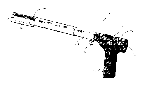
laparoscopic morcellator denoted generally by the reference numeral 10.
Morcellator 10
includes three distinct aspects: handle 14, sheath 12 containing hollow lumen
13 (Fig. 12),
and mouth 20 containing drive shafts 26 with interlocking teeth 24 attached
thereto and
disposed therearound (Fig. 15).
Handle 14 is typically cylindrical and adapted to conform to a user's hand,
though any
structure or conformation is contemplated. Grooves 16 may be disposed on
handle 14 to
provide an ergonomic design. Other types of ridges, grooves, finger
indentations, and the like
17
CA 02900017 2015-07-31
WO 2014/123571
PCT/US2013/050085
are contemplated by the current invention to enhance the comfort of the user
and/or function
of the overall structure.
Handle 14 may be connected to mouth 20 through auger 32 or other cylinder
(e.g., cylinder
15) disposed within the hollow lumen of sheath 12. In an embodiment, handle 14
can be
hollow, as indicated in Figs. 13 and 14, to facilitate this connection to
mouth 20. Thus, handle
14 may be rotatable about sheath 12 and can be detachable from sheath 12 to
allow for other
types of handles to be connected. If handle 14 is connected to mouth 20, then
rotation of
handle 14 would rotate mouth 20 to allow mouth 20 to have flexibility to
contact various
surrounding tissues within the body. Alternatively, handle 14 may include
blade operating
handle or tool (not shown), the rotation of which can rotate mouth 20 or teeth
24 of mouth 20.
Handle guard 18 can be positioned between sheath 12 and handle 14 to protect
the user's
hand and to prevent morcellator 10 from being fully inserted into the body of
the subject.
Sheath 12 is elongate and includes hollow lumen 13, as indicated in Fig. 12,
disposed along
the longitudinal extent of the interior of sheath 12. Sheath 12 further
includes a proximal end
and a distal end, where the proximal end is attached to handle 14 extending
therefrom and
the distal end is in communication with mouth 20 extending therefrom. The
distal end of
sheath 12 telescopically receives mouth 20, such that mouth 20 can retract
within hollow
lumen 13 of sheath 12 and can rotate inside and outside hollow lumen 13 of
sheath 12.
Sheath 12 further includes supplementary channel 28 that has an exit point on
the proximal
end of sheath 12 and an exit point on the distal end of sheath 12.
Supplementary channel 28
has a hollow interior running along the length of sheath 12. Channel doors 29
may be
included on the proximal end of sheath 12 and/or on the distal end of sheath
12. Channel
doors 29 are used to close off either or both exit points of supplementary
channel 28, so the
interior of the body of the subject is not in open communication with the
external environment
through channel 28. For example, this is done so that extraneous materials
cannot
erroneously fall into the body of the subject from the external environment
through channel
28. When open, supplementary channel 28 can be used for a variety of reasons.
For
example, a laparoscopic camera can be inserted through channel 28, so that the
surgical
team can view the procedure without the need to create an additional
laparoscopic port in the
subject's body.
As depicted in Fig. 17, auger 32 includes elongate axle 36 and helicoid 38
disposed
therearound along the length of elongate axle 36, thereby resembling the
threads of a wood
screw, though the current invention contemplates round or abnormally shaped
(e.g.,
hexagonal) augers. Auger 32 is positioned within hollow lumen 13 of sheath 12
along the
18
CA 02900017 2015-07-31
WO 2014/123571
PCT/US2013/050085
length of sheath 12. Auger 32 further extends into the substantially hollow
interior of mouth 20
and is positioned in internal relation to teeth 24 and drive shafts 26
relative to mouth casing
21, such that auger 32 is enclosed within sheath 12 and is nearly enclosed
within mouth 20
but is in communication with the external environment through the spaces among
teeth 24.
Optionally, auger 32 may further extend into the hollow interior of handle 14,
dependent on
the type of handle that is used with morcellator 10.
Auger 32 is structured to rotate about the longitudinal axis within hollow
lumen 13 and the
interior of mouth 20. When tissue or other material comes into contact with
auger 32 during
rotation, the material can be further morcellated by helicoid 38, drawn up
lumen 13 and
transported along the longitudinal extent of sheath 12. The extent of
morcellation and speed
of transportation can be dependent on the rotational force of auger 32 and the
preset pitch of
the ribs of helicoid 38. A sharper pitch would encourage a larger distance of
transportation of
a tissue mass per turn of auger 32. A flatter pitch would encourage a smaller
distance of
transportation of the tissue mass per turn of auger 32.
The proximal end of auger 32 may be coupled to handle 14 such that manual or
automated
rotation of handle 14 would proportionately rotate auger 32. Alternatively,
the proximal end of
auger 32 may be electrically coupled to a drive shaft and motor (not shown)
for automated
activation, deactivation, and control of the rotation of auger 32. Auger 32
drives morcellated
tissue toward the exterior of the body (e.g., toward handle 14) and out of the
body of the
subject. Auger 32 may also be rotated in the opposite direction, such that the
tissue mass can
be directed toward mouth 20.
As depicted in Figs. 10,11, and 15, mouth 20 is disposed on and extends from
the distal end
of sheath 12. Mouth 20 includes mouth casing 21, teeth 24, drive shafts 26,
and window 25,
and contains the proximal end of auger 32. Mouth casing 21 is formed of a
protective material
and protects the substantially hollow interior of mouth 20 on all sides other
than window 25
(Fig. 10). Window 25 leads from the external environment into the
substantially hollow interior
of mouth 20, such that the interior is in communication with the external
environment via
window 25. Auger 32 extends longitudinally from interior 13 of sheath 12 into
the substantially
hollow interior of mouth 20 at the distal end of morcellator 10.
Teeth 24 are disposed within window 25 of mouth 20 and may even protrude from
window 25
to catch additional tissue masses within the body. As indicated in Figs. 6 and
15, there can be
two sets of opposing jaws 27a, 27b, each comprising drive shaft 26 and teeth
24. Teeth 24 of
jaw 27a lie opposed to teeth 24 of jaw 27b.
19
CA 02900017 2015-07-31
WO 2014/123571
PCT/US2013/050085
Teeth 24 of jaws 27a interlock, or otherwise window, teeth 24 of jaw 27b, as
clearly depicted
in Fig. 15, such that teeth 24 that are adjacent to one another along a single
jaw do not
overlap each other. Further, teeth 24 of jaw 27a does not overlap with teeth
24 of jaw 27b
since the teeth window each other. In other words, teeth 24 of jaw 27a are
arranged in a
staggered fashion with teeth 24 of jaw 27b. This allows tissue mass to be
captured and
torn/shredded, while still directing tissue mass into the interior of mouth 20
and ultimately to
auger 32 for exiting morcellator 32.
Teeth 24 are separately mounted securely around corresponding drive shaft 26.
Thus, when
a motor and transmission gears (not shown) are activated, drive shafts 26
rotate
simultaneously (typically counter-rotate), thus also rotating teeth 24 in a
direction suitable for
grasping and cutting tissue mass and pulling tissue mass into the interior of
mouth 20 to
auger 32. In certain embodiments, teeth 24 may be serrated or include a hook
aspect, such
that when teeth 24 rotate, tissue mass can be pinched and cut off. An example
of a hook that
may be used in conjunction with teeth 24 is indicated in Fig. 6 by the
reference numeral 23.
Hooks 23 can draw tissue into mouth 20 for cutting by teeth 24.
In an embodiment, each jaw 27a,27b may be made up of a single piece of
material for ease
of manufacture. For example, each of jaws 27a,27b may resemble a drill bit,
where teeth 24
resemble the cutting edge/lands/margins of the drill bit with flute space
disposed
therebetween. The same principles would apply with teeth 24 of jaws 27a,27b
being
staggered.
As can be seen in Fig. 15, auger 32 lies beneath jaws 27a,27b and is fully
enclosed within
mouth 20 at the distal end of morcellator 20. Auger 32 extends proximally
through sheath 12
and is enclosed within sheath 12. Auger support 33 can be positioned at the
proximal end of
auger 32 to hold auger 32 in place concentrically.
Jaws 27a,27b each extend adjacent to auger 32 proximally along interior 13 of
the extent of
sheath 12. Jaw 27a exits through port 27a' and jaw 27b exits through port
27b', as seen in
Figs. 5 and 12, to a system of motors and gears (not shown) that drive the
rotation of jaws
27a,27b. Though it is shown that jaws 27a,27b and auger 32 are straight and
parallel along
sheath 12 until each exits handle 14 or handle guard 18, it is contemplated
that jaws 27a,27b
and auger 32 can exit morcellator 10 perpendicularly or otherwise at an angle
if structurally
and functionally feasible and more beneficial. For example, this may occur if
the motor shaft
and gears (not shown) require space within morcellator 10. Further, a
collection structure (not
shown) may be placed at the proximal end of morcellator 10, such that tissue
mass directed
by auger 32 toward the distal end of morcellator 10 can fall into and be
collected by the
CA 02900017 2015-07-31
WO 2014/123571
PCT/US2013/050085
collection structure. In this case, only jaws 27a,27b would have an angled
extent and exit
point out of morcellator 10.
Mouth 20 may be beveled when extending from sheath 12 with window 25 being
angled
relative to the line of axis of sheath 12, as indicated in Figs. 1, 3, 4, 8,
and 11. The angle
provides greater surface area for teeth 24 to contact the tissue mass. It is
contemplated that
other embodiments of the current invention may include mouths that are not
angled but rather
lie along the same line of axis as sheath 12.
Mouth 20 may terminate distally in safety tip 30, as seen in Figs. 3, 6, and
8. Safety tip 30 is
used to protect tissue masses in the body from morcellator 10. A user of
morcellator 10 can
push extraneous tissue out of the way of morcellator 10 prior to cutting the
targeted tissue
mass. Thus, the extraneous tissue can be protected from harm by safety tip 30.
As seen in Fig. 6, at the distal end of safety tip 30, fastening mechanisms 31
may be utilized
to affix mouth 20 to sheath 12. Other conventional fastening mechanisms are
contemplated
by the current invention as well.
Conversely, on the proximal end of sheath 12, screw ports 39 may be utilized
to fasten handle
14, handle guard 18, and/or machinery (e.g., motors, gears, etc.) (not shown)
to sheath 12.
Other conventional fastening mechanisms are contemplated by the current
invention as well.
In practice, mouth 20 and a portion of sheath 12 of morcellator 10 is inserted
into the body via
a vagina or a laparoscopic port. Extraneous tissue can be pushed or
manipulated by safety tip
to facilitate targeting of the target tissue mass by the user. Morcellator 10
can then be
25 activated, thus rotating drive jaws 27a,27b (i.e., each drive shaft 26
and each corresponding
set of teeth 24). As a tissue mass is mounted on or otherwise enters window 25
of mouth 20,
teeth 24 grasp the tissue mass and cut the tissue mass, thus pulling the
tissue mass inward
toward auger 32. Thereafter, auger 32 directs the tissue mass proximally up
sheath 12 and
out of the body.
30 As depicted in Fig. 18, morcellator 12 may include cup assembly 40 for
guiding or funneling
tissue mass within the subject's body toward mouth 20 and placing the mass on
mouth 20 for
morcellation. The bottom closed edge of cup assembly 40 can line the edge of
mouth casing
21 that forms the frame of window 25 leading into the interior of mouth 20, as
can be seen at
reference point 41 in Fig. 18. Thus, during insertion into the body, cup
assembly 40 can be
compressed and folded against the edge of mouth casing 21, such that the body
of cup
assembly 40 lies on top of mouth 20, thereby protecting the internal tissue
masses from teeth
21
CA 02900017 2015-07-31
WO 2014/123571
PCT/US2013/050085
24. Subsequently, once mouth 20 is inserted into the body, cup assembly 40 can
be
expanded to guide a tissue mass into mouth 20.
To provide stability in the expanded position of cup assembly 40, as seen in
Fig. 18, the open
edge of cup assembly 40 can be lined with wire frame 44. Thus, in a compressed
position,
wire frame 44 would be folded inwardly and proximally to substantially cover
mouth 20 and
teeth 24. Upon insertion, wire frame 44 can be pushed into an expanded, open,
and stable
position, as indicated in Fig. 18.
A tissue mass can be placed in cup assembly 40 utilizing a laparoscopic
grasper positioned
through the subject's abdominal cavity or even possibly a grasper inserted
through
supplementary channel 28. Thus, cup assembly 40 holds the tissue mass in place
as teeth 24
morcellates the mass and auger 32 directs the mass toward the exterior of the
body. This
prevents any tissue from remaining within the subject's body during or after
the surgical
procedure. Alternatively, cup assembly 40 can be utilized to scoop a tissue
mass within the
subject's body and stably guide the tissue mass to mouth 20 for morcellation
by teeth 24 and
subsequent removal via auger 32.
Cup assembly 40 typically is formed of a flexible material that facilitates
the folding or
manipulation of cup assembly 40 within mouth 20 or sheath 12 for subsequent
deployment
once inside the subject's body.
In an embodiment, as depicted in Figs. 5, 6, and 12, cup assembly 40 can be
folded or
otherwise compressed within supplementary channel 28 during insertion of
morcellator 10
into the subject's body. Alternatively, wire frame 44 may be pulled into
supplementary channel
28 to ensure the cup assembly 40 is stretched taut over mouth 20 to maximize
protection
from teeth 24 and to minimize the size of morcellator 10 entering into the
body.
As can be seen in Fig. 16, deploy apparatus 34 can be used to push or
otherwise deploy cup
assembly 40 from supplementary channel 28 (i.e., compressed position) into the
expanded
position seen in Fig. 18. Deploy apparatus 34 can include grip portion 35 and
shank portion
37. The distal end of shank portion 37 may or may not be attached to the
proximal end of wire
frame 44 of cup assembly 40. Thus, it is contemplated that deploy apparatus 34
can be
pushed distally to expand cup assembly 40 via expansion of wire frame 44, thus
exposing
teeth 24, and can be pulled proximally to compress cup assembly 40 within
supplementary
channel 28, thus covering teeth 24. During expansion and compression, the
bottom edge of
cup assembly 40 would remain attached to the outer edge of mouth casing 21.
Example 2
22
CA 02900017 2015-07-31
WO 2014/123571
PCT/US2013/050085
Referring to Fig. 20, morcellator 100 includes handle 114, hand guard 118,
hollow sheath
112, and auger 132 for cutting and transportation of tissue masses. This
embodiment of the
invention is fabricated with mouth 120 having a longitudinal axis parallel to
that of sheath 112,
rather than being beveled or otherwise angled as in mouth 20 of Example 1 seen
in Fig. 4.
Example 3
Referring to Figs. 21-22, this embodiment of the invention is adapted for the
vaginal port and
referred to by the reference numeral 212. This embodiment is similar to the
one depicted in
Figs. 1-15, but further includes outer sheath 212 slidably positioned in outer
relation to the
inner sheath (not shown) disposed therewithin. As seen in Fig. 22, outer
sheath 212 has a
diameter larger than the inner sheath and is slidably disposed so as to slide
over mouth 220
to safely cover rotating blades 224 and auger 232. The outer diameter of
morcellator 200 with
outer sheath 212 can be about 30 mm, and the diameter of inner sheath (not
shown) can be
about 22 mm. This embodiment would not include a cup assembly that covers the
cutting
blades, such as that seen in Fig. 18.
Outer sheath 212 is retractable and safely houses cutting blades 224 and auger
232 and can
be used as an adjustable blade barrier to allow an increase or decrease in
surface area of the
amount of blades 224 exposed.
Methodologically, the sheath of the morcellator is inserted into the vaginal
port. Once the
proximal end of the sheath is inserted, the cutting blades are engaged in a
rotational motion,
and tissue contact may begin on the opening side of the sheath. Once cut, the
cutting auger
transports the tissue down the sheath to the distal end where it can be
removed. While the
blades and auger are in motion, the surgeon can adjust how much tissue that
needs to be cut
by moving the retractable sheath, thereby permitting an increase or decrease
in the surface
area on the exposed blades. This design reduces surgery time, while reducing
surgical
fatigue of the surgeon.
Example 4
Figs. 23a-28b depict an alternative embodiment of the current invention. The
morcellator of
this embodiment is generally denoted by the reference numeral 300. Fig. 23a
depicts the
exterior aspects of morcellator 300. These exterior aspects include elongate
sheath 312,
handle 314, proximal body 316, mouth 320, and shoot 318. Morcellator 300 may
further
include optional bay sliding sheath assembly 317 that can slide over top mouth
320.
23
CA 02900017 2015-07-31
WO 2014/123571
PCT/US2013/050085
In this embodiment, handle 314 resembles a pistol grip and is positioned
substantially
perpendicular to the longitudinal axis of sheath 312. Proximal body 316
connects handle 314
to the proximal end of sheath 312.
Sheath 312 is straight and is intended to be inserted into a laparoscopic port
or vagina of a
subject. Mouth 320 extends from the distal end of sheath 312 and is concentric
with sheath
312.
Shoot 318 is located at the proximal end of sheath 312 and can be positioned
perpendicular
to the axis of sheath 312. Shoot 318 acts as an exit point for the tissue mass
when the tissue
mass has been excised and traveled proximally through sheath 312. Upon exiting
through
shoot 318, the tissue mass can be collected or discarded.
Apertures 323a,323b on top of and at the rear of proximal body 316,
respectively, are
depicted for purposes of assembling morcellator 300.
Figs. 23b-23d are various angled wireframe views of morcellator 300 that
depict the interior
aspects of morcellator 300. These interior aspects include jaws 327a,327b, and
further
include auger 332, gear assembly 322, and motor unit 324. Each jaw 327a,327b
is formed of
straight or tapered shank 329 enclosed within sheath 312 and shredding body
334 enclosed
within mouth 320 in communication with external environment via window 325.
Each jaw
327a,327b may be formed of a single material and extend proximally in a
straight manner
along the length of sheath 312 and into mouth 320. As seen in Fig. 25c,
shredding body 334
can be formed of a series of lands 336 (lands 336 of each jaw 327a,327b may
interlock as
described in Example 1) and flutes 338 that can cut tissue mass, though other
forms of
cutting or shredding teeth are contemplated by the current invention.
The proximal end of sheath 312 is coupled to gear assembly 322, which will
become clearer
as this specification continues.
Auger 332 has a substantially similar length as each jaw 327a,327b and is
disposed beneath
jaws 327a,327b and within mouth 320. Alternatively, auger 332 may have a
length different
from jaws 327a,327b, as seen in Fig. 24, but the distal end of jaws 327a,327b
and auger 332
are similarly positioned within morcellator 300. Further, the term "beneath"
is used relative to
morcellator 300 being in an upright position, as seen in Fig. 23d, though
morcellator 300 can
be used in any position and at any suitable angle necessary for the surgical
procedure.
Further, auger 332 is substantially centered between jaws 327a,327b when
positioned
beneath jaws 327a,327b. Thus, when tissue mass is grasped and cut by shredding
body 334
of each jaw 327a,327b, the tissue mass is drawn between jaws 327a,327b and
into auger
24
CA 02900017 2015-07-31
WO 2014/123571
PCT/US2013/050085
332. At this point, auger 332 rotates to guide the tissue mass proximally for
exiting the body
and morcellator 300.
The proximal end of auger 332 is coupled to gear assembly 322 as well, which
will become
clearer as this specification continues.
As indicated, the proximal ends of jaws 327a,327b and auger 332 are coupled to
the distal
side of gear assembly 322. The proximal side of gear assembly 322 is coupled
to and is in
mechanical communication with motor unit 324, which may include a battery or
trigger
mechanism (not shown). The battery and/or trigger mechanisms can be
encapsulated by
motor unit 324, can be an additional structure in handle 314, or can be a
standalone box in
electrical communication with motor unit 324.
Fig. 24 depicts several of the external and internal aspects or parts utilized
in this
embodiment of the current invention. These aspects or parts include, but are
not limited to,
handle 314, proximal body 316, rotating disc 346 with keyed channel 348,
driving spur gear
350 attached to the distal face of keyed shaft 352, driven spur gear 354,
conjointly-rotating
driving spur gear 356, driven spur gear 358, sheath 312, jaws 327a,327b, auger
332, and
mouth 320. Each of these aspects are described herein.
Figs. 25a-25c depict mouth 320 and the distal end of sheath 312. As indicated
particularly in
Fig. 25b, the current invention contemplates that sheath 312 is not hollow;
rather, a plurality of
channels can be disposed along the extent of sheath 312, two (2) channels
utilized for jaws
327a,327b and one (1) channel utilized for auger 332. Jaws 327a,327b exit the
distal end of
sheath 312 through ports 327a' and 327b', respectively, and auger 332 exits
the distal end of
sheath 312 through port 333. Once exiting sheath 312, jaws 327a,327b and auger
332 are
contained within mouth 320. This aspect can further be seen in the exploded
view of Fig. 26,
as jaws 327a,327b and auger 332 enter their respective channels on the
proximal end of
sheath 312.
Referring to Fig. 25a, mouth 320 may further include safety tip 330 to protect
extraneous
tissue mass from morcellator 300. Safety tip 330 can be flat, as seen in Fig.
25a, or rounded,
thus allowing the user to push or manipulate extraneous tissue when attempting
to reach the
targeted tissue mass.
As indicated in Fig. 25b, shredding body 334 of each jaw 327a,327b is
contained within
mouth 320 and possibly a portion of sheath 312, though it is contemplated that
a majority of
each jaw 327a,327b enclosed by sheath 312 is straight or tapered shank 329.
CA 02900017 2015-07-31
WO 2014/123571
PCT/US2013/050085
Fastening mechanisms 331 may be utilized on the distal end of mouth 320 to
affix mouth 320
to sheath 312. Other conventional fastening mechanisms are contemplated by the
current
invention as well.
Mouth casing 321 forms the protective outer layer of mouth 320 on all sides
other than
window 325, as directed in Figs. 25a and 26. Window 325 allows open
communication
between the hollow interior of mouth 320 and the external environment, though
the current
invention contemplates various mechanisms of covering window 325. Examples of
these
mechanisms will become clear as this specification continues.
Fig. 25c shows more clearly the positioning of auger 332 relative to jaws
327a,327b. Auger
332 is positioned beneath jaws 327a,327b and substantially centered between
jaws
327a,327b.
Fig. 27 is an exploded view of the proximal end of morcellator 10. Rotating
disc 346 with
keyed channel 348 is positioned within proximal body 316 above perpendicular
handle 314,
which encloses at least a portion of the motor unit (not shown in this
figure). Rotating disc
structure 346,348 is coupled to the motor unit, which, when activated, rotates
rotating disc
structure 346,348. Keyed shaft 352 is keyed into channel 348 of rotating disc
structure
346,348. Thus, when rotating disc structure 346,348 rotates, keyed shaft 352
rotates as well.
Driving spur gear 350 is securely attached to the distal end of keyed shaft
352, such that
when keyed shaft 352 rotates, driving spur gear 350 rotates as well. Spur
gears 354,356,358
are meshably engaged with driving spur gear 350 and/or with each other in a
manner that
when rotating disc structure 346,348 rotates, gears 354,356,358 will rotate as
well. The
purpose of this arrangement is so that jaws 327a,327b and auger 332 will
rotate
simultaneously. The proximal end of auger 332 is securely attached to driving
spur gear 350
in a conventional manner. Jaws 327a,327b are securely attached to conjointly
rotating driving
spur gear 356 and driven spur gear 358, respectively, in a conventional
manner. Considering
the functionality desired of simultaneous rotation of jaws 327a,327b and auger
332, any
suitable arrangement of gears in gear assembly 322 is envisioned.
In an embodiment, as depicted in Figs. 28a and 28b, gear assembly 322 includes
rotating
disc 346 with keyed channel 348, keyed shaft 352, driving spur gear 350,
driven spur gear
354, conjointly-rotating driving spur gear 356, and driven spur gear 358.
Keyed shaft 352
aligns with and is inserted through keyed channel 348, such that rotation of
rotating disc 346
rotates keyed shaft 352 as well. Driving spur gear 350 is securely attached to
the distal end of
keyed shaft 352. As seen in Fig. 28a, the proximal end of keyed shaft 352 is
tool-engageable.
26
CA 02900017 2015-07-31
WO 2014/123571
PCT/US2013/050085
Thus, keyed shaft 352 can be rotated manually or electrically through aperture
323b, if
desired or needed.
Still referring to Figs. 28a and 28b, driving spur gear 350 is meshably
engaged with driven
spur gear 354. Conjointly-rotating driving spur gear 356 is coupled to driven
spur gear 354 in
spaced concentric relation to driven spur gear 354 and distal to driven spur
gear 354.
Conjointly-rotating driving spur gear 356 is meshably engaged with driven spur
gear 358.
Thus, although conjointly-rotating spur gear 356 is driven by rotation of
driving spur gear 350,
conjointly-rotating spur gear 356 becomes a driving gear by actuating the
rotation of driven
spur gear 358.
The proximal end of auger 332 is securely coupled to driving spur gear 350.
The proximal end
of jaw 327b is securely coupled to conjointly-rotating driving spur gear 356.
The proximal end
of jaw 327a is securely coupled to driven spur gear 358.
In an alternative embodiment, a sun and planet gear system may be utilized,
thus eliminating
conjointly-rotating driving spur gear 356 by meshably engaging driven spur
gear 358 with
driving spur gear 350. While the ultimate function of simultaneous rotation of
jaws 327a,327b
and auger 332 should be achieved, the gear assembly used can be altered,
depending on the
needs of the user.
As indicated in Fig. 28a, motor unit 324 is positioned within handle 314
perpendicular to gear
assembly 322. Motor unit 324 is electrically coupled to gear assembly 322 via
power takeoff
shaft 326. Motor unit 324 is capable of activating, deactivating, and
controlling the rotation of
gear assembly 322. Any conventional methodology of controlling the rotation of
rotating disc
346 and/or gear assembly 322 is envisioned by the current invention. For
example, a worm
gear (not shown) may be utilized, but other suitable mechanisms are available
as well.
Methodologically, motor unit 324 is activated to initiate rotation of rotating
disc 346, which
rotates keyed shaft 352. Rotation of keyed shaft 352 rotates driving spur gear
350, which in
turn rotates auger 332, driven spur gear 354, and conjointly-rotating driving
spur gear 356.
Rotation of conjointly-rotating driving spur gear 356 rotates jaw 327b and
driven spur gear
358. Rotation of driven spur gear 358 rotates jaw 327a. Thus, activation of
motor unit 324
results in simultaneous rotation of auger 332, jaw 327b, and jaw 327a.
The current invention contemplates a variety of mechanisms that can cover or
fill window 325
while morcellator 300 is not in use or during insertion of morcellator 300
into the subject's
body. Window 325 can remain covered or filled until just prior to cutting by
shredding body
334 of each jaw 327a,327b. At this point, the cover can be removed to expose
auger 332 and
27
CA 02900017 2015-07-31
WO 2014/123571
PCT/US2013/050085
shredding body 334 of each jaw 327a,327b to the internal tissue masses.
Various non-limiting
mechanisms for covering window 325 are provided in Examples 5-8.
Example 5
Figs. 29a and 29b illustrate an optional addition to morcellator 300, said
addition being cup
assembly 340 with supplementary channel 328 extending along the extent of
sheath 312 for
enclosing and deploying cup assembly 340. As seen more clearly in Fig. 29b,
the bottom
edge of cup assembly 340 is attached to mouth 320, for example by being
attached to mouth
casing 321 or the outer edges of window 325. In particular, this can be seen
in Fig. 29b at
reference numeral 342.
Cup assembly 340 has a compressed position and an expanded position. In a
compressed
position, cup assembly 340 would be enclosed within supplementary channel 328,
though cup
assembly 340 would provide a coverage over window 325 in the compressed
position
because of the attachment of the bottom edge of cup assembly 340 to mouth 320.
Prior to
excising of tissue mass, cup assembly 340 can be deployed into the expanded
position seen
in Figs. 29a and 29b. Cup assembly 340 can scoop or otherwise direct tissue
mass toward
mouth 320 for morcellation by shredding body 334 and transportation (and
further
morcellation) by auger 332.
Cup assembly 340 includes wire frame 344 along its top edge when in its
expanded position.
Wire frame 344 stabilizes cup assembly 340 in an upright position.
Example 6
Figs. 30a-30c depict a mechanism for covering window 325 using bay doors 360.
Fig. 30a
shows mouth 320 when bay doors 360 are closed, whereas Figs. 30b and 30c show
mouth
320 when bay doors 360 are open in the direction of the arrows to expose
shredding body
334 of each jaw 327a,327b. Bay doors 360 may be a single door (not shown) or
multiple
figures as seen in Figs. 30a-30c. Further, bay doors 360 can open in any
direction, for
example transversely (not shown) or longitudinally as seen in Figs. 30a-30c. A
triggering
mechanism (not shown) would be included in proximity to handle 314 or proximal
body 316
for opening and closing bay doors 360.
Methodologically, morcellator 300 would be inserted into the subject's body
with bay doors
360 closed, as in Fig. 30a. Subsequently, prior to contacting the targeted
tissue mass, bay
doors 360 can be opened using triggering mechanism (not shown), thus exposing
the tissue
mass to the shredding body of each jaw 327a,327b for morcellation.
28
CA 02900017 2015-07-31
WO 2014/123571
PCT/US2013/050085
Example 7
Figs. 31a-31c depict a mechanism for covering window 325 using bay rotating
outer sheath
362. Bay rotating outer sheath 362 has a cutout in the shape of window 325.
Fig. 31a shows
mouth 320 when bay rotating outer sheath 362 is rotated to completely cover
window 325.
Fig. 31b depicts rotation of bay rotating outer sheath 362 in the direction of
the arrow to
expose half of window 325. Fig. 31c depicts window 325 when bay rotating outer
sheath 362
has been fully rotated in the direction of the arrow such that the cut out of
outer sheath 362 is
aligned with window 325, thus fully exposing window 325. Bay rotating outer
sheath 362 may
have any suitable length that facilitates its rotation. This rotation may be
achieved manually if
the length extends to proximal body 316 (i.e., outside of the body), or it may
be done
automatically upon activating a trigger mechanism (not shown) in a
conventional manner.
Methodologically, morcellator 300 would be inserted into the subject's body
with bay rotating
outer sheath 362 rotated to close off window 325, as in Fig. 31a.
Subsequently, prior to
contacting the targeted tissue mass, bay rotating outer sheath 362 can be
rotated to expose
as much of shredding body as desired by the user. For example, if the user
desires to
morcellate only a relatively small piece of tissue, the user may desire to
open only half of
window 325, as in Fig. 31b. Alternatively, the user may desire to expose all
of shredding body
by rotating the cutout of bay rotating outer sheath 362 into alignment with
window 325.
Example 8
Figs. 32a-32c depict a mechanism for covering window 325 using bay sliding
outer sheath
364. Bay sliding outer sheath 364 is an outer sheath that typically has no
cutouts but slides
distally to cover window 325 and slides proximally to expose shredding body.
Fig. 32a shows
mouth 320 when bay sliding outer sheath 364 has slid distally to fully cover
window 325. Fig.
32b depicts mouth 320 when bay sliding outer sheath 364 has slid proximally to
expose half
of window 325. Fig. 32c depicts mouth 320 when bay sliding outer sheath 364
has slid
proximally to the proximal edge of window 325 to fully expose window 325. Bay
sliding outer
sheath 364 may have any suitable length that facilitates proximal-distal
movement. This
movement/sliding may be achieved manually if the length extends to proximal
body 316 (i.e.,
outside of the body), or it may be done automatically upon activating a
trigger mechanism (not
shown) in a conventional manner.
Methodologically, morcellator 300 would be inserted into the subject's body
with bay sliding
outer sheath 364 slid distally to the distal edge of window 325, thus closing
off window 325,
as in Fig. 32a. Subsequently, prior to contacting the targeted tissue mass,
bay sliding outer
sheath 364 can be slid proximally to expose as much of shredding body as
desired by the
29
CA 02900017 2015-07-31
WO 2014/123571
PCT/US2013/050085
user. For example, if the user desires to morcellate only a relatively small
piece of tissue, the
user may desire to open only half of window 325, as in Fig. 32b.
Alternatively, the user may
desire to expose all of shredding body by sliding bay sliding outer sheath 364
fully proximally
beyond the proximal edge of window 325.
Definition of Claim Terms
Control apparatus: This term is used herein to refer to any mechanism of
controlling the
rotation of the jaws and auger. Control can be activation, deactivation,
increasing or
decreasing speed of rotation, etc.
Cover apparatus: This term is used herein to refer to any mechanism of
protecting extraneous
tissue from the shredding blades/teeth, typically by covering or filling the
window of the
mouth. Typically the cover apparatus can be removed or manipulated to expose
the
shredding blades/teeth within the mouth.
Deploy apparatus: This term is used herein to refer to any mechanism of
deploying the cup
assembly into an expanded or open position. The deploy apparatus would be
dependent on
the user's positioning of the cup assembly in compressed position. If the cup
assembly is
compressed by partially enclosing it in the supplementary channel, then the
deploy
mechanism of Fig. 16 can be used. However, the deploy apparatus can be manual
(as in Fig.
16 for example), automatic, or electronic.
Distal: This term is used herein to refer to proximity to the patient or
subject. Thus, the distal
end of a structure is closer to the patient or subject than to the user of the
morcellator.
Extraneous tissue: This term is used herein to refer to any tissue within the
body of the
subject or patient that is not being targeted by the user for morcellation.
Gear assembly: This term is used herein to refer to any configuration of gears
that facilitates
the rotation of the jaws and auger.
Gripping apparatus: This term is used herein to refer to any mechanism of
stabilizing or
spatially controlling the morcellator. This mechanism can be manual, for
example using a
handle, or automated/electronic, for example by attachment to machinery (e.g.,
extension
from a robot).
Hook-like protrusion: This term is used herein to refer to a structural part
of the blades/teeth
that are capable of pinching or grasping tissue during rotation of the jaws.
CA 02900017 2015-07-31
WO 2014/123571
PCT/US2013/050085
Inwardly: This term is used herein to refer to the spatial relationship
between the jaws and
auger relative to the line of path taken by tissue undergoing morcellation.
The tissue is cut by
the blades/teeth and proceeds further toward the interior of the mouth where
the auger is
positioned. Thus, the tissue has an inward line of path prior to the auger
transporting the
tissue proximally toward the gripping mechanism.
Morcellator: This term is used herein to refer to any device used for the
removal of tissue from
a patient or subject during laparoscopic surgery.
Motor unit: This term is used herein to refer to any mechanism of mechanically
powering and
controlling the rotation of the jaws and auger.
Opposing blades: This term is used herein to refer to blades/teeth that face
each other from
their respective jaws if two jaws are present, as may be seen in Figs. 6, 15,
22, 25b, and/or
25c. Blades are designed to cut or shred tissue and may have any structure to
achieve such
function.
Outer edges: This term is used herein to refer to the border of the window of
the mouth, or a
surface of the mouth that is in proximity to the border of the window.
Pistol grip: This term is used herein to refer to the portion of the
morcellator that is held by the
user's hand and orients the user's hand in a forward, vertical orientation, or
perpendicular to
the sheath of the morcellator.
Proximal: This term is used herein to refer to proximity to the user (e.g.,
surgeon, surgical
team) of the morcellator. Thus, the proximal end of a structure is closer to
the user of the
morcellator than to the patient or subject.
Snugly fits: This term is used herein to refer to the disposition of an
elongate structure in a
channel, such that the elongate structure is spatially stabilized (i.e.,
minimal movement
around the channel) but is still capable of rotation without damaging the
walls of the channel.
Staggered, interlocking relation: This term is used herein to refer to the
teeth of each jaw
windowing each other, for example as seen in Fig. 15. This configuration of
teeth may
facilitate the shredding of tissue.
Targeted tissue mass: This term is used herein to refer to the tissue intended
to be excised
and/or removed from the patient or subject.
31
CA 02900017 2015-07-31
WO 2014/123571
PCT/US2013/050085
Underneath: This term is used herein to refer to a spatial relationship of the
auger to the jaws
when the morcellator is in an "upright" position or when the sheath is
horizontally oriented, as
in Fig. 23d for example.
Wire frame: This term is used herein to refer to the top edge of the cup
assembly, where the
wire frame facilitates the expansion of the cup assembly and stabilizes the
cup assembly in
an upright position. The wire frame can be formed of any suitable material,
such as a polymer
or metal. Selection of material for the wire frame may be dependent on the
needs of the user.
For example, a light metal may be desired by the user in order for the wire
frame to be able to
fold into the supplementary channel in its compressed position but still
stabilize the cup
assembly in the expanded position.
The advantages set forth above, and those made apparent from the foregoing
description, are
efficiently attained. Since certain changes may be made in the above
construction without
departing from the scope of the invention, it is intended that all matters
contained in the
foregoing description or shown in the accompanying drawings shall be
interpreted as
illustrative and not in a limiting sense.
It is also to be understood that the following claims are intended to cover
all of the generic
and specific features of the invention herein described, and all statements of
the scope of the
invention that, as a matter of language, might be said to fall therebetween.
32