Note: Descriptions are shown in the official language in which they were submitted.
CA 02901377 2015-08-14
WO 2014/124527
PCT/CA2014/000122
Title: METHODS FOR GENERATING HEPATOCYTES AND CHOLANGIOCYTES FROM
PLURIPOTENT STEM CELLS
Field
[0001] The disclosure relates to methods for producing functional
hepatocytes from human
pluripotent stem cells.
Background
[0002] The ability to produce functional hepatocytes from human
pluripotent stem cells (hPSCs;
including embryonic stem cells; hESCs and induced pluripotent stem cells;
hiPSCs) will provide a
source of hepatocytes for drug metabolism studies and cell-based therapy for
the treatment of liver
diseases. Hepatocytes are of particular importance as they are the cells
responsible for drug
metabolism and thus for the control of xenobiotic elimination from the body".
Given this role and the
fact that individuals can differ in their ability to metabolize a particular
drug4, access to functional
hepatocytes from a representative population sample would have a dramatic
impact on drug
discovery and testing within the pharmaceutical industry. In addition to
providing new platforms for
drug testing, hPSC-derived hepatocytes can offer potential new therapies for
patients with liver
disease. Although liver transplantation provides an effective treatment for
end-stage liver disease, a
shortage of viable donor organs limits the patient population that can be
treated with this appr0ach5-7.
Hepatocyte transplantation and bio-artificial liver devices developed with
hPSC-derived hepatocytes
represent alternative life-saving therapies for patients with specific types
of liver disease. These
applications are, however, dependent on the ability to generate mature
metabolically functional cells
from the hPSCs. Reproducible and efficient generation of such cells has been
challenging to date,
due to the fact that the regulatory pathways that control hepatocyte
maturation are poorly understood.
[0003] Given the potential therapeutic and commercial importance of
functional human
hepatocytes, significant effort has been directed towards optimizing protocols
for the generation of
these cells from hPSCs816. Almost all approaches have attempted to
recapitulate the key stages of
liver development in differentiation cultures, including the induction of
definitive endoderm, the
specification of the endoderm to a hepatic fate, the generation of hepatic
progenitors known as
hepatoblasts and the differentiation of hepatoblasts to mature hepatocytes17.
In most studies,
differentiation is induced in a monolayer format with the sequential addition
of pathway agonists and
antagonists that are known to regulate the early stages of development
including endoderm induction
and hepatic specification. With this strategy, it has been possible to
optimize these early
differentiation steps and generate populations that are highly enriched in
definitive endoderm,
hepatoblasts and immature hepatocytes as defined by expression of markers such
as Hex, alpha-
fetoprotein and albumin17. While these early differentiation steps are
reasonably well established,
conditions that promote the maturation of the hepatocytes for example, to
functional cells as defined
by Phase I and Phase II drug-metabolizing enzyme activities, have not been
described. The
populations produced with the different protocols vary considerably in their
maturation status and in
most cases represent immature hepatocytes.
1
CA 02901377 2015-08-14
WO 2014/124527
PCT/CA2014/000122
Summary
[0004] An aspect
of the disclosure includes a method of producing hepatocyte lineage cells
from an extended nodal agonist treated induced endodermal cell population, the
method comprising:
(a) specifying the extended nodal agonist treated induced endodermal cell
population
to obtain a cell population comprising hepatocyte progenitors by contacting
the extended
nodal agonist treated induced endodermal cell population with specification
media comprising
a FGF agonist and a BMP4 agonist and/or active conjugates and/or fragments
thereof;
(b) inducing maturation, optionally further lineage specification and/or
expansion of
the hepatocyte progenitors of the cell population to obtain a population
comprising hepatocyte
lineage cells such as hepatoblasts, hepatocytes and/or cholangiocytes, the
inducing
maturation step comprising generating aggregates of the cell population.
[0005] In an
embodiment, the hepatocyte and/or cholangiocyte lineage cells are
hepatoblasts.
In an embodiment, the method produces an expanded population of hepatoblasts.
In another
embodiment the hepatocyte lineage cells are mature hepatocytes or the
cholangiocyte lineage cells
are mature cholangiocytes.
[0006] In some
embodiments, the extended nodal agonist treated induced endodermal cell
population is induced from pluripotent stem cells (PSCs) such as embryonic
stem cells (ESCs) or
induced pluripotent stem cells (iPSCs).The pluripotent stem cells are
optionally human ESCs (hESCs)
or human iPSCs (hiPSCs).
[0007] The
extended nodal agonist treated induced endoderm population is, in an
embodiment,
obtained by inducing endoderm cells in embryoid bodies (EBs). In another
embodiment, the extended
nodal agonist treated induced endodermal population is obtaining by inducing
endoderm cells that are
in a monolayer. In each case, the induced endodermal population is cultured in
the presence of a
nodal agonist, for example activin, for an extended period to produce an
extended nodal agonist
treated induced endodermal population.
[0008] In an
embodiment, the extended nodal agonist treated induced endodermal population
comprises at least, 80%, 85%, 90%, 95 CXCR4 + and cKIT + positive cells and/or
at least 70%, 75%,
80% SOX17+ cells.
[0009] In an
embodiment, the specifying step comprises contacting an extended nodal agonist
treated (e.g. activin treated) induced endodermal population with
specification media comprising a
FGF and BMP4. The FGF can for example be bFGF, FGF10, FGF2 or FGF4 or
combinations thereof.
The combinations can for example be added sequentially.
[0010] In an
embodiment, the specifying step comprises first contacting an extended nodal
agonist treated induced endodermal population with specification media
comprising FGF10 and
BMP4 for approximately 40 to 60 hours, optionally approximately 40, 42, 44,
46, 48, 50, 52, 54, 56, 58
or 60 hours and then contacting the extended nodal agonist treated induced
endodermal population
2
CA 02901377 2015-08-14
WO 2014/124527
PCT/CA2014/000122
with specification media comprising bFGF and BMP4 for about 4 to 7 days,
optionally about 4, 5, 6 or
7 days.
[0011] In another embodiment, the aggregates are generated from a cell
population comprising
at least 70%, 80%, 85%, or 90% albumin positive cells. In another embodiment,
the aggregates are
generated after 24, 25, 26, 27, or 28 days in culture.
[0012] In some embodiments, aggregates are generated from a monolayer of
the cell
population comprising hepatocyte and/or cholangiocyte progenitors by enzymatic
treatment and/or
manual dissociation.
[0013] Inducing maturation, and optionally further lineage specification
and/or expansion can
comprise one or more additional steps. In a further embodiment, the cell
population comprising
hepatocyte and/or cholangiocyte progenitors and/or the aggregates are cultured
in the presence of
hepatocyte growth factor (HGF), dexamethasone (DEX) and/or Oncostatin M (OSM)
and/or active
conjugates and/or fragments thereof.
[0014] In one embodiment, inducing maturation, and optionally further
lineage specification
and/or expansion further comprises activating the cAMP pathway within the
cells of the aggregates to
induce the maturation of the hepatocyte and cholangiocyte progenitors into
hepatocytes and/or
cholangiocytes. In another embodiment, activating the cAMP pathway comprises
contacting the
aggregates with cAMP and/or a cAMP analog (e.g. such as 8-bromoadensoine-3'5"-
cyclic
monophosphate, dibutyryl-cAMP, Adenosine- 3', 5'-cyclic monophosphorothioate,
Sp- isomer (Sp-
cAMPS) and/or 8-Bromoadenosine-3', 5'-cyclic monophosphorothioate, Sp-isomer
(Sp-8-Br-cAMPS))
and/or any other cAMP agonist.
[0015] For example, in an embodiment, a maturation media comprising a
cAMP agonist and
DEX and optionally HGF is added to the aggregates subsequent to culturing the
pre-aggregate
population in a maturation media comprising HGF, DEX and OSM, for example for
about 10, 11, 12,
13 or 14 days.
[0016] In an embodiment, the population of hepatocytes produced is a
population comprising
functional hepatocytes.
[0017] In embodiments, the hepatocytes, optionally functional
hepatocytes, comprise increased
expression of at least 1, 2, 3, 4, 5, 6, 7, 8, 9, 10 or more genes or protein
selected from the group
consisting of ALB, CPS1, G6P, TDO, CYP2C9, CYP2D6, CYP7A1, CYP3A7, CYP1A2,
CYP3A4,
CYP2B6, NAT2 and UGT1A1 compared to a cell population comprising hepatocyte
and/or
cholangiocyte progenitors, and/or hepatocytes produced from a non-extended
nodal agonist treated
induced endodermal cell population (e.g. from an induced endodermal population
that was not treated
with a nodal agonist such as activin for an extended period of time), produced
without aggregation
and/or cAMP signaling induction. In other embodiments, at least 40%, 50%, 60%,
70%, 80% or 90%
of the hepaiocytes, optionally functional hepatocytes, are ASGPR-1+ cells.
3
CA 02901377 2015-08-14
WO 2014/124527
PCT/CA2014/000122
[0018] In an embodiment, cholangiocyte fate is specified by treating
aggregates of the cell
population with a notch agonist.
[0019] In an
embodiment, the population of cholangiocytes produced is a population of
functional cholangiocytes. The functional cholangiocytes comprise for example
increased expression
of at least 1, at least 2 or 3 genes or proteins selected from Sox9, CK19 and
CFTR (Cystic fibrosis
transmembrane conductance regulator) compared to the cells of the cell
population comprising
hepatocyte and cholangiocyte progenitors and/or compared to a population cells
produced from
aggregates not treated with a notch agonist. In other embodiments, at least
40%, 50%, 60%, 70%,
80% or 90% of the population of cholangiocytes are CK19+ cholangiocytes. In
other embodiments, at
least 40%, 50%, 60%, 70%, 80% or 90% of the functional cholangiocytes are
CFTR+ cholangiocytes.
[0020] As mentioned, the method can be applied to an endodermal cell
population grown in a
monolayer.
[0021]
Accordingly a further aspect includes a method of producing hepatocytes and/or
cholangiocytes from a pluripotent stem cell population, the method comprising:
a) contacting the pluripotent stem cells cultured as a monolayer, with an
induction
media comprising nodal agonist such as ActA and optionally a wnt/beta-catenin
agonist such
as i) Wnt3a and/or ii) a GSK-3 selective inhibitor such as CHIR-99021, to
provide an induced
endodermal cell population;
b) contacting the induced endodermal cell population with a nodal agonist to
provide
an extended nodal agonist treated induced endodermal cell population;
c) specifying the extended nodal agonist treated induced endodermal cell
population
by contacting the extended nodal agonist treated induced endodermal cell
population with a
specification media comprising a FGF agonist and a BMP4 agonist and/or active
conjugates
and/or fragments thereof to obtain a cell population comprising hepatocyte
and/or
cholangiocyte progenitors;
d) optionally contacting the cell population comprising hepatocyte and/or
cholangiocyte progenitors with a maturation media comprising HGF,
dexamethasone and/or
Oncostatin M and/or active conjugates and/or fragments thereof; and
e) inducing maturation, optionally further lineage specification and/or
expansion of
hepatocyte and cholangiocyte progenitors of the cell population into expanded
hepatoblasts,
hepatocytes and/or cholangiocytes, the inducing maturation comprising
generating
aggregates of the cell population.
[0022] Further, the endodermal population can also be comprised in
embryoid bodies.
[0023]
Accordingly, a further aspect of the disclosure provides a method of producing
hepatocytes and/or cholangiocytes from a pluripotent stem cell population, the
method comprising:
4
CA 02901377 2015-08-14
WO 2014/124527
PCT/CA2014/000122
a) forming embryoid bodies (EBs) of the pluripotent stem cells, optionally by
contacting the pluripotent stem cells with a BMP4 agonist;
b) contacting the EBs with an induction media comprising a nodal agonist such
as
ActA and optionally a wnt/beta-catenin agonist such as i) Wnt3a and/or ii) a
GSK-3 selective
inhibitor such as CHIR-99021, to provide an induced endodermal cell
population;
c) dissociating the induced endodermal cell population to provide a
dissociated
induced endodermal cell population;
d) contacting the dissociated induced endodermal cell population with a nodal
agonist
to provide an extended nodal agonist treated induced endodermal cell
population;
e) specifying the extended nodal agonist treated induced endodermal cell
population
by contacting the extended nodal agonist treated induced endodermal cell
population with a
specification media comprising a FOE agonist and a BMP4 agonist and/or active
conjugates
and/or fragments thereof to obtain a cell population comprising hepatocyte
and/or
cholangiocyte progenitors,
f) optionally contacting the cell population comprising hepatocyte and/or
cholangiocyte progenitors with a maturation media comprising HGF,
dexamethasone and/or
Oncostatin M and/or active conjugates and/or fragments thereof; and
g) inducing maturation, further lineage specification and/or expansion of
hepatocyte
and cholangiocyte progenitors of the cell population into hepatocytes and/or
cholangiocytes,
the inducing maturation, further lineage specification and/or expansion
comprising generating
aggregates of the cell population.
[0024] In some
embodiments, the inducing maturation, further lineage specification and/or
expansion step further comprises activating the cAMP pathway within the
aggregates to induce the
maturation of hepatocyte and/or cholangiocyte progenitors of the cell
population into a population
comprising hepatocytes and/or cholangiocytes. In an embodiment, the method
comprises contacting
the aggregates with a cAMP analog and/or cAMP agonist.
[0025] In an
embodiment, the monolayer or EBs are contacted with a nodal agonist in
induction
media for at least about 1 day, 2 days, 3 days or about 4 days.
[0026] In an
embodiment, in a step prior to dissociation of the endodermal population (e.g.
embryoid bodies (EB) stage), the EBs are cultured with a nodal agonist for at
least 36, 38, 42, 44, 46,
48, 50, 52, 56, 58 or 60 hours or for at least about 1 day, 2 days, 3 days or
about 4 days.
[0027]
Accordingly in another aspect of the disclosure relates to a method of
producing
hepatocytes and/or cholangiocytes from pluripotent stem cells (PSCs) such as
embryonic stem cells
(ESCs) or induced pluripotent stem cells (iPSCs), the method comprising:
a) contacting the pluripotent stem cells cultured as a monolayer or formed
into
embryoid bodies, with an induction media comprising nodal agonist such as ActA
and
5
CA 02901377 2015-08-14
WO 2014/124527
PCT/CA2014/000122
optionally a wnt/beta-catenin agonist such as i) Wnt3a and/or ii) a GSK-3
selective inhibitor
such as CHIR-99021 to provide an induced endodermal cell population;
b) contacting the induced endodermal cell population with a nodal agonist to
provide
an extended nodal agonist treated induced endodermal cell population; and
c) specifying the extended nodal agonist treated induced endodermal cell
population
by contacting the extended nodal agonist treated induced endodermal cell
population with a
cspecification media comprising at least one FGF agonist and one BMP4 agonist
and/or
active conjugates and/or fragments thereof to obtain a cell population
comprising
hepatocyte and/or cholangiocyte progenitors, and
d) inducing maturation, further lineage specification and/or expansion of
hepatocyte
and/or cholangiocyte progenitors into hepatocytes and/or cholangiocytes, the
inducing
maturation, further lineage specification and/or expansion comprising:
(i) culturing the cell population comprising hepatocyte and/or cholangiocyte
progenitors with a maturation media comprising HGF, OSM and DEX;
(ii) generating aggregates of the cell population, optionally when the cell
population
comprises at least 70%, 80%, 85%, or 90% albumin positive cells or after about
20
to about 40 days of culture for example after about 24 to about 28 days of
culture;
(iii) culturing the aggregated cells in an aggregated cell maturation media:
and
(iv) activating the cAMP pathway in the aggregated cells, optionally within
about 1
to about 10 days of aggregation, for example within 6 days of aggregation,
optionally after about 27 to about 36 days of culture,.
[0028] In an
embodiment, the aggregated cell maturation media can comprise factors which
promote hepatocyte maturation or factors which promote cholangiocyte
development or both.
[0029] In another
embodiment, aggregated cells are upon aggregation treated with a wnt
agonist such as CIHR 99021, optionally in the presence of a TGFbeta antagonist
such as SB431542.
As demonstrated herein, activation of the Wnt pathway and SMAD pathway at for
example day 26 (or
optionally one or two days later e.g. day 27, in embodiments using EBs),
promotes expansion of an
albumin+/HNF4+ progenitor population. It is demonstrated for example that up
to a 10 fold expansion
of said population can be obtained when a wnt agonist is added.
[0030] In an
embodiment, the aggregated cells are treated with a wnt agonist and optionally
a
TGFbeta antagonist (such as SB431542) for about 6 to about 12 days, preferably
about 8 to about 10
days, optionally for about 9 days.
[0031] In yet a
further embodiment, a Wnt antagonist such as XAV939 (also referred to as XAV
for short) and/or a Mek/Erk antagonist, for example PD0325901 (also referred
to as PD for short) is
added during the cAMP activation step. Addition of a Wnt antagonist and/or a
MEK/Erk antagonist
during activation of cAMP signaling enhances expression of CYP enzymes, for
example up to levels
6
CA 02901377 2015-08-14
WO 2014/124527
PCT/CA2014/000122
or greater than levels seen in adult liver cells. For example, an inhibitor of
MEK/Erk added in the
presence of cAMP, for example, added to about day 28 to about day 32 cultures,
results in
hepatocytes with increased levels of CYP3A4 a. Addition of a MEK/Erk
antagonist in combination with
a Wnt antagonist is shown to also increase levels of CYP1A2. In an embodiment,
the Wnt antagonist
is XAV939. In another embodiment, the MEK/Erk antagonist is PD0325901.
[0032] In an
embodiment, approximately 1 to about 4 days after aggregation, the cells are
treated with a notch agonist. Addition of a notch agonist at such stages
promotes cholangiocyte
maturation. In some embodiments, for example where cholangiocyte maturation is
preferred, inducing
cAMP signaling is omitted.
[0033] Further
inhibiting Notch signaling for example with a Notch antagonist such as gamma-
secretase inhibitor (GSI) L695,458 is demonstrated herein to inhibit
cholangiocyte development and
cells produced retain the characteristics of hepatocytes. In an embodiment,
the method comprises
approximately 1 to about 4 days after aggregation, treating the cells with a
notch antagonist, for
example in embodiments where hepatocyte differentiation is desired.
[0034] In another
embodiment, the method of producing hepatocytes and/or cholangiocytes
from pluripotent stem cells (PSCs), such as embryonic stem cells (ESCs) or
induced pluripotent stem
cells (iPSCs), comprises:
a) contacting the pluripotent stem cells cultured as a monolayer or formed
into
embryoid bodies, with an induction media comprising a nodal agonist such as
ActA
and optionally a wnt/beta-catenin agonist such as i) Wnt3a and/or ii) a GSK-3
selective inhibitor such as CHIR-99021, optionally for about 4 to about 8
days, to
provide an induced endodermal cell population;
b) contacting the induced endodermal cell population with a nodal agonist,
optionally
for about 1, 2, 3, or about 4 days, to provide an extended nodal agonist
treated
induced endodermal cell population;
c) specifying the extended nodal agonist treated induced endodermal cell
population
by contacting the extended nodal agonist treated induced endodermal cell
population
with a specification media comprising at least one FGF agonist and at least
one
BMP4 agonist and/or active conjugates and/or fragments thereof, optionally for
about
4 to about 10 days, to obtain a cell population comprising hepatocyte and/or
cholangiocyte progenitors, and
d) inducing maturation, further lineage specification and/or expansion of the
hepatocyte or cholangiocyte progenitors into hepatocytes and/or
cholangiocytes, the
inducing maturation, further lineage specification and/or expansion
comprising:
(i) culturing the cell population comprising hepatocyte and/or cholangiocyte
progenitors with a maturation media comprising comprising HGF, Dex and/or
OSM, optionally for about 10 to 14 days;
7
CA 02901377 2015-08-14
WO 2014/124527
PCT/CA2014/000122
(ii) generating aggregates of the cell population, optionally when the cell
population comprises at least 70%, 80% 85%, or 90% albumin positive cells
or after about 20 to about 40 days for example after about 24 to about 28
days of culture;
(iii) culturing the aggregates in maturation medium comprising Dex for about
1 to 10 days;
iv) a)
culturing aggreates in a maturation medium comprising
Dex and a cAMP analog and/or cAMP agonist for about 6 days to about 10
days, optionally adding the cAMP analog and/or cAMP agonist within about 1
to about 10 days of the generating aggregates step, for example within 6
days of the generating aggregates step, optionally after about 27 to about 36
days of culture; or
b) culturing the aggregates in a maturation medium comprising a
notch agonist and optionally a cAMP agonist, HGF, and/or EGF for about 6
days to about 20 days, optionally adding the notch agonist within about 1 to
about 10 days of the generating aggregates step, for example within 6 days
of the generating aggregates step, optionally after about 20 to 40 days of
culture.
[0035] In an
embodiment, the method comprises aggregating for example after about 20 days
of culture and/or before 40 days of culture
[0036] The
disclosure also provides a method of inducing maturation, further lineage
specification and/or expansion of cholangiocyte progenitors into
cholangiocytes, the inducing
maturation, further lineage specification and/or expansion comprising:
(i) culturing a cell population comprising cholangiocyte progenitors with a
Notch agonist to induce the maturation of at least one cholangiocyte
progenitor into
a cholangiocyte, optionally a functional cholangiocyte.
[0037] The notch
agonist can for example be any notch ligand bound to a surface such as a
cell, plastic, ECM or bead. In one embodiment, the notch ligand is notch
ligand delta. In one
embodiment, inducing maturation, further lineage specification and/or
expansion comprises
contacting a cell population comprising cholangiocyte progenitors with a notch
signaling donor (e.g.
notch agonist) such as 0P9, OP9delta, and/or 0P9 Jagged1 cells and optionally
in the presence of
EGF, TGFbeta1, HGF and EGF, and/or HGF, TGFbeta1 and EGF for at least or about
5 to about 90
days, to induce the maturation of cholangiocyte progenitors into functional
cholangiocytes.
[0038]
Optionally, contacting a cell population comprising cholangiocyte progenitors
with a
notch agonist (e.g. a notch signaling donor) comprises co-culturing the cell
population comprising
cholangiocyte progenitors with a notch signaling donor such as 0P9, OP9delta,
and/or 0P9 Jagged1
cells and optionally in maturatoin media comprising EGF, TGFbeta1, HGF and
EGF, and/or HGF,
8
TGFbeta1 and EGF, for at least or about 5 to at least or about 90 days,
optionally for at least or about 5 to at
least or about 60 days, at least or about 30 days, at least or about 25 days,
2 at least or about 1 days and/or
at least or about 14 days to induce the maturation of cholangiocyte
progenitors into cholangiocytes, optionally
functional cholangiocytes, optionally wherein the functional cholangiocytes
form branched, cyst, tubular or
sphere type structures.
[0039] In another embodiment, the application provides a method
comprising:
(a) producing a population of cells comprising hepatocytes and/or
cholangiocytes according
to any of the methods described herein; and
(b) introducing the population of cells, or optionally a hepatocyte and/or a
cholangiocyte
enriched or isolated population, into a subject.
[0040] In some embodiments, the method further comprises enriching
or isolating a hepatocyte
and/or cholangiocyte population of cells. Optionally, the hepatocyte and/or
cholangiocyte population of cells
comprises at least 10%, at least 15%, at least 20%, at least 25%, at least
30%, at least 35%, at least 40%, at
least 50%, at least 60%, at least 70%, at least 80% or up to about 95%
hepatocytes and/or cholangiocytes
(e.g. optionally functional hepatocytes and/or cholangiocytes).
[0041] The disclosure also provides the use of the population of
hepatocytes and/or cholangiocytes
for drug discovery, drug metabolism analysis, development of bioartificial
liver devices and/or as cell
replacement therapy for the treatment of liver conditions and disease.
[0042] Other features and advantages of the present disclosure will
become apparent from the
.. following detailed description. It should be understood, however, that the
detailed description and the specific
examples while indicating preferred embodiments of the disclosure are given by
way of illustration only, since
various changes and modifications within the spirit and scope of the
disclosure will become apparent to those
skilled in the art from this detailed description.
[0042a] In another aspect of the invention there is method of
producing hepatocyte and/or
cholangiocyte lineage cells from pluripotent stem cells, the method
comprising: (a) producing an induced
endodermal cell population by either: i) culturing the pluripotent stem cells
as a monolayer in an endoderm
induction medium comprising Activin A and a Wnt/beta-catenin agonist for at
least 3 days, followed by at least
2 days in an endoderm induction medium comprising a FGF agonist selected from
one or more of FGF2,
FGF10, and FGF4 and Activin A; or ii) culturing the pluripotent stem cells
with BMP4 for 1 day to promote
formation embryoid bodies (EBs), and then in an endoderm induction medium
comprising a FGF agonist
selected from one or more of FGF2, FGF10, and FGF4, Activin A, a Wnt/beta-
catenin agonist, and BMP4 for
at least 6 days; (b) contacting the induced endodermal cell population with a
nodal agonist for 1 to 4 days in
a monolayer culture to produce an extended nodal agonist treated induced
endodermal cell population; (c)
producing a hepatoblast population by contacting the extended nodal agonist-
treated induced endodermal
cell population with a specification medium for 4 to 10 days, wherein the
specification medium comprises: i)
a FGF agonist selected from one or more of FGF2, FGF10, and FGF4; and ii) a
BMP4 agonist; (d) inducing
9
Date recue/ date received 2021-12-22
maturation, further lineage specification and/or expansion of the hepatoblasts
population by: i. enzymatically
dissociating the progenitor hepatoblast population; ii. generating aggregates
of the dissociated hepatoblast
population; and iii. culturing the hepatoblast aggregates in a maturation
medium for 1 to 40 days to produce
hepatocyte and/or cholangiocyte lineage cells, wherein the maturation medium
comprises hepatocyte growth
factor (HGF) dexamethasone (DEX) and Oncostatin M (OSM).
Brief description of the drawings
[0043] An embodiment of the present disclosure will now be described
in relation to the drawings
in which:
[0044] Figure 1 shows endoderm induction in hESC-derived embryoid bodies.
Figure 1(a) is a
schematic representation of the differentiation protocol. EBs are trypsinized
at day six and plated as a
monolayer in the presence of activin for two days to generate appropriate-
staged definitive endoderm. The
hepatic lineage is specified from this endoderm population by culture in the
presence of BMP4 and FGF.
Hepatic maturation is induced through a step wise process, first by the
addition of HGF, Dexamethasone
(Dex) and Oncostatin M (OSM) for 12 days followed by the generation of 3D
aggregates that are cultured for
eight days in hepatocyte medium supplemented with Dex and subsequently in this
medium with the addition
of the cAMP analog, 8-br-cAMP for another 12 days (day 32-44). Figure 1(b)
shows flow cytometric analyses
showing the proportion of CXCR4+, CKIT+ (CD117) and EPCAM+ cells in day six
activin- and activin/Wnt3a-
induced populations. Figure 1(c)
9a
Date recue/ date received 2021-12-22
CA 02901377 2015-08-14
WO 2014/124527
PCT/CA2014/000122
shows intracellular flow cytometric analyses showing the proportion of SOX17+
and FOXA2+ cells in
day six activin- and activin/Wnt3a-induced populations. The size of the SOX17+
and FOXA2+
populations was significantly larger in the activinNVnt3a induced EBs (Sox17:
96.2 +/- 1.1%, FoxA2:
88.5 +/- 2.9%) compared to EBs induced with activin alone (Sox17: 91 +/- 1.8%,
FoxA2: 80.3 +/-
2.5%) " P < 0.05 (P = 0.002), "" P < 0.01 (P = 0.005); Student's t-test, n =
4. Figure 1(d) shows RI-
qPCR based analyses of T, SOX17, GSC and FOXA2 expression in activin and
activinNVnt3a-
induced EBs. EBs were analyzed at the indicated time points. Bars represent SD
of the mean of three
independent experiments. Figure 1(e) is a flow cytometric analysis showing the
kinetics of
development of the CXCR4+, CKIT+, SOX17+ and FOXA2+ populations in the
activin/Wnt3a induced
EBs. Figure 1(f) is a flow cytometric analysis showing the proportion of
CXCR4+, CKIT+, EPCAM+,
Sox17+ and FOXA2+ cells in day six EBs induced with activin in with neural
based media.
[0045] Figure 2 (a) RT-qPCR analysis of albumin expression in monolayer
cultures specified
with the indicated cytokines. Cells were treated with the different factors
(bFGF 10 ng/ml; BMP4 50
nen!: HGF 20 ng/ml; or bFGF 20 ng/ml plus BMP4 50 ng/ml) from 6 days to day 12
and then cultured
with DEX, HGF and OSM and analyzed at day 24. Bars represent the standard
deviation (SD) of the
mean of three independent experiments. Values are determined relative to TBP
and presented
relative to expression in bFGF (20 ng/ml) culture, which is set a one. *** P <
0.001 as compared with
the culture treated bFGF. Student's t-test, n = 3. (b) RT-qPCR analysis of
albumin expression in
populations specified in the presence and absence of FGF10. Cultures were
treated (or not) with
FGF10 (50 ng/ml) plus BMP4 (50 ng/ml) between days 6 and 8. At this stage, the
FGF10 was
removed and the cells cultured in bFGF/BMP4 between 8 and 12. Bars represent
the standard
deviation (SD) of the mean of three independent experiments. Values are
determined relative to TBP
and presented relative to expression in FGF10 (-) culture, which is set at
one. * P < 0.05, Student's t-
test, n = 3.
[0046] Figure 3 shows that the duration of activin signaling affects
hepatic development. Figure
3(a) is an intracellular flow cytometric analysis showing the proportion of
SOX17+ and FOXA2+ cells
in day six activin/VVnt3A-induced EBs as well as in monolayer populations
derived from them. The
monolayer populations were cultured either directly in the specification media
(-activin) or for two days
in activin (50 ng/ml) and then in the specification media (+activin).
Populations were analyzed
following two or four days culture in the specification media (total days
eight and 10 for the ¨activin
group and days 10 and 12 for the +activin group). Bars represent standard
deviation (SD) of the mean
of three independent experiments. The proportion of SOX17+ at day 10/12 was
significantly higher in
the activin-treated compared to the non-treated population (73.3 +/- 7.5% vs
45.9 +/- 3.7%). Similarly,
the proportion of FOXA2+ cells at days 8/10 and day 10/12 was significantly
higher in the activin
treated compared to the non-treated population (day 8: 96.1 +/- 0.9% vs 76.5
+/- 10.1%, day 12: 92.7
+/- 2.5% vs 50.2 +/- 6.3%). Figure 3(b) depicts total cell number in activin
treated and non-treated
monolayer cultures. Day six EB-derived cells were cultured directly in hepatic
differentiation media or
in the presence of activin for two days and then in hepatic differentiation
media. Figure 3(c) is a flow
cytometric analysis showing the proportion of CXCR4 and CKIT positive cells in
populations at days 8,
CA 02901377 2015-08-14
WO 2014/124527
PCT/CA2014/000122
10 and 12 culture generated from non- treated cell and activin-treated
endoderm. Figure 3(d) shows
RT-qPCR based expression analyses of hepatic monolayer populations generated
from activin-
treated (Black bars) and non-treated (Grey bars) endoderm. The populations
were analyzed for
expression of the indicated endoderm (HEX, AFP, ALB, and HNF4a) and mesoderm
(MEOX1,
MESP1, CD3/ and CD90) genes. Activin treated populations (grey bars) were
analyzed at days 12,
18 and 26 of total culture, whereas the non-treated population (black bar) was
analyzed at days 10,
16 and 24 of culture. Indicated expression levels are relative to TBP. Bar
represents the standard
deviation (SD) of the mean of three independent experiments. Figure 3(e) is a
flow cytometric analysis
showing the proportion of CD31+ CD90+ and EPCAM+ cells in monolayer
populations derived from
activin treated (day 26) and non-treated (day 24) endoderm. The CD31+ and
CD90+ populations were
significantly larger in non-treated compared to the treated cultures (CD31:
13.6 +/- 2.3% vs 0.49 +/-
0.11%, P < 0.001; CD90: 41.2 +/- 4.7% vs 8.5 +/- 1.19%, P < 0.001, Student's t-
test, n = 3). In
contrast, a higher portion of EPCAM+ cells was detected in the population
derived from the activin-
treated endoderm compared to the population generated from the non-treated
cells (EPCAM: 90.7 +/-
2.7% vs 56.8 +/- 7.3%; P < 0.01, n = 3). Figure 3(f) shows immunostaining
analyses showing the
proportion of albumin positive cells in cultures generated from activin
treated (day 26) and non-treated
(day 24) endoderm. Albumin is visualized with Alexa 488. Scale bars: 200 pm.
(g) Intracellular flow
cytometric analyses indicating the proportion of albumin (ALB) and alpha-
fetoprotein (AFP) cells in
monolayer cultures generated from activin-treated (grey bars; day 26) and non-
treated (black bars;
day 24) endoderm. Bars in figures represent the standard deviation (SD) of the
mean of three
independent experiments. *P < 0.05, P < 0.01, *** P
< 0.001 (Student's t-test; n = 3). AL: adult
liver, FL: fetal liver.
[0047] Figure 4
shows that aggregation promotes hepatic maturation. Figure 4(a) is a phase-
contrast image of hepatic aggregates at day 28 of culture. Scale bar, 200 pm.
Figure 4(b) shows RT-
qPCR based analyses of ALB, CPS1, TAT, G6P and TDO expression in monolayer
(black bar) and
3D aggregate cultures (grey bar) at day 32 of differentiation. Values are
determined relative to TBP
and presented relative to expression in adult liver, which is set a one.
Figure 4(c) is a RT-qPCR based
analysis for CYP7A1, CYP3A7 and CYP3A4 expression at day 32 of differentiation
in monolayer
(black bar) and 3D aggregate culture (grey bar). Expression levels are
relative to TBP. Figure 4(d) is a
flow cytometric analysis showing the proportion of asialo-glycoprotein
receptor-1+ (ASGPR-1) cells in
the monolayer (2D) and aggregate (3D) cultures at day 36. The frequency of
ASGPR-1+ cells was
significantly higher in 3D aggregate cultures (2D: 28.8 +/- 3.1%, 3D: 64.7 +/-
4.26%, P < 0.001, n = 3).
Bars in all graphs represent the standard deviation (SD) of the mean of
samples from three
independent experiments, * P < 0.05, *" P < 0.01, *** P < 0.001, Student's t-
test, AL: adult liver, FL:
fetal liver, PH; primary hepatocytes cultured for two days.
[0048] Figure 5 shows
that CAMP signaling induces maturation of hESC-derived hepatocyte-
like cells. Figure 5(a) is a RT-qPCR analysis of PGC1-a, HNF4a, AFP, ALB, G6P,
and TAT
expression in hepatic aggregates cultured in the presence and absence of 8-Br-
cAMP. Expression
levels are relative to TBP. Figure 5(b) is an intracellular flow cytometric
analysis showing the
11
CA 02901377 2015-08-14
WO 2014/124527
PCT/CA2014/000122
proportion of alpha-fetoprotein (AFP)+ and albumin (ALB) + cells (day 44) in
hepatic aggregates
cultured in the presence and absence of 8-Br-cAMP. The frequency of AFP+ cells
was significantly
lower in the population induced with cAMP compared to the non-induced
population (34.5 +/- 12.4%
vs 56.9 +/- 3.6 %, P < 0.05, mean +/- SD, n = 3). The proportion of ALB
positive cells, on the other
hand, was higher in the cAMP-treated population (89.5 +/- 5.6% vs 82.3 +/-
3.0%, P < 0.05 (mean +/-
SD, n = 3). Figure 5(c) is a RT-qPCR analysis of PGC1-a expression in cAMP
treated pancreatic
aggregates and hepatic aggregates generated from HES2, H9 and 38-2 cells.
Values are determined
relative to TBP and presented as fold change relative to expression in non-
treated cells, which is set
at one. Figure 5(d) shows ICG uptake at day 44 in non-treated and cAMP-treated
aggregates. Bar in
all graphs represent the standard deviation (SD) of the mean of the values
from three independent
experiments. * P < 0.05, *" P < 0.01, *"* P < 0.001, Student's t-test, AL:
adult liver, FL: fetal liver.
[0049] Figure 6
shows that cAMP signaling increases metabolic enzyme activity in hESC-
derived hepatocytes. Figure 6(a) shows expression of CYP3A7, CYP3A4, CYP1A2,
CYP2B6 and
UGT1A1 in hepatic aggregates (day 44) cultured in the presence and absence of
8-Br-cAMP. The
levels in primary hepatocytes (PH) are shown as a control. Values are
determined relative to TBP and
presented as fold change relative to expression in non-treated cells, which is
set at one. Figure 6(b)
shows RT-qPCR analyses showing expression of PGC1a, TAT, HNF4a, CYP1A2 and
CYP3A4 in
untreated (-) and cAMP-treated (+) monolayer populations (day 44). Values are
determined relative to
TBP and presented as fold change relative to expression in non-treated cells,
which is set at one.
Figure 6(c) shows RT-qPCR analyses of CYP1A2 and ALB expression in cAMP
treated aggregates
(day 44) generated from non-treated (-Act) or extended activin treated (+Act)
endoderm. Figure 6(d)
shows RT-qPCR analyses of CYP1A2 expression in aggregates cultured for six
(cAMP +1-) or 12
days in 8-Br-cAMP (cAMP +). Figure 6(e) shows that hESC-derived hepatic cells
display CYP1A2
activity in vitro. Non-treated and cAMP-treated aggregates and primary
hepatocytes were incubated
with phenacetin (200 pM) for 24 hours. Generation of the 0-deethylated
metabolite acetaminophen
from phenacetin was monitored by HPLC. Activity is presented per 10,000 cells.
(* p < 0.05, n = 5).
Figure 6(f) shows that hESC-derived hepatic cells display CYP2B6 activity in
vitro, cAMP-treated
aggregates and primary hepatocytes were incubated with bupropion (1 pM) for 48
hours. Formation of
the metabolite 0-hydroxy-bupropion from bupropion was measured by HPLC.
Activity is presented
per 50,000 cells, (n = 3). Figure 6(g) shows that metabolism of sulfamethazine
(SMZ) to N-acetylated
SMZ indicates the presence of the Phase II enzyme(s) NATI and/or NAT2. cAMP-
treated aggregates
and primary hepatocytes were cultured with SMZ (500 pM) for 48 hr, and N-
acetylated SMZ was
measured by HPLC. Activity is presented per 10,000 cells (n = 3). Figure 6(h)
is an HPLC analysis
showing generation of 4-MU glucuronide (4-MUG) from 4- methylumbelliferone (4-
MU) by the cAMP-
treated aggregates indicative of Total UGT activity, cAMP-treated aggregates
and primary
hepatocytes were cultured with 4-MU for 48 hours. The formation of 4MUG was
measured by HPLC.
Activity is presented per 10,000 cells, (n = 3). Bars in all graphs represent
the standard deviation (SD)
of the mean of samples from three independent experiments, * P < 0.05, ** P <
0.01, *** P < 0.001,
Student's t-test, PH; primary hepatocytes.
12
CA 02901377 2015-08-14
WO 2014/124527
PCT/CA2014/000122
[0050] Figure 7 shows hepatic differentiation from different pluripotent
stem cell lines. Figure
7(a) is a flow cytometric analysis showing the proportion of CXCR4+, CKIT+,
EPCAM+, S0X17+ and
FOXa2+ cells in activinM/nt3a induced day six EBs generated from H9 hESCs, H1
hESCs and 38-2
iPSCs. Figure 7(b) shows RT-qPCR analyses of albumin expression in monolayer
cultures generated
from H9, H1 and 38-2-derived endoderm treated with activin for varying periods
of time. The different
populations were analyzed at the following times: no activin: day 24, 2 day
activin: day 26, 4 day
activin: day 28 of differentiation. Figure 7(c) shows intracellular flow
cytometric analyses showing the
frequency of ALB and AFP positive cells generated from the different hPSC
lines (No activin (-): day
24, 2 day activin: day 26, 4 day activin: day 28 of differentiation). Figure
7(d) is a phase contrast
image showing morphology of H9-derived hepatic cells at day 26 of culture.
Scale bar: 200 pm. Figure
7(e) shows RT-qPCR analyses showing CYP3A7, CYP3A4, CYP1A2, CYP2B6 and UGT1A1
in H9-
and iPSC (38-2)-derived hepatic aggregates (day 44) cultured in the presence
and absence of 8-Br-
cAMP. Values are determined relative to TBP and presented as fold change
relative to expression in
non-treated cells, which is set at one. Bar in all graphs represents the
standard deviation (SD) of the
mean of the values from three independent experiments, *P < 0.05, ** P < 0.01,
*** P < 0.001,
Student's t-test, AL: adult liver, FL: fetal liver, PH: primary hepatocytes.
[0051] Figure 8 (a) (b) (c) demonstrate that CHIR99021 can induce
definitive endoderm cells.
Flow cytometric analysis showing the proportion of CXCR4+, CKIT+ and EPCAM+
cells at day six of
embryoid body induction with either (a) activin/wnt3a or (b) activin/CHIR
99021 or at day seven of
nnonolayer induction with (c) activin/wnt3a or (d) activin/CHIR 99021. (e),
(f) and (g) Ectopic liver
tissue in NSG Mice. Photomicrographs of a H&E stained section of the
intestinal mesentery showing a
cluster of hESC-derived hepatocytes (arrowhead) two months following
transplantation. Magnification
was 5X. Intestine (arrow), engrafted cells (arrow heads). (f) High
magnification (10X)
photomicrographs of H&E stained section from Figure 8 (c). (g) (h)
lmmunohistochemical staining
showing the presence of hESC-derived cells in the intestinal mesentery area
two months following
transplantation. Double staining for human Albumin (Alexa 488: green showing
as an arrow and CK19
(Cy3: red) showing as an arrowhead shows that the transplanted cells have the
potential to
differentiate into the hepatocyte and cholangiocyte lineages. HESC-derived
Hepatocyte-like cells
were observed as Albumin positive cells (Arrow), whereas cholangiocyte like
cells expressed CK19
and were found in duct like structures (Arrowhead).
[0052] Figure 9 depicts factors that influence hepatic progenitor
proliferation and maturation.
Figure 9 (a) shows expansion of the hepatic progenitor population by Wnt
signaling and Smad
signaling. The fold increase in the number of ALB+AFP+ cells following 9 days
of culture of H9-
derived day 27 hepatic progenitors in different concentrations of CHIR99021
(0.3 pM, 1 pM and 3 pM)
and TGFbeta inhibitor SB431542 (6 pM) is shown. Figure 9 (b) and (c) depicts
immunofluorescent
staining showing the presence of ALB positive cells (b) and double positive of
ALB and HNF4a (c)
following 9 days (day 36) of culture of H9-derived day 27 hepatic progenitor
cells. Figure 9 (d) and (e)
shows that inhibition of Wnt/13-catenin and MEK/Erk signaling increases
expression of CYP3A4
(erkinhib +camp enough) and CYP1A2 in day 44 aggregates (all three). The Wnt
inhibitor XAV 939 (1
13
CA 02901377 2015-08-14
WO 2014/124527
PCT/CA2014/000122
pM) and the MEK/Erk inhibitor PD0325901 (1 pM) were added alone or together to
the aggregate
cultures at day 30 of differentiation together with 8-Br-cAMP. Shown is the
expression of CYP3A4 (d)
and CYP1A2 (e) relative to the levels found in adult liver. Addition of the
MEK/ErK inhibitor together
with cAMP induced levels of CYP3A4 expression comparable to those found in the
adult liver
whereas addition of both the Wnt and MEK/ErK inhibitors with cAMP induced the
highest levels of
CYP1A2 expression. Figure 9 (f) shows expression of ALB in day 26 hepatocyte-
like cells culture on
different extra cellular matrix (ECM). Values shown are relative to cells
cultured on gelatin, which is
set to 1.
[0053] Figure 10 shows that the notch signaling pathway in hepatic
progenitor cells influences
the differentiation of the cholangiocyte lineage. (a) high magnification (20X)
photomicrographs of H&E
stained sections from three- dimensional (3D) tissue generated in a
collagen/Matrigel matrix. H9-
derived day 25 hepatic progenitor cells were mixed (aggregated) with 0P9-
delta 1 stromal cells at a
ratio of 5:1, in low cluster culture dishes for 48 hours. The chimeric
aggregates were embedded in a
mixture of type 1 collagen (80%) and Matrigel (20%) to establish a 3D co-
culture. The culture was
maintained in media containing the HGF 20 ng/ml and EGF 50 ng/ml and in the
presence or absence
of GSI for 9 days. The aggregate morphology was maintained in cultures treated
with GSI. These
aggregates contained hepatocyte-like cells that express albumin. In the
absence of GSI, the
aggregates developed extensive branched structures. The cell within the
branches displayed an
epithelial morphology and were organized around an inner lumen. These cells
expressed CK19,
suggesting that they were cholangiocytes and that the branched structures may
represent developing
bile ducts. Figure 10(b) shows that activation of Notch signaling upregulates
expression of CK19 and
the cystic fibrosis transmembrane conductance regulator (CFTR) in the ductal
structures. Values
shown are relative to cells cultured in the presence of GSI. The cells in the
Notch (-) co-culture (i.e.,
treated with GSI) retained the characteristics of hepatocytes as demonstrated
by the expression of
Albumin. Figure 10 (c) shows that the expression of CFTR in 3D co-culture were
highler induced than
those found in monolayer culture. Values shows are relative to cells ultured
in the monolayer
condition.
[0054] Figure Ills a schematic representation of hepatocyte/ cholangiocyte
differentiation protocol.
(a) Monolayer cultures to generate definitive endoderm were also generated in
the presence of Activin
together with wnt3a or CHIR 99021. Endoderm cells at day 5 in monolayer
induction are equivalent to
the cells at day 6 in the EBs. Additional activin treatment beyond day 5 is
also necessary in the
monolayer the cultures for the generation of hepatic progenitor cells and
mature hepatocyte (b) EB
cultures used to generate definitive endoderm. (c) Schematic representation of
the protocol used to
generate cholangiocytes. Definitive endoderm generated in monolayer cultures
was specified to a
hepatic fate resulting in the generation of hepatic progenitors (hepatoblasts)
by day 25 of culture. The
hepatic progenitor cells were dissociated and aggregated with 0P9/0P9 delta
cells in low cluster
dishes for two days. The chimeric aggregates were embedded in a Collagen /
Matrigel gel and
cultured in the medium supplemented with HGF (20 ng/ml) and EGF (50 ng/ml) in
the presence or
14
CA 02901377 2015-08-14
WO 2014/124527
PCT/CA2014/000122
absence of the inhibitor of pan Notch signaling (e.g. notch antagonist), gamma-
secretase inhibitor
(GS!) [-685, 458.
[0055] Figure 12 demonstrates that hESCs-derived endothelial cells enhance
hepatic maturation.(a)
Protocol used for the generation of chimeric aggregates consisting of hESCs-
derived endothelial cells
(RFP-positive) and hepatoblasts. RFP(+)/ CD34(+) endothelial cells were
generated by induction of
EBs with BMP4 for 4 days and then with VEGF and bFGF for an additional 2 days.
FAGS isolated
RFP(+)/ CD34(+) endothelial cells were plated on collagen type I coated wells
and cultured with EGM-
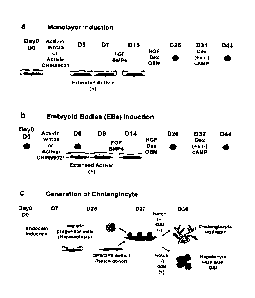
2 medium in the presence of VEGF and bFGF for 6 days. To generate the chimeric
aggregates, the
cultured RFP(+)/ CD34(+) cells were trypsinized, dissociated and placed into
Aggrewell plates at a
cell density of 100 cells per well. Following 2 days of culture, the day 25
hepatoblasts cells were
placed onto the RFP(+)/CD34(+) endothelial aggregates at a cell density of
1000 cells per well. Scale
bar 100um (b) phase contrast and fluorescent images showing RFP positive cells
within
endothelial/hepatic aggregates at day 33. RFP is not detected in hepatic
aggregates generated
without the endothelial cells. Scale bar 100um (c) Flow cytometric analysis
showing the proportion of
RFP positive cells in endothelial/hepatocyte aggregates at day 33. (d) RT-qPCR
analyses showing
CYP3A4 expression in the aggregates with and without endothelial cells at day
44. Values are
determined relative to TBP and presented as fold change relative to expression
of the adult liver
sample, which is set at one.
[0056] Figure 13 demonstrates the effect of 3D gel culture on maturation of
hPSC-derived
hepatocytes. Aggregates consisting of hepatoblasts or hepatoblasts and
endothelial cells (end) were
generated at day 25 of culture and then cultured for an additional 7 days in
liquid in hepatocyte culture
medium supplemented VEGF and bFGF and followed by 12 days of culture in the
same medium
supplemented with cAMP, PD0325901 (PD) and XAV939. To test the effects of
collagen on
maturation, day 32 chimeric aggregates were embedded in a Collagen type 1 gel
and cultured in the
presence of cAMP, PD0325901 and XAV939 for 12 days. All cultures were
harvested at day 44 and
analyzed for expression of the indicated genes by qRT-PCR. Values are
determined relative to TBP.
The expression of ALB and CYP3A4 is presented as fold change relative to their
levels in adult liver.
The expression of AFP and CYP3A7 is presented as fold change relative to their
levels in fetal liver.
AL: Adult liver, FL: Fetal liver.
[0057] Figure 14. Characterization of the hepatoblast stage of development in
hPSC differentiation
cultures. (a) Schematic representation of the differentiation protocol. (b)
Flow cytometric analyses
showing the development of the CXCR4+, CKIT+, and EPCAM + populations at day 7
in the
monolayer induction format. (c) RT-qPCR showing expression of indicated genes
in H9-derived
hepatoblast cells maintained in the culture conditions indicated Figure 14a.
The expression of the
indicated genes was analyzed on days 7, 13, 19 and 25 of culture. Values are
determined relative to
TBP and presented as fold change relative to expression in fetal liver, which
is set at one. AL: Adult
liver, FL: fetal liver.
CA 02901377 2015-08-14
WO 2014/124527
PCT/CA2014/000122
[0058] .. Figure 15. Notch signaling promotes cholangiocyte development from
the hPSC¨derived
hepatoblast-like population. RT-qPCR-based expression analysis of ALB and CK19
in the
hepatoblast-derived cells following co-culture with 0P9 in media supplemented
with HGF (20 ng/ml),
EGF (50ng/m1), and TGFb1 (5 ng/ml) in the presence or absence of the gamma-
secretase inhibitor
(GSI), an antagonist of the Notch pathway. Cells were harvested and analyzed
at days 30, 33, and 36
'10 of culture. Values were determined relative to TBP and presented as
fold change relative to levels of
expression in the day 27 hepatoblast aggregates which is set as 1. (b) RT-qPCR
based expression
analysis of the Notch target genes HES1, HES5 and HEY1 in hepatoblast-derived
cells following
culture with or without 0P9. Cells were assayed at day 36 of culture. For this
analyses, the day 27
hepatoblast aggregates were plated either on 0P9 (0P9+) stromal cells or
Matrigel (0P9-) in media
containing HGF, EGF, and TGFb1 (5 ng/ml) in the presence of absence of the
Notch signaling
antagonist gamma-secretase inhibitor (GS!) Values were determined relative to
TBP and presented
as fold change relative to the levels of expression in the cells at day 36
cultured on Matrigel. This
value was set as 1. Bars in all graphs represent the standard deviation (SD)
of the mean of three
independent experiments. *P<0.05, **, P<0.01, *** P<0.001 (Student's t-test; n
= 3).
[00591 Figure 16. Three-dimensional culture promotes cholangiocyte
maturation: Morphology of
chimeric aggregates consisting of day 25 hESC-derived cells and 0P9 stromal
cells (GFP+). H9-
derived day 25 hepatoblast were mixed (aggregated) with 0P9 stromal cells at a
ratio of 4:1, in low
cluster culture dishes for 48 hours. The chimeric aggregates were embedded in
a mixture of type 1
collagen (1.2 mg/ml) and Matrigel (40 %) to establish a 3D gel culture. The
cultures were maintained
over 2 weeks in the media containing of HGF, EGF and TGFb1 in the presence or
absence of GSI. (b)
Proportion of structures displaying a tubular, cyst or sphere morphology that
develop in the 3D
cultures. Values are presented as proportion of total structure that develop
in the presence or
absence of GSI. The values are representative of 3 independent experiments.
(c) RT-qPCR based
expression analyses of pooled structures that developed in the 3D gels. The
cultures were harvested
at day 44 and the cells analyzed for expression of genes indicative of the
hepatocyte (ALB, AFP and
CYP3A7) and cholangiocyte (CK19, Sox9 and CFTR) lineages. Values are
determined relative to
TBP and presented as fold change relative to levels of expression in the
population treated with GSI,
which is set at one.
[0060] Figure 17, hPSC-derived cholangiocytes form duct-like structures in
vivo. (a-b) Histological
analyses of a cholangiocyte graft in a Matrigel plug 8 weeks following
transplantation of day 25
hepatoblast-derived cells cocultured with 0P9 stromal cells for 9 days in
media containing of HGF,
EGF and TGFb1. Following co-culture, the cells were dissociated and
transplanted (106 per recipient)
into the mammary fat pad of immunodeficient NOD/SCID/ IL2rg -/- (NSG) mice.
Multiple duct structure
were visualized in mammary fat pad at low (a) and high magnification images
(b) (H&E staining). (c-d)
Immunostaining to detect RFP-positive cells in hESC-derived ductal structures
that developed in the
mammary fat pad following transplantation. For these studies cholangiocytes
were generated from
HES2-RFP hESCs that express RFP from the ROSA locus. Cholangiocytes generated
following 9
days of co-culture with 0P9 stromal cells were transplanted into the mammary
fat pad of NSG mice.
16
CA 02901377 2015-08-14
WO 2014/124527
PCT/CA2014/000122
Grafts that developed 8 weeks following transplantation were analyzed for the
presence of RFP+ cell
by innmunohistochemistry. RFP- positive cells were detected within all the
ductal structures,
confirming that the cells were of human origin and derived from the HES2- RFP
cells. RFP+
structures were visible in the images at low (c) and high (d) magnification
[0061] Figure 18. hPSC-derived cyst structures generated in 3D gels contain
functional CFTR
protein (a) Representative confocal microscopy images of calcein-green-labeled
and forskolin/ IBMX
(F/I) stimulated cyst structures generated from H9 (hESC)- and Y2-1 (iPSC)-
derived derived
cholangiocytes. Image was taken 24 hours after F/I stimulation. Scale bar 500
pm (b) Quantification of
the degree of cyst swelling 24 hours after F/I stimulation in the presence or
absence of CFTR
inhibitor. F/I stimulated cyst swelling was quantified using velocity imaging
software. The total size of
the cysts is normalized to that prior to F/I stimulation. Values are from
three individual experiments.
*P<0.05, **, P<0.01, *** P<0.001 (Student's t-test; n = 3).
[0062] Figure 19. The generation of cholangiocytes from cystic fibrosis
patient iPSCs. (a) Phase
contrast images of cholangiocyte like cells derived from CFTR deleted F508 iPS
cells (CF-iPSCs) at
day 44 of 3D gel culture in the presence or absence of forskolin. CF-iPSCs-
derived hepatoblasts and
chimeric hepatoblast/OP9 aggregates were generated using the protocol shown in
Figure 14a. After
embedding in collagen/ Matrigel culture, cyst formation was induced from the
aggregates by the
addition of forskolin for the first week of the two- week culture period
(left). Without forskolin
stimulation, the CF-iPSCs derived cholangiocytes formed branched ductal
structure rather than hollow
cysts (right). (b) Quantification of numbers of cyst structures that developed
from CF-iPSCs and
normal iPSC (Y2-1)-derived cholangiocytes at 7 and 14 days of culture. CF-
iPSCs derived
cholangiocytes were maintained in the 3D gel conditions in the presence or
absence of forskolin for
the first week of the two weeks culture period (left graph). Normal iPS cells-
derived cholangiocytes
were maintained in the presence or absence of CFTR inhibitor for the first
week of the two- week
culture periods (right graph). Addition of forskolin increased the number of
cyst structures that
developed from the CF- iPSCs derived cholangiocytes at both 7 and 14 days of
culture (left graph).
Addition of the CFTR inhibitor to normal iPSC-derived cholangiocytes delayed
cyst formation (right
graph). (c) Histological analyses of cysts derived from normal iPSC- (upper
panel) and CF-iPSC-
(lower panel) cholangiocytes at day 44. Both cholangiocyte populations were
cultured in the presence
of forskolin for the first week of the two weeks culture. Addition of
forskolin to the normal iPSC-
derived cholangiocytes induced the formation of large hollow cysts (upper
panel). The CF-iPSC-
derived cysts were smaller, often containing internal septum (lower panel).
[0063] Figure 20. Restoration of CFTR function in the CF-iPSC-derived
cholangiocytes by
treatment with the small molecule correctors VX-809 and C4. Western blot
analysis shows the
accumulation of mature complex glycosylated form of CFTR (band C) in CF-iPSC-
derived
cholangiocytes treated with VX-809 and C4. The mutant form of the protein
(band B) was
predominant in the uncorrected cells. Human bronchial epithelial cells (HBE)
were used as a positive
control. (b) Representative confocal microscopy images of calcein-green-
labeled and forskolin/ IBMX
17
CA 02901377 2015-08-14
WO 2014/124527
PCT/CA2014/000122
(F/I) stimulated cyst structures generated from CF-iPSCs from two individual
patients (997 CFTR del
and Cl CFTR del ¨ both of which carry the deltaF508 mutation). Images were
taken 24 hours after F/I
stimulation. Scale bar 500 pm (c) Quantification of the degree of swelling
observed in hPSCs-cysts 24
hours following F/I stimulation in the presence or absence of CFTR inhibitor.
F/I stimulated cyst
swelling was quantified using velocity imaging software. The total size of
cyst is normalized to that
before F/I stimulation from each three individual experiment. *P<0.05, **,
P<0.01, '' P<0.001
(Student's t-test; n = 3).
[0064] Figure 21. Intracellular flow cytometric analysis showing the
proportion of ALB+ and CK19+
cells in the hepatoblast-derived population following 9 days of coculture with
0P9. Cells were cultured
in media containing of HGF, EGF and TGFb1 in the presence or absence of GSI.
Ctrl shows isotype
control.
[0065] Figure 22. Hepatic specification and differentiation of hepatoblast
from other hPSCs. RT-
qPCR analyses showing expression of indicated genes in HES2 and Y2-1 iPS cells-
derived
hepatoblast cells maintained as indicated in Figure 14a. The expression of the
indicated genes was
analyzed on days, day 7, 13, 19 and 25 of culture. Values are determined
relative to TBP and
presented as fold change relative to expression in fetal liver, which is set
at one. AL: Adult liver, FL:
fetal liver.
[0066] Figure 23. 3D gels used for the generation of cystic structures from
hPSC-derived
cholangiocytes. (a) Schematic representation of the differentiation protocol
used to generate chimeric
aggregates consisting of day 25 hPSCs derived hepatoblasts and 0P9 cells
(GFP+). Day 25
hepatoblasts were dissociated and co-cultured with 0P9 cells at the ratio of
4:1, in low cluster culture
dishes. The chimeric aggregates were embedded in gel consisting of a mixture
of type 1 collagen (1.2
mg/ ml) and Matrigel (20 %). (b) RT-qPCR based expression analyses of
structures that developed in
the gel in the presence or absence of 0P9 at day 44 of culture in media
containing HGF, EGF and
TGFb1 . Expression of the Notch target genes was significantly upregulated in
the presence of 0P9.
Values are determined relative to TBP and presented as fold change relative to
expression in the cell
cultured in the absence of 0P9, which is set at one. (c) Histological analyses
of cyst structures that
developed from H9 derived cholangiocytes cultured with 0P9 cells in the
presence (right panel) or
absence (left) of GSI at day 44 culture (H&E staining). (d) Western blot
analysis showing the presence
of the mature complex glycosylated form of CFTR protein (Band C) in structures
generated from
normal iPSC-derived cholangiocytes cultured in the presence or absence of 0P9.
Undifferentiated
normal iPSCs were used as negative control. (e) RT-qPCR based expression
analyses of CFTR in
structures generated from normal iPSC-derived cholangiocytes cultured in thQ
presence or absence
of 0P9. Cells were analysed at day 44 of culture.. Values are determined
relative to TBP and
presented as fold change relative to expression value detected in caco-2 cells
(intestinal colon
carcinoma cell line), which set as one. *P<0.05, *", P<0.01, *** P<0.001
(Student's t-test; n = 3).
[0067] Figure 24. Generation of definitive endoderm and hepatoblasts from
cystic fibrosis patient
iPSCs. Flow cytometric analyses showing the development of the CXCR4+, CKIT+,
and EPCAM+
18
CA 02901377 2015-08-14
WO 2014/124527
PCT/CA2014/000122
populations from CF-iPS cells (Cl del CFTR) at day 7 of monolayer culture. (b)
RT-qPCR analyses
showing expression of indicated genes in the CF-iPSC-derived hepatoblast
population maintained in
the culture conditions outlined Figure 14a. The expression of indicated gene
was analyzed on day 7s,
13, 19 and day 25 of culture. Values are determined relative to TBP and
presented as fold change
relative to expression in fetal liver, which is set at one. AL: Adult liver,
FL: fetal liver.
Detailed Description of the Disclosure
[0068] Described
herein is a robustand reliable platform for the efficient generation of
hepatocytes and cholangiocytes from pluripotent stem cells (PSCs) through a
series of steps
described herein and for the generation of metabolicaly functional hepatocytes
and/or cholangiocytes.
It is demonstrated for example that one or more of extended nodal (e.g.
activin) signaling treatment,
inducing aggregation and activtating cAMP signaling for example in combination
with FGF agonist
induction and BMP4 agonist induction optionally in combination with one or
more steps that increases
expansion of a particular cell population and/or specific fate permits the
reproducible generation of
hepatocyte and cholangiocyle lineage cells including for example expanded
hepatoblasts and/or with
further manipulation, functional and mature hepatocytes and cholangiocytes
from definitive endoderm
induced in embryoid bodies or from monolayers.
[0069] An aspect
of the present disclosure includes a method of producing hepatocyte or
cholangiocyte lineaage cells such as hepatoblasts, hepatocytes and/or
cholangiocytes from an
extended nodal agonist treated induced endodermal cell population, the method
comprising: (a)
specifying the extended nodal agonist treated induced endodermal cell
population to obtain a cell
population comprising hepatocyte and/or cholangiocyte progenitors by
contacting the extended nodal
agonist treated induced endodermal cell population with specification media
comprising a combination
of a FGF agonist and a BMP4 agonist and/or active conjugates and/or fragments
thereof to obtain a
cell population comprising hepatocyte and/or cholangiocyte progenitor, and (b)
inducing maturation,
further lineage specification and/or expansion of the hepatocyte and/or
cholangiocyte progenitors of
the cell population to obtain an expanded population of hepatocytes and/or a
population comprising
hepatocytes and/or cholangiocytes, the inducing maturation step comprising
generating aggregates of
the cell population.
[0070] Aggregation is demonstrated herein to be important for and to
promote maturation.
[0071] In an
embodiment, the hepatocyte and/or cholangiocyte progenitors comprise
hepatoblasts and/or immature hepatocytes and/or immature cholangiocytes.
[0072] The term
"contacting" (e.g. contacting an endodermal cell population with a component
or components) is intended to include incubating the component(s) and the cell
together in vitro (e.g.,
adding the compound to cells in culture) and the step of contacting can be
conducted in any suitable
manner. For example the cells may be treated in adherent culture, or in
suspension culture, the
components can be added temporally substantially simultaneously (e.g. together
in a cocktail) or
19
CA 02901377 2015-08-14
WO 2014/124527
PCT/CA2014/000122
sequentially (e.g. within 1 hour, 1 day or more from an addition of a first
component). The cells can
also be contacted with another agent such as a growth factor or other
differentiation agent or
environments to stabilize the cells, or to differentiate the cells further and
include culturing the cells
under conditions known in the art for example for culturing the pluripotent
(and/or differentiated)
population for example as further described in the Examples.
[0073] The terms "endoderm" and "definitive endoderm" as used herein refer
to one of the three
primary germ cell layers in the very early embryo (the other two germ cell
layers are the mesoderm
and ectoderm). The endoderm is the innermost of the three layers. An endoderm
cell differentiates to
give rise first to the embryonic gut and then to derivative tissues including
esophagus, stomach,
intestine, rectum, colon, pharyngeal pouch derivatives tonsils, thyroid,
thymus, parathyroid glands,
lung, liver, gall bladder and pancreas.
[0074] The "induced endodermal cell population" as used herein refers to
a population of
endoderm cells corresponding to "definitive endoderm induction" stage for
example as shown in
Figure la. This population can be for example prepared from embyroid bodies
(EB) that have been
exposed to a nodal agonist, such as activin, or opitionally from EB that have
been exposed to a nodal
agonist and a wnt/beta-catenin agonist such as Wnt3a or a GSK-3 selective
inhibitor such as CHIR-
99021 (StemoleculeTM CHIR99021 Stemgent), 6-bromo-Indirubin-3'-Oxime (B10)
(Cayman Chemical
(cat:13123)), or Stemolecule TM BIO from Stemgent (cat:04003). Alternatively,
the induced endodermal
cell population can be prepared from cells grown in a monolayer. The induced
endodermal cell
population can for example be identified by flow cytometric and molecular
analysis for one or more
markers such as surface markers CXCR4, CKIT and EPCAM and the transcription
factors SOX17 and
FOXA2. The induced endodermal cell population can also for example be
identified by at least or
greater than 70, 80, 90 or 95% of the population co-expressing CXCR4 and CKIT
or CXCR4 and
EPCAM. The induced endodermal cell population can also for example be
identified by greater than
70, 80, 90 or 95% of the population of the population expressing SOX17 and/or
FOXA2. The induced
endodermal cell population can for example be in a 2D (monolayer) or 3D
(Embryoid Body or other
form of aggregates) format. The induced endodermal population can be derived
for example from
hESCs as well as an induced pluripotent cell (iPSC) as demonstrated in Example
1.
[0075] The induced endoderm cell population is for example treated with a
nodal agonist
extended period of time to provide an extended nodal agonist treated induced
endoderm cell
population.
[0076] As described in Example 1 and shown in Figure 3a, culturing day 6
cells (day 5 when the
method comprises monolayer induction) for two additional days in activin prior
to specifying with
FGF/BMP4 results in a higher proportion of SOX17+ FOXA2+ cells as measured at
day 12 compared
to cells not cultured for two additional days in activin (e.g. an example of a
nodal agonist). This step is
also referred to herein as an "extended activin" treatment and is an example
of an "extended nodal
agonist" treatment.
CA 02901377 2015-08-14
WO 2014/124527
PCT/CA2014/000122
[0077] The "extended nodal agonist treated induced endoderm cell
population" as used herein
refers to an induced endodermal cell population that has been treated with a
nodal agonist such as
activin for an extended period, for example from about 1 to about 4 or about
1, 2, 3 or 4 additional
days (e.g. "the extended period" which is in addition to the endoderm
induction phase which can
comprise treatment with a nodal agonist). The extended nodal agonist treatment
as demonstrated
herein resulted in higher levels of expression of genes indicative of hepatic
progenitor (hepatoblast)
development, including HEX, AFP, ALB and HNF4a at day 26 of culture (as shown
in Fig. 3d). The
extended nodal agonist treated induced endoderm population is obtained by
inducing endoderm cells
in ernbryoid bodies (EBs) or by inducing endoderm cells that are in a
monolayer, and wherein the
induced endodermal population is cultured in the presence of a nodal agonist,
for example activin, for
an extended period to produce an extended nodal agonist treated induced
endodermal population.
[0078] The extended nodal agonist treated induced endodermal cell
population is, in an
embodiment, obtained by inducing endoderm cells in embryoid bodies (EBs). In
another embodiment,
the extended nodal agonist treated induced endodermal population is obtaining
by inducing endoderm
cells that are in a monolayer. In each case, the induced endodermal population
is cultured in the
presence of a nodal agonist, for example activin, for an extended period.
[0079] Optionally, the induced endodermal population is subsequently
dissociated, for example
in embodiments where the induced endodermal cell population is derived from
EBs. As used herein,
"dissociated cells" or "dissociated cell populations" refers to cells that are
not in 3D aggregates, for
example, physically separated from one another. Dissociated cells are
distinguished from "cell
aggregates" which refers to clusters or clumps of cells.
[0080] In an embodiment, the induced endodermal population comprises at
least 80%, 85%,
90% CXCR4 + and cKIT + positive cells and/or at least 70%, 75%, 80% SOX17+
cells.
[0081] In some embodiments, the induced endodermal cell population
(and/or the extended
nodal agonist treated induced endodermal cell population) is produced from
pluripotent stem cells
(PSCs) such as embryonic stem cells (ESCs) or induced pluripotent stem cells
(iPSCs). The
pluripotent stem cells are optionally human ESCs (hESCs) or human iPSCs
(hiPSCs).
[0082] The term "pluripotent" as used herein refers to a cell with the
capacity, under different
conditions, to differentiate to more than one differentiated cell type, and
for example the capacity to
differentiate to cell types characteristic of the three germ cell layers.
Pluripotent cells are
characterized by their ability to differentiate to more than one cell type
using, for example, a nude
mouse teratoma formation assay. Pluripotency is also evidenced by the
expression of embryonic stem
(ES) cell marker.
[0083] The term "progenitor cell" refers to cells that have a cellular
phenotype that is at an
earlier step along a developmental pathway or progression than a fully
differentiated cell relative to a
cell which it can give rise to by differentiation. Progenitor cells can give
rise to multiple distinct
21
CA 02901377 2015-08-14
WO 2014/124527
PCT/CA2014/000122
differentiated cell types or to a single differentiated cell type, depending
on the developmental
pathway and on the environment in which the cells develop and differentiate.
[0084] The term "stem cell" as used herein, refers to an undifferentiated
cell which is capable
of proliferation, self-renewal and giving rise to more progenitor cells having
the ability to generate a
large number of mother cells that can in turn give rise to differentiated, or
differentiable, daughter
cells. The daughter cells can for example be induced to proliferate and
produce progeny that
subsequently differentiate into one or more mature cell types, while also
retaining one or more cells
with parental developmental potential.
[0085] The term "embryonic stem cell" is used to refer to the pluripotent
stem cells of the inner
cell mass of the embryonic blastocyst (see, for example, U.S. Pat. Nos.
5,843,780, 6,200,806). Such
cells can also be obtained from the inner cell mass of blastocysts derived
from somatic cell nuclear
transfer (see, for example, U.S. Pat. Nos. 5,945,577, 5,994,619, 6,235,970).
The distinguishing
characteristics of an embryonic stem cell define an embryonic stem cell
phenotype. Accordingly, a cell
has the phenotype of an embryonic stem cell if it possesses one or more of the
unique characteristics
of an embryonic stem cell such that that cell can be distinguished from other
cells. Exemplary
distinguishing embryonic stem cell characteristics include, without
limitation, gene expression profile,
proliferative capacity, differentiation capacity, karyotype, responsiveness to
particular culture
conditions, and the like.
[0086] In one embodiment, the method of producing hepatocytes and/or
cholangiocytes from an
extended nodal agonist treated induced endodermal cell population comprises:
(a) specifying the
extended nodal agonist treated endodermal cell population to a cell population
comprising hepatocyte
and/or cholangiocyte progenitors by contacting the induced endodermal cell
population with
specification media comprising a FGF agonist and a BMP4 agonist and/or active
conjugates and/or
fragments thereof.
[0087] In an embodiment, the specifying step comprises contacting an
extended nodal agonist
treated induced endodermal population with specification media comprising a
FGF agonist and
BMP4. The FGF agonist can for example be bFGF, FGF10, FGF2 or FGF4, active
fragments and/or
combinations thereof. The combinations can be added to the cells for example
sequentially.
[0088] In an embodiment, the specifying step comprises first contacting
an extended nodal
agonist treated induced endodermal population with specification media
comprising FGF10 and
BMP4 for about 40 to about 60 hours for example about 40, 42, 44, 46, 48, 50,
52, 54, 56, 58 or about
60 hours and then contacting the extended nodal agonist treated induced
endodermal population with
specification media comprising bFGF and BMP4 for about 4 to about 7 days, for
example about 4, 5,
6 or about 7 days.
[0089] In one embodiment, the specification media comprises lscove's
Modified Dulbecco's
Medium (IMDM) supplement with 1% vol/vol B27 supplement (lnvitrogen:
A11576SA), ascorbic acid,
22
CA 02901377 2015-08-14
WO 2014/124527
PCT/CA2014/000122
MTG, FGF10 (50 ng/ml) (for example from day 8 to day 10, bFGF (20 ng/ml) (for
example from day
to day 14), and BMP4 (50 ng/ml).
[0090] Optionally, the endodermal cell population is contacted with FGF10
and BMP4 for 1 to 3
days, optionally 2 days and subsequently contacted with bFGF and BMP4 for 2 to
6 days, optionally 3
to 5 days, optionally 4 days. In some embodiments, the endodermal cell
population is incubated in cell
10 culture medium comprising BMP4 and FGF10 or bFGF. Optionally, the
endodermal cell population is
incubated in cell culture medium comprising lscove's Modified Dulbecco's
Medium (IMDM)
supplemented with 1% vol/vol B27, ascorbic acid, monothioglycerol, BMP4 and
FGF10 or bFGF.
[0091] In some embodiments, the endodermal cell population is dissociated
and the monolayer
cells are then contacted with FGF and a BMP4 agonist.
[0092] In other embodiments, the monolayer cells are contacted with activin
for 1 to 4 days,
optionally 1, 2, 3 or 4 days prior to being contacted with FGF and a BMP4
agonist such as BMP4.
Optionally, the monolayer cells are incubated in cell culture medium
comprising activin A, optionally
medium comprising StemPRO-34 supplemented with bFGF, activin A and BMP4.
[0093] In a further embodiment, the specifying step comprises contacting
cells with a
specification media that comprises one or more factors that promote
maturation, further lineage
specification and/or expansion.
[0094] The term "specification media" as used herein refers to culture
medium that is used to
promote or facilitate specification of a cell or a cell population. One
example of a hepatic specification
media includes lscove's Modified Dulbecco's Medium (IMDM) supplemented with 1%
vol/vol B27
(Invitrogen: A11576SA), and ascorbic acid, MTG, a BMP4 agonist and at least
one FGF agonist
selected from FGF10, bFGF, FGF4 and FGF2. For some stages and in some
embodiments, the same
specification media can be used for example for specifying both hepatocyte and
cholangiocyte
lineages. In other stages and in other embodiments, the specification media
comprises one or more
factors that promote specification of hepatocyte and/or cholangiocyte
development, for example a
notch antagonist or a notch agonist. The term "specifying" as used herein
means a process of
committing a cell toward a specific cell fate, prior to which the cell type is
not yet determined and any
bias the cell has toward a certain fate can be reversed or transformed to
another fate. Specification
induces a state where the cell's fate cannot be changed under typical
conditions.
[0095] Specification of the induced endoderm along a hepatic fate can for
example be
confirmed by measuring hepatic and/or cholangiocyte expressed genes, including
for example Tbx3
ALB, AFP, CK19, Sox9, NHF6beta and Notch2 as demonstrated for example in
Example 9. For
example it is demonstrated that Notch 2 expression is upregulated in
HGF/DEX/OSM treated
hepatoblasts. Detection of Notch2 protein and/or expression can be used to
confirm that the cell
population can be specified to cholangiocytes, for example Notch2 can be
detected as described in
Example 9.
23
CA 02901377 2015-08-14
WO 2014/124527
PCT/CA2014/000122
[0096] In the context of a cell, the term "differentiated", or
"differentiating" is a relative term and
a "differentiated cell" is a cell that has progressed further down the
developmental pathway than the
cell it is being compared with. Thus, stem cells can differentiate to lineage-
restricted precursor cells
(such as an induced endodermal progenitor cell), which in turn can
differentiate into other types of
precursor cells further down the pathway and then mature to an end-stage
functional cell, which plays
a characteristic role in a certain tissue type, and may or may not retain the
capacity to proliferate
further. The term "differentiation" as used herein includes steps for
producing an induced endodermal
population and specified cell populations, for example a hepatocyte or
cholangiocyte specified cell
population.
[0097] In another embodiment, the aggregates are generated from a cell
population comprising
at least 70%, 80%, 85%, or at least 90% albumin positive cells In another
embodiment, the
aggregates are generated after 24, 25, 26, 27, or 28 days in culture (for
example where the day PSCs
are obtained is considered day 0).
[0098] Optionally, aggregates are generated by enzymatic treatment and/or
manual
dissociation. In some embodiments, aggregates are generated by dissociating
cells with collagenase
and/or TrypIeLE. In some embodiments, the cells are subsequently cultured in
ultra-low cluster
dishes. In other embodiments, monolayer cultures can be broken apart
mechanically by pipetting, or
can be dissociated enzymatically and aggregated with an incubation in low
attachement plates or by
shaking the population of cells. Aggrewells can optionally be used.
[0099] In an embodiment, the cells (e.g. monolayers and cells prior to
aggregation) are gown on
matrix coated plates, optionally on Matrigel coated plates. Other matrix
coated plates that support the
attachment of hepatoblasts, hepatocytes and/or cholangiocytes can also be
used, for example
laminin, fibronectin and collagen coated plates. Figure 9f for example
demonstrates ALB expression
at day 26 of induction using several different matrix coating substrates.
[00100] Inducing maturation, further lineage specification and/or
expansion can comprise one or
more substeps.
[00101] In a further embodiment, the cell population comprising hepatocyte
and/or cholangiocyte
progenitors and/or the aggregates are cultured in the presence of hepatocyte
growth factor (HGF),
dexamethasone (DEX) and/or Oncostatin M (OSM) and/or active conjugates andior
fragments
thereof. For example, the cell population comprising hepatocyte and/or
cholangiocyte progenitors can
be cultured in a maturation media comprising HGF, DEX and/or OSM for about 10,
11, 12, 13 or 14
days prior to aggregation and/or subsequent to aggregation the aggregates can
be cultured in a
maturation media comprising HGF, DEX and/or OSM for about 6, 7, 8, 9, or 10
days. For example,
addition of about lOng/mL HGF promotes survival of aggregates.
[00102] In one embodiment, inducing maturation, and optionally inducing
further lineage
specification and/or expansion further comprises activating the cAMP pathway
within the cells of the
24
CA 02901377 2015-08-14
WO 2014/124527
PCT/CA2014/000122
aggregates to induce the differentiation and/or maturation of the hepatocyte
and cholangiocyte
progenitors into hepatocytes and/or cholangiocytes.
[00103] The extended nodal agonist treatment and the aggregation of cells
for example at day
25 for monolayer induced cells and day 26 for EB induced cells, produce a
population which is for
example capable of responding to cAMP signaling. As shown herein, activation
of cAMP increases
CYP expression and hepatocyte maturation.
[00104] In another embodiment, activating the cAMP pathway comprises
contacting the
aggregates with a cAMP agonist analog such as 8-bromoadensoine-3'5"-cyclic
monophosphate (8-Br-
cAMP), dibutyryl-cAMP, Adenosine- 3', 5'- cyclic monophosphorothioate, Sp-
isomer (Sp-cAMPS)
and/or 8- Bromoadenosine- 3', 5'- cyclic monophosphorothioate, Sp- isomer (Sp-
8-Br-cAMPS)) and/or
any other cAMP agonist, such as cholera toxin, forskolin, caffeine,
theophylline and pertussis toxin.
Experiments have been conducted for example using Sp-8-Br-cAMP (Biolog: Cat.
No.: B 002 CAS
No.: [127634-20-2]), 8- Br- cAMP and forskolin (FSK)(Sigma:66575-29-9). For
example, the
combination comprising Forskolin (FSK)(Sigma:66575-29-9)+XAV939+PD0325901 was
effective to
increases CYP expression and induce the maturation of the hepatocyte
progenitors into hepatocytes.
As used herein "cAMP agonists" include, cAMP, cAMP analogs that activate cAMP
as well as
molecules such as cholera toxin, forskolin, caffeine, theophylline and
pertussis toxin which activate
cAMP. IBMX which is a phosphodiesterase inhibitor, (phosphodiesterases are
cAMP inhibitors) can
also be used in some embodiments, for example in combination with forskolin.
[00105] It has been found also for example that the addition of 10 ng/ml
HGF (reduced from 20
ng/ml) promotes survival of the aggregates whereas maintaining OSM has an
inhibitory effect on the
induction of expression of Phase 1 CYP enzymes, in particular CYP 3A4.
Accordingly in some
embodiments, aggregates are cultured with cAMP analogs and/or agonists in the
absence of OSM.
[00106] The term "maturation" as used herein means a process that is
required for a cell (e.g.
hepatoblast) to become more specialized and/or attain a fully functional
state, for example its
functional state in vivo. In one embodiment, the process by which immature
hepatocytes or hepatic
progenitors become mature, functional hepatocytes is referred to as
maturation.
[00107] Fig. la refers to "hepatic maturation A" and Fig 14a refers to
hepatoblast differentiation.
The cell population referred to in both cases is a hepatoblast cell
population, that can produce
hepatocytes if continued to be cultured for example in the presence of DEX
optionally in combination
with HGF and OSM and/or cAMP or produce cholangiocytes if cultured in
combination with a Notch
agonist (e.g. a notch signal donor), such as 0P9, 0P9 delta and/or 0P9 Jaggedl
cells in the
presence of EGF, TGB1 HGF and EGF.
[00108] The term "maturation media" as used herein refers to culture
medium that is used to
promote or facilitate maturation of a cell or a cell population and which can
comprise matruation
factors, as well as cell expansion inducers and lineage inducers. One example
of a maturation media
for inducing hepatocyte maturation includes lscove's Modified Dulbecco's
Medium (IMDM)
CA 02901377 2015-08-14
WO 2014/124527 PCT/CA2014/000122
supplemented with 1% volfvol B27 (Invitrogen: A11576SA) as well as ascorbic
acid, Glutamine, MTG
and optionally Hepatocyte growth factor (HGF), Dexamethasone (Dex) and/or
Oncostatin M. Another
example of a maturation media for inducing hepatocyte includes Hepatocyte
culture medium (HCM)
(Lonza: CC-4182) without EGF. The maturation media optionally also comprises a
cAMP analog
and/or cAMP agonist which for example induces expansion of hepatocyte lineage
cells and their
maturation. Maturation media can comprise factors that promote hepatocyte
and/or cholangiocyte
development, further lineage specification and/or expansion and/or further
lineage selection. For
example, Wnt antagonist alone or in combination with TGFbeta antagonists
and/or MEK/Erk
antagonists promote hepatocyte maturation. Notch agonists for example, are
demonstrated herein to
induce cholangiocyte lineage development and are added when cholangiocytes are
desired. Similarly
.. Notch antagonists promote hepatocyte lineages and can be added when
hepatocytes are desired, for
example to inhibit cholangiocyte development.
[00109] Different maturation medias can be used sequentially (e.g. a
monolayer maturation
media (used for example pre-aggregation), an aggregates maturation media (used
for example post
aggregation); a hepatocyte maturation media that for example comprises factors
that promote
hepatocyte development and a cholangiocyte maturation media that for example
promotes
cholangiocyte development.
[00110] For example, in an embodiment, a maturation media comprising a
cAMP analog and/or
cAMP agonist and DEX and optionally HGF is added to the aggregates subsequent
to culturing the
pre-aggregate population in the maturation media comprising HGF, DEX and OSM,
for example for
about 10, 11, 12, 13 or 14 days.
[00111] The term "hepatocyte" as used herein refers to a parenchymal liver
cell. Hepatocytes
make up the majority of the liver's cytoplasmic mass and are involved in
protein synthesis and
storage, carbohydrate metabolism, cholesterol, bile salt and phospholipid
synthesis and the
detoxification, modification and excretion of exogenous and endogenous
substances.
[00112] The term "primary hepatocyte" as used herein is a hepatocyte that
has taken directly
from living tissue (e.g. biopsy material) and established for growth in vitro.
[00113] The term "hepatoblast" as used herein refers to a progenitor cell
which has the capacity
to differentiate into cells of the hepatic and cholangiocyte lineages e.g. a
hepatocyte or a
cholangiocyte. Hepatoblasts are for example a subset of hepatocyte and
cholangiocyte progenitors
which can comprise immature hepatocytes and immature cholangiocytes (e.g.
cells which have a
speficied cell fate and can mature to a hepatocyte only or a cholangiocyte
only). In some
embodiments, hepatoblast cells are defined by expression of markers such as
Hex, HNF4, alpha-
fetoprotein (AFP) and albumin (ALB). For example, hepatoblasts can give rise
to cholangiocyte cells
(e.g. CK19+ cells) when notch signaling is activated in for example day 28
hepatoblast containing
cultures. The term "hepatocyte progenitor" as used herein means cells that
have the capacity to
differentiate into functional hepatocytes which are for example albumin
positive and/or expresses
CYP enzymes.
26
CA 02901377 2015-08-14
WO 2014/124527 PCT/CA2014/000122
[00114] The term
"cholangiocyte progenitor" as used herein means cells that have the capacity
to differentiate into functional cholangiocytes which are for example CK19
positive and/or express
CFIR.
[00115] The terms "immature
hepatocyte" as used herein refers to a hepatocyte lineage cell that
expresses albumin but that does not express appreciable levels of functional
CYP3A4 and/or
CYP1A2 enzyme. In some embodiments, immature hepatocytes must undergo
maturation to aquire
the functionality of mature hepatocytes. In some embodiments, immature
hepatocyte cells are defined
by expression of markers such as Hex, alpha-fetoprotein and albumin.
[00116] A "mature
hepatocyte" as used herein means a hepatocyte lineage cell that expres CYP
enzymes for example CYP3A4 and CYP1A2 and albumin. Optionally, mature
hepatocytes include
functional, or measurable, levels of metabolic enzymes such as Phase I and
Phase II drug-
metabolizing enzymes for example comparable to adult cells. Examples of Phase
I drug-metabolizing
enzymes include but are not limited to cytochromes P450 CYP1A2, CYP3A4 and
CYP286. Examples
of Phase II drug-metabolizing enzymes include but are not limited to arylamine
N-acetyltransferases
NATI and NAT2 and UDP-glucuronosyltransferase UGT1A1. For example, the mature
hepatocyte
can be a metabolically active hepatocyte. Cellular uptake of lndocyanine green
(ICG) is considered to
be a characteristic of adult hepatocytes29 and is used clinically as a test
substrate to evaluate hepatic
function39. A mature hepatocyte is for example an ICG positive staining
hepatocyte. In a population of
hepatocytes, the population of hepatocytes can be considered a mature 50%,
60%, 70%, 80%, 90%
or more of the hepatocytes are ICG. A mature hepatocyte expresses increased
albumin compared to
an "immature hepatocyte" for example at least 5%, 10%, 25%, 50%, 75%, 100% or
200% more
albumin, than an immature hepatocyte.
[00117] In an embodiment, the hepatocyte is a functional hepatocyte.
[00118] The term
"functional hepatocyte" as used herein refers to a hepatocyte cell that
displays
one or more of charactistics of an adult hepatocyte (e.g. a mature hepatocyte)
and/or an immature
hepatocyte that is committed to a hepatic fate and is more differentiationed
than a starting cell (e.g.
compared to an endodermal population cell, a hepatocyte precursor or an
immature hepatocyte),
which for example expresses albumin and/or increased albumin compared to a
starting cell.
Optionally, functional hepatocytes are mature hepatocytes and include
functional, or measurable,
levels of metabolic enzymes such as Phase I and Phase II drug-metabolizing
enzymes for example
comparable to adult cells. For example, the functional hepatocyte can be a
metabolically active
hepatocyte. Cellular uptake of lndocyanine green (ICG) is considered to be a
characteristic of adult
hepatocytes29 and is used clinically as a test substrate to evaluate hepatic
function39. A functional
hepatocyte is for example an ICG positive staining hepatocyte. In a population
of hepatocytes, the
population of hepatocytes can be considered a functional population of
hepatocytes for example at
least 25%, 30%, 35%, 40%, 45% 50%, 60%, 70%, 80%, 90% or more of the
hepatocytes are ICG
positive. In another example, a functional hepatocyte is an albumin secreting
hepatocyte and a
population of hepatocytes can be considered a functional population of
hepatocytes if for example at
27
CA 02901377 2015-08-14
WO 2014/124527
PCT/CA2014/000122
least 25%, 30%, 35%, 40%, 45% 50%, 60%, 70%, 80%, 90% or more of the
hepatocytes are albumin
secreting.
[00119] In embodiments, the hepatocytes, optionally functional hepatocytes
comprise increased
expression of at least 1, 2, 3, 4, 5, 6, 7, 8, 9, 10 or more genes or protein
selected from the group
consisting of ALB, CPS1, G6P, TDO, CYP2C9, CYP2D6, CYP7A1, CYP3A7, CYP1A2,
CYP3A4,
CYP266, NAT2 and UGT1A1 compared to a cell population comprising hepatocyte
and/or
cholangiocyte progenitors, and/or hepatocytes produced from a non-extended
nodal agonist treated
induced endodermal cell population, produced without aggregation and/or cAMP
signaling induction.
In other embodiments, at least 40, 50, 60, 70, 80 or 90% of the hepatocytes,
optionally functional
hepatocytes, are ASGPR-1+ cells.
[00120] In some embodiments, functional hepatocytes display nucleic acid or
protein levels of
CYP1A2, CYP2B6, CYP3A4, CYP2C9 and/or CYP2D6 that are comparable or higher
than those
found in primary mature hepatocytes, optionally levels that are increased at
least 1.1, 2, 3, 4, or 5 fold
or any 0.1 increment between 1.1 fold and 5 fold, optionally increased at
least 50% to 100%, 75% to
125%, 85% to 115%, 90% to 110% or 95% to 105%. For example,increased
expression of 1.15 fold
to 6.1 fold (e.g. CYP1A2 6.1 folds (610%), CYP3A4 13.2 folds (1320%), CYP 2B6
2 folds (200%),
CYP2C9 1.52 folds 152%, UGT1A1 2 folds (200%), CYP2D6 1.15 folds (115%)) of
those found in
primary hepatocytes. In some embodiments, hepatocytes display nucleic acid or
protein levels of ALB,
HNF4, AFP, CPS1, G6P, TD01, NATI, NAT2 and/or UGT1A1 that are comparable or
higher to those
found in primary hepatocytes of similar stage, optionally levels that are at
least 50% to 100%, 75% to
125%, 85% to 115%, 90% to 110% or 95% to 105% of those found in primary
hepatocytes.
[00121] In other embodiments, functional hepatocytes display nucleic acid
or protein levels of
CYP1A2, CYP2B6, CYP3A4, CYP2C9 and/or CYP2D6 that are higher than those in
hepatoblasts
and/or immature hepatocytes, optionally levels that are at least 110%, 125%,
150%, 175%, 200%,
300%, 400% or 500% of those found in primary hepatocytes. In some embodiments,
functional
hepatocytes display nucleic acid or protein levels of ALB, CPS1, G6P, TD01, NA
TI, NAT2 and/or
UGT1A1 that are higher than those in hepatoblasts and/or immature hepatocytes,
optionally levels
that are at least 110%, 125%, 150%, 175%, 200%, 300%, 400% or 500% of those
found in primary
hepatocytes.
[00122] In other embodiments, functional hepatocytes express the receptor
asialo-glycoprotein
receptor 1 (ASGPR1). In other embodiments, at least 40, 50, 60, 70, 80 or 90%
of the hepatocytes,
optionally functional hepatocytes, are ASGPR-1+ cells.
[00123] In further embodiments, functional hepatocytes display CYP1A2
activity in vitro.
Optionally, functional hepatocytes display CYP1A2 activity is comparable or
higher than those found
in primary hepatocytes, optionally levels that are at least 50% to 100%, 75%
to 125%, 85% to 115%,
90% to 110% or 95% to 105% of those found in primary hepatocytes. In some
embodiments, CYP1A2
activity is measured by incubating cells with phenacetin and monitoring the
generation of 0-
deethylated metabolite accumulation in the cells.
28
CA 02901377 2015-08-14
WO 2014/124527
PCT/CA2014/000122
[00124] In further
embodiments, functional hepatocytes display CYP2B6 activity in vitro.
Optionally, functional hepatocytes display CYP2B6 activity that is comparable
or higher than those
found in primary hepatocytes, optionally levels that are at least 50% to 100%,
75% to 125%, 85% to
115%, 90% to 110% or 95% to 105% of those found in primary hepatocytes. In
some embodiments,
CYP2B6 activity is measured by incubating cells with bupropin and monitoring
the formation of the
metabolite 0-hydroxy-bupropion in the cells.
[00125] In
further embodiments, hepatocytes display NATI and/or NAT2 activity in vitro.
Optionally, hepatocytes display NATI and/or NAT2 activity that is comparable
or higher than those
found in primary hepatocytes, optionally levels that are at least 1.1 fold, 2
fold, 3 fold 4 fold, 5 fold, 6
fold or about 50% to 100%, 75% to 125%, 85% to 115%, 90% to 110% or 95% to
105% of those
found in primary hepatocytes. In some embodiments, NATI and/or NAT2 activity
is indicated by the
metabolism of sulfamethazine (SMZ) to N-acetylated SMZ.
[00126] In
further embodiments, hepatocytes display UGT activity in vitro. Optionally,
hepatocytes display UGT activity that is comparable or higher than those found
in primary
hepatocytes, optionally levels that are at least 50% to 100%, 75% to 125%, 85%
to 115%, 90% to
110% or 95% to 105% of those found in primary hepatocytes. In some
embodiments, UGT activity is
indicated by the the generation of 4-MU glucuronide (4-MUG) from 4-
methylumbelliferone (4-MU) in
the cells.
[00127] In
embodiments, the hepatocytes, optionally functional hepatocytes comprise
increased
expression of at least 1, 2, 3, 4, 5, 6, 7, 8, 9, 10 or more genes or protein
selected from the group
consisting of ALB, CPS1, G6P, TDO, CYP2C9, CYP2D6, CYP7A1, CYP3A7, CYP1A2,
CYP3A4,
CYP2B6, NAT2 and UGT1A1 compared to a cell population comprising hepatocyte
and/or
cholangiocyte progenitors, and/or hepatocytes produced from a non-extended
nodal agonist treated
induced endodermal cell population, produced without aggregation and/or CAMP
signaling induction.
In other embodiments, at least 40, 50, 60, 70, 80 or 90% of the hepatocytes,
optionally functional
hepatocytes, are ASGPR-1+ cells.
[00128] In yet
another embodiment, functional hepatocytes display a global gene expression
profile that is indicative of hepatocyte maturation. Optionally, functional
hepatocytes display a global
gene expression profile that is more similar to a primary hepatocyte than a
global gene expression
profile of a hepatoblast and/or a immature hepatocyte. Global gene expression
profiles are obtained
by any mtheod known in the art, for example microarray analysis.
[00129] In an
embodiment, cholangiocyte fate is specified by treating aggregates of the cell
population with a notch agonist.
[00130] The term "cholangiocyte" as used herein refers to the cells that
make bile ducts.
[00131] The term
"cholangiocyte precursor" as used herein refers to cells which have the
capacity to differentiate into a cholangiocyte cell (e.g. hepatoblasts), as
well as immature
cholangiocytes that can mature to functional cholangiocytes. In some
embodiments, cells of the
29
CA 02901377 2015-08-14
WO 2014/124527
PCT/CA2014/000122
cholangiocyte lineages are defined by expression of markers such as CK19,
secretin reseptor (SR),
cystic fibrosis transmembrane conductance regulator (CFTR), and chloride
bicarbonate anion
exchanger 2 (CI(-)/HCO(3)(-) AEs).
[00132] The term "immature cholangiocyte" as used herein refers to a
cholangiocyte lineage cell
which must undergo maturation to aquire the functionality of mature
cholangiocytes. In some
embodiments, immature cholangiocyte cells express CK19 and/or Sox9, optionally
including early
Notch agonist treated cells, optionally treated for at least 1 day, at least 2
days, at least 3 days, or at
least 4 days.
[00133] In an embodiment, the cholangiocyte is a functional cholangiocyte.
[00134] The terms "functional cholangiocyte" as used herein refers to
cholangiocyte cells that
display one or more of the charactistics of adult cholangiocytes (e.g. mature
cholangiocyte) and/or are
CK-19, MDR1 and/or CFTR expressing cholangiocyte lineage cells. For example
functional
cholangiocytes express the MDR1 transporter and can when in cystic structures,
transport a tracer
dye such as rh0damine123, into the structure lumina! space. As another
example, CFTR functional
activity can be assessed for example using a forskolin induced swelling assay
on cystic structures, as
shown for example in Fig 18. In a population of cholangiocytes, the population
can be considered a
functional population if for example at least 25%, 30%, 35%, 40%, 45% 50%,
60%, 70%, 80%, 90%
or more of the cells express secretin reseptor (SR), cystic fibrosis
transmembrane conductance
regulator (CFTR), CK-19 and/or chloride bicarbonate anion exchanger 2 (CI(-
)/HCO(3)(-) AEs).
[00135] The term "mature cholangiocytes" as used herein are cholangiocytes
that express
specified transporter or cell membrane receptor activity, such as secretin
reseptor (SR), cystic fibrosis
transmembrane conductance regulator (CFTR), and optionally chloride
bicarbonate anion exchanger
2 (CI(-)/HCO(3)(-) AEs).
[00136] In an embodiment, the population of cholangiocytes produced is a
population of
functional cholangiocytes. The functional cholangiocyte comprises for example
increased expression
of at least 1, at least 2 or 3 genes or proteins selected from Sox9, CK19 and
CFTR (Cystic fibrosis
transmembrane conductance regulator) compared to the cells of the cell
population comprising
hepatocyte and cholangiocyte progenitors and/or compared to a population cells
produced from
aggregates not treated with a notch agonist. In other embodiments, at least
40, 50, 60, 70, 80 or 90%
of the population of cholangiocytes are CK19+ cholangiocytes. In other
embodiments, at least 40, 50,
60, 70, 80 or 90% of the functional cholangiocytes are CFTR+ cholangiocytes.
[00137] The term "expression" refers to the cellular processes involved in
producing RNA and
proteins and as appropriate, secreting proteins, including where applicable,
but not limited to, for
example, transcription, translation, folding, modification and processing.
"Expression products"
include RNA transcribed from a gene and polypeptides obtained by translation
of mRNA transcribed
from a gene.
CA 02901377 2015-08-14
WO 2014/124527
PCT/CA2014/000122
[00138] The term "cell culture medium" (also referred to herein as a
"culture medium" or
"medium') as referred to herein is a medium for culturing cells containing
nutrients that maintain cell
viability and support proliferation and optionally differentiation. The cell
culture medium may contain
any of the following in an appropriate combination: salt(s), buffer(s), amino
acids, glucose or other
sugar(s), antibiotics, serum or serum replacement, and other components such
as peptide growth
factors, vitamins etc. Cell culture media ordinarily used for particular cell
types are known to those
skilled in the art.
[00139] The suitable culture medium can include a suitable base culture
medium including for
example DMEM (Life Technologies), IMDM, RPMI, CMRL and/or any othe or media
that supports the
growth of endodermal cells to provide for example a base culture medium
composition to which
components and optionally other agents can be added.
[00140] Various days of culture are referred to herein. A person skilled
in the art would recognize
that the culture periods can vary.
[00141] As used herein in some embodiments "day 5" refers generally to
induced endoderm cell
populations derived from for example PSC nnonolayers. Induced endoderm cell
populations derived
from EBs are cultured for about 6 days to arrive at a similar culture point as
they require treatment for
about 24hours to induce EB formation. Hence, day 7 monolayer induction
cultures are equivalent to
day 8 embryoid body induction cultures etc. At this stage the induced
endodermal population can
comprise of cells that express for example Foxa2 and Sox17. Similarly, "day 7"
generally refers to
induced endodermal populations that have been extended nodal agonist treated
for two days (e.g.
which would be day 8 for EB methods). "Day 25" generally refers to the stage
at which cells are
aggregated (if derived from monolayers or Day 26 if derived from EBs). Where
monolayer cells are
used, equivalent methods can be used with EBs with the culture periods
typically delayed 1 day.
Figure 11, provides an example of a schedule using monolayer cells (Fig. 11A)
and an example using
EBs (Fig. 11B). An example schedule for the generation of cholangiocytes is
provided in Figure 11C
and 14A.
[00142] The term "FGF agonist" as used herein means a molecule such as a
cytokine, including
for example FGF, or a small molecule, that activates a FGF signalling pathway,
e.g binds and
activates a FGF receptor.For example, FGF receptor activation can be assessed
by measuring MEK/
ERK , AKT and/or PI3K activity by immuno detection.
[00143] The term "FGF" as used herein refers to any fibroblast growth
factor, for example human
FGF1 (Gene ID: 2246), FGF2 (also known as bFGF; Gene ID: .2247), FGF3 (Gene
ID: 2248), FGF4
(Gene ID: 2249), FGF5 (Gene ID: 2250), FGF6 (Gene ID: 2251), FGF7 (Gene ID:
2252), FGF8 (Gene
ID: 2253), FGF9 (Gene ID: 2254) and FGF10. (Gene ID: 2255) optionally
including active conjugates
and fragments thereof, including naturally occuring active conjugates and
fragments. In certain
embodiments, FGF is FGF10, FGF4 and/or FGF2.
31
CA 02901377 2015-08-14
WO 2014/124527
PCT/CA2014/000122
[00144] As used herein,
"active conjugates and fragments of FGF" include conjugates and
fragments of a fibroblast growth factor that bind and activate a FGF receptor
and optionally activate
FGF signalling.
[00145] The concentration
of FGF can for example range from about 1 ng to about 500 ng/ml for
example from about 1 ng to about 250 ng/ml, from about 10 ng to about 250
ng/m1 from about 10 ng
to about 100 ng/ml. In
another embodiment, the FGF concentration is about 10 ng/ml, about 20 ng/ml,
about 30 ng/ml, about 40 ng/ml, about 50 ng/ml, about 60 ng/ml, about 70
ng/ml, about 80 ng/ml,
about 90 nglml, about 100 ng/ml, about 150 ng/ml, about 200 ng/ml, about 300
ng/ml, about 400
ng/ml, or about 500 ng/ml.
[00146] The concentration
of FGF10 can for example range from about 1 ng to about 500 ng/ml
for example from about 1 ng to about 250 ng/ml, from about 10 ng to about 250
ng/ml from about 10
ng to about 100 ng/ml. In another embodiment, the FGF10 concentration is about
10 ng/ml, about 20
ng/ml, about 30 ng/ml, about 40 ng/ml, about 50 ng/ml, about 60 ng/ml, about
70 ng/ml, about 80
ng/ml, about 90 ng/ml, about 100 ng/ml, about 150 ng/ml, about 200 ng/ml,
about 300 ng/ml, about
400 ng/ml, or about 500 ng/ml.
[00147] The concentration
of bFGF can for example range from about 1 ng to about 500 ng/ml
for example from about 1 ng to about 250 ng/ml, from about 10 ng to about 250
ng/m1 from about 10
ng to about 100 ng/ml. In another embodiment, the bFGF concentration is about
10 ng/ml, about 20
ng/ml, about 30 ng/ml, about 40 ng/ml, about 50 ng/ml, about 60 ng/ml, about
70 ng/ml, about 80
ng/ml, about 90 ng/ml, about 100 ng/ml, about 150 ng/ml, about 200 ng/ml,
about 300 ng/ml, about
400 ng/ml, or about 500 ng/ml.
[00148] In an embodiment,
the BMP4 agonist is selected from the group BMP4, BMP2, and
BMP7. BMP4, BMP7 and BMP2 for example share the same receptors in embryo
development.
[00149] The term "BMP4"
(for example Gene ID: 652) as used herein refers to Bone
Morphogenetic Protein 4, for example human BMP4, as well as active conjugates
and fragments
thereof, optionally including
naturally occuring active conjugates and fragments, that can for example
activate BMP4 receptor signlaing. The concentration of BMP, for example, BMP4
can for example
range from about 1 ng to about 500 ng/ml for example from about 1 ng to about
250 ng/ml, from about
10 ng to about 250 ng/ml from about 10 ng to about 100 ng/ml. In another
embodiment, the BMP
concentration, for example the BMP4 concentration is about 10 ng/ml, about 20
ng/ml, about 30
ng/ml, about 40 ng/ml, about 50 ng/ml, about 60 ng/ml, about 70 ng/ml, about
80 ng/ml, about 90
ng/ml, about 100 ng/ml, about 150 ng/ml, about 200 ng/ml, about 300 ng/ml,
about 400 ng/ml, or
about 500 ng/ml.
[00150] As mentioned, the
method can be applied to an endodermal cell population grown in a
monolayer.
[00151] Accordingly, a further
aspect includes a method of producing hepatocytes and/or
cholangiocytes from a pluripotent stem cell population, the method comprising.
32
CA 02901377 2015-08-14
WO 2014/124527
PCT/CA2014/000122
a) contacting the pluripotent stem cells cultured as a monolayer, with an
induction media
comprising nodal agonist such as ActA and optionally a wnt/beta-catenin
agonist such as i)
Wnt3a and/or a GSK-3
selective inhibitor such as CHIR-99021 to provide an induced
endodermal cell population;
b) contacting the induced endodermal cell population with a nodal agonist to
provide an
extended nodal agonist treated induced endodermal cell population; and
c) specifying by contacting the extended nodal agonist treated induced
endodermal cell
population with a specification media comprising of a FGF agonist and a BMP4
agonist and/or
active conjugates and/or fragments thereof to obtain a cell population
comprising hepatocyte
and/or cholangiocyte progenitors,
d) optionally contacting the cell population comprising hepatocyte and/or
cholangiocyte
progenitors with a maturation media comprising HGF, dexamethasone and/or
Oncostatin M
and/or active conjugates and/or fragments thereof;
e) inducing maturation, and optionally inducing further lineage specification
and/or expansion
of hepatocyte and cholangiocyte progenitors of the cell population into
hepatocytes and/or
cholangiocytes, the inducing maturation step comprising generating aggregates
of the cell
population.
[00152] Further,
the endodermal population can also be comprised in embryoid bodies.
Accordingly a further aspect comprises a method of producing hepatocytes
and/or cholangiocytes
from a pluripotent stem cell population, the method comprising:
a) forming embryoid bodies (EBs) of the pluripotent stem cells, optionally by
contacting the pluripotent stem cells with a BMP4 agonist;
b) contacting the EBs with an induction media comprising a nodal agonist such
as
ActA and optionally a wnt/beta-catenin agonist such as i) Wnt3a and/or ii) a
GSK-3 selective
inhibitor such as CHIR-99021 to provide an induced endodermal cell population;
c) dissociating the induced endodermal cell population to provide a
dissociated
induced endodermal cell population;
d) contacting the dissociated induced endodermal cell population with a nodal
agonist
to provide an extended nodal agonist treated induced endodermal cell
population;
e) specifying by contacting the extended nodal agonist treated induced
endodermal
cell population with a specification media comprising of a FGF agonist and a
BMP4 agonist
and/or active conjugates and/or fragments thereof to obtain a cell population
comprising
hepatocyte and/or cholangiocyte progenitors,
f) optionally contacting the cell population comprising hepatocyte and/or
cholangiocyte progenitors with a maturation media comprising HGF,
dexamethasone and/or
Oncostatin M and/or active conjugates and/or fragments thereof; and
33
CA 02901377 2015-08-14
WO 2014/124527
PCT/CA2014/000122
g) inducing maturation, further lineage specification and/or expansion of
hepatocyte
and cholangiocyte progenitors of the cell population into hepatocytes and/or
cholangiocytes, the
inducing maturation, further lineage specification and/or expansion comprising
generating aggregates
of the cell population. In some embodiments, the inducing maturation, and
optionally inducing further
lineage specification and/or expansion step further comprises activating the
cAMP pathway within the
aggregates to induce the maturation of hepatocyte and/or cholangiocyte
progenitors of the cell
population into a population comprising hepatocytes and/or cholangiocytes. In
an embodiment, the
method comprises contacting the aggregates with a cAMP analog and/or cAMP
agonist.
[00153] Another
aspect includes a method of producing functional hepatocytes and/or
cholangiocytes from pluripotent stem cells (PSCs) such as embryonic stem cells
(ESCs) or induced
.. pluripotent stem cells (iPSCs), the method comprising:
a) contacting the pluripotent stem cells cultured as a monolayer for formed
into
embryoid bodies, with an induction media comprising nodal agonist such as ActA
and
optionally a wnVbeta-catenin agonist such as i) Wnt3a and/or ii) a GSK-3
selective inhibitor
such as CHIR-99021 to provide an induced endodermal cell population;
b) contacting the induced endodermal cell population with a nodal agonist to
provide
an extended nodal agonist treated induced endodermal cell population;
c) specifying by contacting the extended nodal agonist treated induced
endodermal
cell population with a cspecification media comprising at least one FGF
agonist and one
BMP4 agonist and/or active conjugates and/or fragments thereof to obtain a
cell population
comprising hepatocyte and/or cholangiocyte progenitors, and
d) inducing maturation, further lineage specification and/or expansion of
hepatocyte
and/or cholangiocyte progenitors into hepatocytes and/or cholangiocytes, the
inducing
maturation, further lineage specification and/or expansion comprising:
(i) culturing the cell population comprising hepatocyte and/or cholangiocyte
progenitors with a maturation/specification media comprising HGF, OSM and DEX;
(ii) generating aggregates of the cell population, optionally when the cell
population
comprises at least 70%, 80%, 85%, or 90% albumin positive cells or after about
20
to about 40 days of culture for example after about 24 to about 28 days of
culture;
(iii) culturing the aggregated cells in an aggregated cell maturation media:
and
(iv) optionally activating the cAMP pathway in the aggregated cells,
optionally within
about Ito about 10 days of aggregation, for example within 6 days of
aggregation,
optionally after about 27 to about 36 days of culture,.
34
CA 02901377 2015-08-14
WO 2014/124527
PCT/CA2014/000122
[00154] In one embodiment, embryoid bodies (EBs) of the pluripotent stem
cells are formed by
culturing the pluirpotent stem cells in the presence of BMP4 (optionally 1- 5
ng/ml BMP4 or about 5
ng/ml BMP4) for 12 to 36 hours, optionally about 24 hours.
[00155] After the
EBs are formed, they may be recultured in induction medium supplemented
with a nodal agonist for 3 to 10 days, optionally 4 to 8 days or about 5 or 6
days to induce an
endodermal cell population. The nodal agonist is optionally Activin A. The EBs
of are also optionally
contacted with a wnt/beta-catenin agonist such as Wnt3a or a GSK-3 selective
inhibitor such as
CHIR-99021 to induce an endodermal cell population.
[00156] In some
embodiments, the EBs are cultured in the presence of a nodal agonist such as
Activin A for an additional 1 to 4 days (e.g. the extended nodal agonist
treatment) (i.e., prior to
contacting the endodermal cell population with a combination of at least one
FGF agonist and one
BMP4 agonist).
[00157] As
mentioned cells are then treated to induce maturation, further lineage
specification
and/or expansion including for example aggregation and treatment with various
maturation factors.
These steps for example can generate a population of cells that is responsive
to CAMP activation.
Extended nodal agonist treatment and aggregation are steps that generate cells
responsive to cAMP
activation.
[00158] Cells
responsive to cAMP activation are for example after 26 days of culture for
monolayer based methods.
[00159] In an
embodiment, the aggregated cell maturation/specification media can comprise
factors which promote hepatocyte maturation or factors which promote
cholangiocyte development or
both and/or which increase expansion of a precursor population.
[00160] For
example, the aggregates comprise hepatoblasts which as demonstrated can be
specified to hepatocytes or cholangiocytes.
[00161]
Optionally, inducing maturation, further lineage specification and/or
expansion further
comprises contacting the cell aggregates with i) a cAMP signaling activator
(e.g. cAMP analog and/or
agonist) and/or ii) an antagonist of Wnt/beta-catenin signaling (for example,
Wnt inhibitor XAV 939)
and/or an inhibitor of MEK/Erk signaling (for example, MEK/Erk inhibitor
PD0325901). Addition of a
Wnt antagonist and/or a MEK/Erk antagonist during activation of cAMP signaling
enhances
expression of CYP enzymes, for example up to levels or greater than levels
seen in adult liver cells.
For example, inhibition of MEK/Erk in the presence of cAMP, for example added
to about day 28 to
about day 32 cultures, results in hepatocytes with increased levels of CYP3A4.
Addition of a MEK/Erk
antagonist in combination with a Wnt antagonist is shown to also increase
levels of CYP1A2. In an
embodiment, writ antagonists include for example XAV939, IWP2, DKK1, XXX (IWP2
(STEMGENT
04-0034), Dkk-1 (R&D, 5439-DK-010)), IWR-1 endo (Calbiochem 681699-10). Known
antagonists of
Wnt signaling include Dickkopf (Dkk) proteins, Wnt Inhibitory Factor-1 (WIF-
1), and secreted Frizzled-
Related Proteins (sFRPs) and can be used in an embodiment. In another
embodiment, the MEK/Erk
CA 02901377 2015-08-14
WO 2014/124527
PCT/CA2014/000122
antagonist is selected from PD0325901, U0126 (Promega V1121), PD 098059 (Sigma
¨Aldrcih P215-
1MG).
[00162] Optionally, the
cell aggregates are contacted with 0.1 to 10 pM, optionally 0.5 to 2 pM or
about 1 pM XAV 939 and/or PD0325901.
[00163] Concentrations of
other inhibitors/activators are for example concentrations that give
similar activation/inhibition to inhibitors activators described herein.
[00164] In another
embodiment, inducing maturation, further lineage specification and/or
expansion further comprises contacting the cell aggregates with both a cAMP
analog and/or cAMP
agonist and wnt agonist such as a GSK3 selective inhibitor (for example,
CHIR99021) or a TGF-13
antagonist (for example, inhibitor SB431542). Optionally, the cell aggregates
are contacted with 0.1 to
10 pM, optionally 0.2 to 4 pM CHIR99021 and/or about 2 to 10 pM or about 6 pM
SB431542. TGF-I3
inhibitors include SB431542 (Sigma¨Aldrich S4317-5MG), SB 525334 (Sigma-
Aldrich S8822-5MG),
and A83-01 (Tocris, 2929).
[00165] As demonstrated
herein, activation of the Wnt pathway and inhibition of TGF-P/SMAD
pathway at for example day 27, promotes expansion of an albumin+/HNF4+
progenitor population. It
is demonstrated for example that up to a 10 fold expansion of said population
can be obtained when a
wnt agonist is added.
[00166] In an embodiment,
the aggregated cells are treated with a wnt agonist and optionally a
TGFp antagonist (such as S8431542) for about 6 to about 12 days, preferably
about 8 to about 10
days, optionally 9 days. Such treatment, for example results in expansion of
the hepatoblasts. In an
embodiment the method comprises producing an expanded population of
hepatoblasts. These cells
can be used to produce more differentiated population of cells including
mature hepatocytes and/or
cholangiocytes.
[00167] In an embodiment,
the TGF-13 antagonist is selected from SB431542 (Sigma ¨Aldrich
54317-5MG), SB 525334 (Sigma- Aldrich S8822-5MG), A83-01 (Tocris, 2929).
[00168] In an embodiment,
the aggregated cell maturation media comprises one or more factors
which promote maturation, further lineage specification and/or expansion,
optionally:
a wnt agonist such as C1HR 99021, optionally in combination with a TGFbeta
antagonist
such as SB431542 which promotes expansion of an albumin+/HNF4+ progenitor
population; or
a Wnt antagonist such as XAV939 and/or a Mek/Erk antagonist, for example
PD0325901 which is added during the cAMP activation step, which enhances
expression of GYP enzymes and promotes maturation of hepatocyte precursors;
[00169] In another
embodiment, the aggregated cell maturation media comprises one or more
factors which promote maturation, further lineage specification and/or
expansion, optionally:
a Notch agonist which promotes cholangiocyte lineage specification;
36
CA 02901377 2015-08-14
WO 2014/124527
PCT/CA2014/000122
a Notch antagonist such as gamma-secretase inhibitor (GS!) L695,458 which
promotes
hepatocyte lineage specification.
[00170] As
described below, addition of cAMP analog 8-Br-cAMP, did induce significant
levels of
expression of CYP3A4 (16-fold), CYP1A2 (100-fold), and CYP2B6 (10-fold) and
the Phase II enzyme
UGT1A1 (16-fold) in the H9-derived aggregates (Fig. 7e).
[00171] In another embodiment, cell aggregates are generated from a
monolayer of the cell
population comprising hepatocyte and cholangiocyte progenitors by enzymatic
treatment and/or
manual dissociation.
[00172] In an
embodiment, the cell population comprising hepatocyte and/or cholangiocyte
progenitors which have been cultured in maturation/specification media
comprising HGF, OSM and
DEX, optionally prior to cell aggregation, are co-cultured with endothelial
cells, optionally 0D34+
positive endothelial cells. In an embodiment, the endothelial cells are
derived from embryonic ESC,
preferably human. In an embodiment, the endothelial cells are mature
endothelial cells optionally
human, and/or derived from mature endothelial cells.
[00173] CD34+
endothelial cells can for example be generated as described in Example 8 from
hESCs. As described in Example 8, endothelial cells can be generated by
induction with a
combination of BMP4, bFGF and VEGF for about 6 days at which time the CD34+
cells (also CD31+
and KDR+) can be isolated by FACS. The sorted CD34+ cells can be further
cultured for example for 6
days in endothelial cell growth media, optionally EMG2 media, and then used
for the generation of
chimeric aggregates.
[00174] In an embodiment, the cell population comprising hepatocyte and/or
cholangiocyte
progenitors which have been cultured in maturation/specification media
comprising HGF, OSM and
DEX, are co-cultured with 0D34+ positive endothelial cells to form chimeric
aggregates, optionally
using an aggregation vessel (e.g. a vessel that promotes aggregation of a
single cell type or mixed
cell types) such as AggrewellsTM until chimeric aggregation is achieved, for
example for about 1 day,
about 2 days or about 3 days when using Aggrewells. Aggregation can also be
performed using a
method described herein or known in the art. As described in Example 8, the
endothelial cells can be
added to the vessels prior to the hepatic cell population to coat the bottom
of the well. The hepatic
cell population can be added as a single cell suspension, for example day
25/26 hepatoblasts can be
added on top of the endothelial cells and the mixture cultured in the
Aggrewells. Upon suitable
aggregation, the chimeric hepatic/endothelial aggregates can be subsequently
removed from the
Aggrewells and cultured. As shown in Figure 12b, the aggregates cultured
together with the
endothelial cells contained endothelial cells and were larger than those
cultured alone. It is also
demonstrated by gRT-PCR analyses that the chimeric hepatic/endothelial
aggregates cultured for an
additional 12 days expressed substantially higher levels of CYP3A4 message
than the hepatic
aggregates generated without the endothelial cells (Fig. 12d). As these levels
were achieved without
the addition of cAMP, endothelial cells may promote maturation of the hPSC-
derived hepatic cells. In
an embodiment, the hepatic/endothelial chimeric aggregates are cultured for at
least or about 6 days,
37
CA 02901377 2015-08-14
WO 2014/124527
PCT/CA2014/000122
at least or about 8 days, at least or about 10 days, at least or about 12
days, or until a desired or
preselected level of CYP3A4 message is attained.
[00175] In a
further embodiment, the hepatic endothelial chimeric aggregates are cultured
in a
gelatinous matrix, optionally a collagen comprising matrix, optionally a gel.
In an embodiment, the
collagen is collagen I or IV. In an embodiment the gelatinous matrix comprises
Matrigel, laminin,
fibronectin, extracted ECM (e.g. extra cellular matrix from liver tissue)
and/or combinations thereof.
[00176] In an
embodiment, the aggregates cultured in a collagen comprising matrix are
cultured
in the presence of cAMP, PD0325901 and XAV939.
[00177] The
combination of 3D aggregation, cAMP and PD/MV was shown to promote
significant differentiation of the human pluripotent stem cell-derived
hepatocytes (Figure 9), (Figure 9
d and e). Some expression of AFP and fetal CYP3A7 was retained. It is
demonstrated in Example 8,
that treating the hepatic endothelial chimeric aggregates with a combination
of cAMP, PD and XAV in
collagen gels to provide a source of extracellular matrix proteins, promotes
further maturation of the
population. As shown in Figure 13, the addition of endothelial cells to the
aggregates (end) did not
significantly impact the expression levels of ALB, CYP3A4, AFP or CYP3A7 when
the aggregates
were maintained in liquid culture. In contrast, culture of the aggregates in
the collagen gel had a
dramatic effect on AFP and CYP3A7 expression, as both were reduced to almost
undetectable levels,
similar to those found in the adult liver.
[00178] In an
embodiment, for example within approximately 1 to about 4 days after
aggregation
the cells are treated with a notch agonist. Addition of a notch agonist at
such stages promotes
cholangiocyte maturation. In some embodiments, for example where cholangiocyte
maturation is
preferred, inducing cAMP signaling is omitted.
[00179]
Accordingly, the disclosure also provides a method of inducing maturation of
cholangiocyte progenitors into cholangiocytes, the inducing maturation,
further lineage specification
and/or expansion comprising:
(i) culturing a cell population comprising cholangiocyte progenitors with a
Notch agonist to induce the maturation of cholangiocyte progenitors into
holangiocytes, optionally functional cholangiocytes.
[00180] In one
embodiment, the method of producing functional cholangiocytes from pluripotent
stem cells (PSCs) such as embryonic stem cells (ESCs) or induced pluripotent
stem cells (iPSCs):
a) contacting the pluripotent stem cells cultured as a monolayer or formed
into embryoid bodies, with an induction media comprising nodal agonist such as
ActA
and optionally a wnt/beta-catenin agonist such as i) Wnt3a and/or ii) a GSK-3
selective inhibitor such as CHIR-99021 to provide an induced endodermal cell
population;
38
CA 02901377 2015-08-14
WO 2014/124527
PCT/CA2014/000122
b) contacting the induced endodermal cell population with a nodal agonist to
provide an extended nodal agonist treated induced endodermal cell population;
c) specifying by contacting the extended nodal agonist treated induced
endodermal cell population with a specification media comprising at a FGF
agonist
and aBMP4 agonist and/or active conjugates and/or fragments thereof to obtain
a cell
population comprising hepatocyte and/or cholangiocyte progenitors; and
d) inducing maturation and inducing further lineage specification and/or
expansion of cholangiocyte progenitors into cholangiocyte, the inducing
maturation,
further lineage specification and/or expansion comprising:
(i) generating aggregates of the cell population, optionally when the cell
population comprises at least 70%, 80%, 85%, 90% or 95% albumin positive cells
or after about 20 to about 40 days of culture for example after about 24 to
about 28
days of culture;
(ii) culturing the cell population comprising cholangiocyte progenitors with a
maturation media comprising a Notch agonist,
wherein when the Notch agonist is a Notch signaling donor cell, optionally OP-
9, OP-Jagged
1 and/or OP-9delta1 cells, the Notch signaling donor cell is co-aggregated
with the cell
population.
[00181] In a
further embodiment, the aggregates are cultured (or co-cultured when
comprising
Notch signaling donor cells) in a gelatinous matrix, optionally a collagen
comprising matrix, optionally
a gel. In an embodiment, the collagen is collagen I or IV. In an embodiment
the gelatinous matrix
comprises Matrigel, laminin, fibronectin, extracted ECM (e.g. extra cellular
matrix from liver tissue)
and/or combinations thereof.
[00182] Further,
inhibiting Notch signaling for example with a Notch antagonist such as gamma-
secretase inhibitor (GSI) L695,458 (Tocris #2627) DAPT (Sigma _Aldrich D5942)
LY 411575
(Stemgent 04-0054) and L-685458) is demonstrated herein to inhibit
cholangiocyte development and
cells produced retain the characteristics of hepatocytes.
[00183] In another
embodiment, the method of producing hepatocyte lineage cells such as
hepatoblasts, hepatocytes and/or cholangiocytes from pluripotent stem cells
(PSCs) such as
embryonic stem cells (ESCs) or induced pluripotent stem cells (iPSCs), the
method comprises:
a) contacting the pluripotent stem cells cultured as a monolayer for formed
into
embryoid bodies, with an induction media comprising a nodal agonist such as
ActA
and optionally a wnt/beta-catenin agonist such as i) Wnt3a and/or ii) a GSK-3
selective inhibitor such as CHIR-99021, optionally for about 4 to about 8
days, to
provide an induced endodermal cell population;
39
CA 02901377 2015-08-14
WO 2014/124527
PCT/CA2014/000122
b) contacting the induced endodermal cell population with a nodal agonist,
optionally
for about 2, 3, or about 4 days, to provide an extended nodal agonist treated
induced
endodermal cell population; and
c) specifying by contacting the extended nodal agonist treated induced
endodermal
cell population with a specification media comprising at least one FGF agonist
and
one BMP4 agonist and/or active conjugates and/or fragments thereof, optionally
for
about 4 to about 10 days, to obtain a cell population comprising hepatocyte
and/or
cholangiocyte progenitors, and
d) inducing maturation, further lineage specification and/or expansion of
hepatocyte
or cholangiocyte progenitors into optionally an expanded hepatoblast
population and/or
hepatocytes and/or cholangiocytes, the inducing maturation, and optionally
further lineage
specification and/or expansion comprising:
(i) culturing the cell population comprising hepatocyte and/or cholangiocyte
progenitors with a maturation media comprising comprising HGF, Dex and
OSM optionally for about 10 to 14 days;
(ii) generating aggregates of the cell population, optionally when the cell
population comprises at least 70%, 80%, 85%, or 90% albumin positive cells or
after
about 20 to about 40 days of culture for example after about 24 to about 28
days of
culture;
(iii) culturing the aggregates in maturation medium comprising optionally
comprising Dex for Ito 10 days;
iv)
a) culturing aggregates in a maturation medium (e.g. aggregation
maturation medium) comprising Dex and optionally a cAMP analog and/or
CAMP agonist for about 6 days to about 10 days, optionally within about 1 to
about 10 days of generating the aggregates, for example within 6 days of
aggregation, optionally after about 27 to about 36 days of culture ; or
b) culturing the aggregates in a maturation medium comprising a
notch agonist and optionally a cAMP agonist, HOE, and/or EGF for about 6
days to about 20 days, optionally adding the notch agonist within about 1 to
about 10 days of the generating aggregates step, for example within 6 days
of the generating aggregates step, optionally after about 20 to 40 days of
culture.
[00184] The Notch
agonist can for example be any notch ligand bound to a surface such as a
cell, plastic, ECM or bead. In one embodiment, the notch ligand is notch
ligand delta Jagged-1
(EUROGENTEC 188-204), Jagged1 peptide (abcam, ab94375). Recombinant Human Pref-
1/DLK-
CA 02901377 2015-08-14
WO 2014/124527 PCT/CA2014/000122
1/FA1 (R&D 1144-PR). In one embodiment, inducing maturation, and further
lineage specification
and/or expansion comprises contacting a cell population comprising
cholangiocyte progenitors with a
notch signaling donor such as 0P9, OP9delta, and/or 0P9 Jagged1 cells and
optionally in the
presence of EGF, TGFbeta1, HGF and EGF, and/or HGF, TGFbeta1 and EGF, for at
least or about 5
to about 10, about 14 or more days, for example 90 days, optionally for at
least or about 5 to at least
or about 60 days, at least or about 30 days, at least or about 25 days, 2 at
least or about 1 days
and/or at least or about 14 days, to induce the maturation of cholangiocyte
progenitors into functional
cholangiocytes. It has been demonstated that the structures produced can be
maintained in culture
for over 60 days. Accordingly in an embodiment, the cell population comprising
cholangiocyte
progenitors is contacted with a notch signaling donor (notch agonist) such as
0P9, OP9delta, and/or
0P9 Jagged1 cells and optionally in the presence of EGF, TGFbetal, HGF and
EGF, and/or HGF,
TGFbeta1 and EGF, for at least 5 days and optionally up to any day between 5
and 90, or 5 and 60
days.
[00185] Optionally, contacting a cell population comprising cholangiocyte
progenitors with a
notch signaling donor comprises co-culturing the cell population comprising
cholangiocyte progenitors
with a notch signaling donor such as 0P9, OP9delta, and/or 0P9 Jagged1 cells
and optionally in
maturatoin media comprising EGF, TGFbeta1, HGF and EGF, and/or HGF, TGFbeta1
and EGF, for at
least or about 5 to at least or about 90 days, optionally for at least or
about 5 to at least or about 60
days, at least or about 30 days, at least or about 25 days, 2 at least or
about 1 days and/or at least or
about 14 days to induce the maturation of cholangiocyte progenitors into a
cholangiocytes, optionally
functional cholangiocytes.
[00186] In one embodiment, inducing maturation, and optionally further
lineage specification
and/or expansion comprises co-culturing the cell population comprising
cholangiocyte progenitors
with 0P9, OP9delta, and/or 0P9 Jaggedl cells and optionally in the presence of
EGF, TGFbeta1,
HGF and EGF, and/or HGF, TGFbeta1 and EGF, for at least 5, 8, 9, 10, 11, 12,
13 or 14 days or
more, 90 days, optionally for at least or about 5 to at least or about 60
days, at least or about 30 days,
at least or about 25 days, 2 at least or about 1 days and/or at least or about
14 days.
[00187] As used herein, the term "activator of Notch signaling" or "notch
agonist" refers to as
used herein any molecule or cell that activates Notch signaling in a
hepatocyte and/or cholangiocyte
and includes, but is not limited to, notch signaling donors such as 0P9 cells,
a line of bone marrow-
derived mouse stromal cells and notch ligands. OP-9 cells endogenously express
and have been
engineered to overexpress one or more notch ligands. OP-9-Jagged1 are
engineered to overexpress
recombinant/exogenous Jagged 1 notch ligand and 0P9-delta1 are engineered to
overexpress
recombinant deltal notch ligand. In an embodiment, the 0P9 notch signaling
donor is selected from
OP9, 0P9-Jagged1 or OP-delta1 cells. 0P9 cells express notch ligand delta. As
they express notch
ligands they thereby can act as activators of Notch signaling. Also included
are molecules and/or cells
expressing Jagged-1 (EUROGENTEC 188-204), Jagged1 peptide (abcam, ab94375) as
well as
41
CA 02901377 2015-08-14
WO 2014/124527 PCT/CA2014/000122
recombinant Human Pref-1/DLK-1/FA1 (R&D 1144-PR). The "notch agonist" can for
example be
bound to a surface such as a cell, plastic, ECM or bead.
[00188] In one embodiment, the cell population comprising cholangiocyte
progenitors is co-
cultured with 0P9, OP9delta and/or OP9Jagged1 cells in the presence of 10 to
20 ng/ml, optionally
about 20 ng/ml HGF and/or 25 to 75 ng/ml, optionally about 50 ng/ml EGF induce
the differentiation of
at least one cholangiocyte progenitor into a functional cholangiocyte.
[00189] As mentioned, the hepatoblast cells (e.g. stage day 25 or 26 as
shown in Fig. la and 14a
can be aggregated (also referred to as 3D aggregates) as described in step g),
step g) comprising
inducing maturation, further lineage specification and/or expansion of
hepatocyte and cholangiocyte
progenitors of the cell population into hepatocytes and/or cholangiocytes, the
inducing maturation,
further lineage specification and/or expansion comprising generating
aggregates of the cell
population. These 3D aggregates comprise hepatoblast cells that can be
matured/differentiated to
hepatocytes or further specified to cholangiocytes. In embodiments where
cholangiocytes are
desired, further lineage specification is obtained by co-culturing the
aggregates with a Notch
signaling donor such as 0P9, OP9delta and/or 0P9 Jaggedl cells, optionally as
chimeric aggregates
comprising hepatoblast cells and Notch signaling donor cells.
[00190] In an embodiment, the hepatoblast cell population comprising
cholangiocyte progenitors
is co-cultured with 0P9, OP9delta and/or 0P9Jagged1 cells, optionally as
chimeric aggregates, in a
matrix/gel comprising Matrigel and/or collagen.
[00191] In an embodiment, the matrix/gel comprises at least 20%, at least
30%, at least or up to
40%, at least or up to 50%, at least or up to 60%, at least or up to 70%, at
least or up to 80%, at least
or up to 90%, and/or up to 100% Matrigel.
[00192] In an embodiment, the collagen comprises collagen I and/or
collagen IV. In an
embodiment, the matrix/gel comprises from about 0 to about 5 mg/mL collagen I,
optionally about 1.0
mg/mL, about 2 mg/mL, about 3.0 mg/mL, or about 4.0 mg/mL collagen I. In an
embodiment, the
matrix/gel comprises from about 1.0 mg/mL, about 1.2 mg/mL about 1.4 mg/mL,
about 1.6 mg/mL,
about 1.8 mg/mL, about 2.0 mg/mL, about 2.25 mg/mL, about 2.5 mg/mL, about
2.75 mg/mL, or about
3.0 mg/mL collagen I
[00193] As demonstrated in Example 9, cyst structures are obtainable
wherein the co-culture
comprises a Matrigel composition of at least 30% or more. If increased
branched structures are
desired, the Matrigel concentration can be decreased to for example about 20%.
[00194] As demonstrated in Example 9, CFTR expressing cholangiocyte
branched and cyst
structures can be produced using a method described herein. The CFTR is
functional as shown using
swelling assays. Accordingly, in an embodiment, the cholangiocytes produced
and/or isolated are
CFTR expressing cholangiocytes.
42
CA 02901377 2015-08-14
WO 2014/124527
PCT/CA2014/000122
[00195] The term "nodal agonist" as used herein means any molecule that
activates nodal signal
transduction such as "nodal' (for example human nodal such as Gene ID: 4338)
or "activin" in a
hepatocyte lineage cell.
[00196] The term "activin" or "ActA" as used herein refers to "Activin A"
(for example Gene ID:
3624), for example human activin, as well as active conjugates and fragments
thereof, optionally
including naturally occuring active conjugates and fragments, that can for
example activate nodal
signal transduction as well as active conjugates and fragments thereof,
including naturally occuring
active conjugates and fragments. The concentration of activin can for example
range from about 1 ng
to about 500 ng/ml for example from about 1 ng to about 250 ng/ml, from about
10 ng to about 250
ng/ml from about 10 ng to about 100 ng/ml. In another embodiment, the activin
concentration is about
10 ng/ml, about 20 ng/ml, about 30 ng/ml, about 40 ng/ml, about 50 ng/ml,
about 60 ng/ml, about 70
ng/ml, about 80 ng/ml, about 90 ng/nr11, about 100 ng/ml, about 150 ng/ml,
about 200 ng/ml, about 300
ng/ml, about 400 ng/ml, or about 500 ng/ml.
[00197] The term "HGF" as used herein refers to hepatocyte growth factor
(Gene ID: 3082), for
example human HGF, as well as active conjugates and fragments thereof,
including naturally occuring
active conjugates and fragments. The concentration of HGF can for example
range from about 1 ng to
about 500 ng/ml for example from about 1 ng to about 250 ng/ml, from about 10
ng to about 250
ng/ml from about 10 ng to about 100 ng/ml. In another embodiment, the HGF
concentration is about
10 ng/ml, about 20 ng/ml, about 30 ng/ml, about 40 ng/ml, about 50 ng/ml,
about 60 ng/ml, about 70
ng/ml, about 80 ng/ml, about 90 ng/ml, about 100 ng/ml, about 150 ng/ml, about
200 ng/ml, about 300
ng/ml, about 400 ng/ml, or about 500 ng/ml.
[00198] The term "TGFbeta" as used herein means any one of TGFb1, TGFb2
and TGFb3, for
example human TGFb1, TGFb2 and TGFb3, as well as active conjugates and
fragments thereof
including naturally occuring active conjugates and fragments. As described
below, TGFb1, promotes
cholangiocyte branching when hepatoblasts are co-cultured with 0P9. TGFb2 and
TGFb3 have also
been tested and promote branching structures under similar conditions.
[00199] The term "TGFbeta1" as used herein refers to transforming growth
factor beta 1, for
example human TGFbeta1 Gene ID 7040) as well as active conjugates and
fragments thereof
including naturally occuring active conjugates and fragments. The
concentration of TGFbeta1 for
cholangiocyte specification can for example range from about 5ng/m1to about
1Ong/ml.
[00200] The term "a wnt/beta-catenin agonist" as used herein means any
molecule that activates
wnt/beta-catenin receptor signaling in a hepatocyte and incldues for example
Wnt3a and as well as
GSK3 selective inhibitors such as CHIR99021 (StemoleculeTM CHIR99021
Stemgent), 6-bromo-
Indirubin-3'-Oxime (B10) (Cayman Chemical (cat:13123)), or Stemoleculerm BIO
from Stemgent
(cat:04003). CHIR99021 is a selective inhibitor of GSK3. The GSK3 selective
inhibitors contemplated
are for example selective inhibitors for GSK-3a113 in the Wnt signaling
pathway. Wnt/beta receptor
signaling in a hepatocyte can be determined by for example by measuring
increases in Axin2 gene
43
CA 02901377 2015-08-14
WO 2014/124527
PCT/CA2014/000122
expression for example by qPCR and/or measuring beta catenin phosphorylation,
for example using
Cignal TCF/LEF reporter from Qiagen (Cigna! TCF/LEF Reporter (luc) Kit: CCS-
018L).
[00201] The term "Wnt3a" as used herein refers to wingless-type MMTV
integration site family,
member 3A factor (e.g. Gene ID: 89780), for example human Wnt3a, as well as
active conjugates and
fragments thereof, including naturally occuring active conjugates and
fragments. The concentration of
Wnt3a can for example range from about 1 ng to about 500 ng/ml for example
from about 1 ng to
about 250 ng/ml, from about 10 ng to about 250 ng/ml from about 10 ng to about
100 ng/ml. In
another embodiment, the Wnt3a concentration is about 10 ng/ml, about 20 ng/ml,
about 30 ng/ml,
about 40 ng/ml, about 50 ng/ml, about 60 ng/ml, about 70 ng/ml, about 80
ng/ml, about 90 ng/ml,
about 100 ng/ml, about 150 ng/ml, about 200 ng/ml, about 300 ng/ml, about 400
ng/ml, or about 500
ng/ml.
[00202] The term "agonist" as used herein means an activator, for example,
of a pathway or
signaling molecule. For example, a nodal agonist means a molecule that
selectively activates nodal
signaling.
[00203] The term "antagonist" as used herein means a selective inhibitor,
for example of a
pathway or signaling molecule. For example, a TGF beta antagonist is a
molecule that selectively
inhibits TGFbeta signaling, for example by measuring phosphorylation of Smad.
A83-01 is a more
potent inhibitor of smad2 than SB431542.
[00204] The term "selective inhibitor" as used herein means the inhibitor
inhibits the selective
entity or pathway at least 1.5X, 2X, 3X, 4X or 10X more efficiently than a
related molecule. For
example a GSK-3 selective inhibitor inhibits GSK-3 in the wnt pathway at least
1.5X, 2X, 3X, 4X or
10X more efficiently than it is inhibited by for example LiCI or at least
1.5X, 2X, 3X, 4X or 10X more
efficiently than it inhibits other kinases, other GSKs and/or GSK3 in other
pathways. For example,
CHIR 99021 has been shown in in vitro kinase assays to specifically inhibit
GSK3B with an IC50 of
about 5 nM and GSK3a with an IC 50 of 10 nM with little effect on other
kinases. Accordingly, a
selective inhibitor can be exhibit an IC50 that is at least 1.5X, 2X, 3X, 4X
or 10X lower than other for
example, 2 other, 3 other etc. unrelated kinases. Similarly the term
"selective activator" means an
activator that activates the selective entity or pathway at least 1.5X, 2X,
3X, 4X or 10X more efficiently
than a related molecule. The term "active fragments" as used herein is a
polypeptide having amino
acid sequence which is smaller in size than, but substantially homologous to
the polypeptide it is a
fragment of, and where the active fragment has at least 50%, or at least 60%
or at least 70% or at
least 80% or at least 90% or at least 100% effective biological action as
compared to the full length
polypeptide of which it is a fragment of or optionally has greater than 100%,
for example 1.5-fold, 2-
fold, 3-fold, 4-fold or greater than 4-fold effective biological action as
compared to the polypeptide
from which it is a fragment of.
44
CA 02901377 2015-08-14
WO 2014/124527
PCT/CA2014/000122
[00205] The term "active conjugates" as used herein means a polypeptide (or
other molecule"
that is conjugated to a tag such as a fluorescent tag or stabilizing entity
for example for improving
stability under extended storage, heat, enzymes, low pH, stirring etc. that
does not at all or
substantially interfere with the activity of the active portion of the
molecule. For example, the
conjugate can have about at least 50%, or 60% or 70% or at 80% or 90% or 100%
or greater than
100%, for example 1.5-fold, 2-fold, 3-fold, 4-fold or greater than 4-fold
effective biological action (e.g.
receptor activating activity) as compared to the unconjugated polypeptide or
other molecule.
[00206] In one embodiment, the term "active fragments and conjugates"
refers to fragments and
conjugates of a molecule that retain the ability to activate the cognate
receptor of the molecule.
Optinally, active fragments and conguates are at least 60%, 70%, 80%, 90% or
95% as active as the
full length and/or uncojugated molecule.
[00207] Variants such as conservative mutant variants and activating
mutant variants for each of
the polypeptides can also be used.
[00208] The term "Dex" as used herein refers to dexamethasone (Dex). The
concentration of
Dex can for example range from about 1 ng to about 500 ng/ml for example from
about 1 ng to about
250 ng/ml, from about 10 ng to about 250 ng/m1 from about 10 ng to about 100
ng/ml. In another
embodiment, the Dex concentration is about 10 ng/ml, about 20 ng/ml, about 30
ng/ml, about 40
ng/ml, about 50 ng/ml, about 60 ng/ml, about 70 ng/ml, about 80 ng/ml, about
90 ng/ml, about 100
ng/ml, about 150 ng/ml, about 200 ng/ml, about 300 ng/ml, about 400 ng/ml, or
about 500 ng/ml.
[00209] The term "OSM" as used herein refers to oncostatin M. The
concentration of OSM can
for example range from about 1 ng to about 500 ng/ml for example from about 1
ng to about 250
ng/ml, from about 10 ng to about 250 ng/ml from about 10 ng to about 100
ng/ml. In another
embodiment, the OSM concentration is about 10 ng/ml, about 20 ng/ml, about 30
ng/ml, about 40
ng/ml, about 50 ng/ml, about 60 ng/ml, about 70 ng/ml, about 80 ng/ml, about
90 ng/ml, about 100
ng/ml, about 150 ng/ml, about 200 ng/ml, about 300 ng/ml, about 400 ng/ml, or
about 500 ng/ml.
[00210] As mentioned, some embodiments of the present disclosure, comprise
activating the
cAMP pathway within the aggregates to induce hepatocyte and/or cholangiocyte
maturation.
[00211] As used herein, the term "cAMP pathway" refers to the adenyl
cyclase pathway, a G
protein-coupled receptor-triggered signaling cascade used in cell
communication. The cAMP pathway
is optionally the human cAMP pathway.
[00212] As used herein, the term "activating the cAMP pathway" refers to
inducing the pathway
to convert ATP into cAMP e.g increase levels of cAMP. When the cAMP pathway is
activated,
activated GPCRs cause a conformational change in the attached G protein
complex, which results in
the G alpha subunit exchanging GDP for GTP and separation from the beta and
gamma subunits.
The G alpha subunits, in turn, activate adenylyl cyclase, which converts ATP
into cAMP. The cAMP
pathway can also be activated downstream by directly activating adenylyl
cyclase or PKA. Molecules
that activate the cAMP pathway include but are not limited to cAMP, cAMP
analogs such as 8-
CA 02901377 2015-08-14
WO 2014/124527
PCT/CA2014/000122
bromoadenosine-3',5'-cyclic monophosphate (8-Br-cAMP), dibutyry1-.cAMP,
Adenosine-5,5'-cyclic
monophosphorothioate, Sp-isomer (Sp-cAMPS) and/or
8- Bromoadenosine-3',5-cyclic
monophosphorothioate, Sp-isomer (Sp-8-Br-cAMPS)). 8-Br-cAMP, dibutyrl-cAMP and
Sp-cAMPS are
examples of cell permeable analogs of cAMP. A number of other cAMP analogs
that activate cAMP
signaling are also known in the art and can be used. Other compounds that
activate the cAMP
pathway (e.g. cAMP agonists) include, but are not limited to, cholera toxin,
forskolin, caffeine,
theophylline and pertussis toxin. Experiments have been conducted for example
using Sp-8-Br-cAMP
(Biolog: Cat. No.: B 002 CAS No.: [127634-20-2]), 8-Br-cAMP and forskolin
(FSK) (Sigma:66575-29-
9) showing that these compounds can be interchanged.
[00213] In some
embodiments of the present method, the cAMP pathway is optionally activated
by contacting the hepatic aggregates with 0.5 to 50 mM of a cell permeable
cAMP analog such as 8-
Br-cAMP, optionally 1-40, 1-30, 1-20, 5-15, 8-12 or about 10 mM 8-Br-cAMP. The
hepatic aggregates
are optionally contacted with the cell permeable cAMP anolog for example 8-Br-
cAMP for 1, 2, 3, 4, 5,
6, 7, 8, 9, or 10 days.
[00214] Inducing
maturation, further lineage specification and/or expansion can comprise a
series of steps.
[00215] In some
embodiments, the cell population, comprising hepatocyte and/or cholangiocyte
progenitors and/or the aggregates, is cultured in cell culture medium
comprising HGF,
dexamethasone and oncostatin M. In other embodiments, the aggregates are
cultured in cell culture
medium comprising Iscove's Modified Dulbecco's Medium (IMDM) supplemented with
B27, ascorbic
acid, glutamine, MTG, HGF, dexamethasone and oncostatin M. In other
embdoiments, the cells are
cultured in cell culture medium comprising HGF, dexamethasone and oncostatin
M, optionally
Iscove's Modified Dulbecco's Medium (IMDM) supplemented with B27, ascorbic
acid, glutamine,
MTG, HGF, dexamethasone and oncostatin M prior to aggregation and/or during
aggregation. The
cell culture medium is optionally also supplemented with Rho-kinase inhibitor
and BSA. For example,
the cell population comprising hepatocyte and/or cholangiocyte progenitors can
be cultured in a
maturation media comprising HGF, DEX and OSM for 10, 11, 12, 13 or 14 days
and/or the
aggregates can be cultured in a maturation media comprising HGF, DEX and OSM
for 6, 7, 8, 9, 10
days.
[00216] In another
embodiment, the inducing maturation, further lineage specification and/or
expansion step further comprises activating the cAMP pathway within the
aggregates to induce the
maturation of at least one hepatocyte or cholangiocyte progenitor into a
functional hepatocyte and/or
cholangiocyte cell. In another embodiment, activating the cAMP pathway
comprises contacting the
aggregates with a cAMP analog and/or cAMP agonist for example with a cAMP
analog or cAMP
agonist described above.
[00217] For example, in
an embodiment, a maturation media comprising a cAMP analog and/or
CAMP agonist and DEX and optionally HGF is added to the aggregates subsequent
to culturing in the
maturation media comprising HGF, DEX and OSM, for example for about 10, 11,
12, 13 or 14 days.
46
CA 02901377 2015-08-14
WO 2014/124527
PCT/CA2014/000122
[00218] In one embodiment, aggregates are cultured in cell culture medium
comprising HGF,
Dex and OSM until the cAMP pathway is activated. In one embodiment, the
aggregates are cultured
medium containing a cAMP analog and/or cAMP agonist and Dex. OSM is removed
from the medium
when a cAMP analog and/or cAMP agonist is added. In some embodiments, HGF is
also removed
from the medium when a cAMP analog and/or cAMP agonist is added. In another
embodiment, the
amount of HGF in the medium is reduced following the addition of a cAMP analog
and/or cAMP
agonist (for example 10 ng/ml HGF is reduced from 20 ng/ml HGF).
[00219] In another embodiment, aggregates are cultured in HGF, Dex and OSM
for about 6, 7, 8,
9, 10, 11 or 12 days at which point the cAMP analog and/or cAMP agonist is
added. In one
embodiment, OSM and optionally HGF are removed when the cAMP analog and/or
cAMP agonist is
added. In other embodiments, when cAMP analog and/or cAMP agonist is added,
the concentration
of HGF in the media is reduced (for example from about 20 ng/ml to about 10
ng/ml).
[00220] In some embodiments, at least 10%, at least 15%, at least 20%, at
least 25%, at least
30%, at least 35%, at least 40%, at least 45% or at least 50% or at least 60%,
at least 70%, at least
80%, at least 90% or at least 95% of the induced endodermal cell population
differentiates/matures
into functional hepatocytes and/or cholangiocytes.
[00221] Accordingly in an embodiment, the methods induce the production of
greater than about
10%, 20%, 25%, 30%, 35%, 40%, 45%, 50% 55%, 60%, 65%, 70%, 75%, 80%, 85%, 90%,
or about
95% functional hepatocytes and/or cholangiocytes from a population of nodal
agonist treated induced
endodermal cells.
[00222] Maturation for example can be detected by determining the level of
mature hepatocyte
markers. For example, CYP1A2, CYP2B6, CYP2D6, CYP3A4, CYP7A1, CYP2C9, ALB,
CPS1, G6P,
TAT, TD01, NAT2, UGT1A1 and/or ASGPR1 are mature hepatocyte or functional
hepatocyte
markers whose expression can be detected for example by RT-PCR.
Differentiation can also be
detected using antibodies that recognize mature hepatocyte cells, for example
an antibody that
detects ASGPR-1.
[00223] In an embodiment, the endodermal cell population is differentiated
from pluripotent stem
cells (PSCs) such as an embryonic stem cells (ESCs) or an induced pluripotent
stem cells (iPSCs).
[00224] In an embodiment, the pluripotent stem cell is from a mammal, such
as a human. In an
embodiment, the pluripotent stem cell is a human ESC (hESC) or a human iPSC
(hiPSC).
[00225] As used herein, the terms "iPSC" and "induced pluripotent stem
cell" are used
interchangeably and refers to a pluripotent stem cell artificially derived
(e.g., induced or by complete
reversal) from a non-pluripotent cell, typically an adult somatic cell, for
example, by inducing
expression of one or more genes (including POU4F1/0C14 (Gene ID; 5460) in
combination with, but
not restricted to, SOX2 (Gene ID; 6657), KLF4 (Gene ID; 9314), cMYC (Gene ID;
4609), NANOG
(Gene ID; 79923), LIN28/ LIN28A (Gene ID; 79727)).
47
CA 02901377 2015-08-14
WO 2014/124527
PCT/CA2014/000122
[00226] In an embodiment, the method comprises steps for obtaining the
endodermal cell
population. For example, methods are provided herein for inducing a definitive
endoderm in a
pluripotent stem cell such as an ESC or an iPSC.
[00227] In one embodiment, obtaining the endodermal cell population
comprises forming
embryoid bodies from the pluripotent stem cell culture. EBs are formed by any
method known in the
art, for example the method described in Nostro, M.G. et a1.18, wherein EBs
are formed from small
aggregates in culture for 24 hours in low levels of a BMP4 agonist.
[00228] In another embodiment, obtaining the endodermal cell population
comprises obtaining
and/or growing the pluripotent stem cell culture in a monolayer.
[00229] The EBs and/or monolayer cells are subsequently contacted with
high concentrations of
activin A to induce definitive endoderm. Optionally, the EBs and/or monolayer
are exposed to 80 to
120 ng/ml or 90 to 110 ng/ml activin, optionally about 100 ng/ml activin A for
about 1, 2, 3, 4, 5, 6, 7,
8, 9 or 14 days.
[00230] In another embodiment, the EBs and/or monolayer cells are
contacted with a wnt/beta-
catenin agonist such as Wnt3a or a GSK-3 selective inhibitor such as CHIR-
99021, 6-bromo-
Indirubin-3'-Oxime (B10), or StemoleculeTM BIO in addition to activin A. For
example, GSK-3 specific
inhibitor BIO was demonstrated to maintain pluripotency in human and mouse ESC
through activation
of Wnt signaling.53
[00231] Optionally, the EBs and/or the monolayer cells are exposed to from
10 to 40 ng/ml
Wnt3a, or 20 to 30 ng/ml Wnt3A, optionally about 25 ng/ml Wnt3a for about 1,
2, 3, 4, 5, 6, 7, 8, 9 or
10 days. In another embodiment, the EBs and/or the monolayer cells are exposed
to from about 0.03
pM to about 30 pM CHIR-99021, or from about 0.1 pM to about 3 pM, optionally
about 0.3 pM to
about 1 pM CHIR-99021. In an embodiment, the EBs are exposed to from about 0.1
pM to about 2
pM. In another embodiment, the monolayer cells are exposed to from about 1 pM
to about 30 pM, for
example from about 1 pM to about 3 pM CHIR-99021. A person skilled in the art
would be able to
ascertain equivalently useful amounts of other GSK-3 inhibitors.
[00232] In some embodiments, the EBs and/or monolayer cells are first
contacted with 80 to 120
ng/ml activin, or 90 to 110 ng/ml activin, optionally about 100 ng/ml activin
A for about 1, 2, 3, 4, 5, 6,
7, 8, 9 or about 10 days prior to being contacted with 80 to 120 ng/ml
activin, or 90 to 110 ng/ml
activin, optionally about 100 ng/ml activin A and 10 to 40 ng/ml Wnt3a, or 20
to 30 ng/ml Wnt3A,
optionally about 25 ng/ml Wnt3a for about 1, 2, 3, 4, 5, 6, 7, 8, 9 or about10
days to produce an
induced endodermal cell population.
[00233] In an embodiment and as described elsewhere, the induced
endodermal cell population
is cultured with a nodal agonist such as ActA for at least 36, 38, 42, 44, 46,
48, 50, 52, 56, 58 or 60
hours or for about 1 to about 4 days to produce the extended nodal agonist
treated induced
endodermal population.
48
CA 02901377 2015-08-14
WO 2014/124527
PCT/CA2014/000122
[00234] Optionally, the base culture media for inducing definitive endoderm
is any media known
in the art for inducing definitive endoderm, optionally neural base media or
StemPro34. In some
embodiments, the cell culture medium is supplemented with activin A,
glutamine, ascorbic acid, MTG,
bFGF and BMP4. In other embodiments, the cell culture medium is further
supplemented with a
wnt/beta-catenin agonist such as Wnt3a or a GSK-3 selective inhibitor such as
CHIR-99021.
[00235] Other methods of differentiating cells to obtain an induced
endodermal cell population
may also be used.
[00236] The definite endoderm or induced endodermal cell population is
optionally defined by
expression of the surface markers CXCR4, CKIT and EPCAM and the transcription
factors SOX17
and FOXA2 or any combination thereof. In some embodiments, greater than 50%,
60%, 70%, 80%,
85%, 90% or 95% of the endodermal cell population expresses CXCR4, CKIT and
EPCAM following
activin induction. In another embodiment, greater than 50%, 60%, 70%, 80%,
85%, 90% or 95% of
the endodermal cell population expresses SOX17 and/or FOXA2 following activin
induction.
[00237] In certain embodiments, the method further comprises enriching
and/or isolating
functional hepatocytes and/or cholangiocytes to optionally generate an
isolated population of
functional hepatocytes and/or cholangiocytes.
[00238] In an embodiment, the isolating step comprises contacting the
population of cells with a
specific agent that binds functional hepatocytes and/or cholangiocytes.
[00239] The term "isolated population" with respect to an isolated
population of cells as used
herein refers to a population of cells that has been removed and separated
from a mixed or
heterogeneous population of cells. In some embodiments, an isolated population
is a substantially
pure population of cells as compared to the heterogeneous population from
which the cells were
isolated or enriched from. The cells can for example be single cell
suspensions, monolayers and/or
aggregates. In some embodiments, for example comprising cholangiocytes, the
isolated population
can also comprise a notch ligand expressing cells such as OP9, OP9delta and/or
OP9Jagged1 cells.
In some embodiment, for example comprising hepatocytes, the isolated
population can also comprise
endothelial cells. The isolated population, optionally in dissociated cell
suspension and/or aggregates
can be used for example in screening applications, disease modeling
applications and/or
transplanting applications comprising for example scaffold etc.
[00240] The term "substantially pure", with respect to a particular cell
population, refers to a
population of cells that is at least about 65%, preferably at least about 75%,
at least about 85%, more
preferably at least about 90%, and most preferably at least about 95% pure,
with respect to the cells
making up a total cell population. Similarly, with regard to a "substantially
pure" population of
functional hepatocytes and/or cholangiocytes, refers to a population of cells
that contain fewer than
about 30%, fewer than about 20%, more preferably fewer than about 15%, 10%,
8%, 7%, most
preferably fewer than about 5%, 4%, 3%, 2%, 1%, or less than 1%, of cells that
are not functional
hepatocytes and/or cholangiocytes or their progeny as defined by the terms
herein. In some
49
CA 02901377 2015-08-14
WO 2014/124527
PCT/CA2014/000122
embodiments, the present invention encompasses methods to expand a population
of functional
hepatocytes and/or cholangiocytes, wherein the expanded population of
functional hepatocytes
and/or cholangiocytes is a substantially pure population of functional
hepatocytes and/or
cholangiocytes.
[00241] The terms "enriching" or "enriched" are used interchangeably
herein and mean that the
yield (fraction) of cells of one type is increased by at least about 10%, at
least about 20%, at least
about 30%, at least about 40%, at least about 50% or at least about 60% over
the fraction of cells of
that type in the starting culture or preparation. Enriching and partially
purifying can be used
interchangeably.
[00242] The population of cells can be enriched using different methods
such as methods based
on markers such as cell surface markers (e.g. FACS sorting etc).
Cells and Compositions
[00243] As discussed above, functional hepatocytes and/or cholangiocytes
can be isolated using
the methods described herein. Accordingly a further aspect of the application
includes a population of
cells produced according to a method described herein. In an embodiment, the
population of cells is
an enriched, purified or isolated cell population of hepatoblasts, hepatocytes
and/or cholangiocytes,
optionally mature and/or functional hepatocytes and/or cholangiocytes, for
example produced
according to a method described herein, andexpressing for example markers of
mature and/or
functional cells. The enriched, purified or isolated are optionally single
cell suspensions, aggregates,
chimeric aggregates, and/or structures, including branched structures and/or
cysts.
[00244] In an embodiment, the mature and/or functional hepatocytes lack
expression of AFP
and/or fetal CYP3A7.
[00245] In an embodiment, the mature and/or functional cholangioctye
cells express MDR
transporter gene, aquaporin, CFTR and/or a mutant thereof.
[00246] In an embodiment, the cell population is a hepatoblast cell
population, optionally
expressing Notch2.
[00247] In an embodiment, the isolated, purified and/or enriched
population is in vitro produced.
[00248] In an embodiment, the population of cells are comprised in a
composition with a suitable
diluent.
[00249] A suitable dilument includes for example a suitable culture
medium, or freezing medium
containing for example serum, a serum substitute or serum supplement and/or a
suitable
cryoprotectant such as dimethyl sulphoxide (DMSO), glycerol methylcellulose or
polyvinyl pyrrolidone.
A further aspect comprises a culture medium supplement composition comprising
optionally a FGF
and/or a BMP4 agonist which can be used as a supplement for a cell culture
base medium. The
supplement can also include other components discussed herein such as activin
A, Wnt3A, a GSK-3
CA 02901377 2015-08-14
WO 2014/124527 PCT/CA2014/000122
selective inhibitor such as CHIR-99021, HGF, dexamethasone, Oncostatin M,
ascorbic acid,
glutamine and 827 supplement.
[00250] In an embodiment, the fucntional hepatocyte and/or cholangiocyte
is derived from an
iPS of a subject afftected with a liver and/or biliary disease. In an
embodiment, the disease is a
monogenic disease e.g. cystic fibrosis, AlegiIle syndrome, progressive
familial intrahepatic cholestasis
(PFIC types 1, 2 and 3).
[00251] In an embodiment, the disease is cystic fibrosis. In an
embodiment, the subject carries a
mutation, for example in the cystic fibrosis gene, for example deltaF508, 997
CFTR del and/ C1 CFTR
mutation. As shown in Figure 9, the methods described can generate heptoblast
populations from
iPSCs generated/derived from a cystic fibrosis patient.
[00252] In an embodiment, the disease is a complex biliary disease,
optionally primary
sclerosing cholangitis or biliary atresia.
[00253] Another aspect includes an implantable construct or extracorporal
bioartificial liver
device (BAL) comprising a population of cells described herein, prepared
according to a method
described herein.
Uses
[00254] The functional hepatocytes and/or cholangiocytes described herein
and their derivatives
are can be used in one or more applications. For example the methods can used
to produce a
population of hepatic lineage cells from iPSCs derived from or obtained from a
subject affected by a
liver and/or biliary disease.
[00255] Accordingly another aspect is a method for generating a liver
and/or biliary disease cell
model comprising:
i) generating iPSCs from a cell derived or obtained from a subject affected
the liver
and/or biliary disease; and
ii) generating hepatic lineage cells and/or hepatic lineage cell comprising
aggregates
and/or structures optionally branched structures and/or cysts according to a
method
described herein.
[00256] The disease is in an embodiment the disease is a monogenic disease
e.g. cystic fibrosis,
Alagille syndrome, progressive familial intrahepatic cholestasis (PF1C types
1, 2 and 3). In an
embodiment, the disease is complex biliary disease, optionally primary
sclerosing cholangitis or biliary
atresia.
[00257] Another aspect is a method for generating a cystic fibrosis cell
model comprising:
51
CA 02901377 2015-08-14
WO 2014/124527
PCT/CA2014/000122
i) generating iPSCs from a cell derived or obtained from a subject affected by
cystic
fibrosis; and
ii) generating cholangiocyte lineage cells and/or cholangiocyte lineage cell
comprising
structures optionally branched structures and/or cysts according to a method
described
herein.
0
[00258] For example, the functional hepatocyte and/or cholangiocyte cells
can be used for
predictive drug toxicology, drug screenig and drug discovery.
[00259] Accordingly, in an embodiment is provided is an assay comprising:
contacting a
functional hepatocyte and/or cholangiocyte population generated using a method
described herein
with a test compound, and measuring: 1) cell expansion, 2) maturation of
hepatocyte cells and/or
cholangiocyte specification, 3) one or more hepatoblast, hepatocyte and/or
cholangiocyte properties;
and/or 4) restoration and/or amelioration of one or more liver and/or biliary
disease cell model
deficiences and compared to a wildtype cell population and/or other control
tested in the absence of
the test compound.
[00260] In an embodiment, the method further comprises measuring one or
more hepatoblast,
hepatocyte and/or cholangiocyte properties, including for example as measured
in Example 9.
[00261] In an embodiment, the one or more cholangiocyte properties
comprises:
a) hepatoblast/cholangiocyte lineage differentiation capacity compared to
wildtype iPSCs, optionally assessing I) presence and/or number of branched
structures
and/or cysts; II) cholangiocyte marker expression level, form (mature and/or
immature
form) and/or expression pattern;
kinetcs of cholangiocyte lineage formation compared to wildtype iPSCs; and/or
c) transporter activity, optionally CFTR activity.
[00262] CFTR activity can for example be assessed by measuring cyst
swelling, for example
using a cAMP agonist such as forskolin. Example 9 provides an example of a
method that can be
employed to measure CFTR activity.
For example, it was shown that chemical correctors VX809 and Corr-4a could
restore/enhance mutant
CFTR activity in cholangiocyte cysts, as measured by cyst swelling in the
forskolin stimulation
assay(Example 9). Restoration and/or ameiloration of this or another property
could be assessed
when testing with putative or known CFTR treatments, for example providing for
assessment of new
drugs/biologics and/or for assessing patient specific response.
[00263] Another aspect includes a functional CFTR assay comprising:
52
CA 02901377 2015-08-14
WO 2014/124527
PCT/CA2014/000122
i) contacting cholangiocyte lineage cells, optionally in cysts, differentiated
from iPSCs
derived from a patient with CF and/or a CF related disease with a cAMP
activator,
optionally forskolin and IBMX (3-isobuty1-1-methylxanthine)
ii) measuring swelling, optionally in the presence of a test agent, and
iii) comparing to a wildtype cell or other control, optionally in the presence
or absence of
the test agent.
[00264] For
example, the test agent can be compard to and/or tested in the presence of
CFTR channel potentiator such as VX-770. VX-770 is an FDA approved drug (also
known as
Kalydeco) which is used for a patients carrying a particular CF mutation,
G551D.
[00265]
[00266] The cells
described can also be used for cell transplantation. For example, mixed
population of cells, enriched and/or isolated functional hepatocytes and/or
cholangiocytes can be
introduced into a subject in need thereof, for example for treating liver
disease.
[00267]
Accordingly, an aspect includes obtaining cells and/or preparing isolated
hepatocytes
and/or cholangiocytes optionally functional hepatocytes and/or cholangiocytes
according to a method
described herein, and administering said cells to a subject in need thereof,
for example a subject with
liver and/or biliary disease.
[00268] For
example, Yusa et al (55) described correcting a known gene defect in iPSC-
derived hepatocytes and retranspanting them. The corrected cells were re-
transplated back into mice
and showed functionality that was previously absent in the diseased state.
Yusa et al found the most
suitable transposon for their purposes to be piggyBac, a moth-derived DNA
transposon, which can
transpose efficiently in mammalian cells including human embryonic stem (ES)
cells. The mobile
element enables the removal of transgenes flanked by piggyBac inverted repeats
without leaving any
residual sequences. The iPSC-3-G5-A7 generated , had the corrected Al AT, an
intact genome
compared to the parental fibroblast and expressed normal A1AT protein when
differentiated to
hepatocyte-like cells.
[00269] A
transposon is optionally used. Other method of introducing an expression
construct
include lentiviral, adenoviral based methods. Efficient systems for the
transfer of genes into cells both
in vitro and in vivo are vectors based on viruses, including Herpes Simplex
Virus, Adenovirus, Adeno-
associated virus (AAV) and Lentiviruses. Alternative approaches for gene
delivery in humans include
the use of naked, plasmid DNA as well as liposome¨DNA complexes (Ulrich et
al., 1996; Gao and
Huang, 1995). It should be understood that more than one transgene could be
expressed by the
delivered vector construct. Alternatively, separate vectors, each expressing
one or more different
transgenes, can also be delivered to the cell.
53
CA 02901377 2015-08-14
WO 2014/124527
PCT/CA2014/000122
[00270] Cotransfection (DNA and marker on separate molecules) are
optionally employed (see
e.g. US 5,928,914 and US 5,817,492). As well, a marker (such as Green
Fluorescent Protein marker
or a derivative) is useful within the vector itself (preferably a viral
vector).
[00271] Commonly used systems for genome editing in human pluripotent stem
cells are the
transcription activator-like effector nucleases (TALENs) and the CRISPR-Cas9
system for example as
described in Joung and Sander 2013(56) and Ran et al 2013 (57) respectively.
[00272] A further method includes ZFN (Zinc finger nuclease), optionally
combined with TALENs
(transcription activator like effector nuclease for gene editing and
correction of a mutated gene. See
for example Gaj et al (58).
[00273] Accordingly, in an embodiment, the method comprises obtaining
cells, optionally
blood cells, from a patient affected by a liver and/or biliary disease, genome
editing and/or inserting a
construct encoding a functional and/or therapeutic protein; and either before
or after inserting the
construct, inducing hepatoblasts, hepatocytes and/or cholangiocytes according
to a method described
herein.
[00274] In an embodiment, the population of cells administered is an about
day 25/26 population
of cells. In an embodiment, the cells are specified to a hepatocyte or
cholagiocyte fate.
[00275] Also included are uses of said cells and compositions comprising
said cells for
transplanting and/or treating a subject in need thereof, for example a subject
with liver disease.
[00276] It is demonstrated for example in Figure 8 c) to e) and described
in Example 3 that ESC-
derived transplanted hepatocyte cells engraft and are able to differentiate
into hepatocytes and cells
of the cholangiocyte lineage. Similarly Example 9 demonstrates that CFTR
functional cholangiocytes
can be produced in vitro andior in vivo.
[00277] Accordingly, provided in an embodiment is a method of
transplanting or treating a
subject in need thereof with hepatocytes generated according to a method
described herein. In an
embodiment, the disclosure includes use of the cells generated according to a
method described
herein for treating a subject in need thereof of example a subject with liver
disease and/or a biliary
disease.
[00278] In an embodiment, a therapeutically effective amount is
administered.
(00279] Takebe et al have demonstrated the generation of a vascularized
and functional human
livers from human iPSCs by transplantation of liver buds created in vitro.
[00280] In an embodiment, the cells obtained are derived from autologous
cells, for example
iPSCs generated from blood and/or skin cells from a subject. In an embodiment,
the PSC can for
example be iPSCs obtained from a biopsy, blood cells, skin cells, hair
follicles and/or fibroblasts.
[00281] In another embodiment, hepatocytes and/or cholagiocytes generated
using a method
described herein are contacted with a test agent in a toxicity screen. CYPs
are the major enzymes
54
CA 02901377 2015-08-14
WO 2014/124527
PCT/CA2014/000122
involved in drug metabolism and bioactivation. Various assays can be performed
including drug-drug
interaction assays, CYP inhibition assays and CYP induction assays.
[00282] For example, drugs may increase or decrease the activity of
various CYP isozymes,
either by inducing a CYP isozyme (CYP induction) or by directly inhibiting the
activity of a given CYP
(CYP inhibition). Changes in CYP enzyme activity may affect the metabolism
and/or clearance of
various drugs. For example, if one drug inhibits the CYP-mediated metabolism
of another drug, the
second drug may accumulate within the body to toxic levels.
[00283] In other embodiments, CYP inhibition screens can be conducted. As
it is demonstrated
that hepatocytes produced using a method described herein are shown to express
CYP1A2,
CYP2B6, CYP3A4, CYP2B6, CYP2C9, CYP2D6, and/or CYP7A1, screens for inhibition
of one or
more these isozymes for example using LC-MS/MS or fluorescent assays, can be
conducted. CYP
1050 and/or K, can be determined.
[00284] In yet other embodiments, induction of CYP enzymes can be
assessed. For example,
some compounds induce CYP enzymes resulting in increased metabolism of co-
administered drugs
that are substrates for the induced CYP enzymes. Such co-administered drugs
can hence lose
efficacy. CYP enzymes such as CYP1A2, CYP2B6, CYP2C and CYP3A4 are susceptible
to induction.
Catalytic activity and mRNA levels of the CYPs can be measured relative to
controls with the result
being expressed as a fold induction.
[00285] Further in other embodiments, drug metabolites can be assessed,
e.g. the metabolite
spectrum of a drug can be determined.
[00286] In an embodiment, different concentrations of the test agent are
added to cells obtained
using a method described herein, and the cells evaluated for survival, CYP 450
siozyme activity, CYP
450 isozyme mRNA level, and/or metabolite profile. The methods can be used for
example to screen
drugs generally or to assess a patient's specific toxicity to a drug.
[00287] In an embodiment, the functional hepatocytes and/or cholangiocytes
are used in tissue
engineering. For example, access to purified populations of functional
hepatocytes and/or
cholangiocytes allows generation of engineered constructs with defined numbers
of functional
hepatocytes and/or cholangiocytes. In other examples, access to purified
populations of functional
hepatocytes and/or cholangiocytes allows generation of bioartifical liver
devices
[00288] Alternatives to whole organ liver translplantation under
investigation including using
isolated cell transplantation, tissue engineering of implantable constructs
and extracorporal
bioartificial liver devices (BAL) (reviewed in 51). As indicated on page 451
of this reference, "Their
future use will depend on the choice and stabilization of the cellular
component". Although cell lines
and non-human cells have been assessed, there are difficulties with clinical
use. Limitations in human
functional hepatocytes and/or cholangiocyte sources have also hampered
development of such
devices.
CA 02901377 2015-08-14
WO 2014/124527
PCT/CA2014/000122
[00289] As reviewed in 52, the liver is the main source of plasma proteins,
including albumin,
components of the complement system and clotting and fibrinolytic factors.
Liver failure results in the
inability to process low molecular weight substances, some of which are water
soluble (ammonia,
phenylalanine, tyrosine) but many of which are poorly water soluble and are
transported in blood
bound to transport proteins, mainly albumin (middle chain fatty acids,
tryptophan and metabolites of it,
endogenous benzodiazepines and other neuro-active substances, mercaptans,
toxic bile acids,
bilirubin, heavy metals and endogenous vasodilators). This leads to an
accumulation of endogenous
toxins that cause multiple secondary organ dysfunctions via direct cell
toxicity (e.g., acute tubular
necrosis due to jaundice), functional homeostatic alterations (e.g.,
hepatorenal syndrome as a
consequence of hemodynamic dysregulation) or a combination of both (e.g.,
hepatic encephalopathy
and coma). Combined dialysis and plasma exchange, selective plasma filtration
and adsorption27 or
selective plasma exchange therapy techniques have been developed for liver
support therapies.
Plasma exchange techniques utilize for example highly selective membranes and
albumin dialysis to
increase the clearance of albumin-bound toxins along with water soluble
toxins.
[00290] Obtaining functional hepatocytes and/or hepatocytes that produce
albumin can be used
with BAL. For example, if functional hepatocytes are obtained, it may not be
nctessary to perform
albumin dialysis. ES/iPS derived hepatocytes that are able to generate albumin
protein, which is a
major protein secreted from liver, can also be used with BAL and albumin
dialysis. The production of
albumin from a human source is for example important in BAL. Albumin
transports hormones, fatty
acids and other compounds including toxic agents. The benefit of albumin
dialysis is that toxic
compounds binding Albumin can be eliminated from the blood stream. Human serum
albumin, which
is clinically used for liver and kidney disease is only obtained at present
from donated blood.
Generating albumin secreating cells and/or togeher with higher hepatic
function activity, wodul be an
advantage for establishing a BAL system.
[00291] In an embodiment, the methods are applied to patient specific
disease hiPSCs and used
for example to model liver disease. For example, liver or other cells from a
patient with liver disease
can be isolated, treated to obtain hiPSCs which can then be cultured and
induced to differentiate to
functional hepatocyte and/or cholangiocyte cells. These cells can be used to
assess characteristics of
the disease, such as the genes involved in the disease or the response to
patients' immune cells.
[00292] For example, normal cells and patient specific disease hiPSCs can
be induced to
functional hepatocytes and/or cholangiocytes and compared. For example,
genetic, epigenetic and
proteomic analyses of pancreatic progenitors and beta cells from normal and
patient specific hiPSCs
can be conducted. Such detailed analyses can lead to the discovery of
signaling pathways,
transcriptional regulatory networks and/or cell surface markers that regulate
normal human liver
development as well as those that play a role in disease.
[00293] The term "subject" as used herein includes all members of the
animal kingdom including
mammals, and suitably refers to humans.
56
CA 02901377 2015-08-14
WO 2014/124527
PCT/CA2014/000122
[00294] The terms "treat", "treating", "treatment", etc., as applied to an
isolated cell, include
subjecting the cell to any kind of process or condition or performing any kind
of manipulation or
procedure on the cell. As applied to a subject, the terms refer to providing
medical or surgical
attention, care, or management to an individual.
[00295] The term "treatment" as used herein as applied to a subject,
refers to an approach
aimed at obtaining beneficial or desired results, including clinical results
and includes medical
procedures and applications including for example pharmaceutical
interventions, surgery,
radiotherapy and naturopathic interventions as well as test treatments.
Beneficial or desired clinical
results can include, but are not limited to, alleviation or amelioration of
one or more symptoms or
conditions, diminishment of extent of disease, stabilized (i.e. not worsening)
state of disease,
preventing spread of disease, delay or slowing of disease progression,
amelioration or palliation of the
disease state, and remission (whether partial or total), whether detectable or
undetectable.
"Treatment" can also mean prolonging survival as compared to expected survival
if not receiving
treatment.
[00296] As used herein, the terms "administering," "introducing" and
"transplanting" are used
interchangeably in the context of delivering cells (e.g. functional
hepatocytes and/or cholangiocytes)
into a subject, by a method or route which results in at least partial
localization of the introduced cells
at a desired site. The cells can be implanted directly to the liver, or
alternatively be administered by
any appropriate route which results in delivery to a desired location in the
subject where at least a
portion of the implanted cells or components of the cells remain viable.
Kits
[00297] A further aspect includes a kit. The kit can comprise one or more
of the agonists,
antagonists, maturation factors etc e.g. described above, one or more medias,
vessels for growing
cells and the like, which can be used in a method described herein and/or
cells expanded and/or
prepared according to a method described herein. In an embodiment, the kit
comprises instructions
for use according to a method herein. In an embodiment, the kit comprises a
population of cells
produced herein, optionally with instructions, one or more of the agonists,
antagonists, maturation
factors etc e.g. described above, including for example one or more medias,
vessels for growing cells
and the like.
[00298] In understanding the scope of the present disclosure, the term
"comprising" and its
derivatives, as used herein, are intended to be open ended terms that specify
the presence of the
stated features, elements, components, groups, integers, and/or steps, but do
not exclude the
presence of other unstated features, elements, components, groups, integers
and/or steps. The
foregoing also applies to words having similar meanings such as the terms,
"including", "having" and
their derivatives. Finally, terms of degree such as "substantially", "about"
and "approximately" as used
herein mean a reasonable amount of deviation of the modified term such that
the end result is not
significantly changed. These terms of degree should be construed as including
a deviation of at least
5% of the modified term if this deviation would not negate the meaning of the
word it modifies.
57
CA 02901377 2015-08-14
WO 2014/124527
PCT/CA2014/000122
[00299] In understanding the scope of the present disclosure, the term
"consisting" and its
derivatives, as used herein, are intended to be close ended terms that specify
the presence of stated
features, elements, components, groups, integers, and/or steps, and also
exclude the presence of
other unstated features, elements, components, groups, integers and/or steps.
[00300] The recitation of numerical ranges by endpoints herein includes
all numbers and
fractions subsumed within that range (e.g. 1 to 5 includes 1, 1.5, 2, 2.75, 3,
3.90, 4, and 5). It is also
to be understood that all numbers and fractions thereof are presumed to be
modified by the term
"about." Further, it is to be understood that "a," "an," and "the" include
plural referents unless the
content clearly dictates otherwise. The term "about" means plus or minus 0.1
to 50%, 5-50%, or 10-
40%, preferably 10-20%, more preferably 10% or 15%, of the number to which
reference is being
made.
[00301] Further, the definitions and embodiments described in particular
sections are intended to
be applicable to other embodiments herein described for which they are
suitable as would be
understood by a person skilled in the art. For example, in the following
passages, different aspects of
the invention are defined in more detail. Each aspect so defined may be
combined with any other
aspect or aspects unless clearly indicated to the contrary. In particular, any
feature indicated as being
preferred or advantageous may be combined with any other feature or features
indicated as being
preferred or advantageous.
[00302] The above disclosure generally describes the present application.
A more complete
understanding can be obtained by reference to the following specific examples.
These examples are
described solely for the purpose of illustration and are not intended to limit
the scope of the
application. Changes in form and substitution of equivalents are contemplated
as circumstances
might suggest or render expedient. Although specific terms have been employed
herein, such terms
are intended in a descriptive sense and not for purposes of limitation.
[00303] The following non-limiting examples are illustrative of the
present disclosure:
Examples
Example 1
Endoderm induction in EBs
[00304] The strategy used to generate hepatic cells from hESCs using
embryoid bodies (EBs) is
shown in Figure la. Similar to protocols using monolayer culturesle, it
involves specific steps that
recapitulate the critical stages of liver development in the early embryo. EBs
are formed from small
aggregates by culture for 24 hours in low levels of BMP4, as previously
described19. The EBs are
subsequently exposed to high concentrations of activin A (hereafter referred
to as activin) for five
days to induce definitive endoderm, a population defined by expression of the
surface markers
CXCR4, CKIT and EPCAM and the transcription factors SOX17 and FOXA2. As shown
in Figure lb,
greater than 90% of the induced EB population co-expresses CXCR4 and CKIT or
CXCR4 and
EPCAM following five days of activin induction (six days total culture).
Intracellular flow cytometric
58
CA 02901377 2015-08-14
WO 2014/124527
PCT/CA2014/000122
analyses revealed that more than 90% of the population expressed SOX17 and
greater than 80% was
FOXA2+.
[00305] Studies using monolayer induction protocols demonstrated that Wnt
signaling augments
activin-induced endoderm development, likely due to enhancement of anterior
primitive streak
formation10. Addition of Wnt3A to the activin-induced EB cultures led to
reproducible increases in the
proportion of CXCR4+, SOX17+ and FOXA2+ cells within the EBs (Fig. 1c). With
the increase in
CXCR4 expression, the proportion of CKITCXCR4+ and CXCR4+EPCAM+ cells
increased to greater
than 95% of the population. Molecular analyses showed elevated levels of SOX17
and FOXA2
expression in the Wnt induced EBs, confirming the flow cytometric data (Fig.
1d). The addition of Wnt
did not accelerate the decline in expression of 0ct3/4 but did lead to an
increase in T (brachyury)
expression at day three and Goosecoid (GSC) expression at days four and five
(Fig. 1d). Wnt
enhances primitive streak formation as demonstrated in the mouse ESC mode119
and in monolayer
hESC-derived cultures10. Kinetic analyses showed a rapid and dynamic increase
in the proportion of
CKIT+CXCR4+, SOX17+ and FOXA2+ positive cells between days 3 and 6 of
differentiation (Fig. le).
Endoderm induction in the EBs was influenced by the base culture media used.
StemPro34 supported
more efficient endoderm induction than neural basal media that we used
previously (Fig. 10. The
induction of highly enriched endoderm is an important first step in the
efficient and reproducible
generation of hepatocyte-like cells from hPSCs. Induction levels of for
example of at least 90%
CXCR4+CKIT+ and 80% S0X17+ cells were found to result in optimal hepatic
lineage development.
Duration of nodal/activin signaling impacts hepatic development.
(00306] To specify the CXCR4+CKIT+ population to a hepatic fate, day six
EBs were dissociated
and the cells plated as a monolayer on Matrigel coated plates in the presence
of FGF10 and BMP4
for 48 hours and then in bFGF and BMP4 for six days. It has been previously
demonstrated in
mouse20 and human ESC differentiation cultures16 that FGF and BMP are
important for human and
mouse specification. In Si-Tayeb et al, FGF2 and BMP4 were combined and 80-85%
of cells
generated expressed albumin. It was found that the combination of these two
factors was required for
optimal hepatic induction under the tested conditions (Fig. 2a). The
FGF10/BMP4 step was included
as it was found to increase albumin expression compared to bFGF/BMP4 alone in
the differentiation
cultures (Fig. 2b). With these induction conditions, substantial numbers of
albumin positive cells
consistently developed in the cultures between days 12 and 24 of
differentiation.
[00307] While the BMP4/FGF specification step promotes hepatic
cievelopment, analyses of
cultures at day 10 revealed that the proportion of SOX17+ and FOXA2+ cells
within the culture had
decreased significantly, from more than 90% to approximately 50% (Fig. 3a).
Without being bound by
theory, this decrease suggested that either the day six population was
contaminated with non-
endoderm cells that preferentially expanded in the presence of FGF and BMP4 or
that the
endodermal fate of the cells was not yet fixed, and as a consequence some
adopted another fate
under the conditions used. It was previously demonstrated that prolonged
activin/nodal signaling was
required for establishing an endoderm fate from anterior primitive streak
cells in an in vitro mouse
59
CA 02901377 2015-08-14
WO 2014/124527
PCT/CA2014/000122
ESC differentiation system's. It was not known if prolonged signaling would be
useful with human
PSC. The effect of increasing the duration of this signaling pathway was
extended in the human
system by culturing the monolayer cells for two days in activin prior to the
FGF/BMP4 specification
step. When induced with activin for an additional two days, the proportion of
SOX17+ and FOXA2+
cells measured at day 12 was significantly higher than in the non-treated
group (Fig. 3a). Without
being bound by theory, since the total cell number was lower in the treated
population (Fig. 3b) it is
possible that activin signaling at this stage preferentially supports the
survival of endodermal cells.
[00308] The extended activin treatment maintained the CXCReCKIT+
population until day 8 of
culture (Fig. 3c) and resulted in higher levels of expression of genes
indicative of hepatic progenitor
(hepatoblast) development, including HEX, AFP, ALB and HNF4a at day 26 of
culture (Fig. 3d).
Cultures generated from non-treated CXCReCKIT+ endoderm contained
contaminating mesoderm as
demonstrated by the expression of MEOX1, MESP1, CD31 and CD90 and by the
presence of CD90+
mesenchymal cells and CD31+ endothelial cells at day 24 (Fig. 3d,e).
Populations derived from the
activin-treated endoderm showed reduced expression of the mesoderm genes, had
a higher portion
of EPCAM+ cells, no detectable CD31+ cells and a much smaller CD90 population
(Fig. 3e).
Consistent with these differences, a significantly higher proportion of
albumin positive cells was
observed in the treated compared to the non-treated population at day 26 of
culture (Fig. 3f,g).
Interestingly, the proportion of AFP positive cells was not different between
the two groups. Without
being bound by theory, this suggests that at this stage, its expression in the
non-treated population
may not be hepatic specific.
Aggregation promotes hepatic maturation
[00309] Although prolonged activin/nodal signaling promoted hepatic
development, the
expression levels of genes such as albumin and HNF4a were significantly lower
in the hESC-derived
population compared to adult liver, suggesting that the cells in the day 26
cultures are still immature.
Previous studies have shown that cell aggregation can maintain the
differentiated phenotype of
primary hepatocytes21-23 in culture and promotes some degree of maturation of
hESC-derived hepatic
cells24. The role of aggregation on maturation of the day 26 hepatoblast
population derived from
activin-treated endoderm was investigated next. Aggregates were generated from
the monolayer by a
combination of enzymatic treatment and manual dissociation and then cultured
in the presence of
HGF, Dexamethasone (Dex) and Oncostatin M (OSM) for six days (Fig. 4a).
Aggregation did impact
differentiation and led to an increase in the expression of a number of genes
associated with liver
function including ALB (albumin), CPS1 (Carbamonyl-phosphatase synthase 1),
TAT (Tyrosine
aminotransferase), G6P (Glucose 6 phosphatase) and TDO (Tryptophan 2,3-
dioxygenase) (Fig. 4b).
In some instances the levels were similar to (ALB) or higher than (TDO) the
levels found in adult liver
(Fig. 4b). Aggregation also increased the expression of several cytochrome
P450 genes including
CYP7A1, CYP3A7 and CYP3A4. The levels of CYP3A4 were similar to that in found
in primary
hepatocytes but well below that in adult liver (Fig. 4c). Expression of other
P450 genes including
CA 02901377 2015-08-14
WO 2014/124527
PCT/CA2014/000122
CYP1A2 and CYP2B6 as well as the Phase II enzyme UGT1A1 were not induced to
any significant
level.
[00310] The cell
surface marker asialo-glycoprotein receptor-1 (ASGPR-1) is found on mature
hepatocytes and has been shown to mark maturing cells in hESC-differentiation
cultures. Aggregation
resulted in a dramatic increase in the proportion of ASGPR-1+ cells detected
in the culture,
consistently yielding populations that contain greater than 50% positive cells
(Fig. 4d).
lmmunostaining showed that ASGPR-1 and E-cadherin was detected on albumin +
day 32 aggregate
cells. Collectively, these findings show that the simple process of
aggregation into 3-D structures
promotes changes indicative of hepatic maturation.
cAMP signaling induces maturation of hESC-derived hepatocyte-like cells.
[00311] To further mature the cells, the role of cAMP signaling was
investigated. Studies using
hepatic cell lines have shown that activation of this pathway can induce
hepatic gene expression, in
part through the induction of the peroxisome proliferator-activated receptor
gamma coactivator 1-
alpha (PGC1-a), a co-activator that functions together with HNF4a to regulate
the expression of many
genes involved in hepatocyte function25-28. To determine whether cAMP
signaling could promote
maturation of the hESC-derived hepatocyte-like cells, 8-bromoadenosine-3'5"-
cyclic monophosphate
(8-Br-cAMP), a cell permeable analogue of cAMP, was added to the hepatic
aggregates from day 32
to 44 of culture. Treatment with 8-Br-cAMP significantly enhanced the
expression of PGC1-a (15-
fold), but not that of HNF4a in the hESC-derived hepatic cells (Fig. 5a). 8-Br-
cAMP also induced
expression of G6P and TAT an average of 25 and 33 fold respectively, to levels
that approximate
those in the adult liver (Fig. 5a). In contrast, the expression levels of AFP
and ALB were
downregulated by 8-Br-cAMP. Flow cytometric analyses confirmed the AFP
expression analyses and
showed a reduction in the number of AFP positive cells (54% to 26%) in the 8-
Br-cAMP treated
aggregates, compared to the non-treated controls. The proportion of ALB
positive cells was not
reduced in spite of the fact that the levels of mRNA declined (Fig. 5b).
Without being bound by theory,
these differences could reflect differences in RNA vs. protein expression.
Other tissues, such as the
pancreas, also express PGC-1 a. However, in contrast to the observed induction
in hepatic cells,
expression of PGC1-a was not induced by cAMP signaling in hESC-derived insulin
positive
pancreatic cells (Fig. 5c) indicating that this response may be tissue
specific.
[00312] Cellular
uptake of lndocyanine green (ICG) is considered to be a characteristic of
adult
hepatocytes29 and is used clinically as a test substrate to evaluate hepatic
function30. cAMP signaling
dramatically increased the proportion of cells that displayed this activity as
demonstrated by the
observation that almost all treated aggregates stained with ICG (Fig. 5d).
[00313] Confocal
microscopy was used to assess co-expression of ALB and AFP or ALB and
HNF4a in day 44 aggregates cultured in the presence and absence of 8-Br-cAMP.
lmmunostaining
analyses were consistent with the flow cytometry data and showed that cAMP-
treated aggregates
expressed similar levels of ALB but lower levels of AFP compared with the non-
treated ones
61
CA 02901377 2015-08-14
WO 2014/124527
PCT/CA2014/000122
[00314] The levels of albumin (ALB) secreted by hESC-derived monolayer and
aggregate
populations, as well as by HepG2 cells, Huh7 cells and cryopreserved
hepatocytes (PH, lot OSI) was
detected using an ELISA assay. The levels of HNF4aoprotein in both aggregate
populations were
comparable, confirming the PCR analyses. Albumin secretion by the hESC-derived
cells was not
impacted by 8-Br-cAMP treatment but was dramatically enhanced by the
aggregation step. Albumin
secretion was detectable at day 20 in low levels in monolayer cultures but was
dramatically increased
(about 5 fold) in day 32 aggregated cultures. Only low levels were detected in
HepG2, Huh7 and PH
cells. By contrast, the capacity to take up indocyanine green (ICG), a
characteristic of adult
hepatocytes (Stieger et al., 2012) was enhanced by cAMP signaling. ICG uptake
and release by
cAMP-treated and non-treated was measured in day 44 aggregates.
cAMP signaling increases metabolic enzyme activity in hESC-derived
hepatocytes.
[00315] cAMP signaling also induced changes in the expression pattern of
key Phase I
cytochrome P450 genes, notably a reduction in the levels of expression of the
fetal gene CYP3A7,
and a significant increase in expression of the adult genes CYP3A4 (2.5-fold),
CYP1A2 (18-fold) and
CYP2B6 (4.7-fold) (Fig. 6a). UGT1A1, an important Phase II enzyme, was also
significantly induced
(11-fold) by 8-Br-cAMP (Fig. 6a). The induced levels of CYP3A4 and CYP1A2 were
significantly
higher than those found in primary hepatocytes whereas the levels of CYP2B6
were similar in the two
populations. UGT1A1 expression in the hESC-derived population did not reach
the levels found in the
primary hepatocytes. Without being bound by theory, given that expression of
CYP1A2 is restricted to
the liver and only detected after birthal, these findings suggest that cAMP
signaling promotes
differentiation beyond the fetal stage of development.
[00316] The inductive effects of cAMP signaling on the P450 genes were
only observed in cells
in the 3D aggregates, as little increase in expression of CYP1A2 and CYP3A4
was detected when it
was added to monolayer cultures (Fig. 6b). Expression of PGC/-a and TAT was
induced in the
monolayer format, likely due to the fact that the promoter regions of these
genes contain cAMP-
response element binding protein (CREB) sites. To further define variables
that influence the cAMP
response aggregates generated from extended activin treated endoderm were
compared to those
generated from endoderm without the additional two days of activin/nodal
treatment. Little induction of
CYP1A2 and ALB was observed in the aggregates from the non-treated population
(-Act), suggesting
that cAMP signaling is only effective on highly enriched, appropriately
patterned cells (Fig. 6c). For the
above studies, 8-Br-cAMP was included in the cultures for 12 days (day 32-44).
To determine whether
the changes in gene expression are dependent on continuous signaling, cells
induced with 8-Br-
cAMP for six days and then maintained in the absence of 8-Br-cAMP for the
remaining six days were
compared to those cultured for the entire 12 days in 8-Br-cAMP (Fig. 6d).
Expression of CYP1A2 was
maintained following the shorter induction time, indicating that the higher
levels of expression are not
dependent on continuous signaling but rather reflect changes indicative of
hepatocyte maturation.
[00317] To investigate the functional activity of the P450 enzymes, the
ability to metabolize
isozyme-selective marker drugs was measured by high performance liquid
chromatography (HPLC).
62
CA 02901377 2015-08-14
WO 2014/124527
PCT/CA2014/000122
The 8-Br-cAMP-treated cells 0-deethylated the CYP1A2-selective substrate
phenacetin at levels as
high as primary cultured hepatocytes (Fig. 6e). Non-treated cells did not show
detectable activity.
CYP2B6 activity as measured by the hydroxylation of bupropion was also
detected in the 8-Br-cAMP-
treated cells, at levels comparable to those found in primary hepatocytes
(Fig. 6f). Analyses of phase
II metabolic enzymes, including the arylamine N-acetyltransferases NATI and/or
NAT2 (Fig. 6g) and
UDP-glucuronosyltransferase (UGT) (Fig. 6h) revealed activity higher than that
of primary cultured
hepatocytes, indicating that CAMP signaling induced the up-regulation of
expression of a broad range
of enzymes, consistent with maturation of the population. Together, these
observations indicate that
cAMP signaling promotes maturation of the hESC-derived hepatocyte-like cells
in the 3-D aggregates.
[00318] Additionally, the inducibility of the metabolic activity of two of
the key enzymes, CYP1A2
and CYP3A4, was also evaluated. 8-Br-cAMP-treated cells were able to
metabolize the CYP1A2-
selective substrate phenacetin. Induction of the cells with lansoprazole for
72 hours resulted in a 3.4-
fold increase in this activity. The non-treated (8-Br-cAMP) cells had low
levels of activity that were not
inducible. Two independent primary hepatocyte samples showed lower or
comparable levels of basal
metabolic activity, but did display higher levels of induction (18- and 9-
fold). CYP3A4 activity was
measured by the ability of the cells to metabolize testosterone to 6p-hydroxyl
testosterone. 8-Br-Camp
treated cells displayed this activity. Addition of the CYP3A4 inducer
rifampicin increased the activity
2.2-fold, indicating that this enzyme was also inducible in the hESC-derived
cells. As observed with
CYP1A2, little CYP3A4 activity was detected in the non-induced cells. The
primary hepatocytes
showed low but significant levels of CYP3A4 induction.
Hepatic specification and maturation from other hPSC lines
[00319] To determine whether the approach detailed above is broadly
applicable to different
human pluripotent stem cell lines the protocol was used to differentiate the
hESC lines H9 and H1 as
well as an induced pluripotent cell (iPSC) line 38-2 to a hepatic fate. Day
six EBs from all three lines
contained high proportions of CKIVCXCR4. and CKIrEPCAM+ cells following the
Wnt3a/activin
induction step (Fig. 7a). Although the proportion of EPCAM. cells was high in
all EBs, the H9-derived
cells expressed substantially higher levels of EPCAM than cells generated from
the other lines. The
levels of EPCAM expression correlated with the degree of endoderm induction,
as greater than 95%
of the H9-derived population expressed SOX17+ and FOXA2+ whereas only 65-70%
of the iPSC-
derived cells expressed these transcription factors (Fig. 7a). These findings
indicate that surface
marker analysis alone is not sufficient to monitor endoderm development and
that quantitative
analyses of S0X17 and FOXA2 expression is required to measure induction of
this germ layer. As
observed with the HES2 cells, extended activin/nodal signaling improved
hepatic development of the
CKIT+CXCR4+ population from each line (Fig. 7b). However, the duration of
activin treatment
necessary to generate significant levels of ALB expression varied between cell
lines. Whereas higher
levels of ALB expression were achieved following two days of activin treatment
with H9-derived cells,
both H1 and 38-2 cells required four days of additional activin signaling.
With this treatment, it was
possible to generate cultures consisting of 90%, 85% and 70% ALB cells from
the H9, H1 and 38-2
63
CA 02901377 2015-08-14
WO 2014/124527
PCT/CA2014/000122
cell lines, respectively (Fig. 7c). H9-derived cells at day 26 of
differentiation showed a cobblestone
morphology very similar to that of cultured hepatocytes (Fig 7d).
[00320] Addition of 8-Br-cAMP did induce significant levels of expression
of CYP3A4 (16-fold),
CYP1A2 (100-fold), and CYP2B6 (10-fold) and the Phase II enzyme UGT1A1 (16-
fold) in the H9-
derived aggregates (Fig. 7e). The magnitude of induction was substantially
greater than in the HES2-
derived cells and the levels of expression of CYP1A2 and CYP3A4 were
significantly higher than
those in the primary hepatocytes. The reason for the differences in induction
between the two hESC
lines is currently not known. 8-Br-cAMP also induced the expression of these
enzymes in hiPSC-
derived derived aggregates to levels as high as those in primary hepatocytes
(Fig. 7e).
[00321] As observed with the HES2 line, the H9-derived cells possessed
that lansoprazole-
inducible CYP1A2 activity. H9 and iPSC-derived cells also showed CYP3A4
activity that was inducible
with rifampion. Inducible CYP1A2 activity was not detectable in the iPSC-
derived cells, possibly
reflecting suboptimal differentiation of this population.
Microarray analyses of cAMP stimulated hepatic populations.
[00322] To further evaluate the consequence of cAMP induction and to
assess the
developmental status of the hESC-derived hepatic populations relative to
primary hepatocytes, a
microarray analysis was carried out to compare the global expression profile
of the different
populations. A total of 23038 filtered transcripts were used in the final
analysis. A two-way
unsupervised hierarchical cluster analysis revealed that the three groups
appear as distinct
populations. The three cAMP-induced populations were the most similar to one
another, whereas the
three primary hepatocyte populations showed the most divergent expression
patterns. A FDR
corrected ANOVA (q < 0.05) was used to identify 784 transcripts that showed
the most statistically
significant variability across all three sample groups. A hierarchically
clustered visualization of these
data identified clusters of highly expressed transcripts in each of the
biological groups. These clusters
consisted of 181 transcripts in the primary hepatocytes, 106 transcripts in
the 8-Br-cAMP-induced
cells and 80 transcripts in the non-treated cells. Genes enriched in 8-Br-cAMP-
induced cells included
most of the key P450 enzymes, as well as gene ontogeny categories of those
involved in many
aspects of liver function including gluconeogenesis, glucose homeostasis and
lipid metabolism. The
cluster expressed at highest levels in the primary hepatocytes consisted of
immune system,
inflammatory related and MHC genes. The cluster detected in the non-induced
hESC-derived cells did
not contain any enriched gene ontology categories.
[00323] For a more detailed comparison of the populations, selected sets
of transcripts that
encode proteins involved in key aspects of liver function were analyzed. These
included a subset of
Phase I and II drug metabolizing enzymes, transporters, coagulation factors,
lipoproteins, nuclear
receptors and transcription factors and general liver enzymes and other
functional molecules.
Analyses of these data revealed that many of the genes are expressed at
comparable levels in the 8-
Br-cAMP-treated hESC-derived cells and the primary hepatocytes. Select genes
in each category are
expressed at significantly higher levels in the 8-Br-cAMP treated cells
compared to the untreated cells
64
CA 02901377 2015-08-14
WO 2014/124527
PCT/CA2014/000122
or the primary hepatocytes. These include the Phase I enzymes CYP1A2 and
CYP3A4, confirming
the qPCR and functional studies, the Phase II enzyme SULT2A1, the transporter
SLCO1B1, the
general liver enzymes TAT, G6P and TDO (responsible for tyrosine metabolism,
gluconeogenesis and
tryptophan metabolism, respectively), the surface receptor ASGPR-1, and ALB.
Since cellular uptake
of ICG in hepatocytes is regulated by the organic anion transporters SLCO1B1
and SLCO1 83 and the
Nat-independent transporter SLC1 OA 129, induction of their expression is
consistent with the findings
that cAMP treated aggregates showed higher levels of ICG uptake. Taken
together, these data from
the microarray analyses indicate that induction of the hepatoblast-stage
aggregates with CAMP results
in global expression changes indicative of hepatocyte maturation. Based on
these analyses, the
hESC-derived hepatic cells generated by this approach appear to represent a
developmental stage at
least equivalent to that of primary human hepatocytes.
Discussion
[00324] For hPSC-derived hepatocytes to be useful for drug metabolism
analyses and for
transplantation for the treatment of liver disease, the cells must be
relatively mature and display many
characteristics of adult hepatocytes including measurable levels of key Phase
I and Phase II drug-
metabolizing enzymes. To date, a number of different studies have shown that
it is possible to
generate immature hepatic lineage cells from both hESCs and hiPSCs using
staged protocols
designed to recapitulate critical developmental steps in the embryo. The
success of these studies
reflects the fact that the pathways controlling the early stages of
differentiation are reasonably well
defined. In contrast, the factors and cellular interactions that control
hepatocyte maturation are poorly
understood, and as a consequence only a few studies have reported the
development of
metabolically functional cells. Duan et al12 showed that it was possible to
derive hepatic cells from H9
hESCs that displayed levels of CYP1A2, CYP3A4, CYP2C9 and CYP2D6 enzyme
activities
comparable to those found in primary hepatocytes. Duan et al used serum in
their methods which
comprises numerous factors some of which vary between batches of serum. The
factors responsible
were undefined. While these findings indicate that relatively mature hESC-
derived hepatocytes can be
generated, the study did not provide any details on the pathways that promote
maturation nor did it
demonstrate that the strategy is broadly applicable to other hPSC lines. As
shown in Figure 6 of Duan
et al, they measured metabolism drugs in hepatocyte from human ES cells (H9).
Phenacetin induces
and provides an assessment of CYP1A2 activity, Midazolam, Bufuralol and
Diclofenac induces and
provides an assessment of CYP3A4, and CYP2B6 and CYP2D6, respectively. In for
example the
HES2 cell line described herein, et al almost equivalent levels of enzyme
activity (CYP1A2 and
CYP2B6) compared to primary hepatocytes was seen. In qPCR analysis, the level
of expression of
CYP1A2 and CYP2B6 in H9 cells was 5-8 fold higher than those found in HES2
cell line. The
methods described herein result in cells that more closely resemble primary
hepatocytes in terms of
CYP enzymes CYP1A2 and CYP2B6. Similarly almost the same or comparable level
of CYP2D6
expression is seen in cells generated using the present methods compared to
primary hepatocyte.
CA 02901377 2015-08-14
WO 2014/124527
PCT/CA2014/000122
[00325] Several other groups have shown that forced expression of specific
transcription factors
in hESC-derived populations alone or together with co-culture with Swiss 3T3
cells can promote
maturation resulting in the generation of cells that express key metabolic
enzymes32-34. A major
drawback of this approach, however, is the need for viral transduction for
every experiment and the
variability that results from these manipulations including differences in
efficiencies in infection and in
establishing appropriate levels of expression of potent transcription factors.
When compared to
primary hepatocytes, the expression levels in the hESC-derived populations
generated through this
approach were considerably lower than found in primary hepatocytes.
[00326] Herein, insights into pathways that regulate maturation are
provided. It is also
demonstrated that the combination of 3D aggregation and cAMP signaling plays a
pivotal role in the
maturation of hepatoblasts. Further, activin/nodal signaling following
endoderm induction is essential
for the optimal generation of the hepatoblast progenitor population, and
enriched progenitor
populations are useful for the derivation of mature populations. The
combination of these three
distinct steps for example results in the generation of hepatocyte-like cells
that display expression
profiles and levels of functional metabolic enzymes similar to those found in
primary adult
hepatocytes.
[00327] The observation that sustained activin/nodal signaling within the
CXCR4+C-KIT
population is useful for the generation of mature hepatocytes highlights the
importance of appropriate
manipulation of early stage cells for the efficient generation of mature
cells. The effect of extended
activin/nodal signaling between days 6 and 8 of differentiation (for HES2
cells) impacted gene
expression patterns and the proportion of albumin-positive cells detected at
day 26 of culture. This
step also promoted the development of a population of hepatic cells that, in
response to cAMP,
mature to give rise to metabolically functioning hepatocytes. This additional
signaling step is not
compensation for poor endoderm induction, as the day six EB target population
consisted of greater
than 95% CXCR4+CKIT+EPCAM+SOX17+ cells. Rather, without being bound by theory,
it appears to
reduce contaminating mesoderm-derivatives (CD90+ and CD31+ cells), possibly
due to the inability of
activin to induce these lineages or promote their survival in the absence of
BMP or FGF. In addition to
reducing mesodermal contamination, the additional activin culture step may
also play a role in
appropriately patterning the endoderm to a ventral foregut fate, as this
pathway is known to play role
in the anterior-posterior patterning of the gut tube36 36. it was previously
demonstrated that sustained
activin/nodal signaling also impacted pancreatic development from hESCs".
[00328] In particular embodiments, the maturation stage of the protocol
involves two distinct, but
interdependent steps. The first is the generation of 3D aggregates. Previous
studies have shown that
3D culture can improve hepatocyte survival and the maturation of mouse and
human primary fetal
hepatocytes24' 37' 38. Recently, Miki et al. reported that 3D culture in
perfusion bioreactors can improve
the differentiation of hESC-derived hepatocytes indicating that a 3D
environment may be important for
maturation of the cells. The magnitude of these differences was, however,
difficult to interpret, as
comparisons were not made to fetal and adult liver control. The present
studies have extended these
66
CA 02901377 2015-08-14
WO 2014/124527
PCT/CA2014/000122
findings to show that culture in a static 3D format promotes differentiation
as demonstrated by
significant increases in expression of key liver genes such as ALB, CPS1, G6P
and TOO and by a
dramatic increase in the proportion of cells that express ASGPR1, a receptor
found on mature
hepatocytes. Maturation within the aggregates is also important for
responsiveness to cAMP, as
genes such as CYP1A2 and CYP3A4 were not induced in the 2D cultures. The
mechanism by which
aggregation promotes maturation is currently not known, but without being
bound by theory, it could
be related to enhanced cellular interactions and possibly the generation of
polarized epithelial cells
within the 3D structures, mimicking, to some extent the cell morphology of the
hepatocytes within the
liver.
[00329] The second step of the maturation strategy is optionally the
activation of the cAMP
pathway within the 3D aggregates through the addition of the cell permeable
and more slowly
hydrolyzed cAMP analogue 8-Br-cAMP. Specific genes within the liver, including
PGC1-a, TAT and
G6P, contain CREB elements in their promoter regions and as a consequence are
direct targets of
cAMP signaling25, 39, 40. Given this, these target genes were induced in 2D
monolayers as well as in
the 3D aggregates. The PCR and microarray analyses clearly demonstrate that
the effect of cAMP
signaling extends beyond the induction of target genes as activation of the
pathway induced changes
in gene expression patterns associated with different aspects of hepatocyte
function including drug
metabolism, mitochondrial biogenesis, lipid synthesis and glucose metabolism.
These global changes
support the interpretation that cAMP signaling promotes maturation of
hepatoblasts. The fact that
sustained cAMP signaling was not required to maintain the elevated levels of
expression further
supports the interpretation that the effect is one of maturation and not
simply induction and
maintenance of expression of specific genes. Some of the most notable changes
in expression were
observed with key drug-metabolizing enzymes including CYP1A2, CYP3A4 and
UGT1A1 which were
detected at levels as high as or higher than those found in primary human
hepatocytes. The transcript
levels were indicative of function, as the HES2-derived cells displayed levels
of functional enzyme
comparable to that in primary hepatocytes. Similar patterns of induction were
observed in hepatocyte-
like cells from two hESC lines and one hiPSC line indicating that this
maturation strategy is broadly
applicable.
[00330] Endocrine hormones such as insulin and glucagon can influence cAMP
levels in the
adult liver that have acute effects on glucose metabolism as well as chronic
effects via regulating
gene expression. Under conditions of fasting, cAMP levels are upregulated
resulting in the rapid
induction of PGC1-a and genes involved in gluconeogenesis, ensuring an energy
suppiy41, 42. In
addition to conditions of fasting, it has been reported that expression of
PGC1-a is dramatically
upregulated 1 day after birth in the mouse liver43. This upregulation is
thought to rapidly promote
maturation of the neonatal hepatocytes. Without being limited by theory,
through the upregulation of
PGC-1 a expression, the effects of cAMP signaling on the hESC-derived
hepatoblasts may be
recapitulating to some extent, the change observed in the liver during fasting
and/or in hepatocyte
lineage at birth resulting the generation of cells that display many features
of mature cells.
67
CA 02901377 2015-08-14
WO 2014/124527
PCT/CA2014/000122
[00331] In summary, the inventors have, for the first time, defined steps
that promote the
maturation of hepatic lineage cells from hPSCs resulting in the generation of
cells that display gene
expression profiles similar to those of primary human hepatocytes. The
development of metabolically
functional cells is an important end point as it demonstrates that these
advances will enable the
routine production of hPSC-derived hepatocyte-like cells for drug metabolism
analyses in the
pharmaceutical industry. The cAMP-induced cells also provide an ideal
candidate population for the
development of bio-artificial liver devices and ultimately for transplantation
for cell replacement
therapy for the treatment of liver disease.
Materials and Methods:
HPSC culture and differentiation
[00332] HPSCs were maintained on irradiated mouse embryonic feeder cells in
hESC media
consisting of DEME/F12 (50:50; Gibco) supplemented with 20% Knock- out serum
replacement (KSR)
as described previously". Prior to the generation of embryoid bodies (EBs),
hESCs were passaged
onto MatrigelTm-coated plates for 1 day to deplete the population of feeder
cells. At this stage, the
hESCs were dissociated by 0.25% Trypsin-EDTA to generate small cluster as
previously
described44'45 and then cultured in serum free differentiation (SFD) media in
the presence of BMP4 (3
ng/ml) for 24 hours (day 0 to day 1). At day 1, the EBs were harvested and re-
cultured in induction
medium A that consisted of StemPRO-348 supplemented with glutamine (2 mM:),
ascorbic acid (50
pg/ml; Sigma), MTG (4.5 X 10⁻⁴ M; Sigma), basic fibroblast growth factor
(bFGF; 2.5 ng/ml), activin A
(100 ng/ml), Wnt3a (25 ng/ml) and BMP4 (0.25 ng/ml) for 3 days. On day 4, the
EBs were harvested
and re-cultured in StemPRO-34 supplemented with bFGF (10 ng/ml), activin A
(100 ng/ml), Wnt3a
(25 ng/ml) and BMP4 (0.25 ng/ml) (medium B). EBs were harvested at day 6,
dissociated to single
cells and the cells cultured for 2 days on MatrigelTm-coated 12 well plates at
a concentration of 4 x 10⁵
cells in media B without Wnt3A and with activin A at a concentration of 50
ng/ml. At day 8, medium B
was replaced with hepatic specification media that consisted of Iscove's
Modified Dulbecco's Medium
(IMDM) supplement with 1% vol/vol B27 supplement (Invitrogen: A11576SA),
ascorbic acid, MTG,
FGF10 (50 ng/ml) (from day 8 to day 10), bFGF (20 ng/ml) (from day 10 to day
14), and BMP4 (50
ng/ml). Media was changed every 2 days from day 8 to day 14. To promote
maturation of HES2-
derived hepatic cells, they were cultured in maturation media A for 12 days.
Maturation media A
consisted of IMDM with 1% vol/vol B27 supplement, ascorbic acid, Glutamine,
MTG, Hepatocyte
growth factor (HGF) (20 ng/ml), Dexamethasone (Dex) (40 ng/ml) and Oncostatin
M (20 ng/ml).
Aggregates were generated from the population at day 26 of culture. To
generate aggregates the
cells were dissociated with collagenase and TrypIeLE and then cultured in six
well ultra-low cluster
dishes at a concentration of 6 x 10⁵ cells per well in maturation medium A
supplemented with Rho-
kinase inhibitor and 0.1% BSA. Aggregates were maintained under these
conditions until day 32, with
media changes every 3 days. At day 32, the media was changed to maturation
medium B that
consisted of Hepatocyte culture medium (HCM) (Lonza: CC-4182) without EGF. 10
mM 8-Br-cAMP
(Biolab: 6007) was added at this stage. Media was changed every 3 days. To
generate hepatocyte
68
like cells from H9, H1 and IPS cells, the following changes were made to the
hepatic specification
media. The concentration of bFGF was increased to 40 ng/ml and the base media
was switched from
IMDM to H16 DMEM for culture from days 8 to 14 and then to H16 DMEM plus 25%
Ham's F12 from
days 14 to 20. IMDM was replaced with H21 DMEM plus 25% Ham's F12 and 0.1% BSA
for the
maturation media A used from days 20 to 32. All cytokines were human and
purchased from R&D
Systems, unless stated otherwise. EB and monolayer cultures were maintained in
a 5% CO2, 5% 02,
90% N2 environment. Aggregation cultures were maintained in a 5% CO2 ambient
air environment.
Flow cytometry
[00333] Flow cytometric analyses were performed as described
previously45. For cell surface
markers, staining was carried out in PBS with 10% FCS. For intracellular
proteins, staining was
performed on cells fixed with 4% paraformaldehyde (Electron Microscopy
Science, Hatfield, PA, USA)
in PBS. Cells were permeabilized with 90% ice-cold methanol for 20 minutes for
Sox17 and FoxA2
staining as previously described45. Albumin and alpha-fetoprotein staining was
performed in PBS with
10% FCS and 0.5% saponin (Sigma). Stained cells were analyzed using an LSRII
flow cytometer (BD).
Immunostaining
[00334] Immunostaining was carried out as described previously45. Cells
were fixed in the
culture wells with 4% PFA at 37 C for 15 minutes, washed three times in DPBS
(with CaCl2 and MgC12)
+ 0.1% BSA, and then permeabilized in wash buffer with 0.2% TritonTm-X100 for
20 minutes. Following
an additional 3 washes in DPBS (with CaCl2 and MgCl2) + 0.1% BSA, the cells
were blocked with protein
block solution (DAKO; X0909) for 20 minutes at room temperature. For
evaluation of albumin and alpha-
fetoprotein positive cells, the cells were stained for 1 hour at room
temperature with either a goat anti-
ALB antibody (Bethyl) or a rabbit anti-AFP antibody (DAKO). Concentrations of
isotype controls were
matched to primary antibodies. To visualize the signal, the cells were
subsequently incubated for 1 hour
at room temperature with either a donkey anti-goat Alexa 488 antibody
(Invitrogen) or a donkey anti-
rabbit-Cy3 antibody (Jackson Immunoresearch). For Sox17 staining, the cells
were fixed, permeabilized
and blocked as described above. The cells were incubated with goat-anti-
S0X17(R&D) over night at
4 C. The signal was visualized by incubation with donkey anti-goat Alexa 488
(Invitrogen). For ASGPR-
1 staining, aggregates were cultured on MatrigelTm-coated cover glass for 1
day. Following attachment
and spreading, the cells were fixed with 4% PFA at 37 C for 15 minutes and
then permeabilized with
cold 100% methanol for 10 minutes. The cells were washed and blocked as above.
The fixed cells were
incubated with goat anti-ASGPR-1 (Santa Cruz) overnight at 4 C and then with
the rabbit anti ALB
(DAKO) for 1 hour at room temperature. The signals were visualized by
incubation with donkey anti-
goat Alexa 488 antibodies and donkey anti-rabbit CY3 antibodies. Primary and
secondary antibodies
were diluted in DPBS+ 2% BSA+ 0.05% TritonTm-X100. ProLong Gold Antifade with
DAPI (Invitrogen)
was used to counterstain the nuclei. The stained cells were visualized using a
fluorescence microscope
(Leica CTR6000) and images captured using the Leica Application Suite
software.
69
CA 2901377 2020-01-09
CA 02901377 2015-08-14
WO 2014/124527
PCT/CA2014/000122
Quantitative real time-PCR
[00335] Total RNA was prepared with RNAqueoue Micro Kit (Ambion) and
treated with RNase-
free DNase (Ambion). 500 ng to 1 pg RNA was reverse transcribed into cDNA
using random
hexamers and Oligo(dT) with SuperScripte III Reverse Transcriptase
(Invitrogen). qPCR was
performed on a MasterCycler ep realplex (Eppendorf) using A QuentiFast SYBR
Green PCR Kit
(Quiagen) as described previously 45. Expression levels were normalized to the
housekeeping gene
TATA box binding protein (TBP). To measure UGT1A1 expression, relative gene
expression was
calculated using delta-delta CT method relative to the level in 8-Br-cAMP (-)
treatment cells. Total
human adult and fetal liver RNA was purchased from Clontech. Two primary
hepatocyte RNA
samples were provided by Dr Stephen C. Strom (University of Pittsburg) and a
third sample was
purchased from Zenbio (Lot; 2199). Two primary hepatocyte samples were
cultured for two day and
were harvested. One (HH1892) is isolated from a 1-year-old Caucasian male and
the other (HH1901)
is isolated from a 14 months old male, explanted liver due to cholestasis. A
third sample (Zenbio: lot
2199) is isolated from a 48 years old male Caucasian organ donor.
lndocyanine green uptake of hepatic aggregates
[00336] The indocyanine green (ICG, Sigma) solution was added to the cells
at final
concentration of 1 mg/ml ICG in HCM (Lonza). The cells were incubated at 37 C
for 1 hour, washed
3 times with PBS, and then examined with an inverted Microscope (Leica).
Drug metabolism assay by HPLC
[00337] Hepatic aggregates were incubated in HCM containing either the
CYP1A2 substrate
phenacetin (200 pM), the CYP2B6 substrate bupropion (900 pM), the NAT1/2
substrate
sulfamethazine (SMZ) (500 pM), or the total UGT substrate 4-methylumbeliferone
(4-MU) (200 pM)
for either 24 or 48 hours. After incubation, aliquots of the medium were
collected and levels of
metabolites were quantified using individually optimized high-performance
liquid chromatography
assays. Hydroxybupropion level was assayed based on the HPLC method in Loboz
et a146. 4-MU
glucuronide was measured by high-performance liquid chromatography coupled
with tandem mass
spectrometry as described previously47. Cryopreserved hepatocytes were thawed
and plated on
collagen culture dishes at a density of 1 x 10⁴ cells per well for either 24
or 48 hours. Supernatant was
harvested following either 24 or 48 hours of culture and activities for
CYP1A2, CYP2B6, NAT1/2 and
total UGT measured.
Microarray Processing and Data Analysis
[00338] RNA samples were run on Affymetrix Human Gene ST v1.0 chips
following standard
Affymetrix guidelines at the University Health Network Genomics Centre.
Briefly, 300 ng of total RNA
starting material for each sample was used as input to the Ambion WT
Expression Kit. 2.7 pg of
amplified cDNA was then fragmented, labeled and hybridized to Affymetrix Human
Gene ST v1.0
chips for 18 hours (45 C at 60 RPM). Arrays were washed using a GeneChip
Fluidics Station P450
fluidic station and scanned with an Affymetrix GeneChip Scanner 7G. After
scanning, each chip was
CA 02901377 2015-08-14
WO 2014/124527
PCT/CA2014/000122
checked and found to pass Affymetrix quality control guidelines. Raw CEL files
were imported into
Genespring software (Agilent, v11.5.1) and probe level data was summarized
using the ExonRMA16
algorithm based on the HuGene-1_0-st0v1_na31_hg19_2010-09-03 build.
Furthermore, each gene
was normalized to the median value across all samples under consideration. All
statistics were
performed on 1og2 transformed data. In total, 28869 transcripts are
represented on this array.
[00339] As a first step, transcripts were filtered to remove those that
were consistently in the
lower 20th percentile of measured expression across all of the 3 sample
groups. An unsupervised
hierarchical clustering analysis with a Pearson centered distance metric under
average linkage rules
was used to address overall similarity and differences between the samples and
groups. Directed
statistical analysis between the 3 sample groups was performed using an ANOVA
with a Benjamini
and Hochberg False Discovery Rate (FDR, q < 0.05)48. To find sets of
differentially expressed
transcripts with biological meaning, a gene ontology (GO) analysis was
performed using a corrected
Benjamini and Yuketieli hypergeometric test at the q < 0.1 significance
leve148. Two a priori defined
sets of specific transcripts were examined in more detail: transcripts related
to specific liver related
activity of interest; and transcripts found to be expressed and liver specific
based on publicly available
information from the HOMER database.
Example 2
[00340] CHIR99021 is a selective inhibitor of GSK3 that has been reported
to mimic the
canonical Wnt signal pathway. CHIR99021 was tested as a replacement of wnt3a
(e.g. added in
combination with activin) in inducing Embryoid bodies and monolayer induction
for definitive
endoderm cells from hPSCs. As see in Figure 8 a) and b), the proportion of
CKIT+ CXCR4+ and
CXCR4+EPCAM+ cells induced using CHIR99021 to replace Wnt3a is greater than
95% of the
population and is comparable to what is seen with Activin/wnt3a induction.
Figure 8 (a) and (b) demonstrate that CHIR99021 can induce definitive endoderm
cells. Figure 8 (a) is
a flow cytometric analysis showing the proportion of CXCR4+, CKIT+ and EPCAM+
cells in day six
activin/CHIR 99021 of Embryoid body induction with activin/wnt3a. Figure 8 (b)
is a flow cytometric
analysis of showing the proportion of CXCR4+, CKIT+ and EPCAM+ cells in day
six activin/CHIR
99021. Figure 8(c) and (d) show day seven of monolayer induction with (c)
activin/wnt3a or (d)
activin/CHIR 99021.
Methods
Induction of definitive endoderm with GSK3 beta inhibitor.
[00341] For EBs induction, CHIR99021 (0.3 pM) was replaced from Wnt3a for
endoderm
induction.
[00342] For monolayer induction, HPSCs were maintained on irradiated mouse
embryonic
feeder cells in hESC media consisting of DEME/F12 (50:50: Gibco) supplemented
with 20% Knock-
out serum replacement (KSR) as described previously (Kennedy et al., 2007).
Prior to the induction of
71
CA 02901377 2015-08-14
WO 2014/124527
PCT/CA2014/000122
endoderm in monolayer culture, hESCs were passaged onto a Matrigel coated
surface (typically 12
well plates) for 1 day. At day 0, the cells were cultured in a RPM! based
medium supplemented with
glutamine (2 mM), MTG (4.5 X 10 -4 M: Sigma), activin A (100 ng/ml), CHIR99021
(0.3 pM) or Wnt3a
(25 ng/ml). From dayl to day 3, medium was changed every day with RPM'
supplemented with
glutamine (2 mM:), ascorbic acid (50 pg/m1; Sigma), MTG (4.5 X 10⁻⁴ M; Sigma),
basic fibroblast
growth factor (bFGF; 5 ng/ml), activin A (100 ng/ml). From days 3-5 the cells
were cultured in SFD
based medium supplemented with glutamine (2 mM:), ascorbic acid (50 pg/ml;
Sigma), MTG (4.5 X
10-4 M; Sigma), basic fibroblast growth factor (bFGF; 5 ng/ml), activin A (100
ng/ml). The media was
changed every the other day. At day 7, the definitive endoderm was specified
to a hepatic fate by
treatment with FGF and BMP pathway agonists, as described above.
Example 3
Ectopic liver tissue in NSG Mice
[00343] Transplanted hESC-derived hepatoblasts engraft and generate cells
that express
hepatocyte differentiation markers.
[00344]
[00345] Figure 8 (e), (f) and (g) demonstrates engraftment of ES derived
liver cells prepared
using the method described in Example 1 except that the GSK3 inhibitor
CHIR99021 was used to
make the transplanted cells as described in Example 2.
[00346] Figures 8 (e), (f) and (g) (h) Ectopic liver tissue in NSG Mice.
Figure 8(e) is a
demonstrative photomicrograph of H&E staining of the intestinal mesentery
area, showing a cluster of
hESC-derived hepatocyte (arrowhead) 2 months after transplant. Magnification
was 5X. Intestine
(arrow), engrafted cells (arrowhead). Figure 8 (f) shows high magnification
(10X) photomicrographs of
H&E stained section from Figure 8 (e).
[00347] Figure 8 (g) (h) lmmunohistochemical staining shows the presence
of hESC-derived
cells in the intestinal mesentery area two months after transplant. Double
staining for human Albumin
(Alexa 488: green) (showing as an arrow) and CK19 (Cy3: red) (showing as an
arrowhead) shows
that the transplanted cells have the potential to differentiate into the
hepatocyte and cholangiocyte
lineages. HESC-derived hepatocyte-like cells were observed as albumin positive
cells (Arrow),
whereas cholangiocyte-like cells expressed CK19 and were found in duct like
structures (Arrowhead).
Methods
Ectopic liver tissue in NSG Mice
[00348] Six week-old NSG mice were obtained from The Jackson
Laboratories(Bar
Harbor,ME,USA) and housed at UHN animal facility. Aggregates (day 27)
consisting of hESC-derived
72
CA 02901377 2015-08-14
WO 2014/124527
PCT/CA2014/000122
hepatocyte progenitors (hepatoblasts) and hESC-derived CD34+ endothelial cells
were suspended 50
pl Matrigel (BD bioscience) and kept on the ice until transplantation.
Recipient mice were
anesthetized with 1-3% isoflurane and laparotomized. The intestinal mesentery
areas were exposed
and the cells mixture with Matrigel was positioned on the mesentery area and
covered with a
absorbable hemostat agent, Surgicel (Ethicon 360, USA). Two months following
transplantation, the,
.. mice were sacrificed and evaluated for presence of hESC-derived cells by
histological analyses.
Example 4
[00349] A small molecule related to Wnt/f3-catenin pathway can expand
hepatic progenitor cells. ,
Day 27 hepatic progenitor cells (H9) were dissociated and plated on 96 well
Matrigel coated dish at
the density of 1 X10⁴ cell per well. Cells were treated with different
concentrations of CHIR99021 (0.3
pM, 1 pM and 3 pM) and cultured for 9 days. Increases in the ratio of hepatic
progenitor cells was
examined by the counting the cell number compared to day 27 cell number
without treatment (Figure
9a).
[00350] Inhibition of Wnt and MEK/ErK pathway can increase the expression
of gene associated
to Phase I drug metabolism enzyme.
[00351] Gene expression of CYP3A4 in day 44 3D hepatic aggregation
cultured with small
molecule related to the inhibition of Wnt/13-catenin signal (XAV 939: 1 pM)
and MEK/Erk signal
(PD0325901: 1 pM). Together with 8-Br-cAMP, Inhibition of Wnt and MEK/ErK
signal has an impact to
increase gene expression of CYP3A4 (Figure 9d).
[00352] Gene expression of CYP1A2 in day 44 3D hepatic aggregation cultured
with small
molecule related to the inhibition of Wnt/13-catenin signal (XAV 939: 1 pM)
and MEK/Erk signal
(PD032590: 1 pM). Together with 8-Br-cAMP, Inhibition of Wnt and MEIVErK
signal has an impact to
increase gene expression of CYP1A2 (Figure 9d).
[00353] Gene expression of ALB at day 26 hepatocyte-like cells culture on
several different extra
cellular matrix (ECM). The endoderm cells from Embryoid bodies (EBs) were
dissociated and plated
on the ECM at the cell at cell density of 4 X 10⁵ cells and cultured in
hepatic specification and
maturation medium as described above by day 26. Total RNA was extracted at
from day 26 cells and
measured the level of Albumin expression. The expression level was determined
by the fold
difference compared to gelatin coated cultured condition (Figure 90.
Methods
Induction of definitive endoderm with GSK3 beta inhibitor.
[00354] For EBs induction, CHIR99021 (0.3 pM) replaced Wnt3a during
endoderm induction.
[00355] For monolayer induction, HPSCs were maintained on irradiated mouse
embryonic
feeder cells in hESC media consisting of DEME/F12 (50:50: Gibco) supplemented
with 20% Knock-
73
CA 02901377 2015-08-14
WO 2014/124527
PCT/CA2014/000122
out serum replacement (KSR) as described previously (Kennedy et al., 2007). In
prior to the induction
of endoderm in monolayer culture, hESCs were passaged onto 12 wells Matrigel
coated plate for 1
day. At day 0, the cells were cultured in RPM! based medium that supplemented
with glutamine (2
mM), MTG (4.5 X 10⁻⁴ M: Sigma), activin A (100 ng/ml), CHIR99021 (0.3 pM) or
Wnt3a (25 ng/ml).
From day 1 to day 3, Medium was changed every day consisted of RPMI
supplemented with
glutamine (2 mM), ascorbic acid (50 pg/ml; Sigma), MTG (4.5 X 10⁻⁴ M; Sigma),
basic fibroblast
growth factor (bFGF; 5 ng/ml), activin A (100 ng/ml). On day 3, 5, the cells
were cultured SFD based
medium supplemented with glutamine (2 mM), ascorbic acid (50 pg/ml; Sigma),
MTG (4.5 X 10⁻⁴ M;
Sigma), basic fibroblast growth factor (bFGF; 5 ng/ml), activin A (100 ng/ml).
Medium was changed
every the other day. At day 7, the definitive endoderm cells was started to
differentiate with the
hepatic specification medium as described above.
Example 5
Inducing maturation of cell aggregates with cAMP treatment
[00356] Cell aggregates were generated as in Example 1.
[00357] The cell aggregates were cultured in HGF, Dex and OSM until day 32
at which point
cAMP was added. OSM was removed when cAMP analog and/or cAMP agonist is added.
In some
experiments, HGF was also removed from the cultures when cAMP analog and/or
cAMP agonist is
added. In other experiments, the addition of 10 ng/ml HGF (reduced from 20
ng/ml) when cAMP was
added was shown to promote survival of the aggregates.
[00358] Without being bound by theory, it is believed that maintaining OSM
has an inhibitory
effect on the induction of expression of Phase 1 CYP enzymes, in particular
CYP 3A4.
Example 6
The notch signaling pathway in hepatic progenitor cells influences the
differentiation of
cholangiocyte lineage
[00359] To investigate the differentiation of cholangiocyte-like cells, H9-
derived day 27 hepatic
progenitors were co-cultured with OP 9 cells (Notch signaling donor) in the
presence of HGF 20 ng/ml
and EGF 50 ng/ml. The H9- derived day 27 hepatic progenitors were derived as
in Example 1. When
the hepatic progenitor cells received Notch signaling activation from OP-9
cells, the albumin positive
cells were completely diminished and turned into CK19 positive cells with an
organized branching
appearance (Fig 10a,b). In contrast, when Notch signaling was inhibited in the
co-cultured cells with
gamma-secretase inhibitor (GSI) L-685, 458 (10 pM; Tocris), albumin and CK-19
positive cells were
found (Figure 10b).
[00360] Further, an H&E section of the co-cultured cells either in the
presence or absence of GSI
showed that in the presence of GSI, chimeric aggregation was maintained. In
the absence of GSI
.. treatment, cells were arranged in an epithelial duct like structure
containing lumen (Figure 10a).
74
CA 02901377 2015-08-14
WO 2014/124527
PCT/CA2014/000122
[00361] As shown in Figure 10b, increased expression of CK19 and cystic
fibrosis
transmembrane conductance regulator (CFTR) in the 0P9 coculture at day 36 and
in the absence of
GSI. Values shown are relative to cells cultured in the presence of GSI. The
expression of albumin is
seen when Notch signaling is inactivated by culturing in the presence of GSI,
demonstrating the cells
retain characteristics of hepatoblasts.
[00362] Lastly, a 3D co-culture of hepatic progenitors cells with OP 9
cells resulted in increased
expression of CFTR at day 36 compared to a 2D culture (Figure 10c).
[00363] The experiments described above demonstrate that cholangiocyte-
like cells forming a
bile-like structure can be induced from H9- derived hepatic progenitor cells
through the activation of
Notch signaling (for example by co-culturing with 0P9, OP9delta and/or
OP9Jagged1 cells). The
expression of CFTR, a marker of functional cholangiocytes, was higher in 3D
gel co-culture than in
the 2D culture, showing that environment can also influence cholangiocyte
maturation.
Example 7
Exemplary Maturation media formulations
[00364] For HES2 cell line From Day 14(EB)/Day 13 monolayer- to Day
26(EB)/Day
25(Monolayer)
Based medium:
IMDM, 1% vol/vol B27 supplement, glutamine (2 mM:), ascorbic acid (50 pg/ml;
Sigma), MTG (4.5 X
10-4 M; Sigma)
Cytokine and Growth factors:
Hepatocyte growth factor (HGF) (20 ng/ml), Dexamethasone (Dex) (40 ng/ml) and
Oncostatin M (20
ng/ml).
[00365] For H9 and iPS cell line From Day 14(EB)/Day 13 monolayer- to Day
20(EB)/Day 19
(Monolayer)
Based medium:
H16/Ham's F12 (75%/25%), 1% vol/vol B27 supplement, glutamine (2 mM:),
ascorbic acid (50 pg/ml;
Sigma), MTG (4.5 X 10⁻⁴ M; Sigma)
Cytokine and Growth factors:
Hepatocyte growth factor (HGF) (20 ng/ml), Dexamethasone (Dex) (40 ng/ml) and
Oncostatin M (20
ng/ml).
[00366] From Day 20(EB)/Day 19 monolayer- to Day 26(EB)/Day 25(Monolayer)
Based medium:
CA 02901377 2015-08-14
WO 2014/124527
PCT/CA2014/000122
H21/Ham's F12 (75%/25%), 1% vol/vol B27 supplement, glutamine (2 mM:),
ascorbic acid (50 pg/ml;
Sigma), MTG (4.5X 10⁻⁴ M; Sigma)
Cytokine and Growth factors:
Hepatocyte growth factor (HGF) (20 ng/ml), Dexamethasone (Dex) (40 ng/ml) and
Oncostatin M (20
ng/ml).
[00367] Aggregation Stage
From day 26/day 25 to day 32/day 31
Based medium:
IMDM or H21/Ham's F12 (75%/25%), 1% vol/vol B27 supplement, glutamine (2 mM:),
ascorbic acid
(50 pg/ml; Sigma), MTG (4.5 X 10⁴ M; Sigma), Rho-kinase inhibitor (10 pM) and
0.1% BSA.
Cytokine and Growth factors:
Hepatocyte growth factor (HGF) (20 ng/ml), Dexamethasone (Dex) (40 ng/ml) and
Oncostatin M (20
ng/ml).
Aggregation stage with CAMP
From day 32/day 31 to day 44/day 43
Based medium:
Hepatocyte culture medium (HCM) (Lonza: CC-4182) without EGF. 10 mM 8-Br-cAMP
(Biolab: B007),
small molecule related to the inhibition of Wnt/beta.catenin signal (XAV 939:
1 pM) and MEK/Erk
signal (PD032590: 1 pM).
[00368] Cholangiocyte maturation medium
Based medium:
H21/Ham's F12 (75%/25%), 1% vol/vol B27 supplement, glutamine (2 mM), ascorbic
acid (50 pg/ml;
Sigma), MTG (4.5 X 10⁻⁴ M; Sigma),
Cytokine and Growth factors:
Hepatocyte growth factor (HGF) (20 ng/ml), Epidermal Growth factor (EGF) (50
ng/ml)
Example 8
The effect of endothelial cells on hESC-derived hepatic development.
[00369] Given that endothelial cells play an important role in liver
development, this lineage was
assessed for its influence on the growth and/or maturation of the hESC-derived
hepatic cells. For
these studies, CD34+ endothelial cells were generated from hESCs. For these
studies, the HES2
hESC line was used which is engineered to express the red fluorescence protein
(RFP) cDNA from
the ROSA locus to enable us to track the endothelial cells. Endothelial cells
were generated by
76
CA 02901377 2015-08-14
WO 2014/124527
PCT/CA2014/000122
induction with a combination of BMP4, bFGF and VEGF for 6 days at which time
the CD34+ cells (also
CD31+ and KDR+) were isolated by FACS. The sorted CD34+ cells were cultured
for 6 days in EGM2
endothelial cell growth media and then used for the generation of chimeric
aggregates using
AggrewellsTM. The endothelial cells were added to the Aggrewells 2 days prior
to the hepatic cells to
allow them to coat the bottom of the well (Fig 12a). At this point, a single
cell suspension of day 25
hepatoblasts was added on top of the endothelial cells and the mixture
cultured in the Aggrewells for
48 hours. The aggregates were subsequently removed from the Aggrewells and
cultured for an
additional 6 days, at which time they were harvested and analyzed. As shown in
Figure 12b, the
aggregates cultured together with the endothelial cells contained RFP+ cells
and were larger than
those cultured alone. Flow cytometric analysis revealed that the RFP+ cells
represented greater than
30% of the population (Fig. 12c), indicating that significant numbers had
integrated with hepatic cells
in the aggregates. qRT-PCR analyses showed that the chimeric aggregates
cultured for an additional
12 days expressed substantially higher levels of CYP3A4 message than the
hepatic aggregates
without the endothelial cells (Fig. 12d). Importantly, these levels were
achieved without the addition
of cAMP, suggesting that endothelial cells can promote maturation of the hPSC-
derived hepatic cells.
These findings indicate that the interaction with embryonic endothelial cells
influences the survival
and maturation of the hESC-derived hepatocytes.
Maturation of hESC-derived hepatocyte in collagen gels.
[00370] The combination of 3D aggregation, cAMP and PD/XAV did promote
significant
differentiation of the human pluripotent stem cell-derived hepatocytes (Figure
9), (Figure 9 d and e)
The cells did retain some expression of AFP and fetal CYP3A7 indicating they
may not be fully
mature. To promote further maturation of the population, the chimeric
endothelial/hepatic aggregates
were treated with the combination of cAMP, PD and XAV. These aggregates were
maintained either
in liquid culture or in collagen gels to provide a source of extracellular
matrix proteins. As shown in
Figure 13, the addition of endothelial cells to the aggregates (end) did not
significantly impact the
expression levels of ALB, CYP3A4, AFP or CYP3A7 when the aggregates were
maintained in liquid
culture. In contrast, culture of the aggregates in the collagen gel had a
dramatic effect on AFP and
CYP3A7 expression, as both were reduced to almost undetectable levels, similar
to those found in the
adult liver. The findings suggest that signaling pathways, cellular
interactions and the extracellular
.. environment all play a role in the maturation of hPSC-derived hepatocytes.
This demonstrates that it
is possible to generate cells that express little, if any AFP. This expression
pattern suggests that
these cells have progressed to a stage comparable to the hepatocytes in the
adult liver.
[00371]
Example 9
[00372] The development of protocols for the efficient generation of tissue
specific cell types
from human embryonic and induced pluripotent stem cells (pluripotent stem
cells; PSCs) has helped
77
CA 02901377 2015-08-14
WO 2014/124527
PCT/CA2014/000122
towards the establishing of in vitro models of human development and disease
and for designing new
platforms for drug discovery and predictive toxicology. Lineages that comprise
the liver are of
particular importance as hepatocytes as well as cholangiocytes that make up
the biliary system of the
organ are primary targets of the adverse effects of drugs and of a range of
inherited and infectious
diseases. Given the central role of hepatocytes in drug metabolism, most
efforts to date have been
directed at the generation of this cell type from hPSCs. It has been possible
to develop staged
differentiation protocols that promote the generation of cells albeit in low
efficiencies and lacking
metabolic function, that display some characteristics of mature hepatocytes,
including the expression
of functional P450 enzymes. Recent studies have extended this strategy to
patient specific induced
pluripotent stem cells (iPSCs) and to model inherited liver diseases that
affect hepatocyte function.
[00373] Disorders involving the biliary tract are common causes of chronic
liver disease that
result in significant morbidity and often require whole organ transplantation
for definitive management.
The underlying mechanisms of monogenic biliary diseases such as cystic
fibrosis liver and Alagille
syndrome remain incompletely understood, and more complex biliary diseases
such as primary
sclerosing cholangitis and biliary atresia lack appropriate models for
understanding their
pathophysiology or for screening novel pharmacological agents. The ability to
generate functional
cholangiocytes from hPSCs would fulfill these unmet needs.
[00374] The successful derivation of cholangiocytes from hPSCs will be
dependent on the ability
to accurately model the embryonic development of this lineage in the
differentiation cultures.
Cholangiocytes develop early in fetal life and derive from a bipotential
progenitor known as the
hepatoblast that also gives rise to the hepatocyte lineage. Targeting studies
in the mouse have
shown that specification of the cholangiocyte lineage from the hepatoblast is
a Notch dependent
event that is mediated by the interaction of Notch 2 expressed by the
progenitors and Jagged-1
present on the developing portal mesenchyme. The discovery that the pediatric
biliary tract disease
Alagille syndrome is caused by mutations in either Notch 2 or Jagged 1
provides strong evidence that
this pathway is also involved in cholangiocyte development in humans. As the
cholangiocytes mature
they organize to form a polarized epithelium that lines the developing
primitive ductal structures,
which gives rise to the biliary tract.
[00375] As a foundation for investigation of iPSCs from patients with
biliary diseases, a robust
protocol for the directed differentiation and maturation of functional
cholangiocytes from hPSCs is
described. hPSC-derived cholangiocytes could be induced to form epithelialized
cystic structures
that express markers found in mature bile ducts including the cystic fibrosis
transmembrane
conductance regulator (CFTR). CFTR function in these structures was
demonstrated through the
regulation of cyst swelling following stimulation of the CAMP pathway with
forskolin. Cysts generated
from cystic fibrosis patient iPSCs showed a deficiency in the forskolin-
induced swelling assay that
could be rescued by the addition of CFTR correctors. Collectively, these
findings demonstrate that it
is possible to generate cholangiocytes and biliary ductal-like structure from
hPSCs and to use these
derivative cell types to model aspects of cystic fibrosis biliary disease in
vitro.
78
CA 02901377 2015-08-14
WO 2014/124527
PCT/CA2014/000122
Results
[00376] Characterization of the hepatoblast stage of development in hPSC
differentiation
cultures
[00377] To generate cholangiocytes from hPSCs, it was necessary to first
characterize the
hepatoblast stage of development in the differentiation cultures. For these
studies, we used a
modified version of the protocol (Figure 14a) that we developed for the
generation of functional
hepatocytes from hPSCs I. The major difference from our previous approach was
that the endoderm
induction step was carried out in monolayers rather than in 3D embryoid bodies
(EBs). This change
resulted in an acceleration of endoderm development in the cultures as
populations consisting of
greater than 90% CXCR4+ CKIT+ and EPCAM+ cells were generated by day three of
differentiation
(Fig 14b). Comparable populations were not detected until day five of EB
differentiation I. To specify
the endoderm to a hepatic fate, the cultures were treated with a combination
of bFGF and BMP4 at
day 7 of differentiation.
[00378] At the onset of hepatic development in the embryo newly formed
hepatoblasts
delaminate from the from the ventral foregut epithelium and invade the septum
transversum to form
the liver bud. The formation of the bud is dependent on the transcription
factor Tbx3 that is expressed
transiently during the early stages of this process2' 3. As the bud expands,
the progenitor cells
downregulate Tbx3 and maintain and/or upregulate the expression of a
combination of genes that are
normally expressed in the hepatic and/or cholangiocyte lineages including
albumin (ALB), alpha
fetoprotein (AFP) cytokeratin 19 (CK19), Sox9, NHF6p and NOTCH2. RT-qPCR
analyses of the
bFGF/BMP4 treated hESC-derived endoderm population revealed a transient
upregulation of TBX3
expression at day 13 of differentiation, identifying this time as the stage of
hepatoblast specification
(Fig. 14c). Innmunostaining revealed that the majority of the cells in the day
13 population were
TBX3, indicating that hepatoblast specification was efficient. The onset of
SOX9 and HNF6B
expression overlapped with that of TBX3 (Fig 14c). However, unlike TBX3, the
expression of these
genes continued to increase until day 25, the final day of the analyses.
Expression of ALB and AFP
was upregulated at day 19 and also increased at day 25. CK19 showed a biphasic
pattern, with peak
levels of expression detected at days 13 and 25. lmmunofluorescent staining
and flow cytometric
analyses revealed that the majority of the cells at day 25 of differentiation
were ALB, AFP + and
CK19. Together these findings strongly suggest that the cells within the day
25 population are
representative of the expanded hepatoblast stage of development, the
equivalent of the liver bud in
vivo. Notch 2 but not Notch1 expression was also upregulated at day 25,
further supporting the
interpretation that this population contains hepatoblasts capable of signaling
through this pathway.
Notch signaling promotes cholangiocyte development from the hPSC¨derived
hepatoblast-like
population.
[00379] To investigate the effect of Notch signaling on cholangiocyte
development, we co-
cultured the hepatoblast population (day 25) with 0P9 stromal cells that are
known to express
different Notch ligands including Jagged 1 4' 5. The hPSC-derived cells did
not survive well when
79
CA 02901377 2015-08-14
WO 2014/124527
PCT/CA2014/000122
cultured on the stroma as a single cell suspension . To overcome this problem,
we generated 3D
aggregates from the day 25 monolayer cells and cultured them on the 0P9
stromal cells.
lmmunostaining and flow cytometric analyses revealed that the majority of the
cells within the
aggregates prior to co-culture were ALB+AFP+CD19+NOTCH2+ indicating that they
maintained
hepatoblast characteristics This aggregation step appears to select for
hepatoblasts, as aggregates
generated from day 25 populations consisting of only 80% ALB*AFP*CK19+ cells
contained greater
than 90% ALB+AFP+CK19+ cells following 48 hours of culture. When co-cultured
on the 0P9 stroma
(9 days) the aggregates formed distinct clusters of CK19+ cells that no longer
expressed ALB,
suggesting that they had undergone the initial stage of cholangiocyte
specification As studies in the
mouse have shown that HGF, EGF and TGFI31 signaling play a role in bile duct
development 6-8, we
next added these factors, either individually or in combination to the
cultures to determine if activation
of these pathways would promote further development of the CK19+ clusters 6-6.
The addition of EGF
or TGFj31 or the combination of both led to an increase in the size of CK19+
aggregates In contrast,
HGF alone had little effect. Interestingly the combination of either EGF and
HGF or EGF, HGF and
TGF131 induced a dramatic morphological change and promoted the formation of
branched structures
consisting of CK19+ cells. RT-qPCR Figure 15a and flow cytometric analyses
(Fig 21) confirmed the
immunostaining findings and demonstrated a complete absence of ALB* cells
following co-culture with
OP9.
[00380] The addition of gamma secretase inhibitor (GSI), an antagonist of
the Notch pathway,
blocked the downregulation of ALB expression, reduced the proportion of CK19+
cells in the cultures
and inhibited the development of the branched structures indicating that these
effects were mediated
by Notch signaling (Fig 15a). Expression of the Notch targets HES1, HES5 and
HEY1 was
upregulated following nine days of culture on 0P9. (Figure 15b) This increase
in expression was
block by the addition of the y-secretase inhibitor demonstrating that co-
culture with 0P9 effectively
activated the Notch pathway (Fig 15b). Analyses of the differentiation
potential of two other hPSC
lines (ESC HES2 and iPSC Y2-1) revealed similar temporal patterns of TBX3 and
hepatoblast marker
expression, indicating that the transition through these stages is a
characteristic of hepatic
development in vitro. (Figure 22) Aggregates derived from both lines generated
branched structures
consisting of CK19+ALB" cells following co-culture with the 0P9 stromal cells.
As observed with the
H9-derived populations, the downregulation of ALB expression and the
development of these
structures were NOTCH dependent events. Collectively, these findings indicated
that activation of
Notch signaling in the hepatoblast population induces the initial stages of
cholangiocyte development
and the combination of HGF, EGF and TGF31 signaling promotes morphological
changes leading to
the formation of branched structures, possibly reflective of the early stages
of duct morphogenesis.
Three-dimensional culture promotes cholangiocyte maturation
[00381] .. To determine if the branching observed in the OP9 co-cultures is
indicative of the initial
stages of bile duct development, we next established a culture system to
promote the growth of 3D
cellular structures. With this approach, chimeric aggregates consisting of day
25 hESC-derived cells
CA 02901377 2015-08-14
WO 2014/124527
PCT/CA2014/000122
and 0P9 stromal cells (GFP-1-) were cultured in a media mixture consisting of
1.2mg/m1 collagen, 40
% Matrigel and HGF, EGF and TGF131 (Fig 23a). Within 2 weeks of culture in
these conditions, the
aggregates underwent dramatic morphological changes and formed either tubular
structures, hollow
cysts or a mixture of both (Fig 16a,b). The cysts were the most abundant
structures in these cultures
(Fig 16b). Expression of the Notch target genes Hes1, Hes5 and Hey1 was
upregulated in the
populations that developed from the chimeric aggregates compared to the ones
derived from
aggregates without the 0P9 cells, indicating that Notch signaling was active
in the cultures (Figure
23b). Histological analyses revealed that the tubular and cystic structures
had a ductal morphology
with a lumen and were comprised of epithelial-like cells that express CK19 +
and E-CADHERIN+ but
not ALB". ZO-1 (Zonula occuludens 1), the tight junction marker was also
expressed and was found
to be restricted to the apical side of the structures, suggesting that the
cells had acquired apicobasal
polarity, a feature of mature epithelial ducts. The cells in the ducts also
expressed the Cystic fibrosis
transmembrane conductance regulator (CFTR), a transmembrane channel that is
first expressed in
the adult biliary tract. As with ZO-1, the CFTR protein was detected
predominantly on the apical side
of the duct-like structure. Western blot analyses confirmed the presence of
the protein in the
population generated from the chimeric aggregates (Figure 23d). The levels of
CFTR message and
protein were considerably lower in cells derived from aggregates cultured
without 0P9, indicating that
its expression was dependent on Notch signaling (Figure 23d,e). The lack of
hepatic markers,
including ALB, AFP and CYP3A7 and the upregulation of expression the
cholangiocyte markers
CK19, SOX9 and CFTR in these structures was confirmed by RT-qPCR analyses (Fig
16c). Similar
CK19+CFTR+ tubular and cystic structures developed from iPSC-derived
aggregates following culture
under these conditions. Together, these findings show that when cultured in a
mixture of matrigel and
collagen, the hepatoblast population can generate ductal-like structures that
express markers found in
mature bile ducts.
[00382] Addition of GSI prevented the formation of the duct-like
structures and cysts and
promoted the development dense aggregates (Fig 16a,b Spheres) that expressed
ALB and low levels
of CK19, suggesting that, in the absence of Notch signaling, cells with
hepatoblast characteristics
persist in the cultures.. The addition of GSI also led to an increase in the
expression of the
hepatocyte markers (ALB, AFP and CYP3A7) and a decrease in the expression of
the genes
associated with cholangiocyte development (CK19, Sox9 and CFTR) (Figure 16c).
hPSC-derived cholangiocytes form duct-like structures in vivo.
[00383] To evaluate the developmental potential of the hPSC-derived
cholangiocytes in vivo, we
transplanted them (106 cells) in a Matrigel plug into the mammary fat pad of
immunodeficient
NOD/SCID/ 1L2rg -/- (NSG) mice. For these studies, we used dissociated cells
from branched
structures generated by co-culture of a day 25 hepatoblast population derived
from HES2-RFP
hESCs with 0P9 stroma. These hESCs were engineered to express REP from the
ROSA locus 9.
Six to eight weeks following transplantation, multiple duct-like structures
were detected in the Matrigel
plug (Fig 17a,b). The cells within the ducts were RFP+ demonstrating that they
were of human origin,
81
CA 02901377 2015-08-14
WO 2014/124527
PCT/CA2014/000122
derived from the HES2-RFP cells (Fig 17c,d). Additionally the cells expressed
CK19 and CFTR,
indicating that they displayed characteristics of cholangiocytes. As observed
with the structures
generated in vitro, CFTR expression segregated to apical side of the duct.
Teratomas were not
observed in any of the transplanted animals
hPSCs derived cholangiocyte-like are functional
[00384] As a first step to assess function of the hPSC-derived
cholangiocytes, we evaluated their
ability to efflux rhodamine123, a tracer dye used to measure the functional
activity of the MDR1
transporter that is present in normal bile duct cells. The cystic structures
derived from either H9
hESCs or the iPSCs transported dye to the luminal space, indicative of active
transporter activity. In
the presence of 20uM verapamil, an inhibitor of the MDR transporter, rhodamine
did not accumulate
in the lumen of the structures confirming that the movement of the dye
reflected active transport likely
via the MDR transporter protein.
[00385] To demonstrate CFTR functional activity, we next carried out a
forskolin-induced
swelling assay on the cystic structures. With this assay, activation of the
cAMP pathway by the
addition of Forskolin/IBMX increases CFTR function resulting in fluid
transport and swelling of the
cyst. Swelling can be visualized following staining with calcein green, a cell-
permeable fluorescent
dye (Fig 18a). Addition of Forskolin and IBMX to the cultures induced 2.09 +/-
0.21 and 2.65 +/- 3.1
fold increases in the size of the H9- and iPSC-derived cysts respectively when
measured 24 hours
later (Fig18b). Addition of the CFTR inhibitor (CFTRin5-172) blocked the
Forskolin/IBMX induced
swelling indicating that the increase in cyst size was CFTR dependent. The
findings from these
assays demonstrate that the cells in the hPSC-derived cyst/duct-like
structures display properties of
functional cholangiocyte cells found in hepatic bile ducts.
The generation and functional analyses of cholangiocytes from cystic fibrosis
patient iPSCs.
[00386] To demonstrate the utility of this system to model disease in
vitro, we next analyzed cyst
formation from iPSCs generated from two different cystic fibrosis patients
carrying the common F508
deletion (e.g. deltaF508). Both hiPSC lines generated hepatoblast populations
with kinetics similar to
those observed for wild type hPSCs (Fig 24a,b). Although hepatoblast
development was not altered,
cyst formation from the patient iPSCs was clearly impaired as only branched
structures were
observed in the gels following two weeks of culture (Fig. 19a). Cyst formation
from the patient cells
could be induced by the addition of forskolin for the first week of the two-
week culture period (change)
(Fig. 19a,b). However, many of the cysts that developed from the patient iPSC
we not completely
hollow, but rather contained branched ductal structures (Fig 19c). A higher
frequency of hollow cysts,
typical of those that developed from normal iPSCs were detected following
longer periods of culture,
suggesting that maturation of the mutant cells was delayed. Addition of the
CFTR inhibitors to
cultures of normal iPSC-derived cholangiocytes also delayed cyst formation,
indicating that the
generation of these structures was dependent, to some degree, on a functional
CFTR (Fig19b).
82
CA 02901377 2015-08-14
WO 2014/124527
PCT/CA2014/000122
[00387] We next assessed functional restoration of F508 del CFTR in
cholangiocyte like cells
from cystic fibrosis patient iPSCs using the chemical correctors VX-809 and
Corr-4a for 2 days in prior
to CFTR functional assay. Both molecules function to correct folding defects
of the mutant CFTR
protein. The addition of the correctors did not improve cyst formation, but
did result in the
accumulation of detectable levels of CFTR on the apical site of lumen. Unlike
the homogeneous
distribution of CFTR in lumen of wild type cysts, however, the protein
appeared in distinct patches in
the patient-derived cysts treated with the corrector. This pattern may reflect
incomplete rescue of the
trafficking defect of the mutant protein. Western blot analyses also showed an
effect of the addition of
the correctors. The majority of CFTR in normal cells is the larger mature form
identified by the upper
band (C) in lane HBE in Figure 20a. The patient derived cells contained much
less CFTR protein and
the majority was the smaller immature form. Addition of the correctors
dramatically increased the
proportion of mature protein in the patient cells. To determine if the
correctors impact CFTR function,
we next subjected the treated and not treated cysts to the forskolin/IBMX-
induced swelling assay (Fig
b,c). The patient specific cysts showed little swelling in the absence of the
correctors. However,
with the addition of the correctors, the cysts generated from patient Cl
increased by approximately
20 2.18 +/- 0.52 fold where as those from patient 997 increased by 1.64 +/-
0.08 folds 24 hours following
induction with forskolin /IBMX and VX770. Taken together, these findings show
that it is possible to
model aspects of CFTR dysfunction in the patient specific iPSC-derived
cholangiocytes and that
correctors used to treat these patients can rescue the defect.
Discussion
[00388] A system for the directed differentiation of hPSCs into functional
cholangiocyte-like cells
that self-organize into duct-like structures in vitro and in vivo is
described.
[00389] Murine studies suggested that Notch pathway is important for
inducing cholangiocyte
fate decision in vivo, including Jagged 1 interaction with portal mesenchyme
cells and Notch 2 on
hepatocytes.
[00390] Notch signaling provided by 0P9 cells successfully manipulated the
fate decision in not
only monolayer, but also three dimensional gel cultures. Reversed effect was
observed when Notch
signaling was affected in addition to G secretase inhibitor. Previous reports
have shown that
cholangiocyte-like duct structures generated, albeit in low efficiency, from
human ES cells resulted in
showing the function with polarity and rhodamine 123 uptake
[00391] Notch signaling provided by 0P9 promoted the engraftments from
human ES derived
cholangiocyte-like cells and cells formed the RFP-positive duct-like
structures in mouse mammary fat
pad. These structures were not observed in the absence of 0P9. Taken together,
0P9 co-culture
system efficiently provides notch signaling to induce cholangiocyte-like cells
from human PSCs
derived- hepatoblasts both in vitro and in vivo.
83
5
[00392]
Hepatocyte maturation from hPSCs is shown to be enhanced by three dimensional
culture environments herein. Similarly, maturation of cholangiocyte lineage
cells was also promoted by
three dimensional gel culture system. When hepatoblast aggregates stimulated
by Notch signaling via
0P9 cells, gene expression associated with cholangiocyte lineage and
maturation was significantly
increased. Furthermore functional activity as a cholangiocyte was detected in
vitro.
[00393] The
human iPS- derived cholangiocyte-like duct structure demonstrated functional
CFTR activity. These cells can be used for drug screening in a patient-
specific manner.
[00394]
Furthermore, patient-specific cholangiocyte-like duct structures can be
obtained
efficiently and used to validate existing or new therapeutic drugs n other
severe biliary diseases such
as the monogenic conditions progressive familial intrahepatic cholestasis
(PFIC types 1, 2 and 3), and
Alagille syndrome, and the more common and complex biliary diseases, biliary
atresia and primary
sclerosing cholenagitis.
[00395]
While the present application has been described with reference to what are
presently
considered to be the preferred examples, it is to be understood that the
application is not limited to the
disclosed examples. To the contrary, the application is intended to cover
various modifications and
equivalent arrangements included within the spirit and scope of the appended
claims.
84
CA 2901377 2020-01-09
CA 02901377 2015-08-14
WO 2014/124527
PCT/CA2014/000122
REFERNCES:
1. Gebhardt, R. et al. New hepatocyte in vitro systems for drug metabolism:
metabolic capacity
and recommendations for application in basic research and drug development,
standard
operation procedures. Drug Metab Rev 35, 145-213 (2003).
2. Guillouzo, A. Liver cell models in in vitro toxicology. Environ Health
Perspect 106 Suppl 2,
511-532 (1998).
3. Hewitt, N.J. et al. Primary hepatocytes: current understanding of the
regulation of metabolic
enzymes and transporter proteins, and pharmaceutical practice for the use of
hepatocytes in
metabolism, enzyme induction, transporter, clearance, and hepatotoxicity
studies. Drug
Metab Rev 39, 159-234 (2007).
4. Byers, J. et al. An estimate of the number of hepatocyte donors required
to provide
reasonable estimates of human hepatic clearance from in vitro experiments.
Drug Metab Lett
1,91-95 (2007).
5. Kawasaki, S. et al. Living related liver transplantation in adults. Ann
Surg 227, 269-274
(1998).
6. Hashikura, Y. et at. Successful living-related partial liver
transplantation to an adult patient.
Lancet 343, 1233-1234(1994).
7. Miro, J.M., Laguno, M., Moreno, A. & Rimola, A. Management of end stage
liver disease
(ESLD): what is the current role of orthotopic liver transplantation (OLT)? J
Hepatol 44, S140-
145 (2006).
8. Cai, J. et al. Directed differentiation of human embryonic stem cells
into functional hepatic
cells. Hepatology 45, 1229-1239 (2007).
9. Duan, Y. et at. Differentiation and enrichment of hepatocyte-like cells
from human embryonic
stem cells in vitro and in vivo. Stem Cells 25, 3058-3068 (2007).
10. Hay, D.C. et al. Highly efficient differentiation of hESCs to
functional hepatic endoderm
requires ActivinA and Wnt3a signaling. Proc Natl Aced Sci USA 105, 12301-12306
(2008).
11. Basma, H. et at. Differentiation and transplantation of human embryonic
stem cell-derived
hepatocytes. Gastroenterology 136, 990-999 (2009).
12. Duan, Y. et al. Differentiation and characterization of metabolically
functioning hepatocytes
from human embryonic stem cells. Stem Cells 28, 674-686 (2010).
13. Sullivan, G.J. et al. Generation of functional human hepatic endoderm
from human induced
pluripotent stem cells. Hepatology 51, 329-335 (2010).
14. Touboul, T. et al. Generation of functional hepatocytes from human
embryonic stem cells
under chemically defined conditions that recapitulate liver development.
Hepatology 51, 1754-
1765 (2010).
15. Chen, Y.F. et at. Rapid generation of mature hepatocyte-like cells from
human induced
pluripotent stem cells by an efficient three-step protocol. Hepatology 55,
1193-1203 (2012).
16. Si-Tayeb, K. et at. Highly efficient generation of human hepatocyte-
like cells from induced
pluripotent stem cells. Hepatology 51, 297-305 (2010).
17. Si-Tayeb, K., Lemaigre, F.P. & Duncan, S.A. Organogenesis and
development of the liver.
Dev Cell 18, 175-189 (2010).
CA 02901377 2015-08-14
WO 2014/124527
PCT/CA2014/000122
18. Nostro, M.C. et at Stage-specific signaling through TGFbeta family
members and WNT
regulates patterning and pancreatic specification of human pluripotent stem
cells.
Development 138, 861-871 (2011).
19. Gadue, P., Huber, T.L., Paddison, P.J. & Keller, G.M. Wnt and TGF-beta
signaling are
required for the induction of an in vitro model of primitive streak formation
using embryonic
stem cells. Proc Natl Aced Sci U S A 103, 16806-16811 (2006).
20. Gouon-Evans, V. et al. BMP-4 is required for hepatic specification of
mouse embryonic stem
cell-derived definitive endoderm. Nat Biotechnol 24, 1402-1411(2006).
21. Borel Rinkes, I.H., Toner, M., Sheeha, S.J., Tompkins, R.G. & Yarmush,
M.L. Long-term
functional recovery of hepatocytes after cryopreservation in a three-
dimensional culture
configuration. Cell Transplant 1, 281-292 (1992).
22, Roberts, R.A. & Soames, A.R. Hepatocyte spheroids: prolonged hepatocyte
viability for in
vitro modeling of nongenotoxic carcinogenesis. Fundam App! Toxicol 21, 149-158
(1993).
23. Sargiacomo, M. et al. Long-term cultures of human fetal liver cells: a
three-dimensional
experimental model for monitoring liver tissue development. J Hepatol 28, 480-
490 (1998).
24. Miki, T., Ring, A. & Gerlach, J. Hepatic differentiation of human
embryonic stem cells is
promoted by three-dimensional dynamic perfusion culture conditions. Tissue Eng
Part C
Methods 17, 557-568 (2011).
25. Dankel, S.N., Hoang, T., Flageng, M.H., Sagen, J.V. & Mellgren, G. cAMP-
mediated
regulation of HNF-4alpha depends on the level of coactivalor PGC-lalpha.
Biochim Biophys
Acta 1803, 1013-1019 (2010).
26. Benet, M., Lahoz, A., Guzman, C., Castell, J.V. & Jover, R.
CCAAT/enhancer-binding protein
alpha (C/EBPalpha) and hepatocyte nuclear factor 4alpha (HNF4alpha)
synergistically
cooperate with constitutive androstane receptor to transactivate the human
cytochrome P450
266 (CYP2I36) gene: application to the development of a metabolically
competent human
hepatic cell model. J Biol Chem 285, 28457-28471 (2010).
27. Bell, A.W. & Michalopoulos, G.K. Phenobarbital regulates nuclear
expression of HNF-4alpha
in mouse and rat hepatocytes independent of CAR and PXR. Hepatology 44, 186-
194 (2006).
28. Arpiainen, S. et al. Coactivator PGC-1alpha regulates the fasting
inducible xenobiotic-
metabolizing enzyme CYP2A5 in mouse primary hepatocytes. Toxicol App!
Pharmacol 232,
135-141 (2008).
29. Stieger, B., Heger, M., de Graaf, W., Paumgartner, G. & van Gulik, T.
The emerging role of
transport systems in liver function tests. Eur J Pharmacol 675, 1-5 (2012).
30. Huet, P.M. & Villeneuve, J.P. Determinants of drug disposition in
patients with cirrhosis.
Hepatology 3, 913-918 (1983).
31. Hines, R.N. & McCarver, D.G. The ontogeny of human drug-metabolizing
enzymes: phase 1
oxidative enzymes. J Pharmacol Exp Ther 300, 355-360 (2002).
32. Nagamoto, Y. et al. The promotion of hepatic maturation of human
pluripotent stem cells in
3D co-culture using type I collagen and Swiss 3T3 cell sheets. Biomatetials
33, 4526-4534
(2012).
33. Takayama, K. et al. Efficient generation of functional hepatocytes from
human embryonic
stem cells and induced pluripotent stem cells by HNF4alpha transduction. Me/
Ther 20, 127-
137 (2012).
86
CA 02901377 2015-08-14
WO 2014/124527
PCT/CA2014/000122
34. Takayama, K. et at. Efficient and directive generation of two distinct
endoderm lineages from
human ESCs and iPSCs by differentiation stage-specific SOX17 transduction.
PLoS One 6,
e21780 (2011).
35. Green, M.D. et al. Generation of anterior foregut endoderm from human
embryonic and
induced pluripotent stem cells. Nat Biotechnol 29, 267-272 (2011).
36. Spence, J.R. et al. Directed differentiation of human pluripotent stem
cells into intestinal
tissue in vitro. Nature 470, 105-109 (2011).
37, Koyama, T., Ehashi, T., Ohshima, N. & Miyoshi, H. Efficient
proliferation and maturation of
fetal liver cells in three-dimensional culture by stimulation of oncostatin M,
epidermal growth
factor, and dimethyl sulfoxide. Tissue Eng Part A 15, 1099-1107 (2009).
38. Ring, A. et al. Hepatic maturation of human fetal hepatocytes in four-
compartment three-
dimensional perfusion culture. Tissue Eng Part C Methods 16, 835-845 (2010).
39. Montoliu, L., Blendy, J.A., Cole, T.J. & Schutz, G. Analysis of the
CAMP response on liver-
specific gene expression in transgenic mice. Fundam Clin Pharmacol 8, 138-146
(1994).
40. Leopold, J.A. et al. Aldosterone impairs vascular reactivity by
decreasing glucose-6-
phosphate dehydrogenase activity. Nat Med 13, 189-197 (2007).
41. Liu, Y. et at. A fasting inducible switch modulates gluconeogenesis via
activator/coactivator
exchange. Nature 456, 269-273 (2008).
42. Herzig, S. et al. CREB regulates hepatic gluconeogenesis through the
coactivator PGC-1.
Nature 413, 179-183 (2001).
43. Lin, J. et at. PGC-1beta in the regulation of hepatic glucose and
energy metabolism. J Blot
Chem 278, 30843-30848 (2003),
44. Kennedy, M., D'Souza, S.L., Lynch-Kattman, M., Schwantz, S. & Keller,
G. Development of
the hemangioblast defines the onset of hematopoiesis in human ES cell
differentiation
cultures. Blood 109, 2679-2687 (2007).
45. Nostro, M.C., Cheng, X., Keller, G.M. & Gadue, P. Wnt, activin, and BMP
signaling regulate
distinct stages in the developmental pathway from embryonic stem cells to
blood. Cell Stem
Cell 2, 60-71 (2008).
46. Loboz, K.K., Gross, A.S., Ray, J. g, McLachlan, A.J. HPLC assay for
bupropion and its major
metabolites in human plasma. J Chromatogr B Analyt Technol Biomed Life Sci
823, 115-121
(2005).
47. Gagne, J.F. et at, Common human UGT1A polymorphisms and the altered
metabolism of
irinotecan active metabolite 7-ethyl-10-hydroxycamptothecin (SN-38). Mol
Pharmacol 62,
608-617 (2002).
48. Klipper-Aurbach, Y. et al. Mathematical formulae for the prediction of
the residual beta cell
function during the first two years of disease in children and adolescents
with insulin-
dependent diabetes mellitus. Med Hypotheses 45, 486-490 (1995).
49. Benjamini, Y., Drai, D., Elmer, G., Kafkafi, N. & Golani, I.
Controlling the false discovery rate
in behavior genetics research. Behav Brain Res 125, 279-284 (2001).
50. Zhang, F. & Chen, J.Y. HOMER: a human organ-specific molecular
electronic repository.
BMC Bioinformatics 12 Suppl 10, S4(2011).
51. Allen, J.W. Hassanein, T & Bhatia S.N. Advances in Bioartificial Liver
Devices. Hepatology
34:447-455 (2001).
52. Stange, J. Extracorporeal liver support. Organogeneis 7:1, 64-73
(2011).
53. Sato, N et al. Maintenance of pluripotency in human and mouse embryonic
stem cells through
activation of Wnt signaling by a pharmacological GSK-3 specific inhibitor.
Nature Medicine
10:55-63 (2004).
87
CA 02901377 2015-08-14
WO 2014/124527
PCT/CA2014/000122
54. Takebe, T et al. Vascularized and functional human liver from an iPSC-
derived organ bud
transplant. Nature 499:481-4 (2013).
55. Yusa et al Targeted gene correction of alpha1-antityrpsin deficiency in
induced pluripotent
stem cells Nature 478: 391-6 (2011).
56. Joung, JK and Jeffry D Sander TALENs: a wildely applicable technology
for targeted genome
editing Nature Reviews 14: 49-55 (2013).
57. Ran FA et al Genome engineering using the CRISPR-Cas9 system Nature
Protocols 8:2281-
2308 (2013).
58. Gaj et al. ZFN, TALEN and CRISPR/CAs based methods for genome
engineering. Trends in
Biotechnology 31: 397-405 (2013).
88