Note: Descriptions are shown in the official language in which they were submitted.
CA 02902363 2015-08-25
WO 2014/138930
PCT/CA2014/000261
BIOPHOTONIC MATERIALS AND USES THEREOF
FIELD OF THE INVENTION
The present disclosure generally relates to biophotonic materials for
phototherapy.
BACKGROUND OF THE DISCLOSURE
Phototherapy has recently been recognized as having wide range of applications
in both
the medical and cosmetic fields including use in surgery, therapy and
diagnostics. For
example, phototherapy has been used to treat cancers and tumors with lessened
invasiveness, to disinfect target sites as an antimicrobial treatment, to
promote wound
healing, and for facial skin rejuvenation.
Photodynamic therapy is a type of phototherapy involving the application of a
photosensitive agent to target tissue then exposing the target tissue to a
light source after
a determined period of time during which the photosensitizer is absorbed by
the target
tissue. Such regimens, however, are often associated with undesired side-
effects,
including systemic or localized toxicity to the patient or damage to non-
targeted tissue.
Moreover, such existing regimens often demonstrate low therapeutic efficacy
due to, for
example, the poor selectivity of the photosensitive agents into the target
tissues.
Therefore, it is an object of the present disclosure to provide new and
improved
compositions and methods useful in phototherapy.
SUMMARY OF THE DISCLOSURE
The present disclosure provides topical biophotonic materials and methods
useful in
phototherapy.
In particular, the biophotonic materials of the present disclosure include a
cohesive
matrix, and at least one chromophore, wherein the at least one chromophore can
absorb
3C and emit light from within the biophotonic material. In certain
embodiments of any of
the foregoing or following, the biophotonic material is an elastic material.
-1-
CA 02902363 2015-08-25
WO 2014/138930
PCT/CA2014/000261
From another aspect, there is provided a topical biophotonic material
comprising: a
cohesive matrix, and at least one chromophore which can absorb and emit light
from
within the biophotonic material, wherein the topical biophotonic material is a
peelable
film.
From another aspect, there is provided a topical biophotonic material
comprising: a
cohesive matrix, and at least one chromophore which can absorb and emit light
from
within the biophotonic material, wherein the topical biophotonic material is
elastic.
From yet another aspect, there is provided a topical biophotonic material
comprising: a
cohesive matrix, and at least one chromophore which can absorb and emit light
from
within the biophotonic material, wherein the topical biophotonic material is
rigid.
From another aspect, there is provided a topical biophotonic material
comprising: a
cohesive matrix, and at least one chromophore which can absorb and emit light
from
within the biophotonic material, wherein a tear and/or a tensile strength of
the topical
biophotonic material is greater than an adhesive strength of the topical
biophotonic
material to a surface to which it is applied.
From a yet further aspect, there is provided a topical biophotonic material
comprising: a
cohesive matrix, and at least one chromophore which can absorb and emit light
from
within the biophotonic material, wherein the topical biophotonic material has
a well-
defined shape under steady state conditions.
From another aspect, there is provided a topical biophotonic material
comprising: a
cohesive matrix, and at least one chromophore which can absorb and emit light
from
within the biophotonic material, wherein the topical biophotonic material is a
mask or a
dressing. In certain embodiments, the mask and/or the dressing has a pre-
formed
configuration. In certain embodiments, the mask and/or the dressing is
elastic. In certain
embodiments, the mask and/or the dressing is rigid.
From another aspect, there is provided a biophotonic material comprising: a
cohesive
matrix, and at least one chromophore which can absorb and emit light from
within the
biophotonic material, wherein the biophotonic material has a pre-formed
configuration
-2-
CA 02902363 2015-08-25
WO 2014/138930
PCT/CA2014/000261
which is a shape and/or a size corresponding with a shape and/or a size of a
light source
or lamp to which the biophotonic material can be attached.
In certain embodiments of the above aspects, the biophotonic material is a
peelable film.
In some embodiments, the biophotonic material is rigid.
In certain embodiments of any of the foregoing or following, the biophotonic
material
has a tear and/or a tensile strength greater than an adhesive strength of the
biophotonic
material to a surface to which it is applied. The adhesive strength may
comprise a force
required to overcome static friction.
In certain embodiments of any of the foregoing or following, the biophotonic
material is
at least substantially translucent. The biophotonic material may be
transparent. In some
embodiments, the biophotonic material has a translucency of at least about
40%, about
50%, about 60%, about 70%, or about 80% in a visible range. Preferably, the
light
transmission through the material is measured in the absence of the at least
one
chromophore.
In certain embodiments of any of the foregoing or following, the biophotonic
material
has a thickness of about 0.1 mm to about 50 mm, about 0.5 mm to about 20 mm,
or
about 1 mm to about 10 mm.
In certain embodiments of any of the foregoing or following, the biophotonic
material
has a pre-formed configuration. In some embodiments, the pre-formed
configuration is a
shape and/or a size corresponding with a shape and/or a size of a body part to
which the
biophotonic material can be applied. In some embodiments, the body part to
which the
material is applied is a head, scalp, forehead, nose, cheeks, ears, lip, face,
neck, shoulder,
arm pit, arm, elbow, hand, finger, abdomen, chest, stomach, back, sacrum,
buttocks,
genitals, legs, knee, feet, nails, hair, toes, or bony prominences, or
combinations thereof.
In certain embodiments of any of the foregoing or following, the biophotonic
material is
a mask. In some embodiments, the mask is a face mask having at least one
opening for
the eyes, nose or mouth. In certain embodiments, the mask is disposable. The
mask may
-3-
CA 02902363 2015-08-25
WO 2014/138930
PCT/CA2014/000261
also be reusable. The chromophore may at least substantially photobleach after
a single
use or single light illumination.
In certain embodiments of any of the foregoing or following, the biophotonic
material
has a pre-formed configuration and the pre-formed configuration is a shape
and/or a size
corresponding with a shape and/or a size of a light source or lamp to which
the
biophotonic material can be attached.
In certain embodiments of any of the foregoing or following, the biophotonic
material
can be removed without leaving substantially any residue on a surface to which
the
biophotonic material is applied.
In certain embodiments of any of the foregoing or following, the at least one
chromophore included in the biophotonic material is a fluorophore. In certain
embodiments, the chromophore can absorb and/or emit light within the visible
range.
The chromophore may be water soluble. In certain embodiments, the chromophore
can
emit light from around 500 nm to about 700 nm. In some embodiments, the
chromophore or the fluorophore is a xanthene dye. The xanthene dye may be
selected
from Eosin Y, Erythrosine B, Fluorescein, Rose Bengal and Phloxin B In some
embodiments, the chromophore is included in the cohesive matrix. In certain
embodiments of any of the foregoing or following, the cohesive matrix is in
particulate
form.
In certain embodiments of any of the foregoing or following, the cohesive
matrix of the
biophotonic material comprises at least one polymer. In some embodiments, the
polymer
is selected from a cross-linked polyacrylic polymer, a hyaluronate, a hydrated
polymer, a
hydrophilic polymer and a liposoluble polymer. In some embodiments, the
cohesive
matrix comprises sodium hyaluronate. In some embodiments, sodium hyaluronate
is
present in an amount of about 2% to about 8%.
In certain embodiments, the cohesive matrix is a liposoluble polymer, such as
silicone.
The chromophore(s) may be water soluble and be within an aqueous phase within
the
liposoluble polymer. In this case, the biophotonic material comprises an
aqueous phase
containing the chromophore within the liposoluble polymer phase. The aqueous
phase
-4-
CA 02902363 2015-08-25
WO 2014/138930
PCT/CA2014/000261
may comprise about 2 wt% to about 40 wt% of the liposoluble polymer phase. The
aqueous phase may be a liquid or a gel. The biophotonic material may further
comprise a
stabilizing agent such as CMC or gelatin.
In certain embodiments, the cohesive matrix comprises gelatin or chitosan. In
certain
embodiments, the biophotonic material further comprises an oxygen-rich
compound
which may be selected from hydrogen peroxide, carbamide peroxide and benzoyl
peroxide.
In some embodiments, the chromophore is included in a carrier medium which can
form
a cohesive matrix. In some embodiments, the chromophore can absorb and emit
light
within the cohesive matrix when illuminated with light. In some embodiments,
the
carrier medium is at least one polymer or a polymer pre-cursor which can form
the
cohesive matrix by polymerizing, cross-linking or drying.
From another aspect, there is provided a topical biophotonic material
comprising a water
soluble chromophore within an aqueous cohesive matrix, and wherein the aqueous
cohesive matrix is dispersed within a liposoluble polymer. In certain
embodiments, the
liposoluble polymer is silicone. The aqueous phase may be a liquid or a gel.
In certain
embodiments, the aqueous cohesive matrix may be gelatin, water or
carboxymethylcellulose. The chromophore may comprise a fluorophore, such as a
xanthene dye selected from eosin y, fluorescein, erythrosine, Phloxine b and
rose bengal.
The aqueous phase may comprise about 2 wt% to about 40 wt% of the liposoluble
polymer phase. In certain embodiments, the topical biophotonic material may be
used to
treat wounds, or to treat or prevent scarring.
The biophotonic material of any aspects and embodiments of the disclosure may
be used
as a mask, dressing or filter. The biophotonic material of any aspects or
embodiments of
the disclosure may also be used for cosmetic or medical treatment of tissue.
In some
embodiments, the cosmetic treatment is skin rejuvenation and conditioning, and
the
medical treatment is wound healing, periodontal treatment or acne treatment or
treatment
of other skin conditions including acne, eczema, psoriasis or dermatitis. In
some aspects,
33 the topical biophotonic material is used for modulating inflammation, or
for promoting
angiogenesis.
-5-.
CA 02902363 2015-08-25
WO 2014/138930
PCT/CA2014/000261
The present disclosure also provides containers comprising the biophotonic
material or
precursor material according to various embodiments of the disclosure. In some
embodiments, the container comprises a sealed chamber for holding a
biophotonic
material, and an outlet in communication with the chamber for discharging the
biophotonic material from the container, wherein the biophotonic material
comprises at
least one chromophore in a carrier medium which can form a cohesive matrix
after being
discharged from the sealed chamber. In some embodiments, the container is a
spray can.
The container may be opaque.
The present disclosure also provides kits for preparing or providing the
biophotonic
material or precursor according to various embodiments of the disclosure. In
some
embodiments, the kit comprises a first container comprising a first
chromophore; and a
second component comprising a thickening agent, wherein the thickening agent
can form
a cohesive matrix when mixed with the first component. In some embodiments,
the
second container may comprise an oxygen-rich compound.
The present disclosure also provides methods for biophotonic treatment
comprising
applying the topical biophotonic material of the disclosure to a target tissue
and
illuminating the material with light.
From one aspect, there is provided a method for biophotonic treatment of a
skin disorder
wherein the method comprises placing a biophotonic material on or over a
target skin
tissue, wherein the biophotonic material is elastic and comprises at least one
chromophore and a cohesive matrix; and illuminating said biophotonic material
with
light having a wavelength that overlaps with an absorption spectrum of the at
least one
chromophore; wherein said biophotonic material emits fluorescence at a
wavelength and
intensity that promotes healing of said skin disorder. The skin disorder may
be selected
from acne, eczema, psoriasis or dermatitis.
From another aspect, there is provided a method for biophotonic treatment of a
skin
disorder comprising: placing a topical biophotonic material on or over a
target skin
tissue, wherein the biophotonic material comprises at least one chromophore
and a
cohesive matrix, and wherein a tear and/or tensile strength of the topical
biophotonic
material is greater than an adhesive strength of the topical biophotonic
material to a
-6-
CA 02902363 2015-08-25
WO 2014/138930
PCT/CA2014/000261
surface to which it is applied; and illuminating said topical biophotonic
material with
light having a wavelength that overlaps with an absorption spectrum of the at
least one
chromophore; wherein said biophotonic material emits fluorescence at a
wavelength and
intensity that promotes healing of said skin disorder.
From another aspect, there is provided a method for biophotonic treatment of
acne
comprising: placing a topical biophotonic material on or over a target skin
tissue,
wherein the topical biophotonic material is elastic and comprises at least one
chromophore and a cohesive matrix; and illuminating said biophotonic material
with
light having a wavelength that overlaps with an absorption spectrum of the at
least one
chromophore; wherein said topical biophotonic material emits fluorescence at a
wavelength and intensity that treats the acne.
From another aspect, there is provided a method for biophotonic treatment of
acne
comprising: placing a topical biophotonic material on or over a target skin
tissue,
wherein the topical biophotonic material comprises at least one chromophore
and a
cohesive matrix, and wherein a tear and/or tensile strength of the topical
biophotonic
material is greater than an adhesive strength of the topical biophotonic
material to a
surface to which it is applied; and illuminating said biophotonic material
with light
having a wavelength that overlaps with an absorption spectrum of the at least
one
chromophore; wherein said topical biophotonic material emits fluorescence at a
wavelength and intensity that treats the acne.
From another aspect, there is provided a method for promoting wound healing
comprising: placing a topical biophotonic material over or within a wound,
wherein the
topical biophotonic material is elastic and comprises at least one chromophore
and a
cohesive matrix; and illuminating said biophotonic material with light having
a
wavelength that overlaps with an absorption spectrum of the at least one
chromophore;
wherein said topical biophotonic material emits fluorescence at a wavelength
and
intensity that promotes wound healing.
A method for promoting wound healing comprising: placing a topical biophotonic
material over or within a wound, wherein the topical biophotonic material
comprises at
least one chromophore and a cohesive matrix; and wherein a tear and/or tensile
strength
-7-
CA 02902363 2015-08-25
WO 2014/138930
PCT/CA2014/000261
of the topical biophotonic material is greater than an adhesive strength of
the topical
biophotonic material to a surface to which it is applied; and illuminating
said
biophotonic material with light having a wavelength that overlaps with an
absorption
spectrum of the at least one chromophore; wherein said topical biophotonic
material
emits fluorescence at a wavelength and intensity that promotes wound healing.
From another aspect, there is provided a method for promoting skin
rejuvenation
comprising: placing a topical biophotonic material on or over a target skin
tissue,
wherein the topical biophotonic material is elastic and comprises at least one
chromophore and a cohesive matrix; and illuminating said biophotonic material
with
light having a wavelength that overlaps with an absorption spectrum of the at
least one
chromophore; wherein said topical biophotonic material emits fluorescence at a
wavelength and intensity that promotes skin rejuvenation.
From another aspect, there is provided a method for promoting skin
rejuvenation
comprising: placing a topical biophotonic material on or over a target skin
tissue,
wherein the topical biophotonic material comprises at least one chromophore
and a
cohesive matrix; and wherein a tear and/or tensile strength of the topical
biophotonic
material is greater than an adhesive strength of the topical biophotonic
material to a
surface to which it is applied; and illuminating said biophotonic material
with light
having a wavelength that overlaps with an absorption spectrum of the at least
one
chromophore; wherein said topical biophotonic material emits fluorescence at a
wavelength and intensity that promotes skin rejuvenation.
In certain embodiments, the biophotonic material is removed after
illumination. In
certain embodiments, the biophotonic material is peelable and is peeled off
after
illumination. In certain other embodiments, the biophotonic material is not
peelable but
can be removed in one or more pieces. The biophotonic material may be a mask
or a
dressing such a face mask or a wound dressing.
From another aspect, there is provided a method for promoting skin
rejuvenation
comprising: placing a topical biophotonic material which is a mask on or over
a target
skin tissue, wherein the topical biophotonic material comprises at least one
chromophore
and a cohesive matrix; and illuminating said biophotonic material with light
having a
-8-
CA 02902363 2015-08-25
WO 2014/138930
PCT/CA2014/000261
wavelength that overlaps with an absorption spectrum of the at least one
chromophore;
wherein said topical biophotonic material emits fluorescence at a wavelength
and
intensity that promotes skin rejuvenation.
In certain embodiments, the mask is a face mask having at least one opening
for the
eyes, nose or mouth. The mask may be disposable or reusable.
From another aspect, there is provided a method for promoting wound healing
comprising: placing a topical biophotonic material which is a dressing over or
within a
wound, wherein the topical biophotonic material comprises at least one
chromophore
and a cohesive matrix; and illuminating said biophotonic material with light
having a
wavelength that overlaps with an absorption spectrum of the at least one
chromophore;
wherein said topical biophotonic material emits fluorescence at a wavelength
and
intensity that promotes wound healing.
From another aspect, there is provided a method for preventing or treating
scarring
comprising: placing a topical biophotonic material which is a membrane over or
within a
wound, wherein the topical biophotonic material comprises at least one
chromophore
and a cohesive matrix; and illuminating said biophotonic material with light
having a
wavelength that overlaps with an absorption spectrum of the at least one
chromophore;
wherein said topical biophotonic material emits fluorescence at a wavelength
and
intensity that promotes wound healing.
In certain embodiments, the biophotonic material is left in place after
illumination for re-
illumination. In certain embodiments, the chromophore at least partially
photobleaches
after illumination. In certain embodiments, the biophotonic material is
illuminated until
the chromophore is at least partially photobleached.
In certain embodiments, the topical biophotonic material is illuminated with
visible light.
In certain embodiments of any of the foregoing or following, the at least one
chromophore included in the biophotonic material is a fluorophore. In certain
embodiments, the chromophore can absorb and/or emit light within the visible
range.
The chromophore may be water soluble. In certain embodiments, the chromophore
can
emit light from around 500 nm to about 700 nm. In some embodiments, the
-9-
CA 02902363 2015-08-25
WO 2014/138930
PCT/CA2014/000261
chromophore or the fluorophore is a xanthene dye. The xanthene dye may be
selected
from Eosin Y, Erythrosine B, Fluorescein, Rose Bengal and Phloxin B In some
embodiments, the chromophore is included in the cohesive matrix.
In certain embodiments of any of the foregoing or following, the biophotonic
material is
at least substantially translucent. The biophotonic material may be
transparent. In some
embodiments, the biophotonic material has a translucency of at least about
40%, about
50%, about 60%, about 70%, or about 80% in a visible range. Preferably, the
light
transmission through the material is measured in the absence of the at least
one
chromophore. In certain embodiments of any of the foregoing or following, the
biophotonic material has a thickness of about 0.1 mm to about 50 mm, about 0.5
mm to
about 20 mm, or about 1 mm to about 10 mm.
BRIEF DESCRIPTION OF THE DRAWINGS
Further aspects and advantages of the present invention will become better
understood
with reference to the description in association with the following in which:
Figure I illustrates the absorption and emission spectra of donor and acceptor
chromophores. The spectral overlap between the absorption spectrum of the
acceptor
chromophore and the emission spectrum of the donor chromophore is also shown.
Figure 2 is a schematic of a Jablonski diagram that illustrates the coupled
transitions
involved between a donor emission and acceptor absorbance.
Figure 3 is an emission fluorescence spectrum from an activated biophotonic
material
according to an embodiment of the present disclosure (Example 1).
Figure 4 is an emission fluorescence spectrum from a photoactivated
biophotonic
material irradiating fibroblasts and keratinocytes for evaluating protein
regulation and
gene expression (Example 2).
Figures 5a and 5b are emission fluorescence spectra for Eosin Y and
Fluorescein,
respectively, and the activating light passing through the composition, at
different
concentrations of the chromophores (Example 4).
-10-
CA 02902363 2015-08-25
WO 2014/138930
PCT/CA2014/000261
Figures 6a and 6b are absorbance and emission spectra, respectively, of Eosin
and
Fluorescein in a gel (Example 5).
Figures 7a and 7b are absorbance and emission spectra, respectively, of Eosin,
Fluorescein and Rose Bengal in a gel (Example 6).
Figures 8a and 8b are stress-strain curves of cohesive biophotonic materials
according
to embodiments of the present disclosure (Example 10).
DETAILED DESCRIPTION
(1) Overview
The present disclosure provides biophotonic materials and uses thereof.
Biophotonic
therapy using these materials would not involve substantial direct contact of
a
photosensitive agent (or chromophore) with the therapeutic target, which
includes, but is
not limited to, skin, mucous membranes, wounds, hair and nails. Therefore,
undesired
side effects caused by such direct contact may be reduced, minimized, or
prevented.
Furthermore, in certain embodiments, phototherapy using the biophotonic
materials of
the present disclosure will for instance rejuvenate the skin by, e.g.,
promoting collagen
synthesis, promote wound healing, treat skin conditions such as acne, and
treat
periodontitis.
(2) Definitions
Before continuing to describe the present disclosure in further detail, it is
to be
understood that this disclosure is not limited to specific compositions or
process steps, as
such may vary. It must be noted that, as used in this specification and the
appended
claims, the singular form "a", "an" and "the" include plural referents unless
the context
clearly dictates otherwise.
As used herein, the term "about" in the context of a given value or range
refers to a value
or range that is within 20%, preferably within 10%, and more preferably within
5% of
the given value or range.
-11-
CA 02902363 2015-08-25
WO 2014/138930
PCT/CA2014/000261
It is convenient to point out here that "and/or" where used herein is to be
taken as
specific disclosure of each of the two specified features or components with
or without
the other. For example "A and/or B" is to be taken as specific disclosure of
each of (i)
A, (ii) B and (iii) A and B, just as if each is set out individually herein.
"Biophotonic" means the generation, manipulation, detection and application of
photons
in a biologically relevant context. In other words, biophotonic compositions
and
materials exert their physiological effects primarily due to the generation
and
manipulation of photons.
"Biophotonic material" is a material which may be activated by light to
produce photons
for biologically relevant applications. Biophotonic materials, as referred to
herein, may
be cohesive gels, semi-solids or solids. The biophotonic material can be in
the form of,
including, but not limited to, a film or the like, for uses such as a mask, a
dressing or a
light attachment. The biophotonic material can be a composite and include
fibres,
particulates, ribs, supporting structures, networks, non-biophotonic layers or
biophotonic
layers with the same or different compositions.
"Cohesive matrix" refers to a medium which is, or which can form, a self-
supporting
material e.g. a material with a defined shape under steady state conditions.
This may be
due to internal attractive forces. The property of cohesion in a material can
allow the
material to be handled without tearing.
"Topical application" or "topical uses" means application to body surfaces,
such as the
skin, mucous membranes, vagina, oral cavity, internal surgical wound sites,
and the like.
Terms "chromophore" and "photoactivator" are used herein interchangeably. A
chromophore means a chemical compound, when contacted by light irradiation, is
capable of absorbing the light. The chromophore readily undergoes
photoexcitation and
can transfer its energy to other molecules or emit it as light (fluorescence).
"Photobleaching" or "photobleaches" means the photochemical destruction of a
chromophore. A chromophore may fully or partially photobleach.
-12-
CA 02902363 2015-08-25
WO 2014/138930
PCT/CA2014/000261
The term "actinic light" is intended to mean light energy emitted from a
specific light
source (e.g. lamp, LED, or laser) and capable of being absorbed by matter
(e.g. the
chromophore or photoactivator). In a preferred embodiment, the actinic light
is visible
light.
A "peel-off' or "peelable" film, membrane or matrix is one that can be
mechanically
removed, such as by hand, after application. It can be removed as a single
piece, or as a
small number of large pieces.
"Skin rejuvenation" means a process of reducing, diminishing, retarding or
reversing one
or more signs of skin aging or generally improving the condition of skin. For
instance,
increasing luminosity of the skin, reducing pore size, reducing fine lines or
wrinkles,
improving thin and transparent skin, improving firmness, improving sagging
skin (such
as that produced by bone loss), improving dry skin (which might itch),
reducing or
reversing freckles, age spots, spider veins, rough and leathery skin, fine
wrinkles that
disappear when stretched, reducing loose skin, or improving a blotchy
complexion.
According to the present disclosure, one or more of the above conditions may
be
improved or one or more signs of aging may be reduced, diminished, retarded or
even
reversed by certain embodiments of the compositions, methods and uses of the
present
disclosure.
"Wound" means an injury to any tissue, including for example, acute, subacute,
delayed
or difficult to heal wounds, and chronic wounds. Examples of wounds may
include both
open and closed wounds. Wounds include, for example, amputations, burns,
incisions,
excisions, lesions, lacerations, abrasions, puncture or penetrating wounds,
surgical
wounds, amputations, contusions, hematomas, crushing injuries, ulcers (such as
for
example pressure, diabetic, venous or arterial), wounds caused by
periodontitis
(inflammation of the periodontium).
Features and advantages of the subject matter hereof will become more apparent
in light
of the following detailed description of selected embodiments, as illustrated
in the
accompanying figures. As will be realized, the subject matter disclosed and
claimed is
capable of modifications in various respects, all without departing from the
scope of the
claims. Accordingly, the drawings and the description are to be regarded as
illustrative
¨13¨
CA 02902363 2015-08-25
WO 2014/138930
PCT/CA2014/000261
in nature, and not as restrictive and the full scope of the subject matter is
set forth in the
claims.
(3) Biophotonic Materials
The present disclosure provides, in a broad sense, topical biophotonic
materials which
are cohesive and methods of using the biophotonic materials. Biophotonic
materials can
be, in a broad sense, activated by light (e.g., photons) of specific
wavelength. A
biophotonic material according to various embodiments of the present
disclosure
contains a cohesive matrix and at least one chromophore in or on the cohesive
matrix
which is activated by light and accelerates the dispersion of light energy,
which leads to
light carrying on a therapeutic effect on its own, and/or to the photochemical
activation
of other agents contained in the composition (e.g., acceleration in the
breakdown process
of peroxide (an oxidant) when such compound is present in the composition or
in contact
with the composition, leading to the formation of oxygen radicals, such as
singlet
oxygen).
When a chromophore absorbs a photon of a certain wavelength, it becomes
excited. This
is an unstable condition and the molecule tries to return to the ground state,
giving away
the excess energy. For some chromophores, it is favorable to emit the excess
energy as
light when returning to the ground state. This process is called fluorescence.
The peak
wavelength of the emitted fluorescence is shifted towards longer wavelengths
compared
to the absorption wavelengths due to loss of energy in the conversion process.
This is
called the Stokes' shift. In the proper environment (e.g., in a biophotonic
material) much
of this energy is transferred to the other components of the biophotonic
material or to the
treatment site directly.
Without being bound to theory, it is thought that fluorescent light emitted by
photoactivated chromophores may have therapeutic properties due to its femto-,
pico-, or
nano-second emission properties which may be recognized by biological cells
and
tissues, leading to favourable biomodulation. Furthermore, the emitted
fluorescent light
has a longer wavelength and hence a deeper penetration into the tissue than
the
activating light. Irradiating tissue with such a broad range of wavelength,
including in
some embodiments the activating light which passes through the composition,
may have
different and complementary effects on the cells and tissues. In other words,
-14-
CA 02902363 2015-08-25
WO 2014/138930
PCT/CA2014/000261
chromophores are used in the biophotonic materials of the present disclosure
for
therapeutic effect on tissues. This is a distinct application of these
photoactive agents and
differs from the use of chromophores as simple stains or as catalysts for
photo-
polymerization.
The biophotonic materials of the present disclosure may have topical uses such
as a
mask or a wound dressing, or as an attachment to a light source, as a
waveguide or as a
light filter. The cohesive nature of these biophotonic materials may provide
ease of
removal from the site of treatment and hence a faster and less messy
treatment. In
addition the biophotonic materials can limit the contact between the
chromopore and the
tissue. These materials may be described based on the components making up the
composition. Additionally or alternatively, the compositions of the present
disclosure
have functional and structural properties and these properties may also be
used to define
and describe the compositions. Individual components of the biophotonic
materials of
the present disclosure, including chromophores, thickening agents and other
optional
ingredients, are detailed below.
The present disclosure also provides a precursor composition to the material
described
herein, which will become cohesive on drying, heating, light exposure,
application to
tissue or mixing. The precursor composition comprises at least one chromophore
in a
carrier medium, or at least one chromophore and a cohesive matrix.
(a) Chromophores
Suitable chromophores can be fluorescent compounds (or stains) (also known as
"fluorochromes" or "fluorophores"). Other dye groups or dyes (biological and
histological dyes, food colorings, carotenoids, naturally occurring
fluorescent and other
dyes) can also be used. Suitable photoactivators can be those that are
Generally
Regarded As Safe (GRAS). Advantageously, photoactivators which are not well
tolerated by the skin or other tissues can be included in the biophotonic
material of the
present disclosure, as in certain embodiments, the photoactivators are
encapsulated
within the cohesive matrix and may not contact the tissues
In certain embodiments, the biophotonic material of the present disclosure
comprises a
first chromophore which undergoes partial or complete photobleaching upon
application
¨15¨
CA 02902363 2015-08-25
WO 2014/138930
PCT/CA2014/000261
of light. In some embodiments, the first chromophore absorbs at a wavelength
in the
range of the visible spectrum, such as at a wavelength of about 380-800 nm,
380-700,
400-800, or 380-600 nm. In other embodiments, the first chromophore absorbs at
a
wavelength of about 200-800 nm, 200-700 nm, 200-600 nm or 200-500 nm. In one
embodiment, the first chromophore absorbs at a wavelength of about 200-600 nm.
In
some embodiments, the first chromophore absorbs light at a wavelength of about
200-
300 nm, 250-350 nm, 300-400 nm, 350-450 nm, 400-500 nm, 450-650 nm, 600-700
nm,
650-750 nm or 700-800 nm.
It will be appreciated to those skilled in the art that optical properties of
a particular
chromophore may vary depending on the chromophore's surrounding medium.
Therefore, as used herein, a particular chromophore's absorption and/or
emission
wavelength (or spectrum) corresponds to the wavelengths (or spectrum) measured
in a
biophotonic material of the present disclosure.
The biophotonic material disclosed herein may include at least one additional
chromophore. Combining chromophores may increase photo-absorption by the
combined dye molecules and enhance absorption and photo-biomodulation
selectivity.
This creates multiple possibilities of generating new photosensitive, and/or
selective
chromophores mixtures. Thus, in certain embodiments, biophotonic materials of
the
disclosure include more than one chromophore. When such multi-chromophore
materials are illuminated with light, energy transfer can occur between the
chromophores. This process, known as resonance energy transfer, is a widely
prevalent
photophysical process through which an excited 'donor' chromophore (also
referred to
herein as first chromophore) transfers its excitation energy to an 'acceptor'
chromophore
(also referred to herein as second chromophore). The efficiency and
directedness of
resonance energy transfer depends on the spectral features of donor and
acceptor
chromophores. In particular, the flow of energy between chromophores is
dependent on
a spectral overlap reflecting the relative positioning and shapes of the
absorption and
emission spectra. More specifically, for energy transfer to occur, the
emission spectrum
of the donor chromophore must overlap with the absorption spectrum of the
acceptor
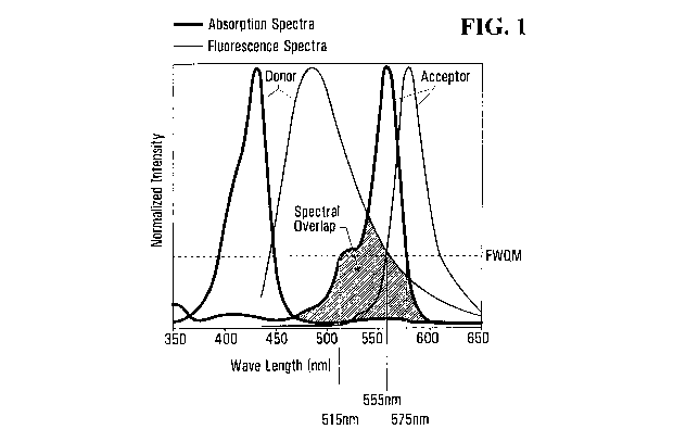
chromophore (Figure 1).
¨16¨
CA 02902363 2015-08-25
WO 2014/138930
PCT/CA2014/000261
Energy transfer manifests itself through decrease or quenching of the donor
emission and
a reduction of excited state lifetime accompanied also by an increase in
acceptor
emission intensity. Figure 2 is a Jablonski diagram that illustrates the
coupled transitions
involved between a donor emission and acceptor absorbance.
To enhance the energy transfer efficiency, the donor chromophore should have
good
abilities to absorb photons and emit photons. Furthermore, the more overlap
there is
between the donor chromophore's emission spectra and the acceptor
chromophore's
absorption spectra, the better a donor chromophore can transfer energy to the
acceptor
chromophore.
In certain embodiments, the biophotonic material of the present disclosure
further
comprises a second chromophore. In some embodiments, the first chromophore has
an
emission spectrum that overlaps at least about 80%, 50%, 40%, 30%, 20% or 10%
with
an absorption spectrum of the second chromophore. In one embodiment, the first
chromophore has an emission spectrum that overlaps at least about 20% with an
absorption spectrum of the second chromophore. In some embodiments, the first
chromophore has an emission spectrum that overlaps at least 1-10%, 5-15%, 10-
20%,
15-25%, 20-30%, 25-35%, 30-40%, 35-45%, 50-60%, 55-65% or 60-70% with an
absorption spectrum of the second chromophore.
% spectral overlap, as used herein, means the % overlap of a donor
chromophore's
emission wavelength range with an acceptor chromophore's absorption wavelength
rage, measured at spectral full width quarter maximum (FWQM). For example,
Figure 1
shows the normalized absorption and emission spectra of donor and acceptor
chromophores. The spectral FWQM of the acceptor chromophore's absorption
spectrum
is from about 60 nm (515 nm to about 575 nm). The overlap of the donor
chromophore's
spectrum with the absorption spectrum of the acceptor chromophore is about 40
nm
(from 515 nm to about 555 nm). Thus, the A) overlap can be calculated as 40nm
/ 60nm
x 100= 66.6%.
In some embodiments, the second chromophore absorbs at a wavelength in the
range of
the visible spectrum. In certain embodiments, the second chromophore has an
absorption
¨17-
CA 02902363 2015-08-25
WO 2014/138930
PCT/CA2014/000261
wavelength that is relatively longer than that of the first chromophore within
the range of
about 50-250, 25-150 or 10-100 nm.
The first chromophore can be present in an amount of about 0.001-40% per
weight of the
biophotonic material. When present, the second chromophore can be present in
an
amount of about 0.001-40% per weight of the biophotonic material. In certain
embodiments, the first chromophore is present in an amount of about 0.001-3%,
0.001-
0.01%, 0.005-0.1%, 0.1-0.5%, 0.5-2%, 1-5%, 2.5-7.5%, 5-10%, 7.5-12.5%, 10-15%,
12.5-17.5%, 15-20%, 17.5-22.5%, 20-25%, 22.5-27.5%, 25-30%, 27.5-32.5%, 30-
35%,
32.5-37.5%, or 35-40% per weight of the biophotonic material. In certain
embodiments,
the second chromophore is present in an amount of about 0.001-3%, 0.001-0.01%,
0.005-0.1%, 0.1-0.5%, 0.5-2%, 1-5%, 2.5-7.5%, 5-10%, 7.5-12.5%, 10-15%, 12.5-
17.5%, 15-20%, 17.5-22.5%, 20-25%, 22.5-27.5%, 25-30%, 27.5-32.5%, 30-35%,
32.5-
37.5%, or 35-40% per weight of the biophotonic material. In certain
embodiments, the
total weight per weight of chromophore or combination of chromophores may be
in the
amount of about 0.005-1%, 0.05-2%, 1-5%, 2.5-7.5%, 5-10%, 7.5-12.5%, 10-15%,
12.5-
17.5%, 15-20%, 17.5-22.5%, 20-25%, 22.5-27.5%, 25-30%, 27.5-32.5%, 30-35%,
32.5-
37.5%, or 35-40.001% per weight of the biophotonic material.
The concentration of the chromophore to be used can be selected based on the
desired
intensity and duration of the biophotonic activity from the biophotonic
material, and on
the desired medical or cosmetic effect. For example, some dyes such as
xanthene dyes
reach a 'saturation concentration' after which further increases in
concentration do not
provide substantially higher emitted fluorescence. Further increasing the
chromophore
concentration above the saturation concentration can reduce the amount of
activating
light passing through the matrix. Therefore, if more fluorescence is required
for a certain
application than activating light, a high 'saturation' concentration of
chromophore can be
used. However, if a balance is required between the emitted fluorescence and
the
activating light, a concentration close to or lower than the saturation
concentration can
be chosen.
Suitable chromophores that may be used in the biophotonic materials of the
present
disclosure include, but are not limited to the following:
Chlorophyll dyes
-18-
CA 02902363 2015-08-25
WO 2014/138930
PCT/CA2014/000261
Exemplary chlorophyll dyes include but are not limited to chlorophyll a;
chlorophyll b; chlorophyllin, oil soluble chlorophyll; bacteriochlorophyll a;
bacteriochlorophyll b; bacterioehlorophyll c; bacteriochlorophyll d;
protochlorophyll;
protochlorophyll a; amphiphilic chlorophyll derivative 1; and amphiphilic
chlorophyll
derivative 2.
Xanthene derivatives
Exemplary xanthene dyes include but are not limited to Eosin B (4',5'-
dibromo,2',7t-dinitr- o-fluorescein, dianion); eosin Y; eosin Y (2',4',5',7-
tetrabromo-
fluoresc- em, dianion); eosin (2',4',5',7'-tetrabromo-fluorescein, dianion);
eosin
(2',4',5',7'-tetrabromo-fluorescein, dianion) methyl ester; eosin (2',4',5',7'-
tetrabromo-
fluorescein, monoanion) p-isopropylbenzyl ester; eosin derivative (2',T-
dibromo-
fluorescein, dianion); eosin derivative (4',5'-dibromo-fluorescein, dianion);
eosin
derivative (2',7'-dichloro-fluorescein, dianion); eosin derivative (4',5'-
dichloro-
fluorescein, dianion); eosin derivative (2`,7'-diiodo-fluorescein, dianion);
eosin
derivative (4',5'-diiodo-fluorescein, dianion); eosin derivative (tribromo-
fluorescein,
dianion); eosin derivative (2',4',5',7`-tetrachlor- o-fluorescein, dianion);
eosin; eosin
dicetylpyridinium chloride ion pair; erythrosin B (2',4',5',T-tetraiodo-
fluorescein,
dianion); erythrosin; erythrosin dianion; erythiosin B; fluorescein;
fluorescein dianion;
phloxin B (2',4',5',7'-tetrabromo-3,4,5,6-tetrachloro-fluorescein, dianion);
phloxin B
(tetrachloro-tetrabromo-fluorescein); phloxine B; rose bengal (3,4,5,6-
tetrachloro-
21,41,5',7'-tetraiodofluorescein, dianion); pyronin G, pyronin J, pyronin Y;
Rhodamine
dyes such as rhodamines include 4,5-dibromo-rhodamine methyl ester; 4,5-
dibromo-
rhodamine n-butyl ester; rhodamine 101 methyl ester; rhodamine 123; rhodamine
6G;
rhodamine 6G hexyl ester; tetrabromo-rhodamine 123; and tetramethyl-rhodamine
ethyl
ester.
Methylene blue dyes
Exemplary methylene blue derivatives include but are not limited to 1-methyl
methylene blue; 1,9-dimethyl methylene blue; methylene blue; methylene blue
(16 1.1M);
methylene blue (14 p,M); methylene violet; bromomethylene violet; 4-
iodomethylene
violet; 1,9-dimethy1-3-dimethyl-amino-7-diethyl-a- mino-phenothiazine; and 1,9-
d imethy1-3-diethylamino-7-dibutyl-amino-phenot- hiazine.
Azo dyes
-19-
CA 02902363 2015-08-25
WO 2014/138930
PCT/CA2014/000261
Exemplary azo (or diazo-) dyes include but are not limited to methyl violet,
neutral red, para red (pigment red 1), amaranth (Azorubine S), Carmoisine
(azorubine,
food red 3, acid red 14), allura red AC (FD&C 40), tartrazine (FD&C Yellow 5),
orange
G (acid orange 10), Ponceau 4R (food red 7), methyl red (acid red 2), and
murexide-
3 ammonium purpurate.
In some aspects of the disclosure, the one or more chromophores of the
biophotonic
materials disclosed herein can be independently selected from any of Acid
black 1, Acid
blue 22, Acid blue 93, Acid fuchsin, Acid green, Acid green 1, Acid green 5,
Acid
magenta, Acid orange 10, Acid red 26, Acid red 29, Acid red 44, Acid red 51,
Acid red
66, Acid red 87, Acid red 91, Acid red 92, Acid red 94, Acid red 101, Acid red
103,
Acid roseine, Acid rubin, Acid violet 19, Acid yellow 1, Acid yellow 9, Acid
yellow 23,
Acid yellow 24, Acid yellow 36, Acid yellow 73, Acid yellow S, Acridine
orange,
Acriflavine, Alcian blue, Alcian yellow, Alcohol soluble eosin, Alizarin,
Alizarin blue
2RC, Alizarin carmine, Alizarin cyanin BBS, Alizarol cyanin R, Alizarin red S,
Alizarin
purpurin, Aluminon, Arnido black 10B, Amidoschwarz, Aniline blue WS,
Anthracene
blue SWR, Auramine 0, Azocannine B, Azocarmine G, Azoic diazo 5, Azoic diazo
48,
Azure A, Azure B, Azure C, Basic blue 8, Basic blue 9, Basic blue 12, Basic
blue 15,
Basic blue 17, Basic blue 20, Basic blue 26, Basic brown 1, Basic fuchsin,
Basic green 4,
Basic orange 14, Basic red 2, Basic red 5, Basic red 9, Basic violet 2, Basic
violet 3,
Basic violet 4, Basic violet 10, Basic violet 14, Basic yellow 1, Basic yellow
2, Biebrich
scarlet, Bismarck brown Y, Brilliant crystal scarlet 6R, Calcium red, Carmine,
Carminic
acid, Celestine blue B, China blue, Cochineal, Coelestine blue, Chrome violet
CG,
Chromotrope 2R, Chromoxane cyanin R, Congo corinth, Congo red, Cotton blue,
Cotton
red, Croceine scarlet, Crocin, Crystal ponceau 6R, Crystal violet, Dahlia,
Diamond green
B, Direct blue 14, Direct blue 58, Direct red, Direct red 10, Direct red 28,
Direct red 80,
Direct yellow 7, Eosin B, Eosin Bluish, Eosin, Eosin Y, Eosin yellowish,
Eosinol, Erie
garnet B, Eriochrome cyanin R, Erythrosin B, Ethyl eosin, Ethyl green, Ethyl
violet,
Evans blue, Fast blue B, Fast green FCF, Fast red B, Fast yellow, Fluorescein,
Food
green 3, Gallein, Gallamine blue, Gallocyanin, Gentian violet, Haematein,
Haematine,
Haematoxylin, Helio fast rubin BBL, Helvetia blue, Hematein, Hematine,
Hematoxylin,
Hoffman's violet, Imperial red, Indocyanin Green, Ingrain blue, Ingrain blue
1, Ingrain
yellow 1, INT, Kermes, Kermesic acid, Kernechtrot, Lac, Laccaic acid, Lauth's
violet,
Light green, Lissamine green SF, Luxol fast blue, Magenta 0, Magenta I,
Magenta II,
-20-
CA 02902363 2015-08-25
WO 2014/138930
PCT/CA2014/000261
Magenta III, Malachite green, Manchester brown, Martius yellow, Merbromin,
Mercurochrome, Metanil yellow, Methylene azure A. Methylene azure B, Methylene
azure C, Methylene blue, Methyl blue, Methyl green, Methyl violet, Methyl
violet 2B,
Methyl violet 10B, Mordant blue 3, Mordant blue 10, Mordant blue 14, Mordant
blue
23, Mordant blue 32, Mordant blue 45, Mordant red 3, Mordant red 11, Mordant
violet
25, Mordant violet 39 Naphthol blue black, Naphthol green B, Naphthol yellow
S,
Natural black 1, Natural green 3(chlorophyllin), Natural red, Natural red 3,
Natural red
4, Natural red 8, Natural red 16, Natural red 25, Natural red 28, Natural
yellow 6, NBT,
Neutral red, New fuchsin, Niagara blue 3B, Night blue, Nile blue, Nile blue A,
Nile blue
oxazone, Nile blue sulphate, Nile red, Nitro BT, Nitro blue tetrazolium,
Nuclear fast red,
Oil red 0, Orange G, Orcein, Pararosanilin, Phloxine B, Picric acid, Ponceau
2R,
Ponceau 6R, Ponceau B, Ponceau de Xylidine, Ponceau S, Primula, Purpurin,
Pyronin B,
phycobi I i ns, Phycocyan ins, Phycoerythrins.
Phycoerythrincyan in (PEG),
Phthalocyanines, Pyronin G, Pyronin Y, Quinine, Rhodamine B, Rosanilin, Rose
bengal,
Saffron, Safranin 0, Scarlet R, Scarlet red, Scharlach R, Shellac, Sirius red
F3B,
Solochrome cyanin R, Soluble blue, Solvent black 3, Solvent blue 38, Solvent
red 23,
Solvent red 24, Solvent red 27, Solvent red 45, Solvent yellow 94, Spirit
soluble eosin,
Sudan III, Sudan IV, Sudan black B, Sulfur yellow S, Swiss blue, Tartrazine,
Thioflavine S, Thioflavine T, Thionin, Toluidine blue, Toluyline red,
Tropaeolin G,
Trypaflavine, Trypan blue, Uranin, Victoria blue 4R, Victoria blue B, Victoria
green B,
Vitamin B, Water blue I, Water soluble eosin, Xylidine ponceau, or Yellowish
eosin.
In certain embodiments, the biophotonic material of the present disclosure
includes any
of the chromophores listed above, or a combination thereof, so as to provide a
synergistic biophotonic effect at the application site.
Without being bound to any particular theory, a synergistic effect of the
chromophore
combinations means that the biophotonic effect is greater than the sum of
their
individual effects. Advantageously, this may translate to increased reactivity
of the
biophotonic material, faster or improved treatment time. Also, the treatment
conditions
need not be altered to achieve the same or better treatment results, such as
time of
exposure to light, power of light source used, and wavelength of light used.
In other
words, use of synergistic combinations of chromophores may allow the same or
better
-21-
CA 02902363 2015-08-25
WO 2014/138930
PCT/CA2014/000261
treatment without necessitating a longer time of exposure to a light source, a
higher
power light source or a light source with different wavelengths.
In some embodiments, the material includes Eosin Y as a first chromophore and
any one
or more of Rose Bengal, Fluorescein, Erythrosine, Phloxine B, chlorophyllin as
a second
chromophore. It is believed that these combinations have a synergistic effect
as they can
transfer energy to one another when activated due in part to overlaps or close
proximity
of their absorption and emission spectra. This transferred energy is then
emitted as
fluorescence or leads to production of reactive oxygen species. This absorbed
and re-
emitted light is thought to be transmitted throughout the composition, and
also to be
transmitted into the site of treatment.
In further embodiments, the material includes the following synergistic
combinations:
Eosin Y and Fluorescein; Fluorescein and Rose Bengal; Erythrosine in
combination with
Eosin Y, Rose Bengal or Fluorescein; Phloxine B in combination with one or
more of
Eosin Y, Rose Bengal, Fluorescein and Erythrosine. Other synergistic
chromophore
combinations are also possible.
By means of synergistic effects of the chromophore combinations in the
material,
chromophores which cannot normally be activated by an activating light (such
as a blue
light from an LED), can be activated through energy transfer from chromophores
which
are activated by the activating light. In this way, the different properties
of
photoactivated chromophores can be harnessed and tailored according to the
cosmetic or
the medical therapy required.
For example, Rose Bengal can generate a high yield of singlet oxygen when
activated in
the presence of molecular oxygen, however it has a low quantum yield in terms
of
emitted fluorescent light. Rose Bengal has a peak absorption around 540 nm and
so can
be activated by green light. Eosin Y has a high quantum yield and can be
activated by
blue light. By combining Rose Bengal with Eosin Y, one obtains a composition
which
can emit therapeutic fluorescent light and generate singlet oxygen when
activated by
blue light. In this case, the blue light photoactivates Eosin Y which
transfers some of its
energy to Rose Bengal as well as emitting some energy as fluorescence.
-22-
CA 02902363 2015-08-25
WO 2014/138930
PCT/CA2014/000261
In some embodiments, the chromophore or chromophores are selected such that
their
emitted fluorescent light, on photoactivation, is within one or more of the
green, yellow,
orange, red and infrared portions of the electromagnetic spectrum, for example
having a
peak wavelength within the range of about 490 nm to about 800 nm. In certain
embodiments, the emitted fluorescent light has a power density of between
0.005 to
about 10 mW/cm2, about 0.5 to about 5 mW/cm2.
(b) Cohesive matrix
The biophotonic materials of the present disclosure comprise a cohesive matrix
made
from one or more thickening agents, or a carrier medium. In other words, the
biophotonic material of the present disclosure comprise one or more thickening
agents,
or a carrier medium which can form a cohesive matrix. These agents are present
in an
amount and ratio sufficient to provide a desired viscosity, flexibility,
rigidity, tensile
strength, tear strength, elasticity, and adhesiveness. The desired properties
may be one of
achieving a peelable film, or a rigid or flexible matrix. The thickening
agents are
selected so that the chromophore can remain photoactive in the cohesive
matrix. The
thickening agents are also selected according to the optical transparency of
the cohesive
matrix which they will form. The cohesive matrix should be able to transmit
sufficient
light to activate the at least one chromophore and, in embodiments where
fluorescence is
emitted by the activated chromophore, the cohesive matrix should also be able
to
transmit the emitted fluorescent light to tissues. It will be recognized by
persons skilled
in the art that the thickening agent is an appropriate medium for the
chromophore
selected. For example, the inventors have noted that some xanthene dyes do not
fluoresce in non-hydrated media, so hydrated polymers or polar solvents may be
used.
The thickening agents should also be selected according to the intended use.
For
example, if the biophotonic material is to be applied onto tissue, the
cohesive matrix is
preferably a biocompatible material, or the cohesive matrix has an outside
layer of a
biocompatible material which will interface the tissue.
Thickening agents
In some embodiments, the content of a thickening agent used to make the
cohesive
matrix is from about 0.001 % to about 40 % (w/w %) of the total weight. In
certain
embodiments, the total content of the thickening agent is about 0.001-0.01%,
about
0.005-0.05%, about 0.01-0.1, about 0.05-0.5% about 0.1-1%, about 0.5-5%, about
1-5%,
about 2.5-7.5%, about 5-10%, about 7.5-12.5%, about 10-15%, about 12.5-17.5%,
or
-23-
CA 02902363 2015-08-25
WO 2014/138930
PCT/CA2014/000261
about 15-20%, or about 15-25%, or about 20-30%, or about 25-35%, or about 30-
40%. It
will be recognized by one of skill in the art that the viscosity, flexibility,
rigidity, tensile
strength, tear strength, elasticity, and adhesiveness can be adjusted by
varying the
content of the thickening material. Methods of determining viscosity,
flexibility, rigidity,
tensile strength, tear strength, elasticity, and adhesiveness are known in the
art.
Thickening agents that can be used to prepare the biophotonic materials of the
present
disclosure include polymers, copolymers, and monomers of: vinylpyrrolidones,
methacrylamides, acrylamides N-vinylimidazoles, carboxy vinyls, vinyl esters,
vinyl
ethers, silicones, polyethyleneoxides, polyethyleneglycols, vinylalcohols,
sodium
acrylates, acrylates, maleic acids, NN-dimethylacrylamides, diacetone
acrylamides,
acrylamides, acryloyl morpholine, pluronic, collagens, polyacrylamides,
polyacrylates,
polyvinyl alcohols, polyvinylenes, polyvinyl silicates, polyacrylates
substituted with a
sugar (e.g., sucrose, glucose, glucosamines, galactose, trehalose, mannose, or
lactose),
acyl am idopropane sulfonic acids,
tetramethoxyorthosilicates,
methyltrimethoxyorthosilicates,
tetraalkoxyorthosilicates, trialkoxyortho silicates,
glycols, propylene glycol, glycerine, polysaccharides, alginates, dextrans,
cyclodextrin,
celluloses, modified celluloses, oxidized celluloses, chitosans, chitins,
guars,
carrageenans, hyaluronic acids, inulin, starches, modified
starches, agarose,
methylcelluloses, plant gums, hylaronans, hydrogels, gelatins,
glycosaminoglycans,
carboxymethyl celluloses, hydroxycthyl celluloses, hydroxy propyl methyl
celluloses,
pectins, low-methoxy pectins, cross-linked dextrans, starch-acrylonitrile
graft
copolymers, starch sodium polyacrylate, hydroxyethyl methacrylates, hydroxyl
ethyl
acrylates, polyvinylene, polyethylvinylethers, polymethyl methacrylates,
polystyrenes,
polyurethanes, polyalkanoates, polylactic acids, polylactates, poly(3-
hydroxybutyrate),
sulfonated hydrogels, AMPS (2-acrylamido-2-methyl-1-propanesulfonic acid), SEM
(sulfoethylmethacrylate), SPM (sulfopropyl methacrylate), SPA (sulfopropyl
acrylate),
N,N-dimethyl-N-methacryloxyethyl-N-(3-sulfopropyl)ammonium betaine,
methacryllic
acid amidopropyl-dimethyl ammonium sulfobetaine, SPI {itaconic acid-bis(I -
propyl
sulfonizacid-3) ester di-potassium salt}, itaconic acids, AMBC (3-acrylamido-3-
methylbutanoic acid), beta-carboxyethyl acrylate (acrylic acid dimers), and
maleic
anhydride-methylvinyl ether polymers, derivatives thereof, salts thereof,
acids thereof,
combinations thereof, and the like.
-24-
CA 02902363 2015-08-25
WO 2014/138930
PCT/CA2014/000261
Thickening agents also include poly (ethylene oxide) polymers (such as POLYOX
from
Dow Chemical), linear PVP and cross-linked PVP, PEG/PPG copolymers (such as
BASF Pluracare L1220), ethylene oxide (E0)--propylene oxide (PO) block
copolymers
(such as polymers sold under the trade mark Pluronic available from BASF
Corporation), ester gum, shellac, pressure sensitive silicone adhesives (such
as BioPSA
from Dow-Corning), or mixtures thereof. In some embodiments, a copolymer
comprises
(PVM/MA). In an embodiment, a copolymer comprises poly
(methylvinylether/maleic
anhydride). In some embodiments, a copolymer comprises poly
(methylvinylether/maleic acid). In some embodiments, a copolymer comprises
poly
(methylvinylether/maleic acid) half esters. In some embodiments, a copolymer
comprises poly (methylvinylether/maleic acid) mixed salts.
Thickening agents can also include carbomers which are a synthetic high
molecular
weight polymer of acrylic acid that is crosslinked with either allylsucrose or
allylethers
of pentaerythritol having a molecular weight of about 3 x 10⁶. The gelation
mechanism
depends on neutralization of the carboxylic acid moiety to form a soluble
salt. The
polymer is hydrophilic and produces sparkling clear gels when neutralized.
Carbomers
are available as fine white powders which disperse in water to form acidic
colloidal
suspensions (a 1% dispersion has approx. pH 3) of low viscosity.
Neutralization of these
suspensions using a base, for example sodium, potassium or ammonium
hydroxides, low
molecular weight amines and alkanolamines, results in the formation of clear
translucent
gels.
In one embodiment of the disclosure, the carbomer is Carbopol . Such polymers
are
commercially available from B.F. Goodrich or Lubrizol under the designation
Carbopol 71G NF, 420, 430, 475, 488, 493, 910, 934, 934P, 940, 971PNF, 974P
NF,
980 NF, 981 NF and the like. Carbopols are versatile controlled-release
polymers, as
described by Brock (Pharmacotherapy, 14:430-7 (1994)) and Durrani
(Pharmaceutical
Res. (Supp.) 8:S-135 (1991)), and belong to a family of carbomers which are
synthetic,
high molecular weight, non-linear polymers of acrylic acid, cross-linked with
polyalkenyl polyether. In some embodiments, the carbomer is Carbopol 974P NF,
980
NF, 5984 EP, ETD 2020NF, Ultrez 10 NF, 934 NF, 934P NF or 940 NF. In certain
embodiments, the carbomer is Carbopol 980 NF, ETD 2020 NF, Ultrez 10 NF,
Ultrez
21 or 1382 Polymer, 1342 NF, 940 NF.
-25-
CA 02902363 2015-08-25
WO 2014/138930
PCT/CA2014/000261
In certain embodiments of the disclosure, the thickening agent or the carrier
medium
may comprise gelatin. For example, the cohesive matrix may comprise at least
about 5%,
about 5 to about 25 weight%, or about 10 to about 20 weight% gelatin within
the
cohesive biophotonic material. Alternatively, a lower weight percentage of
gelatin may
be used together with chemical cross-linkers or any other cross-linking means.
In certain embodiments of the disclosure, the thickening agent or the carrier
medium
may comprise sodium hyaluronate, which may be present in an amount of about 2%
to
about 14%.
The biophotonic material of the present disclosure may be water soluble.
Alternatively,
the biophotonic material of the present disclosure may optionally include a
water-
insoluble or liposoluble substrate. By "water insoluble", it is meant that the
substrate
does not dissolve in or readily break apart upon immersion in water. In some
embodiments, the water-insoluble substrate is the implement or vehicle for
delivering the
treatment composition to the skin or target tissue. A wide variety of
materials can be
used as the water-insoluble substrate. The following non-limiting
characteristics may be
desirable: (i) sufficient wet strength for use, (ii) sufficient softness,
(iii) sufficient
thickness, (iv) appropriate size, (v) air permeability, and (vi)
hydrophilicity.
Non-limiting examples of suitable water-insoluble substrates which meet the
above
criteria include nonwoven substrates, woven substrates, hydroentangled
substrates, air
entangled substrates, natural sponges, synthetic sponges, polymeric netted
meshes, and
the like. Preferred embodiments employ nonwoven substrates since they are
economical
and readily available in a variety of materials. By "nonwoven", it is meant
that the layer
is comprised of fibers which are not woven into a fabric but rather are formed
into a
sheet, mat, or pad layer.
In one embodiment of the disclosure, the thickening agent or the cohesive
agent may
comprise a silicone membrane. In this embodiment, the chromophore or
chromophores
can be included directly within the silicone membrane or if they are water
soluble within
inclusions in the membrane as an aqueous phase. For example, the aqueous phase
may
be present as a micro-emulsion within the silicone. The aqueous phase may be a
liquid or
-26-
CA 02902363 2015-08-25
WO 2014/138930
PCT/CA2014/000261
a semi-solid. The aqueous phase may further comprise a stabilizing agent to
stabilize the
emulsion such as gelatin or CMC. The aqueous phase may comprise up to 40wt% of
the
silicone polymer phase.
Antimicrobials
Antimicrobials kill microbes or inhibit their growth or accumulation, and are
optionally
included in the biophotonic materials of the present disclosure. Exemplary
antimicrobials (or antimicrobial agent) are recited in U.S. Patent Application
Publications 20040009227 and 20110081530. Suitable antimicrobials for use in
the
methods and compositions of the present disclosure include, but not limited
to, hydrogen
peroxide, urea hydrogen peroxide, benzoyl peroxide, phenolic and chlorinated
phenolic
and chlorinated phenolic compounds, resorcinol and its derivatives,
bisphenolic
compounds, benzoic esters (parabens), halogenated carbonilides, polymeric
antimicrobial agents, thazolines, trichloromethylthioimides, natural
antimicrobial agents
(also referred to as "natural essential oils"), metal salts, and broad-
spectrum antibiotics.
Hydrogen peroxide (H202) is a powerful oxidizing agent, and breaks down into
water
and oxygen and does not form any persistent, toxic residual compound. A
suitable range
of concentration over which hydrogen peroxide can be used in the biophotonic
material
is from about 0.1% to about 3%, about 0.1 to 1.5%, about 0.1% to about 1%,
about 1%,
less than about 1%.
Urea hydrogen peroxide (also known as urea peroxide, carbamide peroxide or
percarbamide) is soluble in water and contains approximately 35% hydrogen
peroxide. A
suitable range of concentration over which urea peroxide can be used in the
biophotonic
material of the present disclosure is less than about 0.25 %, or less than
about 0.3%,
from 0.001 to 0.25%, or from about 0.3% to about 5%. Urea peroxide breaks down
to
urea and hydrogen peroxide in a slow-release fashion that can be accelerated
with heat or
photochemical reactions.
Benzoyl peroxide consists of two benzoyl groups (benzoic acid with the H of
the
carboxylic acid removed) joined by a peroxide group. It is found in treatments
for acne,
in concentrations varying from 2.5% to 10%. The released peroxide groups are
effective
-27-
CA 02902363 2015-08-25
WO 2014/138930
PCT/CA2014/000261
at killing bacteria. Benzoyl peroxide also promotes skin turnover and clearing
of pores,
which further contributes to decreasing bacterial counts and reduce acne.
Benzoyl
peroxide breaks down to benzoic acid and oxygen upon contact with skin,
neither of
which is toxic. A suitable range of concentration over which benzoyl peroxide
can be
used in the matrix biophotonic is from about 2.5% to about 5%.
According to certain embodiments, the biophotonic material of the present
disclosure
may optionally comprise one or more additional components, such as oxygen-rich
compounds as a source of oxygen radicals. Peroxide compounds are oxidants that
contain the peroxy group (R-0-0-R). which is a chainlike structure containing
two
oxygen atoms, each of which is bonded to the other and a radical or some
element. When
a biophotonic material of the present disclosure comprising an oxidant is
illuminated
with light, the chromophores are excited to a higher energy state. When the
chromophores' electrons return to a lower energy state, they emit photons with
a lower
energy level, thus causing the emission of light of a longer wavelength
(Stokes' shift). In
the proper environment, some of this energy is transferred to oxygen or the
reactive
hydrogen peroxide and causes the formation of oxygen radicals, such as singlet
oxygen.
The singlet oxygen and other reactive oxygen species generated by the
activation of the
biophotonic material are thought to operate in a hormetic fashion. That is, a
health
beneficial effect that is brought about by the low exposure to a normally
toxic stimuli
(e.g. reactive oxygen), by stimulating and modulating stress response pathways
in cells
of the targeted tissues. Endogenous response to exogenous generated free
radicals
(reactive oxygen species) is modulated in increased defense capacity against
the
exogenous free radicals and induces acceleration of healing and regenerative
processes.
Furthermore, activation of the oxidant may also produce an antibacterial
effect. The
extreme sensitivity of bacteria to exposure to free radicals makes the
biophotonic
material of the present disclosure potentially a bactericidal composition.
Specific phenolic and chlorinated phenolic antimicrobial agents that can be
used in the
disclosure include, but are not limited to: phenol; 2-methyl phenol; 3-methyl
phenol; 4-
methyl phenol; 4-ethyl phenol; 2,4-dimethyl phenol; 2,5-dimethyl phenol; 3,4-
dimethyl
phenol; 2,6-dimethyl phenol; 4-n-propyl phenol; 4-n-butyl phenol; 4-n-amyl
phenol; 4-
_98_
CA 02902363 2015-08-25
WO 2014/138930
PCT/CA2014/000261
tert-amyl phenol; 4-n-hexyl phenol; 4-n-heptyl phenol; mono- and poly-alkyl
and
aromatic halophenols; p-chlorophenyl; methyl p-chlorophenol; ethyl p-
chlorophenol; n-
propyl p-chlorophenol; n-butyl p-chlorophenol; n-amyl p-chlorophenol; sec-amyl
p-
chlorophenol; n-hexyl p-chlorophenol; cyclohexyl p-chlorophenol; n-heptyl p-
chlorophenol; n-octyl; p-chlorophenol; o-chlorophenol; methyl o-chlorophenol;
ethyl o-
chlorophenol; n-propyl o-chlorophenol; n-butyl o-chlorophenol; n-arnyl o-
chlorophenol;
tert-amyl o-chlorophenol; n-hexyl o-chlorophenol; n-heptyl o-chlorophenol; o-
benzyl p-
chlorophenol; o-benxyl-m-methyl p-chlorophenol; o-benzyl-m,m-dimethyl p-
chlorophenol; o-phenylethyl p-chlorophenol; o-phenylethyl-m-methyl p-
chlorophenol;
3-methyl p-chlorophenol 3,5-dimethyl p-chlorophenol, 6-ethyl-3-methyl p-
chlorophenol,
6-n-propy1-3-methyl p-chloropheno1; 6-iso-propy1-3-methyl p-chlorophenol; 2-
ethy1-3,5-
dimethyl p-chlorophenol; 6-sec-butyl-3-methyl p-chlorophenol; 2-iso-propy1-3,5-
dimethyl p-chlorophenol; 6-diethylmethy1-3-methyl p-chlorophenol; 6-iso-propy1-
2-
ethy1-3-methyl p-chlorophenol; 2-sec-amyl-3,5-dimethyl p-chlorophenol; 2-
diethylmethy1-3,5-dimethyl p-chlorophenol; 6-sec-octy1-3-methyl p-
chlorophenol; p-
chloro-m-cresol p-bromophenol; methyl p-bromophenol; ethyl p-bromophenol; n-
propyl
p-bromophenol; n-butyl p-bromophenol; n-amyl p-bromophenol; sec-amyl p-
bromophenol; n-hexyl p-bromophenol; cyclohexyl p-bromophenol; o-bromophenol;
tert-
amyl o-bromophenol; n-hexyl o-bromophenol; n-propyl-m,m-dimethyl o-
bromophenol;
2-phenyl phenol; 4-chloro-2-methyl phenol; 4-chloro-3-methyl phenol; 4-chloro-
3,5-
dimethyl phenol; 2,4-dichloro-3,5-dimethylphenol; 3,4,5,6-tetabromo-2-
methylphenol- ;
5 -methyl-2-pentylphenol; 4-i sopropy1-3 -methylphenol; para-chloro-
metaxylenol
(PCMX); chlorothymol; phenoxyethanol; phenoxyisopropanol; and 5-chloro-2-
hydroxyd iphenylm ethane.
Resorcinol and its derivatives can also be used as antimicrobial agents.
Specific
resorcinol derivatives include, but are not limited to: methyl resorcinol;
ethyl resorcinol;
n-propyl resorcinol; n-butyl resorcinol; n-amyl resorcinol; n-hexyl
resorcinol; n-heptyl
resorcinol; n-octyl resorcinol; n-nonyl resorcinol; phenyl resorcinol; benzyl
resorcinol;
phenylethyl resorcinol; phenylpropyl resorcinol; p-chlorobenzyl resorcinol; 5-
chloro-
2,4-dihydroxydiphenyl methane; 41-chloro-2,4-dihydroxydiphenyl methane; 5-
bromo-
2,4-dihydroxydiphenyl methane; and 4'-bromo-2,4-dihydroxydiphenyl methane.
-29-
CA 02902363 2015-08-25
WO 2014/138930
PCT/CA2014/000261
Specific bisphenolic antimicrobial agents that can be used in the disclosure
include, but
are not limited to: 2,2'-methylene bis-(4-chlorophenol); 2,4,4'trichloro-2'-
hydroxy-
diphenyl ether, which is sold by Ciba Geigy, Florham Park, N.J. under the
tradename
Triclosane; 2,2'-methylene bis-(3,4,6-trichlorophenol); 2,2'-methylene bis-(4-
chloro-6-
bromophenol); bis-(2-hydroxy-3,5-dichlorop- henyl) sulphide; and bis-(2-
hydroxy-5-
chlorobenzyl)sulphide.
Specific benzoie esters (parabens) that can be used in the disclosure include,
but are not
limited to: methylparaben; propylparaben; butylparaben; ethylparaben;
isopropylparaben; isobutylparaben; benzylparaben; sodium methylparaben; and
sodium
propylparaben.
Specific halogenated carbanilides that can be used in the disclosure include,
but are not
limited to: 3,4,4'-trichlorocarbanilides, such as 3-(4-chlorophenyI)-1-(3,4-
dichlorphenyl)urea sold under the tradename Triclocarban by Ciba-Geigy,
Florham
Park, N.J.; 3-trifluoromethy1-4,4'-dichlorocarbanilide; and 3,3',4-
trichlorocarbanilide.
Specific polymeric antimicrobial agents that can be used in the disclosure
include, but
are not limited to: polyhexamethylene biguanide hydrochloride; and
poly(iminoimidocarbonyl iminoimidocarbonyl iminohexamethylene hydrochloride),
which is sold under the tradename Vantocil IB.
Specific thazolines that can be used in the disclosure include, but are not
limited to that
sold under the tradename Micro-Check ; and 2-n-octy1-4-isothiazolin-3-one,
which is
sold under the tradename Vinyzene0 I1-3000 DIDP.
-30-
CA 02902363 2015-08-25
WO 2014/138930
PCT/CA2014/000261
Specific trichloromethylthioimides that can be used in the disclosure include,
but are not
limited to: N-(trichloromethylthio)phthalimide, which is sold under the
tradename
Fungitrol0; and N-trichloromethylthio-4-cyclohexene-1,2-dicarboximide, which
is sold
under the tradename Vancide .
Specific natural antimicrobial agents that can be used in the disclosure
include, but are
not limited to, oils of: anise; lemon; orange; rosemary; wintergreen; thyme;
lavender;
cloves; hops; tea tree; citronella; wheat; barley; lemongrass; cedar leaf;
cedarwood;
cinnamon; fleagrass; geranium; sandalwood; violet; cranberry; eucalyptus;
vervain;
peppermint; gum benzoin; basil; fennel; fir; balsam; menthol; ocmea origanuin;
hydastis;
carradensis; Berberidaceac daceae; Ratanhiae longa; and Curcuma longa. Also
included
in this class of natural antimicrobial agents are the key chemical components
of the plant
oils which have been found to provide antimicrobial benefit. These chemicals
include,
but are not limited to: anethol; catechole; camphene; thymol; eugenol;
eucalyptol; ferulic
acid; farnesol; hinokitiol; tropolone; limonene; menthol; methyl salicylate;
carvacol;
terpineol; verbenone; berberine; ratanhiae extract; caryophellene oxide;
citronellic acid;
curcumin; nerolidol; and geraniol.
Specific metal salts that can be used in the disclosure include, but are not
limited to, salts
of metals in groups 3a-5a, 3b-7b, and 8 of the periodic table. Specific
examples of metal
salts include, but are not limited to, salts of: aluminum; zirconium; zinc;
silver; gold;
copper; lanthanum; tin; mercury; bismuth; selenium; strontium; scandium;
yttrium;
cerium; praseodymiun; neodymium; promethum; samarium; europium; gadolinium;
terbium; dysprosium; holmium; erbium; thalium; ytterbium; lutetium; and
mixtures
thereof. An example of the metal-ion based antimicrobial agent is sold under
the
tradename HealthShielde, and is manufactured by HealthShield Technology,
Wakefield,
Mass.
-31-
CA 02902363 2015-08-25
WO 2014/138930
PCT/CA2014/000261
Specific broad-spectrum antimicrobial agents that can be used in the
disclosure include,
but are not limited to, those that are recited in other categories of
antimicrobial agents
herein.
Additional antimicrobial agents that can be used in the methods of the
disclosure
include, but are not limited to: pyrithiones, and in particular pyrithione-
including zinc
complexes such as that sold under the tradename Octopiroxe; dimethyidimethylol
hydantoin, which is sold under the tradename
Glydantt;
methylchloroisothiazolinone/methylisothiazolinone, which is sold under the
tradename
Kathon CGS; sodium sulfite; sodium bisulfite; imidazolidinyl urea, which is
sold under
the tradename Germall 1150; diazolidinyl urea, which is sold under the
tradename
Germall 110; benzyl alcohol v2-bromo-2-nitropropane-1,3-diol, which is sold
under the
tradename Bronopolt; formalin or formaldehyde; iodopropenyl butylcarbamate,
which
is sold under the tradename Polyphase P100 ; chloroacetamide; methanamine;
methyldibromonitrile glutaronitrile (1,2-dibromo-2,4-dicyanobutane), which is
sold
under the tradename Tektamer0; glutaraldehyde; 5-bromo-5-nitro-1,3-dioxane,
which is
sold under the tradename Bronidox0; phenethyl alcohol; o-phenylphenol/sodium o-
phenylphenol sodium hydroxymethylglycinate, which is sold under the tradename
Suttocide AO; polymethoxy bicyclic oxazolidine; which is sold under the
tradename
Nuosept COD; dimethoxane; thimersal; dichlorobenzyl alcohol; captan;
chlorphenenesin;
dichlorophene; chlorbutanol; glyceryl laurate; halogenated diphenyl ethers;
2,4,4'-
trichloro-2'-hydroxy-diphenyl ether, which is sold under the tradename
Triclosan and
is available from Ciba-Geigy, Florham Park, N.J.; and 2,2'-dihydroxy-5,5'-
dibromo-
diphenyl ether.
Additional antimicrobial agents that can be used in the methods of the
disclosure include
those disclosed by U.S. Pat. Nos. 3,141,321; 4,402,959; 4,430,381; 4,533,435;
4,625,026; 4,736,467; 4,855,139; 5,069,907; 5,091,102; 5,639,464; 5,853,883;
5,854,147; 5,894,042; and 5,919,554, and U.S. Pat. App!. Pub!. Nos.
20040009227 and
20110081530.
-32-
CA 02902363 2015-08-25
WO 2014/138930
PCT/CA2014/000261
(4) Optical properties of the Biophotonic Materials
In certain embodiments, biophotonic materials of the present disclosure are
substantially
transparent or translucent. The % transmittance of the biophotonic material
can be
measured in the range of wavelengths from 250 nm to 800 nm using, for example,
a
Perkin-Elmer Lambda 9500 series UV-visible spectrophotometer. In some
embodiments,
transmittance within the visible range is measured and averaged. In some other
embodiments, transmittance of the biophotonic material is measured with the
chromophore omitted. As transmittance is dependent upon thickness, the
thickness of
each sample can be measured with calipers prior to loading in the
spectrophotometer.
Transmittance values can be normalized according to
t2
FT-corr(A5 t2) = {e¨crt (A)tl]t1 = [FT-corrg, IIAt1
where tactual specimen thickness, t2=thickness to which transmittance
measurements
can be normalized. In the art, transmittance measurements are usually
normalized to 1
cm.
In certain embodiments, the biophotonic materials are substantially opaque. In
these
embodiments, the biophotonic materials may include light transmitting
structures such as
fibres, particles, networks, which are made of materials which can transmit
light. The
light transmitting structures can be waveguides such as optical fibres.
In some embodiments, the biophotonic material has a transmittance that is more
than
about 20%, 30%, 40%, 50%, 60%, 70%, or 75% within the visible range. In some
embodiments, the transmittance exceeds 40%, 41%, 42%, 43%, 44%, or 45% within
the
visible range.
(5) Forms of the Biophotonic Materials
The biophotonic materials of the present disclosure may be in the form of a
cohesive
film or matrix containing at least one chromophore. The cohesive film or
matrix may be
a cohesive gel, or a paste, a putty, a semi-solid, or a solid.
¨33¨
CA 02902363 2015-08-25
WO 2014/138930
PCT/CA2014/000261
The biophotonic materials of the present disclosure may be deformable. They
may be
elastic or non-elastic (i.e. flexible or rigid). The biophotonic materials,
for example, may
be in a peel-off form ('peelable') to provide ease and speed of use. In
certain
embodiments, the tear strength and/or tensile strength of the peel-off form is
greater than
its adhesion strength. This may help handleability of the material. It will be
recognized
by one of skill in the art that the properties of the peel-off biophotonic
material such as
cohesiveness, flexibility, elasticity, tensile strength, and tearing strength,
can be
determined and/or adjusted by methods known in the art such as by selecting
suitable
thickening agents and adapting their relative ratios.
The biophotonic material may be in a pre-formed shape. In certain embodiments,
the
pre-formed shape is in the form of, including, but not limited to, a film, a
face mask, a
patch, a dressing, or bandage. In certain embodiments, the pre-formed shapes
can be
customized for the individual user by trimming to size. In certain
embodiments,
perforations are provided around the perimeter of the pre-formed shape to
facilitate
trimming. In certain embodiments, the pre-shaping can be performed manually or
by
mechanical means such as 3-D printing. In the case of the 3-D printing the
size of the
area to be treated can be imaged, such as a wound or a face, then a 3-D
printer
configured to build or form a cohesive biophotonic material to match the size
and shape
of the imaged treatment area.
A biophotonic material of the disclosure can be configured with a shape and/or
size for application to a desired portion of a subject's body. For example,
the
biophotonic material can be shaped and sized to correspond with a desired
portion of the
body to receive the biophotonic treatment. Such a desired portion of skin can
be selected
from, but not limited to, the group consisting of a skin, head, forehead,
scalp, nose,
cheeks, lips, ears, face, neck, shoulder, arm pit, arm, elbow, hand, finger,
abdomen,
chest, stomach, back, buttocks, sacrum, genitals, legs, knee, feet, toes,
nails, hair, any
boney prominences, and combinations thereof, and the like. Thus, the
biophotonic
material of the disclosure can be shaped and sized to be applied to any
portion of skin on
a subject's body. For example, the biophotonic material can be sock, hat,
glove or mitten
shaped. In embodiments where the biophotonic material is elastic or rigid, it
can be
peeled-off without leaving any residue on the tissue.
-34-
CA 02902363 2015-08-25
WO 2014/138930
PCT/CA2014/000261
In certain embodiments, the biophotonic material is in the form of an elastic
and peelable
face mask, which may be pre-formed. In other embodiments, the biophotonic
material is
in the form of a non-elastic (rigid) face mask, which may also be pre-formed.
The mask
can have openings for one or more of the eyes, nose and mouth. In a further
embodiment, the openings are protected with a covering, or the exposed skin
such as on
the nose, lips or eyes are protected using for example cocoa butter. In
certain
embodiments, the pre-formed face mask is provided in the form of multiple
parts, e.g.,
an upper face part and a lower face part. In certain embodiments, the uneven
proximity
of the face to a light source is compensated for, e.g., by adjusting the
thickness of the
mask, or by adjusting the amount of chromophore in the different areas of the
mask, or
by blocking the skin in closest proximity to the light. In certain
embodiments, the pre-
formed shapes come in a one-size fits all form.
In certain aspects, the mask (or patch) is not pre-formed and is applied e.g.,
by spreading
a composition making up the mask (or patch), on the skin or target tissue, or
alternatively by spraying, smearing, dabbing or rolling the composition on
target tissue.
It can then be converted to a peel-off form after application, by means such
as, but not
limited to, drying, illumination with light, change in temperature or pH upon
application
to the skin or tissue. The mask (or patch) can then be peeled off without
leaving any
flakes on the skin or tissue, preferably without wiping or washing.
In certain aspects, the biophotonic material may have shape memory properties.
For
example, the biophotonic material can include a shape memory material, such as
a shape
memory polymer whose original shape is reverted to on activation by light. The
original
shape can be a flat or concave configuration which allows the film/matrix to
be readily
peeled off the tissue. The shape memory material may be included as a layer
attached to
the biophotonic material, or integrated with the biophotonic material.
In certain aspects, the biophotonic material forms part of a composite and can
include
fibres, particulates, non-biophotonic layers or biophotonic layers with the
same or
different compositions.
In certain embodiments, the biophotonic material may comprise a plurality of
wavegu ides extending at least partially through the biophotonic material or
contained at
-35-
CA 02902363 2015-08-25
WO 2014/138930
PCT/CA2014/000261
least partially within the biophotonic material. The waveguides can be
attached to a light
source to thereby illuminate the biophotonic material from within. The
biophotonic
material may further include the light source attached to the waveguides. The
waveguides can be optical fibres which can transmit light, not only from their
ends, but
also from their body. For example, made of polycarbonate or
polymethylmethacrylate or
any other suitable material.
In a different embodiment, the biophotonic material comprises a layer of a
woven or
non-woven fabric dressing or a mask. Waveguides or a light source may be
included
within the dressing or mask fabric. For example, the dressing or mask fabric
can be in
the form of an envelope which receives the biophotonic material, and which
comprises at
least one light emitting surface.
In certain aspects, the biophotonic material is formed as a filter. For
example, the
biophotonic material can be made to have a shape and a size which can be
connected to,
or spaced from, a light emitting surface of a lamp. In one embodiment, the
lamp can be a
hand-held lamp such as a torch or a dentist's curing lamp. The lamp with the
biophotonic
filter can then be used to treat tissue sites of patient in a contacting or
non-contacting
manner. In this embodiment, the filter has a body having a first end which is
sized and
shaped to be connectable to a light emitting surface, and a second end shaped
to treat
tissues.
In certain aspects, the biophotonic material is formed as a waveguide. In
certain
embodiments, at least one chromophore is included in an elongate solid matrix
having
good light propagation properties and appropriate mechanical properties. The
waveguide
may be flexible. The waveguide can be shaped as an optical fibre. Such an
optical fibre
can be connected to a light source, and the at least one chromophore in the
cohesive
matrix activated by the light source to deliver therapeutic fluorescent light
to hard to
reach places, such as internal cavities and periodontal pockets.
Polymethylmethacrylate
is an example of an appropriate cohesive matrix for use as a biophotonic
waveguide.
Such a waveguide may additionally include a coating to prevent light
dissipation from
along its length.
-36-
CA 02902363 2015-08-25
WO 2014/138930
PCT/CA2014/000261
In other aspects, the biophotonic material comprising at least one chromophore
and a
cohesive matrix is in the form of particulates. Material processing techniques
known in
the art can be used to form particulates of any shape or size. These
particulates can be
contained in semi-solid or liquid preparations. For example, such biophotonic
particulates can be used in skin preparations such as creams, emulsions to
provide
therapeutic effect to the skin. In this case, a biocompatible solid matrix is
used and can
be used to encapsulate all types of chromophores, even those not well
tolerated by the
skin.
The biophotonic materials of the present disclosure may have a thickness of
from about
0.1 mm to about 50 mm, about 0.5 mm to about 20 mm, or about 1 mm to about 10
mm.
It will be appreciated that the thickness of the biophotonic materials will
vary based on
the intended use. In some embodiments, the biophotonic material has a
thickness of from
about 0.1-1 mm. In some embodiments, the biophotonic material has a thickness
of
about 0.5-1.5 mm, about 1-2 mm, about 1.5-2.5 mm, about 2-3 mm, about 2.5-3.5
mm,
about 3-4 mm, about 3.5-4.5 mm, about 4-5 mm, about 4.5-5.5 mm, about 5-6 mm,
about 5.5-6.5 mm, about 6-7 mm, about 6.5-7.5 mm, about 7-8 mm, about 7.5-8.5
mm,
about 8-9 mm, about 8.5-9.5, about 9-10 mm, about 10-11mm, about 11-12 mm,
about
12-13 mm, about 13-14 mm, about 14-15 mm, about 15-16 mm, about 16-17 mm,
about
17-18 mm, about 18-19 mm, about 19-20 mm, about 20-22mm, about 22-24mm, about
24-26mm, about 26-28mm, about 28-30mm, about 30-35mm, about 35-40mm, about 40-
45mm, about 45-50mm.
The tensile strength of the biophotonic materials will vary based on the
intended use.
The tensile strength can be determined by performing a tensile test and
recording the
force and displacement. These are then converted to stress (using cross
sectional area)
and strain; the highest point of the stress-strain curve is the "ultimate
tensile strength." In
some embodiments, tensile strength can be characterized using a 500N capacity
tabletop
mechanical testing system (#5942R4910, Instron ) with a 5N maximum static load
cell
(#102608, Instron). Pneumatic side action grips can be used to secure the
samples
(42712-019, Instron). In some embodiments, a constant extension rate (for
example, of
about 2 mm/min) until failure can be applied and the tensile strength is
calculated from
the stress vs. strain data plots. In some embodiments, the tensile strength
can be
measured using methods as described in or equivalent to those described in
American
-37-
CA 02902363 2015-08-25
WO 2014/138930
PCT/CA2014/000261
Society for Testing and Materials tensile testing methods such as ASTM D638,
ASTM
D882 and ASTM D412.
In some embodiments, the biophotonic material has a tensile strength of from
about 1-50
kPa, 1 to about 1000 kPa, 1 to about 500 kPa, 50 kPa to about 600 kPa. In some
embodiments, the tensile strength is from about 75 kPa to about 500 kPa, from
about 100
kPa to about 200 kPa, 100-300 kPa, 400 kPa, from about 150 kPa to about 350
kPa, or
from about 200 kPa to about 300 kPa.
In some embodiments, the tensile strength is at least about 50 kPa, at least
about 75 kPa,
at least about 100 kPa, at least about 150 kPa, at least about 200 kPa, at
least about 250
kPa, at least about 300 kPa, at least about 350 kPa, at least about 400 kPa,
at least about
450 kPa, at least about 500 kPa, at least about 550 kPa or at least about 600
kPa.
In some embodiments, the tensile strength of the biophotonic material is up to
about 8
MPa.
The tear strength of the biophotonic material will vary depending on the
intended use.
The tear strength property of the biophotonic material can be tested using a
500N
capacity tabletop mechanical testing system (#5942R4910, Instron) with a 5N
maximum
static load cell (#102608, Instron). Pneumatic side action grips can be used
to secure the
samples (#2712-019, Instron). Samples can be tested with a constant extension
rate (for
example, of about 2 mm/min) until failure. In accordance with the invention,
tear
strength is calculated as the force at failure divided by the average
thickness (N/mm).
In some embodiments, the biophotonic material has a tear strength of from
about 0.1
N/mm to about 1 N/mm. In some embodiments, the tear strength is from about
0.20
N/mm to about 0.40 N/mm, from about 0.25 N/mm to about 0.35 N/mm, from about
0.25 N/mm to about 0.45 N/mm, from about 0.35 N/mm to about 0.535 N/mm, from
about 0.45 N/mm to about 0.65 N/mm, from about 0.55 N/mm to about 0.75 N/mm,
from about 0.65 N/mm to about 0.85 N/mm, from about 0.75 N/mm to about 0.95
N/mm.
-38-
CA 02902363 2015-08-25
WO 2014/138930
PCT/CA2014/000261
In some embodiments, the tear strength is at least about 0.10 N/mm, at least
about 0.15
N/mm, at least about 0.20 N/mm, at least about 0.25 N/mm, at least about 0.30
N/mm, at
least about 0.35 N/mm, at least about 0.40 N/mm, at least about 0.45 N/mm, at
least
about 0.55 N/mm or at least about I N/mm.
The adhesion strength of the biophotonic material will vary depending on the
intended
use. Adhesion strength can be determined in accordance with ASTM D-3330-78,
PSTC-
101 and is a measure of the force required to remove a biophotonic material
from a test
panel at a specific angle and rate of removal. In some embodiments, a
predetermined
size of a biophotonic material is applied to a horizontal surface of a clean
glass test plate.
A hard rubber roller is used to firmly apply the piece and remove all
discontinuities and
entrapped air. The free end of the piece of biophotonic material is then
doubled back
nearly touching itself so that the angle of removal of the piece from the
glass plate will
be 180 degrees. The free end of the piece of biophotonic material is attached
to the
adhesion tester scale (e.g. an Instron tensile tester or Harvey tensile
tester). The test plate
is then clamped in the jaws of the tensile testing machine capable of moving
the plate
away from the scale at a predetermined constant rate. The scale reading in kg
is recorded
as the biophotonic material is peeled from the glass surface.
In some embodiments, the adhesion strength can be measured by taking into
account the
static friction of the biophotonic material. For some embodiments of the
cohesive
biophotonic materials of the present disclosure, the adhesive properties are
linked to
their levels of static friction, or stiction. In these cases, the adhesion
strength can be
measured by placing the sample on a test surface and pulling one end of the
sample at an
angle of approximately 0 (substantially parallel to the surface) whilst
applying a known
downward force (e.g. a weight) on the sample and measuring the weight at which
the
sample slips from the surface. The normal force Fn, is the force exerted by
each surface
on the other in a perpendicular (normal) direction to the surface and is
calculated by
multiplying the combined weight of the sample and the weight by the gravity
constant
(g) (9.8m/s2). The biophotonic material with the weight on top is then pulled
away from
a balance until the biophotonic material slips from the surface and the weight
is recorded
on the scale. The weight recorded on the scale is equivalent to the force
required to
overcome the friction. The force of friction (Ff) is then calculated by
multiplying the
weight recorded on the scale by g. Since Ff).1F5 (Coulomb's friction law), the
friction
-39-
CA 02902363 2015-08-25
WO 2014/138930
PCT/CA2014/000261
coefficient )1 can be obtained by dividing Ff / Fn. The stress required to
shear a material
from a surface (adhesion strength) can then be calculated from the friction
coefficient,
by multiplying the weight of the material by the friction coefficient.
In some embodiments, the biophotonic material has an adhesion strength that is
less than
its tensile strength. In some embodiments, the biophotonic material has an
adhesion
strength that is less than its tear strength.
In some embodiments, the biophotonic material has an adhesion strength of from
about
0.01 N/mm to about 0.60 N/mm. In some embodiments, the adhesion strength is
from
about 0.20 N/mm to about 0.40 N/mm, or from about 0.25 N/mm to about 0.35
N/mm.
In some embodiments, the adhesion strength is less than about 0.10 N/mm, less
than
about 0.15 N/mm, less than about 0.20 N/mm, less than about 0.25 N/mm, less
than
about 0.30 N/mm, less than about 0.35 N/mm, less than about 0.40 N/mm, less
than
about 0.45 N/mm, less than about 0.55 N/mm or less than about 0.60 N/mm.
(6) Methods of Use
The biophotonic materials of the present disclosure may have cosmetic and/or
medical
benefits. They can be used to promote skin rejuvenation and skin conditioning,
promote
the treatment of a skin disorder such as acne, eczema or psoriasis, promote
tissue repair,
and promote wound healing including periodontitis pockets. They can be used to
treat
acute inflammation. Acute inflammation can present itself as pain, heat,
redness,
swelling and loss of function. It includes those seen in allergic reactions
such as insect
bites e.g.; mosquito, bees, wasps, poison ivy, or post-ablative treatment.
Accordingly, in certain embodiments, the present disclosure provides a method
for
treating acute inflammation.
In certain embodiments, the present disclosure provides a method for providing
skin
rejuvenation or for improving skin condition, treating a skin disorder,
preventing or
treating scarring, and/or accelerating wound healing and/or tissue repair, the
method
comprising: applying a biophotonic material of the present disclosure to the
area of the
-40-
CA 02902363 2015-08-25
WO 2014/138930
PCT/CA2014/000261
skin or tissue in need of treatment, and illuminating the biophotonic material
with light
having a wavelength that overlaps with an absorption spectrum of the
chromophore(s)
present in the biophotonic material.
In the methods of the present disclosure, any source of actinic light can be
used. Any
type of halogen, LED or plasma arc lamp, or laser may be suitable. The primary
characteristic of suitable sources of actinic light will be that they emit
light in a
wavelength (or wavelengths) appropriate for activating the one or more
photoactivators
present in the composition. In one embodiment, an argon laser is used. In
another
embodiment, a potassium-titanyl phosphate (KTP) laser (e.g. a GreenLightTM
laser) is
used. In yet another embodiment, a LED lamp such as a photocuring device is
the source
of the actinic light. In yet another embodiment, the source of the actinic
light is a source
of light having a wavelength between about 200 to 800 nm. In another
embodiment, the
source of the actinic light is a source of visible light having a wavelength
between about
400 and 600 nm. In another embodiment, the source of the actinic light is a
source of
visible light having a wavelength between about 400 and 700 nm. In yet another
embodiment, the source of the actinic light is blue light. In yet another
embodiment, the
source of the actinic light is red light. In yet another embodiment, the
source of the
actinic light is green light. Furthermore, the source of actinic light should
have a suitable
power density. Suitable power density for non-collimated light sources (LED,
halogen or
plasma lamps) are in the range from about 0.1 mW/cm2 to about 200 mW/cm2.
Suitable
power density for laser light sources are in the range from about 0.5 mW/cm2
to about
0.8 mW/cm2.
In some embodiments of the methods of the present disclosure, the light has an
energy at
the subject's skin surface of between about 0.1 mW/cm2 and about 500 mW/cm2,
or 0.1-
300 mW/cm2, or 0.1-200 mW/cm2, wherein the energy applied depends at least on
the
condition being treated, the wavelength of the light, the distance of the skin
from the
light source and the thickness of the biophotonic material. In certain
embodiments, the
light at the subject's skin is between about 1-40 mW/cm2, or 20-60 mW/cm2, or
40-80
mW/cm2, or 60-100 mW/cm2, or 80-120 mW/cm2, or 100-140 mW/cm2, or 30-180
mW/cm2, or 120-160 mW/cm2, or 140-180 mW/cm2, or 160-200 mW/cm2, or 110-240
mW/cm2, or 110-150 mW/cm2, or 190-240 mW/cm2.
¨41¨
CA 02902363 2015-08-25
WO 2014/138930
PCT/CA2014/000261
The activation of the chromophore(s) within the biophotonic material may take
place
almost immediately on illumination (femto- or pico seconds). A prolonged
exposure
period may be beneficial to exploit the synergistic effects of the absorbed,
reflected and
reemitted light of the biophotonic material of the present disclosure and its
interaction
with the tissue being treated. In one embodiment, the time of exposure to
actinic light of
the tissue or skin or biophotonic material is a period between 1 minute and 5
minutes. In
another embodiment, the time of exposure to actinic light of the tissue or
skin or
biophotonic material is a period between 1 minute and 5 minutes. In some other
embodiments, the biophotonic material is illuminated for a period between 1
minute and
3 minutes. In certain embodiments, light is applied for a period of 1-30
seconds, 15-45
seconds, 30-60 seconds, 0.75-1.5 minutes, 1-2 minutes, 1.5-2.5 minutes, 2-3
minutes,
2.5-3.5 minutes, 3-4 minutes, 3.5-4.5 minutes, 4-5 minutes, 5-10 minutes, 10-
15
minutes, 15-20 minutes, or 20-30 minutes. The treatment time may range up to
about 90
minutes, about 80 minutes, about 70 minutes, about 60 minutes, about 50
minutes, about
40 minutes or about 30 minutes. It will be appreciated that the treatment time
can be
adjusted in order to maintain a dosage by adjusting the rate of fluence
delivered to a
treatment area. For example, the delivered fluence may be about 4 to about 60
J/cm2,
about 10 to about 60 J/cm2, about 10 to about 50 J/cm2, about 10 to about 40
J/cm2,
about 10 to about 30 J/cm2, about 20 to about 40 J/cm2, about 15 J/cm2 to 25
J/cm2, or
about 10 to about 20 J/cm2.
In certain embodiments, the biophotonic material may be re-illuminated at
certain
intervals. In yet another embodiment, the source of actinic light is in
continuous motion
over the treated area for the appropriate time of exposure. In yet another
embodiment,
the biophotonic composition may be illuminated until the biophotonic
composition is at
least partially photobleached or fully photobleached.
In certain embodiments, the chromophore(s) in the cohesive matrix can be
photoexcited
by ambient light including from the sun and overhead lighting. In certain
embodiments,
the chromophore(s) can be photoactivated by light in the visible range of the
electromagnetic spectrum. The light can be emitted by any light source such as
sunlight,
light bulb, an LED device, electronic display screens such as on a television,
computer,
telephone, mobile device, flashlights on mobile devices. In the methods of the
present
disclosure, any source of light can be used. For example, a combination of
ambient light
-42-
CA 02902363 2015-08-25
WO 2014/138930
PCT/CA2014/000261
and direct sunlight or direct artificial light may be used. Ambient light can
include
overhead lighting such as LED bulbs, fluorescent bulbs etc, and indirect
sunlight.
In the methods of the present disclosure, the biophotonic material may be
removed from
3 the skin following application of light. In some embodiments the
biophotonic material is
peeled off from the skin following application of light. In some embodiments,
the
biophotonic material is removed as a single piece from the skin following
application of
light. In other embodiments, the biophotonic material is left on the tissue
for an extended
period of time and re-activated with direct or ambient light at appropriate
times to treat
the condition.
In certain embodiments of the method of the present disclosure, the
biophotonic material
can be applied to the tissue, such as on the face, once, twice, three times,
four times, five
times or six times a week, daily, or at any other frequency. The total
treatment time can
be one week, two weeks, three weeks, four weeks, five weeks, six weeks, seven
weeks,
eight weeks, nine weeks, ten weeks, eleven weeks, twelve weeks, or any other
length of
time deemed appropriate. In certain embodiments, the total tissue area to be
treated may
be split into separate areas (cheeks, forehead), and each area treated
separately. For
example, the composition may be applied topically to a first portion, and that
portion
illuminated with light, and the biophotonic composition then removed. Then the
composition is applied to a second portion, illuminated and removed. Finally,
the
composition is applied to a third portion, illuminated and removed.
In certain embodiments, the biophotonic material can be used following wound
closure
to optimize scar revision. In this case, the biophotonic material may be
applied at regular
intervals such as once a week, or at an interval deemed appropriate by the
physician.
In certain embodiments, the biophotonic material can be used following acne
treatment
to maintain the condition of the treated skin. In this case, the biophotonic
material may
be applied at regular intervals such as once a week, or at an interval deemed
appropriate
by the physician.
In certain embodiments, the biophotonic material can be used following
ablative skin
rejuvenation treatment to maintain the condition of the treated skin. In this
case, the
-43-
CA 02902363 2015-08-25
WO 2014/138930 PCT/CA2014/000261
biophotonic material may be applied at regular intervals such as once a week,
or at an
interval deemed appropriate by the physician.
In the methods of the present disclosure, additional components may optionally
be
included in the biophotonic materials or used in combination with the
biophotonic
materials. Such additional components include, but are not limited to, healing
factors,
antimicrobials, oxygen-rich agents, wrinkle fillers such as botox, hyaluronic
acid and
polylactic acid, fungal, anti-bacterial, anti-viral agents and/or agents that
promote
collagen synthesis. These additional components may be applied to the skin in
a topical
fashion, prior to, at the same time of; and/or after topical application of
the biophotonic
materials of the present disclosure. Suitable healing factors comprise
compounds that
promote or enhance the healing or regenerative process of the tissues on the
application
site. During the photoactivation of a biophotonic material of the present
disclosure, there
may be an increase of the absorption of molecules of such additional
components at the
treatment site by the skin or the mucosa. In certain embodiments, an
augmentation in the
blood flow at the site of treatment can observed for a period of time. An
increase in the
lymphatic drainage and a possible change in the osmotic equilibrium due to the
dynamic
interaction of the free radical cascades can be enhanced or even fortified
with the
inclusion of healing factors. Healing factors may also modulate the
biophotonic output
from the biophotonic composition such as photobleaching time and profile, or
modulate
leaching of certain ingredients within the composition. Suitable healing
factors include,
but are not limited to glucosamines, allantoins, saffron, agents that promote
collagen
synthesis, anti-fungal, anti-bacterial, anti-viral agents and wound healing
factors such as
growth factors.
(i) Skin Rejuvenation
The biophotonic material of the present disclosure may be useful in promoting
skin
rejuvenation or improving skin condition and appearance. The dermis is the
second layer
of skin, containing the structural elements of the skin, the connective
tissue. There are
various types of connective tissue with different functions. Elastin fibers
give the skin its
elasticity, and collagen gives the skin its strength.
-44-
CA 02902363 2015-08-25
WO 2014/138930
PCT/CA2014/000261
The junction between the dermis and the epidermis is an important structure.
The
dermal-epidermal junction interlocks forming finger-like epidermal ridges. The
cells of
the epidermis receive their nutrients from the blood vessels in the dermis.
The epidermal
ridges increase the surface area of the epidermis that is exposed to these
blood vessels
and the needed nutrients.
The aging of skin comes with significant physiological changes to the skin.
The
generation of new skin cells slows down, and the epidermal ridges of the
dermal-
epidermal junction flatten out. While the number of elastin fibers increases,
their
structure and coherence decreases. Also the amount of collagen and the
thickness of the
dermis decrease with the ageing of the skin.
Collagen is a major component of the skin's extracellular matrix, providing a
structural
framework. During the aging process, the decrease of collagen synthesis and
insolubilization of collagen fibers contribute to a thinning of the dermis and
loss of the
skin's biomechanical properties.
The physiological changes to the skin result in noticeable aging symptoms
often referred
to as chronological-, intrinsic- and photo-ageing. The skin becomes drier,
roughness and
scaling increase, the appearance becomes duller, and most obviously fine lines
and
wrinkles appear. Other symptoms or signs of skin aging include, but are not
limited to,
thinning and transparent skin, loss of underlying fat (leading to hollowed
cheeks and eye
sockets as well as noticeable loss of firmness on the hands and neck), bone
loss (such
that bones shrink away from the skin due to bone loss, which causes sagging
skin), dry
skin (which might itch), inability to sweat sufficiently to cool the skin,
unwanted facial
hair, freckles, age spots, spider veins, rough and leathery skin, fine
wrinkles that
disappear when stretched, loose skin, a blotchy complexion.
The dermal-epidermal junction is a basement membrane that separates the
keratinocytes
in the epidermis from the extracellular matrix, which lies below in the
dermis. This
membrane consists of two layers: the basal lamina in contact with the
keratinocytes, and
the underlying reticular lamina in contact with the extracellular matrix. The
basal lamina
is rich in collagen type IV and laminin, molecules that play a role in
providing a
structural network and bioadhesive properties for cell attachment.
-45--
CA 02902363 2015-08-25
WO 2014/138930
PCT/CA2014/000261
Laminin is a glycoprotein that only exists in basement membranes. It is
composed of
three polypeptide chains (alpha, beta and gamma) arranged in the shape of an
asymmetric cross and held together by disulfide bonds. The three chains exist
as
different subtypes which result in twelve different isoforms for laminin,
including
Laminin-1 and Laminin-5.
The dermis is anchored to hemidesmosomes, specific junction points located on
the
keratinocytes, which consist of a-integrins and other proteins, at the basal
membrane
keratinocytes by type VII collagen fibrils. Laminins, and particularly Laminin-
5,
constitute the real anchor point between hemidesmosomal transmembrane proteins
in
basal keratinocytes and type VII collagen.
Laminin-5 synthesis and type VII collagen expression have been proven to
decrease in
aged skin. This causes a loss of contact between dermis and epidermis, and
results in the
skin losing elasticity and becoming saggy.
Recently another type of wrinkles, generally referred to as expression
wrinkles, got
general recognition. These wrinkles require loss of resilience, particularly
in the dermis,
because of which the skin is no longer able to resume its original state when
facial
muscles which produce facial expressions exert stress on the skin, resulting
in expression
wrinkles.
The biophotonic material of the present disclosure and methods of the present
disclosure
promote skin rejuvenation. In certain embodiments, the biophotonic material
and
methods of the present disclosure promote skin condition such as skin
luminosity,
reduction of pore size, reducing blotchiness, making even skin tone, reducing
dryness,
and tightening of the skin. In certain embodiments, the biophotonic material
and
methods of the present disclosure promote collagen synthesis. In certain other
embodiments, the biophotonic material and methods of the present disclosure
may
reduce, diminish, retard or even reverse one or more signs of skin aging
including, but
not limited to, appearance of fine lines or wrinkles, thin and transparent
skin, loss of
underlying fat (leading to hollowed cheeks and eye sockets as well as
noticeable loss of
firmness on the hands and neck), bone loss (such that bones shrink away from
the skin
-46-
CA 02902363 2015-08-25
WO 2014/138930
PCT/CA2014/000261
due to bone loss, which causes sagging skin), dry skin (which might itch),
inability to
sweat sufficiently to cool the skin, unwanted facial hair, freckles, age
spots, spider veins,
rough and leathery skin, fine wrinkles that disappear when stretched, loose
skin, or a
blotchy complexion. In certain embodiments, the biophotonic material and
methods of
the present disclosure may induce a reduction in pore size, enhance
sculpturing of skin
subsections, and/or enhance skin translucence.
In certain embodiments, the biophotonic material may be used in conjunction
with
collagen promoting agents. Agents that promote collagen synthesis (i.e., pro-
collagen
synthesis agents) include amino acids, peptides, proteins, lipids, small
chemical
molecules, natural products and extracts from natural products.
For instance, it was discovered that intake of vitamin C, iron, and collagen
can
effectively increase the amount of collagen in skin or bone. See, e.g., U.S.
Patent
Application Publication 20090069217. Examples of the vitamin C include an
ascorbic
acid derivative such as L-ascorbic acid or sodium L-ascorbate, an ascorbic
acid
preparation obtained by coating ascorbic acid with an emulsifier or the like,
and a
mixture containing two or more of those vitamin Cs at an arbitrary rate. In
addition,
natural products containing vitamin C such as acerola and lemon may also be
used.
Examples of the iron preparation include: an inorganic iron such as ferrous
sulfate,
sodium ferrous citrate, or ferric pyrophosphate; an organic iron such as heme
iron,
ferritin iron, or lactoferrin iron; and a mixture containing two or more of
those irons at
an arbitrary rate. In addition, natural products containing iron such as
spinach or liver
may also be used. Moreover, examples of the collagen include: an extract
obtained by
treating bone, skin, or the like of a mammal such as bovine or swine with an
acid or
alkaline; a peptide obtained by hydrolyzing the extract with a protease such
as pepsin,
trypsin, or chymotrypsin; and a mixture containing two or more of those
collagens at an
arbitrary rate. Collagens extracted from plant sources may also be used.
Additional pro-collagen synthesis agents are described, for example, in U.S.
Patent
Patents 7598291, 7722904, 6203805 , 5529769, etc, and U.S. Patent Application
Publications 20060247313, 20080108681, 20110130459, 20090325885, 20110086060,
etc.
-47-
CA 02902363 2015-08-25
WO 2014/138930
PCT/CA2014/000261
(ii) Skin disorders
The biophotonic materials and methods of the present disclosure may be used to
treat
skin disorders that include, but are not limited to, erythema, telangiectasia,
actinic
telangiectasia, psoriasis, skin cancer, pemphigus, sunburn, dermatitis,
eczema, rashes,
impetigo, lichen simplex chronicus, rhinophyma, perioral dermatitis,
pseudofolliculitis
barbae, drug eruptions, erythema multiforme, erythema nodosum, granuloma
annulare,
actinic keratosis, purpura, alopecia areata, aphthous stomatitis, drug
eruptions, dry skin,
chapping, xerosis, ichthyosis vulgaris, fungal infections, herpes simplex,
intertrigo,
keloids, keratoses, milia, moluscum contagiosum, pityriasis rosea, pruritus,
urticaria, and
vascular tumors and malformations. Dermatitis includes contact dermatitis,
atopic
dermatitis, seborrheic dermatitis, nummular dermatitis, generalized
exfoliative
dermatitis, and statis dermatitis. Skin cancers include melanoma, basal cell
carcinoma,
and squamous cell carcinoma.
(iii) Acne and Acne Scars
The biophotonic materials and methods of the present disclosure may be used to
treat
acne. As used herein, "acne" means a disorder of the skin caused by
inflammation of
skin glands or hair follicles. The biophotonic materials and methods of the
disclosure can
be used to treat acne at early pre-emergent stages or later stages where
lesions from acne
are visible. Mild, moderate and severe acne can be treated with embodiments of
the
biophotonic compositions and methods. Early pre-emergent stages of acne
usually begin
with an excessive secretion of sebum or dermal oil from the sebaceous glands
located in
the pilosebaceous apparatus. Sebum reaches the skin surface through the duct
of the hair
follicle. The presence of excessive amounts of sebum in the duct and on the
skin tends to
obstruct or stagnate the normal flow of sebum from the follicular duct, thus
producing a
thickening and solidification of the sebum to create a solid plug known as a
comedone.
In the normal sequence of developing acne, hyperkeratinazation of the
follicular opening
is stimulated, thus completing blocking of the duct. The usual results are
papules,
pustules, or cysts, often contaminated with bacteria, which cause secondary
infections.
Acne is characterized particularly by the presence of comedones, inflammatory
papules,
or cysts. The appearance of acne may range from slight skin irritation to
pitting and even
the development of disfiguring scars. Accordingly, the biophotonic materials
and
¨48¨
CA 02902363 2015-08-25
WO 2014/138930
PCT/CA2014/000261
methods of the present disclosure can be used to treat one or more of skin
irritation,
pitting, development of scars, comedones, inflammatory papules, cysts,
hyperkeratinazation, and thickening and hardening of sebum associated with
acne.
Some types of acne include, for example, acne vulgaris, cystic acne, acne
atrophica,
bromide acne, chlorine acne, acne conglobata, acne cosmetica, acne
detergicans,
epidemic acne, acne estivalis, acne fulminans, halogen acne, acne indurata,
iodide acne,
acne keloid, acne mechanica, acne papulosa, pomade acne, premenstral acne,
acne
pustulosa, acne scorbutica, acne scrofulosorum, acne urticata, acne
varioliformis, acne
venenata, propionic acne, acne excoriee, gram negative acne, steroid acne, and
nodulocystic acne.
Some skin disorders present various symptoms including redness, flushing,
burning,
scaling, pimples, papules, pustules, comedones, macules, nodules, vesicles,
blisters,
telangiectasia, spider veins, sores, surface irritations or pain, itching,
inflammation, red,
purple, or blue patches or discolorations, moles, and/or tumors.
The biophotonic materials and methods of the present disclosure may be used to
treat
various types of acne. Some types of acne include, for example, acne vulgaris,
cystic
acne, acne atrophica, bromide acne, chlorine acne, acne conglobata, acne
cosmetica,
acne detergicans, epidemic acne, acne estivalis, acne fulminans, halogen acne,
acne
indurata, iodide acne, acne keloid, acne mechanica, acne papulosa, pomade
acne,
premenstral acne, acne pustulosa, acne scorbutica, acne scrofulosorum, acne
urticata,
acne varioliformis, acne venenata, propionic acne, acne excoriee, gram
negative acne,
steroid acne, and nodulocystic acne.
In certain embodiments, the biophotonic material of the present disclosure is
used in
conjunction with systemic or topical antibiotic treatment. For example,
antibiotics used
to treat acne include tetracycline, erythromycin, minocycline, doxycycline,
which may
also be used with the compositions and methods of the present disclosure. The
use of the
biophotonic material can reduce the time needed for the antibiotic treatment
or reduce
the dosage.
(iv) Wound Healing
-49--
CA 02902363 2015-08-25
WO 2014/138930
PCT/CA2014/000261
The biophotonic materials and methods of the present disclosure may be used to
treat
wounds, promote wound healing, promote tissue repair and/or prevent or reduce
cosmesis including improvement of motor function (e.g. movement of joints).
Wounds
that may be treated by the biophotonic materials and methods of the present
disclosure
include, for example, injuries to the skin and subcutaneous tissue initiated
in different
ways (e.g., pressure ulcers from extended bed rest, wounds induced by trauma
or
surgery, burns, ulcers linked to diabetes or venous insufficiency, wounds
induced by
conditions such as periodontitis) and with varying characteristics. In certain
embodiments, the present disclosure provides biophotonic materials and methods
for
treating and/or promoting the healing of, for example, burns, incisions,
excisions,
lesions, lacerations, abrasions, puncture or penetrating wounds, surgical
wounds,
contusions, hematomas, crushing injuries, amputations, sores and ulcers.
Biophotonic materials and methods of the present disclosure may be used to
treat and/or
promote the healing of chronic cutaneous ulcers or wounds, which are wounds
that have
failed to proceed through an orderly and timely series of events to produce a
durable
structural, functional, and cosmetic closure. The vast majority of chronic
wounds can be
classified into three categories based on their etiology: pressure ulcers,
neuropathic
(diabetic foot) ulcers and vascular (venous or arterial) ulcers.
For example, the present disclosure provides biophotonic materials and methods
for
treating and/or promoting healing of a diabetic ulcer. Diabetic patients are
prone to foot
and other ulcerations due to both neurologic and vascular complications.
Peripheral
neuropathy can cause altered or complete loss of sensation in the foot and/or
leg.
Diabetic patients with advanced neuropathy lose all ability for sharp-dull
discrimination.
Any cuts or trauma to the foot may go completely unnoticed for days or weeks
in a
patient with neuropathy. A patient with advanced neuropathy loses the ability
to sense a
sustained pressure insult, as a result, tissue ischemia and necrosis may occur
leading to
for example, plantar ulcerations. Microvascular disease is one of the
significant
complications for diabetics which may also lead to ulcerations. In certain
embodiments,
biophotonic materials and methods of treating a chronic wound are provided
here in,
where the chronic wound is characterized by diabetic foot ulcers and/or
ulcerations due
to neurologic and/or vascular complications of diabetes.
- 0 -
CA 02902363 2015-08-25
WO 2014/138930
PCT/CA2014/000261
In other examples, the present disclosure provides biophotonic materials and
methods for
treating and/or promoting healing of a pressure ulcer. Pressure ulcers include
bed sores,
decubitus ulcers and ischial tuberosity ulcers and can cause considerable pain
and
discomfort to a patient. A pressure ulcer can occur as a result of a prolonged
pressure
applied to the skin. Thus, pressure can be exerted on the skin of a patient
due to the
weight or mass of an individual. A pressure ulcer can develop when blood
supply to an
area of the skin is obstructed or cut off for more than two or three hours.
The affected
skin area can turn red, become painful and necrotic. If untreated, the skin
can break open
and become infected. A pressure ulcer is therefore a skin ulcer that occurs in
an area of
the skin that is under pressure from e.g. lying in bed, sitting in a
wheelchair, and/or
wearing a cast for a prolonged period of time. Pressure ulcers can occur when
a person is
bedridden, unconscious, unable to sense pain, or immobile. Pressure ulcers
often occur
in boney prominences of the body such as the buttocks area (on the sacrum or
iliac
crest), or on the heels of foot.
Additional types of wounds that can be treated by the biophotonic materials
and methods
of the present disclosure include those disclosed by U.S. Pat. Appl. Publ. No.
20090220450, which is incorporated herein by reference.
There are three distinct phases in the wound healing process. First, in the
inflammatory
phase, which typically occurs from the moment a wound occurs until the first
two to five
days, platelets aggregate to deposit granules, promoting the deposit of fibrin
and
stimulating the release of growth factors. Leukocytes migrate to the wound
site and
begin to digest and transport debris away from the wound. During this
inflammatory
phase, monocytes are also converted to macrophages, which release growth
factors for
stimulating angiogenesis and the production of fibroblasts.
Second, in the proliferative phase, which typically occurs from two days to
three weeks,
granulation tissue forms, and epithelialization and contraction begin.
Fibroblasts, which
are key cell types in this phase, proliferate and synthesize collagen to fill
the wound and
provide a strong matrix on which epithelial cells grow. As fibroblasts produce
collagen,
vascularization extends from nearby vessels, resulting in granulation tissue.
Granulation
tissue typically grows from the base of the wound. Epithelialization involves
the
migration of epithelial cells from the wound surfaces to seal the wound.
Epithelial cells
-51-
CA 02902363 2015-08-25
WO 2014/138930
PCT/CA2014/000261
are driven by the need to contact cells of like type and are guided by a
network of fibrin
strands that function as a grid over which these cells migrate. Contractile
cells called
myofibroblasts appear in wounds, and aid in wound closure. These cells exhibit
collagen
synthesis and contractility, and are common in granulating wounds.
Third, in the remodeling phase, the final phase of wound healing which can
take place
from three weeks up to several years, collagen in the scar undergoes repeated
degradation and re-synthesis. During this phase, the tensile strength of the
newly formed
skin increases.
However, as the rate of wound healing increases, there is often an associated
increase in
scar formation. Scarring is a consequence of the healing process in most adult
animal
and human tissues. Scar tissue is not identical to the tissue which it
replaces, as it is
usually of inferior functional quality. The types of scars include, but are
not limited to,
atrophic, hypertrophic and keloidal scars, as well as scar contractures.
Atrophic scars are
flat and depressed below the surrounding skin as a valley or hole.
Hypertrophic scars are
elevated scars that remain within the boundaries of the original lesion, and
often contain
excessive collagen arranged in an abnormal pattern. Keloidal scars are
elevated scars that
spread beyond the margins of the original wound and invade the surrounding
normal
skin in a way that is site specific, and often contain whorls of collagen
arranged in an
abnormal fashion.
In contrast, normal skin consists of collagen fibers arranged in a basket-
weave pattern,
which contributes to both the strength and elasticity of the demiis. Thus, to
achieve a
smoother wound healing process, an approach is needed that not only stimulates
collagen production, but also does so in a way that reduces scar formation.
The biophotonic materials and methods of the present disclosure promote the
wound
healing by promoting the formation of substantially uniform epithelialization;
promoting
collagen synthesis; promoting controlled contraction; and/or by reducing the
formation
of scar tissue. In certain embodiments, the biophotonic materials and methods
of the
present disclosure may promote wound healing by promoting the formation of
substantially uniform epithelialization. In some embodiments, the biophotonic
materials
and methods of the present disclosure promote collagen synthesis. In some
other
¨52-
CA 02902363 2015-08-25
WO 2014/138930
PCT/CA2014/000261
embodiments, the biophotonic materials and methods of the present disclosure
promote
controlled contraction. In certain embodiments, the biophotonic materials and
methods
of the present disclosure promote wound healing, for example, by reducing the
formation
of scar tissue.
In the methods of the present disclosure, the biophotonic materials of the
present
disclosure may also be used in combination with negative pressure assisted
would
closure devices and systems.
In certain embodiments, the biophotonic material is kept in place for up to
one, two or 3
weeks, and illuminated with light which may include ambient light at various
intervals.
In this case, the composition may be covered up in between exposure to light
with an
opaque material or left exposed to light.
(6) Kits
The present disclosure also provides kits for preparing a biophotonic material
and/or
providing any of the components required for forming biophotonic materials of
the
present disclosure.
In some embodiments, the kit includes containers comprising the components or
compositions that can be used to make the biophotonic materials of the present
disclosure. In some embodiments, the kit includes a biophotonic material of
the present
disclosure. The different components making up the biophotonic materials of
the present
disclosure may be provided in separate containers. For example, if the
biophotonic
material is to include an oxygen-rich agent, the oxygen-rich agent is
preferably provided
in a container separate from the chromophore. Examples of such containers are
dual
chamber syringes, dual chamber containers with removable partitions, sachets
with
pouches, and multiple-compartment blister packs. Another example is one of the
components being provided in a syringe which can be injected into a container
of
another component.
In other embodiments, the kit comprises a systemic drug for augmenting the
treatment of
the biophotonic material of the present disclosure. For example, the kit may
include a
-
CA 02902363 2015-08-25
WO 2014/138930
PCT/CA2014/000261
systemic or topical antibiotic, hormone treatment (e.g. for acne treatment or
wound
healing), or a negative pressure device.
In certain embodiments, the kit comprises a first component comprising a first
chromophore; and a second component comprising at least one thickening agent,
wherein the thickening agent can form a cohesive matrix when mixed with the
first
component, when the mixture is applied to skin, or when illuminated with
light.
In other embodiments, the kit comprises a means for applying the components of
the
biophotonic materials.
In certain aspects, there is provided a container comprising a chamber for
holding a
biophotonic material, and an outlet in communication with the chamber for
discharging
the biophotonic material from the container, wherein the biophotonic material
comprises
at least one chromophore in a carrier medium which can form a biophotonic
material
after being discharged from the sealed chamber, for example on contact with
skin or on
illumination with a light. The container can be a pressurized or non-
pressurized spray
can.
In certain embodiments, the kit comprises a first component comprising the
biophotonic
material or a non-cohesive form of the biophotonic material ('precursof ), and
the second
component comprises a dressing or a mask. The dressing or mask may be a porous
or
semi-porous structure for receiving the biophotonic material. The dressing or
mask may
also comprise woven or non-woven fibrous materials. The biophotonic material
or its
precursor can be incorporated, such as by injection, into the dressing before
the
biophotonic material takes on a cohesive form within the dressing or mask.
In certain embodiments of the kit, the kit may further comprise a light source
such as a
portable light with a wavelength appropriate to activate the chromophore the
biophotonic
material. The portable light may be battery operated or re-chargeable.
Written instructions on how to use the biophotonic materials in accordance
with the
present disclosure may be included in the kit, or may be included on or
associated with
the containers comprising the compositions or components making up the
biophotonic
¨54¨
CA 02902363 2015-08-25
WO 2014/138930
PCT/CA2014/000261
materials of the present disclosure. The instructions can include information
on how to
form the cohesive matrix from the thickening agent(s) or matrix precursors
provided
with the kit.
Identification of equivalent biophotonic materials, methods and kits are well
within the
skill of the ordinary practitioner and would require no more than routine
experimentation, in light of the teachings of the present disclosure.
Variations and modifications will occur to those of skill in the art after
reviewing this
disclosure. The disclosed features may be implemented, in any combination and
subcombinations (including multiple dependent combinations and
subcombinations),
with one or more other features described herein. The various features
described or
illustrated above, including any components thereof, may be combined or
integrated in
other systems. Moreover, certain features may be omitted or not implemented.
Examples of changes, substitutions, and alterations are ascertainable by one
skilled in the
art and could be made without departing from the scope of the information
disclosed
herein. All references cited herein are incorporated by reference in their
entirety and
made part of this application.
Practice of the disclosure will be still more fully understood from the
following
examples, which are presented herein for illustration only and should not be
construed as
limiting the disclosure in any way.
EXAMPLES
Example I- Preparation of an exemplary cohesive biophotonic material
A cohesive biophotonic material was prepared according to an embodiment of the
present disclosure and as summarized in Table 1.
Table I. Composition of a cohesive biophotonic material according to an
embodiment of
the present disclosure.
Ingredients % in composition (wt/wt)
Water 60-95
Glycerine 5-15
-55-
CA 02902363 2015-08-25
WO 2014/138930
PCT/CA2014/000261
Propylene Glycol 2-6
Sodium hyaluronate 2-8
Urea peroxide 1-5
Glucosamine sulfate 0.5-4
Carbopol 0.1-2
First Chromophore 0.001-0.01
Second chromophore 0.001-0.01
Phase A was prepared by mixing water, eosin Y, rose bengal and glucosamine
sulphate.
Phase B (water, glycerine, propylene glycol, urea peroxide, carbopol) was then
added to
Phase A, and mixed until a light viscous liquid was obtained. Phase C (sodium
hyaluronate) was then added to the mixture, and mixed until a homogenous thick
cohesive gel was obtained. This cohesive homogenous gel was spread onto a flat
surface,
covered with an aluminum sheet and allowed to dry for 24 hours. After 24
hours, the
resulting membrane was , easy to manipulate, and could be applied to the skin
and
peeled off with little or no residue remaining. A 5-20% weight loss of the
total weight of
the material was found to occur after drying for 24 hours. The membrane could
be stored
between two layers of saran wrap, paraffin etc. On illumination with light
(peak
wavelength between 400-470nm and a power density of about 30-150 mW/cm2) for 5
minutes at a distance of 5 cm from the light source, the film emitted
fluorescent light
which was captured by a photospectrometer (SP-100 spectroradiometer (SP-100,
ORB
Optronix) to measure the power density spectra versus wavelength and is
illustrated in
Figure 3. The emitted fluorescent light was in the green, yellow and orange
portions of
the electromagnetic spectrum. An at least partial photobleaching of the
chromophores
was observed after 5 minutes of illumination.
Example 2 - Angiogenic potential of a biophotonic composition
The angiogenic potential of a biophotonic composition was evaluated using a
human
skin model containing fibroblasts and keratinocytes. The composition was a
transparent
gel comprising fluorescent chromophores, eosin Y and erythrosine. Briefly, the
biophotonic composition was placed on top of the human skin model such that
they were
separated by a nylon mesh of 20 micron pore size. The composition was then
irradiated
with blue light ('activating light') for 5 minutes at a distance of 10 cm from
the light
-56-
CA 02902363 2015-08-25
WO 2014/138930
PCT/CA2014/000261
source. The activating light consisted of light emitted from an LED lamp
having an
average peak wavelength of about 400-470 nm and a power density of about 30-
150
mW/cm2. At a 10cm distance from the LEDs, the activating light had a power at
the peak
wavelength of about 2-3 mW/cm2/nm (about 2.5 mW/cm2/nm), an average power of
about 55-65 mW/cm2, and a fluence in 5 minutes of irradiation of about 15-25
J/cm2
(about 16-20 J/cm2). Upon illumination with the activating light, the
biophotonic
composition emitted fluorescent light, as measured using a SP-100
spectroradiometer
(SP-100, ORB Optronix) and illustrated in Figure 4. As the composition allowed
the
activating light to pass therethrough, the skin model was illuminated
substantially
simultaneously by both the activating light and the fluorescent light.
Since the biophotonic composition was in limited contact with the cells, the
fibroblasts
and keratinocytes were exposed mainly to the activating light and the
fluorescent light
emitted from the biophotonic composition. Conditioned media from the treated
human
3D skin model were then applied to human aortic endothelial cells and diseased
microvascular endothelial cells from diabetic patients previously plated in
MatrigelTm.
The formation of tubes by endothelial cells was observed and monitored by
microscopy
after 24 hours. The conditioned medium from 3D skin models treated with light
illumination induced endothelial tube formation in vitro, suggesting an
indirect effect of
the light treatment (blue light and fluorescence) on angiogenesis via the
production of
factors by fibroblasts and keratinocytes. Plain medium and conditioned medium
of
untreated skin samples were used as a control, and did not induce endothelial
tube
formation.
Example 3- Protein secretion and gene expression profiles of a biophotonic
composition
Wounded and unwounded 3D human skin models (EpiDermETTm, MatTek Corporation)
were used to assess the potential of a composition to trigger distinct protein
secretion and
gene expression profiles. The biophotonic composition comprised fluorescent
chromophores eosin Y and erythrosine. The composition was placed on top of
wounded
and unwounded 3D human skin models cultured under different conditions (with
growth
factors, 50% growth factors and no growth factors). The skin models and the
composition were separated by a nylon mesh of 20 micron pore size. Each skin
model-
composition combination was then irradiated with blue light ('activating
light') for 2
minutes by light having a profile similar to that described in Example 2. The
-57-
CA 02902363 2015-08-25
WO 2014/138930
PCT/CA2014/000261
fluorescence emission is shown in Figure 4. The controls consisted of 3D skin
models
not illuminated with light.
Gene expression and protein secretion profiles were measured 24 hours post-
light
exposure. Cytokine secretion was analyzed by antibody arrays (RayBio Human
Cytokine
antibody array), gene expression was analyzed by PCR array (PAHS-013A,
SABioscience) and cytotoxicity was determined by GAPDH and LDH release.
Results
(Tables 2 and 3) showed that the light treatment is capable of increasing the
level of
protein secreted and gene expression involved in the early inflammatory phase
of wound
healing in wounded skin inserts and in non-starvation conditions.
Interestingly, the effect
of the light treatment on unwounded skin models has a much lower impact at the
cellular
level than on wounded skin insert, which suggests an effect at the cellular
effect level of
the light treatment. It seems to modulate the mediators involved in
inflammation.
Cytotoxicity was not observed in the light treatments.
Table 2 ¨ List of proteins with statistically significant difference secretion
ratio between
treated and untreated control at day 3. Two arrows mean that the ratio was
over 2 folds.
Medium 1X Medium 0.5X Medium OX
Increase ENA78 p=0.04 TT Angiogenin p=0.03
11-1R4/ST2 p=0.02 It CXCL16 p=0.04
MMP3 13-0.01 TT
MCP-2 p=0.04 TT
Decrease BMP6 p=0.0 1 4. BMP6 p=0.02 ,
TNFa p=0.005 4.
Table 3 ¨ List of genes with statistically significant difference expression
ratio between
treated and untreated control during the first 24 hours. Two arrows mean that
the ratio
was over 2 folds.
Medium 1X Medium 0.5X Medium OX
Increase CTGF p=0.02 1 CTGF P=0.04 MMP3 p=0.007
ITGB3 p=0.03 T ITGB3 p=0.05 LAMA I p=0.03
MMP1 p=0.03 I MMP1 p=0.02 TT ITGA2 p=0.03
MMP3 p=O.Ol I MMP10 p=0.003 IT
THBS1 P=0.02 I MMP3 p=0.007 IT
MMP8 p=0.02 TT
THBS1 p=0.03
¨58--
CA 02902363 2015-08-25
WO 2014/138930
PCT/CA2014/000261
Decrease HAS1 p=0.009 11 NCAM1 p=0.02 ,I1
NCAM I p=0.05 VCAN p=0.02
VCAM1 p=0.03 .1,1 LAMC1 p=0.002
COL7A1 p=0.04 COL6A1 p=0.007
CTNNA I p=0.03 1 MMP7 p=0.003
Example 4 - Selecting concentration of chromophore in composition
The fluorescence spectra of biophotonic materials with different
concentrations of
chromophores were investigated using a spectrophotometer and an activating
blue light.
Exemplary fluorescence spectra of Eosin Y and Fluorescein are presented in
Figures 5a
and 5b, respectively. It was found that emitted fluorescence from the
chromophores
increase rapidly with increasing concentration but slows down to a plateau
with further
concentration increase. Activating light passing through the composition
decreases with
increasing chromophore composition as more light is absorbed by the
chromophores.
Therefore, the concentration of chromophores in biophotonic materials of the
present
disclosure can be selected according to a required ratio and level of
activating light and
fluorescence treating the tissue based on this example. The thickness of the
biophotonic
material can also be modulated to control the light treating the tissues, as
well as the
optical properties of the composition such as transparency.
Example 5 - Synergistic combination of Eosin Y and Fluorescein
The photodynarnic properties of (i) Fluorescein sodium salt at about 0.09
mg/mL, (ii)
Eosin Y at about 0.305 mg/mL, and (iii) a mixture of Fluorescein sodium salt
at about
0.09 mg/mL and Eosin Y at about 0.305 mg/mL in a gel (comprising about 12%
carbamide peroxide), were evaluated. A flexstation 384 II spectrometer was
used with
the following parameters: mode fluorescence, excitation 460 nm, emission
spectra 465-
750 nm. The absorption and emission spectra are shown in Figures 6a and 6b,
respectively, which indicate an energy transfer between the chromophores in
the
combination. It is to be reasonably inferred that this energy transfer can
also occur in
biophotonic materials of the present disclosure.
Example 6- Synergistic combination of Eosin Y Fluorescein and Rose Bengal
The photodynamic properties of (i) Rose Bengal at about 0.085 mg/mL, (ii)
Fluorescein
sodium salt at about 0.44 mg/mL final concentration, (ii) Eosin Y at about
0.305 mg/mL,
-59-
CA 02902363 2015-08-25
WO 2014/138930
PCT/CA2014/000261
and (iii) a mixture of (i), (ii) and (iii) in a gel (comprising about 12%
carbamide
peroxide) (Set A), were evaluated. A flexstation 384 II spectrometer was used
with the
following parameters: mode fluorescence, excitation 460 nm, emission spectra
465-750
nm. The absorbance and emission spectra are shown in Figures 7a and 7b,
respectively,
which indicate an energy transfer between the chromophores in the chromophore
combination. It is to be reasonably inferred that this energy transfer can
also occur in
biophotonic materials of the present disclosure.
Energy transfer was also seen between: Eosin Y and Rose Bengal; Phloxine B and
Eosin
Y; Phloxine B, Eosin Y and Fluorescein, amongst other combinations. It is to
be
reasonably inferred that energy transfer can also occur in biophotonic
materials of the
present disclosure.
Example 7¨ Collagen formation potential of a biophotonic composition
A biophotonic composition comprising 0.01% eosin Y and 0.01% fluorescein in a
carrier
matrix (1.8% carbopol gel) was evaluated for its potential to induce collagen
formation.
Dermal human fibroblasts were plated in glass-bottomed dishes with wells
(MatTeke).
There were approximately 4000 cells per well. After 48 hours, the glass-
bottomed dishes
were inverted and the cells were treated through the glass bottom with (i) no
light
(control), (ii) sunlight exposure for about 13 minutes at noon (control),
(iii) the
composition applied to the glass well bottom on the other side of the cells
(no light
exposure), (iv) the composition applied to the glass well bottom on the other
side of the
cells and exposed to sunlight for about 13 minutes at noon, and (v) the
composition
applied to the glass well bottom on the other side of the cells and
illuminated with blue
light. In the case of (iii), (iv) and v), there was no direct contact between
the cells and the
composition. In the case of (iv), the cells were exposed to emitted light from
and through
the Eosin Y and Fluorescein composition when exposed to sunlight. A partial
photobleaching was observed in (iv) and total photobleaching in (v). After the
treatment,
the cells were washed and incubated in regular medium for 48 hours. A collagen
assay
was then performed on the supernatant using the Piero-Sirius red method. This
involved
adding Sirius red dye solution in picric acid to the supernatant, incubating
with gentle
agitation for 30 minutes followed by centrifugation to form a pellet. The
pellet was
washed first with 0.1N HC1 and then 0.5 N NaOH to remove free dye. After
centrifugation, the suspension was read at 540 nm for collagen type I. The
results are
shown in Table 4.
¨60¨
CA 02902363 2015-08-25
WO 2014/138930
PCT/CA2014/000261
Table 4 ¨ A qualitative comparison of collagen type I concentration in a
dermal human
fibroblast supernatant exposed to (i) no light (control), (ii) sunlight
exposure for about 13
minutes at noon (control), (iii) any light emitted from the Eosin Y and
Fluorescein
composition through a glass separation (no activating light exposure), (iv)
any light
emitted from and through the Eosin Y and Fluorescein composition through a
glass
separation when illuminated with sunlight exposure for about 13 minutes at
noon, and
(v) light emitted from and through the composition through a glass separation
when
illuminated with blue light. ++ indicates collagen levels about twice as high
as +, and
_____ I indicates collagen levels about three times as high as +.
No light Sunlight Eosin Y and Eosin Y and Eosin Y and
(control) alone Fluorescein Fluorescein ¨ Fluorescein ¨
(alone) ¨ no light sunlight blue light
Collagen + ++
formation
There was a statistical difference between the collagen levels induced by the
Eosin Y
and Fluorescein composition exposed to sunlight compared to the no light and
sunlight
alone controls. There was also a statistical difference between the collagen
levels
induced by composition exposed to blue light compared to the no light and
sunlight
alone controls. Collagen generation is indicative of a potential for tissue
repair including
stabilization of granulation tissue and decreasing of wound size. It is also
linked to
reduction of fine lines, a decrease in pore size, improvement of texture and
improvement
of tensile strength of intact skin.
It is to be reasonably expected that the same or similar biophotonic effects
can be
obtained with a cohesive biophotonic material of the present disclosure
providing
substantially similar or equivalent light emission properties as the
compositions
described in Examples 2, 3 and 7.
Example 8 ¨ Preparation of an exemplary cohesive biophotonic material based on
silicone
Cohesive biophotonic membranes were made, according to embodiments of the
present
disclosure, comprising a silicone membrane having incorporated therein
chromophores,
specifically water soluble chromophores Eosin Y and Fluorescein. The
biophotonic
¨61¨
CA 02902363 2015-08-25
WO 2014/138930
PCT/CA2014/000261
membranes were based on a colloidal system comprising an aqueous phase of
solubilized chromophores within a solid silicone phase (micro-emulsion). The
cohesive
biophotonic membrane was made by mixing a base (B) comprising (1) dimethyl
siloxane,
dimethylvinyl terminated, (ii) dimethylvinylated and trimethylated silica, and
(iii) tetra
(trimethoxysiloxy) silane in ethyl benzene and with a curing agent (C)
comprising (i)
dimethyl, methylhydrogen siloxane, (ii) dimethyl siloxane, dimethylvinyl
terminated,
(iii) dimethylvinylated and trimethylated silica, and (iv) tetramethyl
tetravinyl cyclotetra
siloxane in ethyl benzene (both in liquid form from a Sylgard 184 silicone
elastomer
kit, Dow Coming Corp, Ltd). When mixed at a ratio of 10 (B): 1 (C), the
mixture cures
to an elastic material. The material obtained was a flexible and
transparent/translucent
elastomer. A stabilizing agent was also used to stabilize the emulsion and
avoid phase
separation. In one example, carboxymethyl cellulose (CMC) was used as the
stabilizing
agent (about 2%). In another example, gelatin was used as the stabilizing
agent.
In one embodiment, 9.4 g of the base was mixed with 0.94 g of the curing
agent, and to
this was added 2 mL of 2% CMC solution (18 wt%) containing 0.327 mg (0.011 wt%
within the aqueous phase) of eosin Y and 0.327 mg (0.011 wt% within the
aqueous
phase) of fluorescein. The whole mixture was emulsified vigorously for about
15
minutes and cast on a petri dish for curing at 35 C for about 16 hours forming
a
translucent/transparent membrane comprising a silicone matrix with embedded
droplets
of the chromophore in CMC phase. In another embodiment, 2mL of gelatin
solution
(5%) was used as the stabilizing agent instead of CMC. This also formed a
translucent/transparent membrane comprising a silicone matrix with embedded
droplets
of the chromophores in the gelatin phase. In both cases, a 2 mm thick membrane
was
achieved, although it will be understood that the thickness of the membrane
can be
controlled by the volume of cast solution. In both cases, the membranes could
be applied
and removed from tissue (human skin) in one piece.
It will be appreciated that other stabilizing agents which can be used which
include but
are not limited to methyl cellulose or hydroxyethylcellulose. Other
concentrations of
gelatin can be used such as from about 1 to about 20 wt%. The total weight
percent of
the aqueous phase can range from about 2 weight% to about 40 weight %.
-62-
CA 02902363 2015-08-25
WO 2014/138930
PCT/CA2014/000261
When the biophotonic membranes were illuminated with blue light, the
chromophores
absorbed and emitted light. An at least partial photobleaching of the
chromophores was
observed with time of illumination. When the water soluble fluorescent
chromophores
were incorporated directly into the silicone (i.e. as a single phase), they
did not absorb or
emit light. It is believed by the inventors that their inclusion in the
silicone membrane as
an aqueous phase provided the appropriate medium to allow biophotonic
activity.
Instead of a liquid phase, the water soluble chromophores could also be
directly
surrounded by any other medium which allows the absorption and emission of
light,
such as a gel or water, or adsorbed on fine solid particles such as, but not
limited to,
silica and hydroxyapatite particles.
The above example can also be demonstrated using any other liposoluble
polymers or
matrices, instead of silicone.
Example 9 ¨ Preparation of an exemplary cohesive biophotonic material based on
gelatin
A cohesive biophotonic material was made, according to another embodiment of
the
present disclosure, comprising a cohesive gelatin matrix incorporating therein
chromophores. In a typical preparation, 10 g of gelatin was dispersed in 50 mL
of de-
ionized water then heated to around 65 C in a hot water bath under continuous
stirring
until complete dissolution of gelatin. While the temperature was decreased to
around
40 C, 0.5 mL of eosin Y solution (10.9 mg/mL) was added to the gelatin
solution, and
the resulting gelatin solution (20% w/v) including eosin Y was cast on a
petridish and
cooled down to room temperature to form a hydrogel membrane of gelatin
containing
eosin Y. A transparent elastic membrane of 2 mm was obtained. The membrane
could be
applied and removed from tissue in one piece. When the gelatin membrane was
illuminated with blue light, the chromophore absorbed and emitted light. An at
least
partial photobleaching of the chromophore within the cohesive membrane was
observed
after illumination. A similarly peelable membrane was also obtained with a
gelatin
matrices having more than 5 wt%. Peelable biophotonic membranes having < about
5
weight % gelatin could be obtained by adding chemical cross-linkers such as
glutaraldehyde or glyoxal. Similar results were also obtained using chitosan
as the
cohesive matrix instead of gelatin.
-63-
CA 02902363 2015-08-25
WO 2014/138930
PCT/CA2014/000261
Example 10 - Measurement of Tensile Strength
The tensile strength of certain embodiments of the silicone and gelatin-based
cohesive
biophotonic materials formed according to Examples 8 and 9 were measured
according
to the following method. Rectangular test samples of 50 mm x 10 mm having a 2
mm
thickness were prepared based on the silicone and gelatin membranes of
Examples 8 and
9 as well the membranes without chromophore(s). Sample length, width and
thickness
were verified at 3 points per dimension using a Vernier caliper and were used
to
calculate the cross-section area of the samples.
Each end of the sample was tightly fixed between a clamp with a 15mm rubber
grip
linked to a 1/16" steel cable. This sample/clamp assembly was installed
vertically in a
rigid scaffold made of steel tubes. The top cable was hung from a manual
ratcheting
device for winching the top cable away from the bottom cable, and the bottom
cable was
attached to a weight. The weight was loaded on a precision balance which was
installed
vertically under the manual ratcheting device. The sample between the clamps
was then
stretched at a steady slow rate using the winch. The force required to deform
the sample
was measured by the decrease of weight measured on the balance relative to a
baseline
length. The baseline was measured by relaxing the sample so that the weight
measured
by the balance was maximal. The top cable was then pulled away from the bottom
cable
via the ratcheting mechanism until a weight decrease was observed on the
scale. This
point was considered baseline and the reading on the balance was recorded and
the
length of the sample (distance between the clamps) was measured with a Vernier
caliper.
This length was defined as the initial length of the sample. The ratchet was
then
activated stepwise to stretch the sample with the balance reading and sample
length
being recorded at every step until rupture of the sample. Absence of grip
slippage was
verified by checking the stabilization of the measured weight and using visual
indicators
on the samples.
Typical stress-strain curves for the silicone-based and the gelatin-based
membranes are
shown in Figures 8a and 8b, respectively. The silicone membranes with and
without
chromophores, and with different thickening agents, had substantially similar
tensile
properties. The gelatin membranes with and without chromophores also had
substantially similar tensile properties. The gelatin-based membranes had a
tensile
strength of about 0.01 MPa ( 10%) (100 kPa) and an Elastic Modulus (slope of
the
-64-
CA 02902363 2015-08-25
WO 2014/138930
PCT/CA2014/000261
stress/strain curve) of about 0.01 MPa ( 10%) (100 kPa). The silicone-based
membranes
were stiffer than the gelatin-based membranes and had an average Elastic
Modulus of
about 1.11 MPa ( 10%) (1110 kPa). This was well within the range reported in
literature
of about 1.2-1.8 MPa) The measured tensile strength was 0.405MPa (826g) due to
grip
slippage but is expected to be up to about 8 MPa based on literature reports
on cured
silicone.
This methodology was based on a similar principle of operation as American
Society for
Testing and Materials tensile testing methods such as ASTM D638, ASTM D882 and
ASTM D412. However, instead of a pneumatic force, in the present example,
gravity
was used for sample extension.
Example 11 - Measurement of Adhesion Strength
The adhesion strength of certain embodiments of the biophotonic materials
formed
according to Examples 8 and 9 were measured according to the following method.
Samples were prepared as described in Example 10. One end of each sample was
fixed
to a clamp with a 15mm rubber grip linked to one end of a 1/16" steel cable.
The other
end of the cable, via a low-friction pulley, was attached to a weight placed
on a balance.
The sample was laid flat on the skin of an inside forearm of a volunteer. A
known
weight, of surface area matching the sample, was then placed on the sample in
order to
apply a homogenous and known downwards force on the sample contacting the
skin.
The normal force Fri (force exerted by each surface on the other in a
perpendicular
direction to the surface) was calculated by multiplying the combined weight of
the
sample and the weight on the sample by the gravity constant, g (9.8m/s2). The
forearm,
with the sample loaded with the weight, was then pulled away from the cable
until the
sample slipped from the skin surface. The weight recorded on the balance at
this time
was calculated by multiplying g to obtain the force of friction (Fr) (force
required to
overcome the friction between the sample and the skin). The friction
coefficient of the
sample can then be calculated using Ff...!_iFn (Coulomb's friction law).
On average, the silicone-based membranes had a friction coefficient of about
1.43, and
the gelatin-based membranes had a friction coefficient of about 1.04. These
values can
be converted to the weight required to shear off a sample from the test
surface by
-65-
CA 02902363 2015-08-25
WO 2014/138930
PCT/CA2014/000261
multiplying the friction coefficient by the sample weight. So, for the
silicone-based
membranes, a weight of 1.50 g is required to shear-off the membranes from
skin. From
Figure 8a, this is equivalent to an elongation of about 0.1% and is well below
its tensile
strength. For the gelatin-based membranes, a weight of about 1.04 g was
required to
shear-off the membranes from skin. From Figure 8b, this is equivalent to an
elongation
of about 1.5% and is well below its tensile strength (equivalent to 24.12 g).
Therefore,
all the silicone-based membranes and gelatin-based membranes of Examples 8 and
9
were peelable.
Example 12 - Demonstration of peelable nature of cohesive biophotonic
materials of the
present disclosure
The biophotonic materials described in Examples 1, 8 and 9 were evaluated for
peelability by applying them to the skin of volunteers and peeling off by
hand. All
membranes could be peeled off, reapplied and peeled off again without damage
to the
membranes and without leaving residues on the volunteer skins.
Example 13 - Cell studies
Certain embodiments of the cohesive biophotonic materials of Example 8 were
evaluated for their ability to modulate inflammation, specifically cytokines
IL6 and IL8.
HaCaT cells were used as an accepted in vitro module for assessing modulation
of these
inflammatory cytokines. A non-toxic concentration of IFNx was used to modulate
the
secretion of IL6 and IL8 by the HaCaT cells.
Silicone membranes containing an aqueous phase of eosin y and fluorescein and
including either CMC or gelatin in the aqueous phase were evaluated. The anti-
inflammatory effect of Dexamethasone was used as a positive control at a
concentration
of 5 [iM. The materials were illuminated with blue light for 90 seconds at a
distance of 5
cm at a fluence of about 11.5 J/cm2. Cytokine quantification was performed by
cytokine
ELISA on the culture supernatant 24 hours after treatment. The quantity of
cytokine
secreted was normalized to cell viability. No toxic effect was observed for
all the test
samples as measured by cell viability using a spectrophotometric evaluation of
viable
cell number 24 hours after treatment. All of the membranes tested, produced a
downward modulation of IL6 and IL8 on IFNI' stimulated HaCaT cells.
-66-
CA 02902363 2015-08-25
WO 2014/138930
PCT/CA2014/000261
It should be appreciated that the invention is not limited to the particular
embodiments
described and illustrated herein but includes all modifications and variations
falling
within the scope of the invention as defined in the appended claims.
-67--