Note: Descriptions are shown in the official language in which they were submitted.
CA 02902650 2015-08-26
WO 2014/131898
PCT/EP2014/053987
A Process for the Production of Adenovirus
The present disclosure relates to a method for the manufacture of certain
adenoviruses, for example
chimeric adenoviruses, in particular replication competent adenoviruses, and
the viral product obtained
therefrom.
Background
At the present time the pharmaceutical field is on the edge of realising the
potential of viruses
as therapeutics for human use. To date a virus derived from ONXY-15 (ONYX
Pharmaceuticals and
acquired by Shanghai Sunway Biotech) is approved for use in head and neck
cancer in a limited number
of countries. However, there are a number of viruses currently in the clinic,
which should hopefully
result in some of these being registered for use in humans.
A number of virus therapies are based on adenoviruses, for example ColoAd1 is
a chimeric
oncolytic adenovirus (WO 2005/118825) currently in clinical trials for the
treatment of colorectal cancer.
These adenoviral based therapeutic agents need to be manufactured in
quantities suitable for
supporting both the clinical trials and demand after registration and under
conditions that adhere to
good manufacturing practice (GMP).
As part of the manufacturing process, the viruses are propagated in mammalian
cells in vitro, for
example in a cell suspension culture. The virus is recovered from these cells
by cell lysis and subsequent
purification. Figure 1 is an extract from Kamen & Henry 2004 (J Gene Med. 6:
pages 184-192) showing a
schematic diagram of the processes involved manufacture of the GMP grade
adenovirus. Notably, after
viral replication, the cells are lysed.
Contaminating DNA from the cells after lysis is a significant problem and must
be removed as far
as possible from the therapeutic adenoviral product. This is described in
detail in the application
WO 2011/045381, which describes lysing the cells, fragmenting or precipitating
the DNA within the cell
suspension and clarifying the same, employing tangential flow. DNA digestion
with DNAse is also shown
as the third step in Figure 1.
Much of the work in the field of GMP manufacture of adenoviruses has been
performed on Ad5.
The prior art indicates that for a batch process a maximum virus titre is
achieved around 40 hours post
infection and thereafter cell death starts to occur. Furthermore, concerns
about reduction in viral
infectivity, which is a measure of the activity of the virus produced, are
usually addressed by keeping
processing times to a minimum in any GMP process.
In short, developing a successful recombinant adenovirus process requires a
detailed
understanding of basic host cell line physiology and metabolism; the
recombinant virus, and the
interaction between the cell line and the virus. Essentially the process
requires adaptation depending on
the particular type of virus or viral vector.
Surprisingly the present inventors have established that chimeric oncolytic
adenovirus can be
prepared by a process that isolates the virus from the cell media and that
avoids the necessity to lyse
the cells and thus significantly reduces the starting level of DNA
contamination in the viral product.
1
CA 02902650 2015-08-26
WO 2014/131898
PCT/EP2014/053987
Summary of the Invention
Thus the present disclosure provides a process for the manufacture of a
chimeric oncolytic
adenovirus having a genome comprising an E2B region, wherein said E2B region
comprises a nucleic acid
sequence from a first adenoviral serotype and a nucleic acid sequence from a
second distinct adenoviral
serotype; wherein said first and second serotypes are each independently
selected from the adenoviral
subgroups B, C, D, E, F or G wherein the process comprises the steps:
a. culturing mammalian cells infected with the adenovirus in the
presence of media
suitable for supporting the cells such that the virus replicates, wherein the
cells are
capable of supporting viral replication, and
b. at the end of the culturing period isolating from the media the adenovirus
from step a)
by filtering
wherein the isolation of virus is not subsequent to a cell lysis step.
Also provided is a process for the manufacture of adenovirus having a fibre
and hexon of
subgroup B (such as Ad11, in particular Ad11p also known as the Slobitski
strain) wherein part of the E4
region is deleted said process comprises the steps:
a. culturing mammalian cells infected with the adenovirus in the presence
of media
suitable for supporting the cells such that the virus replicates, wherein the
cells are
capable of supporting viral replication, and
b. at the end of the culturing period isolating from the media the virus
from step a) by
filtering
wherein the isolation of virus is not subsequent to a cell lysis step. In one
embodiment the virus is
replication competent or replication deficient.
In one embodiment the adenovirus has part or all of the E3 region deleted.
Surprisingly the present inventors have also found that the process can
successfully be extended
to wild-type Ad11 viruses, such as Ad11p, also to viruses having a fibre and
hexon from Ad11, including
Ad11p.
Brief Description of the Figures
Figure 1 is an extract from Kamen and Henry 2004 (J Gene Med. 6: S184-
192) showing a
schematic diagram of the processes involved manufacture of the GMP grade
adenovirus.
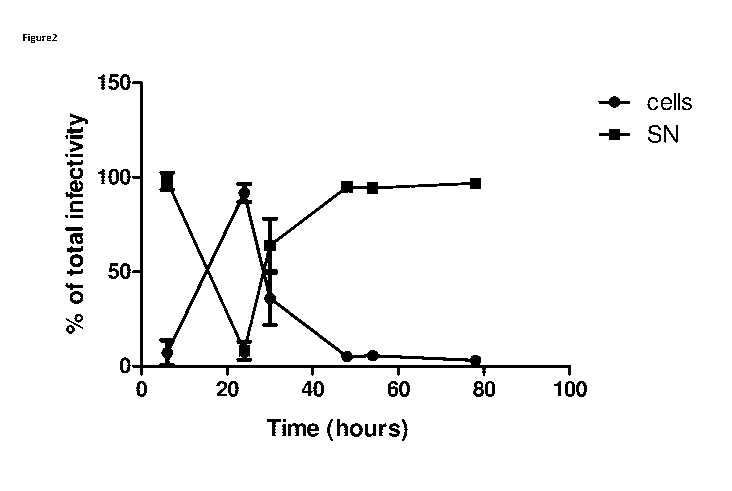
Figure 2 shows the proportion of infectious ColoAd1 particles
associated with the cells and
supernatant (SN) of suspension HEK2935 infected at MOI 10.
Figure 3 shows the proportion of infectious ColoAd1 particles
associated with the cells and
supernatant (SN) of adherent HEK2935 infected at MOI 10 (multiplicity of
infection 10).
Figure 4 shows total viral particle amounts of suspension HEK293
culture in infection condition
testing
Figure 5 Visualisation of cellular and viral DNA in the cell lysate
(Lysate) or supernatant (SN) of
ColoAd1 infected HEK293 cells at 40hrs, 46hrs and 70hrs post-infection
Figure 6 A - Virus distribution (CVL or supernatant),
2
CA 02902650 2015-08-26
WO 2014/131898
PCT/EP2014/053987
B - Total virus production (vp/cell) and
C - Cell viability at each time point post-infection for ColoAd1.
Figure 7 A - Virus distribution (CVL or supernatant),
B -Total virus production (vp/cell), and
C - Cell viability at each time point post-infection for NG135.
Figure 8 A - Virus distribution (CVL or supernatant),
B -Total virus production (vp/cell), and
C - Cell viability at each time point post-infection for NG76.
Figure 9 A - Virus distribution (CVL or supernatant),
B -Total virus production (vp/cell) and
C - Cell viability at each time point post-infection for wild-type Ad5.
Figure 10 A - Virus distribution (CVL or supernatant),
B - Total virus production (vp/cell) and
C - Cell viability at each time point post-infection for wild-type Ad11p.
NG135 as employed herein refers to a derivative of the ColoAd1 virus with a
transgene inserted. The
transgene is a full length antibody. NG135 is SEQ ID 1 with an added transgene
cassette.
NG76 as employed herein refers to a derivative of the ColoAd1 virus with a
transgene inserted. The
transgene is a ScFy antibody fragment. NG76 is SEQ ID 1 with an added
transgene cassette.
Detailed Description of the Disclosure
A process for the manufacture of a chimeric oncolytic virus as employed herein
is intended to
refer to a process wherein the virus is replicated and thus the number of
viral particles is increased. In
particular the manufacturing is to provide sufficient numbers of viral
particles to formulate a
therapeutic product, for example in the range 1-9 x 10⁵to 1-9 x 10²⁰ or more
particles may be produced,
such as in the range of 1-9 x10⁵ to 1-9 x10¹⁵ viral particles, in particular 1
to 9 x101 or 1-9 x 10'5 viral
particles may be produced from a 10L batch.
Part of the E4 region is deleted as employed herein means that at least part,
for example in the
range 1 to 99% of the E4 region is deleted, such as 2, 3, 4, 5, 6, 7, 8, 9,
10, 15, 20, 25, 30, 35, 40, 45, 50,
55, 60, 65, 70, 75, 80, 85, 90, 91, 92, 93, 94 95, 96, 97 or 98% deleted.
"Derived from" as employed herein refers to, for example where a DNA fragment
is taken from
an adenovirus or corresponds to a sequence originally found in an adenovirus.
This language is not
intended to limit how the sequence was obtained, for example a sequence
employed in a virus
according to the present disclosure may be synthesised.
In one embodiment the derivative has 100% sequence identity over its full
length to the original
DNA sequence.
In one embodiment the derivative has 95, 96, 97, 98 or 99% identity or
similarity to the original
DNA sequence.
In one embodiment the derivative hybridises under stringent conditions to the
original DNA
sequence.
As used herein, "stringency" typically occurs in a range from about Tm
(melting temperature)-
50C (5 below the Tm of the probe) to about 20 C to 25 C below Tm.= As will be
understood by those of
3
CA 02902650 2015-08-26
WO 2014/131898
PCT/EP2014/053987
skill in the art, a stringent hybridization can be used to identify or detect
identical polynucleotide
sequences or to identify or detect similar or related polynucleotide
sequences. As herein used, the term
"stringent conditions" means hybridization will generally occur if there is at
least 95%, such as at least
97% identity between the sequences.
As used herein, "hybridization" as used herein, shall include any process by
which a
polynucleotide strand joins with a complementary strand through base pairing"
(Coombs, J., Dictionary
of Biotechnology, Stockton Press, New York, N.Y., 1994).
"Wherein the isolation is not subsequent to a cell lysis step" as employed
herein is intended to
refer to the fact the manufacturing process does not comprise a specific lysis
step. That is to say a step
where the conditions are designed to lyse all or most of the cells in the
culture. For example, the virus is
isolated from the supernatant.
Most as employed herein refers to a large majority, for example 80, 90, 91,
92, 93, 94, 95, 96,
97, 98 or 99%.
"At the end of the culturing period" as employed herein refers to at the end
of a period over
which the virus in the infected cells has been allowed to replicate. End
refers to a selected point in time
selected for harvesting. End as employed herein is not definitive end-point.
In one embodiment the end-
point is chosen to follow a sufficient period of cultivation for the
replicated virus or a significant
proportion thereof to be released into the medium or supernatant. In one
embodiment the harvesting
occurs at multiple time points or is ongoing after it is initiated.
Advantageously, the present process may simplify downstream processing of the
virus because
of the lower starting concentration of contaminating DNA from the cells
because a cell lysis step is
avoided. This may result in cost savings because reagents, equipment and time
employed in
downstream processing may be reduced. It may also result in greater purity
with lower end
concentrations of contaminating DNA and/or a lower concentration of large
fragments of contaminating
DNA.
Furthermore, virus exposure to cell enzymes is minimised by avoiding cell
lysis, which minimises
the exposure of the virus to potential degradants, such as nucleases from the
cell. This may result in
higher virus stability and/or potency as measured, for example by infectivity.
The use of benzonase to degrade cellular DNA may also be avoided or reduced if
desired, which
may be advantageous. In particular, removal of the benzonase and testing to
show the absence of
residual benzonase can be avoided.
Interestingly, after exiting the cells the virus of the present disclosure
does not adhere to the
cells and so can be readily recovered from the supernatant. This may be a
phenomenon which is
characteristic of the oncolytic viruses described herein which facilitates the
current process. In contrast,
wild-type Ad5 is thought to adhere to cells. In fact, results have shown that
substantially no wild-type
Ad5, viral particles are present in the supernatant (see Figure 9 & table 6).
Whilst not wishing to be bound by theory, in one embodiment the ability to
exit the cell and not
adhere thereto, may be associated with the chimeric E2B region.
In one embodiment the ability to exit the cell may be associated with a small
viral genome
and/or a partial deletion in the E4 and/or E3 region.
In one embodiment viruses of the present disclosure further comprise a
transgene.
4
CA 02902650 2015-08-26
WO 2014/131898
PCT/EP2014/053987
In one embodiment the lack of adherence to the cells may be related to the
hexon and fibre of
the oncolytic virus.
Oncolytic viruses are those which preferentially infect cancer cells and
hasten cell death, for
example by lysis of same, or selectively replicate in the cancer cells.
Viruses which preferentially infect cancer cells are viruses which show a
higher rate of infecting
cancer cells when compared to normal healthy cells.
A chimeric adenovirus of the present disclosure can be evaluated for its
preference for a specific
tumor type by examination of its lytic potential in a panel of tumor cells,
for example colon tumor cell
lines include HT-29, DLD-1, LS174T, LS1034, SW403, HCT116, SW48, and
Colo320DM. Any available
colon tumor cell lines would be equally useful for such an evaluation.
Prostate cell lines include DU145 and PC-3 cells. Pancreatic cell lines
include Panc-1 cells. Breast
tumor cell lines include MDA231 cell line and ovarian cell lines include the
OVCAR-3 cell line.
Hemopoietic cell lines include, but are not limited to, the Raji and Daudi B-
lymphoid cells, K562
erythroblastoid cells, U937 myeloid cells, and HSB2 T-lymphoid cells. Other
available tumor cell lines are
equally useful.
Oncolytic viruses including those which are non-chimeric, for example Ad11,
such as Ad11p can
similarly be evaluated in these cell lines.
Viruses which selectively replicate in cancer cells are those which require a
gene or protein
which is upregulated in a cancer cell to replicate, such as a p53 gene.
In one embodiment the chimeric oncolytic virus is apoptotic, that is hastens
programmed cell
death.
In one embodiment the chimeric oncolytic virus is cytolytic. The cytolytic
activity of chimeric
oncolytic adenoviruses of the disclosure can be determined in representative
tumor cell lines and the
data converted to a measurement of potency, for example with an adenovirus
belonging to subgroup C,
preferably Ad5, being used as a standard (i.e. given a potency of 1). A
suitable method for determining
cytolytic activity is an MTS assay (see Example 4, Figure 2 of WO 2005/118825
incorporated herein by
reference).
In one embodiment the chimeric oncolytic adenovirus of the present disclosure
causes cell
necrosis.
In one embodiment the chimeric oncolytic virus has an enhanced therapeutic
index for cancer
cells.
Therapeutic index" or "therapeutic window" refers to a number indicating the
oncolytic
potential of a given adenovirus which may be determined by dividing the
potency of the chimeric
oncolytic adenovirus in a relevant cancer cell line by the potency of the same
adenovirus in a normal
(i.e. non-cancerous) cell line.
In one embodiment the chimeric oncolytic virus has an enhanced therapeutic
index in one or
more cancer cells selected from the group comprising colon cancer cells,
breast cancer cells, head and
neck cancers, pancreatic cancer cells, ovarian cancer cells, hemopoietic tumor
cells, leukemic cells,
glioma cells, prostate cancer cells, lung cancer cells, melanoma cells,
sarcoma cells, liver cancer cells,
renal cancer cells, bladder cancer cells and metastatic cancer cells.
5
CA 02902650 2015-08-26
WO 2014/131898
PCT/EP2014/053987
A chimeric oncolytic adenovirus as employed herein refers to an adenovirus
comprising an E2B
region which has DNA sequence derived from at least two distinct adenovirus
serotypes and wherein
the virus is oncolytic.
There are currently about 56 adenovirus serotypes. Table 1 shows the division
of adenovirus
serotypes:
Subgroup Adenoviral Serotype
A 12, 18, 31
B 3, 7, 11, 14, 16, 21, 34, 35, 50, 55
C 1, 2, 5, 6
D 8-10, 13, 15, 17, 19, 20, 22-30, 32, 33, 36-39, 42-51,
53, 54, 56
E 4
F 40,41
G 52
The E2B region is a known region in adenoviruses and represents about 18% of
the viral
genome. It is thought to encode protein IVa2, DNA polymerase and terminal
protein. In the Slobitski
strain of Ad11 (referred to as Ad11p) these proteins are encoded at positions
5588-3964, 8435-5067 and
10342-8438 respectively in the genomic sequence and the E2B region runs from
10342-3950. The exact
position of the E2B region may change in other serotypes but the function is
conserved in all human
adenovirus genomes examined to date as they all have the same general
organisation.
In one embodiment the virus of the present disclosure, such as a chimeric
oncolytic virus has a
subgroup B hexon.
In one embodiment the virus of the disclosure, such as a chimeric oncolytic
virus has an Ad11
hexon, such as an Allp hexon.
In one embodiment the virus of the disclosure, such as a chimeric oncolytic
virus has a subgroup
B fibre.
In one the virus of the disclosure, such as a chimeric oncolytic virus has an
Ad11 fibre, such as an
Allp fibre.
In one embodiment the virus of the disclosure, such as a chimeric oncolytic
virus has fibre and
hexon proteins from the same serotype, for example a subgroup B adenovirus,
such as Ad11, in
particular Ad11p.
In one embodiment the virus of the disclosure, such as a chimeric oncolytic
virus has fibre,
hexon and penton proteins from the same serotype, for example Ad11, in
particular Ad11p, for example
found at positions 30811-31788, 18254-21100 and 13682-15367 of the genomic
sequence of the latter.
A virus of a distinct serotype to a first virus may be from the same subgroup
or a different
subgroup but will always be from a different serotype. In one embodiment the
combinations are as
follows in (first Ad serotype: second Ad serotype): AA, AB, AC, AD, AE, AF,
AG, BB, BC, BD, BF, BG, CC,
CD, CE, CF, CG, DD, DE, DF, DG, EE, EF, EG, FF, FG and GG.
In one embodiment the chimeric E2B region is derived from Ad3 and Ad11 (in
particular Ad11p).
In one embodiment the E2B region is the sequence shown in SEQ ID NO: 2 herein.
6
CA 02902650 2015-08-26
WO 2014/131898
PCT/EP2014/053987
Mammalian cells are cell derived from a mammal. In one embodiment the
mammalian cells are
selected from the group comprising HEK, CHO, COS-7, HeLa, Viro, A549, PerC6
and GMK, in particular
HEK293.
In one embodiment the cells are grown in adherent or suspension culture, in
particular a
suspension culture.
Culturing mammalian cells as employed herein refers to the process where cells
are grown
under controlled conditions ex vivo. Suitable conditions are known to those in
the art and may include
temperatures such as 37 C. The CO2 levels may need to be controlled, for
example kept at a level of 5%.
Details of the same are given in the text Culture of Animal Cells: A Manual of
Basic Techniques and
Specialised Applications Edition Six R. Ian Freshney, Basic Cell Culture
(Practical Approach) Second
Edition Edited by J.M. Davis.
Usually the cells will be cultured to generate sufficient numbers before
infection with the
adenovirus. These methods are known to those skilled in the art or are readily
available in published
protocols or the literature.
Generally the cells will be cultured on a commercial scale, for example 5L,
10L, 15L, 20L, 25L,
30L, 35L, 40L, 45L, 50L, 100L, 200L, 300L, 400L, 500L, 600L, 700L, 800L, 900,
1000L or similar.
Media suitable for culturing mammalian cells include but are not limited to EX-
CELL media
from Sigma-Aldrich, such as EX-CELL 293 serum free medium for HEK293 cells, EX-
CELL ACF CHO media
serum free media for CHO cells, EX-CELL 302 serum free media for CHO cells,
EX-CELL CD hydrolysate
fusion media supplement, from Lonza RMPI (such as RMPI 1640 with HEPES and L-
glutamine, RMPI 1640
with or without L-glutamine, and RMPI 1640 with UltraGlutamine), MEM and DMEM,
SFMII medium.
In one embodiment the medium is serum free. This is advantageous because it
facilitates
registration of the manufacturing process with the regulatory authorities.
The viruses of the present disclosure, such as chimeric oncolytic viruses have
different
properties to those of adenoviruses used as vectors such as Ad5, this includes
the fact that they can be
recovered from the medium without the need for cell lysis. Thus, whilst not
wishing to be bound by
theory, the viruses appear to have mechanisms to exit the cell.
Furthermore, the viruses of the present disclosure, such as chimeric oncolytic
adenoviruses do
not seem to associate or adhere the cells after exiting the same, which also
facilitates recovery from the
supernatant, in particular when the cell culturing conditions are optimised.
In addition the chimeric oncolytic viruses do not appear to degrade, even when
the culturing
process is extended to 70 hours or more. The degradation of the virus can be
checked by assaying the
infectivity of the virus. The infectivity of the virus decreases as the viral
particles degrade.
In one embodiment the culturing period is in the range 30 to 100 hours, for
example 35 to 70
hours, for example 40, 45, 50, 55, 60 or 65 hours.
In one embodiment the culturing period is 65, 70, 75, 80, 85, 90, 95 hours or
more.
In one embodiment over 90% of the chimeric oncolytic virus is present in the
supernatant at the
64 hour timepoint, for example, 91, 92, 93, 94, 95, 96, 97, 98, 99 or 100%,
such as 95% or more,
particularly 98% or more.
In one embodiment significant amounts of virus are in media post 38 hours. For
example, over
50%, particularly over 70% of the virus is in the media post 38 hours.
7
CA 02902650 2015-08-26
WO 2014/131898
PCT/EP2014/053987
In one embodiment the maximum total virus production is achieved at about 40
to 60 hours
post-infection, for example 49 hours post-infection. In one embodiment the
decrease in virus
production following the maximum is slow.
In one embodiment the maximum total virus production is achieved at about 70
to 90 hours
post-infection.
Surprisingly, the present inventors have found that, when employing the
process, the cells
maintain high viability (such as 80 to 90% viability) post-infection for over
90 hours. Thus in one
embodiment the harvesting and process may continue as long as the cells remain
viable.
Maximum total virus production as employed herein means the total number of
viral particles
produced per cell and encompasses viral particles in the supernatant and the
cell.
In one embodiment the virus production in the supernatant for ColoAd1 at 49
hours post-
infection is about 20000 to 30000 viral particles per cell (vpicell). For
example 26000 voicell.
In one embodiment the virus production in the supernatant for NG135 at 49
hours post-
infection is about 20000 to 30000 voice'', for example 26000 voicell.
In one embodiment the virus production for NG76 at 49 hours post-infection is
about 6000 to
10000 voice'', for example 8000 voicell.
In one embodiment there is less than 10% detectable virus in the CVL pellet at
the 64 hour
timepoint, i.e. post 64 hours, such as 9, 8, 7, 6, 5, 4, 3, 2, 1% detectable
virus. Example 6 describes how
the CVL was obtained.
CVL as employed herein means the crude viral lysate.
Culturing cells may employ a perfusion culture, fed batch culture, batch
culture, a steady state
culture, a continuous culture or a combination of one or more of the same as
technically appropriate, in
particular a perfusion culture.
In one embodiment the process is a perfusion process, for example a continuous
perfusion
process.
In one embodiment the culture process comprises one or more media changes.
This may be
beneficial for optimising cell growth, yield or similar. Where a medium change
is employed, it may be
necessary to recover virus particle from the media being changed. These
particles can be combined with
the main virus batch to ensure the yield of virus is optimised. Similar
techniques may also be employed
with the medium of a perfusion process to optimise virus recovery.
In one embodiment the culture process does not include a medium change step.
This may be
advantageous because no viral particles will be lost and therefore yield may
be optimised.
In one embodiment the culture process comprises one or more cell additions or
changes. Cell
addition change as employed herein refers to replenishing some or all of the
cells and optionally
removing dead cells.
In one embodiment the chimeric oncolytic adenovirus during culture is at
concentration in the
range 20 to 150 particles per cell (ppc), such as 40 to 100 ppc, in particular
5Oppc.
Lower values of virus concentrations, such as less than 100ppc, in particular
5Oppc may be
advantageous because this may result in increased cell viability compared to
cultures with higher virus
concentrations, particularly when cell viability is measured before
harvesting.
8
CA 02902650 2015-08-26
WO 2014/131898
PCT/EP2014/053987
Low cell viability can result in cell lysis which may expose the cell to
enzymes, which with time
may attack the virus. However, in a dynamic process such as cell culturing a
percentage, such as a small
percentage of cells may be unviable. This does not generally cause significant
problems in practice.
In one embodiment cell viability is around 85 to 95% during the process, for
example at the 96
hour timepoint (i.e. 96 hours post-infection) when infected with ColoAd1, such
as 90% viability.
In one embodiment cell viability is around 80 to 90% during the process, for
example at the 96
hour timepoint (i.e. 96 hours post-infection) when infected with NG76, such as
83% viability.
In one embodiment cell viability is around 80 to 90% during the process, for
example at the 96
hour timepoint (i.e. 96 hours post-infection) when infected with NG135, such
as 85% viability.
In one embodiment cell viability is around 80 to 90% during the process, for
example at the 96
hour timepoint (i.e. 96 hours post-infection) when infected with Ad11. For
example 85% viability.
In one embodiment the medium and/or cells are supplements or replenished
periodically.
In one embodiment the cells are harvested during the process, for example at a
discrete
timepoint or timepoints or continuously.
In one embodiment of the process the mammalian cells are infected with a
starting
concentration of virus of 1-9 x 10⁴ \Wml or greater, such as 1-9 x 10⁵, 1-9 x
106, 1-9 x 10⁷, 1-9 x 10⁸, 1-9 x
109, in particular 1-5 x 10⁶ \Wm! or 2.5-5 x 10⁸ vp/ml.
In one embodiment of the process the mammalian cells are infected at a
starting concentration
of 1x10⁶cells/m1 at about 1 to 200ppc, for example 40 to 120ppc, such as
5Oppc.
Ppc as employed herein refers to the number of viral particles per cell.
In one embodiment the process is run at about 35 to 39 C. For example 37 C.
In one embodiment the process run at about 4-6% CO2. For example 5% CO2.
In one embodiment the media containing the virus, such as the chimeric
oncolytic viral particles
is filtered to remove the cells and provide crude supernatant for further
downstream processing.
In one embodiment a tangential flow filter is employed.
In one embodiment medium is filtered employing Millipore's Millistak+ POD
system with
cellulose based depth filters. Millistak+ depth filter medium is offered in a
scalable, disposable format,
the Pod Filter System. It is ideal for a wide variety of primary and secondary
clarification applications,
including cell cultures.
Millistak+ Pod filters are available in three distinct series of media grades
in order to meet
specific application needs. Millistak+ DE, CE and HC media deliver optimal
performance through
gradient density matrix as well as positive surface charge properties. In one
embodiment the filtration is
effected using tangential flow technology, for example employing the CogentTM
M system comprising a
Pellicon Mini cassette membrane holder, pressure sensors, 10 litre recycle
tank with mixer, retentate
flow meter, weigh scale, feed pump, transfer pump, piping and valves. Control
and operation of the
system is manual with an exception of semi-automatic
diafiltration/concentration. The operator has
manual control of pump speeds, all valves and operational procedures. The
virus can also, if desired, be
formulated into the final buffer in this step.
Thus in one embodiment in the filtration step, concentrated and conditioned
adenovirus
material is provided in a final or near final formulation.
In one embodiment the process comprises two or more filtration steps.
9
CA 02902650 2015-08-26
WO 2014/131898
PCT/EP2014/053987
In one embodiment the downstream processing comprises Millistak+POD system 35
CE and 50
CE cassettes followed by an opticap XL 10 express 0.5/0.2 um membrane filter
in series.
In one embodiment the process further comprises a purification step, selected
from a CsCI
gradient, chromatography step such as size exclusion chromatography, ion-
exchange chromatography in
particular anion-exchange chromatography, and a combination thereof.
Ion exchange chromatography binds DNA very strongly and typically is the place
were any
residual DNA is removed. The ion exchange resin/membrane binds both the virus
and the DNA and
during salt gradient elusion the virus normally elutes off the column first
(low salt gradient) and the DNA
is eluted at a much higher salt concentration since the interaction of the DNA
with the resin is stronger
than the virus.
In one embodiment the chromatography step or steps employ monolith technology,
for
example available from BIA Separations.
In one embodiment Sartobind Q (quaternary amine membrane purification process)
is
employed as a purification step.
In one embodiment Source Q RESIN is employed in a purification step.
In one embodiment Sartobind Q is employed followed by Source Q RESIN in
downstream
processing of the isolated virus.
In one embodiment Source Q is employed in the purification step.
In one embodiment after purification the virus prepared contains less than
8Ong/mL of
contaminating DNA, for example between 6Ong/mL and lOng/mL.
In one embodiment substantially all the contaminating DNA fragments are 700
base pairs or
less, for example 500bp or less, such as 200bp or less.
In one embodiment the residual benzonase content in the purified virus product
is lng/mL or
less, such as 0.5ng/mL or less.
In one embodiment the residual host cell protein content in the purified virus
product in
2Ong/mL or less, for example 15ng/mL or less, in particular when measured by
an ELISA assay.
In one embodiment the residual tween in the purified virus product is 0.1mg/mL
or less, such as
0.05mg/mL or less.
In one embodiment the virus has a hexon and fibre from a group B adenovirus,
for example
Ad11 and in particular wherein the virus is ColoAdl.
In one embodiment there is provided isolated purified ColoAdl wherein the
contaminating DNA
content is less than 8Ong/mL.
ColoAdl is disclosed in WO 2005/118825 and the full sequence for the virus is
provided herein,
namely SEQ ID No: 1.
Alternative chimeric oncolytic viruses include OvAdl and OvAd2, which are SEQ
ID NO: 2 and 3
respectively disclosed in WO 2008/080003 and incorporated herein by reference.
In one embodiment the virus is replication competent. Replication competent
virus as
employed herein refers to a virus that is capable of replication without the
assistance of a
complementary cell line encoding an essential viral protein, such as that
encoded by the El region (also
referred to as a packaging cell line) and virus capable of replicating without
the assistance of a helper
virus.
CA 02902650 2015-08-26
WO 2014/131898
PCT/EP2014/053987
In one embodiment the virus of the disclosure, such as the chimeric oncolytic
virus of the
present disclosure comprises one or more transgenes, for example one or more
transgenes encoding
therapeutic peptide(s) or protein sequence(s).
In one embodiment the chimeric oncolytic virus encodes at least one transgene.
Suitable
transgenes include so called suicide genes such as p53; polynucleotide
sequences encoding cytokines
such as IL-2, IL-6, IL-7, IL-12, IL-15, IL-18, IL-21, GM-CSF or G-CSF, an
interferon (eg interferon I such as
IFN-alpha or beta, interfon II such as IFN-gamma), a TNF (eg TNF-alpha or TNF-
beta), TGF-beta, CD22,
CD27, CD30, CD40, CD120; a polynucleotide encoding a monoclonal antibody, for
example trastuzamab,
cetuximab, panitumumab, pertuzumab, epratuzumab, an anti-EGF antibody, an anti-
VEGF antibody and
anti-PDGF antibody, an anti-FGF antibody.
A range of different types of transgenes, and combinations thereof, are
envisaged that encode
molecules that themselves act to modulate tumour or immune responses and act
therapeutically, or are
agents that directly or indirectly inhibit, activate or enhance the activity
of such molecules. Such
molecules include protein ligands or active binding fragments of ligands,
antibodies (full length or
fragments, such as Fv, ScFv, Fab, F(ab)'2 or smaller specific binding
fragments), or other target-specific
binding proteins or peptides (e.g. as may be selected by techniques such as
phage display etc), natural
or synthetic binding receptors, ligands or fragments, specific molecules
regulating the transcription or
translation of genes encoding the targets (e.g. siRNA or shRNA molecules,
transcription factors).
Molecules may be in the form of fusion proteins with other peptide sequences
to enhance their activity,
stability, specificity etc (e.g. ligands may be fused with immunoglobulin Fc
regions to form dimers and
enhance stability, fused to antibodies or antibody fragments having
specificity to antigen presenting
cells such as dendritic cells (e.g. anti-DEC-205, anti-mannose receptor, anti-
dectin). Transgenes may also
encode reporter genes that can be used, for example, for detection of cells
infected with the "insert-
bearing adenovirus", imaging of tumours or draining lymphatics and lymph nodes
etc.
In one embodiment the cancer cell infected with the chimeric oncolytic virus
is lysed releasing
the contents of the cell which may include the protein encoded by a transgene.
In one embodiment 40 to 93% or more of the total virus replicated in the cells
is recoverable
from the media, for example 41, 42, 43, 44, 45, 46, 47, 48, 49, 50, 51, 52,
53, 54, 55, 56, 57, 58, 59, 60,
61, 62, 63, 64, 65, 66, 67, 68, 69, 70, 71, 72, 73, 74, 75, 76, 77, 78, 79,
80, 81, 82, 83, 84, 85, 86, 87, 88,
89, 90, 91 or 92% of the total virus is recoverable, such as 94, 95, 96, 97,
98, 99 or 100% of the total
virus recoverable.
In one embodiment the process is a GMP manufacturing process, such as a cGMP
manufacturing process.
In one embodiment the process further comprises the step formulating the virus
in a buffer
suitable for storage.
In one embodiment the present disclosure extends to virus or viral
formulations obtained or
obtainable from the present method.
Known methods for cell lysis include employing lysis buffer (pH 8.0)
containing MgC12 and
detergent, e.g. 1% Tween-20. Cell lysis is performed without pH or p02
controls. Rocking and heating
are used. Lysis is continued for 1.5-2 hours.
Freeze-thawing multiple times is also a routine method of cell lysis.
11
CA 02902650 2015-08-26
WO 2014/131898
PCT/EP2014/053987
Benzonase (Merck), 100 U/ml, is used to digest host cell DNA. Benzonase
treatment is done for
30 min in +37 C. Benzonase is stopped with high salt incubation for 1 hour at
RT.
Pulmozyme may also be employed in cell lysis.
Alternative methods for cell lysis include centrifuging cell suspension at
1000 x g, 10 min at 4 C.
Resuspending the cell pellet into 1 ml of Ex-Cell medium 5 % glycerol and
releasing the viruses from the
cells by freeze-thaw by freezing tubes containing the responded cells from the
pellet in liquid nitrogen
for 3 - 5 minutes and thaw at +37 C water bath until thawed. Generally the
freeze and thaw step is
repeated twice more. This cycle releases viruses from the cells. After the
last thaw step remove the cell
debris by centrifugation 1936 x g, 20 min at +4 C.
In the context of the resent application, medium and media may be used
interchangeably.
In the context of this specification "comprising" is to be interpreted as
"including".
Aspects of the invention comprising certain elements are also intended to
extend to alternative
embodiments "consisting" or "consisting essentially" of the relevant elements.
Where technically appropriate, embodiments of the invention may be combined.
Embodiments are described herein as comprising certain features/elements. The
disclosure
also extends to separate embodiments consisting or consisting essentially of
said features/elements.
Technical references such as patents and applications are incorporated herein
by reference.
Any embodiments specifically and explicitly recited herein may form the basis
of a disclaimer
either alone or in combination with one or more further embodiments.
The present invention is further described by way of illustration only in the
following examples,
which refer to the accompanying Figures, in which:
EXAMPLES
Example 1
Suspension HEK2935 (1 x 10⁶ cells/mL in 125mL shake flasks at 10Orpm) were
infected at MOI 10 and fed
with CD293 media 2 hours after infection with ColoAd1. Samples were taken 6,
24, 30, 48, 54 and 78
hours after infection. The supernatant was separated from the cells by
centrifugation and the cell pellet
resuspended in cell lysis buffer. The amount of infectious ColoAd1 particles
in the cells and supernatant
were determined by immunostaining infectivity assay and expressed as a
proportion of the total at each
time point. N = 1, error bars (SD) show triplicate infections. Results are
shown in Figure 2.
Example 2
Adherent HEK2935 (1 x 10⁶cells/mL in 1mL of 24-well plate) were infected at
MOI 10 (in 2% FCS
containing media). At 6, 24, 30, 48, 54, 72 and 78 hours after infection with
ColoAd1 supernatant was
removed, and cells detached from the surface before re-suspending in cell
lysis buffer. The amount of
infectious ColoAd1 particles associated with the cells and supernatant were
determined by
immunostaining infectivity assay and expressed as a proportion of the total at
each time point. N = 1,
error bars (SD) show triplicate infections. Results shown in Figure 3.
Example 3 ColoAd1 cultured in a HEK 293 suspension culture
Infection conditions for oncolytic virus ColoAd1 were tested in a small scale
suspension HEK293 culture.
Cells were cultured for 96 hours prior to infection using Ex-Cell ¨ 6 mM L-
glutamine ¨ 50 g/m1/501U/m1
Penicllin/Streptomycin at +37 C and 5% CO2.
12
CA 02902650 2015-08-26
WO 2014/131898
PCT/EP2014/053987
Cell counting was performed using BOrker cell hemacytometer and Trypan Blue
stain (Invitrogen, 15250-
061). For larger dilutions (dilution factor 3 or greater) cell culture medium
with Trypan blue was used.
Two virus dilutions (50 and 100 particles per cell) and six harvesting time
points (40, 43, 36, 49, 64, and
70 h) were tested. All the testing was done in duplicate shaker flasks. The
viral particle concentrations
of the samples were analysed with AEX-HPLC and the results are shown in Tables
3 and 4.
Table 3. AEX-HPLC results of the cell samples (intracellular virus
concentrations) of suspension HEK239
culture in infection condition testing.
Infection AEX-HPLC titer Volume Average Produced
ppc time (h) (vp/ml) (ml) Total vp
total vp vp/cell
6.99E+11 1.05 7.34E+11
40 1.12E+12 70914
1.25E+12 1.20 1.50E+12
6.89E+11 1.05 7.24E+11
43 6.52E+11 89169
5.05E+11 1.15 5.81E+11
5.69E+11 1.20 6.82E+11
46 6.39E+11 99401
4.96E+11 1.20 5.96E+11
7.43E+11 1.15 8.55E+11
49 8.77E+11 155253
7.50E+11 1.20 9.00E+11
6.18E+11 1.20 7.42E+11
64 7.25E+11 228787
5.89E+11 1.20 7.07E+11
6.28E+11 1.20 7.54E+11
70 8.34E+11 262976
7.62E+11 1.20 9.14E+11
6.37E+11 1.20 7.64E+11
40 4.57E+11 60914
1.25E+11 1.20 1.50E+11
8.16E+11 1.10 8.97E+11
43 9.10E+11 107440
9.23E+11 1.00 9.23E+11
5.77E+11 1.20 6.92E+11
46 7.20E+11 155171
6.23E+11 1.20 7.48E+11
100
7.38E+11 1.20 8.85E+11
49 8.65E+11 215871
7.05E+11 1.20 8.45E+11
7.06E+11 1.10 7.76E+11
64 8.35E+11 321975
7.45E+11 1.20 8.94E+11
6.69E+11 1.20 8.03E+11
70 7.82E+11 351378
5.85E+11 1.30 7.60E+11
15
13
CA 02902650 2015-08-26
WO 2014/131898
PCT/EP2014/053987
Table 4. AEX-HPLC results of the medium samples and total intracellular and
extracellular virus amount
of suspension HEK239 culture in infection condition testing.
Infection AEX-HPLC titer Volume Average Total vp in
% in
ppc Total vp cells + in
time (h) (vp/ml) (ml) total vpmed
medium
ium
3.84E+10 28.0 1.08E+12 1
1
I 40 1.01E+12 2.13E+12 47
3.42E+10 27.5 9.42E+12
7.23E+10 27.5 1.99E+12
43 2.02E+12 2.68E+12 76
7.22E+10 28.5 2.06E+12
1.06E+11 27.5 2.93E+12
46 2.34E+12 2.98E+12 79
6.21E+10 28.3 1.76E+12
1.33E+11 27.5 3.67E+12
49 3.78E+12 4.66E+12 81
1.37E+11 28.3 3.89E+12
2.18E+11 27.3 5.95E+12
64 6.13E+12 6.86E+12 89
2.24E+11 28.3 6.33E+12
2.57E+11 27.3 7.03E+12
70 7.05E+12 7.89E+12 89
2.52E+11 28.1 7.09E+12
4.63E+10 28.0 1.30E+12
40 1.37E+12 1.83E+12 75
5.25E+10 27.5 1.44E+12
7.44E+10 27.5 2.05E+12
43 2.31E+12 3.22E+12 72
9.05E+10 28.5 2.58E+12
1.37E+11 27.7 3.79E+12
46 3.94E+12 4.66E+12 85
1.42E+11 28.7 4.08E+12
100
1.92E+11 27.9 5.36E+12
49 5.61E+12 6.48E+12 87
2.06E+11 28.5 5.86E+12
3.12E+11 27.4 8.55E+12
64 8.82E+12 9.66E+12 91
3.21E+11 28.3 9.10E+12
3.49E+11 26.9 9.40E+12
70 9.76E+12 1.05E+13 93
3.55E+11 28.5 1.01E+13
The highest amount of viral particles was produced when infecting the cells
for 70 hours with 100 ppc
5 (the average results of duplicate flasks 1.05E+13vp, Table 4). At that
time point, 93 % of the viral
particles were in the medium. It can be seen from Figure 5, that the total
amount of virus increased up
to 70 hours, but the curve seemed to start approaching plateu after 64 hours.
Already at 43 hours, over
half of the virus is in the culture medium, however, in the suspension
production process, also the viral
particles in the medium can be captured during purification. In 70 hours, MOI
of 100 ppc produced
10 2.6E+12 more viral particles than 50 ppc (and 2.8E+12 more in 64 h). But
even with 50 ppc, the
production capacity of the cells appeared to be close to the maximum: the
intracellular virus amount
remained fairly constant during the time range of 40-70 hours.
Example 4 Example of ColoAdl cultured in adherent HEK 293 cells
Adherent HEK293 cells were seeded at 4.8 x 10⁶ cells per flask in 185 CM2 cell
culture flasks (24 pieces) 72
15 hours prior to infection. Cell culturing was performed using DMEM - 10%
FBS - 2 mM L-glutamine at
+37 C and 5% CO2. Cell number was counted from one cell culture flask on the
day of infection resulting
14
CA 02902650 2015-08-26
WO 2014/131898
PCT/EP2014/053987
40.6 x 10⁶ cells/ flask. The tested particles per cell (ppc) were 200, 100 and
50. After infection the cells
may be cultured for between 35 to 70 hours.
Example 5 Visualisation of cellular and viral DNA in the cell lysate
(Lysate) or supernatant (SN) of
ColoAdl infected HEK293 cells at 40hrs, 46hrs and 70hrs post-infection.
HEK293 cells infected with ColoAd1 at 50 particles per cell were harvested 40
hours, 46 hours or 70
hours post-infection. The culture supernatants and the cell lysates were
collected and total DNA
extracted. Equivalent volumes of purified lysate or supernatant DNA were
loaded in duplicate onto a
0.7% agarose gel and the DNA was separated electrophoresis. Significant
cellular DNA could be
detected at the top of the gel and as a smear in all lanes containing DNA
extracted from cell lysates,
however only very low levels of cellular DNA could be detected in lanes
containing DNA extracted from
supernatant (SN). In contrast, viral DNA could be detected in all samples and
the total detectable viral
DNA observably increased in the supernatant over time. Results are shown in
Figure 5.
Example 6
ColoAd1, NG-135, NG-76, Ad5 and Ad11p (referred to in the figures and tables
as Ad11) were compared
for the relative levels of expression of virus particles associated with the
cell pellet (CVL) or in the
supernatant.
Table 5. Total adenoviral particle concentration of virus (by HPLC assay).
ADC HPLC 1:10 1:100
Viruses used
titer vp/rni Dilution Dilution
1=,2, 1 3.CCE-12 3 CCE+11 3
\G 135 3ancieci,:f=,1 2.9E-:L2 ? r59z-11 2 r59:-.:C
G 76-C3 3a-ide51:7., 15 91E+11 6 6 91E-1:9
AcI5 3a-idPci 1.59E+12 1 59E-L1 1 593--'3C
AcIL1 Sanded 2.30E+11 2,3CE-1C 2.3CE-C9
Suspension HEK293 cells (293f) were cultured in duplicate shaker flasks
containing 40 ml working
volume of SFMII media supplemented with 4 mM L-glutamine and 50 pg/m1/501U/m1
Penicillin/Streptomycin and infected at 106 cells/ml with viruses at a ratio
of 50 virus particles per cell
(ppc).
The cell expansion was started by thawing one vial of cells and continued cell
expansion for 3 weeks
until a total of 4.8 x 10⁸ cells required for this study was achieved. Three
days before infection, the HEK
293 suspension cells were seeded in a single one litre shaker flask using 4 x
105 cells/ml in 428 ml of
SFMII medium per flask (3.4 x 10⁸ cells/ flask) and incubated in a shaker
incubator at + 37 C, 5 % CO2
&115 rpm.
On the day of infection the cell count was performed and based on cell
density, 225 ml of cell
suspension at 2.15 x 10⁶cell/m1 was used for the study. Remaining cells were
discarded.
HEK 293 suspension cells were infected with one of the four different viruses
(see Table 5 ) at 50 ppc in
duplicate.
A 1:100 dilution of each virus was performed in SFMII growth medium prior to
infection of cells (for
virus concentration refer to Table 5).
CA 02902650 2015-08-26
WO 2014/131898 PCT/EP2014/053987
Table 6. Infection calculation and dilution of virus prior to infection
Volume total vp Diluted required
Virus cells/nil (m1) total cells nix needed
vpiml (m1) Notes
Total 2x 67 ul of
ColoAdl 1 CCE-C6 4) 4 CCE-C7 50 2.00E+09
3.00E+10 0.067 used for infec: an& 2 f as-(s
Total 2x 74 .4 _
NG 135 40 4 CCE-C7 50 2 2.69E-1C
0.074 used for infection Of 2 f aSKS
To:a 2x 297_ J
1.--1,211:LCC (1C-99C;
NG 76-03 _ : 40 4.0,0E+07 50 2. C L.E +09
6.9:7E+09 0.29C used for :.f cucc of 2
To:a 2 17.15 -
C-99C;
Ad5 CCE+C.6 40 4.00E+07 50 2 CCE-C9 1
59E+10 0.126 used for -cc f 2 f as4s
Total 2x KT. j 1r
_:LCC ,:LC-99C;
Ad11 1 OOE-CÃ 40 4.00E+07 50 2 COE-C9 2.3CE-C9
C 87c. Used for infection or 2 flasks
Infection of suspension HEK293 cells with virus diluted as follows:
225 ml of cell suspension at 2.15 x 10⁶ was centrifuged and the cell pellets
was resuspended in 480 ml
media to adjust the cell concentration to 1x 10⁶ cells/ml,
Thereafter, 10 shaker flasks with working volume of 40 ml per shaker flask
were prepared. Dual flask
were labelled as ColoAd1¨A1A2, NG 135-131132, NG 76-C1C2, Ad5-D1D2 and
Ad11E1E2 respectively.
All labelled flasks were infected with 50 ppc virus in accordance with Table
6.
All shaker flasks were placed in a shaking incubator at + 37 C, 5 % CO2 &120
rpm until harvested.
At 40, 46, 49, 64, 70, 73 and 89 hours post infection, 2.5m1 samples were
taken from each flask and the
duplicates pooled to provide 5.0m1 samples. At 96 hours post-infection, all
the cells were harvested. Cell
viabilities were assessed using 0.5ml and the remaining 4.5ml volume was then
centrifuged and the
virus distribution between the supernatant and cell pellet for each virus was
determined in the
following way:
The cells were centrifuged at 1000 x g, 10 min at 4 C
After centrifugation, the supernatant was gently poured into a sterile
container and 0.5 ml of
50% glycerol was added to the sample 1m1 aliquots were stored at -80 C until
analysis.
The cell pellet was suspended in 1m1 of SFMII medium containing 5% glycerol,
Intracellular virus was released from the cells by freeze-thaw as follows:
The centrifuge tubes were frozen in liquid nitrogen for 3-5 minutes and then
transferred to a
water bath set at +37 C until thawed.
The freeze and thaw process was repeated twice more as described in step a,
above.
After the final thaw step, the cell debris is removed from the crude viral
lysate (CVL) by
centrifugation at 1936 x g for 20 min at +4 C and transfer of the supernatant
(CVL) to a new
container.
The CVL was aliquoted as 100 ul samples and stored at -80 C until analysis.
Total viral particle concentration (vp) from Crude Viral Lysate (CVL) and
supernatant (SN) samples were
analysed by AEX-HPLC assay. These values were then used to calculate the total
number of virus
particles per culture and the percentage in the SN and CVL for each sample
time point. These are
represented as bar graphs, together with the viability of the HEK293 cells,
for ColoAd1, NG-135, NG-76,
Ad5 and Allp in Figs 6-10 respectively. For ColoAd1, NG-135,NG-76 and Ad11p
the majority of virus was
in the culture supernatant whereas for Ad5 virus was entirely in the cell
lysate (CVL), being undetectable
16
CA 02902650 2015-08-26
WO 2014/131898
PCT/EP2014/053987
in the supernatant. For all cultures, the viability of the HEK293 cells
remained high over the 96 hours of
culture.
For ColoAd1 (Figure 6 A), 84 % was present in the supernatant at 40hrs
timepoint, and 98%
present in the supernatant at 64h timepoint with no detectable virus in the
CVL (pellet) sample.
For Next Gen 135 (Figure 7A) and Next Gen 76 (Figure 8A) the trend of virus
distribution is
similar to ColoAd1. For NG135, 77 % of virus was present in the supernatant at
40hrs timepoint. This
increased to 97% virus present in the the supernatant at 64h timepoint. For
the same time points, 56 %
and 98% of the virus for Next Gen 76 was present in the supernatant.
For Ad11 (Figure 10A), 31 % of the virus was present in the supernatant at
40hrs timepoint, 88%
present in the supernatant at 64h timepoint and 98% of the virus present in
the supernatant at 96h
timepoint, respectively.
For Ad5 (Figure 9A), 100 % of the virus was detected in CVL (pellet) sample
with no virus
detected in the supernatant.
For all viruses assessed in this study, the maximum level of virus was
observed at 49 hrs post
infection and there was a slow decrease in virus level observed thereafter in
subsequent timepoints
For ColoAd1 (Figure 68) and NG135 (Figure 78) maximum virus level (27000
vp/cell) was
observed at 49hrs timepoint, while in NG76 (Figure 88) the maximum virus level
(9200 vp/cell) was
observed at same timepoint.
For Ad5 (Figure 98) the maximum virus level(11000 vp/cell) was observed at 89
hrs timepoint
while in Ad11p (Figure 1013) the maximum virus level (30000 vp/cell) was
observed at same timepoint.
The total viral particle concentrations of all the supernatant and CVL samples
analysed with AEX-
HPLC are shown in Tables 7 and 8 and in Figures 6-10.
17
Table 7. AEX-HPLC assay results of ColoAd1, NG135 and NG76
0
AEX-
AEX-HPLC
Total vp Total vp Produced Produced
c,)
Virus InfectionHPLC Volume Avg Total Produced
Cell Total vp Total vp Total vp
Sample Detail DF titer Total vp
(% in (% in vp/cell vp/cell
used time (h) titer (m1) cells/ml cells
vp/cell via bility% (SN) (CVL) (SN+CVL)
vp/ml
SN) CVL) (in SN) (SN+CVL)
vp/ml
SN 40 h ColoAd1
40 2.03E+10 1.11 2.25E+10 40.00 9.00E+111.00E+06 4.00E+07 22492 55
9.00E+11 3.74E+11 1.27E+12 71 29 22492 31830
A1A2
SN 46 h ColoAd1
46 2.31E+10 1.11 2.57E+10 37.50 9.63E+111.00E+06 3.75E+07 25671 86
9.63E+11 1.61E+11 1.12E+12 86 14 25671 29977
A1A2
SN 49 h ColoAd1
49 2.34E+10 1.11 2.60E+10 35.00 9.11E+111.00E+06 3.50E+07 26019 NA
9.11E+11 1.52E+11 1.06E+12 86 14 26019 30350 0
A1A2
0
oo SN 64 h ColoAd1
0
64 1.81E+10 1.11 2.01E+10 32.50 6.52E+111.00E+06 3.25E+07 20058 85
6.52E+11 2.70E+10 6.79E+11 96 4 20058 20888
A1A2
0
SN 70 h ColoAd1
ColoAd1 70 1.73E+10 1.11
1.92E+10 30.00 5.77E+111.00E+06 3.00E+07 19238 87 5.77E+11 3.39E+10
6.11E+11 94 6 19238 20366
A1A2
SN 73 h ColoAd1
73 1.71E+10 1.11 1.90E+10 27.50 5.22E+111.00E+06 2.75E+07 18989 NA
5.22E+11 2.42E+10 5.46E+11 96 4 18989 19869
A1A2
SN 89 h ColoAd1
89 1.77E+10 1.11 1.97E+10 25.00 4.92E+111.00E+06 2.50E+07 19660 NA
4.92E+11 2.70E+10 5.18E+11 95 5 19660 20739
A1A2
SN 96 h ColoAd1
96 1.84E+10 1.00 1.84E+10 22.50 4.15E+111.00E+06 2.25E+07 18428 90
4.15E+11 6.10E+09 4.21E+11 99 1 18428 18699
A1A2
CVL 40 h ColoAd1
40 2.10E+10 0.44 9.34E+09 40.00 3.74E+111.00E+06 4.00E+07 9339
A1A2
oo
J
CVL 46 h ColoAd1
46 9.69E+09 0.44 4.31E+09 37.50 1.61E+11 1.00E+06 3.75E+07 4306
A1A2
0
CVL 49 h ColoAd1
49 9.75E+09 0.44 4.33E+09 35.00 1.52E+11 1.00E+06 3.50E+07 4331
A1A2
CVL 64 h ColoAd1
oe
64 1.87E+09 0.44 8.30E+08 32.50 2.70E+10 1.00E+06 3.25E+07 830 oe
A1A2
CVL 70 h ColoAd1
70 2.54E+09 0.44 1.13E+09 30.00 3.39E+10 1.00E+06 3.00E+07 1129
A1A2
CVL 73 h ColoAd1
73 1.98E+09 0.44 8.80E+08 27.50 2.42E+10 1.00E+06 2.75E+07 880
A1A2
CVL 89 h ColoAd1
89 2.43E+09 0.44 1.08E+09 25.00 2.70E+10 1.00E+06 2.50E+07 1079
A1A2
o
CVL 96 h ColoAd1
0
96 6.10E+09 0.04 2.71E+08 22.50 6.10E+09 1.00E+06 2.25E+07 271
A1A2
O
o
40 SN 40 h NG135 B1B2
1.66E+10 1.11 1.85E+10 40.00 7.39E+11 1.00E+06 4.00E+07 18468 76
7.39E+11 5.02E+11 1.24E+12 60 40 18468 31009
46 SN 46 h NG135 B1B2
2.31E+10 1.11 2.56E+10 37.50 9.60E+11 1.00E+06 3.75E+07 25597 92
9.60E+11 2.49E+11 1.21E+12 79 21 25597 32230
49 SN 49h NG135 B1B2
2.26E+10 1.11 2.51E+10 35.00 8.78E+11 1.00E+06 3.50E+07 25075 NA
8.78E+11 2.81E+11 1.16E+12 76 24 25075 33106
64 SN 64 h NG135 B1B2
1.70E+10 1.11 1.88E+10 32.50 6.12E+11 1.00E+06 3.25E+07 18840 85
6.12E+11 4.80E+10 6.60E+11 93 7 18840 20317
NG-135
70 SN 70 h NG135 B1B2
1.44E+10 1.11 1.60E+10 30.00 4.80E+11 1.00E+06 3.00E+07 15984 92
4.80E+11 4.97E+10 5.29E+11 91 9 15984 17640
73 SN 73 h NG135 B1B2
1.29E+10 1.11 1.44E+10 27.50 3.95E+11 1.00E+06 2.75E+07 14369 NA
3.95E+11 2.56E+10 4.21E+11 94 6 14369 15299
89 SN 89 h NG135 B1B2
1.03E+10 1.11 1.15E+10 25.00 2.87E+11 1.00E+06 2.50E+07 11463 NA
2.87E+11 1.58E+10 3.02E+11 95 5 11463 12094
96 SN 96 h NG135 B1B2
1.10E+10 1.00 1.10E+10 22.50 2.46E+11 1.00E+06 2.25E+07 10954 85
2.46E+11 5.31E+09 2.52E+11 98 2 10954 11190 cA)
oe
CVL 40 h NG135
40 2.82E+10 0.44 1.25E+10 40.00 5.02E+11 1.00E+06 4.00E+07 12541
B1B2
0
CVL 46 h NG135
46 1.49E+10 0.44 6.63E+09 37.50 2.49E+11 1.00E+06 3.75E+07 6633
B1B2
CVL 49 h NG135
oe
49 1.81E+10 0.44 8.03E+09 35.00 2.81E+11 1.00E+06 3.50E+07 8031 oe
B1B2
CVL 64 h NG135
64 3.32E+09 0.44 1.48E+09 32.50 4.80E+10 1.00E+06 3.25E+07 1477
B1B2
CVL 70 h NG135
70 3.73E+09 0.44 1.66E+09 30.00 4.97E+10 1.00E+06 3.00E+07 1656
B1B2
CVL 73 h NG135
73 2.09E+09 0.44 9.30E+08 27.50 2.56E+10 1.00E+06 2.75E+07 930
B1B2
o
CVL 89 h NG135
0
89 1.42E+09 0.44
6.31E+08 25.00 1.58E+10 1.00E+06 2.50E+07 631
B1B2O
0
o
CVL 96 h NG135
96 5.31E+09 0.04 2.36E+08 22.50 5.31E+09 1.00E+06 2.25E+07 236
B1B2
40 SN 40 h NG76 C1C2
4.20E+09 1.11 4.66E+09 40.00 1.86E+11 1.00E+06 4.00E+07 4657 74 1.86E+11
3.26E+11 5.12E+11 36 64 4657 12807
46 SN 46 h NG76 C1C2
5.90E+09 1.11 6.54E+09 37.50 2.45E+11 1.00E+06 3.75E+07 6545 90 2.45E+11
4.08E+10 2.86E+11 86 14 6545 7633
49 SN 49 h NG76 C1C2
7.24E+09 1.11 8.04E+09 35.00 2.81E+11 1.00E+06 3.50E+07 8035 NA 2.81E+11
9.52E+10 3.76E+11 75 25 8035 10755
NG-76
64 SN 64 h NG76 C1C2
5.48E+09 1.11 6.09E+09 32.50 1.98E+11 1.00E+06 3.25E+07 6088 88 1.98E+11
1.02E+10 2.08E+11 95 5 6088 6401 1-3
70 SN 70 h NG76 C1C2
4.93E+09 1.11 5.48E+09 30.00 1.64E+11 1.00E+06 3.00E+07 5477 87 1.64E+11
2.02E+10 1.85E+11 89 11 5477 6151
73 SN 73 h NG76 C1C2
5.40E+09 1.11 6.00E+09 27.50 1.65E+11 1.00E+06 2.75E+07 5998 NA 1.65E+11
8.61E+09 1.74E+11 95 5 5998 6311
oe
89 SN 89 h NG76 C1C2 3.64E+09
1.11 4.04E+09 25.00 1.01E+11 1.00E+06 2.50E+07 4036 NA 1.01E+11 7.83E+09
1.09E+11 93 7 4036 4349
96 SN 96 h NG76 C1C2 4.20E+09
1.00 4.20E+09 22.50 9.44E+10 1.00E+06 2.25E+07 4195 83 9.44E+10 1.73E+09
9.61E+10 98 2 4195 4272 0
40 CVL 40 h NG76 C1C2 1.83E+10
0.44 8.15E+09 40.00 3.26E+11 1.00E+06 4.00E+07 8150
46 CVL 46 h NG76 C1C2 2.45E+09
0.44 1.09E+09 37.50 4.08E+10 1.00E+06 3.75E+07 1089 oe
oe
49 CVL 49 h NG76 C1C2 6.12E+09
0.44 2.72E+09 35.00 9.52E+10 1.00E+06 3.50E+07 2720
64 CVL 64 h NG76 C1C2 7.04E+08
0.44 3.13E+08 32.50 1.02E+10 1.00E+06 3.25E+07 313
70 CVL 70 h NG76 C1C2 1.52E+09
0.44 6.74E+08 30.00 2.02E+10 1.00E+06 3.00E+07 674
73 CVL 73 h NG76 C1C2 7.04E+08
0.44 3.13E+08 27.50 8.61E+09 1.00E+06 2.75E+07 313
89 CVL 89 h NG76 C1C2 7.04E+08
0.44 3.13E+08 25.00 7.83E+09 1.00E+06 2.50E+07 313
o
96 CVL 96 h NG76 C1C2 1.73E+09
0.04 7.71E+07 22.50 1.73E+09 1.00E+06 2.25E+07 77
o
O
o
oe
Table 8. AEX-HPLC assay results of Ad5 and Ad11
AEX-HPLC AEX-HPLC
Total vp Produced Produced-. 0
Virus InfectionVolume Avg Total Produced
Cell Total vp Total vp Total vp Total
vp n.)
Sample Detail titer DF titer
Total vp (% in vp/cell vp/cell o
1-,
used time (h) (ml) cells/ml cells
vp/cell viability% (SN) (CVL) (SN+CVL) (% in SN)
4=.
vp/ml vp/ml
CVL) (SN) (SN+CVL)
c...)
1-,
oe
SN 40h Ad5
40 0.00E+00 1.11 0.00E+00 40.00 0.00E+00 1.00E+06
4.00E+07 0 80 0.00E+00 5.37E+11 5.37E+11 0 100
o 13426 oe
D1D2
SN 40h Ad5
46 0.00E+00 1.11 0.00E+00 37.50 0.00E+00 1.00E+06
3.75E+07 0 89 0.00E+00 6.44E+11 6.44E+11 0 100
0 17165
D1D2
SN 49h Ad5
49 0.00E+00 1.11 0.00E+00 35.00 0.00E+00 1.00E+06
3.50E+07 0 NA 0.00E+00 7.65E+11 7.65E+11 0 100
0 21846
D1D2
SN 64h Ad5
P
64 0.00E+00 1.11 0.00E+00 32.50 0.00E+00 1.00E+06
3.25E+07 0 90 0.00E+00 5.54E+11 5.54E+11 0 100
0 17058 0
D1D2
0
1.,
0,
n.) SN 70h Ad5
0
70 0.00E+00 1.11 0.00E+00 30.00 0.00E+00 1.00E+06
3.00E+07 0 89 0.00E+00 5.43E+11 5.43E+11 0 100
0 18086 "
0
D1D2
1-
u,
1
Ad5
0
00
1
SN 73h Ad5
"
73 0.00E+00 1.11 0.00E+00 27.50 0.00E+00 1.00E+06
2.75E+07 0 NA 0.00E+00 4.21E+11 4.21E+11 0 100
0 15322 0,
D1D2
SN 89h Ad5
89 0.00E+00 1.11 0.00E+00 25.00 0.00E+00 1.00E+06
2.50E+07 0 NA 0.00E+00 6.47E+11 6.47E+11 0 100
0 25885
D1D2
SN 96h Ad5
96 0.00E+00 1.00 0.00E+00 22.50 0.00E+00 1.00E+06
2.25E+07 0 81 0.00E+00 2.35E+11 2.35E+11 0 100
0 10455
D1D2
IV
n
= ,-i
CVL 40h Ad5
40 3.02E+10 0.44 1.34E+10 40.00 5.37E+11 1.00E+06
4.00E+07 13426 M
D1D2
IV
n.)
o
1-,
4=.
CVL 46h Ad5
46 3.86E+10 0.44 1.72E+10 37.50 6.44E+11 1.00E+06
3.75E+07 17165 -a-,
un
D1D2
oe
I ,..4
CVL 49h Ad5
49 4.92E+10 0.44 2.18E+10 35.00 7.65E+11 1.00E+06 3.50E+07 21846
D1D2
0
CVL 64h Ad5
64 3.84E+10 0.44 1.71E+10 32.50 5.54E+11 1.00E+06 3.25E+07 17058
D1D2
oe
CVL 70h Ad5
70 4.07E+10 0.44
1.81E+10 30.00 5.43E+11 1.00E+06 3.00E+07 18086 oe
D1D2
CVL 73h Ad5
73 3.45E+10 0.44 1.53E+10 27.50 4.21E+11 1.00E+06 2.75E+07 15322
D1D2
CVL 89h Ad5
89 5.82E+10 0.44 2.59E+10 25.00 6.47E+11 1.00E+06 2.50E+07 25885
D1D2
CVL 96h Ad5
96 2.35E+11 0.04 1.05E+10 22.50 2.35E+11 1.00E+06 2.25E+07 10455
D1D2
o
cA) SN 40h Ad11
0
40 6.68E+09 1.11
7.41E+09 40.00 2.97E+11 1.00E+06 4.00E+07 7415 73 2.97E+11 1.49E+12
1.78E+12 17 I 83 I 7415 I 44601
O
E1E2o
o
SN 46h Ad11
46 1.05E+10 1.11 1.16E+10 37.50 4.36E+11 1.00E+06 3.75E+07 11639 91
4.36E+11 1.04E+12 1.48E+12 30 70 11639 39340
E1E2
SN 490h Ad11
49 1.13E+10 1.11 1.25E+10 35.00 4.37E+11 1.00E+06 3.50E+07 12498 NA
4.37E+11 1.37E+12 1.81E+12 24 76 12498 51629
E1E2
Ad11p
SN 64h Ad11
64 1.40E+10 1.11 1.55E+10 32.50 5.03E+11 1.00E+06 3.25E+07 15492 89
5.03E+11 1.52E+11 6.56E+11 77 23 15492 20170
E1E2
SN 70h Ad11
1-3
70 1.25E+10 1.11 1.39E+10 30.00 4.17E+11 1.00E+06 3.00E+07 13891 89
4.17E+11 1.16E+11 5.33E+11 78 22 13891 17768
E1E2
SN 73h Ad11
73 1.09E+10 1.11
1.21E+10 27.50 3.33E+11 1.00E+06 2.75E+07 12098 NA 3.33E+11 7.74E+10
4.10E+11 81 19 12098 14913
E1E2
cA)
oe
SN 89h Ad11
89 9.30E+09 1.11 1.03E+10 25.00 2.58E+11 1.00E+06 2.50E+07 10320 NA
2.58E+11 2.45E+10 2.83E+11 91 9 10320 11301
E1E2
0
SN 96h Ad11
96 9.96E+09 1.00 9.96E+09 22.50 2.24E+11 1.00E+06 2.25E+07 9965 85
2.24E+11 1.10E+10 2.35E+11 95 5 9965 10453
E1E2
oe
CVL 40h Ad11
40 8.37E+10 0.44 3.72E+10 40.00
1.49E+12 1.00E+06 4.00E+07 37186 oe
E1E2
CVL 46h Ad11
46 6.23E+10 0.44 2.77E+10 37.50 1.04E+12 1.00E+06 3.75E+07 27702
E1E2
CVL 49h Ad11
49 8.80E+10 0.44 3.91E+10 35.00 1.37E+12 1.00E+06 3.50E+07 39130
E1E2
CVL 64h Ad11
64 1.05E+10 0.44 4.68E+09 32.50 1.52E+11 1.00E+06 3.25E+07 4678
E1E2
o
CVL 70h Ad11
0
70 8.72E+09 0.44 3.88E+09 30.00
1.16E+11 1.00E+06 3.00E+07 3877
O
E1E2o
o
CVL 73h Ad11
73 6.33E+09 0.44 2.81E+09 27.50 7.74E+10 1.00E+06 2.75E+07 2815
E1E2
CVL 89h Ad11
89 2.21E+09 0.44 9.81E+08 25.00 2.45E+10 1.00E+06 2.50E+07 981
E1E2
CVL 96h Ad11
96 1.10E+10 0.04 4.89E+08 22.50 1.10E+10 1.00E+06 2.25E+07 489
E1E2
J
oe