Note: Descriptions are shown in the official language in which they were submitted.
CA 02902880 2015-08-26
WO 2014/133856 PCT/US2014/017298
DISTAL TIP FEATURES FOR END EFFECTOR OF SURGICAL INSTRUMENT
BACKGROUND
[00011 In some settings, endoscopic surgical instruments may be preferred
over
traditional open surgical devices since a smaller incision may reduce the post-
operative
recovery tim.e and complications. Consequently, some endoscopic surgical
instruments
may be suitable for placement of a distal end effector at a desired surgical
site through
the cannula of a trocar. These distal end effectors may engage tissue in a
number of
ways to achieve a diagnostic or therapeutic effect (e.g., endocutter, grasper,
cutter,
stapler, clip applier, access device, drug/gene therapy delivery device, and
energy
delivery device using ultrasound, RF, laser, etc.). Endoscopic surgical
instruments may
include a shaft between the end effector and a handle portion, which is
manipulated by
the clinician. Such a shaft may enable insertion to a desired depth and
rotation about the
longitudinal axis of the shaft, thereby facilitating positioning of the end
effector within
the patient. Positioning of an end effector may be further facilitated through
inclusion of
one or more articulation joints or features, enabling the end effector to be
selectively
articulated or otherwise deflected relative to the longitudinal axis of the
shaft.
(0002) Examples of endoscopic surgical instruments include surgical
staplers. Some
such staplers are operable to clam.p down on layers of tissue, cut through the
clamped
layers of tissue, and drive staples through the layers of tissue to
substantially seal the
CA 02902880 2015-08-26
WO 2014/133856 PCT1US2014/017298
severed layers of tissue together near the severed ends of the tissue layers.
Merely
exemplary surgical staplers are disclosed in U.S. Pat. No. 4,805,823, entitled
"Pocket
Configuration for Internal Organ Staplers," issued February 21, 1989; U.S.
Pat. No.
5,415,334, entitled "Surgical Stapler and Staple Cartridge," issued May 16,
1995; U.S.
Pat. No. 5,465,895, entitled "Surgical Stapler Instrument," issued November
14, 1995;
U.S. Pat. No. 5,597,107, entitled "Surgical Stapler Instrument," issued
January 28, 1997;
U.S. Pat. No. 5,632,432, entitled "Surgical instrument," issued May 27, 1997;
U.S. Pat.
No. 5,673,840, entitled "Surgical Instrument," issued October 7, 1997; U.S.
Pat. No.
5,704,534, entitled "Articulation Assembly for Surgical Instruments," issued
January 6,
1998; U.S. Pat. No. 5,814,055, entitled "Surgical Clamping Mechanism," issued
September 29, 1998; U.S. Pat. No. 6,978,921, entitled "Surgical Stapling
Instrument
Incorporating an E-Beam Firing Mechanism," issued December 27, 2005; U.S. Pat.
No.
7,000,818, entitled "Surgical Stapling Instrument Having Separate Distinct
Closing and
Firing Systems," issued February 21, 2006; U.S. Pat. No. 7,143,923, entitled
"Surgical
Stapling Instrument Having a Firing Lockout for an Unclosed Anvil," issued
December
5, 2006; U.S. Pat. No. 7,303,108, entitled "Surgical Stapling Instrument
Incorporating a
Multi-Stroke Firing Mechanism with a Flexible Rack," issued December 4, 2007;
U.S.
Pat. No. 7,367,485, entitled "Surgical Stapling Instrument Incorporating a
Multistroke
Firing Mechanism Having a Rotary Transmission," issued May 6, 2008; U.S. Pat.
No.
7,380,695, entitled "Surgical Stapling Instrument Having a Single Lockout
Mechanism
for Prevention of Firing," issued June 3, 2008; U.S. Pat. No. 7,380,696,
entitled
"Articulating Surgical Stapling Instrument Incorporating a Two-Piece E-Beam
Firing
Mechanism," issued June 3, 2008; U.S. Pat. No. 7,404,508, entitled "Surgical
Stapling
and Cutting Device," issued July 29, 2008; U.S. Pat. No. 7,434,715, entitled
"Surgical
Stapling instrument Having Multistroke Firing with Opening Lockout," issued
October
14, 2008; U.S. Pat. No. 7,721,930, entitled "Disposable Cartridge with
Adhesive for Use
with a Stapling Device," issued May 25, 2010; U.S. Pub. No. 2010/0264193,
entitled
"Surgical Stapling Instrument with An Articulatable End Effector," published
October
21, 2010; and U.S. Pub. No. 2012/0239012, entitled "Motor-Driven Surgical
Cutting
Instrument with Electric Actuator Directional Control Assembly," published
September
2
20, 2012.
100031 While the surgical staplers referred to above are described as
being used in
endoscopic procedures, it should be understood that such surgical staplers may
also be
used in open procedures and/or other non-endoscopic procedures. By way of
example
only, a surgical stapler may be inserted through a thoracotomy and thereby
between a
patient's ribs to reach one or more organs in a thoracic surgical procedure
that does not
use a trocar as a conduit for the stapler. Such procedures may include the use
of the
stapler to sever and close a vessel leading to a lung. For instance, the
vessels leading to
an organ may be severed and closed by a stapler before removal of the organ
from the
thoracic cavity. Of course, surgical staplers may be used in various other
settings and
procedures.
[0004] While various kinds of surgical stapling instruments and associated
components
have been made and used, it is believed that no one prior to the inventor(s)
has made or
used the invention described in the appended claims.
SUMMARY OF THE INVENTION
[0004A] According to one embodiment of the invention there is provided an
apparatus
comprising a body, a shaft extending from the body and an end effector. The
end effector
is in communication with the shaft. The end effector is operable to compress,
staple, and
cut tissue. The end effector includes an anvil and a cartridge. The anvil is
movable
between an open position and a closed position. The anvil has a distal tip.
The end
effector defines a longitudinal axis intersecting the distal tip of the anvil
when the anvil is
in the closed position. The cartridge defines a sight line extending along a
distal surface
of the cartridge from a first side of the cartridge toward the anvil. The
first side of the
cartridge is opposite to the anvil. The distal surface of the cartridge is
neither parallel to
nor perpendicular to the longitudinal axis. The sight line intersects the
longitudinal axis
near the distal tip when the anvil is in the closed position. A segment of the
sight line and
a segment of the longitudinal axis define an angle 0. The segment of the sight
line is on a
3
Date Recue/Date Received 2020-04-09
cartridge side of the longitudinal axis. The segment of the longitudinal axis
is distal to
the sight line. The angle 0 is larger than 90 . The apparatus includes at
least one of: the
anvil comprising an anvil ball groove and a distal tip of the cartridge
comprising a
cartridge ball tip, wherein at least a portion of the cartridge ball tip is
configured to fit in
the anvil ball groove; and the cartridge comprising a cartridge ball groove
and the distal
tip of the anvil comprising an anvil ball tip, wherein at least a portion of
the anvil ball tip
is configured to fit in the cartridge ball groove.
BRIEF DESCRIPTION OF THE DRAWINGS
[0005] The accompanying drawings illustrate embodiments of the invention,
and,
together with the general description of the invention given above, and the
detailed
description of the embodiments given below, serve to explain the principles of
the
present invention.
[0006] FIG. 1 depicts a perspective view of an exemplary articulating
surgical stapling
instrument;
100071 FIG. 2 depicts a side elevational view of the instrument of FIG. 1;
[0008] FIG. 3 depicts a perspective view of an opened end effector of the
instrument of
FIG. 1;
3a
Date Recue/Date Received 2020-04-09
CA 02902880 2015-08-26
WO 2014/133856 PCT/US2014/017298
100091 FIG. 4A depicts a side cross-sectional view of the end effector of
FIG. 3, taken
along line 4-4 of FIG. 3, with the firing beam in a proximal position;
1000101 FIG. 4B depicts a side cross-sectional view of the end effector of
FIG. 3, taken
along line 4-4 of FIG. 3, with the firing beam in a distal position;
[00011] FIG. 5 depicts an end cross-sectional view of the end effector of
FIG. 3, taken
along line 5-5 of FIG. 3;
1000121 FIG. 6 depicts an exploded perspective view of the end effector of
FIG. 3;
[000131 FIG. 7 depicts a perspective view of the end effector of FIG. 3,
positioned at
tissue and having been actuated once in the tissue;
[000141 FIG. 8 depicts a schematic view of an exemplary control circuit for
use in the
instrument of FIG. 1;
1000151 FIG. 9 depicts a perspective view of the handle assembly of the
instrument of
FIG. 1, with a housing half removed;
1000161 FIG. 10 depicts a perspective view of drive assembly components
from. the handle
assembly of FIG. 9;
[000171 FIG. 11 depicts a perspective view of an elongate member from the
drive
assembly of FIG. 10;
[000181 FIG. 12 depicts a side, elevational view of an alternative version
of an end
effector with an angled anvil and an angled cartridge;
[000191 FIG. 13 depicts an enlarged, side view of the end effector of FIG.
12;
1000201 FIG. 14 depicts an enlarged side view of the cartridge of FIG. 12
showing an
angled tip;
4
CA 02902880 2015-08-26
WO 2014/133856 PCT/US2014/017298
100021] FIG. 15A depicts a side, elevation view of an exemplary alternative
version of an
end effector with an anvil with a ball tip and a cartridge with a ball tip,
with the anvil in
the closed position;
1000221 FIG. 15B depicts a side, elevation view of the end effector of FIG.
15A, with the
anvil in an open position;
100023.1 FIG. 16 depicts a side perspective view of the anvil of the end
effector of FIG.
15A showing the inner portion with a ball groove and recesses;
[000241 FIG. 17 depicts a side, elevation view of an exemplary alternative
version of an
end effector with a colored portion; and
[000251 FIG. 18 depicts a side, perspective view of an exemplary
alternative version of an
end effector with an opening formed in the anvil.
[000261 The drawings are not intended to be limiting in any way, and it is
contemplated
that various embodiments of the invention may be carried out in a variety of
other ways,
including those not necessarily depicted in the drawings. The accompanying
drawings
incorporated in and forming a part of the specification illustrate several
aspects of the
present invention, and together with the description serve to explain the
principles of the
invention; it being understood, however, that this invention is not limited to
the precise
arrangements shown.
DETAILED DESCRIPTION
[000271 The following description of certain examples of the invention
should not be used
to limit the scope of the present invention. Other examples, features,
aspects,
embodiments, and advantages of the invention will become apparent to those
skilled in
the art from the following description, which is by way of illustration, one
of the best
modes contemplated for carrying out the invention. As will be realized, the
invention is
capable of other different and obvious aspects, all without departing from the
invention.
Accordingly, the drawings and descriptions should be regarded as illustrative
in nature
and not restrictive.
1000281 I. Exemplary Surgical Stapler
[00029] FIGS. 1-7 depict an exemplary surgical stapling and severing
instrument (10) that
is sized for insertion, in a nonarticulated state as depicted in FIG. 1,
through a trocar
cannula to a surgical site in a patient for performing a surgical procedure.
By way of
example only, such a trocar may be inserted in a patient's abdomen, between
two of the
patient's ribs, or elsewhere. In some settings, instrument (10) is used
without a trocar.
For instance, instrument (10) may be inserted directly through a thoracotomy
or other
type of incision. Instrument (10) of the present example includes a handle
portion (20)
connected to a shaft (22). Shaft (22) distally terminates in an articulation
joint (11),
which is further coupled with an end effector (12). It should be understood
that terms
such as "proximal" and "distal" are used herein with reference to a clinician
gripping
handle portion (20) of instrument (10). Thus, end effector (12) is distal with
respect to
the more proximal handle portion (20). It will be further appreciated that for
convenience and clarity, spatial terms such as "vertical" and "horizontal" are
used herein
with respect to the drawings. However, surgical instruments are used in many
orientations and positions, and these terms are not intended to be limiting
and absolute.
1000301 In some versions, shaft (22) is constructed in accordance with at
least some of the
teachings of U.S. Pat. Pub. No. U520140239040, entitled "Surgical Instrument
with
Multi-Diameter Shaft," filed on even date herewith. Other suitable
configurations for
shaft (22) will be apparent to those of ordinary skill in the art in view of
the teachings
herein.
1000311 Once articulation joint (11) and end effector (12) are inserted
through the cannula
passageway of a trocar, articulation joint (11) may be remotely articulated,
as depicted in
phantom in FIG. 1, by an articulation control (13), such that end effector
(12) may be
deflected from the longitudinal axis (LA) of shaft (22) at a desired angle
(a). End
6
Date Recue/Date Received 2020-04-09
effector (12) may thereby reach behind an organ or approach tissue from a
desired angle
or for other reasons. In some versions, articulation joint (11) enables
deflection of end
effector (12) along a single plane. In some other versions, articulation joint
(11) enables
deflection of end effector along more than one plane. Articulation joint (11)
and
articulation control (13) may be configured in accordance with the teachings
of any of
the numerous references that are cited herein. Alternatively, articulation
joint (11) and/or
articulation control (13) may have any other suitable configuration. By way of
example
only, articulation control (13) may instead be configured as a knob that
rotates about an
axis that is perpendicular to the longitudinal axis (LA) of shaft (22).
1000321 In some versions, articulation joint (11) and/or articulation
control (13) are/is
constructed and operable in accordance with at least some of the teachings of
U.S. Pat.
Pub. No. US2014243801, entitled "Surgical Instrument End Effector Articulation
Drive
with Pinion and Opposing Racks," filed on even date herewith. Articulation
joint (11)
may also be constructed and operable in accordance with at least some of the
teachings of
U.S. Pat. Pub. No. US20140239040 . Other suitable forms that articulation
joint (11) and
articulation control (13) may take will be apparent to those of ordinary skill
in the art in
view of the teachings herein.
1000331 End effector (12) of the present example includes a lower jaw (16)
and a pivotable
anvil (18). In some versions, lower jaw (16) is constructed in accordance with
at least
some of the teachings of U.S. Pat. Pub. No. U520140239044, entitled
"Installation
Features for Surgical Instrument End Effector Cartridge," filed on even date
herewith.
Anvil (18) may be constructed in accordance with at least some of the
teachings of U.S.
Pat. Pub. No. U520140239042, entitled "Integrated Tissue Positioning and Jaw
Alignment Features for Surgical Stapler," filed on even date herewith; at
least some of
the teachings of U.S. Pat. Pub. No. U520140239036, entitled "Jaw Closure
Feature for
End Effector of Surgical Instrument," filed on even date herewith; and/or at
least some of
the teachings of U.S. Pat. Pub. No. U520140239037, entitled "Staple Forming
Features
for Surgical Stapling Instrument," filed on even date herewith. Other suitable
forms that
7
Date Recue/Date Received 2020-04-09
lower jaw (16) and anvil (18) may take will be apparent to those of ordinary
skill in the
art in view of the teachings herein.
1000341 Handle portion (20) includes a pistol grip (24) and a closure
trigger (26). Closure
trigger (26) is pivotable toward pistol grip (24) to cause clamping, or
closing, of the anvil
(18) toward lower jaw (16) of end effector (12). Such closing of anvil (18) is
provided
through a closure tube (32) and a closure ring (33), which both longitudinally
translate
relative to handle portion (20) in response to pivoting of closure trigger
(26) relative to
pistol grip (24). Closure tube (32) extends along the length of shaft (22);
and closure ring
(33) is positioned distal to articulation joint (11). Articulation joint (11)
is operable to
communicate/transmit longitudinal movement from closure tube (32) to closure
ring (33).
1000351 Handle portion (20) also includes a firing trigger (28). An
elongate member
(136) (shown in FIG. 11) longitudinally extends through shaft (22) and
communicates a
longitudinal firing motion from handle portion (20) to a firing beam (14) in
response to
actuation of firing trigger (28). This distal translation of firing beam (14)
causes the
stapling and severing of clamped tissue in end effector (12), as will be
described in
greater detail below. Thereafter, triggers (26, 28) may be released to release
the tissue
from end effector (12).
1000361 FIGS. 3-6 depict end effector (12) employing an E-beam form of
firing beam (14)
to perform a number of functions. It should be understood that an E-beam form
is just a
merely illustrative example. Firing beam (14) may take any other suitable
form,
including but not limited to non-E-beam forms. As best seen in FIGS. 4A-4B,
firing
beam (14) includes a transversely oriented upper pin (38), a firing beam cap
(44), a
transversely oriented middle pin (46), and a distally presented cutting edge
(48). Upper
pin (38) is positioned and translatable within a longitudinal anvil slot (42)
of anvil (18).
Firing beam cap (44) slidably engages a lower surface of lower jaw (16) by
having firing
beam (14) extend through lower jaw slot (45) (shown in FIG. 4B) that is formed
through
lower jaw (16). Middle pin (46) slidingly engages a top surface of lower jaw
(16),
8
Date Recue/Date Received 2020-04-09
cooperating with firing beam cap (44). Thereby, firing beam (14) affirmatively
spaces
end effector (12) during firing.
1000371 Some non-E-beam forms of firing beam (14) may lack upper pin (38),
middle pin
(46) and/or firing beam cap (44). Some such versions of instrument (10) may
simply rely
on closure ring (33) or some other feature to pivot anvil (18) to a closed
position and hold
anvil (18) in the closed position while firing beam (14) advances to the
distal position.
By way of example only, firing beam (14) and/or associated lockout features
may be
constructed and operable in accordance with at least some of the teachings of
U.S. Pat.
Pub. No. U520140239041, entitled "Lockout Feature for Movable Cutting Member
of
Surgical Instrument," filed on even date herewith. Other suitable forms that
firing beam
(14) may take will be apparent to those of ordinary skill in the art in view
of the
teachings herein.
[00038] FIG. 3 shows firing beam (14) of the present example proximally
positioned and
anvil (18) pivoted to an open position, allowing an unspent staple cartridge
(37) to be
removably installed into a channel of lower jaw (16). As best seen in FIGS. 5-
6, staple
cartridge (37) of this example includes a cartridge body (70), which presents
an upper
deck (72) and is coupled with a lower cartridge tray (74). As best seen in
FIG. 3, a
vertical slot (49) is formed through part of staple cartridge (37). As also
best seen in
FIG. 3, three rows of staple apertures (51) are formed through upper deck (72)
on one
side of vertical slot (49), with another set of three rows of staple apertures
(51) being
formed through upper deck (72) on the other side of vertical slot (49). Of
course, any
other suitable number of staple rows (e.g., two rows, four rows, any other
number) may
be provided. Referring back to FIGS. 4A-6, a wedge sled (41) and a plurality
of staple
drivers (43) are captured between cartridge body (70) and tray (74), with
wedge sled (41)
being located proximal to staple drivers (43). Wedge sled (41) is movable
longitudinally
within staple cartridge (37); while staple drivers (43) are movable vertically
within staple
cartridge (37). Staples (47) are also positioned within cartridge body (70),
above
corresponding staple drivers (43). In particular, each staple (47) is driven
vertically
within cartridge body (70) by a staple driver (43) to drive staple (47) out
through an
9
Date Recue/Date Received 2020-04-09
associated staple aperture (51). As best seen in FIGS. 4A-4B and 6, wedge sled
(41)
presents inclined cam surfaces that urge staple drivers (43) upwardly as wedge
sled (41)
is driven distally through staple cartridge (37).
[00039] In some versions, staple cartridge (37) is constructed and operable
in accordance
with at least some of the teachings of U.S. Pat. Pub. No. US20140239042. In
addition or
in the alternative, staple cartridge (37) may be constructed and operable in
accordance
with at least some of the teachings of U.S. Pat. Pub. No. U520140239044. Other
suitable
forms that staple cartridge (37) may take will be apparent to those of
ordinary skill in the
art in view of the teachings herein.
1000401 With end effector (12) closed as depicted in FIGS. 4A-4B by
distally advancing
closure tube (32) and closure ring (33), firing beam (14) is then advanced in
engagement
with anvil (18) by having upper pin (38) enter longitudinal anvil slot (42). A
pusher
block (80) (shown in FIG. 5) is located at the distal end of firing beam (14),
and is
configured to engage wedge sled (41) such that wedge sled (41) is pushed
distally by
pusher block (80) as firing beam (14) is advanced distally through staple
cartridge (37)
when firing trigger (28) is actuated. During such firing, cutting edge (48) of
firing beam
(14) enters vertical slot (49) of staple cartridge (37), severing tissue
clamped between
staple cartridge (37) and anvil (18). As shown in FIGS. 4A-4B, middle pin (46)
and
pusher block (80) together actuate staple cartridge (37) by entering into
vertical slot (49)
within staple cartridge (37), driving wedge sled (41) into upward camming
contact with
Date Recue/Date Received 2020-04-09
CA 02902880 2015-08-26
WO 2014/133856 PCT/US2014/017298
staple drivers (43) that in turn drive staples (47) out through staple
apertures (51) and into
forming contact with staple forming pockets (53) (shown in FIG. 3) on the
inner surface
of anvil (18). FIG. 4B depicts firing beam (14) fully distally translated
after completing
severing and stapling of tissue. It should be understood that staple forming
pockets (53)
are intentionally omitted from the view in FIGS. 4A-4B; but staple forming
pockets (53)
are shown in FIG. 3. It should also be understood that anvil (18) is
intentionally omitted
from the view in FIG. 5.
1000411 FIG. 7 shows end effector (12) having been actuated through a
single stroke
through tissue (90). As shown, cutting edge (48) (obscured in FIG. 7) has cut
through
tissue (90), while staple drivers (43) have driven three alternating rows of
staples (47)
through the tissue (90) on each side of the cut line produced by cutting edge
(48).
Staples (47) are all oriented substantially parallel to the cut line in this
example, though it
should be understood that staples (47) may be positioned at any suitable
orientations. In
the present example, end effector (12) is withdrawn from the trocar after the
first stroke
is complete, spent staple cartridge (37) is replaced with a new staple
cartridge, and end
effector (12) is then again inserted through the trocar to reach the stapling
site for further
cutting and stapling. This process may be repeated until the desired amount of
cuts and
staples (47) have been provided. Anvil (18) may need to be closed to
facilitate insertion
and withdrawal through the trocar; and anvil (18) may need to be opened to
facilitate
replacement of staple cartridge (37).
1000421 It should be understood that cutting edge (48) may sever tissue
substantially
contemporaneously with staples (47) being driven through tissue during each
actuation
stroke. In the present example, cutting edge (48) just slightly lags behind
driving of
staples (47), such that a staple (47) is driven through the tissue just before
cutting edge
(48) passes through the sam.e region of tissue, though it should be understood
that this
order may be reversed or that cutting edge (48) may be directly synchronized
with
adjacent staples. While FIG. 7 shows end effector (12) being actuated in two
layers (92,
94) of tissue (90), it should be understood that end effector (12) may be
actuated through
a single layer of tissue (90) or more than two layers (92, 94) of tissue. It
should also be
11
understood that the formation and positioning of staples (47) adjacent to the
cut line
produced by cutting edge (48) may substantially seal the tissue at the cut
line, thereby
reducing or preventing bleeding and/or leaking of other bodily fluids at the
cut line.
Furthermore, while FIG. 7 shows end effector (12) being actuated in two
substantially
flat, apposed planar layers (92, 94) of tissue, it should be understood that
end effector
(12) may also be actuated across a tubular structure such as a blood vessel, a
section of
the gastrointestinal tract, etc. FIG. 7 should therefore not be viewed as
demonstrating
any limitation on the contemplated uses for end effector (12). Various
suitable settings
and procedures in which instrument (10) may be used will be apparent to those
of
ordinary skill in the art in view of the teachings herein.
1000431
It should be understood that instrument (10) may be configured and operable in
accordance with any of the teachings of U.S. Pat. No. 4,805,823; U.S. Pat. No.
5,415,334; U.S. Pat. No. 5,465,895; U.S. Pat. No. 5,597,107; U.S. Pat. No.
5,632,432;
U.S. Pat. No. 5,673,840; U.S. Pat. No. 5,704,534; U.S. Pat. No. 5,814,055;
U.S. Pat. No.
6,978,921; U.S. Pat. No. 7,000,818; U.S. Pat. No. 7,143,923; U.S. Pat. No.
7,303,108;
U.S. Pat. No. 7,367,485; U.S. Pat. No. 7,380,695; U.S. Pat. No. 7,380,696;
U.S. Pat. No.
7,404,508; U.S. Pat. No. 7,434,715; U.S. Pat. No. 7,721,930; U.S. Pub. No.
2010/0264193; and/or 2012/0239012. Additional exemplary modifications that may
be
provided for instrument (10) will be described in greater detail below.
Various suitable
ways in which the below teachings may be incorporated into instrument (10)
will be
apparent to those of ordinary skill in the art. Similarly, various suitable
ways in which
the below teachings may be combined with various teachings of the
patents/publications
cited herein will be apparent to those of ordinary skill in the art. It should
also be
understood that the below teachings are not limited to instrument (10) or
devices taught
in the patents cited herein. The below teachings may be readily applied to
various other
kinds of instruments, including instruments that would not be classified as
surgical
staplers.
Various other suitable devices and settings in which the below
12
Date Recue/Date Received 2020-04-09
CA 02902880 2015-08-26
WO 2014/133856 PCT/US2014/017298
teachings may be applied will be apparent to those of ordinary skill in the
art in view of
the teachings herein.
[00O44 II. Exemplary Motorized Drive Features
1000451 in the present example, instrument (10) provides motorized control
of firing beam
(14). FIGS. 8-11 show exemplary components that may be used to provide
motorized
control of firing beam (14). In particular, FIG. 8 shows an exemplary control
circuit
(100) that may be used to power an electric motor (102) with electric power
from a
battery pack (104) (also shown in FIGS. 1-2). Electric motor (102) is operable
to
translate firing beam (14) longitudinally as will be described in greater
detail below. It
should be understood that the entire control circuit (100), including motor
(102) and
battery pack (104), may be housed within handle portion (20). FIG. 8 shows
firing
trigger (28) as an open switch, though it should be understood that this
switch is closed
when firing trigger (28) is actuated. Circuit (100) of this example also
includes a safety
switch (106) that must be closed in order to complete circuit (100), though it
should be
understood that safety switch (106) is merely optional. Safety switch (106)
may be
closed by actuating a separate button, slider, or other feature on handle
portion (20).
1000461 Circuit (100) of the present example also includes a lockout switch
(108), which
is configured to be closed by default but is automatically opened in response
to a lockout
condition. By way of example only, a lockout condition may include one or more
of the
following: the absence of a cartridge (37) in lower jaw (16), the presence of
a spent (e.g.,
previously fired) cartridge (37) in lower jaw (16), an insufficiently closed
anvil (18), a
determination that instrument (10) has been fired too many times, and/or any
other
suitable conditions. Various sensors, algorithms, and other features that may
be used to
detect lockout conditions will be apparent to those of ordinary skill in the
art in view of
the teachings herein. Similarly, other suitable kinds of lockout conditions
will be
apparent to those of ordinary skill in the art in view of the teachings
herein. It should be
understood that circuit (100) is opened and thus motor (102) is inoperable
when lockout
switch (108) is opened. A lockout indicator (110) (e.g., an LED, etc.) is
operable to
13
provide a visual indication of the status of lockout switch (108). By way of
example
only, lockout switch (108), lockout indicator (110), and associated
components/functionality may be configured in accordance with at least some of
the
teachings of U.S. Patent No. 7,644,848, entitled "Electronic Lockouts and
Surgical
Instrument Including Same," issued January 12, 2010.
1000471 Once firing beam (14) reaches a distal-most position (e.g., at the
end of a cutting
stroke), an end-of-stroke switch (112) is automatically switched to a closed
position,
reversing the polarity of the voltage applied to motor (102). This reverses
the direction
of rotation of motor (102), it being understood that the operator will have
released firing
trigger (28) at this stage of operation. In this operational state, current
flows through a
reverse direction indicator (114) (e.g., an LED, etc.) to provide a visual
indication to the
operator that motor (102) rotation has been reversed. Various suitable ways in
which
end-of-stroke switch (112) may be automatically switched to a closed position
when
firing beam (14) reaches a distal-most position will be apparent to those of
ordinary skill
in the art in view of the teachings herein. Similarly, various suitable forms
that reverse
direction indicator (114) may take will be apparent to those of ordinary skill
in the art in
view of the teachings herein.
1000481 Handle portion (20) of the present example also includes a manual
return switch
(116), which is also shown in circuit (100). Manual return switch (116) is
configured to
act as a "bailout" feature, enabling the operator to quickly begin retracting
firing beam
(14) proximally during a firing stroke. In other words, manual return switch
(116) may
be manually actuated when firing beam (14) has only been partially advanced
distally.
Manual return switch (116) may provide functionality similar to end-of-stroke
switch
(112), reversing the polarity of the voltage applied to motor (102) to thereby
reverse the
direction of rotation of motor (102). Again, this reversal may be visually
indicated
through reverse direction indicator (114).
1000491 In some versions, one or more of switches (28, 106, 108, 112, 116)
are in the form
of microswitches. Other suitable forms will be apparent to those of ordinary
skill in the
14
Date Recue/Date Received 2020-04-09
art in view of the teachings herein. In addition to or in lieu of the
foregoing, at least part
of circuit (100) may be configured in accordance with at least some of the
teachings of
U.S. Pat. No. 8,210,411, entitled "Motor-Driven Surgical Instrument," issued
July 3,
2012.
[00050] FIGS. 9-11 show various mechanical components that may be used to
provide
motorized translation of firing beam (14). In particular, FIG. 9 shows motor
(102)
housed in pistol grip (24) of handle portion (20). It should be understood
that battery
pack (104) (shown in FIGS. 1-2) may also be located in pistol grip (24) (e.g.,
below
motor (102)) and/or elsewhere within handle portion (20). Motor (102) has a
drive shaft
(120) that is coupled with a gear assembly (122). Gear assembly (122) has an
external
casing (not shown) and is operable to drive an upper gear (126), which is
shown in FIG.
10. Upper gear (126) meshes with a pinion (128), which is rotatably supported
by a pin
(129) secured in handle portion (20). It should therefore be understood that
activation of
motor (102) will ultimately rotate pinion (128) within handle portion (20).
1000511 As also shown in FIGS. 9-10, a translating rack (130) includes
teeth (132) that
mesh with pinion (128), such that rack (130) translates longitudinally when
pinion (128)
rotates. As shown in FIG. 11, rack (130) is coupled with an elongate member
(136),
which extends through shaft (22) and includes a distal end (138) that couples
with the
proximal end of firing beam (14). Elongate member (136) translates within
shaft (22),
such that elongate member (136) communicates longitudinal motion of rack (130)
to
firing beam (14). It should therefore be understood that activation of motor
(102) will
ultimately translate firing beam (14) within end effector (12). In particular,
motor (102)
may drive firing beam (14) distally to sever tissue (90) and drive staples
(47) into tissue
(90). A switch actuation arm (134) extends laterally from rack (130), and is
positioned to
engage end-of-stroke switch (112) when firing beam (14) reaches a distal-most
position
(e.g., after tissue (90) has been severed and staples (47) have been driven
into tissue
(90)). As noted above, this engagement of end-of-stroke switch (112)
automatically
reverses motor (102) to return firing beam (14) from the distal-most position
to the
Date Recue/Date Received 2020-04-09
proximal position, enabling anvil (18) to be pivoted away from lower jaw (16)
to release
tissue (90).
1000521 Use of the term "pivot" (and similar terms with "pivot" as a base)
should not be
read as necessarily requiring pivotal movement about a fixed axis. In some
versions,
anvil (18) pivots about an axis that is defined by a pin (or similar feature)
that slides
along an elongate slot or channel as anvil (18) moves toward lower jaw (16).
In such
versions, the pivot axis translates along the path defined by the slot or
channel while
anvil (18) simultaneously pivots about that axis. In addition or in the
alternative, the
pivot axis may slide along the slot/channel first, with anvil (18) then
pivoting about the
pivot axis after the pivot axis has slid a certain distance along the
slot/channel. It should
be understood that such sliding/translating pivotal movement is encompassed
within
terms such as "pivot," "pivots," "pivotal," "pivotable," "pivoting," and the
like. Of
course, some versions may provide pivotal movement of anvil (18) about an axis
that
remains fixed and does not translate within a slot or channel, etc.
1000531 In addition to or in lieu of the foregoing, the features operable
to drive firing beam
(14) may be configured in accordance with at least some of the teachings of
U.S. Pub.
No. 2012/0239012; and/or in accordance with at least some of the teachings of
U.S. Pub.
No. 2012/0239012. Other suitable components, features, and configurations for
providing motorization of firing beam (14) will be apparent to those of
ordinary skill in
the art in view of the teachings herein. It should also be understood that
some other
versions may provide manual driving of firing beam (14), such that a motor may
be
omitted. By way of example only, firing beam (14) may be actuated in
accordance with
at least some of the teachings of any other patent/publication reference cited
herein.
1000541 III. Exemplary End Effector with Visualization and Lead-In
Features
16
Date Recue/Date Received 2020-04-09
CA 02902880 2015-08-26
WO 2014/133856 PCT/US2014/017298
100055j It will be understood that in some instances, it may be desirable
to provide the
user with better visualization of end effector (12). In particular, as end
effector (12) is
inserted into a surgical site, the user may rotate shaft (22) of instrument
(10) during the
procedure. As a result, end effector (12) also rotates. As end effector (12)
rotates, it may
be desirable for the user to have visual access to the surgical site. For
instance, the user
may wish to see the interface or contact between tissue (90) and end effector
(12). Since
end effector (12) may be rotated about the longitudinal axis (LA.) relative to
handle
portion (20), it will be understood that the user may view the surgical site
such that lower
jaw (16) of end effector is visible rather than anvil (18). Alternatively, end
effector (12)
could be rotated such that when the user views end effector (12), anvil (18)
is visible by
the user. It may be desirable to provide visibility of the surgical site for
the user beyond
what is possible in instrument (10) of FIG. 1. For instance, in the case of
some surgical
procedures where fluid carrying vessels are transected and stapled, it may be
desirable to
have visual confirmation that anvil (18) and lower jaw (16) completely cover
the vessel
to be cut, such that the vessel may be fully cut and stapled in one single
actuation. In
other words, the user may wish to avoid cutting and stapling only a portion of
a vessel.
Thus, some means of visual monitoring and/or feedback may be desirable so that
the user
will know that end effector (12) has been positioned properly within the
surgical site for
anvil (18) and lower jaw (16) to fully clamp the vessel. One potential way of
monitoring
the surgical site may include improving visualization of the area adjacent to
the distal tip
of lower jaw (16) and anvil (18). Furthermore, it will be understood that not
only
visualization of the distal end of end effector (12) may be desirable. It may
also be
desirable to construct end effector (12) such that as end effector (12) is
urged through
tissue, end effector (12) has features operable to promote smooth and
atraumatic
movement of end effector (12) through tissue.
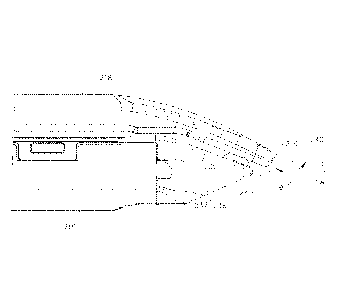
[00056] FIG. 12 depicts an exemplary end effector (212) comprising an anvil
(218) and a
lower jaw (216). It will be appreciated that end effector (212) may be used
interchangeably with end effector (12) of instrument (10). End effector (212)
may be
17
CA 02902880 2015-08-26
WO 2014/133856 PCT/US2014/017298
integrally formed with instrument (10) or in the alternative may be
interchangeable with
end effector (12) of instrument (10).
100057f Anvil (218) is operable to pivot relative to lower jaw (216). Anvil
(218) and lower
jaw (216) may clamp tissue (90) similarly to clamping performed by anvil (18)
and lower
jaw (16) shown in FIG. 1. End effector (212) further comprises a cartridge
(237) operable
to be placed in lower jaw (216) similarly to cartridge (37) shown in FIG. 3.
[00058] Anvil (218) as can be seen in FIGS. 12-13 has an elongated shape
where the distal
portion of anvil (218) angles toward cartridge (237). The distal portion of
anvil (218)
angles toward cartridge (237) such that the distal most tip (219) of anvil
(218) extends
distally longitudinally further than cartridge (237). Though it will be
understood that in
some versions, distal tip (219) may extend to a distance longitudinally equal
to cartridge
(237) or proximal relative to the distal most point on cartridge (237).
Furthermore, anvil
(218) angles toward cartridge (237) through a gentle slope. By way of example,
anvil
(218) is shaped in FIG. 12 similarly to an inverted ski tip. It will be
understood that the
angled shape of anvil (218) may provide easier insertion of end effector (212)
into a
surgical site. For instance, the gentle slope or inverted ski tip shape of
anvil (218) may
provide an atraumatic tissue deflection surface as anvil (218) contacts or
moves through
tissue. Once placed into a surgical site, it will be understood that the
angled shape of
anvil (218) may provide better maneuverability of end effector (212). Other
suitable
variations of anvil (218) will be apparent to one of ordinary skill in the art
in view of the
teachings herein.
1000591 Cartridge (237) is operable to hold staples similar to staples (47)
shown in FIG.
4A for driving into tissue. As shown in FIG. 13, the distal end of cartridge
(237) has a
triangular profile. In particular, the distal end of cartridge (237) comprises
an upper
tapered surface (239) and a lower tapered surface (238). Upper tapered surface
(239) and
lower tapered surface (238) lead to the distal most end of cartridge (237).
Lower tapered
surface (238) defines a sight line (240) such that once end effector (212) is
inserted into a
surgical site, the user can see along sight line (240). Sight line (240),
which can be seen
18
CA 02902880 2015-08-26
WO 2014/133856 PCT/US2014/017298
more clearly in FIG. 14, extends along the edge of lower tapered surface
(238). it will be
appreciated that the planar shape of lower tapered surface (238) may be
operable to allow
the user to visualize and/or nearly visualize the distal tip (219) of anvil
(218). In
particular, sight line (240) intersects longitudinal axis (LA), which extends
longitudinally
through end effector (212), to form a viewing angle (0).
1000601 It will be understood that viewing angle (0) may establish the
relative visibility
that a user has regarding distal tip (219). In particular, the user can see in
front of distal
tip (219) along any line of sight that passes through the intersection of
sight line (240)
and longitudinal axis (LA) within viewing angle (0). For instance, as viewing
angle (0)
increases, the user would have greater visibility of the area immediately in
front of distal
tip (219) from proximal vantage points; whereas as viewing angle (0)
decreases, the user
has less visibility of the area in front of distal tip (219) from proximal
vantage points. In
some versions, viewing angle (0) defines an angle greater than 90 degrees.
Additionally,
in some versions, viewing angle (0) defines an angle greater than 135 degrees.
Other
suitable angles for viewing angle (0) will be apparent to one of ordinary
skill in the art in
view of the teachings herein. In the illustrated version, it will be
understood that the user
generally looks along sight line (240) or along some other line of sight
within viewing
angle (0), thus, the user has visibility along sight line as well as any area
within viewing
angle (0). The underside of distal tip (219) is ftirther slightly rounded to
aid in the
visibility of the intersection of longitudinal axis (LA) and sight line (240).
1000611 When tissue (90) is clamped between a closed cartridge (237) and
anvil (218) as
seen in FIG. 13, the user can look along sight line (240) or elsewhere within
viewing
angle (0) to see, for instance, precisely where anvil (218) has clamped tissue
(90).
Furthermore, the user would be able to determine whether the tissue is
completely
clamped between anvil (218) and cartridge (237) such that tissue does not
spill over the
end of end effector (212). It will be understood that the user may be able to
also visualize
the quality of the clamp between anvil (218) and cartridge (237) against
tissue (90). It
will be appreciated that in some instances, end effector (212) may be rotated
before,
during, or after clamping tissue (90). As a result, the tapered shape of anvil
(218) may
19
CA 02902880 2015-08-26
WO 2014/133856 PCT/US2014/017298
also provide more accessible viewing of distal tip (219) or substantially
adjacent distal tip
(219). It will be understood that the taper of anvil (218) along with lower
tapered surface
(238) of cartridge (237) may further promote easy insertion of end effector
(212) into
tissue in an atraumatic manner. Furthermore, it may be easier to fit end
effector (212)
through a trocar or other devices operable to introduce end effector (212)
into a surgical
site due to the tapered end of end effector (212). For instance, once distal
tip (219) is fit
into a trocar, lower tapered surface (238) and the tapered shape of anvil
(218) may
provide a lead-in, guiding the rest of end effector (212) into the trocar.
[000621 FIG. 15A shows an exemplary alternative version of an end effector
(312)
comprising an anvil (318) and a lower jaw (316). It will be appreciated that
end effector
(312) may be used in place of end effector (12) shown in FIG. 1. In some
versions, end
effector (312) may be integrally formed with shaft (22) or alternatively may
be separately
formed and then combined. Anvil (318) is operable to pivotally open and close
in relation
to cartridge (337). For instance, FIG. 15B shows anvil (318) in the open
position and
FIG. 15A shows anvil (318) in the closed position. Anvil (318) has an angled
distal
portion (320) and an anvil ball tip (319) at the distal most portion of angled
distal portion
(320). Cartridge (337) has a cartridge ball tip (322). It will be understood
that cartridge
ball tip (322) fits into the underside of anvil (318) as will be described in
further detail
below. It will be understood that, similar to end effector (212) shown in FIG.
12, the
distal taper of anvil (318) and cartridge (337) provides improved visibility
of the distal
end of end effector (312).
[000631 FIG. 16 shows an enlarged view of the underside of anvil (318). In
addition to
anvil ball tip (319) and angled distal portion (320), anvil (318) comprises a
plurality of
transversely extending recesses (324) and a ball pocket (326). Plurality of
recesses (324)
line angled distal portion (320). In the illustrated version, plurality of
recesses (324)
include three recesses, but it will be appreciated other suitable numbers of
recesses (324)
may be used. For instance, four, five, six, or more recesses (324) may be
used. It will be
appreciated that recesses (324) may be equally spaced apart. Furthermore,
recesses (324)
may be spaced apart by a known distance. For instance, the user may be aware
that each
CA 02902880 2015-08-26
WO 2014/133856 PCT/US2014/017298
of recesses (324) is spaced I mm apart from each other. As a result, when
tissue is
grasped between anvil (318) and cartridge (337), the tissue may cover a
portion of
recesses (324). Due to the tissue covering recesses (324), the user would be
able to
determine the approximate length of the tissue over recesses (324) by simply
multiplying
the number of covered recesses (324) by the distance between recesses (324).
Alternatively, the user could assess the number of exposed recesses (324) to
calculate the
approximate distance from anvil ball tip (319) to tissue. Furthermore, it will
be
appreciated that recesses (324) are also operable to promote gripping of
tissue such that
anvil (318) and cartridge (337) are operable to maintain a more secure grip on
tissue.
Anvil (318) of the exemplary version also comprises staple apertures (351) and
vertical
slot (349). It will be understood that staple apertures (351) and vertical
slot (349)
function substantially similar to apertures (51) and vertical slot (349) of
FIG. 3.
[000641 It will be appreciated that ball pocket (326) has a shape that
complements
cartridge ball tip (322). While pocket (326) and tip (322) have partially
spherical shapes
in the present example, it should be understood that any other suitable shapes
may be
used. In the present example, when anvil (318) closes onto cartridge (337),
cartridge ball
tip (322) engages ball pocket (326) and promotes alignment and stabilization
of anvil
(318) with cartridge (337) due to the complementary configurations of tip
(322) and
pocket (326). While ball pocket (326) is positioned in one of recesses (324)
in the
exemplary version, it will be understood that ball pocket (326) may be
positioned in any
suitable place along anvil (318). In some versions, ball pocket (326) or ball
tip (322) may
have a surface that is polished and/or painted such that when anvil (318) is
inserted into
tissue, the polished surface of ball pocket (326) or ball tip (322) provides
the user with
greater visibility of ball pocket (326) or ball tip (322). As a result, the
user can better
determine whether ball tip (322) or ball pocket (326) is sufficiently close to
the targeted
tissue area. Furthermore, while the exemplary version shows anvil (318) as
having
recesses (324) and ball pocket (326), it will be understood that cartridge
(337) may in
addition or in the alternative comprise a ball pocket similar to ball pocket
(326) for
21
CA 02902880 2015-08-26
WO 2014/133856 PCT/US2014/017298
receiving anvil ball tip (319) and may also have recesses substantially
similar to recesses
(324).
1000651 In addition to facilitating tip visibility of anvil (218) to the
user, ball tip (322) may
be used as a blunt dissection tool (e.g., as a button dissector) or as a tool
for moving
tissue around within a surgical site. It will be understood that the rounded
shape of ball
tip (322) is operable to provide an atraumatic surface to engage tissue. While
the
exemplary version shows a spherical shape for ball tip (322), it will be
appreciated that
any suitable atraumatic shape for ball tip (322) may be used as would be
apparent to one
of ordinary skill in the art in view of the teachings herein. By using ball
tip (322), it will
be understood that the user may be able to minimize tissue trauma as end
effector (312)
is pushed or otherwise moved through tissue.
1000661 In some instances it may be desirable to have an end effector with
a more visible
tip such that the user has improved visibility of the distal end of end
effector as it is
inserted, for instance, into a surgical site. FIGS. 17 and 18 depict end
effectors (412, 512)
designed to promote improved visibility at the distal end of end effectors
(412). In FIG.
17, end effector (412) comprises an anvil (418) and lower jaw (416) where
lower jaw
(416) holds a carb-idge (437). It will be appreciated that anvil (418), lower
jaw (416), and
cartridge (437) ftmction in a substantially similar manner to anvil (18),
lower jaw (16),
and cartridge (37) of FIG. 3. Anvil (418) differs due to the angled shape of
the distal end
of anvil (418), and due to anvil (418) comprising a cap portion (440). Cap
portion (440)
comprises a rubber, plastic, or otherwise synthetic material operable to cover
the end of
anvil (418). For instance, cap portion (440) could include an insert, an.
overmold, a
coating; or take any other suitable form as would be apparent to one of
ordinary skill in
the art in view of the teachings herein. In the exemplary version, cap portion
(440) covers
the underside of anvil (418). As a result, it will be appreciated that as
tissue is squeezed
by anvil (418), the amount of visible cap portion (440) may indicate to the
user how
much of tissue is being clamped by anvil (418). It will also be appreciated
that cap
portion (440) may comprise a contrasting color distinguishable from tissue.
For instance,
cap portion (440) may comprise a bright red, yellow, blue, green, etc. color
such that
22
CA 02902880 2015-08-26
WO 2014/133856 PCT/US2014/017298
once anvil (418) is inserted into tissue, the user can quickly determine the
position of
anvil (418) within the tissue. While in the exemplary version, anvil (418)
comprises cap
portion (440), it will be understood that in addition to or in the
alternative, cartridge (437)
may have a similar colored portion as well. Furthermore, while the illustrated
version
shows cap portion (440) as only covering a portion of anvil (418), in some
version, the
entire anvil (418) may be covered.
1000671 FIG. 18 depicts an alternative exemplary end effector (512)
comprising an anvil
(518) and lower jaw (516) where lower jaw (516) holds a cartridge (537). Anvil
(518)
defines an opening (540) at the distal end of anvil (518). It will be
appreciated that
opening (540) extends completely through anvil (518) such that as anvil (518)
closes on
tissue, the user can look to see whether tissue is visible through opening
(540). In the
event that tissue is visible, the user has at least one form of confirmation
that tissue has
been clamped between anvil (518) and cartridge (537). In some instances, it
will be
appreciated that the user may desire to see tissue through opening (540) only
briefly,
followed by no tissue. As a result, the user would have confirmation that
tissue has been
positioned deeply enough between anvil (518) and cartridge (537) to ensure
that when
the tissue is clamped and cut, clamping and cutting occurs across the entirety
of the tissue
between anvil (518) and cartridge (537) in a single firing of end effector
(512). Such
confirmation may be desirable, for instance, in the cutting of vessels.
Furthermore, it will
be appreciated that opening (540) also provides a disturbance in the surface
of anvil
(518) that may provide increased grip between anvil (518) and the tissue.
While in the
exemplary version anvil (518) forms opening (540), it will be appreciated that
opening
(540) may be formed on cartridge (537) or any other suitable portion of end
effector
(512) as would be suitable for providing visibility or gripping of tissue in
between anvil
(518) and cartridge (537). Furthermore, while opening (540) in the illustrated
version is
shown as a circular opening formed perpendicular to the surface of anvil
(518), opening
(540) may have any suitable shape and may be formed at any suitable angle
through anvil
(518) that promotes the visibility of tissue through opening (540).
23
[00068] In some versions, end effector (512) may be combined with end
effector (412). In
particular, opening (540) could provide an anchoring point for applying an
overmold or
securing an additional feature such as cap portion (440). Such an overmold or
cap
portion (440) may be formed of a material that is different from the material
forming
anvil (518). By way of example only, an overmold or cap portion (440) could be
formed
of plastic or other polymer while anvil (518) is formed of metal. It should
also be
understood that anvil (518) and/or any other feature of end effector (512) may
include an
applied coating or surface treatment. While the illustrated version is shown
to include
opening (540) on anvil (518), it will be understood that opening (540) could
be
positioned anywhere on the distal end of end effector (512) for receiving an
overmold
component such as cap portion (440). For instance, opening (540) could be
positioned on
the side of anvil (518) or on any suitable location on cartridge (537).
[00069] IV. Miscellaneous
1000701 It should be understood that any one or more of the teachings,
expressions,
embodiments, examples, etc. described herein may be combined with any one or
more of
the other teachings, expressions, embodiments, examples, etc. that are
described herein.
The above-described teachings, expressions, embodiments, examples, etc. should
therefore not be viewed in isolation relative to each other. Various suitable
ways in
which the teachings herein may be combined will be readily apparent to those
of ordinary
skill in the art in view of the teachings herein. Such modifications and
variations are
intended to be included within the scope of the claims.
[00071] intentionally left blank
1000721 Versions of the devices described above may have application in
conventional
medical treatments and procedures conducted by a medical professional, as well
as
application in robotic-assisted medical treatments and procedures. By way of
example
only, various teachings herein may be readily incorporated into a robotic
surgical system
such as the DAVINCITm system by Intuitive Surgical, Inc., of Sunnyvale,
California.
24
Date Recue/Date Received 2020-04-09
Similarly, those of ordinary skill in the art will recognize that various
teachings herein
may be readily combined with various teachings of any of the following: U.S.
Pat. No.
5,792,135, entitled "Articulated Surgical Instrument For Performing Minimally
Invasive
Surgery With Enhanced Dexterity and Sensitivity," issued August 11, 1998; U.S.
Pat. No.
5,817,084, entitled "Remote Center Positioning Device with Flexible Drive,"
issued
October 6, 1998; U.S. Pat. No. 5,878,193, entitled "Automated Endoscope System
for
Optimal Positioning," issued March 2, 1999; U.S. Pat. No. 6,231,565, entitled
"Robotic
Arm DLUS for Performing Surgical Tasks," issued May 15, 2001; U.S. Pat. No.
6,783,524, entitled "Robotic Surgical Tool with Ultrasound Cauterizing and
Cutting
Instrument," issued August 31, 2004; U.S. Pat. No. 6,364,888, entitled
"Alignment of
Master and Slave in a Minimally Invasive Surgical Apparatus," issued April 2,
2002;
U.S. Pat. No. 7,524,320, entitled "Mechanical Actuator Interface System for
Robotic
Surgical Tools," issued April 28, 2009; U.S. Pat. No. 7,691,098, entitled
"Platform Link
Wrist Mechanism," issued April 6, 2010; U.S. Pat. No. 7,806,891, entitled
-Repositioning and Reorientation of Master/Slave Relationship in Minimally
Invasive
Telesurgery," issued October 5, 2010; U.S. Pub. No. 2013/0012957, entitled
"Automated
End Effector Component Reloading System for Use with a Robotic System,
published
January 10, 2013; U.S. Pub. No. 2012/0199630, entitled "Robotically-Controlled
Surgical Instrument with Force-Feedback Capabilities," published August 9,
2012; U.S.
Pub. No. 2012/0132450, entitled "Shiftable Drive Interface for Robotically-
Controlled
Surgical Tool," published May 31, 2012; U.S. Pub. No. 2012/0199633, entitled
"Surgical
Stapling Instruments with Cam-Driven Staple Deployment Arrangements,"
published
August 9, 2012; U.S. Pub. No. 2012/0199631, entitled "Robotically-Controlled
Motorized Surgical End Effector System with Rotary Actuated Closure Systems
Having
Variable Actuation Speeds," published August 9, 2012; U.S. Pub. No.
2012/0199632,
entitled "Robotically-Controlled Surgical Instrument with Selectively
Articulatable End
Effector," published August 9, 2012; U.S. Pub. No. 2012/0203247, entitled
"Robotically-
Controlled Surgical End Effector System," published August 9, 2012; U.S. Pub.
No.
2012/0211546, entitled "Drive Interface for Operably Coupling a Manipulatable
Surgical
Tool to a Robot," published August 23, 2012; U.S. Pub. No. 2012/0138660,
entitled
Date Recue/Date Received 2020-04-09
"Robotically-Controlled Cable-Based Surgical End Effectors," published June 7,
2012;
and/or U.S. Pub. No. 2012/0205421, entitled "Robotically-Controlled Surgical
End
Effector System with Rotary Actuated Closure Systems," published August 16,
2012.
[00073]
Versions of the devices described above may be designed to be disposed of
after a
single use, or they can be designed to be used multiple times. Versions may,
in either or
both cases, be reconditioned for reuse after at least one use. Reconditioning
may include
any combination of the steps of disassembly of the device, followed by
cleaning or
26
Date Recue/Date Received 2020-04-09
CA 02902880 2015-08-26
WO 2014/133856 PCT/US2014/017298
replacement of particular pieces, and subsequent reassembly. In particular,
some
versions of the device may be disassembled, and any number of the particular
pieces or
parts of the device may be selectively replaced or removed in any combination.
Upon
cleaning and/or replacement of particular parts, some versions of the device
may be
reassembled for subsequent use either at a reconditioning facility, or by a
user
immediately prior to a procedure. Those skilled in the art will appreciate
that
reconditioning of a device may utilize a variety of techniques for
disassembly,
cleaning/replacement, and reassembly. Use of such techniques, and the
resulting
reconditioned device, are all within the scope of the present application.
[000741 By way of example only, versions described herein may be sterilized
before
and,/or after a procedure. In one sterilization technique, the device is
placed in a closed
and scaled container, such as a plastic or TYVEK bag. The container and device
may
then be placed in a field of radiation that can penetrate the container, such
as gamma
radiation, x-rays, or high-energy electrons. The radiation may kill bacteria
on the device
and in the container. The sterilized device may then be stored in the sterile
container for
later use. A device may also be sterilized using any other technique known in
the art,
including but not limited to beta or gamma radiation, ethylene oxide, or
steam.
[000751 Having shown and described various embodiments of the present
invention,
further adaptations of the methods and systems described herein may be
accomplished by
appropriate modifications by one of ordinary skill in the art without
departing from the
scope of the present invention. Several of such potential modifications have
been
mentioned, and others will be apparent to those skilled in the art. For
instance, the
examples, embodiments, geometries, materials, dimensions, ratios, steps, and
the like
discussed above are illustrative and are not required. Accordingly, the scope
of the
present invention should be considered in terms of the following claims and is
understood
not to be limited to the details of structure and operation shown and
described in the
specification and drawings.
27