Note: Descriptions are shown in the official language in which they were submitted.
POLYMER BASED OXYGEN SENSORS
[0001]
TECHNICAL FIELD
[0002] The present disclosure is in the field of luminescent dyes, polymers
and biosensors.
BACKGROUND
[0003] Diagnosis, treatment and management of some medical conditions
require monitoring of oxygen concentration in the afflicted organ or tissue.
For
example, Peripheral Arterial Disease (PAD), a disease that is characterized by
plaque
buildup in arteries that carry blood to the extremities, head, or organs, if
left untreated,
can lead to complete blockage of lower extremity arteries and requires either
open
bypass surgery or endovascular intervention. Annually, at least 140,000 such
revascularization procedures are conducted in the US alone to restore blood
flow to
ischemic tissues. Thus, ensuring that blood and oxygen flow are adequately
restored
and maintained during and after the revascularization technique is highly
desirable.
Current monitoring methods are expensive, cumbersome, time consuming, and do
not
provide accurate, continuous tissue oxygenation information. Thus, there is
clearly a
need for a better long-term oxygen tissue monitoring system. Doing so non-
invasively
with minimal user maintenance is essential, and sensor longevity of days to
months is
crucial in actual user environments.
[0004] Such real-time, continuous measurement of oxygen concentration
(partial pressure) in tissues can be achieved by the use of sensors inserted
or
implanted into the tissue and measuring the signal generated by the sensor by
a device
located outside the body. Luminescence provides a useful tool for the design
of such
sensors. Luminescent oxygen sensors are based on the phenomenon that oxygen
has a
quenching effect on the molecular luminescence of various chemical compounds
and
that this effect can be employed for measuring oxygen concentrations (partial
pressure) in vivo. The sensors, which are monitored optically through the
skin, require
1
Date Recue/Date Received 2021-09-03
CA 02904127 2015-09-03
WO 2014/160258
PCT/US2014/026183
a highly stable dye with excitation and emission spectra in the near-infrared
(NIR)
optical window of the skin. These dye properties are crucial for the
successful design
of a luminescent oxygen sensor that can be implanted deep into tissue.
Monitoring
non-invasively through the skin requires the use of dyes with excitation and
emission
wavelengths in the optical window of the skin (approximately 550 to 1000 nm)
to
minimize light scattering and absorbance, and achieve a high signal-to-noise
ratio.
However, commercially available NIR dyes can be prone to photobleaching.
Palladium porphyrins, such as tetracarboxyphenyl porphyrin (Pd-TCPP) have a
very
large Stokes shift and emission in the NIR. However, they unfortunately
require
excitation with green light (525 nm), which is largely absorbed by the skin
and the
underlying tissue. Additionally, currently available sensors, made of rigid
materials
that vastly differ from the mechanical properties of tissue in which they are
implanted,
are bulky and inconvenient, and induce a series of biological events upon
implantation that ultimately culminate in the formation of a fibrous capsule
that walls
it off from the body.
[0005] Thus, until the present invention there remains a clear need in
the art to
provide improved stable, near-IR luminescent compounds and sensors for direct,
rapid
and accurate measurement of oxygen levels in tissue, particularly in vivo.
SUMMARY
[0006] Disclosed herein are luminescent dyes, polymers comprising said
dyes,
and sensors comprising the polymers of the present invention.
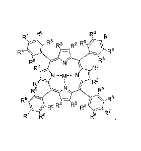
[0007] In one embodiment, the present invention relates to a compound
of
Formula 1:
2
CA 02904127 2015-09-03
WO 2014/160258
PCT/US2014/026183
1
R6 R6 R1
R
R6 R3 R2 R6
R6 R6
R6 R6
R2 R3
R3 R2
R6 R6
R6 R6
R6
0
R6 R2 IA3 R6
R7
R1 R6
wherein:
M is H, Pd, Zn, Pt, Gd or Yb;
each RI is same or different and independently C(0)X-(CH2).-YC(0)C(R4)CH2
, C(0)X-(CH2CH20)õICH2CH2-YC(0)C(R4)CH2 or COOH;
R7 is C(0)X-(CH2).-YC(0)C(R4)CH2 or C(0)X-(CH2CH20),,CH2CH2-
YC(0)C(R4)CH2;
R2 and R3 are hydrogen or are fused, in each case, to form a cycloalkenyl,
aryl,
or heteroaryl group;
X is 0 or NRs;
Y is 0 or NH;
R5 and R4 are independently H or C1-C4 alkyl;
each R6 is the same or different and independently H or F;
n is 1-10; and
m is 1-300.
[0008] In another aspect, the present invention relates to a polymer
comprising as a monomer repeat unit, the residue of the compound of Folinula
1. The
polymers provided herein can be luminescent biocompatible hydrogels.
[0009] In further embodiments, the present invention relates to
various
luminescent sensors comprising the polymers provided herein for detecting an
analyte, e.g., oxygen, in vivo or in vitro. The sensors can be in the foim of
a powder,
fabric (e.g., wound dressing), sutures, needle, rod, disk or any other
suitable form.
3
100101 In another aspect, the luminescent sensors provided herein are
tissue-
integrating or comprise a tissue-integrating scaffold and produce a detectable
signal
in the presence of the analyte; and further wherein the sensors provide
detection of the
analyte when placed (e.g., implanted) into the tissue of a subject. The tissue-
integrating sensors as described herein can provide long-term detection of the
analyte(s).
BRIEF DESCRIPTION OF THE DRAWINGS
[0011] Figure 1 depicts Compound 2 (Pd-BP) absorption and emission
spectra. Spectra were taken of covalently bound Pd-BP in pHEMA hydrogel.
Excitation at 633 inn gave 805 mri emission, confirming shift into the NIR.
[0012] Figure 2 demonstrates that Compound 2 (Pd-BP) incorporated
into a
pHEMA hydrogel sensor enables brighter signals from deeper within the tissue.
Images above show intensity of NIR Pd-BP (A) and green-ex Pd-TCPP (B)
subcutaneous hydrogel implants measured in a rat carcass. Pd-BP is
significantly
brighter than TCPP due to the NIR excitation and emission wavelengths, which
allow
much greater light penetration into the skin, enabling deeper sensor
placement.
[0013] Figure 3 depicts luminescence signal of pHEMA 02 sensor
implanted
in a mouse brain.
100141 Figure 4 depicts luminescence of oxygen sensors implanted in
rat skin
(170 days). Intensity varies as a function of implantation depth (data
normalized to
baseline fluorescence) and tissue oxygen concentration. Inhaled oxygen was
modulated between 100% and 12% and images were collected every 30 s in a
Calipeirm
IVIS (Ex = 640 nm, Em=800 nm). Regions of interest (ROIs) were drawn around
the
sensors and the data plotted versus time. Data is shown in Figure 6.
[0015] Figure 5 shows a SEM image of tissue-integrating porous
hydrogel
scaffold.
4
Date Recue/Date Received 2021-09-03
CA 02904127 2015-09-03
WO 2014/160258
PCT/US2014/026183
[0016] Figure 6 demonstrates determination of photostability Pd-BP.
Gels in
PBS (pH 7.4, 37 C) were illuminated using a 525 nm LED at a 40% duty cycle.
Both
lifetime signal remains constant.
[0017] Figure 7, panels A and B, depicts that the response of the
porous,
tissue-integrated sensors (A) is rapid (-30 seconds), while the response of
the solid
sensor (B) response is much slower (plateau not even reached after 5 minutes).
The
solid sensors are the same rod-shape and material composition.
[0018] Figure 8, panels A and B, depicts dynamic response of Pd-BP
hydrogels to 02 (A) and a Stern-Vomer plot of 02 quenching efficiency with Pd-
BP
(B). The response is linear with good sensitivity and rapid response time.
[0019] Figure 9, panels A and B, depicts dynamic response of G0x/Pd-BP
gel to glucose (A) and normalized glucose dose-response curve (B).
[0020] Figure 10, panels A and B, depicts detectable modulating sensor
signal from the 02 sensor (A) and histological analysis of pig biopsy
containing the
sensor (B).
[0021] Figure 11, panels A to D, depicts solid sensor response to
deoxygenation (0.12 FI02) and re-oxygenation (1.00 FI02) (A), fluorescent
micrographs of solid sensors and surrounding tissue samples at 7 and 28 days
after
implantation, porous, tissue-integrating sensor response to deoxygenation
(0.12 FI02)
and re-oxygenation (1.00 FI02) (C), fluorescent micrographs of porous sensors
and
surrounding tissue samples at 7 and 28 days after implantation (D).
DETAILED DESCRIPTION
[0022] Described herein are polymerizable luminescent dyes useful for
incorporation into polymers and polymers comprising as monomeric units
residues of
the dyes of the present invention. The dyes and the polymers are useful, for
example,
in sensing and imaging applications, for example, accurate and optionally long
term
measurements of oxygen in vivo and in vitro.
5
CA 02904127 2015-09-03
WO 2014/160258
PCT/US2014/026183
[0023] Additionally, described herein are sensors comprising the
polymers of
the present invention. The sensors can be implanted into a tissue of a subject
and used
for long-term or short-teim continuous and semi-continuous collection of data
of
various biochemical analytes, optionally without the use of implantable
hardware of
any type and/or enzymatic and electrochemical detection methods. In one
aspect, the
sensors are tissue integrating, e.g., allow capillaries to grow in close
proximity to all
regions of the sensor (e.g., on the surface and inside), which results in
accurate
analyte measurements, including over long tem'. In another aspect, in addition
to the
luminescent dyes and/or the polymers of the present invention, the sensors
comprise
an oxidase, such as, but not limited to, glucose oxidase, and the luminescent
dyes
and/or their residues incorporated as monomeric units into the polymers
measure the
consumption of oxygen by the oxidase, thus, the sensors can provide detection
of a
number of analytes other than oxygen, such as, but not limited to, glucose.
[0024] Advantages of the dyes and luminescent polymers provided herein
include, but are not limited to: (1) excitation and emission wavelengths in
the optical
window of the skin (approximately 550 nm to 1000 nm) allowing detection of
analytes deep within a tissue or an organ; (2) high signal-to-noise ratio; (3)
large
Stokes shifts and emission; (4) photostablity, e.g., the dyes and/or polymers
do not
undergo rapid photobleaching.
[0025] Advantages of the sensors described herein include, but are not
limited
to: (1) providing devices that generate stable signal over a long period of
time (e.g.,
greater than a week, greater than a month, greater than 6 months), (2)
providing
devices that are placed or implanted and integrate into the subject's tissue
(e.g.,
through tissue and/or capillary in-growth); (3) providing devices which can be
implanted through syringe injection or trocar injection, meaning that no
surgery is
required to put the sensing media in place in the body; (4) providing devices
that do
not include sensor electronics in the body; (5) providing devices that
accurately assess
analyte (e.g., oxygen) concentration for long periods of time (e.g., greater
than a
week, typically weeks, months or years) and/or (6) providing devices of small
dimensions which will give result in increased patent comfort and better
acceptance
by the body.
6
CA 02904127 2015-09-03
WO 2014/160258
PCT/US2014/026183
[0026] It must be noted that, as used in this specification and the
appended
claims, the singular forms "a", "an", and "the" include plural referents
unless the
content clearly dictates otherwise. Thus, for example, reference to a sensor
comprising "a sensing moiety" includes devices comprising of two or more
sensing
moieties. Likewise, reference to "an analyte" refers to two or more analytes.
Definitions
[0027] The term "tissue integrating" refers to a material (e.g.,
scaffold) which,
when integrated into living tissue remains in close proximity with the blood
vessels of
the tissue (e.g., capillaries).
[0028] By "long-terni" is meant that the implant senses the analyte
for greater
than about 7 days, for example weeks, months, or years.
[0029] By "biodegradable" or "bioabsorbable" is meant that the
material is
capable of being broken down by the subject's body over a period of time,
ranging
from days to weeks to months or years.
[0030] By "hydrogel" is meant a material that absorbs a solvent (e.g.
water),
undergoes rapid swelling without discernible dissolution, and maintains three-
dimensional networks capable of reversible deformation.
[0031] The term "stimuli-responsive" refers to substrances, e.g.,
polymers,
that change their physical state, e.g., undergo a phase transition, when
exposed to an
external stimulus or according to the environment they are in. Non-limiting
examples
of such polymers are "smart polymers" (Kumar A. et al., Smart polymers:
Physical
forms and bioengineering applications. Frog Polym. S'ci. 32 (2007) 1205-1237).
A. Luminescent MR dyes
[0032] In one aspect, this invention provides a compound of Folutulal
:
7
CA 02904127 2015-09-03
WO 2014/160258
PCT/US2014/026183
R1 R6 R6 Ri
R6 R3 R2 R6
R6 R6
R6 R6
R2 R3
R3 R2
R6 R6
R6 R6
Rs R2 R3 Rs
A R7
R1 R6 R-
wherein
M is H, Pd, Zn, Pt, Gd or Yb;
each R1 is same or different and independently C(0)X-(CH2)õ-
YC(0)C(R4)CII2 , C(0)X-(CH2CH20),,CH2CH2-YC(0)C(R4)CH2 or COOH;
R7 is C(0)X-(CH2)õ-YC(0)C(R4)CH2 or C(0)X-(CH2CH20)m CH2CH2-
YC(0)C(R4)CH2;
R2 and R3 are hydrogen or are fused, in each case, to foun a cycloalkenyl,
aryl,
or heteroaryl group;
X is 0 or NR5;
Y is 0 or NH;
R5 and R4 are independently H or Cl -C4 alkyl;
each R6 is same or different and, independently, H or F;
n is 1-10; and
m is 1-300.
[0033] In one embodiment, M is Pd. In another embodiment, RI and R7
are
both C(0)NH(CH2)20C(0)C(CH3)CH2. In another embodiment, R1 is
C(0)NH(CH2)20C(0)C(CH3)C112 and R7 is COOH. In yet another embodiment, two
of the Rl are C(0)NH(CH2)20C(0)C(CH3)CH2, one of the le is COOH and R7 is
COOH. In another embodiment, one of the R1 is C(0)NH(CH2)20C(0)C(CH3)CH2,
two of the Rl are COOH, and R7 is COOH. In one embodiment, all le and R7 are
COOH.
8
CA 02904127 2015-09-03
WO 2014/160258
PCT/US2014/026183
[0034] In
another embodiment, R1 and R7 are both C(0)X-(CH2CH20).
CH2CH2-YC(0)C(R4)CH2. In another embodiment, R1 is C(0)X-(C112CH20),/,
CH2CH2-YC(0)C(R4)CH2 and R7 is C0011. in yet another embodiment, two of the R1
are C(0)X-(CH2CH20). CH2C1-12-YC(0)C(R4)CH2, one of the R1 is COOH and R7 is
COOH. In another embodiment, one of the le is C(0)X-(CH2CH20)m CH2CH2-
YC(0)C(R4)CH2, two of the RI are COOH, and R7 is COOH. In one embodiment, all
121 and R7 are COOH.
[0035] In
another embodiment, RI and R7 are both C(0)X-(CH2)-
YC(0)C(R4)CH2. In another embodiment, Rl is C(0)X-(CH2)õ-YC(0)C(R4)CH2 and
R7 is COOH. In yet another embodiment, two of the R1 are C(0)X-(CH2)n-
YC(0)C(R4)CH2, one of the R1 is COOH and R7 is COOH. In another embodiment,
one of the R1 is C(0)X-(CH2)11-YC(0)C(R)CH2, two of the R1 are COOH, and R7 is
COOH. In one embodiment, all RI and R7 are COOH.
[0036] In one
embodiment, R2 and R3 are fused to form a heteroaryl group. In
one embodiment, R2 and R3 are fused to form a cycloalkenyl group. In one
embodiment, R2 and R3 are fused to form a tetracyclohexeno group. In one
embodiment, R2 and R3 are fused to form an aryl group. In one embodiment, the
aryl
group is perfluorinated. In one embodiment, R2 and R3 are fused to form a
benzo
group. In another embodiment, R2 and R3 are fused to form a naphtho group.
[0037] In one
embodiment, RI comprises an oligoethyene glycol linker having
2-300 ethylene units. In another embodiment, R7 comprises an oligoethyene
glycol
linker having 2-300 ethylene units.
[0038] In one
specific embodiment, M is Pd, R1 and R7 are both
C(0)NH(CH2)20C(0)C(CH3)CH2, and R2 and R3 are H.
[0039] In one specific
embodiment, M is Pd, R1 and R7 are both
C(0)NH(CH2)20C(0)C(CH3)CH2, and R2 and R3 are fused to form a benzene ring.
[0040] In one
embodiment, the compound of Formula 1 is a near-IR
luminescent dye. In one embodiment, the compound of Formula 1 has an
absorption
maximum between 500 nm and 800 nm. In one specific embodiment, the compound
of Formula 1 has an absorption maximum between 500 nm and 700 nm. In one
9
CA 02904127 2015-09-03
WO 2014/160258
PCT/US2014/026183
embodiment, the compound of Formula 1 has an emission maximum between 500 and
1000 nm. In one embodiment, the compound of Formula 1 has an emission maximum
between 650 and 900 mm In one specific embodiment, the compound of Formula 1
has an emission maximum between 800 and 900 rim. In one embodiment, the
compound of Formula 1 of the present invention is photostable and has
excitation and
emission spectra in the NIR optical window of the skin.
[0041] For example, in a preferred embodiment, as illustrated by
FIGURE 1,
the Compound 2 of Formula 2 has an absorption maximum at 633 nm and an
emission maximum at 805 rim when co-polymerized with HEMA into a hydrogel.
0, _
0
H
0
/ 11
141111 N-
H IP
o
0
0 0
Compound 2
Formula 2
[0042] In some embodiments, the dyes of the present invention are
encapsulated into a solid, oxygen-impermeable nanosphcre. The nanosphcres can
be
used for luminescent, non-oxygen sensitive applications.
B. Polymers
[00431 The fluorescent dyes of the present invention comprise
polymerizable
groups, e.g., residue of acrylic or methacrylic acid, and can be co-
polymerized with
other monomers to provide polymers comprising near-IR luminescent groups. When
the compounds have 2 or more polymerizable groups, the polymers obtained from
their co-polymerization with other monomers can be crosslinked. Alternatively,
another crosslinking monomer can be added into the polymerization mixture to
achieve a higher degree of crosslinking of the resulting polymer.
CA 02904127 2015-09-03
WO 2014/160258
PCT/US2014/026183
[0044] Polymers
described herein can be prepared in any suitable manner.
Suitable synthetic methods used to produce the polymers provided herein
include, by
way of non-limiting example, cationic, anionic and free radical
polymerization. In
certain embodiments, polymer synthesis is performed neat or in any suitable
solvent.
Suitable solvents include, but are not limited to, pentane, hexane,
dicbloromethane,
chloroform, water, ethylene glycol, propylene glycol, DMSO or dimethyl
formamide
(DMF). In certain embodiments, the polymer synthesis is performed at any
suitable
reaction temperature, including, e.g., from about -50 C to about 100 C, or
from about
00C to about 70 C.
[0045] Preferably the polymers are prepared by the means of a free
radical polymerization. When a free radical polymerization process is used,
(i) the
monomer, (ii) optionally, the co-monomer(s), and (iii) an optional source of
free
radicals are provided to trigger a free radical polymerization process. In
some
embodiments, the source of free radicals is optional because some monomers may
self-initiate upon heating at high temperature. In certain instances, after
forming the
polymerization mixture, the mixture is subjected to polymerization conditions.
Such
conditions are optionally varied to any suitable level and include, by way of
non-
limiting example, temperature, pressure, light, atmosphere, ratios of starting
components used in the polymerization mixture and reaction time. The
polymerization is carried out in any suitable manner, including, e.g., in
solution,
dispersion, suspension, emulsion or bulk.
[0046] In some
embodiments, initiators are present in the reaction
mixture. Any suitable initiator is optionally utilized if useful in the
polymerization
processes described herein. Such initiators include, by way of non-limiting
example,
one or more of alkyl peroxides, substituted alkyl peroxides, aryl peroxides,
substituted
aryl peroxides, acyl peroxides, alkyl hydroperoxides, substituted alkyl
hydroperoxides, aryl hydroperoxides, substituted aryl hydroperoxides,
heteroalkyl
peroxides, substituted heteroalkyl peroxides, heteroalkyl hydroperoxides,
substituted
heteroalkyl hydroperoxides, heteroaryl peroxides, substituted heteroaryl
peroxides,
heteroaryl hydroperoxides, substituted heteroaryl hydroperoxides, alkyl
peresters,
substituted alkyl peresters, aryl peresters, substituted aryl peresters, or
azo
11
compounds. In specific embodiments, benzoylperoxide (BPO) and/or AIBN are used
as initiators.
[00471 In some
embodiments, polymerization processes are carried out
in a controlled (living) mode. Preferred controlled (living) polymerization
processes
include reversible addition-fragmentation chain transfer (RAFT) polymerization
processes and Atom Transfer Radical Polymerization (ATRP).
100481 In
certain embodiments, the polymer of the present invention is a
hydrogel. For example, the hydrogel can be prepared by reacting hydroxyethyl
methacrylate (HEMA), to form poly(hydroxyethyl methacrylate), pHEMA.
Furthermore, various comonomers can be used in combination to alter the
hydrophilicity, mechanical and swelling properties of the hydrogel (e.g. PEG,
NV?,
MAA). Non-limiting examples of polymers include 2-Hydroxyethyl methacrylate,
polyacrylamide, N-vinylpyrrolidone, N,N-Dimethylacrylamide, poly(ethylene
glycol)
monomethacrylate (of varying molecular weights), diethylene glycol
methacrylate, N-
(2-hydroxypropyl)methacrylamide, glycerol monomethacrylate, 2,3-
dihythoxypropyl
methacrylate and combinations thereof. Non-limiting examples of cross-linkers
include tetraethylene glycol dimethacrylate, poly(ethylene glycol) (n)
diacrylate (of
varying molecular weights), ethoxylated trimethylolpropane triacrylate,
bisacrylamide
and combinations thereof. Non-limiting examples of initiators include
Irgacur7Series
(UV), Azobisisobutyronitrile (AIBN) (thermal), Ammonium Persulfate (APS)
(thermal).
100491 In a
specific embodiment, the polymer is a luminescent hydrogel
prepared by co-polymerization of HEMA and compound of Formula 1. In a
preferred
embodiment, the hydrogel is prepared by co-polymerization of various molar
amounts
of compound of Formula 2 mixed with 2-hydroxyethyl methacrylate (HEMA)
monomer, tetraethylene glycol dimethacrylate (TEGDMA) crosslinker, Irgacure
651
initiator, water and co-solvent, followed by UV-initiated polymerization. In
another
embodiment, the polymer contains 1 mM final concentration of Compound of
Formula 1. In a specific embodiment, the polymer is an oxygen sensing poly(2-
hydroxyethyl methacrylate) (pITEMA) scaffold prepared by co-polymerization of
HEMA (2-hydroxyehtyl methacrylate) (50 Wt %), TEGDMA (triethyleneglycol-
12
Date Recue/Date Received 2021-09-03
CA 02904127 2015-09-03
WO 2014/160258
PCT/US2014/026183
dimethacrylate) (1 Wt %1), ethylene glycol (20 Wt%), water (25.5 Wt %), the
photoinitiator Irgacure 651(0.5% vol/vol) and 3% of Compound 2.
[0050] The polymer of the present invention may be degradable, either
by the
body (biodegradable) or by the application of an external initiator to start
or speed up
the degradation process (e.g. UV, ultrasonics, radio frequency, temperature,
or other
exogenous sources to initiate degradation.). For example, the polymer may be
biodegradable or bioresorbable or may comprised any biodegradable or
bioresorbable
segments, including but not limited to degradable forms of alginates,
poly(lactic acid),
poly(vinyl alcohol), polyanhydrides, poly(glycolic acid), microporous
polyesters,
microporous polyethers and cross-linked collagen. One specific example is UV-
photopolymerization of poly(ethylene glycol)-diacrylate and acrylated protease-
degradable peptides and VEGF as described by Phelps, et al (2010) Proc. Nat'l.
Acad.
Sci. USA 107(8):3323-3328
[0051] In one embodiment, polymers provided herein are bioeompatible.
In
another aspect of the invention, the polymers are biodegradable. Degradable
hydrogels can be synthesized using Atom Transfer Radical Polymerization (ATRP)
through co-polymerization of the HEMA with polymerizable luminescent dyes of
the
present invention. Porous sensor scaffolds, based on non-degradable and
degradable
oxygen-sensing hydrogels, can be generated by using a sphere-templating
fabrication
technique. Degradable and non-degradable HEMA reagents and polymerizable dye
will be polymerized over templating microspheres, which are subsequently
dissolved
away with solvent to generate desirable non-degradable and degradable
scaffolds.
Briefly, using controlled ATRP, HEMA will be polymerized in the presence of bi-
functional degradable PCL-based ATRP initiator and cross-linker. In this
synthesis
scheme, pHEMA chains grow at the same rate from both sides of degradable
initiator,
resulting in degradation products with a MW that is half that of the parent
polymer.
By controlling the MW of the parent polymer and the PEG and PCL units in the
initiator and/or crosslinker, the degradation rate of the polymers can be
varied.
Limiting the MW of the parent polymer to 10kDa results in degradation products
that
can be cleared by the body and an increased degradation rate while still
preserving the
hydrogel's mechanical strength.
13
CA 02904127 2015-09-03
WO 2014/160258
PCT/US2014/026183
[0052] In
certain embodiments the polymers provided herein are stimuli-
responsive, e.g., temperature or pH-sensitive polymers. One non-limiting
example of
such a stimuli-responsive polymer is a temperature-sensitive polymer derived
from
co-polymerization of NIPAM. Such polymers are useful for implantation of the
sensor
comprising said polymers in a desired location within tissue by first
dissolving the
polymer in a suitable for injection media at a lower than body temperature and
then
injecting the resulting solution into the tissue and/or at desired location of
the body.
As the polymer is subjected to a higher (e.g., body) temperature, it
precipitates in or
near the site of the injection where monitoring of oxygen is required.
C. Sensors
[0053] In some
embodiments, the polymer of the present invention is
incorporated into a sensor useful for detection of an analyte. The detection
of the
analyte can be in vitro or in vivo. The remaining sentences of this paragraph
describe
how the word "polymer" is used in section titled "C. Sensors". The polymer may
have
the molecules of Formula 1 and/or Formula 2 covalently bound to the polymer
backbone. The molecules of Formula 1 and/or Formula 2 maybe attached to (e.g.
via
a covalent bond or other means) or contained within nanoparticle carriers or
microparticle carriers or other carriers that are attached to or contained
within the
polymer. Such carriers may be covalently bound to the polymer backbone. The
word
polymer can be used interchangeably with the word sensor.
[0054] In one
non-limiting example, the polymer is incorporated into an
oxygen-sensing wound dressing that can be used to monitor the process of wound
healing, e.g. to constantly and non-invasively assess one of the critical
factors of
healing (i.e. oxygenation).
[0055] In another embodiment, the polymer is incorporated into a powder,
which is used directly in the wound as a sensor for wound-healing monitoring.
The
sensor of the present invention can also be in the form of an injectable,
implant, a
mesh or sutures to be used in applications which benefit from monitoring of
oxygenation of skin or the underlying tissue, including, but not limited to
wound
healing monitoring, skin closure, hernia repair, flap transfer surgeries,
reconstructive
surgery, and other plastic surgery applications. The sensor of the present
invention
can also be used for measurement for microcirculatory dysfunction and
peripheral
14
artery disease. Specifically in re-vascularization procedures or upon
administration of
drug, tissue oxygen may be directly monitored. The sensor of the present
invention
can also be used in oncology applications to determine the degree of hypoxia
in a
tissue or an organ. In one embodiment, the sensor is used to monitor tumor
growth in
animal, including but not limited to, mouse or rat models used in oncology
pharmaceutical and diagnostic research and discovery, e.g., cancer therapy
dosing or
monitoring of tumor metabolism. The sensor of the present invention can also
be used
in monitoring the state of pulmonary function, for example in COPD and asthma
disease states. In yet another embodiment, the sensor is used for exercise or
training
optimization, e.g., soldier and athlete performance or personal exercise
programs. The
sensor can also be in the form of an oxygen-sensing tattoo.
[0056] Yet in another embodiment, the sensors of the present invention are
used in neuroscience monitoring applications, where currently there are no
tools
available for continuous monitoring of oxygen, for example, in subarachnoid
hemorrhage monitoring.
[0057] In one embodiment, the sensor of the present invention is a
solid
material that could be in form of a slab, rod, cylinder, particle or powder.
In a specific
embodiment, the sensor is in the form of a rod. In another embodiment, the
sensor is
in the form of a cylinder.
[0058] In another embodiment, the polymer of the present invention is
incorporated into a tissue-integrating scaffold to provide a tissue-
integrating sensor (as
described in the US patent application 2012/0265034). The sensors described
herein
typically comprise a tissue-integrating scaffold (also referred to as a
matrix) material.
Preferably, the tissue-integrating scaffold of the invention may be
constructed with
materials and/or micro-architecture such that the scaffold promotes tissue-
integration
and/or vascularization. For example, porous scaffolds provide tissue
biomaterial
anchoring and promote in growth throughout the pores. The resulting "hallway"
or
"channel" pattern of tissue growth are healthy, space-filling masses that
persist over time
and promote host cell integration. Most or all of the pores of the
biomaterials described
herein are preferably interconnected (co-continuous). The co-continuous pore
structure
of the biomaterials promotes space-filling in-growth of cells in the implant,
which in turn
Date Recue/Date Received 2021-09-03
limits the foreign body response and leads to long-term (greater than one week
and up
to years) persistence of the implant's ability to act as a sensor. Alternative
structures
that provide tissue integrating scaffolds include fibers (e.g., 1 to 10 or
more microns
in diameter, such as 5,6, 7, 8, 9, 10 or more microns), which may be arranged
in non-
random or random configuration. Tissue-integrating scaffolds (in any
configuration)
can also be formed by multiphoton polymerization techniques. Kaehr et al.
(2008)
Proc. Nat'l. Acad. ScL USA 105(26):8850-8854; Nielson et al. (2009) Small
1:120-
125; Kasprzak, Doctoral Dissertation, Georgia Institute of Technology, May
2009.
100591 The polymer of the invention, preferably in the form of a
tissue-
integrating scaffold, may comprise any material in combination with the
compound of
Formula 1 or Formula 2, including but not limited to synthetic polymers,
naturally-
occurring substances, or mixtures thereof. Exemplary synthetic polymers
include, but
are not limited to polyethylene glycol (PEG), 2-hydroxyethyl methacrylate
(HEMA),
silicone rubber, polyaepsilon]-caprolactone) dimethylacrylate, polysulfone,
(poly)methy methacrylate (PMMA), soluble Teflon-TM
AF, (poly) ethylenetetrapthalate
(PET, Dacron, Nylon, polyvinyl alcohol, polyacrylamide, polyurethane, and
mixtures
thereof. Exemplary naturally-occurring materials include, but are not limited
to,
fibrous or globular proteins, complex carbohydrates, glycosarninoglycans,
extracellular matrix, or mixtures thereof. Thus, the polymer scaffold may
include
collagens of all types, elastin, hyaluronic acid, alginic acid, desmin,
versican,
TM
matricelluar proteins such as SPARC (osteonectin), osteopontin, thrombospondin
1
and 2, fibrin, fibronectin, vitronectin, albumin, chitosan etc. Natural
polymers may be
used as the scaffold or as an additive.
100601 In certain embodiments, the polymer of the invention,
preferably in the
form of a tissue-integrating scaffold, comprises a hydrogel. For example, the
polymer
may comprise a hydrogel, for example by reacting hydroxyethyl methacrylate
(HEMA), poly (hydroxyethyl methacrylate), pHEMA. Furthermore, various
comonomers can be used in combination to alter the hydrophilicity, mechanical
and
swelling properties of the hydrogel (e.g. PEG, NVP, MAA). Non-limiting
examples
of polymers include 2-hydroxyethyl methacrylate, polyacrylamide, N-
vinylpyrrolidone, N,N-dimethylacrylamide, poly(ethylene glycol)
monomethacrylate
(of varying molecular weights), diethylene glycol methacrylate, N-(2-
16
Date Recue/Date Received 2021-09-03
CA 02904127 2015-09-03
WO 2014/160258
PCT/US2014/026183
hydroxypropyl)methacrylamide, glycerol monomethacrylate, 2,3-dihydroxypropyl
methacrylate and combinations thereof. Non-limiting examples of cross-linkers
include tetraethylene glycol dimethacrylate, poly(ethylene glycol) (n)
diacrylatc (of
varying molecular weights), ethoxylated trimethylolpropane triacrylate,
bisacrylamide
and combinations thereof. Non-limiting examples of initiators include irgacure
Series
(UV), Azobisisobutyronitrile (AIBN) (thermal), Ammonium Persulfate (APS)
(thermal).
[0061] The polymer of the invention, preferably in the form of a
tissue-
integrating scaffold, may be a sphere-templated hydrogel, for instance an
inverse
colloid crystal, for example as described in U.S. Patent Publication No.
2008/0075752
to Ratner, et al. or other tissue integrating materials.
[0062] The polymer of the invention, preferably in the fount of a
tissue-
integrating scaffold, may be degradable, either by the body (biodegradable) or
by the
application of an external initiator to start or speed up the degradation
process (e.g.
UV, ultrasonics, radio frequency, or other exogenous sources to initiate
degradation.).
For example, the polymer may be comprised of any biodegradable or
bioresorbable
polymers, including but not limited to degradable forms of alginates,
poly(lactic acid),
poly(vinyl alcohol), polyanhydrides, poly(glycolic acid), microporous
polyesters,
microporous polyethers and cross-linked collagen. One specific example is UV-
photopolymerization of poly(ethylene glycol)-diacrylate and acrylated protease-
degradable peptides and VEGF as described by Phelps, et al (2010) Proc. Nat'l.
Acad.
Sci. USA 107(8):3323-3328.
[0063] Other specific examples are polymers described by Kloxin et al
(2009)
Science 324:59-63 and U.S. Patent No. 6,013,122 whose degradation is
controlled
through exposure to exogenous energy fauns, as well as by Alexeev et al.
(2003)
Anal. Chem. 75:2316-2323; Badylak et al. (2008) Seminars in Immunology 20:109-
116; Bridges et al. (2010) 94(1):252-258; Isenhath et al. (2007) Research
83A:915-
922; Marshall et al. (2004) Polymer Preprints, American Chemical Society,
Division
of Polymer Chemistry 45:100-101; Phelps et al. (2010) Proc Nat'l Acad Sci USA.
107(8):3323-8; Ostendorf and Chichkov (2006) Two Photon Polymerization: A New
Approach to MicroMachining, Photonics Spectra; Ozdemir et al. (2005)
Experimental
and Clinical Research, Plast. Reconstr. Surg. 115:183; U.S. Patent Publication
No.
17
CA 02904127 2015-09-03
WO 2014/160258
PCT/US2014/026183
20080075752; Sanders et al. (2003) Journal of Biomedical Materials Research
Part A
67A(4):1181-1187; Sanders et al. (2002) Journal of Biomedical Materials
Research
62(2):222-227; Sanders et al. (2003) Journal of Biomedical Materials Research
65(4):462-467; Sanders et al. (2005) Biomaterials 26:813-818; Sanders et al.
(2005)
Journal of Biomedical Materials Research Part A 72(3):335-342; Sanders (2003)
Journal of Biomedical Materials Research 67(4):1412-1416; Sanders et al.
(2000)
Journal of Biomedical Materials Research 52(1):231-237; and Young Min Ju et
al.
(2008) J Biomed Mater Res 87A:136-146.
[0064] In certain embodiments, the polymer of the invention,
preferably in the
form of a tissue-integrating scaffold, is constructed such that tissue
response modifiers
are released from the scaffold material to promote or enhance tissue-
integration and
vascularization.
[0065] In addition, the polymer of the invention, preferably in the
form of a
tissue-integrating scaffold, may be constructed such that it has conduits,
pores or
pockets that are hollow or filled with degradable, angiogenic, or other
substances (e.g.
stem cells). As noted above, once in the body, the biodegradation of the
material
filling the conduits, pores or pockets, creates space for tissue, including
capillaries to
integrate with the material. The degradable material that initially fills the
conduits,
pores, or pockets may enhance vessel growth or tissue growth within the
scaffold.
This architecture promotes new vessel formation and maintains healthy viable
tissue
within and around the implant.
[0066] The polymer of the invention, preferably in the form of a
tissue-
integrating scaffold, may be constructed such that it is permeable to analytes
of
interest (e.g., oxygen can diffuse into a tissue-integrating hydrogel scaffold
and reach
the sensing moieties that are embedded within the hydrogel matrix).
[0067] The polymer of the invention, preferably in the form of a
tissue-
integrating scaffold, can be of any suitable form, including, but not limited
to block-
like (or any thickness), cube-like, disk-shaped, cylindrical, oval, round,
random or
non-random configurations of fibers and the like. In certain embodiments, the
sensor
comprises one or more fibers, which may be organized in a non-random fashion
(e.g.,
grid, layered grid, etc.) or in a random fashion.
18
CA 02904127 2015-09-03
WO 2014/160258
PCT/US2014/026183
[0068] The
polymer of the invention, preferably in the faun of a tissue-
integrating scaffold, described herein are typically combined with (or made up
of)
sensing moieties that detect one or more analytes. In one embodiment, the
sensing
moiety is the residue of compound of Foimula 1 and/or 2 incorporated into the
tissue-
integrating scaffold.
[0069] In
another embodiment, the polymer of the invention, preferably in the
foun of a tissue-integrating scaffold, comprises, in addition to the residue
of
compound of Formula 1 and/or Formula 2, a second sensing moiety that produces
or
consumes oxygen, e.g., an oxidase, and the residue of compound of Folinula 1
and/or
Foimula 2 is used to detect the change in the oxygen concentration generated
by the
second sensing moiety. The second sensing moiety can comprise an enzyme, for
example glucose oxidase (G0x), which is specific for the substrate glucose.
The
reaction of glucose via enzymatic interaction with glucose oxidase causes
oxygen to
be proportionally consumed and converted to H202. The reduction of 02 in the
vicinity of the enzyme can be measured by using an 02-sensitive fluorescent
dye, such
as the molecules of Fottnula 1 and Formula 2. These dye molecules are quenched
in
the presence of 02, so the reduction of 02 by the action of G0x, causes an
increase in
fluorescence. The amount of fluorescence emitted from the 02 calibration
moieties is
thus proportional to the concentration of glucose in the sensor. Oxidases
besides
glucose oxidase for detection of other analytes besides glucose may include
billirubin
oxidase, ethanol oxidase, lactate oxidase, pyruvate oxidase, histamine oxidase
or other
oxidase to provide specificity to other analytes of interest.
[0070] The
concentration of 02 in the tissue can also vary physiologically,
thereby changing or limiting the reaction of the oxide enzyme in the sensing
moieties.
Therefore, the 02 concentration in the sensor can be measured independent of
the
oxidase target concentration. This may be accomplished through physical
separation
on some nanometer, micro on mm scale of 02 reference moieties from the enzyme-
02
detection moieties to avoid cross talk. Such a reference measurement of 02
would
allow corrections to be made to the glucose-specific signal from the oxidase
sensing
moieties.
[0071] In
another embodiment, the polymer of the invention, preferably in the
form of a tissue-integrating scaffold, may be a multi-analyte sensor where
oxygen is
19
one of two or more analytes detected and reported. In this embodiment, the
polymer
comprises a residue of compound of Formula 1 and/or Formula 2 for detection of
oxygen, and a second sensing moiety for detection of another substance. Non-
limiting
examples of analytes that may be detected by the sensing moieties include
oxygen,
reactive oxygen species, glucose, lactate, pyruvate, cortisol, creatinine,
urea, sodium,
magnesium, calcium, potassium, vasopressin, hormones (e.g., Luteinizing
hormone),
pH, cytokines, chemokines, eicosanoids, insulin, leptins, small molecule
drugs,
ethanol, myoglobin, nucleic acids (RNAs, DNAs), fragments, polypeptides,
single
amino acids and the like.
100721 In another embodiment, the polymer of the invention, preferably in
the
form of a tissue-integrating scaffold, may be a sensor where the oxygen
signal, as
detected by Formula 1 and/or Formula 2, is used as a reference to correct or
calibrate
the signal for one or more other analytes. The oxygen signal may or may not be
reported. It may be used only in internal algorithms to calibrate or correct
the signal
of the other analyte. The use of the oxygen signal as a reference in this
embodiment
helps to overcome physiological fluctuations, which may alter the analyte
availability
at the site of the sensor (e.g. blood flow variations).
100731 In still further embodiments, the sensing moieties, in
addition to the
residue of compound of Formula 1 and/or Formula 2 comprise a second
luminescent
analyte sensing moiety, and the residue of the compound of Formula 1 and/or
Formula 2 is used as a reference molecule. The non-oxygen sensing moieties may
utilize analyte-specific moieties such as competitive binding assays (e.g. a
li2and
receptor moiety and an analyte analogue moiety such as Concanavalin A and
dextran), reversible luminescent binding molecules (e.g. boronic acid based
sensing
chemistry for glucose detection), binding proteins such as glucose binding
proteins.
To measure an analyte such as glucose in the tissue, the polymer is
illuminated from a
patch reader on top of the skin above the implant with 650mn light at desired
intervals
over the long-term life of the implant (e.g., every 5-60 minutes over a period
of 90
days or more). The amount of luminescent signal (e.g., from a molecule such as
Alexafluormr 647) detected is proportional to the concentration of analyte
(e.g. glucose)
in the tissue. The amount of luminescent signal (e.g. from Formula 1 or
Formula 2
molecule) detected is proportional to the concentration of 02 in the tissue.
The
Date Recue/Date Received 2021-09-03
CA 02904127 2015-09-03
WO 2014/160258
PCT/US2014/026183
concentration of 02 in the tissue is indicative of acute and or chronic
physiological
changes around the sensor, and may be used to correct or adjust the glucose
signal or
other analyte signal through a porportionality algorithm.
[00741 In
another embodiment, internal reference control materials can be
employed that facilitate correcting for tissue optical variation. The tissue-
integrating
implanted bio sensor typically resides 3-4 mm under the surface of the scan.
It is well
known that in skin excitation light and emitted fluorescent light in the near
infrared
range are highly scattered as the light traverses the tissue between the
reader patch
and the implant. The extent of absorption and scattering is affected by
physical
properties such as temperature or by tissue composition, including but not
limited to
variations in blood perfusion, hydration, and melanin concentration. Skin
variations
can occur between users or between different time points for a single patient,
and
these variations can affect the fluorescence excitation and emissions signals
causing
in accurate signals for the analyte-specific signal.
Accordingly, a separate
fluorescence molecule with emission spectra distinguishable from the analyte-
specific
fluorescence can be immobilized into the scaffold. The fluorescence from the
molecule can be measured separately from the analyte-specific fluorescence to
measure a signal that infoims about variations in tissue composition. The dye
selected is based on having a similar response to tissue variations as the
analyte-
specific dye. Formula 1 or Formula 2 may have the oxygen sensing capabilities
greatly reduced or eliminated, for example, by incorporation in a non-oxygen
diffusive environment such as embedding in highly crosslinked PAN or inside a
silica
shell. In this format, the dye molecules of this invention may serve as the
stable
internal reference control materials described above.
[0075] Tissue-integrating sensors comprised of one or more cylindrical
shaped
elements (e.g., fibers) eliminate or greatly reduce the foreign body response
as
compared to currently available implants. Moreover, the average diffusion
distances
from the capillary supply to all parts of the sensing media are comparable to
native
tissue, unlike other known sensors.
[0076] It will be apparent that the overall dimensions of the sensing media
(implantable sensor) will vary according to the subject and/or the analyte(s)
to be
measured. Typically, the implant will be between about .001 mm to 2 mm in
21
CA 02904127 2015-09-03
WO 2014/160258
PCT/US2014/026183
thickness (or any value therebetween) and between 1 mm and 1 cm in diameter
(or an
equivalent cross sectional area of a non-circular shape, for example
length/width) and
15 mm in length or less, for example, a disk shaped sensor that is 2 mm or
less thick
and 10 mm or less in diameter. In certain embodiments, the approximate sensor
size
is approximately 100-1000 microns in diameter and has the length of between
0.25
mm and 10 mm. The size of the tissue-integrating sensing media in disk form is
typically 2 mm or less thick and 10 mm or less in diameter.
[0077] Another aspect of the present invention is a tissue-integrating
biosensor system for semi-continuous, continuous and/or long-teim use within a
mammalian body.
[0078] One advantageous property of the polymers of the present
invention is
their stability. In one aspect of the invention, the sensor is stable in a
mammalian
tissue for a long period of time, e.g., longer than a week, longer than a
month, longer
than 6 months. In one exemplary embodiment, as shown by the FIGURE 2, the
sensor
is stable and produces a stable signal when implanted into the rat skin for
170 days.
EXAMPLES
[0079] NMR spectroscopic data were recorded on a 300 MHz instrument at
room temperature. NMR spectra were calibrated to the solvent signals of
deuterated
DMSO-d6 or CDC13. The following abbreviations are used to indicate the signal
multiplicity: s (singlet), d (doublet), t (triplet), q (quartet), br (broad),
m (multiple .
Analytical IIPLC-MS data were recorded on a HPLC system with a C18 reverse
column coupled to an electrospray ionization (ESI) spectrometer. 2-Aminoethyl
methacrylate hydrochloride and tetraethylene glycol dimethacrylate were
purchased
from Polysciences, Inc. All other chemicals were purchased from Sigma Aldrich.
Example 1: Synthesis of a polymerizable near-IR luminescent dye.
[0080] Scheme 1 describes the synthesis of one exemplary near-IR
luminescent dye, Compound 2 (also referred to as Pd-BP):
22
CA 02904127 2015-09-03
WO 2014/160258 PCT/US2014/026183
Me00C COOMe
1) it CHO
NO2- MeO2C
= ....._
NCõCO2Et N
NH
(-....r., 1111)111
_..- KOH C
el NH HN = __________________________________________________________
C----.9 ethylene glycol
THE, influx, 16h c02Et RT 2) 13F3-0Et, it, 2h 1h
\ fi ....-
- 3) DDQ, 16h
3 4 10 = =
Me00C COOMe
HOOG Me00C Me00C COOMe
is 00H Mk is COOMe
* it * = *
IN '. --- KOH / 'Ili - N-
- DDQ / PdC12
0 N- -Pd--N lb ' 1110 N- -c/ 1,--N ---0 ...,õ
THF IP -c1 11,- -N 1110
THF/Nle0H
influx, 2h \ fi reflux, 20 mm. \ fi refl
,..- PhCN
ux, 10 Thin.
HOOC COON Me00C COOMe Me00C COOMe
8 7
6
-cr._Fri 0* * *0
...,_,
0 N 0
H
0
H2N /
______________ ..- 00 N--P:c1--N 0
EDC/HOBt \ }I ---
DGM/DMF
it 16h H
0 N 0 0
0 0 0)r-
Compound 2
Scheme 1
5 [0081] Compound 3
was prepared as described in Niedermair et al, J. Inorg.
Chem., 2010, 49, p. 9333. Briefly, to 90 mL of anhydrous THF was added 1-
nitrocyclohexenene (2.66 mL), ethyl isocyanoacetonitrile (2.6 mL), and DBU
(3.53
mL). The reaction was refluxed at 70 C under argon for 18 hours. Brown
precipitate
fanned as soon as heating began. THF was evaporated, the residue was dissolved
in
methylene chloride, and the product was purified by flash chromatography on
silica
gel in methylene chloride. Product-containing fractions were evaporated under
vacuum to remove most of the solvent, and to the residual solution hexanes
were
added to facilitate crystallization of the product. After 48 hr at 4 C, the
precipitate was
collected to by filtration to yield 2 g of the product as fine yellow needles.
The mother
liquor was partially evaporated to yield additional 1.4 g of the product; 75%
total
yield.
23
100821 Compound 5: Compound 3 (1.40 g, 7.2 mmol) was suspended in 30
mL of anhydrous ethylene glycol, and KOH pellets (0.73 g, 13.0 mmol) were
added to
the solution. The mixture was refluxed under argon for 1 hr. The resulting
clear brown
solution was cooled to 0 C, and 100 mL of dichloromethane was added to the
solution. Dichloromethane layer was separated, washed with water (2X100 mL),
and
brine (2X100 mL) and dried over anhydrous sodium sulfate. The product was
purified
by flash chromatography on silica gel in dichloromethane. Fractions containing
the
fast-running component were pooled and diluted with dichloromethane to 1000
mL.
To the resulting solution was added methyl-4-formyl benzoate, under argon, the
solution was stirred at room temperature for 10 min, and BF3.0Et2 (0.19 mL,
1.3
mmol) was added. The mixture was stirred for 2 hr, then 1.73 g (7.6 mmol) of
DDQ
was added, and the mixture was allowed to stir overnight. The mixture was
washed
sequentially with 10% aq. Na2CO3, 1M HC1, and brine, then dried over anhydrous
sodium sulfate. After purification by silica gel chromatography using stepwise
gradient of Me0H in dichloromethane (0-2%), 430 mg (24%) of the product as
green
powder.
[0083] Compound 6: Compound 5 as a free base (0.43 g, 0.40 mmol) was
dissolved in 50 mL of benzonitrile. To the solution, PdC12 was added under
argon,
and the mixture was refluxed for 10 min. The color of the solution changed
from
green to red. The mixture was cooled to room temperature, diluted with 200 mL
of
TM
dichloromethane, and filtered through Celite. Dichloromethane was evaporated
under
vacuum, and benzonitrile was distilled off. The product was purified by flash
chromatography on silica gel in dichloromethane, and the final purification
was
achieved by flash chromatography on silica gel in hexanes:ethyl acetate (1:1)
to yield
0.109 mg (60%) of the product as a red powder.
100841 Compound 7: Compound 6 (0.105 g, 0.09 mmol) was dissolved in
20
mL of anhydrous THF, and DDQ (0.327 g, 1.44 tnmol) was added to the solution.
The
mixture was refluxed for 20 min, and the reaction was stopped when no starting
material was detected in the mixture by TLC. THF was removed under vacuum, the
residue was diluted with dichloromethane and washed sequentially with 10%
Na2SO4,
water, and brine.
24
Date Recue/Date Received 2021-09-03
CA 02904127 2015-09-03
WO 2014/160258
PCT/US2014/026183
[0085] Compound
8: The ester 7 was hydrolyzed as described in Finikova et
al., J. Phys. Chem., 2007, 111, p. 6977. Briefly, 0.074 g (0.064 mmol) of
Compound 7
were dissolved in 110 mL of THF. To the solution, Me0H (10 mL) was added,
followed by a solution of 0.573 g of KOH in 2 mL of Me0H. Green precipitate
formed in the solution, and the solution became almost colorless. The
precipitate was
collected by centrifugation and dissolved in 10 mL of water. The solution was
acidified with 0.2 mL of concentrated HCI, and the resulting precipitate was
collected
by centrifugation. Yield: 0.070 g (86%).
[0086] Compound
2: Compound 8, 30 (70 mg, 63.9 nmol) in DMF (10 mL)
and CH2C12 (10 mL) at 0 C was added 1-hydroxybenzotriazole hydrate (43.17 mg,
0.32 mmol), N-(3-dimethylaminopropy1)-N'-ethylcarbodiimide hydrochloride
(61.25
mg, 0.32 mmol), and triethylamine (90 pt, 0.64 mmol). After 20 min., 2-
aminoethyl
methacrylate hydrochloride (53.23 mg, 0.3195 mmol) was added, and the reaction
was stirred for 16 h at room temperature. The CH2C12 was evaporated under
reduced
pressure, and ethyl acetate/hexanes mixture was added to precipitate the crude
product
from residual DMF. The solvent was decanted, and the precipitated residue was
dissolved in CH2C12, washed sequentially with sat. NaHCO3 and brine, dried
over
Na2SO4, filtered, and concentrated in vacuo. The crude product was purified by
flash
chromatography on silica gel (gradient of 0 ¨ 4% methanol in CH2C12) to yield
Compound 2 as a green powder (16 mg, 16% yield). 1H NMR (300 MHz, CDC13) 6
8.40 (d, J = 8.1 Hz, 8H), 8.32 (d, J = 8.1 Hz, 8H), 7.22 (br s, 8H), 7.10 (hr
s, 8H), 6.28
(s, 4H), 5.71 (s, 4H), 4.61 (t, J = 5.4 Hz, 8H), 4.03 (q, J = 5.1 Hz, 8H),
2.06 (s, 12H).
LC-MS (ESI): calcd for C881173N8012Pd: 1539.4403 [M+H]+, found 1539.4405
[M+H]+, Rt= 11.8 mm.
[0087] Compound 9 was synthesized analogously to Compound 2 by reacting
commercially available tetracarboxyphenyl porphyrin with ainoethyl
methacrylate in
the presence of HOBt and EDC as shown in Scheme 2:
CA 02904127 2015-09-03
WO 2014/160258 PCT/US2014/026183
HO 0 N 0
40 0 lis
0
,
N N¨
O N N¨ 0 ______________________________________ * NH
HO = \
N / -"N 0 EDC/HOBt TEA, DCWDMF
HN
N 0
1
\1
410
HO 0
0
0
Compound 9
Scheme 2
Example 2: Production of an oxygen sensing media with oxygen sensitive
5 luminescent dye immobilized in a tissue-integrating hydrogel scaffold
[0088] The following describes one method for making a tissue-
integrating
sensor as described herein. This method involves the use of non-crosslinked
PMMA
templating microspheres and pHEMA as the scaffold material. The PMMA
microsphere template was prepared using monodispersed PMMA spheres (20-100
10 urn, preferably 80 urn) and placing the template beads between two glass
slides with
Teflon spacers. The sintering process included sonicating for at least 10
minutes (one
or more times) to closely pack the beads. Following sonication, the template
is heated
to a sufficient temperature for a sufficient time to fuse the beads
(typically, to 140-
180 C for 20 ¨ 32 hours, for example, heat to approximately 177 C for 24
hours). For
15 each lot of the beads, the temperature and heating times are optimized.
[0089] The general preparation of an oxygen sensing poly(2-
hydroxyethyl
methacrylate) (pHEMA) scaffold was performed as follows: HEMA (2-hydroxyehtyl
methacrylate) (50 Wt %), TEGDMA (triethyleneglyeol-dimethacrylate) (1 Wt %1),
ethylene glycol (20 Wt%), water (25.5 Wt %), the photoinitiator Irgacure
651(0.5%
20 vol/vol) and 3% of Palladium-tetramethacrylate-benzoporphyrin (Compound 2,
polymerizable 02 sensitive dye) were mixed, yielding a final concentration of
1 mM
Compound 2 in the polymer precursor solution. Polymer, solvents and sensing
reagents were mixed to achieve sufficiently high sensing chemistry
concentration to
measurably detect a change in signal through tissue.
26
[0090] The pre-mixed monomer solution was filled into the PMMA mold.
The solution was placed under vacuum to remove any bubbles and to completely
infiltrate the PMMA-mold. Polymerization was initiated by exposing the mold to
UV
light (280 ¨ 320 nm, 10 ¨ 300 mW/cm2) for 5-10 minutes. Next, the PMMA
microspheres were dissolved out of the resulting polymer by frequent exchange
of
dichloromethane or other solvent system for 24-48 hours using a Soxhlet
extractor by
manual volume changes.
[00911 The following describes preparation of the rod hydrogel
sensors. 100
of a 10 mM solution of Compound 2 in DMSO, was added to a polymer precursor
solution [2-hydroxyethyl methacrylate (0.5 mL, 4.1 nunol), tetraethyleneglycol
dimethacrylate (10 gL, 34 gmol), ethylene glycol (0.2 mL), water (185 pL) and
2,2-
dimethoxy-2-phenylacetophenone (5 mg, 2 funo1)1, yielding a final
concentration of 1
mM Compound 2. The dye and polymer precursor mixture was injected into a
poly(methyl methacrylate) (PMMA) bead-containing glass mold, as previously
described by Marshall, A.J. et al. (Biomaterials with Tightly Controlled Pore
Size that
Promote Vascular In-Growth. ACS Polymer Preprints 45, 100-101 (2004)). The
mold
was placed under vacuum to remove any bubbles and to ensure complete filling.
Polymerization was initiated by exposing the mold to CV light (280-320 nm)
using a
Dymar2000-EC Flood Curing System equipped with a 400 Watt Mercury bulb for 2
minutes per side at a distance of approximately 6". The glass plates were
removed and
the hydrogel was soaked in 50 mL of CH2C12 (exchanged twice) with shaking for
24
hours to extract out the PMMA beads. The hydrogel was transferred into water
and
placed under vacuum for 5 minutes to fully hydrate the porous scaffold. For
implantation, the hydrogels were cut into rods (10 mm in length with a 750 gm
X 750
gm cross-section), disinfected by exposure to 70% ethanol, and thcn stored in
sterile
pH 7.4 PBS at 4 C before use. Non-porous (i.e., solid) hydrogel sensors were
prepared analogously but without the use of templating beads.
109921 Hydrogels comprising Glucose oxidase (G0x) were also prepared
as
described above except GOx was also included in the polymerization mixture
used to
prepare the scaffold (FIGURE 5).
Example 3: Determination of excitation and emission wavelengths of Compound
2 incorporated into a hydrogel.
27
Date Recue/Date Received 2021-09-03
CA 02904127 2015-09-03
WO 2014/160258
PCT/US2014/026183
[0093] The absorption and emission spectra of the dye-containing
hydrogels
generated in Example 2 were measured in pH 7.4 PBS at ambient atmosphere using
a
fluorescence plate reader (FIGURE 1). The absorption spectra contained a Sorct
band
at 445 nm and a Q band at 633 nm. Excitation at 633 nm gave an emission peak
at
805 nm, thus confirming that Pd-BP (Compound 2) exhibits both absorption and
emission in the NIR.
Example 4: Determination of optimal dye concentration in hydrogel.
[0094] To determine the minimum dye concentration required to achieve
a
maximum intensity signal, a series of pHEMA hydrogels containing various
concentrations of Pd-BP (Compound 2) were made. Solid and porous pHEMA
hydrogels containing covalently-bound Pd-BP (Compound 2) at 0.01, 0.1, 1, 2,
and 3
InM dye concentrations were prepared. All gels were ¨ 1 mm thick; porous gels
contained an average pore size of ¨ 70 um. While in pH 7.4 PBS in ambient air,
the
fluorescence emission of each gel was measured at 805 nm (633 nm excitation)
using
a fluorescence plate reader. From these data, the optimal dye concentration
was
determined to be 1 mM, since signal saturation was observed at higher
concentrations.
Example 5: Characterization of photobleaching of NIR benzoporphyrin.
[0095] Hydrogels containing covalently-bound Compound 2 were used in
photobleaching studies to determine the photostability of Compounds 2 and 9.
The
hydrogels were tested in a custom-built flow-through system intended to
simulate
physiological conditions (pH 7.4 PBS, 37 C, 21% 02) while being illuminated by
LED. The excitation light was directly delivered to the bottom face of the gel
samples
via 1 mm diameter fiber optic cables. Hydrogels containing Compound 9 were
excited with a 525 nm LED source (power = 127 mW/cm2) having a pulse duration
(LED "on time") of 2 seconds and a pulse period of 5 seconds to achieve an
overall
duty cycle of 40%, while Compound 2 containing hydrogels were excited with a
630
IlM LED source (power = 143 mW/cm2) with the same duty cycle. The experiment
was run for 15 continuous hours under these conditions. However, less than 5%
change in the lifetime signal of Compound 2 was observed. The resulting data
from
this experiment is used to estimate the expected degree and rate of
photobleaching
which can occur during long-term in vivo use.
28
CA 02904127 2015-09-03
WO 2014/160258
PCT/US2014/026183
[0096] Gels
containing the dye were extremely photostable when tested under
simulated use conditions (FIGURE 6). These data indicate that measurement of
the
lifetime signal is a preferable strategy to achieve long-tem]. (5 months)
stability in
vivo. Photostability of the Pd-BP compound may be further improved using
techniques elsewhere disclosed, e.g. changing the metal core, or fluorinating
or
perfluorinating the base compound.
Example 6: Implantation
[0097] A tissue
integrating sensor produced in rods that are 300-500 um in
diameter and 5 mm long are placed in a 19-23 Gauge insertion needle, trochar,
modified biopsy device or other devices engineered for injection under the
skin. The
sensor is optionally dehydrated or compressed before insertion to allow for
the use of
a smaller insertion needle.
[0098] Upon
insertion, skin is pinched up so that the insertion needle is placed
parallel to the surface of the skin up to 4 mm beneath the surface. Fluid or a
reverse
displacement plunger (or trochar) is used to leave the sensor in the tissue as
the
syringe is withdrawn. Insertion site may include any subcutaneous or dermal
area,
typically the abdomen, arm and thigh (FIGURE 4). In research models, the
dorsal
skin, abdomen, hindlimb and brain (FIGURE 3) have all been explored. The
following describes an example of hydrogel implantation, in-vivo fluorescent
imaging, and data analysis in a rat model.
[0099] Hydrogel
implantation and in vivo fluorescent imaging. Hydrogel
sensors (n = 3 to 4 porous and n = 3 to 4 solid), were injected into the
subcutaneous
tissue of 12 adult male CD rats (Charles River Labs, 150-250 g) for 1 week, 4
weeks,
or 170 days. Rats were anesthetized with 2-3% isoflurane (v/v in oxygen)
during
sensor injection. Porous and solid hydrogel rods (10 mm long, 750 im x 750
1.1m
cross-section) were loaded into 18 gauge needles and then inserted into the
dorsal
subcutaneous space perpendicular to the midline. Sensors were ejected from the
needle by inserting a stainless steel plunger through the cannula. Hydrogel
sensors
were implanted approximately 1.5 cm apart. Rats grew normally and showed no
discomfort during the weeks following the sensor injection.
29
[00100] Oxygen sensors were fluorescently imaged once every 30 seconds
in
vivo with the IVIS Spectrug or Kinetic imaging system (Perkin Elmer, Waltham,
MA, USA). Rats were anesthetized at 2% isoflurane in 1.00 FI02 for 30 minutes
prior
to imaging. During in vivo imaging, the FI02 was at least twice modulated down
to
0.12 (v/v balance N2) for 5-10 minutes and then returned to 1.00 for 10-15
minutes.
The relative response (intensity) of each sensor was quantified by identifying
regions
of interest (ROIs) surrounding the sensors and measuring the average radiant
efficiency in the ROI using the Living Image Software included with the IVIS
System.
TM
1001011 On the day of implantation, the Oxford Optronics OxyLite system was
used as a reference for tissue oxygenation. A needle-encased OxyLite probe was
inserted subcutaneously in the dorsum of the rat on the day of sensor
injection (DO)
and was allowed 10-15 minutes for the signal to reach a steady-state before
data
collection as described by Braun, et. al. (Comparison of tumor and normal
tissue
oxygen tension measurements using OxyLite or microelectrodes in rodents. Am J
Physiol Heart Circ Physiol 280, 112533-2544 (2001)).
[00102] Data analysis and statistical tests. The data for each sensor,
as
defined by the ROI, was normalized to the maximum and minimum average radiant
efficiency and inverted to have a positive correlation between the
fluorescence data
and tissue oxygenation. This normalization ensured that data for every sensor
for each
separate experiment fell between 0 and 1, which were the maximum and minimum
intensity of the sensor, respectively.
[00103] The sensors often did not reach a plateau during hypoxia
testing
because animal health concerns necessitated the short exposure times (5-10
min).
Therefore, to calculate the response time of the sensors, the time to achieve
90% of
the fluorescent intensity change (190%) during either the 10 min hypoxic (FI02
=
0.12) or the 15 min hyperoxic (FI02 = 1.00) event was determined. The sensors
were
declared to have reached a steady state if there was less than 10% of the
total change
over the last 3 minutes of the FI02 change event. Data was tested for
statistical
significance using the non-parametric Wilcoxon rank-sum test (p <0.05).
Date Recue/Date Received 2021-09-03
[00104] Histological analysis. Rats were sacrificed and the sensors
and
surrounding tissue were explanted and frozen immediately in liquid nitrogen
and
stored at ¨80 C. Frozen tissue samples were cryosectioned at 10 um thickness
on a
LeielauCM1850 cryostat and mounted on poly L-lysine coated glass slides.
Sections
were iminunostained for rat CD31 (BD Biosciences, San Jose, CA). Briefly,
slides
were fixed in acetone for 20 min at room temperature, rinsed in lx PBS,
blocked with
staining buffer (5% normal donkey serum in lx PBS) for 30 ruin, incubated with
mouse-derived rat CD31 primary antibody at 1:200 in staining buffer for 1 h,
and
incubated with anti-mouse Alexa Fluor 488 (Jackson ImmunoResearch) for 30 min,
and stained with Hoechs733342 (Invitrogen) for 5 min at room temperature.
Samples
were fixed in 4% paraformaldehyde and imaged on the same day. Samples were
fluorescently imaged using a Zeiss AxioSk0711+ fluorescence microscope
equipped
with a 12 bit CCD camera (QImaging) and an automated scanning stage
(Marzhauser)
TM
driven by a Ludl Mac500TOdriving unit (Loll). An array of micrographs was
acquired
using a 5x objective (NA 0.25, Zeiss) and then stitched together to form a
montage
TM
using Metamorph software. Exposures were set at low illumination intensities
with
1 xl binning (pixel size of 1.36 gm x 1.36 gm) and a typical acquisition
period of 100
ms. The results of the experiment are depicted in FIGURE 11.
Example 7: Measurement
[00105] Data from the sensor is collected by a fluorescent reader placed on
the
surface of skin directly above the sensor location, and the data is processed
and
displayed on a smart phone, other hand-held device, computer screen or other
visualization format, for example using commercially available data display
devices.
Raw data is converted to an analyte concentration or some non-quantitative
representation of the analyte concentration (e.g. high, low, within range).
Values at
any given point in time or trends (graphs over time) or summary statistics
over a
period of time are provided. An indication of the quality of the data is
optionally
provided. Hydrogels prepared from co-polymerization of HEMA with NIR Pd-BP and
green-ex Pd-TCPP were subcutaneously implanted in a rat carcass, and their
emission was measured (FIGURE 2). Pd-BP was significantly brighter than Pd-
TCPP
due to the NIR excitation and emission wavelengths, which allow much greater
light
penetration into the skin, enabling deeper sensor placement. Deeper placement
is
desirable for better immunological response, but was not possible previously
because
31
Date Recue/Date Received 2021-09-03
the original green Pd-TCPP signal was largely blocked, e.g., scattered and/or
absorbed, by the skin. Only shallow dermal implants were possible.
Additionally, Pd-
BP hydrogel sensors produced bright detectable signal when implanted deep
under a
mouse skull (inside mouse brain).
Example 8: Stability of sensors implanted in rat skin.
1001061 Oxygen sensors were implanted in rat skin and the intensity of
their
signal was monitored for 170 days. FIGURE 4 shows fluorescence of the sensor
implanted in a mouse skin for 170 days. Intensity varied as a function of
implantation
depth (data was normalized to baseline fluorescence). Inhaled oxygen was
modulated
between 100% and 12% and images were collected every 30 s in a Caliper IVIS
(Spectrum, Ex = 640 mu, Em=800 nm, 20 nm bandwidth). Regions of interest
(ROls)
were drawn around the sensors and the data plotted versus time (FIGURE 6).
This
data illustrate that the sensors made with the dyes of the present invention
maintain
function for many months in vivo. Additionally, the tissue-integrating sensor
was
.. compared to a solid sensor. The tissue-integrating sensor produced a faster
kinetic
response to changes in oxygen levels than the solid sensor, which illustrates
another
advantageous property of the tissue-integrating sensor.
Example 9: In-vitro oxygen detection in low oxygen concentrations.
1001071 To characterize oxygen sensitivity of Pd-BP, the intensity and
luminescence lifetime of the dye in a porous HENIA hydrogel at various 02
levels
(0%, 12%, and 20% 02) was measured (FIGURE 8). The hydrogels were tested in a
custom-built flow-through system (pH 7.4 PBS, 37 C) while being monitored with
the
TM
TauTheta fiber-optic instrument The dye showed good reversibility, as well as
good
02 sensitivity as indicated by the Stem-Volmer plot.
Example 10: Preparation and characterization of glucose sensor.
10010811 Glucose oxidase (G0x) was entrapped in a pHEMA hydrogel
containing covalently bound Pd-BP as described above. The porous morphology of
the resulting sensor was confirmed with SEM (FIGURE 5). The G0x-Pd-BP sensors
were tested for glucose response in a flow-through system (PBS, 37 C). The
luminescence intensity and lifetime of Pd-BP within the gel were monitored
during a
series of glucose excursions spanning the physiological range (FIGURE 9). The
slight
32
Date Recue/Date Received 2021-09-03
dip in intensity and lifetime during the plateaus (where glucose concentration
was
held constant) are due to consumption of glucose within the test reservoir by
G0x.
Example 11: Implantation of 02 sensor into pig skin
[00109] Explant
specimens were obtained from acute pig experiment during
which time the 02 sensors prepared from the polymers comprising Compound 2
were
injected into a pig. Sensor signals were obtained. Fluorescence lifetime and
intensity
measurements were collected. After sensor signal measurements were obtained,
the
pig was sacrificed and specimens were fixed in 10% Foiiiialin and stained with
Hematoxylin and Eosin (H&E). Images and depth measurements were obtained using
a NikonTM microscope at 40X magnification and the InfinitylTm microscopy
camera and
software (Version 6.1.0, Luminera Corp.) Sequential overlapping images were
obtained to create the final composite images. FIGURE 10 depicts a sensor that
was
found to have been implanted at 8 mm in depth under the surface of the skin.
FIGURE 10 shows that modulating sensor signal was still detectable at the
depth of
sensor implantation of 8 mm under the surface of the skin.
[00110] While
preferred embodiments of the present invention have been
shown and described herein, it will be obvious to those skilled in the art
that such
embodiments are provided by way of example only. Numerous variations, changes,
and substitutions will now occur to those skilled in the art without departing
from the
invention. It should be understood that various alternatives to the
embodiments of the
invention described herein may be employed in practicing the invention. It is
intended that the following claims define the scope of the invention and that
methods
and structures within the scope of these claims and their equivalents be
covered
thereby.
[00111] Although
disclosure has been provided in some detail by way of
illustration and example for the purposes of clarity of understanding, it will
be apparent
to those skilled in the art that various changes and modifications can be
practiced
without departing from the spirit or scope of the disclosure. Accordingly, the
foregoing
descriptions and examples should not be construed as limiting.
33
Date Recue/Date Received 2021-09-03