Note: Descriptions are shown in the official language in which they were submitted.
CA 02904799 2015-09-09
Description
In vitro method for predictive assessment of the
prospects of success of an implant and/or transplant
FIELD OF APPLICATION AND PRIOR ART
[0001] The present invention relates to an in vitro
method for prognostically assessing tissue regeneration
capacity and/or cellular potency and/or the prospects
of success of an implantation and/or transplantation.
[0002] The creation of gene expression profiles or the
analysis of the transcriptome has, with the
establishment of microarray technology, taken hold to
become an important tool in biomedical science.
[0003] Particularly the development of second-
generation RNA sequencing methods (next-generation
sequencing, NGS) has not only resulted in a drastic
lowering of the costs for carrying out a transcriptome
analysis, but has also increased the accuracy in
identifying hitherto unknown gene activities. Examples
of application areas of gene expression profiles are
the diagnosis and prognosis of diseases, the aftercare
analysis of therapies, the analysis of genetic
predispositions, the investigation of pharmacological
mechanisms of action and also the qualitative and
quantitative investigation of growth and
differentiation processes of cells and tissues.
[0004] A customary method for evaluating gene
expression data is differential analysis, by means of
which both the expression of known genes is
investigated and the detection of unknown genes can be
carried out. In said method, the expression data of the
sample to be investigated are aligned or compared with
the gene expression pattern of reference samples or
CA 02904799 2015-09-09
- 2 -
else with the expression data of selected genes. For
example, when investigating the expression of
pathophysiologically relevant genes, the expression
data of healthy tissue (reference sample) are compared
with the expression data of diseased tissue
(measurement sample) such as tumour tissue for example.
On the basis of this comparison, information can be
provided in relation to the qualitative (yes/no answer)
or the quantitative expression (increase or decrease in
expression) of selected genes and this in turn can be
assigned to a particular state, for example a
pathological state.
[0005] DE 10 2010 033 565 Al discloses various markers
for the in vitro determination of the pharmaceutical
identity, purity or potency of chondrocytes (cartilage
cells), by means of which the chondrocytes can be
tested for their suitability for an expectedly
successful chondrocyte transplantation. The
establishment of said markers was borne by the fact
that chondrocytes can vary greatly with respect to
their suitability for use as autologous cells for an
implantation for cartilage regeneration, specifically
not only chondrocytes from one donor in relation to
chondrocytes from another donor, but also chondrocytes
from the same donor. Furthermore, it was taken into
account that the culturing of chondrocytes can alter
their properties such that they are no longer as
suitable for an implantation as directly after
isolation from the donor.
[0006] Although a selective analysis of a few genes,
especially those involved in cellular metabolism, can
definitely lead to powerful results in the quality
assurance of cells to be transplanted, the results of
such an approach are nevertheless limited in their
statistical meaningfulness, especially since
CA 02904799 2015-09-09
- 3 -
chondrocyte differentiation is merely one parameter for
assessing cure-related success.
OBJECT AND ACHIEVEMENT
[0007] Against this background, it is therefore an
object of the present invention to provide a method
which circumvents shortcomings known from the prior art
and allows in particular more valid individual
prognostics.
[0008] This object is achieved by an in vitro method
having the features of independent claim 1 and also by
a use having the features of independent claim 21.
Preferred embodiments of the method are specified in
dependent claims 2 to 20. The wording of all claims is
hereby incorporated in the description by express
reference.
[0009] The invention proposes an in vitro method for
prognostically assessing or prognosing tissue
regeneration capacity and/or cellular potency and/or
the prospects of success of an implantation, preferably
cell implantation, and/or transplantation, more
particularly for prognostically assessing or prognosing
a failure of implantation and/or transplantation.
[0010] The method is particularly notable for the fact
that the transcriptome of cells, more particularly
cells from a patient (patient cells), and/or the gene
expression of cells, more particularly cells from a
patient (patient cells), said gene expression
originating from the transcriptome or being based on
the transcriptome, are/is analyzed in vitro.
[0011] In the case of the transcriptome analysis
and/or the analysis of a gene expression based on the
CA 02904799 2015-09-09
- 4 -
transcriptome, it is especially advantageously possible
to capture the transcription and/or translation
behavior of all the genes of a cell and not only - as
known from the prior art - the transcription or
translation of a few genes, especially those
specifically selected on the basis of their
significance for cellular metabolism. The transcriptome
analysis envisaged according to the invention makes it
possible to create in particular a complete metabolic
profile, expressed in the gene expression activity of
the cells in question, preferably patient cells.
[0012] It has now been found that, surprisingly, the
transcriptome and/or gene expression profiles obtained
as part of the transcriptome analysis of patient cells
can be assigned a therapeutic significance in terms of
the tissue regeneration capacity and the prospects of
success of an implantation- and/or transplantation-
related measure in the patient(s) in question. It was
possible for the inventors to successfully verify this
as part of a retrospective clinical follow-up using the
example of a matrix- or support-assisted autologous
chondrocyte transplantation (MACT).
[0013] Since the profiles obtained are based on an in
vitro analysis of the transcriptome and/or on a gene
expression based on the transcriptome, individual
prognostics which is more valid compared to generic
methods, i.e., individual prognostics with greater
statistical meaningfulness, is possible.
[0014] In other words, the invention therefore
proposes a method for prognosticating tissue
regeneration, cellular potency, a success or failure of
implantation and/or a success or failure of
transplantation.
CA 02904799 2015-09-09
- 5 -
[0015] In the context of the present invention, the
expression "cell implantation" relates to an
implantation using an implant loaded or inoculated with
cells.
[0016] In the context of the present invention, the
expression "transcriptome" encompasses at least the sum
total of the genes transcribed from DNA to mRNA
(messenger RNA) in a cell at a particular time point.
However, in the context of the present invention, the
expression "transcriptome" preferably encompasses the
sum total of the genes transcribed from DNA to RNA in a
cell at a particular time point, i.e., the entirety of
all RNA molecules produced in a cell.
[0017] In the context of the present invention, the
expression "transcriptome profile" (or transcriptome
pattern) denotes the profile (or pattern) of all the
transcripts of cells that are preferably capturable by
means of hybridization-based and/or sequence-based
methods, more particularly second-generation sequencing
methods.
[0018] In the context of the present invention, the
expression "gene expression" encompasses the synthesis
of RNA, more particularly mRNA (primary gene product),
regulatory RNA and/or further RNA types, that takes
place over the course of transcription and/or the
translation to proteins (secondary gene products) that
is based on mature mRNA sequences. Examples of
regulatory RNA include microRNA (miRNA), small
interfering RNA (siRNA) and/or small nuclear RNA.
Examples of the further RNA types additionally
mentioned in this paragraph are ribosomal RNA (rRNA)
and/or transfer RNA (tRNA), which are likewise counted
among the primary gene products.
CA 02904799 2015-09-09
- 6 -
[0019] In the context of the present invention, the
expression "gene expression profile" (or gene
expression pattern) denotes the interpretation of the
data preferably generated by means of hybridization-
based and/or sequence-based methods, more particularly
second-generation sequencing methods, as a profile (or
pattern) of the gene activities of the cells
investigated.
[0020] In the context of the present invention, the
expression "tissue regeneration" can fundamentally
encompass the regeneration of any body tissue or
patient tissue. However, the expression "tissue
regeneration" preferably encompasses the regeneration
of supporting tissue, preferably regeneration of
cartilage tissue, particularly preferably regeneration
of articular cartilage, and/or regeneration of
intervertebral disk tissue.
[0021] Accordingly, in the context of the present
invention, the expression "tissue regeneration
capacity" can fundamentally encompass the capacity for
regenerating any body tissue or patient tissue, more
particularly the capacity for regenerating supporting
tissue, preferably for regenerating cartilage tissue,
particularly preferably for regenerating articular
cartilage, and/or for regenerating intervertebral disk
tissue.
[0022] In the context of the present invention, the
expression "cellular potency" is to be understood to
mean the capacity of tissue cells to develop tissue-
specific properties and/or to maintain or resume the
development of tissue-specific properties, especially
after a preceding in vitro culturing. For example, the
potency of chondrocytes is to be understood to mean
their capacity to produce extracellular matrix and/or
CA 02904799 2015-09-09
- 7 -
to resume the production of extracellular matrix,
especially when the chondrocytes are implanted into a
defective site to be treated.
[0023] In the context of the present invention, the
expression "matrix- or support-assisted cell
implantation" or "matrix- or support-assisted cell
transplantation" means the implantation or
transplantation of an implant provided or inoculated
with autologous cells.
[0024] The cells can fundamentally be of human and/or
animal origin. In other words, the cells can be human
and/or animal cells.
[0025] Preferably, the cells originate from a human
patient.
[0026] More particularly, the cells can be endogenous
or autologous cells.
[0027] Preferably, the cells are extracted from a
patient in the form of a tissue sample. Depending on
the nature or origin of the sample, it may be
advantageous to process the sample before carrying out
the transcriptome analysis. A suitable processing of
the sample can comprise steps such as centrifugation,
concentration, homogenization, in vitro multiplication
and also further processing steps fundamentally known
to a person skilled in the art.
[0028] In a preferred embodiment, the cells originate
from a patient tissue, the regeneration capacity of
which and/or the cellular potency of which is to be
assessed.
CA 02904799 2015-09-09
- 8 -
[0029] More particularly, the cells originate from a
patient tissue having a defect which is to be treated
by means of the implantation and/or transplantation.
[0030] Preferably, the cells originate from a
supporting tissue, more preferably cartilage tissue,
particularly preferably articular cartilage tissue,
and/or intervertebral disk tissue.
[0031] In a further embodiment, the cells are
supporting tissue cells, preferably chondrocytes
(cartilage cells), and/or precursor cells thereof,
particularly preferably articular chondrocytes,
intervertebral disk cells, more particularly nucleus
cells and/or annulus cells, and/or precursor cells
thereof.
[0032] In a further embodiment, the cells are healthy
cells or cells originating from healthy tissue parts or
areas.
[0033] In a particularly preferred embodiment, the
implantation in the context of the present invention is
a matrix- or support-assisted autologous cell
implantation, preferably matrix- or support-assisted
autologous chondrocyte implantation (MACI). Suitable
matrices are, in particular, collagen supports. A
preferred matrix or a preferred collagen support is a
multilayered implant composed of a pericardium membrane
and a collagen sponge, the collagen sponge preferably
having column-shaped pores which are oriented
perpendicularly or substantially perpendicularly in
relation to the pericardium membrane and can be formed
by means of one-sided lyophilization. Such a collagen
support is commercially sold by the applicant, for
example under the name Novocart Basic or Novocart 3D.
With regard to further features and advantages of such
CA 02904799 2015-09-09
- 9 -
a collagen support, reference is additionally made to
EP 1 824 420 B1, the disclosure content of which with
respect to the implant described therein relating to
the repair of a cartilage defect is hereby incorporated
in the present description by express reference.
[0034] In a further embodiment, the transplantation in
the context of the present invention is an autologous
cell transplantation, preferably autologous chondrocyte
transplantation.
[0035] Preferably, the cells are cultured and, in
particular, multiplied in vitro before carrying out the
transcriptome analysis and/or the analysis of the gene
expression originating from the transcriptome. The
culturing can, for example, take place in a culture
medium which is preferably enriched with autologous or
homologous serum.
[0036] The cells can, in particular, be cultured over
a period of from 14 days to 30 days, more particularly
from 17 days to 24 days, preferably from 19 days to 21
days.
[0037] In a preferred embodiment, the prognostic
assessment is performed on the basis of a transcriptome
profile obtained by means of the transcriptome analysis
and/or a gene expression profile originating from the
transcriptome profile.
[0038] In a further embodiment, the transcriptome
profile and/or the gene expression profile originating
from the transcriptome profile are/is compared with a
transcriptome profile and/or gene expression profile of
the same cell type, the latter profile(s) being
indicative of, or specific for or characteristic of, a
successful tissue regeneration, successful implantation
CA 02904799 2015-09-09
- 10 -
and/or successful transplantation and/or the presence
of cellular potency.
[0039] As an alternative or as a supplement to the
preceding embodiment, the transcriptome profile and/or
the gene expression profile originating from the
transcriptome profile are/is compared with a
transcriptome profile and/or gene expression profile of
the same cell type, the latter profile(s) being
indicative of, or specific for or characteristic of, an
unsuccessful or less promising tissue regeneration,
unsuccessful or less promising implantation and/or
unsuccessful or less promising transplantation and/or
the absence of cellular potency.
[0040] The indicative, or specific or characteristic,
transcriptome and/or gene expression profiles mentioned
in the two preceding embodiments enable, with
particular advantage, a (more) reliable prognosis of a
possible tissue regeneration success, implantation
success or implant success and/or transplantation
success or - in other words - of a possible tissue
regeneration failure, implantation failure or implant
failure and/or transplantation failure.
[0041] The transcriptome profile and/or gene
expression profile which are/is indicative of, or
specific for or characteristic of, a successful tissue
regeneration, successful implantation and/or successful
transplantation and/or the presence of cellular potency
are/is preferably determined by evaluating
transcriptome profiles and/or gene expression profiles,
originating from the transcriptome profiles, of the
same cell type from patients for whom the tissue
regeneration, implantation and/or transplantation has
proceeded successfully, and/or for whom the cell type
was potent.
CA 02904799 2015-09-09
- 11 -
[0042] The transcriptome profile and/or gene
expression profile which are/is indicative of, or
specific for or characteristic of, an unsuccessful or
less promising tissue regeneration, unsuccessful or
less promising implantation and/or unsuccessful or less
promising transplantation and/or the absence of
cellular potency are/is preferably determined by
evaluating transcriptome profiles and/or gene
expression profiles, originating from the transcriptome
profiles, of the same cell type from patients for whom
the tissue regeneration, implantation and/or
transplantation has proceeded unsuccessfully or failed,
and/or for whom the cell type was not potent.
[0043] The indicative transcriptome profile and/or
gene expression profile mentioned in the preceding
embodiments are/is preferably determined as part of a
retrospective clinical follow-up or analysis of therapy
results.
[0044] Furthermore, it is preferred when the
indicative transcriptome profile and/or gene expression
profile are/is determined by means of a search
algorithm, preferably a computer-based search
algorithm. This can be done using any (commercially)
available evaluation software for transcriptome data,
as presented in the application example, for example.
[0045] In a useful embodiment, RNA is isolated from
the cells in order to carry out the transcriptome
analysis. To this end, the cells are generally lysed in
a chemical environment in which RNases (ribonucleases)
are quickly denatured. Subsequently, the RNA is
separated from the other cellular constituents such as,
for example, DNA, proteins, sugars, lipids or the like.
The isolation of the RNA can be based on an extraction
or purification. For example, RNA can be isolated by
CA 02904799 2015-09-09
- 12 -
means of the so-called guanidinium thiocyanate method
with subsequent phenol/chloroform extraction.
[0046] The isolated RNA can be subjected to a quality
analysis and/or quantity analysis. A qualitative
determination of the isolated RNA can, for example, be
achieved using a photometer, which usually requires
only a very low sample amount in order to create a
nucleic acid spectrum generally between 220 nm and
450 nm. Typically, what is measured is, firstly, the
260 nm/280 nm absorbance ratio and, secondly, the 260
nm/230 nm absorbance ratio. The 260 nm/280 nm
absorbance ratio should be between 1.8 and 2Ø It
allows, in particular, conclusions to be drawn about
protein contamination. The 260 nm/230 nm absorbance
ratio should be above 1.8 and indicates, in particular,
contamination with solvents, salts and proteins. A
further suitable method for quality analysis and/or
quantity analysis is electrophoretic analysis, in which
isolated RNA is separated by capillary electrophoresis
in a special chip to obtain a so-called RNA
electropherogram.
[0047] In a further embodiment, noncoding RNA, more
particularly noncoding and nonregulatory RNA, is
removed as part of the transcriptome analysis.
[0048] Preferably, ribosomal RNA (rRNA) and/or
transfer RNA (tRNA) are/is removed as part of the
transcriptome analysis. This achieves, with particular
advantage, an enrichment of coding RNA and/or
regulatory RNA and allows the transcriptome analysis to
be carried out without disruptive interference from
other RNA. In other words, preference is given to
performing the transcriptome analysis solely on the
basis of coding RNA and/or regulatory RNA. Particularly
preferably, a depletion of rRNA is performed.
CA 02904799 2015-09-09
- 13 -
[0049] For the removal of ribosomal RNA (rRNA),
preparation kits from various manufacturers are
fundamentally available. For example, rRNA can be
removed by using the RiboMinusTM Eukaryote Kit (from
Life Technologies), which is based on the selective
removal of frequently occurring large ribosomal RNA
molecules from the pool of total RNA. This is achieved
by a hybridization of these rRNAs to sequence-specific
biotin-labeled oligonucleotide probes. The hybridized
complex is then immobilized and removed by
streptavidin-coated magnetic beads. The rRNA-depleted
product is generally subsequently additionally
concentrated.
[0050] After removal of noncoding and, in particular,
nonregulatory RNA, preferably ribosomal RNA (rRNA)
and/or transfer RNA (tRNA), a quality analysis and/or
quantity analysis can be carried out (again). In this
respect, reference is made in full to the quality
and/or quantity analyses described above in connection
with the isolated RNA.
[0051] In a particularly preferred embodiment, the
transcriptome analysis is carried out solely on the
basis of mRNA (messenger RNA). mRNA is processed RNA
which, inter alia, has already passed through so-called
splicing, i.e., no longer contains introns (noncoding
segments) in contrast to pre-mRNA or natural DNA.
[0052] In a further embodiment, the transcriptome
analysis comprises a fragmentation of RNA, preferably
mRNA. The fragmentation can be achieved by means of an
enzymatic digest, generally by means of an RNase
(ribonuclease) such as RNase III for example, and/or by
physical means, for example by means of ultrasound.
Fragments suitable for the method according to the
invention can comprise 30 to 1000 nucleotides.
CA 02904799 2015-09-09
- 14 -
Preferably, the fragmentation is carried out after
removal of rRNA.
[0053] In a further embodiment, the transcriptome
analysis comprises carrying out a reverse
transcription, i.e., the transcription of RNA, more
particularly mRNA, into cDNA (complementary DNA). The
transcription is preferably performed after a
fragmentation of the RNA. Generally, the transcription
is achieved using the enzyme reverse transcriptase. The
product primarily obtained in the reverse transcription
is a cDNA strand which is hybridized to the original
RNA strand. The latter can then be degraded using RNase
H. In a further step, a DNA-dependent DNA polymerase
(via a primer) is used to synthesize a DNA strand
complementary to the already existing single cDNA
strand, with double-stranded cDNA being obtained.
[0054] In a further embodiment, the transcriptome
analysis comprises the replication or amplification of
double-stranded cDNA. Preferably, the cDNA is
replicated or amplified by means of the polymerase
chain reaction (PCR), more particularly emulsion
polymerase chain reaction (emulsion PCR).
[0055] A reverse transcription and a subsequent
amplification of the cDNA obtained as part of the
reverse transcription make it possible, with particular
advantage, to create cDNA libraries.
[0056] In useful embodiments, a size selection of the
double-stranded cDNA by means of polyacrylamide gel
electrophoresis (PAGE) can be carried out prior to the
replication or amplification.
[0057] In a further embodiment, the cDNA is subjected
to a sequencing method.
CA 02904799 2015-09-09
- 15 -
[0058] Preferably, the transcriptome analysis
comprises a hybridization-based microarray or
macroarray method, also referred to as DNA
hybridization array method, or a sequence-based method,
preferably a second-generation sequencing method.
[0059] Both the microarray or macroarray method and
the sequence-based method allow, in each case, the
expansion of gene expression analysis to a genomewide
approach, by allowing the simultaneous detection of the
differences in expression of several thousand genes in
one experiment.
[0060] In terms of its functional principle, the
microarray or macroarray method resembles conventional
hybridization techniques in molecular biology such as,
for example, Northern or Southern blot analyses. These
methods utilize the property of nucleic acids to
hybridize to one another in a sequence-specific manner.
Hybridization is understood to mean the noncovalent
bonding of two nucleic acid single strands
complementary to one another, said bonding being
primarily based on the formation of hydrogen bonds
between the heterocyclic bases of the nucleic acid
molecules.
[0061] In the microarray or macroarray method or the
DNA hybridization array method, nucleic acids of known
sequence, so-called probes, are applied to and
immobilized on a support in a spatially resolved manner
in a large number and at a high density, generally with
the aid of a robot. These DNA hybridization arrays are
subsequently hybridized to labeled nucleic acids. For
the labeling, it is, for example, possible to
incorporate radioactively or fluorescently labeled
nucleotides during the reverse transcription of RNA,
generally mRNA, into cDNA. Since a hybridization only
CA 02904799 2015-09-09
- 16 -
takes place between complementary nucleic acid
molecules, the intensity of the measured signal is
proportional to the frequency of the hybridizations
achieved. Since each position of a probe corresponds to
a particular gene or gene segment, the signal intensity
measured at said position provides a measure of the
relative expression level of said gene. Depending on
the number of available gene probes and the density at
which they are applied to the support, it is possible
using such arrays to simultaneously analyze several
thousand genes.
[0062] The second-generation sequencing methods are no
longer based on a separation of DNA via capillary
electrophoresis, as in the case of the so-called Sanger
method, but instead on a coupling of cDNA fragments to
solid supports and the complementary binding of
individual nucleotides or oligonucleotides, the binding
thereof being confirmed using a high-resolution camera.
[0063] In a preferred embodiment of the method
according to the invention, the transcriptome analysis
comprises a second-generation sequencing method
selected from the group comprising pyrosequencing,
sequencing by synthesis, and sequencing by ligation.
[0064] In the case of pyrosequencing, DNA fragments
are hybridized via linker molecules, generally in the
form of oligo-peptide adapters, onto beads (one
fragment per bead). The DNA fragments are then
replicated by means of a polymerase chain reaction
(PCR). To this end, the beads enter an emulsion
containing PCR reagents. The newly formed DNA copies as
a consequence of the polymerase chain reaction are
likewise caught on the beads. For the sequencing, the
beads are subsequently distributed on appropriate titer
plates, preferably PicoTiter plates, having wells
CA 02904799 2015-09-09
- 17 -
-
containing enzymes and primers required for carrying
out the sequencing. One after another, the four
nucleotides deoxyadenosine triphosphate (dATP),
deoxyguanosine triphosphate (dDTP), deoxycytidine
triphosphate (dCTP) and deoxythymidine triphosphate
(dTTP) are then added. With each incorporation of
nucleotide, pyrophosphate is released, which, as ATP,
stimulates for example the enzyme luciferase to convert
luciferin into oxyluciferin and light. The
corresponding wells of the titer plates light up. Since
only one nucleotide is added per sequencing step, the
sequence of the DNA fragments can thus be determined on
the basis of the signal.
[0065] In the case of the sequencing method
"sequencing by synthesis", reversible terminator
nucleotides are used. The DNA fragments to be sequenced
are bound to the glass surface of a flow cell and
replicated by means of a polymerase chain reaction
(PCR). The PCR copies are fixed around the original DNA
fragment, resulting in a group of identical molecules.
The sequencing involves - similar to the Sanger method
- reversible terminator nucleotides. The synthesis
reagents (primer, DNA polymerase and four different
fluorescent dye-labeled reversible terminator
nucleotides) are added to the flow cell. If one of the
four terminator nucleotides attaches to a DNA fragment,
the fluorophore blocks further synthesis. The reaction
stops briefly, dye and terminator nucleotide are
cleaved, and the light signal is documented before a
new round begins.
[0066] In the case of the sequencing method
"sequencing by ligation", the actual sequencing
reaction takes place after an emulsion polymerase chain
reaction (emulsion PCR) on beads. In a first round (of
five in total), both universal sequencing primers
CA 02904799 2015-09-09
- 18 -
(length n) and a mixture of four different octamer
oligonucleotides are added to the reaction. Positions 1
and 2 of said octamers have defined bases (four of 16
possible dinucleotide pairs; in the five rounds, all 16
possible dinucleotides are used) which are coded by one
of four fluorescent dyes. The appropriate octamer
oligonucleotide hybridizes onto the PCR fragment and is
ligated to the likewise hybridized sequencing primer.
The fluorescence signal is measured and the dye
together with the last three nucleotides removed. These
steps are repeated several times, depending on DNA
length (in the case of 30-35 bases, this would be 6-7
rounds, and, in the next cycle, bases 6/7, then 11/12,
are interrogated). Lastly, all ligated oligo-primer
constructs are removed (reset). A new round starts with
a new sequencing primer of length n-1 and four other
fluorescently labeled dinucleotides. Now, in the first
cycle round, bases n-1 and I are thus identified, then
bases 5/6, 10/11, etc. After three further rounds
(primers n-2, n-3 and n-4), the sequence is available;
each base has been checked by two different
oligonucleotides.
[0067] With regard to an overview of the currently
established high-throughput sequencing methods,
reference is made to the publications by Niedringhaus
et al. (Landscape of Next-Generation Sequencing
Technologies, Anal. Chem. 2011, 83, 4327-4341) and Hurd
et al. (Advantages of next-generation sequencing versus
the microarray in epigenetic research, BRIEFINGS IN
FUNCTIONAL GENOMICS AND PROTEOMICS. VOL 8. NO. 3. 174-
183) and also to the article by Hollricher
(Hochleistungs-Sequenzieren [High-
performance
sequencing], Laborjournal 2009, 4, 44-48), the
disclosure content of which with respect to the
sequencing methods described therein is in each case
CA 02904799 2015-09-09
- 19 -
incorporated in the present description by express
reference.
[0068] In a further embodiment, the transcriptome
analysis comprises an assembly or a joining together of
the sequenced cDNA or cDNA fragments. This allows
conclusions to be drawn about functional Or
evolutionary relationships and thus about the original
sequence. The assembly can be carried out by means of
appropriate bioinformatic methods familiar to a person
skilled in the art.
[0069] In a further embodiment, the assembled cDNA
fragments are subjected to a gene annotation, which
allows an identification of information-bearing
sequences, especially of differentially regulated
genes. It is useful for the gene annotation to be
supported by bioinformatic methods, by means of which
patterns or profiles and relationships can be
discovered and related to known knowledge, especially
concerning metabolic and regulatory networks.
Fundamentally, the analysis of these data requires a
normalization before the actual processing, for example
by clustering methods. If the data are in the form of
quotients composed of measured values via a treatment
experiment and a reference experiment, a normalization
is generally achieved by logarithm formation. Other
normalizations are, for example, based on vector norm,
hierarchy, uniform variance or the so-called z-score.
The last one is a method for deciding whether a
particular value is significantly below, on or above a
mean value. In this connection, a negative value is an
indicator for values smaller than a mean value and a
positive value is an indicator for values greater than
a mean value. The analysis of the standard deviation
then delivers additionally the significance of this
deviation. Available for a visualization of these data
CA 02904799 2015-09-09
- 20 -
are various software systems, which generally allow,
firstly, a structuring of the data on the basis of
different functional categories and, secondly, a
visualization according to the categorization done. The
assignment of function, or categorization, can be
fundamentally achieved on the basis of available
annotations in conjunction with known search algorithms
or else by a combination of available annotations and
individually found search algorithms. The basis of such
search algorithms is usually formed by difference
analyses, in which the gene expression pattern of a
sample to be investigated is compared with reference
samples depicting a particular pathophysiological
phenotype. On the basis of these data, it is then
possible to program search algorithms specifically
tailored to the cell states to be identified.
[0070] Furthermore, the invention relates to the use
of the transcriptome analysis, more particularly of
transcriptome profiles and/or of gene expression
profiles originating from a transcriptome or based on a
transcriptome, for prognostically assessing tissue
regeneration capacity and/or cellular potency and/or
the prospects of success of an implantation, more
particularly cell implantation, and/or transplantation.
To avoid unnecessary repetition, reference is made in
full to the description so far with regard to further
features and advantages.
[0071] Further features and advantages of the
invention are revealed by the below-described
embodiments with reference to figures, figure
descriptions, one example and also the dependent
claims. Here, individual features of the invention can
be realized alone or in combination with one another.
The described embodiments merely serve to elucidate the
invention and to provide a better understanding of the
CA 02904799 2015-09-09
- 21 -
invention and are not to be understood to be limiting
in any way.
FIGURE DESCRIPTIONS
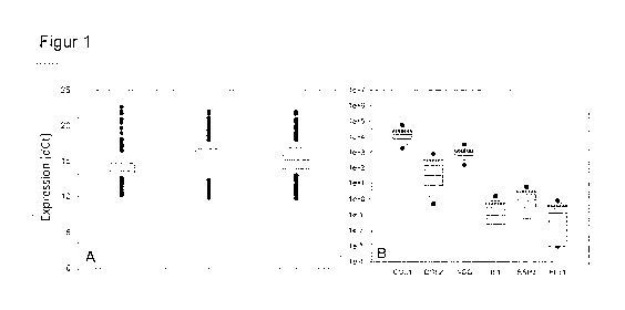
[0072] Figure 1 shows expression values of GAPDH (A)
and six selected marker genes (B) determined by means
of qRT-PCR (quantitative real-time PCR). For each gene,
the box plot shows the 25%-75% range (gray box), the
5%/95% range (horizontal lines above and below the
box), the median (line within the box) and also
outliers (black dots). A: The GAPDH expression values
were standardized to the total mRNA expression in 3
repetitive measurements (each using the entire 422
patient samples). The differences between the three
datasets are not statistically significant (simple
ANOVA), indicating the reliability of the qRT-PCR
method. B: The expression of the six marker genes is
shown as a negative dCt value in relation to GAPDH. The
data were gathered as part of a routine quality control
at the time of harvesting of the monolayer cell
cultures directly before the colonization of the
support (Novocart Basic). The mRNA expression value
obtained for each patient was standardized to the cDNA
standard of the patient in question (according to the
information from the manufacturer concerning the cDNA
synthesis kit), and so a direct comparability of the
expression data for each of the six selected genes is
ensured. COLl: COL1A2, collagen type I alpha-2 chain;
COL2: COL2A1, collagen type lib alpha-1 chain; AGG:
ACAN, aggrecan; ILl: interleukin-1P;
BSP2: bone
sialoprotein 2; FLT-1: vascular endothelial growth
factor receptor 1.
[0073] Figure 2 shows the distribution of the
expression levels of different protein families in
cultured human chondrocytes. The RPKM expression values
CA 02904799 2015-09-09
- 22 -
shown on a logarithmic scale were gathered for each of
the 20 samples as part of a transcriptome analysis. For
each gene, the box plot shows a 25%-75% range (gray
box), the median (line within the box) and also the
entire range (horizontal lines above and below the
box). Triangles label genes to which positive (e.g.,
FGF-2) or anabolic properties (e.g., ACAN) with respect
to cartilage can be assigned. Diamonds label genes to
which undesired (e.g., collagen I) or negative or
catabolic properties (e.g., interleukin-1, ADAM-TS5)
are attributed. Gene designations correspond to the
NCBI nomenclature.
[0074] Figure 3 shows the correlation analysis between
gRT-PCR and RNA sequencing (NGS, next-generation
sequencing) on the basis of the expression data of
COL1A2, COL2A1, ACAN and IL-113. The expression values
for COL1A2, COL2A1, ACAN and IL-i3 were obtained,
firstly, by means of conventional gRT-PCR from 422
samples (cf. table 1) and, secondly, by means of NGS-
based RNA sequencing from 20 samples. L,Ct values and
RPKM expression values are shown logarithmically. The
secondary figure shows the plot of the RPKM expression
values before their logarithmic conversion, their very
good correlation being even more clearly discernible.
[0075] Figure 4 shows the distribution of the
numerical values for the gene expression ratios of the
3114 most highly expressed genes in the form of a
histogram. The numerical values were obtained from the
ratio of the averaged RPKM values of each transcribed
gene from the group having good clinical results (Si to
S10) in relation to the mean value from the group
having implant failure (S11 to S20). Numerical values >
1 correlate with good clinical results. Numerical
values < 1 correlate with implant failure. Each bar
CA 02904799 2015-09-09
- 23 -
represents 1/100 of the entire captured range (smallest
numerical value: 0.006; largest numerical value: 4.9).
[0076] Figure 5 shows the hierarchical clustering on
the basis of the genomewide expression data of the 20
samples (Si to S20). The clustering is based on a
Pearson correlation between the expression values of
all the genes of a sample using the neighbor joining
algorithm. The samples are referred to as "positive" or
"negative" according to the clinical result of the
implantation in the patients in question. While the
variance among the negative samples is considerably
higher than among the positive samples, a clear
separation between the underlying clinical results can
be registered. This separation indicates that the
clinical results - transplantation which proceeded
positively or negatively - can be assigned to a
transcriptome phenotype of the cells used for the
implantation.
[0077] Figure 6 shows two heatmap evaluations, A and
B, of the Pearson correlation cluster analysis from the
RPMK numerical values of samples Si to S20. The values
were arranged according to their relationship, with
closely related expression patterns being close
together. A: What was considered here were the complete
genes with all their exons. B: The exons were analyzed
individually and independently of the mRNA structure.
Apart from S2 and S11, it was possible with this
evaluation to achieve a separation of samples Si to S20
according to the underlying clinical progression (S1-
S10: positive progression; S11-520: implant failure).
EXAMPLE SECTION
1. Material and methods
CA 02904799 2015-09-09
- 24 -
1.1 Structure of the study
[0078] What was carried out was a retrospective survey
of initial results, adverse effects and changes in the
starting state with regard to pain, functioning and
swellings in a patient population as defined below.
Further analyses were performed in order to investigate
the influences of patients, production and cell biology
properties on safety and patient results. The clinical
and surgical procedures, including indications and
rehabilitation, were defined in standard operating
procedures (SOPs). Surgeons were trained in the
surgical techniques for cartilage recovery and
implantation surgery before they used the implant for
the first time. After the patients had given their
informed consent, the treatment indication was
confirmed by arthroscopy. In the affected joint, two to
three cartilage-bone pieces were removed from the fossa
intercondylaris (a non-stressed region) using a sterile
and validated standard trephine (Aesculap AG,
Tuttlingen, Germany, cutting diameter: 4 mm).
1.2 Clinical data collection
[0079] The surgeon of each patient was asked to
complete a data collection sheet which comprised
medical history (etiology), basic demographic data
(age, gender) and period between surgical procedure and
last contact with patient. Together with the patient,
pain intensity, functioning and swellings were assessed
on a 10-point scale both in the consultation before the
procedure and after the procedure. The result
assessment scale was adapted from the visual analog
scale (VAS). Higher values indicate better results
(i.e., 10 means "no pain", "no swelling", "no
functional impairment"). This 10-point result grading
was carried out by each surgeon in a patient
CA 02904799 2015-09-09
- 25 -
consultation and was used as an early indicator for
further clinical progression. The participating
surgeons were also asked to specify all adverse effects
which occurred and, similarly, any treatment which was
subsequently required. The questionnaire did not
investigate whether the patients responded inadequately
to an earlier arthroscopic or other surgical cartilage
repair method. In extreme cases, the undesired effect
was an implant failure as a result of nonhealing or
tear-out.
1.3 Study population
[0080] The survey was carried out at 61 centers in
Germany, in which 433 patients were treated. Data were
reported back for a total of 422 patients (97.4%). The
remaining 11 patients were excluded from the rest of
the study and the RNA analyses. Among the 422 patients,
there were 140 women and 282 men. Their average age was
33.4 years (minimum: 14.7 years, maximum: 66.3 years).
The majority of the primary cartilage defects (damage
from grade III to IV according to the classification of
the International Cartilage Repair Society (ICRS)) were
established on the medial femoral condyle of the knee
(68.5%), followed by the lateral condyle (14.9%), the
retropatellar region (10.2%), the trochlea (5%) and the
tibia (0.2%). In six patients (1.4%), the defect was
established on the talus. The majority of the patients
had a single defect (93%). The defect size varied
between 1 and 20 cm2. The average size was 5.9 cm2. The
average duration of patient aftercare was 6.9 months.
In the case of a total of 83 patients, at least one
year elapsed since the surgical procedure up to the day
of the last visit registered by the surgeon (table 1).
Absolute Percent
number
CA 02904799 2015-09-09
- 26 -
Gender Male 282 66.8%
Female 140 33.2%
Age Mean 33.4
SD 10.0
Range 14.7-66.3
Duration of Mean 210
patient after- SD 185
care in days Range 0-921
Diagnosis Chronic damage 24 5.7%
Degenerative defect 144 34.1%
Osteochondritis 123 29.1%
dissecans
Traumatic defect 137 32.5%
Location of Lateral femoral 63 14.9%
primary defect condyle
Medial femoral 289 68.5%
condyle
Patella 43 10.2%
Talus 6 1.4%
Tibia 1 0.2%
Trochlea 21 5.0%
Primary defect Mean 5.9
size (cm2) SD 3.1
Range 1-20
Number of 1 391 92.7%
defects 2 29 6.9%
treated No data 2 0.5%
Table 1: Study population (422 patients)
[00811 Table 1 shows the study population with the
corresponding patient features. The features are
assigned absolute patient numbers and, where
applicable, percentage shares. A multiple diagnosis was
possible for one patient, with the defects affecting
more than one region.
CA 02904799 2015-09-09
- 27 -
1.4 Culturing
[0082] After removal from bone, mineralized cartilage
and superficial cartilage, chondrocytes were isolated
from the remaining cartilage by mechanical and
enzymatic extraction, conditioned according to standard
methods, and multiplied in vitro as a primary culture
in equipment permitted according to good manufacturing
practice (GMP) (TETEC AG, Reutlingen, Germany). The
steps comprised, after cell isolation, a multiplication
of cells in culture medium enriched with autologous
serum, harvesting by trypsinization and colonization in
the scaffold (Novocart Basic), and this took place 21
days after the arthroscopy for cartilage recovery. The
scaffold was colonized with a mean cell dose of 1.36 x
106 cells per cm2. Typically, cells were directly
applied from the primary culture. Under special
circumstances (e.g., patient disease), cells were
cryopreserved before use and administered from
secondary cultures (less than 10% of cases).
1.5 High-throughput RNA sequencing
[0083] 20 RNA samples from the batches described in
the preceding section were analyzed, and of these, ten
came from clinically successful treatments and ten were
obtained from patients with negative results. Each
sample was assigned a unique barcode sequence and
aliquots of the barcoded samples were, depending on the
patient result (positive results, negative results),
combined into two groups.
[0084] From each sample, 1 to 10 pg of the total RNA
of chondrocytes was used as starting material. The
total RNA was analyzed with regard to quantity and
integrity using an Agilent RNA 6000 Nano Chip (5067-
1511) on instrument model Bioanalyzer 2100. A depletion
CA 02904799 2015-09-09
- 28 -
of ribosomal RNA (rRNA) was carried out using 2-3 runs
with the RiboMinusTM Eukaryote Kit for RNA-Seq (A10837-
08, life technologies) according to the protocol from
the manufacturer. A concentration was carried out using
the RiboMinus Concentration Module (K1550-05, life
technologies). The efficiency of rRNA removal was
checked and determined in an aliquot run on an Agilent
RNA 6000 Nano Chip (5067-1511) on instrument model
Bioanalyzer 2100, with a complete elimination of rRNA
peaks being seen.
[0085] A library was created using the SOLiDTM Total
RNA-Seq Kit (4445373, life technologies) according to
the protocol from the manufacturer. In brief, 10 to
100 ng of RNA after ribosome depletion were firstly
fragmented by a 10-minute long digest at 37 C with
RNase III and purified again using the RiboMinus
Concentration Module (K1550-05, life technologies) and
eluted in 12 pl of RNase-free water. The fragment size
was checked for the optimal size range for the SOLiD4
instrument (150 to 200 nucleotides), and again an
aliquot run was performed on an Agilent RNA 6000 Nano
Chip (5067-1511) on instrument model Bioanalyzer 2100.
[0086] For the construction of cDNA libraries, the RNA
fragments were linked to adapters in a strand-specific
manner and converted into double-stranded cDNA
libraries, with use being made of a reverse
transcription followed by a size selection using PAGE
and amplification by FCR. During the PCR amplification,
barcode sequences were inserted, with the 3'-adapter
primer bearing a specific barcode-specific overhang.
This method avoids barcode distortions which have been
reported when barcode adapters are directly linked to
the library. Once again, the fragment size was checked
for the region of 150 to 200 nucleotides in an aliquot
CA 02904799 2015-09-09
- 29 -
run on an Agilent DNA 1000 Chip (5067-1504) on
instrument model Bioanalyzer 2100.
[0087] The concentration measurements of the
Bioanalyzer were used for the calculation and equimolar
mixing of barcoded samples S1-S10 (good clinical
results) and S11-S20 (negative clinical results) (in
each case barcodes 1-10). An emulsion PCR (emPCR) was
carried out for each combined library, in each case on
an E20 scale according to the recommendations from the
manufacturer with a final library concentration of
0.5 pM. The breaking of the emulsion and the washing of
beads and also the enrichment of beads were performed
manually according to the protocol from the
manufacturer. The quality and quantity of the template
beads were measured in a workflow analysis (WFA) run of
15 million enriched template beads on a SOLiD4.0
instrument. In both libraries, the signal-to-nose ratio
was 4% and the P2/21 ratio was 100%, indicating a high
percentage of monoclonal beads. 158 million template
beads of the libraries were applied in each case to a
quad field (1/4) of a SOLiD4.0 carrier and sequenced by
ligation from adapter P1, and so a 50 bp fragment
reading record was obtained.
1.6 Data creation
[0088] Data relating to adverse effects and patient
results were collected and entered into an Excel
spreadsheet. Quality control data of production and
also covariables (features relating to demography,
patient and product release) were entered into the same
database. All the entered data were checked for
agreement.
1.7 Statistics and covariant analysis of individual
parameter data
CA 02904799 2015-09-09
- 30 -
[0089] Summary statistics with mean values, standard
deviations, ranges and confidence intervals were used
in order to show the amounts of adverse effects,
efficacy results and patient characteristics. The
release characteristics of each product were compared
with the survey data in order to identify associations
between implant production features and patient
results. A number of chi-squared tests and regression
models was created in order to check for associations
between production characteristics or release
characteristics and the five result measurements in the
patient safety survey comprised: implant-related
adverse effects (defined as pooling of implant failure,
detachment, hypertrophy, arthrofibrosis, adhesions,
chondromalacia, articular infections and appearance of
free articular bodies), further operations (any reason)
and changes in the starting state with regard to pain,
functioning and swellings. Regressions relating to the
modeling of the association between biomarkers and an
interesting result were simultaneously fitted both for
each biomarker and all biomarkers. A logistic
regression modeling was used in order to determine the
influence of the independent covariants on the
appearance of implant-related adverse effects and the
appearance of further operations. Linear models were
used in order to model the changes in the starting
state with regard to pain, functioning and swellings.
At 0.05, p-values were assumed as significant. All the
regression models comprised the period from the
surgical procedure up to the last medical appointment
with the patient, since this was suspected of being a
significant independent predictive value for the
results.
1.8 Statistical analysis of all transcriptome data
CA 02904799 2015-09-09
- 31 -
[0090] Using the Whole Transcriptome Analysis Pipeline
of the BioScope 1.32 software (Applied Biosystems),
reading sequences were mapped onto the human genome
(UCSC version HG 19). Proceeding from the annotations
of the UCSC refSeq (downloaded on 7 November 2010),
RPKM values of expression for genes were calculated. In
brief, RPKM values (reads per kilobase exon model per
million mapped) are the number of sequenced segments
mapping onto the exons of a given transcription,
normalized by the sequencing depth per sample (total
segment number) and the length (bp) of all exons. These
values were used for the initial determination. The
scale normalization method described by Bullard et al.
(J.H. Bullard, E. Purdom, K.D. Hansen, S. Dudoit,
Evaluation of statistical methods for normalization and
differential expression in mRNA-Seq experiments. BMC
Bioinformatics 11: 94 (2010)) was used in order to
eliminate sample-specific technical distortions and the
expression values obtained were mapped onto a
logarithmic scale. Samples were clustered using the
neighbor joining algorithm (N. Saitou and M. Nei, The
neighbor-joining method: a new method for
reconstructing phylogenetic trees. Mol. Biol. Evol. 4
(4): 406-425 (1987)), which is based on the Pearson
correlation distance between their entire transcriptome
expression profiles. The nonparametric rank product
method (R. Breitling, P. Armengaud, A. Amtmann, P.
Herzyk, Rank products: a simple, yet powerful, new
method to detect differentially regulated genes. FEBS
Letters 573 (1): 83-92 (2004)) was used in order to
check for differential expression, yielding pfp values
(percent false positive values, a measure comparable
with FDR-corrected p-values), and transcripts with pfp
< 0.05 and an absolute value of fold change greater
than 0.9 (i.e., doubled or halved expression) were
considered to be significant differential expression.
The differentially expressed protein-
encoding
CA 02904799 2015-09-09
- 32 -
transcripts were used in order to check for an
enrichment of specific functional categories
(overrepresentation analysis, hypergeometric test, p-
values corrected for false discovery rate). For all the
analyses, use was made of the technique described by
Mayday (F. Battke, S. Symons, K. Nieselt, Mayday -
Integrative analytics for expression data. BMC
Bioinformatics 11 (1): 121 (2010)). Using the
Integrative Genomics Viewer IGV (J.T. Robinson, H.
Thorvaldsdottir, W. Winckler, M. Guttman, E.S. Lander,
G. Getz, J.P. Mesirov, Integrative genomics viewer.
Nat. Biotech. 29: 24-26 (2011)), the segments of
individual genes of interest were studied in detail.
2. Results
2.1 Changes in the starting state for the patient
results
(0091) Prior to a surgical procedure and in following
patient visits, the physicians determined articular
pain, functioning and swellings on a 10-point scale,
with higher values indicating better results. Patients
who gave answers both in relation to questions before
the procedure and after the procedure achieved
significant improvements with respect to the starting
state (p < 0.0001, Wilcoxon signed-rank test) in all
three measurements. The mean duration since the
surgical procedure was 6.9 months (range: 2 to 30
months), and this is a noticeably shorter time scale
than specified in the majority of other summaries of
ACI results. The retrospective study contributed
important aspects to the present knowledge relating to
the repair of articular cartilage. In the subgroup of
83 patients with at least 12 months since their
surgical procedure, it was reported that they exhibited
stronger changes on average in all three result
CA 02904799 2015-09-09
- 33 -
measurements than the entire patient population (table
2).
All patients Investigation
(N-422) period >1 year
___________________________________________ (N=83)
Result measurement Min Mean Max SD N Min Mean Min SD N
Pain Pre- 1 3.4 10 1.3 412 2 3.4 9 1.2 82
operative
Post- 1 7.0 10 2.0 382 2 7.2 10 1.9 80
operative
Change -5 3.8 8 2.1 376 2 3.9 8 2.1 80
Swelling Pre- 1 5.3 10 2.4 411 1 5.1 10 2.4 82
operative
Post- 1 7.7 10 1.9 382 4 8.1 10 1.6 80
operative
Change -7 2.5 9 2.6 375 -3 3.1 8 2.2 80
Function- Pre- 1 4.2 10 1.8 412 2 4.4 10 2.0 82
ing operative
Post- 2 7.3 10 1.8 380 2 7.8 10 1.6 80
operative
Change -7 3.2 8 2.3 374 -7 3.6 8 2.4 80
Table 2: Individual result measurement
2.2 Appearance of adverse effects
[0092] Table 3 shows the appearance of reported
adverse effects. The appearance of implant failure was
3.1% in the entire patient population and 6% in the
subgroup of patients for whom at least 12 months had
elapsed since their procedure. In general, the reported
numbers of cases of implant-related complications were
low for the entire patient population. Detachment
(delamination), arthrofibrosis and hypertrophy were
observed in 1.7%, 2.4% and 0.7%, respectively.
Altogether 36 patients (8.5%) required a further
CA 02904799 2015-09-09
- 34 -
operation and/or correction. The most common adverse
effects reported in patients who required further
operations were implant failure (13, the ten samples
further investigated by transcriptome analysis
originated from these cases), detachment (6),
arthrofibrosis (7), synovitis (7), adhesions (5) and
pain (6). The subgroup of 83 patients for whom at least
12 months had elapsed since their procedure exhibited a
further operation rate of 13.3%. The majority of the
further operations were carried out arthroscopically.
All patients (N-422) Investigation period
>1 year (N-83)
Complication Cases % 95% CI** Cases % 95% CI**
Implant-related
complications***
Implant failure 13 3.1% 1.7% 5.2% 5 6.0% 2.0% 13.5%
Delamination 7 1.7% 0.7% 3.4% 2 2.4% 0.3% 8.4%
Hypertrophy 3 0.7% 0.2% 2.1% 1 1.2% 0.0% 6.5%
Arthrofibrosis 10 2.4% 1.1% 4.3% 2 2.4% 0.3% 8.4%
Adhesions 7 1.7% 0.7% 3.4% 2 2.4% 0.3% 8.4%
Free articular 1 0.2% 0.0% 1.3% 1 1.2% 0.0% 6.5%
bodies
Deep (artic- 3 0.7% 0.2% 2.1% 0
ular) infection
Chondromalacia 2 0.5% 0% 1.7% 0
Further
complications
Effusion 32 7.6% 5.2% 10.5% 6 7.2% 2.7% 15.1%
Pain 29 6.9% 4.7% 9.7% 7 8.4% 3.5% 16.6%
Synovitis 14 3.3% 1.8% 5.5% 3 3.6% 0.8% 10.2%
Hematoma/ 7 1.7% 0.7% 3.4% 5 6.0% 2.0% 13.5%
hemarthrosis
Stiffening 1 0.2% 0.0% 1.3% 1 1.2% 0.0% 6.5%
Superficial 2 0.5% 0.0% 1.7% 0
infection
CA 02904799 2015-09-09
- 35 -
Table 3: Reported complications
2.3 Association between results and independent risk
factors
[0093] The size of all treated defects did not show
any correlation with any of the measured patient
results. For the change in the starting state in the
case of the functioning of the patient, a significant
association with patient age was found (i.e., in the
case of a lower age, greater improvements on average
were apparent, p - 0.004, not shown). Table 4 specifies
the results of chi-squared tests relating to the
association of the location of the primary defect to
the appearance of implant-related adverse effects. It
was found that both the appearance of implant-related
adverse effects and of further operations is largely
independent of the location of the primary defect, with
the exception of patellar defects. In any case, for an
affected individual, the probability of such an event
was greater when the primary defect was situated on the
patella (p < 0.0001).
Implant-related complications*
depending on location of defect
Defect in this Defect not in p-value
position this position
Location Cases N % Cases N %
Medial femoral condyle 22 289 7.6% 14 133 10.5% 0.32
Lateral femoral condyle 3 63 4.8% 33 359 9.2% 0.25
Trochlea 1 21 4.8% 35 401 8.7% 0.53
Patella 11 43 25.6% 25 379 6.6% <0.0001
Tibia 0 1 0.0% 36 421 8.6% 0.76
Talus 0 6 0.0% 36 416 8.7% 0.45
CA 02904799 2015-09-09
- 36 -
Further operations depending on
location of defect**
Defect in this Defect not in p-value
position this position
Location Cases N % Cases N %
Medial femoral condyle 20 289 6.9% 16 133 12.0% 0.08
Lateral femoral condyle 3 63 4.8% 33 359 9.2% 0.25
Trochlea 1 21 4.8% 35 401 8.7% 0.53
Patella 12 43 27.9% 24 379 6.3% <0.0001
Tibia 0 1 0.0% 36 421 8.6% 0.76
Talus 0 6 0.0% 36 416 8.7% 0.45
Table 4: Implant-related complications and further
operations depending on the location of the
defect
[0094] Table 5 specifies the results of chi-squared
tests relating to the association between the
classification of cartilage injuries and the appearance
of implant-related adverse effects and further
operations. In the case of implant-related adverse
effects, for an affected individual, the probability of
such an event was greater when the cartilage defect was
classified as degenerative (p = 0.005). However, for
patients, the probability of such an event was lower
when there was a cartilage defect caused by
osteochondritis dissecans (p = 0.04).
Implant-related complications*
depending on nature of defect
Defect of this Other defect p-value
type
Nature of defect Cases N % Cases N %
Chronic damage 1 24 4.2% 35 398 8.8% 0.43
Degenerative defect 20 144 13.9% 16 278 5.8% 0.005
Traumatic defect 11 137 8.0% 25 285 8.8% 0.80
CA 02904799 2015-09-09
- 37 -
Osteochondritis 5 123 4.1% 31 299 10.4%
0.04
dissecans
Further operations depending on nature
of defect
Defect of this Other defect p-value
type
Nature of defect Cases N % Cases N %
Chronic damage 1 24 4.2% 35 398 8.8% 0.43
Degenerative defect 17 144 11.8% 19 278 6.8% 0.08
Traumatic defect 13 137 9.5% 23 285 8.1%
0.63
Osteochondritis 5 123 4.1% 31 299 10.4%
0.04
dissecans
Table 5: Implant-related complications and further
operations depending on the nature of the
defect
[0095] Table 6 shows associations between patient
results and cell culture variables, on the one hand,
and mRNA expression values of six selected marker genes
(cf. section 2.4.1), on the other hand, as multivariant
p-values. Significant relationships and marginal trends
are highlighted. A temporary cryopreservation of the
cells before an implantation did not show any
association with an intensified appearance of implant-
related complications (IRC) or with any of the other
result measurements (further operation FO, pain,
functioning, swelling). with regard to cell viability,
the same results were observed. However, a low number
of administered cells was significantly associated with
a further operation (FO) (p = 0.06; table 6).
Cell culture variables IRC FO Pain Funct-
Swelling
ioning
Cryopreservation 0.98 0.98 0.59 0.78 0.13
CA 02904799 2015-09-09
- 38 -
Cell viability upon 0.56 0.13 0.55 0.38 0.76
harvesting
Cell count (log 10) 0.30 0.06 0.47 0.37 0.36
PCR expression
measurement
Aggrecan 0.14 0.44 0.61 0.50 0.62
BSP-2 0.11 0.14 0.76 0.78 0.90
Collagen I 0.49 0.70 0.53 0.79 0.27
Collagen II 0.08 0.08 0.42 0.90 0.29
Interleukin-113 0.08 0.20 0.26 0.19 0.24
FLT-1 0.02 0.03 0.21 0.67 0.81
Table 6: Relationship between patient results and cell
culture variables or mRNA expression values
of six selected marker genes
2.4 Expression data
2.4.1 Specific gene expression
[0096] Quantitative real-time PCR (qRT-PCR) was used
to determine the expression of six selected marker
genes: aggrecan (ACAN), an Integral constituent of the
extracellular cartilage matrix; type I collagen
(COL1A2), which usually does not occur in cartilage
tissue; cartilage-specific type II collagen (COL2A1);
interleukin-113 (IL-1p), an inflammatory cytokine;
FLT-1, an isoform of a vascular endothelial growth
factor receptor; and bone sialoprotein BSP-2, a bone
growth factor. The reason for selecting the last two
mentioned genes was because complications can result
especially from a vascularization of the implanted
cartilage tissue or from a formation of osteophytes
within the implanted cartilage tissue.
CA 02904799 2015-09-09
- 39 -
[0097] The expression values (dCt) of these six marker
genes are depicted in Figure 1 and show significant
differences. The most stable mRNA levels, which vary
within one order of magnitude among all 422 samples,
were found for aggrecan. The greatest variations,
within three orders of magnitude, with an altogether
very low detection level, occurred for FLT-1. For type
I collagen for example, it was possible to calculate on
average 1000 mRNA transcripts per cell, whereas in the
case of FLT-1, only 10 mRNA transcripts per 1000 cells
were calculated. For comparison, there are, for
example, at least 500 times more FLT-1 mRNA transcripts
present in human venous endothelial cells cultured in
vitro.
[0098] Despite these variations, it was nevertheless
possible to observe significant or at least marginal
correlations between some of these marker genes and the
clinical result (tables 6 and 7). For instance, it was
possible to establish a-significant association between
implant-related complications (IRC) and elevated FLT-1
expression levels in multiplied chondrocytes (p = 0.02,
table 6). In contrast, it was possible to determine
only a marginal relationship for elevated IL-113 levels
and lowered type II collagen levels (both p = 0.08,
table 6). However, taking into account all the marker
genes in the calculation models, it was possible to
find for IL-l3 (p = 0.02, table 6) and FLT-1 (p = 0.08,
table 6) a significant or marginal relationship with
respect to IRC.
[0099] According to table 7, which shows for the six
marker genes not only the regression coefficients but
also the multivariant p-values taking into account the
elapsed time since the procedure, it was not possible
to classify the expression of any of the marker genes
as significant with respect to pain, functioning and
CA 02904799 2015-09-09
- 40 -
swelling. The coefficients for the patient results IRC
and FO (further operation) were calculated by means of
logistic regression; the coefficients for pain,
functioning and swelling were calculated by means of
linear regression. The p-values (second row) Indicate
the significance of the relationship in question, and
significant relationships and marginal trends are
highlighted.
Variable IRC FO Pain Funct- Swelling
ioning
Time since 1.057 1.064 0.0084 0.055 0.060
procedure
0.037 0.017 0.644 0.0049 0.0059
PCR
expression
measurement
Aggrecan 1.141 0.962 0.105 -0.099 0.087
p= 0.492 0.836 0.343 0.397 0.506
BSP-2 1.086 1.078 -0.018 -0.013 -0.0085
P= 0.161 0.177 0.607 0.730 0.837
Collagen I 0.901 0.973 0.015 0.049 0.057
p= 0.426 0.835 0.849 0.564 0.556
Collagen II 1.095 1.112 -0.034 0.015 -0.054
p= 0.166 0.107 0.347 0.698 0.216
Interleukin-113 1.151 1.112 -0.044 -0.049 -0.054
p= 0.029 0.083 0.197 0.177 0.179
FLT-1 1.106 1.102 -0.033 -0.0096 -0.0041
P= 0.062 0.070 0.222 0.739 0.901
Table 7: Multivariate analysis of the relationship
between patient results and the time since
the procedure or the mRNA expression values
of six selected marker genes
2.4.2 Genomewide expression
CA 02904799 2015-09-09
- 41 -
[0100] The entirety of the captured expression data
comprised about 30 000 genes with a large variety of
exon data and yielded more than 200 million individual
data segments. For the group S1-10 (good clinical
results), 42 716 671 segments (75.3%) were
unambiguously mapped, 3 828 740 (6.7%) were spanning
exon junctions, 3 791 685 of the exon junctions were
known and 37 055 were new. For the group S11-20
(implant failure), altogether 43 429 958 segments
(76.0%) were unambiguously mapped, 3 991 606 segments
(7.0%) were spanning exon junctions, 3 958 041 of the
exon junctions were known and 33 565 were new. The
mapped segments were used for calculating RPKM values
("reads per kilobase per million mapped reads" values)
with Bioscope 1.3 for each exon present in the UCSC
refSeq database (downloaded on 07.11.2010). A first
RPKM survey was performed using gene families, with
family members "typical" for chondrocytes being
considered, for example collagens (with type II, IX and
XI collagens as "cartilage collagens") or proteoglycans
(with aggrecan (ACAN) as typical representative)
(Figure 2). The data are shown graphically, with the
standard deviations indicating the exon variability. In
addition, the expression values spanned several orders
of magnitude and could only be shown on a logarithmic
scale, as in Figure 2. The results immediately show
that the classic "chondrocytic" phenotype does not
dominate the results. A further analysis of other gene
families, including for example IGF-related or
PDGF/VEGF-related genes, confirms this presumption. An
expression of the interleukin cytokines was
diversified. The expression of these growth factors
became even more complex when complementary binding
proteins or the associated receptors were used.
Equally, the transcription of the expanded clusters of
metalloproteinases is very broad (Figure 2).
CA 02904799 2015-09-09
- 42 -
2.5 Calibration of transcriptome data by gRT-PCR
[0101] To estimate the comparability of data in the
two methods, a correlation analysis was performed on
the basis of the expression data of COL1A2, COL2A1,
ACAN and IL-l3 (cf. Figure 1). These genes show typical
expression values in chondrocytes within the range of
seven powers of ten. The correlation of the values from
the two methods was successful and yielded a
correlation coefficient of 1.0 in an exponential
function (Figure 3).
2.6 Correlation between transcriptome data and
clinical results
[0102] To obtain information about specific patterns
which are (hypothetically) related to the clinical
results, two evaluation methods were selected. In a
first approach, the expression ratio for the averaged
RPKM value of each transcribed gene from the group with
good clinical results (S1 to S10) in relation to the
mean value from the group with implant failure (S11 to
S20) was determined. This yielded a list of numerical
values > 1, which have a positive correlation with good
results, in relation to numerical values < 1, which
have a positive correlation with implant failure.
Numerical values close to 1 indicate genes which behave
neutrally in relation to the results. The results were
shown as a histogram in Figure 4. The histogram
contains values for the 3114 most strongly expressed
genes. 1803 values were > 1, 1311 values were < 1.
Among these, 62 values were > 2, meaning a twofold
stronger expression in the group with good results Sl-
10, and 70 values were < 0.5, meaning a twofold
stronger expression in the group with implant failure.
In other words, these two gene clusters are candidates
for a restricted list of quality parameter genes for
CA 02904799 2015-09-09
- 43 -
following studies. Said list does not contain
regulatory RNAs.
[0103] In a second approach, a correlation cluster
analysis was carried out from the RPKM numerical
values: the Pearson correlation coefficient was
calculated for each sample pair (S1-S20) in the genes
and plotting was carried out of a so-called "heatmap"
of the correlation matrix with the integrated heatmap
function. The hypothesis behind this correlation
assumes that the calculation yields a sorted list for
the 20 clinical results which separates the good
results from the clinical failure cases when the
relations outlined in the data cited above are
statistically significant. The result is shown in
Figure 5. In Figure 6A, the data for the complete genes
with all their exons were compiled. In Figure 6B, the
exons were analyzed Individually and independently of
the mRNA structure. In other words, alternative
splicing is disregarded in Figure 6A, whereas it is
shown in Figure 6B. It became apparent that the data
were sorted corresponding to their origin, with the
group S1-10 being separated from the group S11-20.
Exceptions are sample S2, which was classified in the
transition zone between the two groups, when it was
analyzed for genes, and migrated further into the
failure group, when it was analyzed for exons, and
sample S11, which remained stably in the "wrong"
cluster. Altogether, the heatmap yielded a correct
"prediction- of the clinical result in 18 of 20 cases,
or with a probability of 90%.