Note: Descriptions are shown in the official language in which they were submitted.
CA 02904896 2015-09-09
WO 2014/164330 PCT/US2014/021932
RADIATION DIAGNOSTIC AND TREATMENT DEVICES AND METHODS
DESCRIPTION
Technical Field
[0001] The present subject matter relates to devices, systems and procedures
used
during radiation diagnosis. When retained in place during treatment, the
subject matter
encompasses in-place marking and tracking of radiation treatment patterns and
effectiveness. The overall technical field involves radiation oncology
procedures with
respect to a wide variety of cancerous conditions. Diagnosis, marking, mapping
and
evaluation are carried out by implements incorporating expandable component
technology in combination with other technologies, including treatment
technologies,
which together enhance the precision and accuracy of cancer treatment
diagnosis,
mapping, marking and tracking before, during and after radiation treatment.
Background
[0002] Diagnostic and marking systems, devices and methods are known and used
by
medical professionals in dedicated units of numerous hospitals and free-
standing
cancer treatment centers, and some incorporate balloons to achieve and
maintain
proper placement during diagnosis.
[0003] Diagnostic and marking tasks often utilize devices of the type intended
to be
inserted into living body cavities through existing body orifices or into
surgically
executed openings for treatment under the skin of a patient. For example, once
a
catheter and its balloon are inserted in a prescribed manner into a body
cavity, its
balloon can be inflated to mark the boundary of the body cavity during
radiographic
examination, and the inflated balloon may also be used to move, push or
otherwise
manipulate body tissue during the diagnostic procedure.
[0004] Various devices, systems and methods have been developed, each
typically
being designed for a specific diseased body organ, area or part and/or for one
or more
treatment locations. Whether a treatment regimen involves a one-step or a
multi-step
CA 02904896 2015-09-09
WO 2014/164330 PCT/US2014/021932
protocol, it is important to maintain a good balance among radiation dosage,
placement
and timing. To do so requires precision in diagnosis so that the target
location or
locations are treated with the radiation source while protecting as much as
possible
areas of the body that are disease-free and otherwise could be vulnerable to
unintended
treatment if positioning with respect to the treatment locations is not
modified during
diagnosis, marking and treatment.
[0005] Proper, precise and accurate marking, diagnosis and manipulation
procedures
can precede and be reproduced or maintained during carcinoma treatment
procedures,
such as when following high dose rate (HDR) brachytherapy. At times, the
diagnoses
by the radiation oncologist will be intended for regimens using low dose rate
(LDR)
brachytherapy, typically based on cesium delivery as in 137Cs. For HDR
brachytherapy
regimens, 192Ir is frequently used because of its high specific activity.
Diagnoses for
using other isotopes are available and used as warranted. The degree of
treatment
measurement is in terms of units of radiation exposure (in roentgens or Gray
or Gy),
and often these are prescribed at specific locations and points. Details in
this regard
are known to radiation oncologists, medical physicists and other medical
professionals
experienced in brachytherapy and cancer treatment in general. An objective
also is to
provide reasonably constant and predictable dose rates at each specific
location that
diagnosis and marking have determined are most beneficial for the patient.
[0006] Intracavitary and percutaneous radiation treatment diagnoses need to be
exacting and specific at each radiation target location. Typically important
is protection
of tissue that is not diseased. Pre-treatment diagnoses also are important for
developing a plan for dose rate and duration specifics, for example.
[0007] In terms of protecting non-diseased tissue, an example is presented
relating to
intrauterine diagnosis and treatment where it typically is important to
minimize, if not
eliminate, radiation exposure to the bladder and the rectum. Generally,
marking and
diagnosis devices, as well as brachytherapy devices, are visible (or can be
rendered
visible) under x-ray images or other imaging technologies in order to insure
intended
placement and to allow the medical physicist or radiation oncology
professional to
generate a radiation treatment plan specific for this placement and for the
particular
2
CA 02904896 2015-09-09
WO 2014/164330 PCT/US2014/021932
anatomy and disease location and severity for the particular patient and for
each
particular treatment event.
[0008] It will be appreciated that radiation delivery systems can be used in
treatments
that are applied manually or remotely using remote afterloading systems. In
remote
afterloading systems, the radioactive materials are delivered from a safely
contained
access location to distal reaches of the delivery tubes at treatment portions
or locations.
Radioactive material can be in the form of wires, seeds, liquids or other
species. In
such systems, the radioactive material typically is delivered via remote
control, such as
by operation of a motor, after the medical professionals are out of view from
the
treatment room. Such remote delivery equipment can move the radioactive dose
into
the applicator already positioned within the body cavity, the accuracy of
which is
facilitated by the marking and diagnosis device or catheter.
Summary
[0009] There are several aspects of the present subject matter that may be
embodied
separately or together in the systems, devices and methods described herein
and
claimed below. These aspects may be employed alone or in combination with
other
aspects of the subject matter described herein, and the description of these
aspects
together is not intended to preclude the use of these aspects separately or
the claiming
of such aspects separately or in different combinations as may be set forth in
the claims
appended hereto.
[00010] In one aspect, systems, devices and methods provide a significantly
improved diagnostic tool and procedure before, during and/or after radiation
therapy in
or near body cavities accessible through existing orifices or percutaneously
when
desired.
[00011] In another aspect, improvements in systems, devices and methods
provide the medical professional with a view of the body cavity or target
percutaneous
location while imaging, such as radiographic viewing or CT scanning, and if
desired
during radiation treatment itself.
[00012] In another aspect, the medical professional diagnostician or
treatment
physician applies or utilizes the system, device or method to move, push or
otherwise
3
CA 02904896 2015-09-09
WO 2014/164330 PCT/US2014/021932
manipulate body tissue for the purpose of improved diagnosis or marking and
during
radiation therapy.
[00013] Another aspect is to enable introduction of radiographic fluids or
air into a
body cavity, a surgically opened cavity, a percutaneous site, or other
treatment site of
the patient without subjecting the patient to risk of direct contact with
radiographic
andfor radiopaque fluids, while providing real-time detection of and reporting
upon
treatment specifics.
[00014] In another aspect, the system, device and method maintain a desired
positioning of a diagnostic expandable component having detecting capabilities
through
the use of securement components such as balloons, clips, templates, tethers,
other
expandable components or the like while detecting and/or formulating specifics
for
treatment.
[00016] In a further aspect, the system, device and method are especially
suitable
for use in conjunction with the bladder or other body locations by providing
an elongated
insertion catheter having drainage characteristics in combination with
detection
capabilities.
[00016] Yet another aspect of the system, device and method includes
providing
radiopaque reference lines, or reference designations otherwise visible to the
attending
medical professional, at desired locations within the body. Some can be
tailored for one
or more of a variety of body cavities and along portions of the device that
can be viewed
on components of the device that are external of the body when inserted during
diagnosis, mapping, marking or treatment, in order to facilitate re-
positioning of
detectors and balloons for a future insertion in that same patient.
[00017] In an added aspect, the physician is provided with equipment and
techniques for diagnosing, mapping and marking in preparation for treatment of
any of a
wide variety of cancers such as those inside or in the proximity of body
cavities
including the bladder, vagina, rectum, subglottic, superglottic or glottic
regions,
stomach, bronchial tubes, nasopharynx or larynx regions, eye sockets, and
other
intracavity areas. Interstitial insertion of devices through tissue and
percutaneous
procedures also are encompassed, such as the treatments of the breast, central
nervous system, prostate, lung lesions and liver lesions, insertion being
through a
4
CA 02904896 2015-09-09
WO 2014/164330 PCT/US2014/021932
surgically made opening or percutaneous entry. Treatment can proceed while the
diagnostic, mapping and marking device is within the body and later retrieved
or
removed and subsequently reinserted depending upon the treatment protocol
being
followed. Catheter-type channels can be used for delivery of radioactive
solutions, such
as to the expandable component. Also, microdiodes can be incorporated to
achieve
real-time treatment capabilities, and hyperthermia components can be included
for
enhancing diagnosis and effect of subsequent treatment.
[00018] In a further aspect, a system, device and method includes at least
one
intracavitary expandable component that is sized, shaped, positioned and
adapted to
impart a space separation between the radiation source emanating (or to be
emanating)
from the device and an internal location within the body at which radiation
treatment is
not desired. Each expandable component can be a separate unit provided in
association with or secured to the device. in other approaches, one or more
expandable components are secured to a component of the device, which can be
used
for delivery of radioactive material, solutions or the like. Catheter-type
channels can be
used for delivery of radioactive solutions, such as to the expandable
component.
Typically microdiodes are incorporated to achieve real-time reporting and
marking
capabilities during mapping, marking, analyses and radiation treatment, and
hyperthermia components can be included.
[00019] Another aspect facilitates long-term, low dose rate radiation by
enabling
introduction of nutrients, fluids, air or other gasses and/or enabling
evacuation of wastes
and/or gasses through a diagnostic device itself. Catheter-type channels can
be used
for the delivery of marking solutions, such as through the expandable
component,
microdiodes being incorporated to achieve real-time treatment capabilities,
and
hyperthermia components can be included.
[00020] Another aspect provides a system, device and method suitable for
use in
marking, mapping and diagnosing bladder carcinoma by providing an elongated
insertion catheter having drainage characteristics. Catheter-type channels can
be used
for delivery of radioactive solutions, such as through the expandable
component,
microdiodes are incorporated to achieve real-time treatment tracking, and
hyperthermia
components can be included.
CA 02904896 2015-09-09
WO 2014/164330 PCT/US2014/021932
[000211 Another aspect permits the physician to tailor the size, placement
and
duration of radiation treatment specifics to the particular therapeutic
requirements of the
diseased tissue to be treated. Catheter-type channels can be used for
positioning of
radiopaque material or solutions, such as to the expandable component,
microdiodes
are incorporated to achieve real-time treatment assessment, and hyperthermia
components can be included.
[00022] Another aspect maintains the positioning of devices including
expandable
component such as balloons through the use of a relatively small or secondary
or
placement expandable component or balloon located within a larger therapeutic
or
diagnostic expandable component. Catheter-type channels can be used for
delivery of
radiopaque solutions, such as to the large expandable component, microdiodes
are
incorporated to achieve real-time treatment assessment, and hyperthermia
components
can be included.
[00023] An additional embodiment concerns a system, device and method for
diagnosing, mapping and marking in advance of and/or during radiation therapy
wherein
a radiation detector and a radiation data receiver are included to provide
real-time
feedback, including during treatment, or after-treatment recording of
treatment specifics.
In a particular embodiment, at least one radiation detector is positioned on
or in a
expandable component, which expandable component is sized, shaped, adapted and
positioned to provide positioning, visible by way of appropriate imaging, and
confirmation of separation and/or positioning with respect to the radiation
source during
treatment.
[00024] Yet a further embodiment concerns a system, device and method for
diagnosis, mapping or marking in conjunction with brachytherapy that includes,
in
combination, a hyperthermia sub-system and at least one radiation detector,
both
positioned in the close vicinity of the radiation delivery location or
anticipated radiation
delivery location along the catheter-like component. A radiation data receiver
is located
external of the body within which the brachytherapy is expected or is
proceeding.
Alternatively, the detector may be fixed and its data later able to be
analyzed.
6
CA 02904896 2015-09-09
WO 2014/164330 PCT/US2014/021932
Brief Description of the Drawings
[00025] FIG. 1 is an elevation view of an embodiment including an
expandable
component that is inflatable and deflatable along a distal portion of a
catheter;
[000261 FIG. 2 is a cross-sectional view along the line 2-2 of FIG. 1;
[00027] FIG. 3 is a somewhat schematic view, partially in cross-section, of
a
further embodiment for use within the rectum;
[00028] FIG. 4 is a somewhat schematic view, partially in cross-section, of
a
further embodiment for use within the bladder; and
[00029] FIG. 5 is a cross-sectional view along the line 5-5 of FIG. 4.
Description of the Illustrated Embodiments
[00030] The embodiments disclosed herein are exemplary only, and the
subject
matter described herein can be embodied in various forms. Therefore, specific
details
described herein are not to be interpreted as limiting the subject matter as
defined in the
accompanying claims.
[00031] Certain of the illustrated embodiments utilize a catheter for
insertion into a
body cavity. An expandable component is secured to a tubular catheter body,
the
expandable component being positioned and sized for insertion into a
particular type of
body cavity to be treated. The proximal end of the catheter has one or a
plurality of
passageways to enable fluid communication through one or more channels in the
catheter body, depending upon the embodiment. Such passageway typically
utilizes
one-way or two-way valves, regulators, hypodermic syringes or other devices
for
introduction, control and/or withdrawal of fluids into and out of one or more
expandable
components and/or body cavities. When the expandable component is of the type
that
can be inflated, the fluid for manipulating the expandable component in
certain
embodiments may be filled with a biocompatible gas, such as air, or a
biocompatible
liquid, such as saline solution, or with a radiopaque fluid to facilitate
viewing. In some
embodiments, the fluid can itself have a treatment function. The catheter
expandable
component is sized, shaped and adapted in order to move, expand or otherwise
7
CA 02904896 2015-09-09
WO 2014/164330 PCT/US2014/021932
manipulate the body component to be treated or to be positioned in order to
prevent or
minimize treatment, all with the objective of providing more effective and
safer radiation
treatment.
[00032] One or more detectors and/or hyperthermia components typically are
associated with the expandable component, which association can modify the
positioning of such detector or hyperthermia component being controlled
external of the
subject's body within which the device is inserted. For example, the
expandable
component can contain receiving members to hold the detector and/or
hyperthermia
component within the material of the expandable component, or strips of
elastomeric or
adhesive material along the inside surface or along the outside surface of the
expandable component can be provided. Other holding approaches can be followed
such as placement within the wall thickness of the expandable component, on or
in the
catheter tube or by way of bowing or free-floating arms or spokes.
[00033] For example, the detector or hyperthermia components can be
elongated
treatment members, or treatment components positioned on elongated members,
typically inside of the expandable component, and that are secured at one or
both of
their respective end portions. When desired, same can generally follow end
portions of
the expandable component. In such approaches, the elongated members bow out
when within the expandable component as the expandable component is expanded
or
inflated when same has inflation properties. Or the elongated members may be
positioned immediately inside a neck of the expandable component where
attached to
the catheter and are otherwise freely suspended within the expandable
component, not
necessarily secured to the expandable component at both proximal and distal
portions.
Even in that event, the elongated treatment members can be secured together at
their
respective distal end portions to facilitate bowing out of the elongated
members.
Alternatively, one or both end portions of the elongated members can be
located within
the polymeric material of the expandable component or of the catheter, or
between
material layers of the expandable component or catheter, in order to provide a
gathering
function for the portions of the elongated members at an attachment location.
[00034] Different embodiments can utilize one or more of a variety of
approaches
to secure the catheter device during the marking, mapping, diagnosing or
positioning
8
CA 02904896 2015-09-09
WO 2014/164330 PCT/US2014/021932
function or functions and also during subsequent radiation therapy. These
securement
embodiments and approaches include, for example, a placement expandable
component that can be considered as an inner expandable component, a distal
placement expandable component, a template, a catheter lead, and one or more
secondary outer expandable components, with or separate from tether catheters.
In
some embodiments, one or more of these various optional expandable components
can
take the form of a balloon or other inflatable device or component.
[000351 When provided, an inner expandable component, which can be a
secondary or inner balloon, usually is substantially smaller than the main
expandable
component. Same assists in holding the catheter device in place within the
body cavity
or location of interest, typically located generally within and at the
proximal end portion
of the main expandable component. Upon inflation or expansion, the secondary
expandable component secures the catheter device within the body cavity or
location of
interest by restricting movement of the device at the body orifice or surgical
opening. A
secondary outer expandable component, if and when included, is located distal
of the
main expandable component. When inflated or expanded, same anchors the
catheter
device at a location downstream or distal of the main expandable component.
[00036] Some embodiments lend themselves to include a template to secure
the
catheter device at a location external of the body, such as a natural body
cavity orifice
or in areas surrounding a body cavity that is a surgical opening. Such a
template may
be secured by one or combinations of approaches. The template can be sutured
to
tissue in the vicinity of the body insertion location, or same can be adhered
to tissue in
the vicinity of the body insertion location, for example, using adhesive or
glue. The
template can be secured by attaching secondary catheters secured in orifices
near the
body insertion location. Securement may also be provided by a distally
extending
catheter lead which anchors the catheter device by slipping the distal end
lead through
a narrow section of the body, such as at the cervix or duodenum when a body
cavity is
treated. Devices of this type assist in avoiding unintended movement of the
catheter
device during marking and diagnosis and during treatment following the marking
or
diagnosis.
9
[00037] Some embodiments can incorporate a drainage catheter function, such
as
in conjunction with radiation therapy in the bladder. When provided, such a
drainage
catheter enables liquids or gasses, including those produced by the body
before and
while the catheter device is inserted in the body. This eliminates or reduces
potentially
disruptive distortions caused by gas or liquid build-up and/or dissipation
that can
change expandable component, detector and hyperthermia component placement
during marking, diagnosis and treatment.
[00038] One or more detectors, such as a diode or a microdiode, facilitate
treatment and evaluation of the radiation therapy regimen, typically in
association with a
hyperthermia treatment. Each detector senses and, if desired, leads to
recordal of
dose amounts and an indication of location for marking, mapping, diagnosis or
detection. Detectors can be embedded in another component such as an
expandable
component, a balloon or a catheter, or they can be positioned on or in such
component.
In many instances, it is advantageous to provide detectors in a symmetrical
array, for
example, evenly spaced from each other or from a reference location. Detectors
also
can be movable and/or removable. Positioning can be anterior, posterior, right
plane or
left plane, for example.
[00040] Further details concerning devices and approaches noted herein,
including device securement, expandable component or balloon size adjustment,
detectors, hyperthermia, for example, are noted in copending U.S. application
Serial
No. 13/786,649, filed March 6, 2013, and U.S. Patents No. 5,520,646, No.
5,653,683
and No. 5,720,717. Certain specific embodiments now are described.
[00041] FIG. 1 shows a diagnostic catheter, generally designated at 10,
having a
body or tube member or catheter 23. An expandable component or diagnostic
expandable component 20 is secured to the catheter 23, being positioned over
and
sealed onto a distal end length or portion of the body member or catheter 23.
This
distal end length and the expandable component 20 are intended to be inserted
by the
medical professional or physician into the body of the patient during a
diagnostic
procedure that can include marking and/or mapping and/or reporting during
treatment
Date Recue/Date Received 2020-05-13
CA 02904896 2015-09-09
WO 2014/164330 PCT/US2014/021932
for immediate and/or post-treatment analysis. Expandable component 20 can be
of the
inflatable type, such as a balloon.
[00041] Expandable component 20, when of the inflatable type, typically is
made
of a polymer that has elastorneric properties, although for some uses, the
polymeric
expandable component need not be elastomeric but only need be expandable from
a
collapsed condition to an expanded condition, such as would be the case for a
balloon
of polyethylene terephthalate, for example. The catheter body 23 typically is
made of a
polymeric material, a metallic material, or a combination of polymeric
material with
metallic material, such as strands of metal embedded in a polymer in order to
create the
desired balance of flexibility and rigidity.
[00042] It is possible for the length and profile of the expandable
component (or
multiple expandable components when provided) to be adjustable by means of an
adjustment member or assembly 21. Illustrated in this regard in this
embodiment is a
slidable clip. Although the expandable component usually will already be
sealed to the
catheter body member 23 at its proximal end, as well as at its distal end in
most
embodiments, the adjustment member 21 allows the physician or other medical
professional to select a location for the proximal end of the expandable
component. For
example, when the adjustment member is a slidable clip, same can be of a type
that is a
cuff that is variable in circumference, the circumference being increased to
facilitate
movement of the slidable clip either distally or proximally in order to, in
effect, adjust the
length of the expandable component 20, after which the cuff circumference is
reduced
and locked in place. When a desired expandable component length is thus
provided,
the reduced and locked circumference provides a temporary seal between the
expandable component and the catheter at the location of the slidable clip, at
which
time the slidable clip is locked into place by any suitable mechanism. In this
manner,
when the expandable component is inflatable such as a balloon, the inflatable
component will inflate in the proximal direction only up until the location of
this
adjustment member 21.
[00043] At its proximal end, catheter body 23 may juncture into a plurality
of
branches. Each branch contains a separate, isolated passageway which
communicates
through the catheter and to an appropriate component. For example, one such
11
CA 02904896 2015-09-09
WO 2014/164330 PCT/US2014/021932
passageway includes a fitting 14 that connects with a pressurized fluid
source, which
may be a biocompatible gas or liquid, which may or may not be radiopaque to
enhance
image visibility. As an example, a means of pressurization is provided that is
a
hypodermic syringe 24. This passageway extends the length of the catheter body
23,
including its length internal of the expandable component 20 within which one
or more
fluid orifices 22 are provided. This forms a passageway between each orifice
22 and
the pressurized fluid source 24. When the expandable component 20 is an
inflatable
component such as a balloon, same is inflated or deflated (or reduced in
inflation) to
vary the size of the inflatable component in accordance with the needs of a
particular
case. For example, it often is desirable in this embodiment to have the
inflatable
expandable component engage or become as close as possible to the diseased
tissue.
This action also can be used to modify the location of the detector or
detectors
(discussed elsewhere herein) with respect to either diseased tissue to be
targeted for
treatment, or tissue that is not intended to be directly radiation treated.
This action also
can be used to adjust spacing between a detector and a radiation source and/or
between a radiation source and the tissue to be treated and/or between a
detector and
the tissue to be treated and/or to vary the placement of fluid inside the
catheter such as
radiopaque material or low-grade radiation treatment, or radiation shielding.
[000441 The embodiment of FIG. 1 includes a further passageway 26, same
functioning primarily as a drain or dissipater of fluids, such as urine or
other bodily fluids
or liquids, or gasses found in or developed in the body cavity being treated
or in the
vicinity of the treatment location. Passageway 26 runs from within an input
tube 27,
continuing through a proximal tube 28, which can terminate at a connector 29,
such as
a luer-lock allowing connection to a suitable collector, such as a urine bag
(not shown in
this embodiment).
[00045] The diagnosis expandable component 20 may be shaped so as to be
generally round, oblong, semi-circular or curved along one side and flat along
another
side, such as being generally D-shaped in cross-section. Different expandable
component cross-sectional shapes can tailor the device for specific radiation
treatment
sites.
12
CA 02904896 2015-09-09
WO 2014/164330 PCT/US2014/021932
[00048] A plurality of detectors 31 are positioned inside the expandable
component 20 in this embodiment. Detectors in this regard are diodes,
microdiodes,
mini-dosimeters or other data collecting devices that can be used to transmit
data for
"real-time" measurement, mapping, marking, observation and/or recordal of such
data.
For example, when the device is implanted or inserted for marking, mapping or
diagnosis purposes, the detectors provide information on any radiation
existing at that
time, such as residue from a previous treatment, both in terms of location and
magnitude. When the diagnostic catheter 10 is in place during radiation
treatment, the
detectors 31 will observe and transmit location and magnitude information on
treatment
radiation. Treatment radiation can be of various types. For example, same can
be
external beam radiation and/or can be brachytherapy radiation on a body member
or
cavity that is different from the one within which the diagnostic catheter 10
is inserted,
thereby monitoring for any possible radiation spillover. In addition, the
diagnostic
catheter 10 can be implanted or inserted into the body cavity or percutaneous
location
that is undergoing or soon to undergo brachytherapy or external beam
radiation, or the
diagnostic catheter 10 can be at a location immediately adjacent to the
brachytherapy
procedure or external beam target.
[00047] It will be appreciated that the detector or detectors will
communicate with
appropriate data receptors, which communication can be wireless or can enlist
the use
of a transmission wire or lead (not shown). A wireless data receptor 32 is
shown in FIG.
1. Data received thereat is processed, displayed and/or stored in accordance
with
practices known in the art.
[00048] FIG. I also incorporates a hyperthermia system by which heat can be
applied within the expandable component and thus to the detectors 31 and/or to
surrounding tissue, whether during marking, mapping, diagnosis and/or
treatment. The
illustrated hyperthermia system includes a delivery tube 34 having a distal
end portion
outlet 35 and continues external of the diagnostic expandable component 20 and
catheter 23, which passageway can be selectively opened and closed by a valve
36.
Details of the placement of the hyperthermia delivery tube 34 and other
components of
this embodiment are seen in FIG. 2. Multiple hyperthermia tubes can be
provided.
When desired, the hyperthermia tube or tubes can be used for or in association
with low
13
CA 02904896 2015-09-09
WO 2014/164330 PCT/US2014/021932
dose rate (LDR) or high dose rate (HDR)/radiation treatment from whatever
source is
associated with the diagnostic catheter. For example, same can be based on
microwave, ultrasound and/or radiant energy, or some other type of method.
Hyperthermia application in this manner is for enhancing the effect of
diagnoses and
improving radiation treatment effectiveness that can be observed via the
detector
system.
[00049] FIG. 3 illustrates a diagnostic catheter with a catheter tube or
body 40 that
incorporates a positioning expandable component 42, which is considerably
smaller
than the primary or diagnostic expandable component 39 of this embodiment, as
well as
an adjustment member 41 functioning in the manner of member 21. The distal
portion
of this diagnostic catheter is inserted into the patient's rectal cavity 43 in
this
embodiment. The adjustment member, e.g., slidable clip 41, and secondary
expandable component 42 are positioned so as to be located at the orifice of
the
patient, with the portion of the expandable component distal of the adjustment
member
41 being within the rectal cavity in this illustration. To facilitate
location, the adjustment
member 41 can be radiopaque for marking purposes.
[00050] Proximal and distal ends of the positioning expandable component 42
are
adhered to the catheter body 40, and the adjustment member 41 may be secured
anywhere along the length of the positioning expandable component 42. When the
expandable components are inflatable, separate respective inflation
passageways 44,
44a are illustrated for controlling the inflation of the respective expandable
components
through respective catheter holes 45, 45a. If desired, the two holes 45a could
themselves be separately filled by another inflation passageway 44a or by
bifurcating
such passageway. Drainage of bodily fluids, liquids and/or gasses can be
achieved by
provision of passageway 46 having input tube 47.
[00061] A plurality of detectors 48 are positioned within the walls of the
primary
expandable component 39, each being provided for transmission of radiation
data to a
receptor (not shown). In addition, a hyperthermia delivery tube 49 opens into
the
primary expandable component 39. Detectors 48 and hyperthermia components
including tube 49 are functional in the manner generally discussed hereinabove
with
respect to the embodiment of FIG. 1 and FIG. 2. In addition, supplemental
detectors 38
14
CA 02904896 2015-09-09
WO 2014/164330 PCT/US2014/021932
are shown positioned within the secondary or positioning expandable component
42,
either secured to the expandable component or to the catheter tube 40.
Alternatively,
the detectors can be relatively free-floating, being secured by way of a
tether 37. When
associated with the positioning balloon 42, the detector 38 can be on the
inside surface
of the expandable component, on the outside surface of the expandable
component, or
embedded within the wall of the expandable component 42. However secured in
place,
the supplemental detector 38 indicates radiation in the vicinity of the
positioning
expandable component 42 and can indicate differences in radiation immediately
within
and immediately without of the body cavity undergoing diagnosis.
[00052] The volume between the expanded expandable component 30, which can
be an inflated diagnostic balloon in some embodiments, and the diagnostic
catheter
tube 40 can be filled with material that is multi-functional. For example,
when the
expandable component is inflatable, this material inflates or deflates the
balloon or other
inflatable component. In addition, the multi-functional material can
incorporate contrast
liquid or fluid, water, saline solution, or a liquid radioisotope when low-
grade radiation
treatment is desired, or combinations thereof.
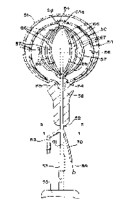
[00053] FIG. 4 illustrates a catheter, generally designated at 50, for
diagnosis in a
bladder 51, while treatment use is also possible. The particular illustrated
bladder is a
male bladder; however, this embodiment is suitable for use in female bladders
as well.
This catheter includes a catheter tube or body 52 having a drainage passageway
tube
53 through which fluid, liquid or gas can escape after entering at input tube
54. A
detachable urine bag 55 is shown. This catheter includes an expandable
component 56
secured to the catheter tube 52. A radiopaque reference line 57 is provided on
the
expandable component in this embodiment.
[00054] The catheter 50 of FIG. 4 and FIG. 5 is inserted through the
urinary tract
58 into the bladder 51. Catheter tube 52 exhibits a plurality of passageways.
Passageway 59 continues into branch 61 having the drainage catheter tube 53.
When
the expandable component is an inflatable member in some embodiments,
passageway
62 continues into branch 63 through which the inflation and deflation of the
inflatable
expandable component 56 proceeds.
CA 02904896 2015-09-09
WO 2014/164330 PCT/US2014/021932
[00055] Expandable component 56, when inflatable, is inflated by way of one
or
more holes 64, and the inflatable expandable component such as a balloon is
secured
to the catheter at proximal neck 65 and distal neck 65a. A plurality of
detectors 66 are
positioned in an array along a plurality of spokes 67. In this embodiment, all
of the
detectors and spokes are within the inflatable expandable component.
[00056] In this illustrated embodiment, the distal end portions of the
spokes 67 join
a slidable hub 68 at a location along the catheter 52. A manipulation wire 69
is joined at
its distal end to the hub 68. Manipulation wire 69 continues proximally
through the
catheter tube 52 and to branch 70 thereof, this branch having a passageway 71
that
slidingly receives the manipulation wire 69. Inserting the manipulation wire
69 further
into the passageway 71 moves the hub 68 distally, thereby reducing the bowing
of the
spokes 67 and thereby moving the detectors 66 generally radially inwardly.
Moving the
manipulation wire 69 outwardly pulls the hub 68 in a proximal direction,
thereby
increasing the bowing of the spokes 67 and moving the detectors generally
radially
outwardly. This action allows the medical professional to modify the detector
array from
an external position, thereby varying detector positioning in order to probe
for changes
or in order to modify radiation location and magnitude without having to
remove the
catheter 50 from the body cavity.
[00057] While FIG. 4 shows use of this catheter 50 within the bladder, same
can
be suitable for use elsewhere as well, which may or may not involve a
modification in
the shape of the catheter and of the expandable component. For example, some
embodiments can have a more elongated expandable component for body
passageways that are not as symmetrical as that illustrated in FIG. 4. Also,
hyperthermia components can be included in such an embodiment as generally
disclosed herein with respect to other embodiments. Although the spokes are
shown in
FIG. 4 to be generally uniformly positioned and spaced, variations can be
provided in
order to better conform to body cavity shapes for an expected use of the
device. For
example, the spokes can have separate sliding lengths, and modification of the
bowing
of the spokes can be independently generated by providing a plurality of
manipulation
components, for example one for each spoke.
16
CA 02904896 2015-09-09
WO 2014/164330 PCT/US2014/021932
[00058] As a general proposition, chemotherapy materials can be included in
conjunction with one or more of the radiation treatment devices described
herein. Such
delivery can be, for example, practiced by way of delivery tubes such as those
shown
herein for a hyperthermia function in those instances where separate tubing is
desired
for chemotherapy delivery. Additionally or alternatively, one or more of the
expandable
components, or catheter in some embodiments, can have impregnated into,
infused
onto, coated on, or otherwise carry chemotherapy materials separate and apart
from
being able to be delivered from the outside after insertion into the body.
Chemicals or
drugs along these lines can be provided in the form of microspheres or other
organically
bound or chemically bound substances as alternative chemotherapy or
radioactive
delivery systems. For example, delivery of Bacillum calmette-guerin (BCG) for
bladder
cancer treatment can be used. In other embodiments, the substance delivered by
any
of these means can be useful for pain maintenance, such as analgesic materials
and
pain or narcotic materials to provide pain relief during procedures,
especially when the
device protocol requires insertion within the body for extended time periods.
These can
include delayed release analgesics and the like.
[00059] It will be understood that the embodiments described above are
illustrative
of some of the applications of the principles of the present subject matter.
Numerous
modifications may be made by those skilled in the art without departing from
the spirit
and scope of the claimed subject matter, including those combinations of
features that
are individually disclosed or claimed herein. For these reasons, the scope
hereof is not
limited to the above description but is as set forth in the following claims,
and it is
understood that claims may be directed to the features hereof, including as
combinations of features that are individually disclosed or claimed herein.
17