Note: Descriptions are shown in the official language in which they were submitted.
SOFT TISSUE FIXATION SYSTEM
[0001] This application claims priority to U.S. Patent Application Serial No.
13/832,201 filed
March 15, 2013, U.S. Patent Application Serial No. 61/786,736 filed March 15,
2013, U.S.
Patent Application Serial No. 13/800,868, filed March 13, 2013, and U.S.
Patent Application
Serial No. 61/780,077 filed March 13, 2013.
BACKGROUND
[0002] Common injuries can involve tears of soft tissue, such as tendons and
ligaments, and
detachments of soft tissue from one or more underlying bones. As one example,
the tendons at
the ends of the rotator cuff muscles can become torn, leading to pain and
restricted movement of
the musculoskeletal system. The soft tissue can be conventionally re-attached
to bone
arthroscopically, for instance by driving bone anchors into the bone at a
desired locations, and
attaching separate strands of suture to each of the bone anchors and the soft
tissue. Each of the
separate strands of suture is then tied off to draw the soft tissue against
the bone, thereby
allowing reattachment of the soft tissue to the bone. What is desired is an
improved method and
apparatus for attaching soft tissue to bone.
SUMMARY
[0003] In accordance with one embodiment, a bone anchor includes a bone anchor
body
defining a proximal end, a distal end that is spaced from the proximal end
along a longitudinal
axis, and a perimeter that extends between the proximal end and the distal
end. The bone anchor
body is configured to receive a strand of suture. At least a portion of the
bone anchor body
comprises a deformation material that is responsive to an applied energy so as
to deform and,
1
Date Recue/Date Received 2020-08-11
CA 02904987 2015-09-09
WO 2014/165036
PCT1US2014/024196
when the strand of suture is received by the anchor body, capture the strand
of suture with
respect to movement relative to the bone anchor body.
BRIEF DESCRIPTION OF THE DRAWINGS
100041 The foregoing summary, as well as the following detailed description of
an example
embodiment of the application, will be better understood when read in
conjunction with the
appended drawings, in which there is shown in the drawings example embodiments
for the
purposes of illustration. It should be understood, however, that the
application is not limited to
the precise arrangements and instrumentalities shown. In the drawings:
[00051 Fig. I is a schematic top plan view of a soft tissue fixation assembly
anchored to bone
and retaining a soft tissue against the bone, the soft tissue fixation
assembly constructed in
accordance with one embodiment.
100061 Fig. 2A is a perspective view of a bone anchor of the soft tissue
fixation assembly
illustrated in Fig. 1, showing the bone anchor in accordance with one
embodiment;
100071 Fig. 2B is a side elevation view of the bone anchor illustrated in Fig.
2A;
100081 Fig. 2C is a sectional side elevation view of the bone anchor
illustrated in Fig. 2A;
100091 Fig. 2D is a bottom plan view of the bone anchor illustrated in Fig.
2A;
10010) Fig. 2E is a top plan view of the bone anchor illustrated in Fig. 2A;
100111 Fig. 3A is a perspective view of a bone anchor constructed in
accordance with an
alternative embodiment;
[00121 Fig. 3B is a sectional side elevation view of the bone anchor
illustrated in Fig. 3A;
[00131 Fig. 3C is an exploded perspective view of the bone anchor illustrated
in Fig. 3A;
[00141 Fig. 4A is a perspective view of a suture extending through opposed
channels of the
bone fixation element illustrated in Fig. 2A;
[00151 Fig. 4B is a perspective view of a suture extending through adjacent
channels of the
bone fixation element illustrated in Fig. 2A;
100161 Fig. 5A is a schematic sectional side elevation view of a soft tissue
fixation system
constructed, including an actuation assembly shown operably engaged with an
initial bone
anchor of the soft tissue fixation assembly illustrated in Fig. 1;
[00171 Fig. 5B is a schematic sectional side elevation view of the soft tissue
fixation system
illustrated in Fig. 5A, shown after activation of the actuation assembly;
2
CA 02904987 2015-09-09
WO 2014/165036
PCT1US2014/024196
100181 Fig. SC is a schematic side elevation view of a soft tissue fixation
system illustrated in
Fig. 5B, showing the actuation assembly operably engaged with a subsequent
bone anchor of the
soft tissue fixation assembly illustrated in Fig. I;
100191 Fig. SD is a schematic sectional side elevation view of the soft tissue
fixation system
illustrated in Fig. 5C, shown after activation of the actuation assembly;
[00201 Fig. 6A is a schematic sectional side elevation view of a portion of
the bone fixation
assembly I, showing a single strand of suture sequentially connected
continuously to a plurality
of the bone anchors;
100211 Fig. 6B is a schematic sectional side elevation view of a portion of
the bone fixation
assembly I, showing a pair of strands of suture each sequentially connected
continuously to a
plurality of the bone anchors;
[00221 Fig. 6C is a schematic sectional side elevation view of a portion of
the bone fixation
assembly 1, showing the pair of strands of suture each sequentially connected
continuously to a
plurality of the bone anchors, one of the strands of suture configured to
attach to an auxiliary
implant;
100231 Fig. 7A is a schematic plan view of the soft tissue fixation assembly
as illustrated in
Fig. I, but showing the bone anchors in one arrangement;
[00241 Fig. 7B is a schematic plan view of the soft tissue fixation assembly
as illustrated in
Fig. 7A, but showing the bone anchors in another arrangement as illustrated in
Fig. 1, shown as
an open arrangement;
[00251 Fig. 7C is a schematic plan view of the soft tissue fixation assembly
as illustrated in
Fig. 7B, but showing the bone anchors in a closed arrangement in accordance
with one
embodiment;
100261 Fig. 7D is a schematic plan view of the soft tissue fixation assembly
as illustrated in
Fig. 7C, but showing the bone anchors in a closed arrangement in accordance
with another
embodiment; and
[00271 Fig. 7E is a schematic plan view of the soft tissue fixation assembly
as illustrated in
Fig. 7B, but showing the bone anchors in an open arrangement in accordance
with another
embodiment.
DETAILED DESCRIPTION
[00281 Referring initially to Figs. 1 and 8B, a soft tissue fixation assembly
20 is configured to
secure a soft tissue 22 against a surface of an underlying bone 24 so as to
promote attachment of
3
CA 02904987 2015-09-09
WO 2014/165036
PCT1US2014/024196
the soft tissue 22 to the bone 24. The soft tissue 22 can be detached from the
bone due to an
anatomical defect, trauma, or the like, and can be configured as any
anatomical soft tissue in the
human or other animal body, such as a tendon or ligament. For instance, in
accordance with one
embodiment, the tendon can be a rotator cuff tendon or any other tendon as
desired.
[00291 The fixation assembly 20 can include at least one bone anchor 26, such
as first and
second bone anchors 26a and 26b, respectively, and a strand of suture 28 that
is configured to
attach to each of the bone anchors 26a and 26b. For instance, the strand of
suture 28 is
configured to be received by each of the bone anchors 26 and 26b. The first
bone and second
bone anchors 26a and 26b can, for instance, be driven into the bone 24, such
that the strand of
suture 28 extends from the first bone anchor 26a to the second bone anchor
26b. One of the first
and second bone anchors 26a and 26b, for instance the second bone anchor 26b
as illustrated, can
be driven through the soft tissue 22 and into the bone 24, referred to as a
trans-tendon technique.
The other of the first and second bone anchors 26a and 26b, for instance the
first one anchor 26a
as illustrated, can be driven into the bone 24 at a location spaced from the
soft tissue 22. *Thus,
the strand of suture 28 can extend from the first bone anchor 26a along an
outer surface of the
bone 24, across an interface 30 between the bone 24 and the soft tissue 22, to
the second bone
anchor 26b. The strand of suture 28 can be under tension between the first and
second bone
anchors 26a and 26b so as to apply a force against the soft tissue soft 22
toward the bone 24,
thereby causing the soft tissue 22 to maintain contact with the bone 24. It
should be recognized
that in most instances it may not be recommended, during rotator cuff repair,
to insert the bone
anchors 26 in the intraarticular space of the shoulder, but rather to position
the bone anchors
about the joint.
100301 It will be appreciated from the description below that the fixation
assembly 20 can
include any number of bone anchors 26, such as a plurality of bone anchors 26,
as desired. One
or more of the plurality of bone anchors 26 can be driven through the soft
tissue 22 and into the
bone 24. One or more others of the plurality of bone anchors can be driven
into the bone 24 at a
location spaced from the soft tissue 22, so as to define any geometric pattern
as desired. In
accordance with one embodiment as illustrated in Fig. 1, the soft tissue
fixation assembly 20 can
include a third bone anchor 26a driven through the soft tissue 22 and the bone
24, and a fourth
bone anchor 26d driven through the bone 24 at a location spaced from both the
soft tissue 22 and
the first bone anchor 26a. The strand of suture 28 can extend continuously
from each of the
plurality of bone anchors 26 to at least one adjacent one of the plurality of
bone anchors 26.
Thus, the plurality of bone anchors 26 can define at least one row 32, such as
a first row 32a that
4
CA 02904987 2015-09-09
WO 2014/165036
PCT1US2014/024196
can be defined by at least some of the plurality of bone anchors 26, and a
second row 32b that
can be defined by at least others of the plurality of bone anchors 26, the
strand of suture 28
connected between the first and second rows 32a and 32b. The rows 32a and 32b
can extend
substantially linearly, curvilinearly, or can define any other suitable
arrangement, and can be
parallel with each other or aliened to intersect each other as desired. The
fixation assembly 20
can define as many rows as desired.
100311 Referring now to Figs. 2A.-2E, each of the bone anchors 26 can include
an anchor body
34 that defines a proximal end 36a and a distal end 36b that is spaced from
the proximal end 36a
along a distal direction of insertion into bone. It should be appreciated that
the term "distal" and
derivatives thereof can refer to a direction from the proximal end 36a to the
distal end 36b, and
the term "proximal" and derivatives thereof can refer to a direction from the
distal end 36b to the
proximal end 36a. The anchor body 34 can extend, for instance can be elongate,
along a
longitudinal axis 38, such that the proximal end 36 and distal end 36b are
spaced from each other
along the longitudinal axis 38. The longitudinal axis 38 can be substantially
straight, or can be
curved or assume any alternative shape as desired. The anchor body 34 can
further define an
outer perimeter 40 extends between the proximal end 36a and the distal end
36b. The perimeter
40 can be round, for instance substantially circular, as illustrated, or can
define any alternative
shape whatsoever, such as a rectilinear shape. The distal end 36b can define a
tip 37 that can be
tapered inwardly, and thus toward the longitudinal axis 38, as it extends
along the distal
direction.
[00321 The anchor body 34 can further define at least one channel 42, such as
a plurality of
(e.g., at least two) channels 42, that extend into the perimeter 40, for
instance toward the
longitudinal axis 38. Each of the channels 42 can be configured to receive the
strand of suture
28 (see Figs. 4A.-B). For instance, respective ones of the channels 42 are
configured to receive
respective portions of the strand of suture 28. Each channel 42 can define a
central axis 43a, and
a first or proximal end 43b, and a second or distal end 43e that is spaced
from the proximal end
43b along the central axis 43a. The channels 42 can extend along an entirety
of the length of the
anchor body 34 from the proximal end 36a to the distal end 36b, or can extend
along a portion of
the length of the anchor body 34. Thus, it can be said that the channels 42
can extend between
the proximal and distal ends 43b and 43c, respectively, for instance from the
proximal end 43b to
the distal end 43c. The channels 42 can, for instance, be open to a proximal-
most outer surface
54 of the anchor body 34, the proximal-most outer surface 54 extending from
the outer perimeter
40 toward the longitudinal axis 38. The central axis 43a can extend parallel
to the longitudinal
CA 02904987 2015-09-09
WO 2014/165036
PCT1US2014/024196
axis 38, or can be angularly offset with respect to the longitudinal axis 38,
and can define any
shape as desired. The anchor body 34 can include any number of channels 42,
such as a first
channel 42a and a second channel 42b, that can be disposed on opposite sides
with respect to the
longitudinal axis 38, and thus be opposite each other with respect to the
longitudinal axis 38, or
can be angularly offset with respect to the longitudinal axis 38 at any angle
of separation as
desired.
100331 Thus, a first line 35a perpendicular to the longitudinal axis 38 that
intersects both the
longitudinal axis 38 and the central axis 43a of the first channel 42a can
define the angle of
separation with respect to a second line 35b that is perpendicular to the
longitudinal axis 38 and
intersects both the longitudinal axis 38 and the central axis 43a of the
second channel 42b. The
angle of separation al can be substantially 180 degrees, or any angle between
zero and 180
degrees. In accordance with the illustrated embodiment, the anchor body 34 can
include a third
channel 42c and a fourth channel 42d that can define an angle of separation
with respect to each
other, for instance 180 degrees as described above with respect to the first
and second channels
42a and 42b. Further, the channels 42 can be equidistantly spaced from each
other about the
perimeter 40 as illustrated, or can be variably spaced from each other about
the perimeter 40 as
desired. Thus, adjacent ones of the channels 42, such as the first channel 42a
and either or both
of the third and fourth channels 43e and 43d, respectively, can define an
angle of separation a2
of substantially 90 degrees. The channels 42 that arc spaced from each other
by an angle of
separation that is greater than 90 degrees and less than or equal to 180
degrees can be referred to
as opposed channels. The channels 42 that are spaced from each other by an
angle of separation
that is greater than between 0 degrees and less than or equal to 90 degrees
can be referred to as
adjacent channels.
100341 As will be appreciated from the description below, at least a portion
up to all of the
anchor body 34 can be made from a deformation material 44, which can be
polymeric, that
comprises a deformation material responsive to an applied energy that causes
the deformation
material, and thus the anchor body 34 at the deformation material, to deform.
The applied
energy can. be a laser or any suitable alternative energy, such as an
electrical current,. In
accordance with one embodiment, the deformation material 44 can define at
least a portion of the
anchor body 34, including at least a portion up to an entirety of one or more
up to all of the
channels 42. For instance, the deformation material 44 can be located in a
region as illustrated in
Figs. 3A-C that can be spaced from one or both of the proximal end 36a and the
distal end 36b.
Alternatively, the deformation material 44 can comprise an entirety of the
anchor body 34, from
6
the proximal end 36a to the distal end 36b, as illustrated in Figs. 2A-2E. The
deformation
material 44 can be responsive to the applied energy so as to deform at least a
portion of the
anchor body 34 and capture the received portion of the strand of suture 28
therein with respect to
movement relative to the anchor body 34. Deformation of the deformation
material 44 in
response to the applied energy can further cause the anchor body 34 to
similarly deform and
become fixed to the bone 24. In accordance with one embodiment, the
deformation that causes
the anchor body 34 to become fixed to the bone can similarly cause the anchor
body 34 to
capture the received portion of the suture strand 28 therein with respect to
movement relative to
the anchor body 34.
[0035] For instance, in accordance with one embodiment, the deformation
material 44 is
configured to deform in response to the applied energy and close at least a
portion of each of the
channels 42 and capture the respective received portion of the strand of
suture 28 in the channels
42 with respect to movement relative to the anchor body 34. Deformation of the
deformation
material 44 in response to the applied energy can further cause the anchor
body 34 to similarly
deform and become fixed to the bone 24. In accordance with one embodiment, the
deformation
that causes the anchor body 34 to become fixed to the bone can similarly cause
the channels 42
to close and capture the received strand of suture 28 therein. In accordance
with one
embodiment, the deformation material 44 can be disposed in a middle region of
the anchor body
34 that is spaced from the proximal end 36a and the distal end 36b. For
instance, the middle
region can include a location that is disposed midway between the proximal end
36a and the
distal ends 36b. The bone anchor body 34 can be constructed so as to prevent
the applied energy
from traveling to the distal end 36b. For instance, the bone anchor body 34
can define any color
as desired, or be made of any one or more suitable materials as desired, that
can prevent the
applied energy from traveling to at least one select region of the bone anchor
body 45, such as
the distal end 36b. Examples of deformation materials of the type described
herein are disclosed
in U.S. Patent Application Publication No. 2010/0241229 Al, published
September 23, 2010,
and U.S. Patent Application Publication No. 2012/0129131 Al, published May 24,
2012.
[0036] As described above, each of the channels 42 can define a central axis
43a, and a first or
proximal end 43b, and a second or distal end 43c that is spaced from the
proximal end 43b along
the central axis 43a. In accordance with one embodiment, the distal ends 43c
of the channels 42
can be open to each other, such that the strand of suture 28 can extend
distally along one of the
channels 42 out the distal end 43c of the channel, into the distal end 43c of
another one of the
7
Date Recue/Date Received 2020-08-11
CA 02904987 2015-09-09
WO 2014/165036
PCT1US2014/024196
channels 42 and proximally along the other one of the channels 42. For
instance, the distal ends
43c of the channels 42 can be open to each other at the distal end 36b of the
anchor body 34. In
accordance with one embodiment, the anchor body 34 can define a void 46 that
extends along
the proximal direction into the tip 37. The void 46 can be aligned with the
longitudinal axis 38,
and can be open to the distal ends 43c of each of the channels 42, including
the first channel 42a,
the second channel 42b, the third channel 42c, and the fourth channel 42d.
While the void 46 is
open through the tip 37 along the distal direction as illustrated, it should
be appreciated that the
tip 37 can alternatively be enclosed, such that the tip 37 defmes a distal
boundary of the void 46.
Nevertheless, the void 46 can be open to the outer perimeter 40 and to each of
the distal ends 43c
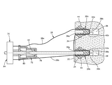
of the channels 42.
100371 Thus, the distal ends 43c of each of the third and fourth channels 42c
and 42d are open
to both each other and the distal ends 43c of the first and second channels
42a and 42b, for
instance at the distal end 36b. Similarly, the distal ends 43c of each of the
first and second
channels 42a and 42b are open to both each other and the distal ends 43c of
the third and fourth
channels 42c and 42d, for instance at the distal end 36b. For instance, the
distal ends 43c can be
open to each other via the void 46, such that strands of suture extending from
the distal end 43c
of one of the channels 42 to the distal end 43c of another one of the channels
42 can extend
across the void 46. Alternatively, the distal ends 43c of one or more up to
all of the channels 42
can be continuous with each other.
[00381 Each channel 42 can be defined by at least one wall that can define a
base 45a of the
anchor body 34 and opposed side walls 45b of the anchor body 34 that extend
from the base 45a
toward, for instance to, the perimeter 40. The void 46 can be at least
partially defined by a floor
51 that extends toward the perimeter 40, and the anchor body 34 can defme
respective interface
49 between the bases 45a and the floor 51. The interface 49 can be beveled as
desired.
[00391 With continuing reference to Figs. 2A-E, the anchor 26 can define an
insertion aperture
52 that extends into the anchor body 34 and is configured to receive an energy
emitting
instrument 70 (see Fig. 6A) that is configured to apply the energy that causes
the deformation
material 44 to deform as described herein. For instance, the anchor body 34
can define the
insertion aperture 52 that extends into the proximal end 36a, and in
particular the proximal-most
surface 54, substantially along the distal direction. For instance, the
insertion aperture 52 can be
centrally disposed with respect to the outer perimeter and the longitudinal
axis 38, and thus can
extend substantially along the longitudinal axis 38. As will be described in
more detail below,
the insertion aperture 52 can be sized to receive an energy emitting
instrument 70 (see Fig. 5A)
8
CA 02904987 2015-09-09
WO 2014/165036
PCT1US2014/024196
that is configured to apply the energy that causes the deformation material 44
to deform in the
manner described herein. The insertion aperture 52 can be round, such as
cylindrical, in shape,
or can define any suitable alternative shape as desired. The insertion
aperture 52 can be defined
by a base 53a of the anchor body 34 and at least one inner side wall 53b that
can extend from the
base 53a along the proximal direction, for instance to the proximal outer
surface 54 of the
proximal end 36a. In accordance with one embodiment, the proximal end 36a that
defines the
insertion aperture 52 can be integral and monolithic with one or more up to
all of the distal end
36b, the base 45a of one or more up to all of the channels 42, the side walls
45b of one or more
up to all of the channels 42, the floor 51, and the interfaces 49.
100401 Alternatively, as illustrated in Figs. 3A-C, the bone anchor can
include an insert 56 that
defines an insert body 59 having a proximal end 56a, a distal end 56b spaced
from the proximal
end 56a along the distal direction, and an outer perimeter 56c that extends
from the proximal end
56a to the distal end 56b. The insert 56 can define the insertion aperture 52
that is configured to
receive the energy emitting instrument 70 (see Fig. 6A) that is configured to
apply the energy to
the deformation material 44. The bone anchor 26 can define an aperture 60 that
extends into the
anchor body 34 and is sized and configured to receive the insert 56. For
instance, the body 34
can define the aperture 60 that extends into the proximal end 36a
substantially along the distal
direction. For instance, the insertion aperture 60 can be centrally disposed
with respect to the
outer perimeter and the longitudinal axis 38, and thus can extend
substantially along the
longitudinal axis 38. The insert 56 can be insertable in the aperture 60 in
any manner as desired.
For instance, the perimeter 56c of the insert 56 can define a cross-sectional
dimension
substantially equal to that of the aperture 60, such that the insert 56 can be
press-fit into the
aperture 60. Alternatively, the insert 56 can be secured to the anchor body 34
in the aperture 60
using any known adhesive, fastener, or the like. The insert 56 can define a
head 61 that extends
out from the proximal end 56a of the body 59 and is configured to abut the
proximal most
surface 54. The head 61 can define channels 57 that are aligned with the
channels 42 and extend
through the head 61 along the distal direction. Thus, the channels 42 and 57
can combine to
define the respective channels that are configured to receive the strands of
suture 28 as described
herein. The insert 56 can be made from a material different than the
deformation material, such
that the insert 56 does not deform in response to application of the energy.
100411 Referring now to Fig. 5A, a soft tissue fixation system 62 can include
an actuation
assembly 64 and the soft tissue fixation assembly 20. The soft tissue fixation
assembly 20 can
include at least one bone anchor, such as a plurality (such as a pair or more)
of bone anchors 26
9
CA 02904987 2015-09-09
WO 2014/165036
PCT1US2014/024196
and at least one strand of suture 28. The actuation assembly 64 can include
the energy emitting
instrument 70 and a tensioner 72 that supports the energy emitting instrument
70. The energy
emitting instrument 70 is configured to apply energy to the anchor body 34,
thereby causing the
deformation material 44 to deform. As the deformation material 44 deforms, the
tensioner 72
can maintain tension in the suture 28.
[00421 The energy emitting instrument 70 can include an energy source 74 and
an energy
conduit 76 that extends from the enemy source 74 and is configured to be
inserted into the
insertion aperture 52 so as to be configured to apply energy to the implant
body 34 in sufficient
quantity that the deformation material 44 deforms in response to the applied
energy, or can
otherwise be operably coupled to the implant body 34 so as to be configured to
apply energy to
the implant body 34 in sufficient quantity that the deformation material 44
deforms in response
to the applied energy. In accordance with the illustrated embodiment, the
energy emitting
instrument 70 is a laser, the energy source 74 is a laser source configured to
emit energy in the
form of a laser beam. The energy conduit 76 can define a light pipe that
extends from the laser
source, the light pipe configured to communicate the laser beam from the laser
source to the
implant 26, and apply the energy in the form of the laser beam to the implant
body 34.
100431 The tensioner 72 can include a first support member 78 configured to
support the
energy conduit 76, and a second support member 80 that is spaced from the
first support member
along a direction that can be parallel, such as coincident, with the
longitudinal axis 38. In
accordance with the illustrated embodiment, the second support member 80 can
be spaced from
the first support member 78 along the proximal direction, such that the first
support member 78
is disposed between the second support member 80 and the bone anchor 26 that
receives the
energy conduit 76. The second support member 80 is configured to attach to the
suture 28 that
extends through at least one of the channels 42 of the bone anchor 26. The
tensioner 72 can
include a biasing member 82, for instance a spring such as a coil spring that
is connected
between the first and second support members 78 and 80, respectively. The
tensioner 72 is
configured to bias one of the first and second support members 78 and 80,
respectively, to move
relative to the other of the first and second support members 78 and 80,
respectively.
100441 During operation, the biasing member 82 applies a force to the second
support member
80 that biases the second support member to move along the proximal direction
away from the
bone anchor 26, and also away from the first support member 78. Alternatively,
the second
support member 80 can be disposed distal of the first support member 78, and
thus between the
first support member 78 and the bone anchor 26, such that the biasing member
82 applies a force
CA 02904987 2015-09-09
WO 2014/165036
PCT1US2014/024196
to the second support member 80 that biases the second support member 8010
move along the
proximal direction away from the bone anchor 26 and toward the first support
member 78.
100451 With continuing reference to Fig. 5A, a method of anchoring suture to
bone can include
the step of inserting the strand of suture 28 into at least one channel 42 of
an initial bone anchor,
which can be defined as a first bone anchor 26. In particular, the strand of
suture 28 can be
inserted into first and second select ones of the channels 42, such that the
strand of suture 28
extends from, the proximal end 36a, distally along the first select one of the
channels 42, out the
distal end of the first select one of the channels 42 as described above, into
the distal end of the
second select one of the channels 42, proximally along the second select one
of the channels 42,
and out the proximal end 36a of the anchor body. The first and second select
channels can be
opposed channels or adjacent channels as described above.
[00461 After the strand of suture 28 has been inserted into the at least one
channel 42, the bone
anchor 26 can be driven into the bone 24. A pilot hole can be drilled or
otherwise formed in the
bone, and the initial anchor 26 can be driven into the pilot hole, or the
anchor 26 can be driven
into the bone without first forming the pilot hole. Next, the energy conduit
76 is inserted into the
insertion aperture 52 or otherwise operably coupled to the anchor body 34. At
least one end of
the stran.d of suture 28, for instance first and second opposed ends 28a and
28b that extend out
from different ones of the channels 42 of the initial bone anchor, can be
fixedly attached to the
second support member 80 prior to deformation of the initial bone anchor.
[00471 Referring now to Fig. 5B, the energy source 74 s then actuated so as to
cause the energy
emitting instrument 70, for instance the energy source 74, to emit energy,
which can be in the
form of a laser beam, to the conduit 76. The energy emitting instrument, for
instance at the
conduit 76, can apply the energy to the bone anchor body 34 so as to cause the
deformation
material 44 to deform, thereby causing the anchor body to deform and I) fix to
the bone 24, and
2) capture the strand of suture 28 therein with respect to movement relative
to the bone anchor
26. For instance, the energy emitting instrument, for instance at the conduit
76, can apply the
energy to the bone anchor body 34 so as to cause the deformation material 44
to deform and
close at least the portion of the channel 42 that is defined by the
deformation material 44, thereby
capturing the strand of suture 28 therein with respect to movement relative to
the bone anchor
26. Thus, the strand of suture 28 is unable to move relative to the bone
anchor 26 at the portion
of the channel 42 that has been closed. In accordance with the illustrated
embodiment, both
channels within which the suture 28 resides can be closed.
11
CA 02904987 2015-09-09
WO 2014/165036
PCT1US2014/024196
10048i It is appreciated that prior to deformation of the first bone anchor,
the first bone anchor
26 defines a first length Ll along the longitudinal axis 38 between the
proximal end 36a and the
distal end 36b. As the deformation material 44 deforms, the distal end 36b can
be drawn toward
the proximal end 36a, thereby shortening the length of the bone anchor to a
second length L2
along the longitudinal axis 38 between the proximal end 36a and the distal end
36b, wherein the
second length L2 is greater than Li. For instance, the biasing force of the
biasing member 82
causes the second support member 80, which is attached to the suture 28, to
translate proximally
as the deformation material 44 of the anchor body 34 softens and deforms in
response to the
applied energy, thereby promoting deformation of the anchor body 34 as
described above.
Furthermore, translation of the second support member 80 can cause the first
and second ends
28a and 28b to likewise translate proximately, thus maintaining a desired
level of tension in the
strand of suture 28 through completion of the application of energy to the
anchor body 34 and
through completion of the resulting deformation. =lt is further appreciated
that a maximum width
of the anchor body 34 along a direction perpendicular to the longitudinal axis
38 can increase in
response to deformation of the deformation material 44, thereby securely
anchoring the bone
anchor in the bone 24. It should be appreciated. of course, that the biasing
force can be produced
by any suitably constructed tensioner 72, or manually, as desired.
[00491 Referring now to Fig. 5C, it is appreciated that the first end 28a is
fixed to the initial
bone anchor. Accordingly, the first end 28a can be cut at a location adjacent
to the proximal end
36a of the anchor body 34, or the first end 28a can define a free end of the
strand of suture 28
that extends out from the bone 24. The free end can be loosely attached to the
second support
member 80, or can be free from the second support member 80. As is described
in more detail
below, the free end can be attached to a final one of the bone anchors 26. The
second end 28b
can then be inserted into first and second select channels of a subsequent
bone anchor, which can
be define as a second bone anchor 26b, in the manner described above with
respect to the first
anchor 26a. The first and second channels 42 can be opposed channels or
adjacent channels as
described above. The second end 28b can extend out the anchor body 34 and can
be attached to
the second support member 80 in the manner described above.
100501 After the strand of suture 28 has been inserted into the channels 42 of
the second bone
anchor 26b, the second bone anchor 26b can be driven into the bone 24 in the
manner described
above. It should be appreciated that the second bone anchor 26 can be driven
through the soft
tissue 22 and into the bone 24. Next, as illustrated in Fig. 5D, the method
can include the step of
applying energy to the second bone anchor 26b so as to cause the deformation
material 44 of the
12
CA 02904987 2015-09-09
WO 2014/165036
PCT1US2014/024196
second bone anchor 26b to deform, thereby capturing the strand of suture 28
therein with respect
to movement relative to the second bone anchor 26b. For instance, deforming
the deformation
material 44 can close the portion of the channels 42 that is defined by the
deformation material
44, thereby capturing the received strand of suture 28 therein with respect to
movement relative
to the second bone anchor 26b. The biasing member 82 can apply a biasing force
to the second
support member 80 that causes the second support member 80 to move away from
both the first
support member 78 and the first and second bone anchors 26a and 26b, thereby
promoting
deformation and maintaining tension in the strand of suture 28 as the length
of the second bone
anchor 26b decreases in the manner described above with respect to the first
bone anchor 26a. It
should be appreciated that the strand of suture 28 is continuous, and is
fixedly attached to the
first and second bone anchors 26a and 26b. Thus, the strand of suture 28
extends continuously
from the first bone anchor 26a, over the bone 24, across the interface 30
between the soft tissue
22 and the bone 24, over the soft tissue 22, to the second bone anchor 26b.
Because the strand of
suture 28 is in tension and is anchored to the first and second bone anchors
26a and 26b inside
the bone 24, the strand of suture 28 forces the soft tissue 22 into contact
with the bone 24.
[00511 It should be appreciated that the method steps of inserting the strand
of suture 28 into at
least one such as a pair of channels, driving the bone anchor into the bone,
for instance possibly
through the soft tissue 22 and into the bone 24, and applying the enemy to the
anchor body can
be sequentially repeated for at least one additional bone anchor, such a third
bone anchor 26c as
illustrated in Fig. 6A. For instance, the method steps of inserting the strand
of suture 28 into at
least one such as a pair of channels, driving the bone anchor into the bone,
for instance possibly
through the soft tissue 22 and into the bone 24, and applying the energy to
the anchor body can
be sequentially repeated for a plurality of additional bone anchors 26c. In
accordance with one
embodiment, the energy is applied to a given one of the bone anchors 26 prior
to inserting the
strand of suture 28 into a channel 42 of another one of the bone anchors 26
that is to be
subsequently anchored to the bone 24.
[00521 As described above, at least one strand of suture can be inserted into
the channels 42 of
the anchors 26 prior to driving the anchors into bone and applying the energy
to the respective
anchor bodies 34. For instance, as illustrated in Fig. 6A, a single continuous
strand of suture 28
can be fixedly attached sequentially to at least two, such as three, bone
anchors 26a, 26b, and
26c. Alternatively, as illustrated in Fig. 6B, it should be appreciated that
the steps of inserting a
strand of suture into at least one channel of the bone anchors 26 can include
the step of inserting
a pair or more, and thus a plurality, of strands of suture 28 into the at
least one channel.
13
CA 02904987 2015-09-09
WO 2014/165036
PCT1US2014/024196
Accordingly, the step of applying energy to the implant body causes the anchor
body, and thus
the anchor, to deform so as to secure each of the plurality of strands of
suture 28 to the respective
anchor body. Deformation of the anchor body can further cause the anchor body
to fix to the
bone 24, and can cause the portion of the channels defined by the deformation
material to close
so as to secure the strands of suture 28 therein with respect to movement
relative to the anchor
body. A free end of the single strand of suture 28, or of a second strand of
suture 28, can extend
out the proximal end of the bone anchor 26, and out the bone 24, such that it
can be attached to
an auxiliary implant, such as a soft tissue or bone graft, and then secured to
itself or the other
strand of suture 28, for instance at a knot 29, thereby attaching the
auxiliary implant to the soft
tissue 22 or bone 24.
100531 Referring now to Figs. 7A-7E in general, it is appreciated that the
soft tissue fixation
assembly 20 can include as many bone anchors 26 as desired, disposed in any
arrangement as
desired. For instance, as illustrated in Fig. 7A, the first bone anchor 26a is
shown inserted into
the bone without being driven through the soft tissue 22, and the second one
anchor 26b is
shown inserted through the soft tissue 22 and into the bone 24. The first and
second bone
anchors 26a and 26b can define a row 32 of bone anchors. Referring to Fig. 7B,
the plurality of
bone anchors 26c, in combination with the first and second bone anchors 26a
and 26b can define
a pair of rows, such as a first row 32a and a second row 32b. For instance,
the row defined by
the first and second bone anchors 26a and 26b can define the first row 32a,
and the plurality of
bone anchors 26c can define the second row 32b that is adjacent the first row
32a. The strand of
suture 28 can extend from the first row 32a to the second row 32b. For
instance, the strand of
suture 28 can extend through adjacent channels 42 (see Fig. 2D) of one of the
bone anchors, such
as the second bone anchor 26b illustrated in Fig. 7B, so as to extend from the
first row 32a along
a column direction toward the second row 32b. The strand of suture 28 can then
extend through
adjacent channels of a subsequent one of the bone anchors, such as one of the
plurality of bone
anchors 26c illustrated in Fig. 7B, so as to extend from the column direction
to the row direction,
for instance along the second row 32b.
100541 With continuing reference to Fig. 7B, the step of driving a final bone
anchor 26d of the
plurality of bone anchors 26c can include the step of driving the final bone
anchor 26d into the
bone 24 at a location spaced from the first bone anchor 26a. Thus, the strand
of suture 28 is not
connected directly from the final bone anchor 26d to the first bone anchor
26a. Because the
strand of suture 28 is not directly connected between the bone anchors 26 of
every adjacent pair
of bone anchors 26, the arrangement defined by the bone anchors 26 can be
referred to as an
14
CA 02904987 2015-09-09
WO 2014/165036
PCT/US2014/024196
open arrangement. As described above, the soft tissue fixation assembly 20 can
include as many
bone anchors as desired, configured in any arrangement as desired. For
instance, the soft tissue
fixation assembly 20 can define as many rows 32 as desired.
[00551 Referring to Fig. 7E, the suture 28 of the soft tissue fixation
assembly 20 can extend
from the second bone anchor 26b to a first one of the plurality of bone
anchors 26c along the first
row 32a. In accordance with one embodiment, the strand of suture 28 can be
inserted into
opposed channels of the second bone anchor 26b, wherein the opposed channels
are spaced from
each other along a direction that is substantially parallel with the row 32a.
The suture 28 can
extend from the second bone anchor 26b toward a first one of the plurality of
bone anchors 26c.
The strand of suture 28 can be inserted into adjacent columns of the first one
of the plurality of
bone anchors 26c. Thus, the suture 28 can extend from the second bone anchor
26b along the
row 32a to the first one of the plurality of bone anchors 26c, and along a
column direction from
the first one of the plurality of bone anchors 26c to a second one of the
plurality of bone anchors
26c that lies along the second row 32b. The strand of suture 28 can be
inserted into adjacent
columns of the second one of the plurality of bone anchors 26c so as to extend
from the first one
of the plurality of bone anchors 26c to the second one of the plurality of
bone anchors 26c along
the column direction, and along the second row 32b from the second one of the
plurality of bone
anchors 26c to another one of the plurality of bone anchors.
[00561 Of course, it should be appreciated that the strand of suture 28 can be
inserted into any
channels of the bone anchor 26 as desired so as to extend along a respective
row 32, or to extend
from a respective row along the column direction, or to extend from a column
direction to a
respective row. It should be ftuther appreciated that the soft tissue fixation
assembly 20 can
define as many bone anchors as desired arranged along a given column
direction. It should be
further appreciated still that the soft tissue fixation assembly 20 can define
a chain configuration
having a plurality of rows of bone anchors 26, each row defined by two or more
bone anchors
26, and the rows of adjacent pairs of rows (thus rows that are partially
defined by a common one
of the anchors 26) angularly offset from each other at an angle between 90
degrees and 180
degrees, or alternatively between 0 degrees and 90 degrees (and thus an angle
other than 90
degrees). Thus, the first anchor 26a can be disposed at a first terminal end
of the chain, and the
final bone anchor 26d can be disposed at a second terminal end of the chain.
[00571 Referring now to Fig. 7C, the suture 28 can be connected between the
final bone anchor
26d and the first bone anchor 26a. For instance, as described above with
reference to Fig. 5C,
the first end 28a of the strand of suture 28 can define a free end that
extends out from the first
CA 02904987 2015-09-09
WO 2014/165036
PCT1US2014/024196
bone anchor 26a and the bone 24. Similarly, the second end 28b of the strand
of suture 28 can
define a free end that extends out from the final bone anchor 26d and the bone
24. The free ends
that are defined by the first and second ends 28a and 28b can be attached to
each other in
accordance with any suitable embodiment, so as to directly attach the suture
28 to the last bone
anchor 26d and the first bone anchor 26a. For instance, the free ends can be
tied to each other, or
secure to each other via any suitable fastener. Because the strand of suture
28 is directly
connected between the bone anchors 26 of every adjacent pair of bone anchors
26, the
arrangement defined by the bone anchors 26 can be referred to as a closed
arrangement
[00581 Referring now to Fig. 7D, the bone anchors 26 can define a closed
arrangement in
accordance with an alternative embodiment. For instance, the plurality of
additional bone
anchors 26c can define a select bone anchor 26e that is driven into the bone
24 and deformed
before the final bone anchor 26d is driven into the bone. As described above,
the first end 28a of
the strand of suture 28 can define a free end that extends out from the first
bone anchor 26a and
the bone 24 as described above. Furthermore, the second end 28b of the strand
of suture 28 can
define a free end that extends out from the select bone anchor 26e and the
bone 24.
[00591 The free end defined by the first end 28a of the strand of suture 28
can be inserted into
at least one channel of the final bone anchor 26d. For instance, the free end
defined by the first
end 28a of the strand of suture 28 can be inserted into a first channel 42 of
the final bone anchor
26d, and out of a second channel 42 of the final bone anchor 26d. The first
and second channels
of the final bone anchor 26d can be adjacent channels or opposed channels as
described above.
Similarly, the free end defined by the second end 28b of the strand of suture
28 can be inserted
into at least one channel of the final bone anchor 26d. For instance, the free
end defined by the
second end 28b of the strand of suture 28 can be inserted into a first channel
42 of the final bone
anchor 26d, and out of a second channel 42 of the final bone anchor 26d. The
first and second
channels can be adjacent channels or opposed channels as described above. In
accordance with
one embodiment, the first channel of the free end that receives the first end
28a can define the
second channel that receives free end defined by the second end 28b.
Similarly, the first channel
that receives second end 28b can define the first channel that receives the
first end 28a. It should
be appreciated, of course, that the free ends defined by the first and second
ends 28a and 28b,
respectively, can be inserted into different channels of the final bone anchor
28d. In accordance
with the embodiment illustrated in Fig. 2D, the first and second bone anchors
26a and 26b can
each be driven through the soft tissue 22 and into the bone 24, and the select
and final ones 26e
16
CA 02904987 2015-09-09
WO 2014/165036
PCT1US2014/024196
and 26d, respectively, of the plurality of bone anchors 26c can be inserted
into the bone 24
without being driven through the soft tissue 22.
100601 The foregoing description is provided for the purpose of explanation
and is not to be
construed as limiting the soft tissue fixation system, or components thereof.
While various
embodiments have been described with reference to preferred embodiments or
preferred
methods, it is understood that the words which have been used herein are words
of description
and illustration, rather than words of limitation. Furthermore, although the
embodiments have
been described herein with reference to particular structure, methods, and
embodiments, the
disclosure is not intended to be limited to the embodiments specifically
described herein. For
instance, it should be appreciated that structure and methods described in
association with one
embodiment are equally applicable to all other embodiments described herein
unless otherwise
indicated. Those skilled in the relevant art, having the benefit of the
teachings of this disclosure,
may effect numerous modifications to the embodiments as described herein, and
changes may be
made without departing from the scope of the present invention, for instance
as set forth by the
appended claims.
17