Note: Descriptions are shown in the official language in which they were submitted.
Surgical implant
The invention relates to a surgical implant, in particular to
a surgical implant having an areal basic shape and comprising
a mesh-like, flexible basic structure. The implant can be
applied, e.g., as a ventral hernia device in the intraperito-
neal space, but may be useful also for other indications of
ventral hernia defects (like umbilical hernia defects, inci-
sional hernia defects), for hernia prevention and for repair-
ing a tissueor muscle wall defect in general.
EP 2 032 073 A discloses an implantable medical device com-
prising a tissue repair material having two sides (faces) and
an outer perimeter (periphery) with at least one side adapted
for ingrowth of cells. A cuff is formed from the outer perime-
ter to overlap onto a side of the tissue repair material, cre-
ating an opening between the cuff and the tissue repair mate-
rial. The cuff forms a fixation area at the outer edge of the
device for use in joining the device to tissues of a patient.
US 2008/0147099 A shows a bi-layer patch device for hernia re-
pair including a first layer and a second layer. The first
layer is cut to form locating flakes. The edges of the first
layer and the second layer are connected to form a pocket. The
second layer further comprises an auxiliary layer. The patch
can be attached to a cavity of the peritoneum for repairing a
hernia.
US 2011/0118851 A discloses an implantable prosthesis for re-
pairing or augmenting anatomical weaknesses or defects, and is
particularly suitable for the repair of soft tissue and muscle
wall openings. The prosthesis includes a repair fabric that is
constructed and arranged to allow tissue ingrowth and is sus-
ceptible to erosion and the formation of adhesions with tissue
and organs. One or more regions of the prosthesis may be con-
Date Recue/Date Received 2020-06-19
- 2 -
figured to inhibit erosion and/or the formation of adhesions.
The prosthesis may include an erosion-resistant edge, which
can be provided along an opening that is adapted to receive a
tube-like structure, such as the esophagus.
Pocket-shaped implants which are currently available on the
market show some disadvantages. The pockets are formed by
placing various material layers on top of each other, followed
by, for example, a seam connection at the edges. Sometimes,
support rings are included as well in the edge areas. Implant
fixation is only permitted inside these edge connections. This
leads to non-fixated edge material which may result in bulged
and/or folded edge areas. Furthermore, stiff materials, for
example support rings, tend to fail (by bending or breaking).
Problems concerning tissue ingrowth as well as organ irrita-
tions or injuries can result from the above-mentioned disad-
vantages. As a consequence of the assembling process (posi-
tioning of different material layers on top of each other),
the outer edges of some pocket-shaped implants are not covered
with anti-adhesive materials and pose a potential risk for ad-
hesions.
Nowadays, the fixation of such pocket-shaped implants is often
performed with staplers or tackers. Due to the shape of the
current open IPOM (Intra-Peritoneal Onlay-Mesh technique) de-
vices (oval, circular or rectangular with significantly round-
ed edges), predefined positions of the first fixation points
cannot be found.
Furthermore, a correct placement in terms of orientation and
centering of the implant after insertion into the abdominal
cavity is often difficult.
WO 2011/159700 A describes a composite implant which can be
used for repairing hernias, especially incisional hernias,
Date Recue/Date Received 2020-06-19
- 3 -
particularly for intraperitoneal applications. This implant
includes an alignment marker, which is asymmetric and Is
adapted to show the center of the implant and the preferred
placement direction for the implant.
WO 2003/037215 A discloses an areal implant having a mesh-like
basic structure and a marking in a central region that indi-
cates the center of the implant. A marking line runs through
the central marking. The central marking and the marking line
can be used for aligning the implant over a surgical opening
for reinforcing the tissue.
These implants may give an indication on the orientation of
the implant. However, they do not clearly inform the surgeon
on the actual position of the periphery of the implant, which
might be concealed by bodily tissue. Such information is im-
portant because, generally, the implant is fixed to bodily
tissue in its peripheral area.
The object of the invention is to provide a surgical implant,
in particular useful for hernia repair, which can be easily
handled and which facilitates the surgical procedure.
The surgical implant according to the invention comprises a
mesh-like, flexible basic structure having a center area and
an outer periphery. The outer periphery comprises a polygonal
shape having N corners, wherein N is at least 3. The term
"corner" also includes somewhat rounded forms. Preferably, the
surgical implant has a hexagonal or an octagonal shape, i.e.
N=6 or N=8.
According to one aspect of the invention, there is provided a
surgical implant comprising a mesh-like, flexible basic struc-
ture. The flexible basic structure has a center area and an
Date Recue/Date Received 2020-06-19
- 4 -
outer periphery. The
outer periphery comprises a polygonal
shape having N corners. N is at least 3. The implant com-
prises at least two pockets. Each pocket extends from a pe-
ripheral line which connects two corners of the outer periph-
ery of the basic structure towards the center area of the
basic structure.
A polygonally shaped implant has a generally well-defined and
compact form, which assists the surgeon in estimating the
boundaries of the implant, which often is not straightforward
because part of the implant may be hidden by bodily tissue.
In advantageous embodiments according to a main aspect of the
invention, the surgical implant comprises at least two pock-
ets, wherein each pocket (or pouch) extends from a peripheral
line or edge line (which connects two corners of the outer pe-
riphery of the basic structure) towards the center area of the
basic structure. Preferably, the implant comprises N pockets.
The pockets might be separately manufactured and attached to
the basic structure. In advantageous embodiments, however, the
mesh-like basic structure and the pockets are folded about
fold lines from a common blank, wherein the fold lines are lo-
cated at the outer periphery of the basic structure. The pock-
ets are accessible via respective edges opposite to the fold
lines. In this terminology, the common blank is made of mesh-
like material and comprises the material of the basic struc-
ture plus the material of the pocket flaps folded back towards
the basic structure along the fold lines; and one face of a
pocket is formed from the pocket flap, whereas the opposite
face of the pocket is part of the basic structure.
When the surgical implant according to the invention is used,
e.g., for hernia repair, the face including the pocket flaps
(parietal side, fixation layer) is pointing to the surgeon,
Date Recue/Date Received 2020-06-19
- 5 -
whereas the opposite face (visceral side, repair layer) of the
implant is facing the patient's interior.
In advantageous embodiments of the invention, material of a
given pocket overlaps with material of an adjacent pocket in a
respective overlap area, wherein in the overlap area material
of the adjacent pockets is connected to each other. The over-
lap areas can be design, e.g., strip-like, curved, zigzag-
shaped, asymmetric or diamond-shaped. In the overlap areas,
the material of adjacent pockets can be connected, e.g. by
welding, gluing or sewing. A material used for gluing (e.g. a
film material) or sewing (e.g. a thread material) can be per-
manent (non-absorbable), absorbable (resorbable) or partially
absorbable.
The material of a pocket flap can also be connected to the
basic structure outside of an overlap area. This may be advan-
tageous if the edge of the pocket flap, via which the pocket
is accessible, is relatively long, because it stabilizes the
pocket when used for fixing the implant to bodily tissue. For
example, this connection can be point-like, and it is advanta-
geous when a directional indicator (see below) aims at such
point-like connection area.
On the other hand, it is also conceivable that adjacent pock-
ets are not attached to each other in the overlap areas or
even that the pocket flaps are formed in a way that there is
no overlap between adjacent pockets. In such cases, the shape
of the pockets may nevertheless be rigid enough to enable the
function of a fixation layer for the implant.
In advantageous embodiments of the invention, an anti-adhesive
film (or, more general, anti-adhesive layer) is located at the
face of the basic structure facing away from the pockets, i.e.
on the visceral side of the implant. The anti-adhesive layer
Date Recue/Date Received 2020-06-19
- 6 -
resists and prevents ingrowth of bodily tissue into the mesh-
like basic structure and acts anti-adhesive. Preferably the
anti-adhesive film/layer is absorbable so that it exhibits its
effect during the initial healing period, when this is im-
portant. The anti-adhesive film/layer may cover the area of
the basic structure and also extend beyond the outer periphery
of the basic structure where the anti-adhesive film/layer is
folded back together with the material (flaps) of the pockets.
Preferably, less than 50% of the area of the pocket flap mate-
rial is covered by the anti-adhesive layer. In this way, the
edges of the surgical implant are also protected from general-
ly undesired adhesion to bodily tissue.
Suitable materials for the anti-adhesive film/layer are, e.g.
poly-p-dioxanone (PDS), c-caprolactone, copolymers of gly-
colide and c-caprolactone (e.g. MONOCRYLTM film of Ethicon),
oxygenized regenerated cellulose (ORC), collagens or combina-
tions thereof, but other anti-adhesive and bio-compatible ma-
terials known in the art can be considered as well. The anti-
adhesive film can have any thickness in the range of, e.g., 2
pm to 1000 pm. Typical thicknesses are in the ranges of 5 pm
to 100 pm and preferably of 8 pm to 30 pm.
The anti-adhesive film or layer can be connected to the mate-
rial of the basic structure over the full surface of the anti-
adhesive film/layer or over part of the surface of the anti-
adhesive film/layer, e.g. by laminating, welding, gluing
and/or sewing (e.g., lamination of a bi-layer film comprising
a MONOCRYL film and PDS film). Additional material used for
laminating, gluing and/or sewing may be permanent (non-
absorbable), absorbable or partially absorbable.
In another main aspect of the invention, the surgical implant
provides to the surgeon clear indications to its location and
orientation, even if the boundaries of the implant are con-
Date Recue/Date Received 2020-06-19
- 7 -
cealed by bodily tissue. To this end, the center area of the
mesh-like basic structure is marked by a center marking, and
directional indicators point from the center marking to at
least two corners of the outer periphery of the basic struc-
ture. Preferably, directional indicators point from the center
marking to all of the corners of the outer periphery of the
basic structure. The center marking can be a marking indicat-
ing a particular point like the center of gravity of the im-
plant, e.g. a cross, but it can also be an extended marking
arrangement which marks the central area of the implant in a
different, but unambiguous way. Such center marking and direc-
tional indicators can also be used with mesh-like basic struc-
tures without pockets.
In advantageous embodiments of the invention, the directional
indicators are provided as line marks (e.g., continuous lines
or dashed lines, etc.) extending from the center marking up to
the respective corner of the outer periphery of the basic
structure, which tends to maximize the desired effect of a
clear indication of the position and orientation of the im-
plant during surgery.
The center marking and/or the directional indicators can be
shaped from a film structure connected to the basic structure.
They may also be formed from a threaded structure connected to
the basic structure, e.g. embroidered on the basic structure
or sewn on the basic structure. It is also conceivable that
the center marking and/or the directional indicators are made
in one piece with the basic structure; e.g. incorporated in a
warp-knitting process. Preferably, the center marking and/or
the directional indicators are dyed, but they could be undyed
as well, provided there is a good contrast to the rest of the
surgical implant. The center marking and/or the directional
indicators can be made from absorbable or from non-absorbable
materials.
Date Recue/Date Received 2020-06-19
- 8 -
Advantageous materials for the basic structure include, e.g.,
polypropylene, fluorinated polyolefines, blends of polyvinyli-
dene fluoride and copolymers of vinylidene fluoride and hexa-
fluoropropene (e.g. PRONOVATM Poly(Hexafluoropropylene-VDF) ma-
terial of Ethicon), which are all non-absorbable, or poly-p-
dioxanone (PDS), copolymers of glycolide and lactide, copoly-
mers of glycolide and lactide in the ratio 90:10 (e.g. VICRYLim
(polyglactin 910) synthetic absorbable filaments of Ethicon),
copolymers of glycolide and c-caprolactone (e.g. MONOCRYL ab-
sorbable material of Ethicon), which are all absorbable. Other
biocompatible materials for the basic structure, as generally
known in the art, are conceivable as well. Moreover, the basic
structure can comprise a mixture of different materials, in-
cluding a mixture or absorbable and of non-absorbable materi-
als.
In advantageous embodiments, the basic structure is macro-
porous, e.g. having a pore size of at least 1 mm. Preferably,
it is a light-weight construction having an areal weight of
less than 50 g/m2, but it could also be heavier. The basic
structure can comprise, e.g., a warp-knit, a weft-knit, a cro-
chet-knit, a woven fabric and/or a perforated film. If it in-
cludes filaments, the filaments may be bio-absorbable or non-
absorbable, and the filaments can comprise mono-filaments
and/or multi-filaments (including multi-filaments made from
different materials). Tape yarns and/or drawn film tapes are
conceivable as well.
In summary, the surgical implant comprising pockets, in par-
ticular when designed as "Ventral Hernia Device" (VHD) with
tissue-separating properties (anti-adhesive film), offers a
plurality of advantages compared to the prior art. In contrast
to the implants which are currently available on the market,
repair layer and fixation layer (pocket) of the VHD can be
Date Recue/Date Received 2020-06-19
- 9 -
formed by folding only one flexible blank. In this context,
the fixation layer consists of several folded flaps which can
overlap at the edges with the adjacent flaps. Fusing these
preferably strip-like material doublings leads to palpable ar-
eas which are oriented to the corners of the implant. There-
fore, the fused strips allow a better tactile control and
guidance to the corners which lead to an improved intra-
operative handling of the device. Furthermore, the pouch
(pocket) formation out of only one flexible blank enables fix-
ation at the outermost positions of the device edges and cor-
ners. Non-fixated edge areas with disadvantageous consequences
are avoided. The VHD can be covered on the visceral side (re-
pair layer) with an anti-adhesive, resorbable layer which ex-
tends to the parietal side (fixation layer). The covered edges
provide additional protection regarding adhesion formation.
The folding of the surgical implant from one flexible blank
also results in a significant material reduction and in an in-
creased area available for tissue ingrowth, because seams
along the periphery of the basic structure can be avoided. The
stiffness of the fixation layer can be influenced by the shape
of the overlap areas of the pockets and the kind of connection
in the overlap areas.
Moreover, the repair layer may contain a marking guide (center
marking and the directional indicators), which is linked to
the outer shape of the implant and indicates the implant cen-
ter and the position of the corners. This marking guide as-
sists the surgeon in knowing the current position and orienta-
tion of the implant, enables a controlled fixation with evenly
placed staples and offers the possibility for a standardized
fixation approach during the surgery (first fixation points
are predefined and can be found intuitively by following the
marking guide).
Date Recue/Date Received 2020-06-19
- 10 -
A surgical implant comprising a center marking and directional
indicators according to the invention can also be useful in
designs without pockets.
In the following, the invention is explained in more detail by
means of embodiments. The drawings show in
Figure 1 in parts (a) to (d) several views of a first embodi-
ment of the surgical implant according to the inven-
tion, i.e. in part (a) an explosion view of the com-
ponents of the implant, in part (b) an isometric view
of the partially finished implant, in part (c) a plan
view of the implant and in part (d) an isometric view
of the implant,
Figure 2 in parts (a) to (f) several views of a second embodi-
ment of the surgical implant according to the inven-
tion, i.e. in part (a) an isometric view of a blank
for the implant, in part (b) an explosion view of the
blank and an anti-adhesive film, in part (c) a plan
view of the blank with anti-adhesive film illustrat-
ing melt-glue areas, in part (d) an isometric view of
the partially finished implant, in part (e) a plan
view of the implant and in part (f) an isometric view
of the implant,
Figure 3 in parts (a) to (e) several views of a third embodi-
ment of the surgical implant according to the inven-
tion, i.e. in part (a) an isometric view of a blank
for the implant, in part (b) an explosion view of the
blank and an anti-adhesive film, in part (c) an iso-
metric view of the partially finished implant, in
part (d) a plan view of the implant and in part (e)
an isometric view of the implant,
Date Recue/Date Received 2020-06-19
- 11 -
Figure 4 in parts (a) to (e) several views of a fourth embodi-
ment of the surgical implant according to the inven-
tion, i.e. in part (a) an isometric view of a blank
for the implant, in part (b) an explosion view of
several layers used in the assemblage of the implant,
in part (c) an isometric view of the partially fin-
ished implant, in part (d) a plan view of the implant
and in part (e) an isometric view of the implant,
Figure 5 a schematic plan view of an illustrative embodiment
of the surgical implant according to the invention
which shows several designs for connecting the over-
lap areas between adjacent flaps, and
Figure 6 in parts (a) to (c) several schematic representations
as examples for the arrangement of center markings
and directional indicators in surgical implants ac-
cording to the invention.
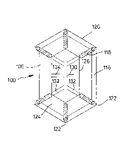
Figure 1 illustrates a first embodiment of a surgical implant,
which is designated by reference numeral 100, as well as a
process of manufacturing the implant 100.
As shown in the explosion view of Figure 1(a), the implant 100
is made of three parts. One part is a blank 102 consisting of
a mesh-like material, in the embodiment a knitted, undyed mon-
ofilament polypropylene mesh (PROLENETM Polypropylene Mesh of
Ethicon; non-absorbable) comprising a filament thickness of 89
pm. The blank 102 defines a basic structure 104 of the implant
100 plus six flaps 106.
On top of the blank 102, Figure 1(a) shows a marking layer 108
composed of a dyed (violet) film of poly-p-dioxanone (PDS)
having a thickness of 50 pm (absorbable) and comprising a to-
tal of eight openings 110. The openings 110 can be punched or
cut, e.g. by laser-cutting.
Date Recue/Date Received 2020-06-19
- 12 -
The third part visible in Figure 1(a) is the blank of an anti-
adhesive film 112. In the embodiment, the anti-adhesive film
112 is a MONOCRYL film (undyed) of 20 pm thickness. MONOCRYL
material (Ethicon) is an absorbable copolymer of glycolide and
c-caprolactone and has anti-adhesive properties.
The three layers 102, 108 and 112 are placed on top of each
other and laminated by heat. In that step, the poly-p-
dioxanone of the layer 108 melts or gets very soft and pene-
trates through the pores of the blank 102 so that it well at-
taches to the blank 102 and additionally glues the anti-
adhesive film 112 to the blank 102 so that part of the area of
the anti-adhesive film 112 adheres to the blank 102.
Afterwards, as shown in Figure 1(b), the flaps 106 are folded
about respective fold lines 114, which run through the edge
areas of the anti-adhesive film 112 so that zones 116 of the
anti-adhesive film 112 are formed which are folded as well but
do not completely cover the flaps 106.
Adjacent flaps 106 overlap somewhat in their common edge
zones, so that overlap areas 118 are provided. In the overlap
areas 118, the material of adjacent flaps 106 is connected to
each other, in the embodiment fused by ultrasonic welding in a
strip-like shape.
Figure 1(c) illustrates the finished implant 100 in a top plan
view. The fold lines 114 define an outer periphery 120 of the
basic structure 104. In the embodiment, the basic structure
104, i.e. the finished implant 100, has a hexagonal shape in-
cluding six corners 122. Six pockets 124 are formed by the
flaps 106 and the opposing material of the basic structure
104. These pockets are accessible via edges 126 defined by
part of the outer edge of the original blank 102.
Date Recue/Date Received 2020-06-19
- 13 -
The marking layer 108 provides a center marking 130, six di-
rectional indicators 132 pointing to and extending up to the
corners 122, and additionally a middle line indicator 134
which also extends up to the outer periphery 120 of the im-
plant 100. In Figure 1(c), the overlap areas 118 are hatched,
but since the mesh material of the blank 102 is translucent,
the directional indicators 132 and also the middle line indi-
cator 134 are well visible through the flaps 106 when the im-
plant 100 is viewed from its top side displayed in Figure
1(c).
Figure 1(d) is an isometric view of the implant 100 from the
top.
The surgical implant 100 is flexible and comprises a mesh-like
areal structure. The center marking 130 and the directional
indicators 132 permit a certain assessment of the position and
orientation of the implant 100, even if its outer periphery
120 is not or not completely visible. In the overlap areas
118, the material has double thickness, which provides a tac-
tile response, thus facilitating the surgical procedure. More-
over, the hexagonal shape of the implant 100 with six well-
defined corners 122 (which nevertheless could be somewhat
rounded in an atraumatic manner) additionally improves the
handling properties of the implant 100.
Since material of the marking layer 108 is also present in the
area of the fold lines 114, the outer periphery 120 of the im-
plant 100 is also well visible (unless hidden by bodily tis-
sue).
Preferably, the implant 100 is attached to bodily tissue by
introducing a stapling instrument into the pockets 124 and ex-
pelling the staples so that they penetrate just the flaps 106,
Date Recue/Date Received 2020-06-19
- 14 -
pointing away form the basic structure 104. This procedure can
be performed in a well-defined way, e.g. one staple can be
placed in the area of each corner 122 so that it penetrates
the material of the flaps 106 in the overlap areas 118, which
are stronger because of double-thickness.
A second embodiment of the surgical implant, designated by
reference numeral 200, is illustrated in Figure 2. The implant
200 is similar to implant 100. For that reason, the parts and
components of the implant 200 are not explained again in de-
tail. In the following, only the differences between the im-
plants 100 and 200 are pointed out. In Figure 2, the respec-
tive associated reference numerals from Figure 1 have been in-
creased by 100.
Blank 202 is cut from a knitted partially absorbable mesh ma-
terial made from undyed monofilaments of polypropylene (89 pm
thick; PROLENE Polypropylene) and dyed (violet) monofilaments
of poly-p-dioxanone (81 pm thick; PDS).
In implant 200, a center marking 230 and directional indica-
tors 232 are not provided via a film-like marking layer, but
by threads 208 of absorbable violet poly-p-dioxanone monofila-
ments (109 pm thick; PDS), which are sewn to blank 202 in or-
der to form the center marking 230, six directional indicators
232, a middle line indicator 234 as well as a hexagonal line
along the outer periphery 220 of the implant 200, see Figure
2(a).
The flexible blank 202 and an anti-adhesive, absorbable layer
212 (see Figure 2(b)), in the embodiment a 20 pm thick undyed
Monocryl film, are connected partially in the area of the sewn
marking threads 208 of PDS and in zones 216, see Figure 2(c).
To this end, the assembly is heated in pre-selected areas
(hatched in Figure 2(c)) so that the PDS material of the blank
Date Recue/Date Received 2020-06-19
- 15 -
202 and the marking threads 208 melts in these areas and acts
as a melt-glue, similar to the first embodiment. In overlap
areas 218, the folded flaps 206 are fused by ultrasonic weld-
ing in a strip-like shape, see Figure 2(d). Figures 2(e) and
2(f) display the finished implant 200.
A third embodiment of the surgical implant, designated by ref-
erence numeral 300, is illustrated in Figure 3. The implant
300 is similar to implant 200. Again, the respective associat-
ed reference numerals have been increased by 100. In implant
300, the material for blank 302 and sewn marking threads 308,
respectively, is the same as in implant 200.
In contrast to implant 200, anti-adhesive film 312 (again of
undyed MONOCRYL film) is only 10 pm thick and is connected
across its entire surface to blank 302. To achieve the latter,
during the lamination process the heat is controlled to suffi-
ciently soften or melt the PDS filaments in the blank 302 op-
posing the anti-adhesive film 312 and not just in the marking
threads 308. In the overlap areas 318, the folded flaps 306
are fused by ultrasonic welding in a strip-like shape, as in
the other embodiments.
Figure 3(a) shows the blank 302 with the marking threads 308,
Figure 3(b) additionally the anti-adhesive film 312, Figure
3(c) the partially finished implant 300, and Figures 3(d) and
3(e) show the finished implant 300.
Figure 4 illustrates a fourth embodiment of the surgical im-
plant, designated by reference numeral 400. Because of the
similarity to the other embodiments, the respective associated
reference numerals again have been increased by 100.
Blank 402 is made from a knitted mesh of 89 pm thick polypro-
pylene monofilaments (PROLENE Polypropylene; dyed and undyed),
Date Recue/Date Received 2020-06-19
- 16 -
which includes in the central area a marking guide 408 sewn
with dyed polypropylene monofilaments (89 pm thick; PROLENE
Polypropylene), see Figure 4(a).
The flexible blank 402 and an anti-adhesive, resorbable layer
412 of oxidized regenerated cellulose (ORC; undyed) are con-
nected by using an intermediate layer 411 made of an undyed
PDS film of 20 pm thickness and an additional layer 413 made
of an undyed, absorbable PDS film of 5 pm thickness as a hot-
melt adhesive, see Figure 4(b). In the overlap areas 418, the
folded flaps 406 are fused by ultrasonic welding in a strip-
like shape, as in the other embodiments. Figure 4(c) shows the
partially finished implant 400, and Figures 4(d) and 4(e) show
the finished implant 400.
Figure 5 illustrates various shapes of overlap areas 518
(hatched), in which adjacent flaps 506 of a surgical implant
500 are connected to each other. By designing the size and
shape of the overlap areas 518, the properties of pockets 524
(in particular in terms of flexibility) can be influenced.
Usually, the shape will be the same for all overlap areas in a
given implant, like in the surgical implants 100, 200, 300 and
400, but assemblies like that in Figure 5 are conceivable as
well, in particular when symmetric with respect to axes of
symmetry.
In different embodiments of the surgical implant, adjacent
flaps are not connected in the overlap areas or do not overlap
at all.
Figure 6 displays some schematic representations of basic
structure shapes and center markings and directional indica-
tors for embodiments of the surgical implant.
Date Recue/Date Received 2020-06-19
- 17 -
In Figure 6(a), a preferred orientation of polygonal implants
with three to six corners with respect to the anatomical envi-
ronment is indicated.
Figure 6(b) shows examples of equilateral polygons, in which
the center marking 630 is point-like and indicates the center
of gravity of the respective implant. The directional indica-
tors 632 extend up to the corners of the implant. Additional
marking lines 636 are provided along the periphery of the im-
plant.
The implants in the examples of Figure 6(c) have a hexagonal
shape like the implants 100, 200, 300 and 400. In two cases,
marking lines 636 are included along the periphery of the im-
plant. In the other cases, there are no such marking lines. In
all cases, the directional indicators 632 extend up to the
corners. In one case, the corners are additionally marked by
extended dots 638. However, the directional indicators will be
helpful even if they do not reach the corners. In some cases,
additional midline indicators 634 are provided. It is evident
from Figure 1(c) that the center area of each implant is easi-
ly assessable, even if a center marking 630 is not point-like.
Many other examples for the arrangement of the marking lines
are conceivable as well.
The embodiments described above illustrate the general concept
of the surgical implant according to the invention, when de-
signed as a Ventral Hernia Device (VHD) in preferred variants.
In summary:
The Ventral Hernia Device (VHD) is a pocket-shaped device with
tissue-separating properties for the reinforcement and bridg-
ing of the abdominal wall in ventral, incisional and larger
umbilical hernia repair, using an open intra-peritoneal onlay
mesh technique.
Date Recue/Date Received 2020-06-19
- 18 -
VHD comprises a polygonal flexible basic structure with a cen-
tral area (repair layer) and peripheral flaps (fixation lay-
er). The flaps form pockets, but do not extend up to the geo-
metric center of the fixation layer so that the fixation layer
has a central opening and the pockets form a kind of pouch
which is easily accessible via this central opening. Flap
folding and fusion at overlap areas leads to a preferably
strip-like doubling of the fixation layer in such a manner
that the strips are oriented to the corners; the areas of dou-
bling may cover between 1% and 50% of the fixation layer area,
preferably 1% to 20%. The fused strips allow for a better tac-
tile control and guidance to the corners, which leads to an
improved intra-operative handling of the implant.
The central opening in the fixation layer enables entry of a
finger or surgical instrument such as a stapler into the space
created between the visceral and parietal side, i.e. into the
pockets. The pocket formation by folding of only one flexible
blank enables a fixation at the outermost position of the pe-
riphery and corners of the implant, which results in a flatly
spread out implant. Non-fixated edge material (as in the case
of products with supporting rings or seams to connect differ-
ent layers) is avoided so that bulging or folding of non-
fixated edge material usually does not occur.
On the visceral side (repair layer), the implant is covered
with an anti-adhesive, absorbable layer, which is fixated on
the basic structure from one side and also covers the edges at
the periphery of the implant where it extends from the viscer-
al side to the parietal side, which leads to a partially cov-
ered fixation layer. Preferably, less than 50% of the fixation
layer is covered with the absorbable layer, which allows very
good tissue ingrowths. Moreover, such an extended absorbable
Date Recue/Date Received 2020-06-19
- 19 -
layer provides additional edge protection regarding adhesion
formation.
The surgical implant having mesh pockets can be used in the
so-called open-IPOM technique for the repair of ventral herni-
as. For the fixation of the mesh pockets, it has been found
that it is advantageous to start the fixation of the mesh
pockets in the axis of cranial/caudal, then to fix the other
vertices, and then to complete the fixation according to the
usual known techniques. In this procedure, starting with the
fixation in the axis of cranial/caudal, it was observed that
the tendency for forming folds in the implant, which is caused
by uneven fixation of the mesh pockets on the relatively soft
abdominal wall, is clearly reduced, and the implants are inte-
grated significantly better (tissue ingrowths) in the ab-
dominal wall.
A marking guide (center marking and directional indicators) at
the repair layer, which is linked to the outer shape of the
implant and indicates the implant center and the position of
the corners, helps the surgeon to know the actual device posi-
tion and orientation without additional lifting of bodily tis-
sue or implant manipulation and enables a controlled fixation
with evenly placed staples or clips. This also offers to the
surgeon the possibility for a standardized fixation approach
(e.g., the initial fixation points are predefined and can be
found intuitively by following the marking guide). The shape
of the mesh pockets and a better visualization of the mesh
edges/corners improve the intra-operative handling to a con-
siderable extent.
The marking guide (center marking and directional indicators)
is also very useful in areal surgical implants (in particular
mesh-like structures) without pockets.
Date Recue/Date Received 2020-06-19
- 20 -
Generally, the outer contour or shape of implant meshes ac-
cording to the prior art does not provide reference to the
surgeon to consider direction-dependent properties of the re-
pair mesh, such as achieving a certain coverage or overlap of
the defect, the stretch properties of the mesh, the orienta-
tion during fixation, a guide to the first fixation points, or
a clear identification of the area where to fixate. On the
other hand, a correct placement in terms of orientation and
centering of the implant over the defect is crucial for a suc-
cessful repair. And wrinkling and folding of the implant dur-
ing fixation lead to wrinkle cavities (and in consequence to
seroma formation), which results in a poor integration of the
implant into the abdominal wall.
Such problems can be avoided when the mesh-like basic struc-
ture of the implant has a polygonal shape and when the implant
comprises a marking guide, i.e. a center marking and direc-
tional indicators pointing from the center marking to the cor-
ners. The link between shape and marking guide helps the sur-
geon to know the current position and orientation of the im-
plant just by looking in the center of the defect without ad-
ditional lifting of the tissue or implant manipulation. The
implant can be positioned in a better way, compared to prior
art, for example when a corner tip of the implant is put under
osseous structures like the sternum.
Starting the fixation in the vertices (corner areas) of the
implant by following the continuous marking guide with the
fixation device from the implant center to the vertices leads
to a flat spreading of the implant with evenly placed staples.
This offers to the surgeon the possibility for a structured
and standardized fixation approach, e.g. in that the first
fixation points are predefined and can be found intuitively
following the marking guide. A hexagonal shape of the implant
is particularly advantageous.
Date Recue/Date Received 2020-06-19
- 21 -
For example, during implantation for repair of an incisional
hernia with a tetragonal mesh (square or rectangular), a mark-
ing guide pointing to the vertices or corners "1" and "3", as
shown in Figure 6(a), can indicate the cranial-caudal direc-
tion. For a hexagonal shape, the corners "1" and "4" indicate
the cranial-caudal direction. The cranial-to-caudal orienta-
tion can be displayed in a pentagonal mesh shape through the
corner point "1" and the center of the basis between corner
points 3 and 4.
Date Recue/Date Received 2020-06-19