Note: Descriptions are shown in the official language in which they were submitted.
CA 02905598 2015-09-11
WO 2014/138995
PCT/CA2014/050241
SYSTEM AND METHOD FOR LOW X-RAY DOSE BREAST DENSITY EVALUATION
CROSS-REFERENCE TO RELATED APPLICATIONS
[0001] This
application claims the benefit of U.S. Provisional Patent Application
Serial No. 61/783,786, filed on March 14, 2013, and entitled "SYSTEM AND
METHOD
FOR LOW X-RAY DOSE BREAST DENSITY EVALUATION."
BACKGROUND OF THE INVENTION
[0002] The
field of the invention is systems and methods for x-ray
mammography. More particularly, the invention relates to systems and methods
for
estimating breast density from an x-ray mammogram.
[0003]
Mammographic density, hereafter referred to as breast density, describes
the relative amount of fibroglandular tissue in the breast compared to the
total amount
of breast tissue, which is mostly composed of fibroglandular tissue and fat or
adipose
tissue. Mammographic breast density has been identified as an independent risk
factor
for breast cancer, and studies have identified a 4-5 fold increase in risk for
developing
breast cancer in women with dense breast tissue versus women with less dense
breast
tissue (i.e., breast tissue with more fat). The addition of breast density
quantification to
mammographic examination has the potential to greatly improve the accuracy of
breast
cancer risk assessment, especially for those without hereditary or familial
risk factors.
The inclusion of accurate breast density measurements can also be potentially
helpful
for women by suggesting that other imaging modalities such as magnetic
resonance
imaging ("MRI") or ultrasound be used for initial screening instead of
mammography
because mammography's accuracy is known to be reduced in women with very dense
breasts. Because the reporting of breast density is required in some
jurisdictions, there
is a desire to provide an accurate and reproducible quantitative density
measurement
method that is simple to implement on conventional digital mammography
machines.
To date, nearly all work in measuring breast density has used film-screen
mammograms.
[0004]
Quantitative methods, such as computer-assisted planimetry, can be very
reproducible and are the best validated method for association with breast
cancer risk,
but they generally require at least some manual intervention and, thus, are
time-
consuming to use. With the increased utilization of digital mammography,
automated
computerized measurement of breast density is now becoming widely available,
but has
-1-
CA 02905598 2015-09-11
WO 2014/138995
PCT/CA2014/050241
not yet been well validated. One major advantage of newer software methods
applied
to digital mammograms is that pixel signal levels can be measured objectively,
yielding
information about the composition of the breast tissue (e.g., volumetric
breast density).
The use of digital mammograms will allow automation and reduce variability.
Automated breast density measurements that are both reproducible and
demonstrated
to be accurate could be an important addition to breast cancer risk
assessment.
[0005] The
current gold standard quantitative method of measuring breast
density is the 20 Cumulus program developed by Dr. Martin Yaffe from Toronto.
This
method is a computer-assisted thresholding technique similar to planimetry.
First, a
film-screen mammogram is digitized. The pixels representing the total breast
area and
those representing dense breast area are then defined by a radiologist through
an
interactive program. The 2D Cumulus program yields the percent area density of
a
breast. There are, however, several limitations of the 20 Cumulus program.
[0006] The 2D
Cumulus program uses binary information and a two-dimensional
image of the breast. The binary nature of the procedure means that each pixel
is
counted as representing either one hundred percent breast tissue or one
hundred
percent fat, with no ability to represent a mixture of the two tissue types,
or to account
for the height of the column of tissue above the pixel. Furthermore, simple
thresholding
methods, such as 2D Cumulus, may cause dense tissue not to be included in the
thin
periphery of the breast, or may treat fatty tissue as being dense in regions
where the
compressed breast is thicker than average. Use of the 2D Cumulus program is
also
cumbersome, as it requires a radiologist or a trained scientist to visually
select the
division between fat and breast tissue, a process that can take as long as one
minute per
image. Because of these limitations, 2D Cumulus has only been used in the
research
setting and not in clinical practice.
[0007] For a
risk model to be useful in clinical practice, breast density
measurement should be automated, reproducible, accurate, precise, and,
ideally,
measured on a continuous scale. This eliminates observer bias and provides
maximal
discrimination.
[0008] The work
of Shepherd, et al., (as described in U.S. Patents Nos. 6,516,045;
6,654,445; and 7,873,198) calculates mass density, which is related to, but
not
equivalent to mammographic density. The process described by Shepherd requires
that
calibration materials of one-hundred percent fat and one-hundred percent
glandular
-2-
CA 02905598 2015-09-11
WO 2014/138995
PCT/CA2014/050241
materials be placed on the breast support, and also requires that radio-opaque
markers
be present in each image to enable thickness measurements.
[0009] Standard
compression methods for mammography use a movable, semi-
rigid clear plastic compression paddle. The breast is placed on a bottom
breast platform
that is flat, and the paddle is then lowered onto the breast, usually while
the
technologist is holding the breast in place to ensure proper tissue coverage
in the image
receptor's field of view. A significant patient concern in mammography is the
discomfort the patient feels when the breast is compressed with sufficient
force to
spread out the breast tissues. The reasons for using such high compression
include: (1)
to make the breast thinner and thereby reduce patient radiation exposure; (2)
to
improve image quality by reducing the amount of scattered radiation; (3) to
make the
breast more uniform in thickness in the direction of the x-ray flux, leading
to a more
uniform exposure over the entire breast image; (4) to immobilize the breast
during the
x-ray exposure, thereby reducing image blurring; and (5) to bring breast
tissues out
from the chest wall into the exposure area and thus image more tissue. A
problem with
the calculation of volumetric breast density is the requirement that thickness
must be
known accurately in order to relate the attenuation to a given mammographic
density.
For instance, two millimeters of error in the path length on the breast could
result in an
error of five percent or more in the breast density measurement.
[0010] On most
mammography machines, the paddles and readout systems are
not designed to produce uniform compression. All paddles show some deflection
when
compressed on a breast, often as much as 3 mm in the centre. They also deflect
from
front-to-back due to flexion of the mechanical components, which varies with
the
compression force. Some compression paddles also tilt, further introducing the
possibility for variations in compressed breast thickness.
[0011] The
electronic readout of thickness can be incorrectly calibrated, or the
error may change with compression force applied. Even for those mammographic
units
that report thickness compensated for compression force, there can be errors
as large
as two millimeters. These errors can be much greater if the breast is not
centered on
the breast support plate. Calibrating for variations in thickness is time
consuming, and
not always accurate. As noted above, accurate measurement of compressed breast
thickness is an important factor in determining breast density; however, the
measurement of thickness provided by commercial mammography systems can differ
-3-
CA 02905598 2015-09-11
WO 2014/138995
PCT/CA2014/050241
by as much as one centimeter from the actual thickness due to deflection of
the breast
compression plate and the inaccuracies in the readout system.
[0012]
Therefore, there remains a need to provide a system and method for
quantifying volumetric breast density that can more accurately determine the
thickness
of the breast, thereby addressing the drawbacks of currently available
methods.
SUMMARY OF THE INVENTION
[0013] The
present invention overcomes the aforementioned drawbacks by
providing a system and method for quantifying the proportion or density of a
tissue
using a lower level of compression force and a lower x-ray dose.
[0014] It is an
aspect of the invention to provide a compression assembly for
compressing a breast in use with a mammography system. The compression
assembly
includes a rigid plate and at least one spacer. The at least one spacer is
coupled to the
rigid plate and sized such that the spacer maintains the rigid plate in a
substantially
parallel alignment at a fixed distance from a breast support plate of an x-ray
mammography system, thereby providing a region for receiving and compressing a
breast at a substantially uniform thickness. The at least one spacer may be
composed of
a material having a known density and/or attenuation value.
[0015] It is
another aspect of the invention to provide a method for measuring a
density of a subject's breast with a mammography system. The method includes
compressing the subject's breast to a defined uniform thickness using a rigid
compression paddle that is parallel to a breast support plate and spaced apart
from the
breast support plate at a fixed distance by at least one spacer. Data is then
acquired
with the mammography system by exposing the compressed breast to x-rays that
traverse the rigid compression paddle before traversing the compressed breast
and
impinging upon an image plane of an x-ray detector. Attenuation values are
determined
from the acquired data at all points in the image plane and are then converted
to
mammographic density values. Breast volume and volumetric density content are
then
estimated by calculating the total volume of dense tissue by summing the
mammographic density values across the total volume of the breast, and
dividing the
total volume of dense tissue by the total volume of the breast.
[0016] The
foregoing and other aspects and advantages of the invention will
appear from the following description. In the description, reference is made
to the
-4-
CA 02905598 2015-09-11
WO 2014/138995
PCT/CA2014/050241
accompanying drawings which form a part hereof, and in which there is shown by
way
of illustration a preferred embodiment of the invention. Such embodiment does
not
necessarily represent the full scope of the invention, however, and reference
is made
therefore to the claims and herein for interpreting the scope of the
invention.
BRIEF DESCRIPTION OF THE DRAWINGS
[0017] FIG. 1 is an example of one configuration of a compression assembly
of
the present invention;
[0018] FIG. 2 is a front view of the compression assembly of FIG. 1;
[0019] FIG. 3 is an elevational view of the compression assembly of FIG. 1;
[0020] FIG. 4 is an illustration of different spacers that may form a part
of the
compression assembly of the present invention;
[0021] FIG. 5A is a mammography system incorporating one configuration of
the
compression assembly of the present invention;
[0022] FIG. 513 is another configuration of the compression assembly of the
present invention, which may form a part of a mammography system;
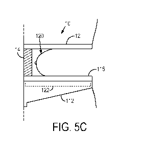
[0023] FIG. 5C is a schematic representation of a breast under compression
with
one configuration of the compression assembly of the present invention;
[0024] FIG. 6A is a configuration of the compression assembly of the
present
invention as used in a mammography system that implements a curved compression
paddle and a curved breast support;
[0025] FIG. 6B is another configuration of the compression assembly of the
present invention as used in a mammography system that implements a curved
compression paddle and a curved breast support;
[0026] FIG. 7 is a schematic representation of a breast under compression,
illustrating different x-ray beam paths through regions of uniform thickness,
but
different material compositions;
[0027] FIG. 8 is a block diagram of an example of a computer system that
can be
configured to implement some embodiments of the present invention;
[0028] FIG. 9 is a plot showing a comparison between volumetric breast
density
values computed from conventional mammography images and from low-dose images
in accordance with some embodiments of the present invention; and
[0029] FIG. 10 is a plot showing a comparison between two human readers
-5-
CA 02905598 2015-09-11
WO 2014/138995
PCT/CA2014/050241
estimating breast density.
DETAILED DESCRIPTION OF THE INVENTION
[0030] Accurate
breast density results are difficult to obtain, and the currently
existing, non-automated methods for measuring breast density are very
subjective. The
currently existing automated techniques also have their problems, including a
lack of
accuracy when estimating breast thickness, and tilt and bulging of the
compression
paddle, which decrease the accuracy of breast thickness estimation. In
addition, the
conventional method requires that the patient receive a radiation dose that is
equal to
that from a standard mammographic exposure. This requirement reduces the
likelihood that a density measurement would be made unless the patient was
receiving
a mammogram for another purpose.
[0031] The
present invention provides a system and method for measuring
volumetric breast density using a low dose of radiation. For instance, this
low-dose
image can be added to a standard mammographic screening protocol with less
than a
two percent increase in radiation dose imparted to the subject. If only a
density
measurement is performed, then the dose imparted to the subject is less than
two
percent of that imparted in a standard mammographic examination. The breast
density
measurement obtained with the present invention is more accurate than
measurements
that can be obtained with existing systems and methods. The present invention
also has
the added benefit that it can be readily implemented on a conventional digital
x-ray unit
with low cost.
[0032] The
present invention is capable of self calibration using one or more
objects having x-ray attenuation characteristics that are similar to those of
breast tissue.
These objects are used to establish the spacing between a compression paddle
and a
breast support, and can provide direct signal numbers representing those
density
values.
[0033] A
calibration and correction for breast support deflection can also be
added for different thickness breasts. This Feature can be achieved by
compressing
different size phantoms containing a homogeneous gel or liquid, and using the
resultant
image signal values to calculate the different path lengths through this
material from
that presented by a rigid flat phantom.
[0034] A breast
compression assembly for use with an x-ray mammography
-6-
CA 02905598 2015-09-11
WO 2014/138995
PCT/CA2014/050241
system that is capable of compressing the breast to an accurate and constant
thickness.
This compression assembly may be attached to any mammography system, and can
be
used to accurately measure volumetric breast density using a low-dose x-ray
exposure,
as will be described below in more detail.
[0035] With
reference now to FIGS. 1-3, the compression assembly 10 of the
present invention generally includes a paddle 12 and, optionally, one or more
spacers
14. The compression assembly 10 can be a standalone device that is configured
to be
coupled to a mammography system, or the compression assembly 10 can be a
device
that is configured to be coupled to a breast compression paddle that forms a
part of a
commercially available mammography system. In the latter configuration, the
compression assembly 10 of the present invention is configured to be coupled
to the
commercially available breast compression paddle using mechanical clips; self-
adhering
straps, such as hook-and-loop (VelcroTM) strips; or so on. As an example, the
compression assembly 10 can be configured to be coupled to the commercially
available
mammography system using the existing mounting hardware or mechanism available
for that mammography system. In this way, the compression assembly 10 can be
configured to be incorporated into pre-existing mammography systems without
the
need for specialized hardware.
[0036] The
paddle 12 is generally a flat and rigid plate that is configured to be in
parallel alignment with a breast support platform of a mammography system.
Preferably, the paddle 12 is rounded on all edges that make contact with the
patient to
improve patient comfort The paddle 12 may be composed of any suitable
materials,
but may preferably be composed of a transparent material to allow visual
confirmation
of the location of the patient's breast during mammographic examination. By
way of
example, the paddle 12 can be composed of clear poly(methyl methacrylate)
("PMMA").
Although one or more spacers 14 can be used to maintain the paddle 12 in
parallel
alignment with a breast support platform, other approaches for maintaining
this
parallelism can also be used. For instance, the mounting for the breast
support plate
can be modified such that it does not deflect more than one millimeter. As
another
example, parallel linkages or a straight line system could be used when
coupling the
paddle 12 and/or the breast support plate to the mammography system.
[0037] The
paddle 12 is preferably about one centimeter thick, or more. This
thickness is chosen such that the distortion of the paddle 12, the entrance
exposure, and
-7-
CA 02905598 2015-09-11
WO 2014/138995
PCT/CA2014/050241
the dose imparted to the patient are all reduced relative to their values that
would occur
for the native compression paddles used in conventional mammography systems.
For
example, the paddle 12 is thick enough to reduce the entrance exposure to the
breast to
less than ten percent of the normal entrance exposure received in a regular
examination. An example of a paddle 12 configuration that achieves this result
is a
paddle 12 composed of 2.54 centimeter (one inch) thick PMMA, which attenuates
the
incident x-rays by about ninety percent (for x-rays whose effective energy is
20 keV).
[0038] The
thickness of the paddle 12 is also chosen to make the paddle 12 very
rigid, unlike the 1.5-3 mm polycarbonate or PMMA compression paddles used in
current mammography systems, which bulge and deflect during the compression of
a
patient's breast. In some configurations, the paddle 12 may be thinner than
one
centimeter. In these configurations, an increased amount of x-ray filtration
in the x-ray
tube port can be used to decrease the effective dose imparted to the patient's
breast.
[0039] The
thicker paddle 12 hardens the x-ray beam provided by the
mammography system, and thus absorbs most of the lower energy photons. In
addition,
the thicker paddle 12 absorbs a significant amount of the primary radiation.
The
combination of the attenuation provided by the thick paddle 12 and the beam
hardening
enables the use of a similar time of exposure as used for conventional
mammography
while reducing the dose to less than two percent of the dose imparted in a
conventional
mammography examination. When added to a mammography study, total dose will
thus be below the MQSA limit for all systems. Using the compression assembly
10 of the
present invention, imaging may be carried out with increased kV relative to
that used
for conventional mammography. This increased kV further reduces the effective
dose
imparted to the breast.
[0040] In one
configuration, the paddle 12 is configured to be coupled to an
existing compression paddle of a mammography system. In another configuration,
however, the paddle 12 is configured to replace the compression paddle on an
existing
mammography system in its entirety.
[0041]
Preferably, the compression assembly 10 also includes one or more
spacers 14. The spacers 14 can be coupled to the paddle 12 and positioned
between the
paddle 12 and the breast support plate of a mammography system. For example,
the
spacers 14 can be placed in or near the corners of the paddle 12 that are
located away
from the chest wall of the patient. The spacers 14 are sized to have a known
thickness.
-8-
CA 02905598 2015-09-11
WO 2014/138995
PCT/CA2014/050241
By way of example, the spacers 14 can be constructed to have dimensions that
differ by
an integer number of centimeters, such as 2cm x 3 C17/ x 4 cm , 4 em x 5 cm x
6c717 , or
3 cm x 5 CM X7 cm . The spacers 14 can be embedded with radioopaque or
radiolucent
materials so that their height can be identified on the images. For instance,
the spacers
14 can be embedded with ball bearings that are arranged in a certain pattern
that
identifies the known height of the spacer 14. As illustrated in FIG. 4,
however, the
spacers 14 need not be rectangular, but can generally be constructed as having
a cross-
section of any suitable polygonal or otherwise arbitrary shape, including a
circular
cross-section, so long as the face of the spacer 14 that makes contact with
the breast
support plate 116 is parallel to the lower face of the paddle 12.
[0042] The
spacers 14 are composed of a material with an attenuation equivalent
to that of a breast tissue of known density. It is not necessary that the
spacers 14 be
composed of a material that mimics pure fat or pure fibroglandular tissue.
When more
than one spacer 14 is used, it may be preferable to have the different spacers
14 be
composed of different materials. By having more than one material, the
accuracy may
be improved. A reference material, however, is not required because the
spacers 14
provide an exact measurement of how thick the breast is under compression and
of
attenuation values.
[0043]
Referring now to FIG. SA, the compression assembly 10 of the present
invention can be implemented in an x-ray imaging system 100, such as a
mammography
system. The x-ray imaging system includes an arm 102 that is rotatably mounted
to a
base support 104 through a rotatable coupling 106. An x-ray source assembly
108 is
coupled to a first end 110 of the arm 102, and an x-ray detector assembly 112
is coupled
proximate an opposing end 114. The x-ray source assembly 108 extends
substantially
perpendicular to the arm 102 and is directed toward the x-ray detector
assembly 112.
The x-ray detector assembly 112 also extends from the arm 102 such that the x-
ray
detector assembly 112 receives x-ray radiation produced by the x-ray source
assembly
108, transmitted through the breast, and incident on the x-ray detector
assembly 112. A
breast support plate 116, and a breast compression plate 118, are positioned
between
the x-ray source assembly 108 and the x-ray detector assembly 112. In this
configuration, the compression assembly 10 is coupled to the existing breast
compression plate 118 of the x-ray imaging system 100. The compression
assembly 10,
however, can also replace the breast compression plate 118, as illustrated in
FIGS. 5B
-9-
CA 02905598 2015-09-11
WO 2014/138995
PCT/CA2014/050241
and SC.
[0044] As
discussed above, when the paddle 12 of the compression assembly 10
is sized to have a thickness less than one centimeter, additional filtering is
preferably
added to the x-ray source assembly 108 such that the mean glandular dose
imparted to
the breast is decreased.
[0045]
Referring now to FIG. SC, the compression assembly 10 of the present
invention is shown in use during a mammography session. The patient's breast
120 is
positioned between the compression paddle 12 of the compression assembly 10
and the
breast support plate 116 of the mammography system 100. One or more spacers 14
are
also positioned between the compression paddle 12 and the breast support plate
116
such that, under compression, the breast 120 is maintained at a uniform and
known
thickness.
[0046] In
operation, x-rays emitted by the x-ray source assembly 108 are
attenuated by the compression assembly 10, the patient's breast 120, and the
spacers
14 that form a part of the compression assembly 10. The attenuated x-rays are
detected
by an x-ray detector 122 in the x-ray detector assembly 112 as signals that
represent
transmission values, and can be converted to attenuation values at each pixel.
For
example, the transmission values can be converted to attenuation values by
dividing the
signal at each pixel by the average signal in a region outside of the breast,
that is, a
region in which primary radiation experiences little to no attenuation. The x-
ray
detector 122 may include any suitable x-ray detector, including an imaging
plate, such
as an accumulative phosphor sheet, or a flat panel detector, in which an array
of x-ray
detecting elements are arranged on an x-ray detection surface.
[0047] Because
the compression paddle 12 is applied on the top of the breast
120, but supported by the spacers 14 with known dimensions, a uniform and
known
thickness of the breast 120 under compression is achieved. As an example, if
the
patient's breast thickness was measured to be 4.5 cm during a regular full-
field digital
mammography ("FFDM"), then the compression paddle 12 can be positioned on top
of
the breast 120 and a set of 5.0 cm spacers 14 coupled underneath the
compression
paddle 12, or placed on top of the breast support plate 116. The compression
paddle 12
is used to lightly compress the breast 120 against the breast support plate
116. For
instance, the compression pressure will typically be below that used for
current
mammographic examinations.
-10-
CA 02905598 2015-09-11
WO 2014/138995
PCT/CA2014/050241
[0048] When the
compression paddle 12 is lowered to apply force to the breast
120, the compression paddle 12 becomes parallel to the breast support plate
116 and
spaced apart from the breast support plate 116 by the height of the spacers
14, which in
the example discussed here is 5.0 cm. With this geometry, the imaged breast
120 is
slightly compressed to a constant thickness of 5.0 cm. The breast 120 can then
be
imaged using the same target, filter, kV, and closest mAs technique as used
for the FFDM
method. The volumetric breast density VBD of the breast 120 can then be
accurately
measured with calibrated data discussed below using the same algorithms as
used on
the high dose image. Breasts of other thicknesses will similarly be compressed
using
spacers 14 that are the next higher thickness than the thickness to which the
breasts
were compressed during the clinical mammogram.
[0049] In some
configurations, such as those illustrated in FIGS. 6A and 6B, the
compression assembly 10 can be implemented in a mammography system that makes
use of a compression paddle 12 and breast support 116 having curved surfaces.
In
these configurations, the contact faces of the spacers 14, which are the faces
of the
spacers 14 that make contract with the compression paddle 12 and the breast
support
116, can be suitably curved so as to provide uniform contact with the
compression
paddle 12 and breast support 116. Accordingly, the contact faces of the
spacers 14 may
be suitably convex or concave. In this configuration, the spacers 14 are able
to maintain
the parallel relationship between the compression paddle 12 and breast support
116
such that the surfaces of the compression paddle 12 and breast support 116 are
parallel
curved surfaces.
[0050] A
digital mammography system can be characterized by measuring the
effective attenuation of fat and glandular tissue for given tissue path
lengths. The
relationship of attenuation to tissue composition can be determined by
calibrating the
system using materials of known attenuation and known thicknesses. This can be
achieved by using spacers 14 of known material composition and thicknesses, as
illustrated in the example shown in FIG. 7. In this example, x-rays travelling
along path
L1 will pass through substantially only air, x-rays travelling along path L2
will pass
through substantially only fat tissues in region 150 of the breast 120, x-rays
travelling
along path L3 will pass through an approximately 50-50 mixture of fat and
fibroglandular tissue, and x-ray travelling along path L4 will pass through
substantially
only fibroglandular tissues in region 152 of the breast 120. By using the
spacers 14 of in
-11-
CA 02905598 2015-09-11
WO 2014/138995
PCT/CA2014/050241
the compression assembly 10, each of the x-ray paths will traverse a similar,
uniform
path length, Ax.
[0051] To aid
the calibration of the system, four spacers 14a, 14b, 14c, and 14d
can be used in this example, each being composed of a different material
having a
known attenuation characteristic. For instance, spacer 14a can be composed of
a
material having attenuation characteristics similar to fat tissue, spacer 14b
can be
composed of a material having attenuation characteristics similar to
fibroglandular
tissue, spacer 14c can be composed of a material having attenuation
characteristics
similar to a mixture of fat and fibroglandular tissue, and spacer 14d can be
composed of
a material having attenuation characteristics similar to air. As an example,
spacer 14c
can be composed of a material having attenuation characteristics similar to a
50-50
mixture of fat and fibroglandular tissue.
[0052] A
typical mammography system acquires a projection image of the
compressed breast. Typically, two views are taken of each breast, one from
above
(cranial-caudal, or "CC") and one from the side (mediolateral-oblique, or
"MLO"). In
each view, the breast is compressed to reduce patient motion and scatter,
separate
overlapping structures in the breast, make the thickness of the imaged breast
more
uniform, and provide more uniform x-ray exposure. Using the compression
assembly
of the present invention, an additional low-dose image is added for each
breast. This
low-dose image can be obtained in the CC view or MLO view. Two low-dose
images, one
in each of the CC view and the MLO view, can also be obtained. The positioning
of the
breast would be the same as for the full-dose mode except that the compression
assembly 10 would be inserted before lightly compressing the breast for the
low-dose
image. The low-dose exposure is preferably taken at a fixed x-ray technique,
but could
also utilize a technique in which automatic exposure control is employed
without
impairing accuracy.
[0053] The
linearity of pixel values reported by modern FFDM systems in
arbitrary digital units ("ADU") per mAs is very stable. An mAs-normalized
arbitrary
digital unit ("NADU") is defined here to be the mean pixel value of a region-
of-interest
("ROI") normalized by the mAs for the unprocessed ("RAW") digital image. When
only
air is imaged the NADU value is designated as NADUõ,,,.. For a given target,
filter, kV,
and thickness combination, attenuation, a, can be defined as,
-12-
CA 02905598 2015-09-11
WO 2014/138995
PCT/CA2014/050241
NADU
a = (1).
NADU õõ
[0054] Logarithmic attenuation can thus be defined as,
NADU \
LA= log (a) = logIO _______________________________________________ (2).
NADU . )
,
[0055] In logarithmic space, the logarithmic attenuation of different
breast
density tissue is linear with respect to percentage of fibroglandular tissue
("PFG"),
which is a measure of the proportion of fibroglandular tissue to the sum of
fibroglandular and fatty tissue. That is to say, fatty tissue has a PFG of
zero percent, an
equal admixture of fatty tissue and fibroglandular tissue has a PFG of fifty
percent, and
fibroglandular tissue has a PFG of one hundred percent. A given FFDM system
can be
calibrated with potential combinations of target, filter, kV, and expected
thicknesses.
For each of these combinations, three PFG values (0, 50, 100) are measured
with a block
phantom, or the like.
[0056] An example of a calibration procedure includes obtaining
measurements
from a calibration phantom, such as a phantom that contains distinct regions
of having
different, known PFG values. Alternatively, the function of the calibration
phantom can
be replicated using appropriately configured spacers 14 in the compression
assembly
10. For instance, the compression assembly 10 can be designed to include
multiple
spacers 14 having different, known PFG values. As an example, three different
spacers
14 can be used, in which the spacers 14 would be designed to have PEG values
of 0, 50,
and 100. When the spacers 14 are present in a given image, they can be used as
consistent calibration points. As an added advantage, due to the significant
amount of
beam hardening from the compression assembly 10, or added filtration, only a
few
calibration points are necessary to characterize the thickness-attenuation
relationship.
[0057] Once the calibration is obtained, the effective attenuation of a
breast can
also be calculated from a single, low-dose image. With accurate knowledge of
the breast
thickness using the compression assembly 10 of the present invention, the
amount and
proportion of dense breast tissue, represented as volumetric breast density,
can be
determined from the low-dose image. The system of the present invention can
thus be
used to produce a report indicating breast density. This report can form a
part of the
patient's clinical record. By way of example, this report could be a DICOM
structured
-13-
CA 02905598 2015-09-11
WO 2014/138995
PCT/CA2014/050241
report, or an HL7 report. In addition to calculating and reporting breast
density, other
quantitative metrics can also be determined and reported. For instance, the
proportion
of fibroglandular materials that define the breast can be calculated based on
the
summation of the attenuation values at the known compressed thickness of the
breast.
Also, the area of the breast that is making contact with the compression
paddle 12 can
be analyzed and an appropriate correction for thickness changes in the breast
can be
applied to regions outside the contact area.
[0058]
Referring now to FIG. 8, a block diagram of an example computer system
800 that can be in communication with a mammography system, such as the
mammography system illustrated in FIG. SA, and configured or otherwise
programmed
to process medical images in accordance with embodiments of the invention
described
above. The image or images to be processed, which are preferably digital
images, are
provided to the computer system 800 by a mammography system, and are received
in
an image processing unit ("IPU") 802.
[0059] In some
embodiments, the IPU 802 can include one or more processing
units. As an example, the IPU 802 may include one or more of a digital signal
processor
("DSP") 804, a microprocessor unit ("MPU") 806, and a graphics processing unit
("GPU")
808. The DSP 804, MPU 806, and GPU 808 can be any suitable, commercially
available
processor unit. The IPU 802 also preferably includes an image acquisition unit
810 that
is configured to electronically receive an image to be processed. The DSP 804,
MPU 806,
GPU 808, and image acquisition unit 810 are all coupled to a communication bus
812.
As an example, the communication bus 812 can be a group of wires or a hardwire
used
for switching data between the peripherals or between any component in the IPU
802.
[0060] The DSP
804 can be configured to receive an image and processes the
image to yield a digital image. The MPU 806 and GPU 808 can be configured to
process
the image in conjunction with the DSP 804. As an example, the GPU 808 can
process
image graphics. Also as an example, the MPU 806 can be configured to control
operation
of components in the IPU 802 and can include instructions to perform
processing of the
image on the DSP 804.
[0061] In some
embodiments, the DSP 804 can be configured to process an image
received by the IPU 802 in accordance with the breast density estimation
algorithms
described herein. Thus, the DSP 804 can be configured to derive or otherwise
compute
breast density data, such as volumetric breast density measurements, from low-
dose x-
-14-
CA 02905598 2015-09-11
WO 2014/138995
PCT/CA2014/050241
ray images.
[0062] The IPU
802 preferably includes a communication port 814 in electronic
communication with other devices, which may include a storage device 816, a
display
818, and one or more input devices 820. Examples of an input device 820
include, but
are not limited to, a keyboard, a mouse, and a touch screen through which a
user can
provide an input.
[0063] The
storage device 816 is configured to store digital images, whether
those provided to the IPU 802 or those processed or enhanced images generated
by the
IPU 802. The display 818 is used to display images, such as images that may be
stored in
the storage device 816. Thus, in some embodiments, the storage device 816 and
the
display 818 can be used for displaying the digital image and for outputting
other
information.
[0064] The IPU
802 can also be in electronic communication with a network 822
to transmit and receive data, including images and reports using DICOM, XML,
or other
protocols. The communication port 814 can also be coupled to the IPU 802
through a
switched central resource, for example the communication bus 812.
[0065] The IPU
802 can also include a temporary storage 824 and a display
controller 826. As an example, the temporary storage 824 can store temporary
information. For instance, the temporary storage 824 can be a random access
memory.
[0066] Having
described systems and methods that implement the present
invention, generally, several non-limiting examples of the present invention
in use are
now provided.
Example 1: Measuring Breast Density
[0067] In this
example, the inventors demonstrate that breast density can be
accurately measured under lower dose conditions than conventional mammography
when implementing the systems and methods of the present invention.
[0068] In this
example, thirty healthy volunteers and two women with previous
surgery on a single breast were scanned using both a conventional, flexible
mammogram paddle and the rigid paddle system described above. The mean age of
the
healthy volunteers was 63 years (SO - 81 yrs).
Example 1 Materials and Methods
[0069] Each
volunteer was scanned using a conventional four view screening
mammogram using a flexible paddle. A technician arbitrarily selected the side
to be
-15-
CA 02905598 2015-09-11
WO 2014/138995
PCT/CA2014/050241
imaged. The thickness of the compressed breast was determined by a technician
from a
crania-caudal ("CC") view of screen mammogram and the appropriately sized
spacers
were selected for a follow on scan using the rigid paddle system described
above. The
spacers were chosen to be either the same as the noted thickness (if an
integer) or next
centimeter higher. A low-dose image was then acquired using the rigid paddle
system
described above. In this example, the rigid paddle was 2.5 centimeters thick
and
composed of poly(methyl methacrylate) ("PMMA"). Images were acquired using a
35
kVp tube voltage setting, a 10 mAs setting, and with a Rhodium-Rhodium target-
filter
setup or a Molybdenum-Rhodium target-fitter setup.
Example 1 Results
[0070] Images
were automatically analyzed using the Cumulus Volume
("Cumulus V") program developed by Martin Yaffe at the University of Toronto.
The
resultant density was plotted with the screening image density derived from
the
conventional mammography images on the bottom axis and the low-dose density on
the
other axis, as seen in FIG. 9.
[0071] Two
human readers ("NE" and "MY") analyzed area density and a
comparison of their results are illustrated in FIG. 10. The R-value for the
human readers
is 0.81 as compared to the 0.90 achieved using the low-dose technique
described herein,
demonstrating that the method of the present invention is at least as reliable
as using
trained human estimators of breast density.
[0072] The low-
dose volumetric density matches that obtained by using standard
dose screening mammograms to within a few percent. The R-value is
significantly
better with the automatic technique than having humans read and score the
images,
however.
[0073] The
present invention has been described in terms of one or more
preferred embodiments, and it should be appreciated that many equivalents,
alternatives, variations, and modifications, aside from those expressly
stated, are
possible and within the scope of the invention.
-16-