Note: Descriptions are shown in the official language in which they were submitted.
EXTRACORPOREAL LIFE SUPPORT SYSTEM AND METHODS OF USE
THEREOF
FIELD OF THE INVENTION
The present invention relates to the field of neonatal care. More
specifically,
the invention provides apparati and methods for the maintenance of homeostasis
in
to the pre-viable fetus outside of the womb.
BACKGROUND OF THE INVENTION
Several publications and patent documents are cited throughout the
specification in order to describe the state of the art to which this
invention pertains.
Full citations of these references can be found throughout the specification.
In cases of extreme prematurity, survival outside the womb is complicated by
inadequate organogenesis, including insufficient lung growth and maturation to
permit gas exchange. Furthermore, in the event of congenital anomalies
affecting the
growth and development of the lungs, such as congenital diaphragmatic hernia
and
other causes of pulmonary hypoplasia, insufficient pulmonary function may
limit
long-term survival. The development of an extracorporeal system to support
ongoing
fetal growth and development without the perturbations induced by postnatal
intensive care, would offer a chance far survival of such infants with reduced
mortality and long term morbidity. The ability to maintain homeostasis in the
pre-
viable fetus for weeks or months may also alter the current standards for
assessment
of viability outside the womb.
1
CA 2905619 2019-01-15
SUMMARY OF T'HE INVENTION
In accordance with the instant invention, an extracorporeal membrane
oxygenation system (artificial placenta) is provided. In a particular
embodiment,
the system is pumpless and comprises a very low resistance oxygenator. The
system/apparatus may further comprise an incubation chamber for holding the
subject and sterile liquid in which to submerge the subject. The
system/apparatus
may further comprise a pump and filtration system for the sterile liquid.
In accordance with another aspect of the instant invention, methods for
the extracorporeal oxygenation of a subject (e.g., maintaining a fetus in an
extrauterine setting to allow for growth and maturation) are provided. In a
particular embodiment, the method comprises connecting the subject to the
extracorporeal membrane oxygenation system of the instant invention. The
subject may be connected to the oxygenator via vessels in the neck. In a
particular embodiment, the subject is a premature fetus, extreme premature
fetus,
or a pre-viable fetus. In a particular embodiment, the oxygenator is primed
with
fetal blood. The subject may also be maintained submerged in an incubation
chamber comprising sterile liquid, particularly where the sterile liquid is
heated
and continually pumped through a filtration system.
In accordance with one embodiment of the present invention, there is
provided an incubation apparatus configured to maintain a fetus in an
extrauterine environment. The incubation apparatus comprises: an incubation
chamber configured to hold a volume of a sterile liquid and the fetus, the
volume
of the sterile liquid sufficient such that the fetus can be submerged in the
sterile
liquid in the incubation chamber, the incubation chamber including an inlet
port
and an outlet port, the inlet port configured to provide a path for the
sterile fluid
into the incubation chamber, the exit port configured to provide a path for
the
sterile liquid out of the incubation chamber. The incubation chamber is a bag
or
sac. A pumpless oxygenation circuit is provided including an oxygenator having
an inflow port and an outflow port. The pumpless oxygenation circuit
configured
to be coupled to the fetus such that the pumpless oxygenation circuit defines
a
path that includes: I) a first portion that provides arterial outflow for the
fetus to
the oxygenator inflow port, and 2) a second portion that provides return flow
2
CA 2905619 2019-01-15
from the outflow port to the fetus. A supply tank is configured to store a
supply
of sterile liquid greater than the volume of the sterile liquid in the
incubation
chamber. A drain is configured to receive used liquid from the incubation
chamber. A pump is configured to pump the sterile liquid from the supply tank
through the inlet port, into the incubation chamber, and through the outlet
port.
BRIEF DESCRIPTION OF THE DRAWINGS
Figure 1 provides a photo of an example of an apparatus of the instant
invention.
Figure 2 provides a photo of an example of the incubation chamber.
Four ports in the chamber are clearly identified. Two of the ports are for
circulation of the sterile liquid in the chamber (labeled "amniotic in" and
"amniotic out"). The other two ports are for circulating warm liquid into an
enclosed unit within the chamber to maintain the subject's body temperature.
Figure 3 provides a photo of an example of a dry chamber comprising an
oxygenator.
Figure 4 provides a photo of a filtration system.
Figure 5 provides a photo of a circuit design with the oxygenator.
Figure 6A provides a photo of a premature lamb connected to the
apparatus of the instant invention. Figure 6B provides a photo of the lamb
after 5
days of growth.
30
2a
CA 2905619 2019-01-15
CA 02905619 2015-09-10
WO 2014/145494
PCT/US2014/030277
Figure 7A provides a schematic diagram of an example of the apparatus of the
instant invention. Figure 7B provides a photograph of an example of the
apparatus of
the instant invention. Figure 7C provides a a photograph of an example of the
circuit
of the apparatus of the instant invention.
Figures 8A-8F show the stability of fetal biochemical and hemodynamic
parameters. Carotid arterial sampling for pH (Fig. 8A) and partial pressure of
CO2
and 02 (Fig. 8B) are shown. Further, the recorded fetal heart rate (Fig. 8C),
systolic
blood pressure (Fig. 8D), Fi02 delivery to oxygenator (Fig. 8E), and circuit
flow rates
(Fig. 8F) over the course of 300 hours are shown. Error bars represent five
independent experiments.
Figure 9 provides a graph showing a linear relationship between systolic blood
pressure (mmHg) and circuit flow rates (ml/min). Error bars represent four
independent experiments.
Figures 10A-10D show fetal growth and metabolism with the instant
invention. Figure 10A shows weight gain over the course of fetal incubation.
Figure
10B shows fetal breathing response to increased arterial PaCO2 levels. Figure
10C
shoes the patency of the ductusarteriosis (white arrow) confirmed by fetal
echocardiography. Figure 10D shows the fetal oxygen consumption over the
course of
the incubation.
Figures 11A-11D show the growth and development of the lamb. Figure 11A
provides a photograph of the fetal lamb on day 1 (GA 120 days). Figure 11B
provides a photograph of the fetal lamb on day 14 (GA 134 days) Figure 11C
provides an image of hematoxylin and eosin (H&E) staining of paraffin-embedded
fetal lung following day 14. Figure 11D provides a photograph of the normal
growth
and development of the lamb 6 months after incubation.
DETAILED DESCRIPTION OF THE INVENTION
Respiratory failure remains the major challenge to survival in the critically
premature infant. The development of an extrauterine system to support ongoing
fetal
growth and development would represent a changing paradigm in the management
of
such patients. The concept of the artificial placenta was first introduced
over 50 years
ago, but numerous studies employing conventional pump-supported extracorporeal
oxygenation systems have had limited success due to circulatory overload and
cardiac
failure. A pumpless oxygenation circuit has long been speculated to promise
3
CA 02905619 2015-09-10
WO 2014/145494
PCT/US2014/030277
advantages over current ECMO technology, including reduced priming and
distribution volumes, shorter exposure of blood to thrombogenic surfaces, and
achieving innate regulation of blood flow and pressure by the fetal heart
itself.
However, the development of such a circuit remains elusive, with several well-
designed studies resulting in rapid circulatory failure. Herein, complete
physiologic
support of extrauterine fetal lambs is reported with a pumpless artificial
placenta, with
stable hemodynamics, maintenance of fetal circulation, and normal growth and
metabolism. This is the first successful demonstration of long-term
maintenance of a
fetus in an extrauterine environment with autoregulation of systemic
circulation in a
m manner analogous to the fetal-placental circuit.
Premature birth may occur due to any one of a multitude of reasons. For
example, premature birth may occur spontaneously due to preterm rupture of the
membranes (PROM), structural uterine features such as shortened cervix,
secondary
to traumatic or infectious stimuli, or due to multiple gestations. Preterm
labor and
.. delivery is also frequently encountered in the context of fetoscopy or
fetal surgery,
where instrumentation of the uterus often stimulates uncontrolled labor
despite
maximal tocolytic therapy.
The 2010 CDC National Vital Statistics Report notes birth rates at a
gestational age of less than 28 weeks in the U.S. over the past decade have
remained
stable at approximately 0.7%, or 30,000 births annually. Similarly, birth
rates at
gestational ages 28-32 weeks over the past decade in the U.S. have been stable
at
1.2%, or 50,000 births annually. Patients with pulmonary hypoplasia secondary
to
congenital diaphragmatic hernia, oligohydramnios, or abdominal wall defects
are also
significant. The National Birth Defects Prevention Network reports an annual
incidence of congenital diaphragmatic hernia between 0.9 to 5.8 per 10,000
live births
in the U.S., or approximately 375-2,500 births annually. The incidence of
other
causes of pulmonary hypoplasia is not well documented.
The major physiologic limitation of the preterm infant affecting survival is
pulmonary insufficiency due to insufficient pulmonary growth and maturation to
permit gas exchange. The development of a system for extracorporeal
oxygenation of
the fetus would represent a major milestone towards a complete artificial
placenta.
Previous attempts to achieve adequate oxygenation of the fetus in animal
models have
employed traditional extracorporeal membrane oxygenation (EMOC) with pump
support, and have been limited by circulatory overload and cardiac failure in
treated
4
CA 02905619 2015-09-10
WO 2014/145494
PCT/US2014/030277
animals. Reoma et al. (J. Ped. Surg. (2009) 44:53-59) describe an aterio-
venous
extracorporeal life support system using a low resistance oxygenator (MC3; Ann
Arbor, MI). However, the pumpless system of Reoma et al. was unsuccessful as 2
of
7 fetuses dies within three hours and the remainder of the fetuses exhibited
hemodynamic instability with fetal hypotension, bradycardia and acidosis
within 4
hours. During the four hour period, Reoma et al. observed reduced device flow,
reduced oxygen delivery, and reduced aortic flow over time and ultimately
concluded
that the inclusion of a pump was needed for adequate long term support.
The system of the instant invention allows for the support and ongoing growth
and organ maturation of the fetus while maintaining fetal physiology in an
extrauterine setting. The system substantially reduces the mortality,
morbidity and
costs associated with prematurity and complex lung lesions. Indeed, a 2007
report by
the Institute of Medicine (Behrman et al., ed., Institute of Medicine (US)
Committee
on Understanding Premature Birth and Assuring Healthy Outcomes; Washington DC:
National Academies Press; 2007) estimates the cost associated with preterm
birth to
be in excess of $26.2 billion in 2005 alone, with the majority of cost
incurred during
the initial medical management in the intensive care setting.
In accordance with the instant invention, the fetal heart is used to drive
flow
through the circuit and oxygenator (i.e., it is a pumpless system). The use of
a
pumpless system avoids exposure of the fetal heart to excess preload
encountered in
non-pulsatile pump-assisted circuits. The pumpless system also permits
intrinsic fetal
circulatory regulation of flow dynamics. The oxygenator of the instant
invention is
preferably very low resistance, has low priming volume and low transmembrane
pressure drops, and provides efficient gas exchange. In a particular
embodiment, the
oxygenator has a pressure drop of less than about 50 mmHg or about 40 mmHg at
1.5
1/min of blood flow. In a particular embodiment, the priming volume of the
oxygenator is less than about 100 ml, particularly less than about 85 ml. In a
particular embodiment, the oxygenator has a blood flow range up to about 2.0
Umin,
about 2.5 I/min, about 2.8 1/min, or greater. In a particular embodiment, the
oxygenator has a gas transfer rate of about 150 ml/min, about 160 ml/min,
about 180
ml/min, or greater for 02. In a particular embodiment, the oxygenator is a
hollow
fiber membrane oxygenator (e.g., a polymethyl pentene hollow fiber membrane
oxygenator). In a particular embodiment, the oxygenator is lined with anti-
clotting
measures/compounds (e.g., immobilized polypeptide and/or heparin). In a
particular
5
CA 02905619 2015-09-10
WO 2014/145494
PCT/US2014/030277
embodiment, the oxygenator is the Quadrox-iDTM pediatric oxygenator (Maquet;
Wayne, NJ).
The subjects of the instant invention may be infants, including term and
preterm infants. The preterm infant may be premature infants (i.e., less than
37 weeks
estimated gestational age, particularly 28 - 32 weeks), extreme premature
infants (i.e.,
24 - 28 weeks), or pre-viable fetuses (e.g., 20 - 24 weeks). The gestation
periods are
provided for humans, though corresponding preterm infants of other animals may
be
used. In a particular embodiment, the preterm infant has no underlying
congenital
disease. In a particular embodiment, the term or preterm infants has limited
capacity
.. for pulmonary gas exchange, for example, due to pulmonary hypoplasia or a
congenital anomaly affecting lung development, such as congenital
diaphragmatic
hernia. In a particular embodiment, the subject is a preterm or term neonate
awaiting
lung transplantation, for example, due to congenital pulmonary disease (e.g.,
bronchioalveolar dysplasia, surfactant protein B deficiency, and the like).
Such
.. transplantation surgeries are currently rarely performed in the U.S.
(Huddleston et al.
(2002) Ann Surg., 236:270-6). However, the number of transplantation surgeries
would be increased with the more stable method for pulmonary support provided
by
the instant invention. The subject may also be a candidate for ex utero
intrapartum
treatment (EXIT) delivery, including patients with severe airway lesions and a
long
expected course before definitive resection. The subject may also be a fetal
surgical
or fetoscopic procedure patient, particularly with preterm labor precipitating
early
delivery. The subject may be maintained in the apparatus of the instant
invention for
as long as needed (e.g., for days, weeks or months).
In a particular embodiment of the instant invention, cannulae are placed in
the
great neck vessels (e.g., carotid) of the subject to connect the circulatory
system of the
subject to the oxygenator. The placement in the great neck vessels avoids
issues of
vasospasm and cannula instability in umbilical vessels. The connective tubing
(e.g.,
silicone) between the oxygenator and the cannulae is preferably as short and
narrow
as feasible in order to reduce blood volume outside the subject. However, the
potential movements of the subject should be considered in the length of the
tubing.
In a particular embodiment, the tubing is about 12 inches or less from the
cannula to
the oxygenator. In a particular embodiment, the tubing is lined with anti-
clotting
measures/compounds (e.g., immobilized polypeptide and/or heparin) (i.e., the
tubing
is clot resistant). As explained hereinbelow, the external portion of the
cannulas may
6
CA 02905619 2015-09-10
WO 2014/145494
PCT/US2014/030277
be fitted with a sleeve (e.g., to permit increased tension of the stabilizing
sutures).
The sleeve may be made of silicone and may be, for example, about 1-10 cm in
length, particularly about 3-5 cm in length. The cannulae may be sutured to
the
animal (e.g., via the fitted sleeve) to secure them to the neck of the animal.
In a particular embodiment of the instant invention, the oxygenator device is
primed with blood. The oxygenatoer device may be primed with, for example,
maternal blood and/or fetal blood. The priming of the oxygenator with fetal
hemoglobin permits optimal oxygen exchange across the membrane. Indeed, the
fetal
oxygen dissociation curve is shifted to the left meaning that fetal arterial
oxygen
pressures are lower than adult arterial oxygen pressures. In a particular
embodiment,
the blood comprises heparin.
In a particular embodiment, the gas inflow to the oxygenator is mixed medical
air and oxygen.
In a particular embodiment, the subject is placed with an incubator. In a
particular embodiment, the incubator is a chamber filled with a sterile liquid
such that
the subject is submerged (e.g., approximating the in utero environment). The
incubation chamber may be sealed to prevent contamination of the sterile
liquid on
the inside. In a particular embodiment, the top of the chamber is removable or
is a lid
to allow access to the subject. However, the top should be sealable to the
remainder
of the chamber (e.g., via a gasket). In a particular embodiment, the chamber
is a rigid
structure such as a box or bowl made out of glass, metal, or an inert medical
grade
plastic or silicone. In a particular embodiment, the chamber is a bag or sac
(e.g.,
made out of an inert medical grade plastic or silicone; water-tight), thereby
replicating
the amniotic sac. The chamber may comprise a hanging or suspended mesh or
hammock to place the subject on within the chamber (see, e.g., Figs. 11A and
11B).
The hanging/suspended mesh (e.g., a sling or hammock) reduces fetal anxiety,
thereby reducing fetal movement and possible disruption or disconnection of
attached
probes or carmulae. The hanging/suspended mesh (e.g., sling or hammock) may be
made out of a sterile and inert medical grade material such as metal or nylon.
The
incubation chamber may also comprise glove ports to allow sterile access to
the
subject (e.g., for swaddling of the subject to calm or to gain access to
objects within
the interior of the chamber).
The sterile liquid within the incubation chamber may be amniotic fluid,
sterile
artificial/synthetic amniotic fluid, Lactated Ringer's solution, or the like.
The sterile
7
CA 02905619 2015-09-10
WO 2014/145494
PCT/US2014/030277
liquid may contain antibiotics (e.g., penicillin) and/or lysozyme. The sterile
liquid
and/or incubator may be heated to maintain the body temperature of the
subject. The
sterile liquid may be heated outside of the incubator and pumped in warm
and/or may
be heated within the chamber. In a particular embodiment, a warm liquid (e.g.,
water)
is pumped into a closed unit (e.g., tubing (e.g., silicone), particularly in a
loop or
coiled shape) within the chamber and returned to a heater before being pumped
in
again. In a particular embodiment, the warm liquid is pumped into the heating
coil at
about 50 C.
In a particular embodiment, the fluid within the incubator chamber is
connected to a pump and one or more filters (e.g., to remove particulate
matter
excreted from the subject in the chamber). In a particular embodiment, the
apparatus
comprises a series of filters which may optionally have pressure gauges in
between
them to allow for rapid identification of any filter clogs. The filtration
system may
also comprise a UV fluid filter. An example of a filter system is shown in
Figure 4.
Depicted in Figure 4 is a gross debris filter connected in succession with a
30 micron
filter, a 5 micron filter, and a 0.15 micron filter. The filtration system may
comprise
any number of filters of varying pore size. For example, the filtration system
may
comprise a gross debris filter connected in succession with a 1 micron filter,
a 30
micron filter, a 1 micron filter, a 5 micron filter, 0.2 micron filter, and a
0.15 micron
filter. An example of another filter system is shown in Figure 7A wherein a
pump is
connected to a 100 [tm filter and a UV light.
In a particular embodiment, the fluid within the incubator chamber is
exchanged about 1 to about 10 times daily, particularly about 1 to about 5
times daily
or about 2 to about 4 times daily. Sterile fluid may be pumped into the
chamber by at
least one port. Fluid may be removed from the chamber through at least one
port,
wherein the fluid may be removed from the chamber with the assistance of a
pump.
An example of the exchange system is shown in Figure 7A, wherein a pump moves
sterile fluid into the chamber at a first port and a second pump removes old
fluid from
the chamber through a second port. In a particular embodiment, the apparatus
and
methods of the instant invention use fluid exchange and/or filtration for
maintaining
the sterility of the fluid (e.g., fluid exchange may be used without
supplemental
filtration, although the combined use will increase sterility).
In a particular embodiment, the subject receives nutritional support through
feeding tubes or IV while in the incubation chamber. The subject may also be
8
CA 02905619 2015-09-10
WO 2014/145494
PCT/US2014/030277
administered sedatives in order to limit movements, but the instant invention
does
allow for some movements within the chamber so it may not be necessary. The
subject may also be administered antibiotics (e.g., ampicillin, gentamycin,
etc.). The
subject may also be administered an anticoagulant (e.g., heparain). The
subject may
also be administered a prostaglandin (e.g., prostaglandin-El or E2). The
subject's
vital signs, weight, liver function, and blood flow are also typically
monitored.
Bilirubin levels may also be monitored.
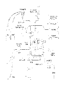
Figures 1 and 7 show examples of the apparatus of the instant invention.
Figure 7A provides a schematic of an example of an apparatus of the instant
invention. The apparatus may be a single unit or comprise separate housing
units
with interconnecting tubing. Also, the apparatus shown is an appropriate size
for a
lamb. The apparatus can be sized appropriately for the size of the subject.
For
example, the apparatus may about 1/3 the size for a human subject.
Figure 2 provides a close-up of the incubation chamber. The incubation
chamber may comprise any number of inlets and outlets. In a particular
embodiment,
the chamber comprises at least one inlet and one outlet to circulate the
sterile liquid
within. The incubation chamber may also comprise glove ports to allow sterile
access
to the subject (e.g., for swaddling of the subject to calm or to gain access
to objects
within the interior of the chamber). The chamber may also comprise at least
one port
for an IV line to the subject. The chamber may also have a variety of ports
(e.g.,
resealable ports) to allow access for any of a variety of monitoring devices.
For
example, the chamber may have ports to allow access for an ultrasound device
and/or
a dialysis unit. The chamber may also comprise a UV light unit (e.g., to
treat/inhibit
jaundice).
Figure 3 provides an image of the oxygenator contained within an optional dry
chamber. Figures 5 and 7C show an example of the circuit design with the
oxygenator without the dry chamber. While this chamber is shown as being
separated
from the incubator chamber by a divider in a single unit, the two can be
separated into
individual units (albeit connected by the necessary tubing and the like). The
cannulas
from the subject may connected directly to the oxygenator within the dry
chamber or
through ports in the dry chamber. The dry chamber may also comprise at least
one
port to attach the gas flow to the oxgenator. The port may connect to the
oxygenator
via tubing within the dry chamber. Figure 3 also shows additional ports for
the
addition of a heater (e.g., via a warm liquid and tubing) to the dry chamber
to help
9
CA 02905619 2015-09-10
WO 2014/145494
PCT/US2014/030277
maintain the temperature of the blood being circulated through the tubing and
the
oxygenator. Additionally, the tubing to and/or from the oxygenator may be
attached
to monitors (e.g., temperature monitors, gas content monitors, etc.).
Figure 7A provides a schematic of an example of an apparatus of the instant
invention. The chamber is depicted as a bowl located within a water bath to
maintain
the temperature of the system. The incubation chamber is depicted with two
glove/hand ports on the top of the chamber (though the ports could be located
anywhere, including the sides of the chamber) for sterile access to the
interior of the
chamber. The chamber comprises an inlet port for the pumping in of sterile
amniotic
fluid and an exit port for the removal of used/old amniotic fluid. The chamber
is also
depicted as having a filtration system comprising a pump, at least one filter,
and a UV
filter for elimination of bacteria and contaminants. The chamber also depicts
a mesh
upon which the subject may be placed. Figure 7A also depicts the oxygenator
(Ox) in
fluid connection with the blood system of the subject, particularly through
cannulae
into the neck vessels of the subject. The oxygenator is connected to a gas
mixer
drawing upon air and oxygen. The tubing of the oxygentor system may comprise
at
least one port for the introduction of compounds (e.g., nutrients,
antibiotics, drugs,
etc.) into the blood stream of the subject. Monitors such as for determining
pressure,
flow, and temperature may connected to the oxygentor system and/or the subject
directly.
Definitions
The singular forms "a," "an," and "the" include plural referents unless the
context clearly dictates otherwise.
As used herein, the terms "host," "subject," and "patient" refer to any
animal,
particularly mammals including humans.
The following examples are provided to illustrate various embodiments of the
present invention. The examples are illustrative and are not intended to limit
the
invention in any way.
CA 02905619 2015-09-10
WO 2014/145494
PCT/US2014/030277
EXAMPLE 1
A pre-term lamb (28 weeks) was maintained in the apparatus using the
methods of the instant invention. 8F to 1OF arterial ECM() cannulas were
placed in
carotid artery and internal jugular vein (size chosen at time of surgery).
Approximately 12 inches of ECM tubing was used on outflow and inflow to
connect
to the cannulae. The nutritional support provided was Total Parenteral
Nutrition.
Figure 6A provides an image of the lamb connected to the apparatus and Figure
68
shows the lamb after 5 days of growth. The growth of the premature lamb for
five
days demonstrates the ability of the instant invention to maintain a fetus
outside of the
womb.
EXAMPLE 2
The complications of preterm birth result in significant morbidity and
mortality, with one third of all infant deaths attributed to prematurity and
chronic
sequelae affecting most major organ systems in survivors. In 2010, 12.0% of
all US
births were preterm (less than 37 weeks completed gestation), and 3.5% were
early
preterm (less than 34 weeks gestation) (Martin et al. (2013) MMWR Surveill.
Summ.,
62(Suppl 3):136-138). Respiratory failure represents the most common and
challenging problem faced by these patients, as gas exchange in critically
preterm
neonates is impaired by structural and functional immaturity of the lungs.
Advances
in neonatal intensive care including antenatal steroid administration,
surfactant
replacement, pulmonary vasodilatory therapy, and high-frequency oscillatory
ventilation, have achieved improved outcomes and pushed the limitations of
viability
to the transition from the canalicular to the saccular phase of lung
development at 22
to 24 weeks gestation. However, incomplete development of most major organ
systems remains a limitation to survival and optimal functional outcomes in
many
patients. The development of an extrauterine system to support ongoing fetal
growth
and development without the perturbations induced by postnatal intensive care
would
offer a chance for survival of critically preterm infants, with potentially
reduced
mortality and long-term morbidity.
The concept of extracorporeal membrane oxygenation (ECMO) of the fetus is
appealing due to the similarities to innate fetal physiology, in which
extracorporeal
gas exchange is performed by the placenta. The artificial placenta has been a
subject
of experimental trials since the 1960's, with a series of short experiments in
which
11
CA 02905619 2015-09-10
WO 2014/145494
PCT/US2014/030277
fetal lambs were cannulated via the umbilical vessels and perfused by first-
generation
bubble membrane oxygenators, with perfusion supported for 40 minutes to 2 days
(Callaghan et al. (1963) Circulation 27:686-690; Zapol et al. (1969) Science
166:617-
618). Due to substantial improvements in oxygenator and pump technology over
the
following two decades, the duration of extrauterine fetal life support has
been
increased using conventional pump-driven ECMO circuits, with runs lasting up
to
three weeks before onset of circulatory failure (Kuwabara et al. (1986)
Artificial
Organs 11:224-277; Kuwabara et al. (1989) Artificial Organs 13:527-531; Unno
et al.
(1993) Artificial Organs 17:996-1003; Unno et al. (1997) Artificial Organs
21:1239-
1246). Despite these extensions in survival time, these studies were limited
by
circulatory overload and cardiac failure, resulting in the development of a
fluid
overloaded state and death of experimental animals.
Several features of the conventional pump-driven venous-arterial ECM
circuit are felt to represent challenges to the application of this technology
to support
of the fetus. The large priming volume of the circuit is substantially higher
than the
excess of the innate placental reserve, resulting in an increased volume of
distribution.
Pump-supported non-pulsatile flow also represents a departure from innate
fetal
physiology, with the potential for significantly increased cardiac afterload
and
resulting myocardial strain, in addition to a loss of innate autoregulation of
flow.
Finally, the large surface area of such circuits results in a requirement for
high levels
of systemic anticoagulation. A pumpless circuit may offer advantages over
current
ECM technology including reduced priming and distribution volumes, shorter
exposure of blood to thrombogenic surfaces, and achieving innate regulation of
blood
flow and pressure by the fetal heart itself.
The development of a pumpless extracorporeal oxygenation system remains
elusive. Only five attempts to achieve fetal oxygenation in a pumpless system
have
been reported in the literature, and all were ultimately unsuccessful trials,
with fetal
demise within several minutes to up to 29 hours secondary to declining rates
of blood
flow and the requirement for pressor support to artificially prolong perfusion
(Awad
et al. (1995) J. Invest. Surg., 8:21-30, Reoma et al. (2009) J. Pediatr.
Surg., 44:53-59;
Mirua et al. (2012) Pediatr. Res., 72:490-494; Schoberer et al. (2014)
Artificial
Organs 38:208-14).
Recent technologic advances in extracorporeal membrane technology have
resulted in the generation of exceptionally low resistance devices with low
priming
12
CA 02905619 2015-09-10
WO 2014/145494 PCT/US2014/030277
volume and highly efficient gas exchange, better recapitulating the properties
of the
placenta itself. In particular, the MaquetQuadrox-ID Pediatric Oxygenator
supports
the possibility of achieving pumpless oxygenation of fetal blood using the
native heart
to drive flow through the circuit. This oxygenator has been applied as a
pumpless
artificial lung in pediatric patients with good success (Boston et al. (2013)
J. Thorac.
Cardiovasc. Surg., 146:e42-e43). Herein, an approach was sought to maintain
fetal
blood oxygenation and stable hemodynamics using a modified pumpless circuit
that
permits the fetal heart to act as the pump, replicating innate fetal
hemodynamics.
Major challenges included achieving stable perfusion autoregulated by the
innate fetal
circulation, replicating the sterile fluid-immersed intrauterine environment,
and
facilitating appropriate fetal growth and development.
Herein, the first demonstration of pumpless extrauterine fetal life support
(PEFLS) resulting in stable long-term incubation of the mammalian fetus for up
to
three weeks, with normal growth, metabolism, and maintenance of autoregulated
fetal
circulation is provided.
METHODS
Surgical procedure
Time-dated pregnant ewes were used at gestational ages 120 to 135 days (term
= 145 days). Animals were treated according to approved protocols by the
Institutional Animal Care and Use Committee of the Children's Hospital of
Philadelphia.
Ewes were anesthetized with 15 mg/kg of intramuscular ketamine, with
maintenance of general anesthesia with 2-4% inhaled isoflurane in 02.
Intraoperative
hemodynamic monitoring included pulse oximetry, with a constant infusion of
isotonic saline administered via a central venous line placed in the right
jugular vein
to maintain maternal fluid balance.
A lower midline laparotomy was created to expose the uterus, with a small
hysterotomy performed to expose the fetal sheep head and neck. In the setting
of twin
lambs, fetal blood was collected from the donor animal to prime the circuit.
Donor
animals underwent creation of a small right neck incision to expose the
jugular vein,
and were administered one intramuscular dose of buprenorphine (0.005 mg/kg)
and
one intravenous dose of sodium heparin (150 USP units, APP Pharmaceuticals,
13
CA 02905619 2015-09-10
WO 2014/145494 PCT/US2014/030277
Schaumburg, IL), followed by creation of a small right neck incision to expose
the
jugular vein and placement of a catheter to permit collection of the entire
blood
volume of the animal. In the setting of singleton lambs, maternal blood was
collected
to prime the circuit. In all animals, maternal blood was collected and stored
for
subsequent transfusion requirements during the run.
Experimental lambs underwent creation of a small right neck incision to
expose the carotid artery and jugular vein. Animals received one intramuscular
dose
of buprenorphine (0.005 mg/kg) and one intravenous dose of sodium heparin (300
USP units). After determination of the maximal cannula size accommodated by
each
vessel, ECM() cannulae were placed (size range 8-12Fr, Avalon Laboratories,
LLC,
Rancho Dominguez, CA), with stabilizing sutures placed along the external
length of
cannulae at the neck. In a subset of animals, the external portion of the
cannulas were
fitted with a silicone 'sleeve' of 3-5 cm to permit increased tension of the
stabilizing
sutures. A subset of animals also underwent placement of insulated multi-
stranded
stainless steel wire electrodes to measure ocular electromyography (EOG) and
electroencephalography (EEG) activities. EMG wire electrodes were implanted
subcutaneously in the superior and inferior margins of the muscle overlying
the orbit
of one eye, and a pair of EEG electrodes were placed on the dura over the
parasagittal
parietal cortex and secured with cyanoacrylate glue, with a reference
electrode sewn
over the occiput.
Following construction and blood priming of the oxygenator circuit as
described below, connection of the cannulas to the circuit was performed under
continuous ultrasonographic visualization of the fetal heart. Occlusion of the
umbilical cord was performed immediately following establishment of blood flow
through the circuit, with administration of additional blood volume and/or
atropine
(0.1 mg) and/or epinephrine (0.1 mg) in a subset of animals demonstrating poor
cardiac contractility immediately after establishment of circuit flow. After
confirmation of stable cardiac function and circuit blood flow, stay-sutures
were
placed along the external length of the cannulas to secure them to the neck of
the
animal. Subsequently fetal lambs were weighed, washed in a warm sterile saline
bath,
and transferred to a sterile fluid incubator for further management as
described.
In order to generate baseline data of the ovine fetus in utero, two time-dated
pregnant ewes at 118 days gestational age underwent laparotomy for
implantation of
fetal vascular catheters and electrodes as described (Crossley et al. (1997)
Reprod.
14
CA 02905619 2015-09-10
WO 2014/145494 PCT/US2014/030277
Fertil. Dev., 9:767-73). Briefly, after induction of general anesthesia and
exteriorization of the fetus, catheters were implanted in the fetal carotid
artery and
jugular vein, in addition to a reference catheter placed in the amniotic sac,
followed
by placement of BOG and dural EEG wire electrodes as described above.
Additionally for these studies, EMG wire electrodes were placed in the nuchal
and
diaphragm muscles. The posterior left diaphragm was approached from a mid-
axillary incision with two electrodes sewn 1.0 cm apart, while a single
electrode was
sewn into a long muscle of the neck to record nuchalactivity. Fetal catheters
and
electrodes were exteriorized through the maternal flank, and the uterus and
abdomen
were closed. Following a 48-72 hour recovery period, ewes were transferred to
a
holding cage for fetal monitoring.
Circuit
The pumpless extrauterine fetal life support (P-EFLS) circuit consisted of a
low-resistance hollow fiber oxygenator (Quadrox-ID, Maquet, Rastatt, Germany)
connected to the ECM() cannulae with 3/16" BIOLINE heparin-coated Medtronic
tubing (Medtronic, Minneapolis Minesotta). Connections were established as an
arterial-venous extracorporeal oxygenation circuit, with the carotid arterial
outflow
under pulsatile systemic pressure connected to the oxygenator inflow port and
return
flow connected to the outflow port. The priming volume was 81 mL, and
heparinized
fetal blood was used when available, with maternal blood utilized in singleton
pregnancies. Circuit flow was measured with a flow probe (Transonic Systems
Inc,
Ithaca NY), and sweep gas supplied to the oxygenator was a blended mixture of
medical air and oxygen titrated to fetal blood gas values.
Fluid incubation
Trial incubator designs included a 30-liter heated stainless steel reservoir
filled
with sterile synthetic amniotic fluid ("still reservoir"), a 40-liter
polycarbonate tank
with continuous recirculation of fluid through a series of sterile filters
("recirculated
filtration"), and finally a 60-liter tank within flow and outflow tubing
mounted on a
double-head peristaltic pump to facilitate continuous exchange of sterile
fluid
("continuous exchange"). In the latter system, the complete volume of the tank
is
replaced three-fold with sterile inflow from a 180-liter reservoir over a 24-
hour
period. Synthetic amniotic fluid was composed of a balanced salt solution
containing
CA 02905619 2015-09-10
WO 2014/145494 PCT/US2014/030277
Na + (109 mM), Cl" (104 mM), HCO3" (19 mM), K+ (6.5 mM), Ca2+ (1.6 mM), pH 7.0-
7.1, osmolarity 235.8 mOsm/kg water. Antibiotics were added to a final
concentration of 18 mg/L gentamycin and 30 mg/L ciprofloxacin, and a
submersible
UV sterilizer pump was placed in the fluid reservoir continuous recirculated
and
sterilization throughout the run.
Fetal lamb maintenance on circuit
Following stabilization and transfer of animals to the fluid incubator, a
continuous infusion of heparin (80-200 USP units per hour) and prostaglandin
El (0.1
mcg/kg/min, Pfizer Inc, New York, NY) were administered intravenously. Blood
was
drawn every 1-4 hours for blood gas, electrolyte and coagulation values using
an i-
Stat System (Abbott Point of Care Inc, Princeton, NJ), with titration of the
heparin
infusion to a target Activated Clotting Time of 180-200 seconds (100-400 USP
units
per hour), and titration of the oxygenator sweep gas to target fetal partial
pressures of
02 (Pa02 20-30 mmHg) and CO2 (PaCO2 35-45 mmHg) (Fi02 21-55%, sweep gas
0.125-1.5 L/min). Stored whole maternal blood was used to maintain fetal
hemoglobin levels above 9 mg/dL. Analgesics (buprenorphine, 0.005 mg/kg IV
every
3-5 hours as needed) and anxiolytics (midazolam, 0.4 mg/kg IV every 3-5 hours
as
needed) were administered during periods of perceived fetal distress (restless
repetitive fetal movements, tachycardia, hypertension). Total parenteral
nutrition was
administered throughout the duration of fetal incubation, with a dosage of 3.5
g/kg
amino acids (TrophAmine 10%), 5-10% dextrose, and 3 g/kg lipids (Intralipide
20%).
Data acquisition
Fetal blood pressure, heart rate, circuit flow rates, trans-membrane pressure
differential, sweep gas flow rates, and bath fluid temperature were
continuously
recorded with input sampling every 0.1 seconds (LabChart 5, ADInstrumentsInc,
Colorado Springs CO). Oxygen consumption and respiratory quotients were
calculated daily, with measurement of the oxygenator exhaust gas content for
oxygen
and carbon dioxide. The following formulas were used:
Blood oxygen content (02C) = 1.34 x Hgb x Sa02/100 + 0.003 x Pa02 (mmHg)
Oxygen Delivery (OD) = Post-membrane 02C x Circuit Flow/100/body weight
16
CA 02905619 2015-09-10
WO 2014/145494
PCT/US2014/030277
Oxygen Consumption (OC) = (Post-membrane 02C ¨ Pre-membrane 02C) x Circuit
Flow/100
Oxygen Extraction Rate (OER) = (0C/OD) x 100.
Monitoring of catheterized fetal lambs in utero was started 48-72 hours after
surgery and continued in 24-hour intervals on alternating days until
completion of the
experimental protocol at 140 days gestational age. Continuous polygraphic
recordings of EEG, EMG and arterial pressures were recorded with data captured
every 0.1 seconds (LabChart 5, ADInstrumentsInc, Colorado Springs CO).
Decannulation
Following completion of the planned incubation period, animals were
transitioned from the fluid bath, with endotracheal intubation and suctioning
to
remove excess fluid from the lungs. General anesthesia was maintained with 2-
4%
inhaled isoflurane in 100% 02, andintraoperative hemodynamic monitoring
included
pulse oximetry, with a constant infusion of isotonic saline administered via a
peripheral venous cannula. The arterial and venous ECMO cannulas were removed
with ligation of the vessels, and the neck incision was closed with a running
absorbable suture. Anesthesia was then reversed, with animalsextubated upon
initiation of spontaneous respiration with arterial blood gases demonstrating
adequate
gas exchange (Pa02 > 75 mmHg, PaCO2 < 50 mmHg on inhaled medical air, FiO2
21%).
RESULTS
Pilot studies
A total of 5 pilot experiments were conducted to determine fetal stability on
the PEFLS circuit (Table 1). Fetal gestational ages ranged from 120 to 140
days, with
weights ranging from 3.20 to 4.89 kg. All animals demonstrated remarkable
hemodynamic stability during support on the circuit, with no evidence of
acidosis or
increasing lactate, decreasing circuit flows, or circulatory failure.
Unexpectedly, two
animals displayed bradycardia immediately upon initial opening of fetal
circulation
onto the PEFLS circuit, requiring the administration of epinephrine and
atropine to
restore normal cardiac function. Following this initial event, no animals
required
17
CA 02905619 2015-09-10
WO 2014/145494
PCT/US2014/030277
vasopressor support at any time in the run. All animals were maintained on
systemic
anticoagulation and total parenteral nutrition.
18
-
Animal Length Priming Cannula Average Flow probe
Average Fluid Survived to Complications Pathology
Number- of Run Size flow rate pH (SD) incubation
decannulation during run 0
n.)
GA (h) (SD)
1--L
(weight,
4.,
Lril
.r-
.r-
1-140 23 Fetal 8Fr 135.8-/- Transsonic 7.41(7.28-
Still reservoir No Cardiac arrest on Closure DA,
(3.62) (carotid), MARCUS- T201; pre-
7.62) opening circuit, pulmonary
(June) 8Fr check oxygenator
bacterial inflammation,
(jugular) calibration?
infection of fluid pulmonary
hemorrhage
2-135 71 Maternal 8Fr 336.3(297- Transsonic
7.24(6.77- Recirculated No Cardiac arrest on Closure DA, diffuse
(4.89) (carotid), 396) T201; pre-
7.51) filtration opening circuit, pulmonary
(Charlotte 8 Fr oxygenator
bacterial inflammation
) (jugular)
infection of fluid,
bacteremia
0
0
3-135 96 Maternal 12Fr 492.5(450- Transsonic
7.31(7.10- Recirculated Yes Bacterial Patent DA,
diffuse ,0
0
0
(3.49) (carotid), 10 520) T201; pre-
7.56) filtration infection of fluid, pulmonary 0
0
1-L (Lily) Fr (jugular) oxygenator
bacteremia inflammation 1-
0
0
01
4-130 51 Fetal 10Fr 435.6(250- Transsonic
7.19 (6.60- Continuous No Clot formation in Patent
DA, diffuse '
0
(4.24) (carotid), 12 470) T201; post-
7.46) excha nge circuit tubing, shower emboli in
0
(Little Fr (jugular) oxygenator
cardiac I ungs, heart, liver,
Alan)
a rrythmias, bowel
acidosis
, . _
- 120 108 Maternal 10Fr 387.4(290- Transsonic
7.38(7.24- Continuous No Traumatic Normal organ
(3.20) (carotid), 12 430) H7XL; pre-
7.57) exchange decannulation histology
(Eddie) Fr (jugular) oxygenator
*0
Table 1: Study Animals.
n
ci)
LV
0
I¨k
.F.,
Ge4
0
ls.)
¨.1
--.1
C-11
CA 02905619 2015-09-10
WO 2014/145494 PCT/US2014/030277
Several obstacles to long-term fetal survival were identified, with bacterial
contamination of the fluid incubator noted in four of five animals (Table 1).
The
initial pilot study employed a warmed open incubator filled with sterile
amniotic fluid
that was refilled only to compensate for evaporative losses, with no internal
mixing
and no antibiotics. Within 12 hours, this incubator fluid developed
significant
bacterial overgrowth, and after 23 hours on the circuit, the animal developed
massive
pulmonary hemorrhage. Histology confirmed significant diffuse inflammatory
changes throughout the lungs, consistent with severe bacterial pneumonia. The
fluid
incubator was subsequently designed to a closed system with recirculation of
amniotic
fluid containing antibiotics (Ciprofloxacin 30 mg/L, Gentamycin 18 mg/L)
through a
series of filters with increasingly fine pore size (1 mm, 10 ptm, 5 ptm, 2 m,
0.2 ilm) to
eliminate gross debris and maintain sterility. Two studies were performed with
this
fluid incubator. In both cases, bacterial growth was confirmed on culture of
sampled
bath fluid within 48 hours of incubation. One animal developed increasing Fi02
requirements over the course of the incubation period and was found on
echocardiography to have significant constriction of the ductusarteriosus,
resulting in
impaired mixing of oxygenated blood via the superior vena cava. The second
animal
had no complications during the 96-hour incubation period and was delivered
and
decannulated at 140 days gestational age (term), but was found to have
inadequate gas
exchange despite vigorous spontaneous respiratory effort. Both animals were
found
to have significant pulmonary inflammation consistent with pneumonia as well
as
bacteremia. A closed sterile system with continuous fluid exchange was
designed,
with synthetic amniotic fluid passing through a series of 0.22 tim filters to
enter the
incubator, with the rate of inflow matched to outflow and complete exchange of
the
60 liter incubator volume 2-4 times daily (Figure 7).
Additional technical limitations identified included premature closure of the
ductusarteriosus in two animals exposed to epinephrine, clot formation in a
circuit
including a segment of non-antithrombogenic-coated tubing, and traumatic
decannulation in one animal with resulting hemorrhagic demise (Table 1). These
findings led to the addition of a continuous infusion of prostaglandin-E2 for
maintenance of fetal shunts, implementation of a circuit employing exclusively
antithrombogenic-coated tubing, and the addition of a sterile silicone sleeve
overlay
on each cannula to permit increased stabilization of anchoring stay-sutures.
CA 02905619 2015-09-10
WO 2014/145494
PCT/US2014/030277
Fetal biochemical and hemodynamic parameters
Following implementation of the system refinements noted above, five
experimental animals were maintained on the PEFLS circuit for 343.8 +/- 93.5
hours
(Table 2). Trends in fetal blood gases, hemodynamics and circuit flow rates
over the
course of five independent experiments are summarized in Figure 8. The pH
remained stable throughout the duration of the incubation period (Figure 8A),
and the
partial pressures of oxygen and carbon dioxide were maintained at target fetal
levels
throughout the course of the experiment (Figure 8B). While the basal heart
rate was
stable throughout the duration of incubation (Figure 8C), the animals'
systolic blood
pressure increased steadily (Figure 8D), consistent with expected rates of
growth.
Similarly, circuit flow rates increased in proportion to the increase in blood
pressure
(Figure 8E), and the concentration of supplied oxygen required to maintain
target
arterial blood oxygenation levels increased steadily as well, reflecting
increased fetal
metabolic demand (Figure 8F).
21
Animal Length of Priming Cannula Size Average Average
GA at Complications Outcome and Pathology
Number-GA Run (h) flow rate pH (SD)
decannulation during run 0
(weight, kg) (SD) (weight, kg)
i...4
o
1¨,
.6.
1-125 ( 3.17) 209 Fetal 10Fr (ca roti d), 350.7 +/- 7.38 +/-
134 (3.9) Hypotension after Appropriate gas
exchange on ventilator support .6
uni
(Willow) 10Fr (jugular) 137.2 0.21
i nitiation of with good spontaneous respiratoryeffort.
.6
mid azolam infusion
Eutha nized DOL2 - ge neralized hypotonia. .6
with resulting
Pathology: normal organ histology. Appropriate
peri od of hypoxia
lung maturation.
>8 hours
2-120 (3.20) 360 Fetal 10E r (ca rob d), 380.1+!- 7.34+!-
135 (4.2) Bacterial Appropriate gas exchange on ventilator s upport
(Se i nne) 10 Fr (jugular) 154.7 0.27
overgrowth in fluid with good spontaneous respiratoryeffort.
incubator
Eutha nized DOLldue to inabilityto wean from
ventilator. Pathology:extensive pulmonary
inflammation, mucus plu y: ing of d istala irways.
Appropriate lung maturation.
0
0
3-120(3.30) 372 Maternal 10Fr (ca roti d), 374.6 =1-
7.40 +/- 136 (4.5) Bacterial Abrupt onset
of hypotension o
o
(Bowie) 12 Fr (jugular) 149.9 0.32
overgrowth in fluid following the development of lactic acidosis
o
incubator
associated with fluid incubator infection.
Eutha nized DOLO due to severityof acidosis..
,..,
o
1,..)
Pathology: mild pulmonary inflammation. 17,1
IQ
Appropriate I ung maturation. 0
o
IL
4- 120 (3.20) 288 Maternal 10Fr (carotid), 390.6 +/-
7.47 +/- 134 (3.7) Small ca nnula bleed Long-term survivor; MRI
head/chest/abdomen:
( I ggy) 10 Fr (jugular) 201.2 0.29
(suture tra uma) normal organ structures
5-120 (2.9) 490 Ma ternal 10Fr (Carotid), 334.2 +/-
7.48 +/- 140(4.12) GI hemorrhage Appropriate gas exchange on
ventilator support
(Manson) 12Fr (jugular) 121.2 0.17
fol I owingsystemic with good spontaneous respiratory effort.
a nti coagulation,
Eutha nized DOL4 duet inabilityto wean from
resulting in
ventilator. Histology: *0
complete post-natal
n
bowel obstruction.
1-3
ri)
Table 2: Study Animals.
Cs)
0
4,..-
0
C.4
0
t=4
-41
-.1
CA 02905619 2015-09-10
WO 2014/145494
PCT/US2014/030277
Flow characteristics on PEFLS
Fetal flows during PEFLS are linearly related to the systolic blood pressure
of
the animal (Figure 9) and are pulsatile and directly correlate with the
measured
cardiac output. Autoregulation of circuit flow was consistently demonstrated
in
response to hypoxia, with compensatory increases in systolic blood pressure
and
circuit flow in response to reduced sweep gas flow to the oxygenator and
normalization to baseline flow and blood pressure after restoration of sweep
gas flow.
Fetal growth and metabolism in PEFLS
Over the course of five independent experiments ranging from 209 to 490
hours, animals gained an average of 930 +/- 278 grams (Figure 10A). Fetal
breathing
movements were noted regularly throughout the incubation period, and were
correlated with the partial pressure of carbon dioxide measured in the
systemic
circulation (Figure 10B). Echocardiography was performed to confirm patency of
fetal circulatory shunts, including the ductusarteriosus (Figure 10C). Total
oxygen
consumption increased steadily over the course of five independent experiments
(Figure 10D), consistent with growth and proportionate increases in fetal
metabolic
demand. Following normalization to estimated fetal weight as extrapolated from
the
growth curve generated from pre- and post-PEFLS weights, oxygen consumption
rates were found to remain stable throughout the course of incubation.
Sleep-wake cycles, breathing movements and whole-body movements were
recorded in two chronically catheterized fetal lambs by EEG, EOG and EMG, and
compared to two lambs maintained in the artificial placenta over the same
range of
gestation. Recordings from a catheterized fetal lamb in utero were made at 125
and
140 days gestational age, and recordings from a fetal lamb in the artificial
placenta
were made at the same gestational ages. A developmental progression from
fragmented to consolidated sleep between the two gestational ages is apparent
in both
in utero and PEFLS animals.
Fetal growth and development was consistently observed over the course of
the incubation period, with the eyelids noted to progress from fused to open,
increased
wool growth, and increased level of activity and alertness (Figure 11A and
11B).
Histologic evidence of lung maturation was also consistently demonstrated,
with
thinning of the alveolar walls and secondary septations acquired upon
completion of
23
CA 02905619 2015-09-10
WO 2014/145494
PCT/US2014/030277
PEFLS runs over 200 hours compared to control lungs of 120 day gestation lambs
(Figure 11C, Table 2).
One animal was maintained for 288 hours in the absence of infection, and was
successfully delivered from the artificial placenta and transitioned to
postnatal life
(Figure 5D). Magnetic resonance imaging confirmed normal structure of the
brain,
thoracic and abdominal viscera, and the animal displayed appropriate growth
and
development over eight months before transport to a long-term adoptive
facility.
A pumpless circuit to permit long-term support of the extrauterine fetus may
be the optimal design for an artificial placenta. However, previous efforts
have been
limited by low flow rates and poor perfusion. Herein, a system for pumpless
extrauterine fetal life support (PEFLS) is described which results in stable
long-term
incubation of the mammalian fetus for up to three weeks or more. By employing
a
superior oxygenator with extremely low resistance and low priming volumes, a
circuit
has been created which more closely approximates the placenta itself. The
reported
placental blood volume of the sheep is 23.1 to 48.1 ml/kg (Creasy et al.
(1970) Circ.
Res., 27:487-494), with placenta blood flow reported as 199 +/- 20 ml/min/kg
(Faber
et al. (1972) J. Pysiol., 223:375-393). The circuit described herein requires
a priming
volume of 80 to 90 mls, or 27 ml/kg of an average 120-day 3 kg fetal lamb, and
flow
rates in the system averaged 120-140 ml/min/kg.
In applications of extracorporeal oxygenation to the fetus, priming of the
oxygenator circuit with fetal blood might provide advantages with respect to
oxygen
unloading and maintaining peripheral perfusion. However, priming of the
circuit was
completed with fetal blood in the case of twin gestations and with maternal
blood in
the event of singleton lambs (Tables 1 and 2), and no differences were
observed in
arterial oxygen content, lactic acid production, or cardiac function over the
course of
long-term fetal incubation.
A pumpless circuit in which innate hemodynamics can be maintained by
autoregulation may be advantageous for normal fetal development. Prior to the
instant invention, the longest extrauterine fetal incubation experiments was
with
animals maintained on a circuit employing a silicone hollow fiber membrane
oxygenator and a roller pump and animals cannulated via the umbilical vessels
(Kuwabara et al. (1986) Artificial Organs 11:224-277; Kuwabara et al. (1989)
Artificial Organs 13:527-531). In 6 animals maintained with the fetal
circulation
24
CA 02905619 2015-09-10
WO 2014/145494 PCT/US2014/030277
under direct regulation of the roller pump, the maximum duration of incubation
was 8
hours, with rapid onset of cardiac failure accounting for fetal demise in the
majority
of experiments. Run times were significantly prolonged in this study by the
addition
of a blood reservoir, which was filled passively by the umbilical arterial
outflow, and
which automatically regulated flow rates through the pump according to the
rate of
reservoir filling. Flow rates were maintained between 100 to 200 ml/min in all
animals, with fetal gases maintained within target ranges throughout the
incubation
period. Although not a pumpless system, this modification did permit flow
rates to
better mirror the innate circulation, and did extend survival time to up to
165 hours.
However, ultimately cardiac failure and subcutaneous edema developed in all
animals
which did not succumb to iatrogenic complications such as bleeding or
embolism. In
a subsequent study, this reservoir-supported circuit was modified by the
addition of
hemodialysis to improve fluid and electrolyte balance, achieving incubation
lengths of
up to 236 hours with flow rates between 50 to 100 ml/min/kg. Again, while
three
animals died due to catheter malfunctions, cardiac failure was the cause of
all non-
technical deaths, with progressive circulatory depression and eventual demise
on the
circuit. Based upon the suspicion that fetal movement may have contributed to
fluid
imbalance, flow disturbances and bleeding complications, a follow-up study
employing this circuit with the administration of continuous paralytics
resulted in the
stable support of two preterm goat fetuses for 494 and 543 hours respectively,
with
flow rates between 80 to 180 ml/min/kg and successful delivery from the
incubator to
mechanical ventilation (Unno et al. (1993) Artificial Organs 17:996-1003).
However,
animals failed to demonstrate adequate spontaneous respiratory effort and
expired due
to respiratory insufficiency. Further analyses of the performance of this
circuit, with
modifications including the placement of an occlusion tube to create a fixed
point of
resistance on the arterial outflow, achieved incubation times up to 236 hours,
but all
animals died due to circulatory failure characterized by declining flows and
blood
pressure as well as recurrent arrhythmias (Unno et al. (1997) Artificial
Organs
21:1239-1246).
Additional studies attempting to achieve improved circulatory outcomes
following prolonged extrauterine fetal incubation resulted in similar findings
of
demise due to circulatory failure. In 1998, four goat fetuses cannulated via
the
umbilical vessels and maintained on a circuit comprised of a hollow fiber
oxygenator
and a centrifugal pump were studied (Yasufuku et al. (1998) J. Pediatr. Surg.,
33:442-
CA 02905619 2015-09-10
WO 2014/145494
PCT/US2014/030277
448). This circuit permitted the delivery of pulsatile flow at higher rates
compared to
previous published reports, with flow rates ranging from 113 to 193 ml/min/kg.
The
total duration of support ranged from 87 to 237 hours, with all animals
succumbing to
hydrops secondary to circulatory failure. In 2002, 12 goat fetuses cannulated
via the
umbilical vessels and supported by a circuit employing a roller pump with
manual
control of the flow rate, in an attempt to increase the rate of umbilical
artery drainage,
were studied. In this study, 3 animals died due to cannula problems and one
due to
hypoxia secondary to clot formation in the circuit, while the remaining 8
animals
developed circulatory failure. Flows ranged from 103.0 +/-17.0 to 176.0 +/-
15.0
(ml/min/kg) and adequate gas exchange was achieved, however drainage of the
umbilical artery by the roller pump was felt to impose increased afterload to
the
myocardium.
The continued demonstration of circulatory overload in pump-supported fetal
ECM() suggests an unacceptable afterload imposed by these circuits, resulting
in
eventual cardiac insufficiency. Moreover, the ideal artificial placenta will
permit the
fetus to maintain circulation analogous to that achieved in the intact fetal-
placental
unit, where perfusion is determined by fetal cardiac output. However, previous
attempts to design a pumpless system for fetal perfusion have yielded
discouraging
results. For example, the use of a pumpless circuit in a series of lambs with
surgically created congenital diaphragmatic hernias was reported (Awad et al.
(1995)
J. Invest. Surg., 8:21-30). Animals were perfused for up to 6 hours, but
circuit flow
rates did not exceed 75 ml/min and oxygenation levels were inadequate to
sustain
stable long-term incubation. In 2009, a pumpless extracorporeal circuit using
a
hollow-fiber oxygenator and umbilical cannulation in four near-term lambs was
reported (Reoma et al. (2009) J. Pediatr. Surg., 44:53-59). Animals were
supported
for up to four hours in this system, with a gradual decline in circuit flows
and systolic
blood pressure over this short course of incubation. It was concluded that a
pump-
driven system would be required to maintain adequate flow and perfusion of the
fetus.
In 2012, a pumpless extracorporeal circuit in lambs at a gestational age of
130 +/- 1.6
days, with cannulation of a single umbilical artery and the umbilical vein,
was
reported (Mirua et al. (2012) Pediatr. Res., 72:490-494). The 5 animals
studied
survived for an average of 18.2 +/- 3.2 hours, but developed a progressive
lactic
acidosis resulting in cardiac failure and death. Administration of pressors to
increase
cardiac contractility and ionotropes to induce peripheral vasodilation did not
achieve
26
CA 02905619 2015-09-10
WO 2014/145494
PCT/US2014/030277
long-term survival in this system. Lastly, the development of a miniaturized
low-
volume oxygenator, with a priming volume of 12 mL and gas exchange surface
area
of 0.12 m2, was reported (Schoberer et al. (2014) Artificial Organs 38:208-
14).
Animals were cannulated via the umbilical vessels but were maintained on
mechanical ventilation in addition to extracorporeal oxygenation. In this
system, 6 of
the 7 animals studied were maintained on pumpless extracorporeal support for 6
hours, the defined endpoint, however all animals developed a metabolic
acidosis,
elevated blood lactate, and a continuous decline in blood pressure, and three
animals
ultimately required catecholamines to reach the experimental endpoint. In the
system
of the instant invention, no animals required pressor support at any time to
maintain
stable hemodynamics and perfusion over up to three weeks of support.
Long-term sterility of the fetal fluid incubation system was achieved in this
study. Previous reports employing fluid immersion systems have not extensively
described rates of bacterial contamination and strategies to improve
sterility. Notably,
an incubator filled with synthetic amniotic fluid and antibiotics, with daily
in-line
filtration and complete exchange every other day, has been described (Kuwabara
et al.
(1989) Artificial Organs 13:527-531). In this study, the longest duration of
fetal
incubation was 236 hours. In the instant study, with the final incubator
design,
infection did not commonly occur before day 12, or 288 hours. The prolonged
length
of the incubation periods likely increases the difficulty in maintaining
sterility.
Several refinements of the incubator design were made throughout the series of
experimental animals, including the addition of a mounted glove to facilitate
manipulation and position changes of the animal without breaching sterility,
as well
as the placement of an internally-fixed and sealed suction device to permit
removal of
waste products and debris. In addition, an aquarium model UV filtration system
was
placed within the fluid reservoir to provide an additional level of
antimicrobial
protection. With these modifications, sterility was maintained for up to three
weeks
of fetal incubation, representing a significant period for growth and
development of a
critically preterm infant.
One advantage of a pumpless system for fetal perfusion is the maintenance of
regulation of cerebral blood flow by innate autoregulatory pathways. Ensuring
adequate oxygenation of the developing brain is a key consideration in the
design of
an artificial placenta, with cerebral autoregulation felt to represent one
important
component of cerebral perfusion. Cerebral autoregulation has been well
described in
27
CA 02905619 2015-09-10
WO 2014/145494
PCT/US2014/030277
the population of neonatal infants who require intensive care, but is poorly
understood. Significant individual variability exists with respect to measured
ranges
of cerebral artery blood flow rates, vasoreactivity, and autoregulatory
thresholds, to
the extent that normative values for these patients remain undefined (Vutskis,
L.
(2014) Pediatr. Anesth., 24:22-29). Cerebral autoregulation has also been
observed in
the human fetus, with increased middle cerebral artery peak systolic velocity
described in fetuses with intrauterine growth restriction (Hanif et al. (2007)
Am. J.
Perinatol., 24:501-505). The effects of conventional pump-supported ECM() on
cerebral perfusion in neonates have demonstrated a well-documented loss of
.. autoregulation in response to fetal hypoxia in a number of systems,
including lamb
models of veno-arterial ECMO (Short et al. (1993) Pediatr. Res., 33:289-294;
Stolar
et al. (1988) J. Pediatr. Surg., 23:1163-1168), studies of newborn lambs
supported on
veno-venous ECM() (Walker et al. (1996) Crit. Care Med., 24:2001-2006), as
well as
infants supported on veno-arterial ECM() (Papademetriou et al. (2013) Adv.
Exp.
Med. Biol., 765:203-209), suggesting significant alterations in cerebral
perfusion in
the setting of pump-supported ECMO flow. Notably, numerous studies indicate
that
the long-term neurodevelopmental impact of ECMO is impaired functional outcome
in these patients (Kumar et al. (1994) Pediatrics 93:951-955; lisselstiin et
al. (2014)
Semin. Perinatol., 38:114-121). In the instant study, EEG studies revealed
waveforms
consistent with those observed in chronically catheterized lambs in utero.
Maintenance of cerebral autoregulation likely achieves optimal brain perfusion
and
development in the artificial placenta, improving outcomes in this population
with
potentially devastating neurodevelopmental sequelae following conventional
management.
The implications of the total extrauterine fetal life support (TEFLS) system
extend beyond clinical innovations, and provide a basis for addressing
fundamental
questions regarding the role of the placenta in fetal development. For the
first time,
long-term stable maintenance of a fetus amputated from the maternal-placental
axis
has been achieved, making it possible to study the relative contribution of
this organ
to fetal maturation. The system can also be used to bridge the transition from
fetal to
postnatal life, which may be applied to models of congenital lung disease to
expand
the window of opportunity for therapeutic interventions. The TEFLS system
therefore represents a capability that has not been previously available for
research in
28
CA 02905619 2015-09-10
WO 2014/145494 PCT/US2014/030277
fetal physiology, and represents a powerful new resource for numerous
translational
clinical applications.
While certain of the preferred embodiments of the present invention have been
described and specifically exemplified above, it is not intended that the
invention be
limited to such embodiments. Various modifications may be made thereto without
to departing from the scope and spirit of the present invention, as set
forth in the
following claims.
29