Note: Descriptions are shown in the official language in which they were submitted.
NANOSCALE ARTIFICIAL ANTIGEN PRESENTING CELLS
[01] This paragraph has been deleted intentionally.
TECHNICAL FIELD
[02] This disclosure relates to immunotherapy.
BRIEF DESCRIPTION OF THE DRAWINGS
[03] FIGS. IA-C. Synthesis and Characterization of Iron-Dextran Nano-aAPC .
Nano-
aAPC were synthesized in one of two ways: FIG. IA, Direct chemical coupling of
soluble MHC-Ig Dimer (Signal 1) and B7.1-Ig (Signal 2) in a 1:1 molar ratio to
the
surface of a paramagnetic iron-oxide, dextran-coated particle. FIG. IB,
Binding of
biotinylated MHC-Ig dimer (Signal 1) and biotinylated anti-CD28 (Signal 2) in
a 1:1
molar ratio to anti-biotin coated particles. FIG. IC, Nanoparticle Tracking
Analysis
confirms that Nano-aAPC are a monodisperse mixture of particles with a mean
diameter of 50-100 nm suspended at a concentration of 8.3 nM.
[04] FIGS. 2A-F. Nano aAPC Induced Proliferation is Antigen-Specific and Dose-
Dependent. FIG. 2A, Antigen specific nano-aAPC induce proliferation. TCR
transgenic 2C (grey) and pMEL (white) T cells proliferated only when incubated
with
anti-biotin coated particles bearing cognate MHC/peptide, and not in the
presence of
particles bearing either non-cognate peptide or non-cognate MHC. FIG. 2B,
Addition
of both Signal 1 and Signal 2 leads to optimal T cell expansion. At a dose of
10 [iL
particles per 1*106 T cells, only anti-biotin particles bearing both MHC-Ig
and anti-
CD28 induced robust T cell proliferation. FIG. 2C, Proliferation of CD8+ CTL
induced by LD and HD particles at dose equivalent concentrations by Day 3 CFSE
dilution. Decreased fluorescence indicates increased proliferation. Equivalent
volumes of HD particles induces greater proliferation than LD particles, with
0.5 uL
LD particles inducing almost no expansion. FIG. 2D, Fold expansion on day 7 of
samples in (A) shows a similar pattern. Proliferation is dose-dependent and 2-
4 fold
greater for HD particles compared to an equivalent dose of LD particles. FIG.
2E,
Day 3 CFSE dilution of CD8+ CTL induced by LD and HD particles at protein
1
Date Recue/Date Received 2020-06-26
CA 02906514 2015-09-14
WO 2014/160132 PCT/US2014/025889
equivalent concentrations . When particle doses are normalized to equivalent
protein
concentrations, particles induce similar amounts of proliferation. FIG. 2F,
Fold
expansion on day 7 of samples in (C) demonstrates equivalent expansion for HD
and
LD particles at an equivalent protein dose. A threshold of about 0.5 uL LD
particles
or 0.08 uL HD particles is required to induce detectable expansion.
[05] FIGS. 3A-F. T cell Functional Characterization. FIG. 3A, CD8+ T cells
were
expanded using HD and LD particles. Particle doses were chosen to induce
equivalent
expansion by HD and LD particles (3.5 uL and 20 uL, respectively) and to
induce
more robust expansion (HD 20 uL). Samples were re-stimulated on day 7 and
assessed for effector function by intracellular cytokine staining assay. 20 uL
HD
sample (black circles), 3.5 uL HD sample (black filled square), and 20 uL LD
samples
(unfilled square) all induced robust, equivalent, and dose-dependent (FIG. 3B)
degranulation measured by CD107a and (FIG. 3C) IFNy production. FIG. 3D,
Memory effector phenotype measured by staining of surface proteins CD44 and
CD62L. T cells can be classified as naive (CD62Lhi, CD441o), Central Memory
(CD62Lhi, CD44hi), or Effector Memory (CD62L1o, CD44hi). E) Representative
FACS plot shows three populations seven days after nano-aAPC stimulation. FIG.
3F, T cells were stimulated with 2, 10 and 504 of LD or HD iron-oxide nano-
aAPC
and characterized seven days later. Bar plots show percentage of Naive
(unfilled), Ton
(grey fill), and Tern (black fill) cells generated after stimulation.
[06] FIGS. 4A-B. Antigen-specific Human T cell Expansion From Endogenous
Precursors. FIG. 4A, PBMC were incubated with increasing doses of iron-dextran
nano-aAPC bearing A2-M1 MHC-Ig and assessed for antigen-specificity by
tetramer
staining before stimulation (PBMC, top row) or after one (middle row) or two
(bottom
row) weeks of stimulation. Numbers in top left represent percentage of CD8+
cells
that were tetramer+ (gated).The size of the MI specific population increases
with
repeated rounds of stimulation (top to bottom) and increasing dose of nano-
aAPC (left
to right). Plots are representative of results from three separate
experiments,
summarized in panel B. FIG. 4B, Percentage of CD8+ PBMC binding Ml tetramer
increases with repeated stimulation and increasing dose of nano-aAPC (left
panel).
The total number of tetramer-positive cells (right panel) similarly increases
with
2
CA 02906514 2015-09-14
WO 2014/160132 PCT/US2014/025889
rounds of stimulation and particle dose, expanding up to 800-fold of the
initial
precursor population.
[07] FIGS. 5A-B. Synthesis and Characterization of Quantum Dot Nano-Aapc. FIG.
5A,
Quantum Dot (Qdot) Nano-aAPC were constructed by avidin-biotin mediated
coupling of soluble MHC-Ig Dimer (Signal 1) and anti-CD28 antibody (Signal 2)
in a
1:1 ratio to the surface of a polymer-coated quantum dot particle. FIG. 5B,
Qdot
Nano-aAPC expansion in whole CD8+ T cells. Fold expansion on Day 7 is dose
dependent and antigen-specific. Non cognate particles did not induce any
expansion,
whereas the highest dose of cognate quantum dot aAPC induced 14.6 fold
expansion
of CTL.
[08] FIGS. 6A-B. Nano-aAPC Mediate Tumor Rejection In Vivo. FIG. 6A, quantum
dot
aAPC. B16 Tumors were injected subcutaneously on day 0, with injection of
naive
pMEL T cells on the same day. One day later, quantum dot aAPC were injected
intravenously (iv). Tumor size was measured as surface area (mm2) on indicated
days,
with Area Under Curve (AUC) shown at right. Mice treated with pMEL T cells and
cognate quantum dot aAPC (black bars) had less tumor growth compared to no
treatment (white), T cells alone (light grey), and T cells + noncognate
quantum dot
aAPC (checkered) (4 mice per group). Significance was characterized over
entire
experiment by AUC (p<0.02) for treatment group compared to non-cognate quantum
dot aAPC. FIG. 6B, Iron-Dextran aAPC. Naive pMEL T cells were injected
intravenously on day -7. One day later, quantum dot aAPC were injected either
iv or
subcutaneously (sc) on the right flank. B16 tumors were injected sc on right
flank on
day 0. Mice in treatment arms were given an additional injection on day 4 post
tumor
injection either iv or sc, to form four treatment groups: noncognate aAPC iv
(day -6)
then sc (day 4) (checkered), cognate aAPC iv then iv (light grey), cognate
aAPC iv
then sc (dark grey), and cognate aAPC sc then sc (black). Mice treated with
pMEL T
cells and cognate Iron-Dextran aAPC iv/sc or sc/sc (filled squares) had less
tumor
growth compared to noncognate aAPC (7 mice per group, * p<0.01 for AUC).
[09] FIG. 7A. CFSE, a dye whose intensity is reduced after T cell
proliferation, shows that
T cell populations including activated cells (CD44 mixed) proliferate in
response to 6
ng of micro- or nano-aAPC based stimulation, but naive CD44 low cells do not.
FIG.
7B.When micro- and nano-aAPC are titrated to doses that induce equivalent fold
3
expansion (about 17-fold) in CD8+ (activated) cells, nano-aAPC cannot expand
naive
T cells.
[10] FIG. 8A. Schematic of magnetic enrichment strategy for enhanced T cell
activation.
Low-frequency precursors cells are bound to nano-aAPC carrying specific
antigen of
interest. Antigen-specific cells are enriched by positive magnetic selection,
enhancing
subsequent expansion. FIG. 8B. The frequency of antigen specific T cells (y
axis) is
enhanced by magnetic enrichment using nano-aAPC. FIG. 8C. Increased frequency
of antigen specific cells after seven days of nano-aAPC mediated expansion
post
enrichment. FIG. 8D. As a complementary approach, cells are activated in a
magnetic field after pre-binding to nano-aAPC. Culture in a magnetic field
boosts cell
proliferation. FIG. 8E. CFSE staining three days after activation shows magnet
induced boosting after 1-3 hours of activation. FIG. 8F, This leads to
enhanced
expansion measured seven days after activation, with magnetic stimulation
providing
a boost at all doses considered.
[11] FIGS. 9A-G. Nano-aAPC Bind to Naive and Activated Cells. FIG. 9A,
Schematic
of nano-aAPC synthesis by coupling MHC-Ig dimers and co-stimulatory anti-CD28
to
iron-dextran nanoparticles. FIG. 9B, Proliferation of naive (left) and
activated (right)
pmcl T cells measured by CFSE dilution 3 days after stimulation with nano-aAPC
presenting 8 ng of Db-GP100. Unstimulated controls in dashed lines. FIG. 9C,
Fold
expansion of naive and activated cells seven days after nano-aAPC
stimulation. Nano-aAPC presenting 8 ng or less of MHC-Ig induced minimal
proliferation in naive cells (*, p < 0.01) compared to activated T cells. FIG.
9D,
Disassociation of Kb-SIY nanoparticles bound to 2C T cells (half-lives
significantly
different p<0.02 by paired Student's t-test). See Table 1. FIG. 9E, Mean TCR-
MHC
contacts made between Kb-STY dimers (MHC-Ig) and Kb-SIY nanoparticles
(Particle) with naive and activated cells as estimated from
disassociation
data (p<0.05 by ANOVA with Tukey's post-test, see Table 1). FIG. 9F,
Equilibrium
binding of increasing doses of nano-aAPC (measured by total MHC-Ig presented)
to
naive and activated cells (p<0.0001 by two-way ANOVA). FIG. 9G, A
binding model that explains increased equilibrium binding and particle off-
rate: naive
cells bind more beads with fewer contacts per bead than activated cells.
4
Date Recue/Date Received 2020-06-26
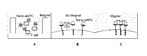
[12] FIGS. 10A-G. Clustering of aAPC and CD3 t Induced by a Magnetic Field.
FIG.
10A-C, Schematic of magnet-induced clustering. FIG. 10D, aAPC and CD3
aggregation immediately after nano-aAPC binding (Time 0) and after incubation
in
the presence or absence of a magnetic field. Cells were labeled with
antibodies against
LFA-1 , MHC-Ig on nano-aAPC , and CDR . Representative
images are shown for cells prior to incubation (Time 0, top left), cells
incubated with
non-cognate particles (Non-Cognate, top right), cells incubated with cognate
nano-
aAPC (No Magnet, bottom left), and cells incubated with cognate nano-aAPC in a
magnetic field (Magnet, bottom right). FIG. 10E, Aggregate detection shown for
representative images from Time 0 group (two on left) and Magnet group (two on
right). White outlines represent borders of CD3 clusters identified by
algorithm. FIG. 10F, Average cluster area identified with cluster detection
algorithm
(15 cells/group). The No Magnet group had significantly larger clusters than
Time 0
(*, mean difference 0.22 tm2), and the Magnet group had significantly larger
clusters
than both Time 0 (**, mean difference 0.46 [tm2, p <0.0001 by ANOVA with Tukey
post-test) and No Magnet (**, mean difference 0.24 [an2). FIG. 10G, Cells in
No
Magnet group had fewer clusters per cell than Time 0 (*, mean difference 5.8
clusters) and Magnet group cells had fewer clusters per cell than No Magnet
(**,
mean difference 1.9 clusters, p <0.001 by ANOVA with Tukey post-test).
[13] FIGS. 11A-G. Magnet-enhanced Nano-aAPC Stimulation Leads to Robust T cell
Proliferation In Vitro. FIG. 11A, Pmel T cell proliferation by CFSE dilution
three
days after stimulation with nano-aAPC in the presence or absence
of a
0.2 T external magnetic field. FIG. 11B, Fold expansion of samples described
in A
seven days after stimulation. FIG. 11C, Pmel T cells incubated with 5 ng MHC-
Ig
dose of nano-aAPC and 0.2 T magnetic field for 0-24 hours. Proliferation
assessed by
CFSE dilution at day 3. FIG. 11D, Fold expansion of samples from C seven days
after stimulation. (*, p<0.001 by ANOVA with Tukey post-test) FIG. 11E, Pmel T
cells incubated with 5 ng MHC-Ig dose of nano-aAPC and magnetic fields of
increasing maximal strength (0.15-0.225 T) generated by neodymium magnets of
increasing thickness for twenty-four hours. FIG. 11F, Proliferation of samples
from E
seven days after stimulation (* greater than no magnet, ** greater than 0.15 T
magnet,
p<0.001 by ANOVA with Tukey post-test). FIG. 11G, Antigen-specific expansion
of
endogenous CD8+ lymphocytes from wild type mice after stimulation with Kb-Trp2
Date Recue/Date Received 2020-06-26
nano-aAPC in the presence or absence of a 0.2 T magnetic field for twenty-four
hours. After seven days, populations were stained with cognate Kb-Trp2 (top
row) or
non-cognate Kb-SIINF (bottom row) MHC-Ig dimer.
[14] FIGS. 12A-F. Magnet-Enhanced T Cell Expansion In Vivo and Increased
Efficacy of Adoptive Immunotherapy. FIG. 12A, Schematic of adoptive
immunotherapy model. CD441o, CD8+ T cells from Thy1.1+ pmel TCR transgenic
mice were stimulated in vitro for 24 hours in the presence or absence of nano-
aAPC
(5 ng total MHC-Ig) and magnetic field prior to being adoptively transferred
into wild
type, Thy1.2+ B6 recipient mice (6 mice per group). FIG. 12B, Representative
frequencies of Thy1.1 cells from spleens 7 days after transfer and day lymph
nodes 21
days after transfer. FIG. 12C, Frequencies of Thy1.1+ cells were significantly
higher
in mice given T cells stimulated with nano-aAPC in a magnetic field
compared
to nano-aAPC with no magnet and no stimulation (p<0.001 for
treatment effect by two-way ANOVA for day 7 and 21). FIG. 12D, Total Thy1.1+
cells in all organs combined on Day 7 and Day 21. Five-fold more cells were
observed in the nano-aAPC + Magnet group than nano-aAPC alone group on day 7
(p
<0.05 by student's t-test), but did not reach significance on Day 21 (p =
0.15). FIG.
12E, Schematic of treatment of established tumors with magnetic field enhanced
adoptive immunotherapy. SC tumors were administered on Day 0, partial
myeloablation on Day 9, and CD441o, CD8+ pmel T cells stimulated for 24 hours
with either nano-aAPC (5 ng total MHC-Ig) in a magnetic field or nano-aAPC
with no magnet were transferred on Day 10. T cell alone and
untreated
(unfilled) groups were used as control (8 mice per group). FIG. 12F, Treatment
with
magnet-enhanced nano-aAPC activated T cells attenuated tumor growth compared
to
no magnet and control groups (p<0.0001 for treatment effect by two-way ANOVA).
Arrow indicates timepoint of adoptive transfer (day 10). Mice were censored if
dead
or tumors were greater than 150 mm2. Treatment led to increased survival in T
cells +
nano-aAPC + Magnet group (p<0.001 by Mantel-Cox log-rank test).
[15] FIGS. 13A-D. Characterization of Protein Bound to Nano- and Micro-aAPC By
Fluorescence. FIG. 13A, Mean fluorescence intensity (MF1) of antibody bound to
nanoparticles and controls. Nano-aAPC and Micro-aAPC (cell-sized) particles
were
incubated with excess of monoclonal anti-mouse IgG1 (for MHC-Ig) and anti-
6
Date Recue/Date Received 2020-06-26
antibody conjugated with PE for 30 minutes, and washed on a magnetic column.
Fluorescent antibody bound to particles was detectable above background
samples,
including micro- and nano- particles not stained with anti-IgG1 (No Ab) and
particles
which were not coupled to protein and stained with anti-IgG1 (Blank). Protein
concentration in solution was determined by comparison to an IgGl-PE standard
curve. Fluorescence is shown for anti-IgG1 and is representative of three
experiments.
HD ¨ High Density. LD ¨ Low Density. FIG. 13B, Particles in solution do not
interfere with antibody fluorescence. Soluble anti-IgG1 PE antibody was
titrated and
measured for fluorescence. Similar fluorescence emission was observed when
soluble
antibody was measured in the presence of blank micro- and nano-particles. FIG.
13C,
Washing in magnetic column was sufficient to remove free antibody. After three
washes (Fraction 3), fluorescence is not detectable above background.
Fluorescence
of 0.63 ug/ml free antibody is provided for comparison. FIG. 13D, Nanoparticle
concentration was characterized by iron absorbance at 405 nm. Particle
concentrations
were determined by Nanoparticle Tracking Analysis. Titrations of nanoparticles
were
measured for absorbance and a standard curve was calculated to determine
particle
concentration.
[16] FIGS. 14A-E. pMEL T cell Proliferation Induced by Micro-aAPC. FIG. 14A,
CD8+ pM EL splenocytes include a population of memory-phenotype, CD44 positive
cells (representative percentage shown as percentage of CD8, left). CD4410
naive
cells were isolated by a no-touch negative selection enrichment with anti-CD44
antibody in a magnetic enrichment column. FIG. 14B, Proliferation of Naive
CD4410
(left) and activated (right) cells by CFSE dilution stimulated three days with
micro-
aAPC and nano-aAPC or
unstimulated . Micro- and nano-aAPC were used at doses presenting
equivalent total amount of MHC-Ig (8 ng). Nano-aAPC data are re-produced from
FIG. 1. FIG. 14C, Proliferation of naive and active cells
seven days after
stimulation with indicated doses of micro-aAPC. FIG. 14D, Effect of MHC-Ig
density on micro-aAPC induced stimulation. High density (HD ) and low
density
(LD ) micro-aAPC were normalized for total MHC-Ig (4-16 ng). See Table
1 for
density. Proliferation assessed by CFSE dilution three days after activation.
FIG.
14E, Fold expansion of samples shown in FIG. 14D seven days after activation,
representative of three experiments.
7
Date Recue/Date Received 2020-06-26
[17] FIGS. 15A-D. FIG. 15A, Kb-SIY nanoparticle binding to cognate 2C T cells.
Binding to activated cells, seven days after peptide activation (activated,
MFI
89) as compared to naive, CD4410 isolated 2C T cells (naive, MFI 179) and
control non-cognate CD4410 pmel T cells (non-specific binding, MFI 21).
Binding is characterized as mean fluorescence intensity of Alexa 647 labeled
particles
bound to cells. FIG. 15B, Surface TCR expression of naive (MFI 137) and
activated
(WI 128) cells measured with fluorescent anti-TCR13. FIG. 15C, Disassociation
of
Kb-STY MHC-Ig dimers from activated and naive cells.
Disassociation of nano-aAPC from activated and naive cells
are
reproduced from FIG. 1 for comparison. FIG. 15D, Disassociation curves of nano-
aAPC bound to naive CD44low cells before and after one hour of
incubation in a magnetic field. FIG. is representative of 2 experiments.
[18] FIGS. 16A-E. TCR Clustering and Expansion by Micro-aAPC in a Magnetic
Field. FIG. 16A, Micro-aAPC aggregation in a magnetic field. Representative
confocal images of micro-aAPC shown
before (left) and after (right) application
of a magnetic field. FIG. 16B, Micro-aAPC magnetic aggregation does not induce
CD3 aggregation. Cells were labeled with antibodies against LFA-1 , MHC-Ig
on micro-aAPC , and CDR . Micro-
aAPC displayed auto-fluorescence
in all three channels. Representative
images are shown for cells incubated with cognate micro-aAPC (No Magnet), both
not in contact (top) and in contact (bottom) with micro-aAPC, and cells
incubated
with cognate nano-aAPC in a magnetic field (Magnet). FIG. 16C, Average cluster
area and clusters per cell identified with cluster detection algorithm (20
cells/group,
divided evenly between cells in contact and not in contact with particles).
Control
samples include cells prior to incubation (Time 0) and cells incubated with
non-
cognate microparticles (Non-Cognate) (p>0.05 by ANOVA). FIG. 16D, Pmel T cells
incubated with 5 ng (left) and 10 ng (right) MHC-Ig dose of micro-aAPC with
and without a 0.2 T magnetic field for 3 days. Proliferation assessed
by CFSE
dilution at day 4. FIG. 16E, Fold expansion of pmel T cells incubated with
increasing
doses of micro-aAPC with and without a 0.2 T magnetic field seven days after
stimulation (p>0.05 by two-way ANOVA).
8
Date Recue/Date Received 2020-06-26
CA 02906514 2015-09-14
WO 2014/160132 PCT/US2014/025889
[19] FIG. 17. Magnetic Field Strength Generated in Culture by Neodynium Disk
Magnets. Density plots of field strength in culture as estimated by finite
element
analysis with FEMM (Finite Element Method Magnetics) software. Disk magnets
(magenta) 3/4, 'A", and %" in thickness were used to generate fields of up to
0.225 T,
0.200 T, and 0.150 T, respectively.
SUMMARY
[20] This disclosure provides a nano-scale artificial antigen presenting cell
(nano-aAPC)
comprising a nanoparticle; at least one lymphocyte affecting molecule on the
surface
of the nanoparticle; and at least one molecular complex on the surface of the
nanoparticle that, when bound to an antigen, engages a unique clonotypic
lymphocyte
receptor, i.e., an antigen-specific lymphocyte receptor.
[21] This disclosure provides a nano-aAPC comprising a nanoparticle; at least
one B cell
affecting molecule on the surface of the nanoparticle; and at least one
molecular
complex on the surface of the nanoparticle that engages B cell surface
immunoglobulins or MHC-antigen complexes on a B cell surface.
[22] This disclosure provides a nano-aAPC comprising a nanoparticle; at least
one T cell
costimulatory molecule on the surface of the nanoparticle; and at least one
MHC class
I molecular complex on the surface of the nanoparticle. The at least one MHC
class I
molecular complex comprises at least two fusion proteins. A first fusion
protein
comprises a first MHC class I a chain and a first immunoglobulin heavy chain
and
wherein a second fusion protein comprises a second MHC class I a chain and a
second immunoglobulin heavy chain. The first and second immunoglobulin heavy
chains associate to form the MHC class I molecular complex. The MHC class I
molecular complex comprises a first MHC class I peptide binding cleft and a
second
MHC class I peptide binding cleft.
[23] This disclosure provides a preparation comprising a plurality of nano-
aAPCs
described in the three paragraphs above.
[24] This disclosure provides a method of inducing the formation of antigen-
specific T
cells. The method comprises contacting an isolated preparation comprising a
plurality
of precursor T cells with at least one first nano-aAPC which comprises a T
cell
9
CA 02906514 2015-09-14
WO 2014/160132 PCT/US2014/025889
affecting molecule and an antigen presenting complex that comprises at least
one
antigen binding cleft. An antigen is bound to the antigenic binding cleft.
Members of
the plurality of precursor T cells are thereby induced to form a first cell
population
comprising antigen-specific T cells that recognize the antigen. The number or
percentage of antigen-specific T cells in the first cell population is greater
than the
number or percentage of antigen-specific T cells that are formed if precursor
T cells
are incubated with a nano-aAPC that comprises an antibody that specifically
binds to
CD3 but does not comprise an antigen presenting complex.
[25] This disclosure provides a method of increasing the number or percentage
of antigen-
specific T cells in a population of cells. The method comprises incubating a
first cell
population comprising antigen-specific T cells with at least one first nano-
aAPC
which comprises a T cell affecting molecule and an antigen presenting complex
that
comprises at least one antigen binding cleft. An antigen is bound to the
antigen
binding cleft. The incubation is carried out for a period of time sufficient
to form a
second cell population comprising an increased number or percentage of antigen-
specific T cells relative to the number or percentage of antigen-specific T
cells in the
first cell population.
[26] This disclosure provides a method of regulating an immune response in a
patient. The
method comprises administering to a patient a preparation comprising (A) a
plurality
of particles and (B) a pharmaceutically acceptable carrier. Members of the
plurality of
particles comprise (1) at least one T cell affecting molecule; and (2) at
least one
antigen presenting complex. The at least one antigen presenting complex
comprises at
least one antigen binding cleft. An antigen is bound to the at least one
antigen binding
cleft.
[27] This disclosure provides a method of suppressing an immune response in a
patient.
The method comprises administering to a patient a preparation comprising (A) a
plurality of particles and (B) a pharmaceutically acceptable carrier. Members
of the
plurality of particles comprise (1) at least one apoptosis-inducing molecule;
and (2) at
least one antigen presenting complex. The at least one antigen presenting
complex
comprises at least one antigen binding cleft. An antigen is bound to the at
least one
antigen binding cleft.
CA 02906514 2015-09-14
WO 2014/160132 PCT/US2014/025889
[28] This disclosure provides a method of increasing the number or percentage
of
antibody-producing B cells in a population. The method comprises contacting an
isolated preparation comprising a plurality of precursor B cells with at least
one first
nano-aAPC which comprises a nanoparticle; at least one B cell affecting
molecule on
the surface of the nanoparticle; and at least one molecular complex on the
surface of
the nanoparticle that engages B cell surface immunoglobulins or MHC-antigen
complexes on a B cell surface. Members of the plurality of precursor B cells
are
thereby induced to form a first cell population comprising antibody-producing
B cells
that produce antibodies that specifically bind to the antigenic peptide.
[29] This disclosure provides a method of increasing the number or percentage
of
antibody-producing B cells in a population. The method comprises incubating a
first
cell population comprising antibody-producing B cells with at least one first
nano-
aAPC which comprises a nanoparticle; at least one B cell affecting molecule on
the
surface of the nanoparticle; and at least one molecular complex on the surface
of the
nanoparticle that engages B cell surface immunoglobulins or MHC-antigen
complexes on a B cell surface. The incubating is carried out for a period of
time
sufficient to form a second cell population comprising an increased number or
percentage of antibody-producing B cells relative to the number or percentage
of
antibody-producing B cells in the first cell population.
[30] This disclosure provides a method of increasing the number or percentage
of
antibody-producing B cells in a population. The method comprises contacting an
isolated preparation comprising a plurality of precursor B cells with a
preparation of
nano-aAPCs. The nano-aAPCs comprise a nanoparticle, at least one B cell
affecting
molecule on the surface of the nanoparticle; and at least one molecular
complex on
the surface of the nanoparticle that engages B cell surface immunoglobulins or
MHC-
antigen complexes on a B cell surface. Members of the plurality of precursor B
cells
are thereby induced to form a first cell population comprising antibody-
producing B
cells that produce antibodies that specifically bind to the antigenic peptide.
[31] This disclosure provides a method of regulating an immune response in a
patient. The
method comprises administering to a patient a preparation comprising (A) a
plurality
of particles and (B) a pharmaceutically acceptable carrier. Members of the
plurality of
particles comprise (1) at least one B cell affecting molecule; and (2) at
least one
11
CA 02906514 2015-09-14
WO 2014/160132 PCT/US2014/025889
molecular complex that engages B cell surface immunoglobulins or MHC-antigen
complexes on a B cell surface.
[32] This disclosure provides a method of enriching antigen-specific T cells
in a polyclonal
T cell population. The method comprises incubating the polyclonal T cell
population
with a nano-aAPC comprising a nanoparticle; at least one lymphocyte affecting
molecule on the surface of the nanoparticle; and at least one molecular
complex on
the surface of the nanoparticle that, when bound to an antigen, engages
antigen-
specific lymphocyte receptors. The nano-aAPC further comprises a cross-linking
antibody or an oligomerizing molecule.
[33] This disclosure provides a method of activating T cells. The method
comprises
incubating in the presence of a magnetic field a population of T cells with a
nano-
aAPC which comprises a T cell affecting molecule and an antigen presenting
complex
that comprises at least one antigen binding cleft. The nano-aAPC is
paramagnetic.
[34] This disclosure provides a method of providing a population of antigen-
specific T
cells to a patient in need thereof, comprising:
(1) contacting an isolated population of T cells with a plurality of
nano-scale artificial antigen presenting cells (nano-aAPCs) in the presence of
a
magnetic field of sufficient strength to generate antigen-specific T cells,
wherein nano-aAPCs of the plurality are paramagnetic nanoparticles which
comprise on their surface (i) at least one T cell affecting molecule and (ii)
at
least one antigen presenting complex, wherein the antigen presenting complex
comprises at least one antigen binding cleft and wherein the antigen binding
cleft comprises an antigen;
(2) isolating complexes of antigen-specific T cells bound to nano-
aAPC from the isolated population of T cells; and
(3) administering the complexes to the patient.
In some variations of this method, the isolated population of T cells
comprises naïve
T cells. In some variations of these methods, complexes are isolated using a
magnetic
enrichment column, flow cytometry, or differential centrifugation. In some
variations
12
CA 02906514 2015-09-14
WO 2014/160132 PCT/US2014/025889
of this method, the complexes are administered by a route of administration
selected
from the group consisting of intravenous administration, intra-arterial
administration,
subcutaneous administration, intradermal administration, intralymphatic
administration, and intra-tumoral administration.
[35] This disclosure provides a method of providing a population of antigen-
specific T
cells to a target area in a patient in need thereof, comprising:
(1) administering to the patient a plurality of nano-scale artificial
antigen presenting cells (nano-aAPCs) in the presence of a magnetic field of
sufficient strength to stimulate antigen-specific T cells, wherein nano-aAPCs
of the plurality are paramagnetic and comprise on their surface (i) a T cell
affecting molecule and (ii) an antigen presenting complex, wherein the antigen
presenting complex comprises an antigen binding cleft, wherein binding of an
antigen to the antigen binding cleft engages a unique antigen-specific
lymphocyte receptors; and
(2) applying to the target area a magnetic field, wherein the target area
comprises the antigen which engages unique antigen-specific lymphocyte
receptors., thereby directing the nano-aAPCs to the target area.
[36] In some variations of the method described in paragraph [35], nano-aAPC
are
administered by a route of administration selected from the group consisting
of
intravenous administration, intra-arterial administration, subcutaneous
administration,
intradermal administration, intralymphatic administration, and intra-tumoral
administration.
[37] In some variations of the methods described in paragraphs [34] and [35],
the at least
one antigen presenting complex comprises an MHC class I peptide binding cleft.
[38] In some variations of the methods described in paragraphs [34] and [35],
the at least
one antigen presenting complex is an MHC class I molecule. In some of these
variations, the at least one antigen presenting complex is an MHC class I
molecular
complex comprising at least two fusion proteins, wherein a first fusion
protein
comprises a first MHC class I a chain and a first immunoglobulin heavy chain
and
wherein a second fusion protein comprises a second MHC class I a chain and a
13
CA 02906514 2015-09-14
WO 2014/160132 PCT/US2014/025889
second immunoglobulin heavy chain, wherein the first and second immunoglobulin
heavy chains associate to form the MHC class I molecular complex, wherein the
MHC class I molecular complex comprises a first MHC class I peptide binding
cleft
and a second MHC class I peptide binding cleft.
[39] In some variations of the methods described in paragraphs [34] and [35],
the at least
one antigen presenting complex comprises an MHC class II peptide binding
cleft. In
some of these variations, the antigen presenting complex is an MHC class II
molecule. In some of these variations, the antigen presenting complex is an
MHC
class II molecular complex comprising at least four fusion proteins, wherein
(a) two
first fusion proteins comprise (i) an immunoglobulin heavy chain and (ii) an
extracellular domain of an MHC class 1113 chain; and (b) two second fusion
proteins
comprise (i) an immunoglobulin light chain and (ii) an extracellular domain of
an
MHC class ha chain, wherein the two first and the two second fusion proteins
associate to form the MHC class II molecular complex, wherein the
extracellular
domain of the MHC class 1113 chain of each first fusion protein and the
extracellular
domain of the MHC class Ha chain of each second fusion protein form an MHC
class
II peptide binding cleft. In some of these variations, the immunoglobulin
heavy chain
comprises a variable region.
[40] In some variations of the methods described in paragraphs [34] and [35],
an antigenic
peptide is bound to the at least one antigen binding cleft. In some of these
variations,
the antigenic peptide is selected from the group consisting of a peptide of a
tumor-
associated antigen, a peptide of an autoantigen, a peptide of an alloantigen,
and a
peptide of an infectious agent antigen.
[41] In some variations of the methods described in paragraphs [34] and [35],
nano-APCs
comprise at least two antigen presenting complexes. In some of these
variations, an
identical antigen is bound to each antigen binding cleft of the at least two
antigen
presenting complexes. In other of these variations, different antigens are
bound to
each antigen binding cleft of the at least two antigen presenting complexes.
In some
variations, a first antigen presenting complex comprises at least one MHC
class I
peptide binding cleft and wherein a second antigen presenting complex
comprises at
least one MHC class 11 peptide binding cleft.
14
CA 02906514 2015-09-14
WO 2014/160132 PCT/US2014/025889
[42] In some variations of the methods described in paragraphs [34] and [35],
the at least
one antigen presenting complex is a non-classical MHC-like molecule. In some
of
these variations, the non-classical MHC-like molecule is a CD1 family member.
The
non-classical MHC-like molecule can be selected from the group consisting of
CD1a,
CD1b, CD1c, CD1d, and CD1e.
[43] In some variations of the methods described in paragraphs [34] and [35],
the at least
one T cell affecting molecule is a T cell costimulatory molecule. The T cell
costimulatory molecule can be selected from the group consisting of CD80 (B7-
1),
CD86 (B7-2), B7-H3, 4-1BBL, CD27, CD30, CD134 (0X-40L), B7h (B7RP-1),
CD40, LIGHT, an antibody that specifically binds to CD28, an antibody that
specifically binds to HVEM, an antibody that specifically binds to CD4OL, an
antibody that specifically binds to 0X40, and an antibody that specifically
binds to 4-
1BB.
[44] In some variations of the methods described in paragraphs [34] and [35],
the at least
one T cell affecting molecule is an adhesion molecule.
[45] In some variations of the methods described in paragraphs [34] and [35],
the adhesion
molecule is selected from the group consisting of ICAM-1 and LFA-3.
[46] In some variations of the methods described in paragraphs [34] and [35],
the at least
one T cell affecting molecule is a T cell growth factor. The T cell growth
factor can
be selected from the group consisting of a cytokine and a superantigen. The
cytokine
can be selected from the group consisting of IL-2, IL-4, 1L-7, IL-10, IL-12,
IL-15, and
gamma interferon. The T cell growth factor can be selected from the group
consisting
of (A) a first molecular complex comprising at least two fusion proteins,
wherein a
first fusion protein comprises a first cytokine and an immunoglobulin heavy
chain and
wherein a second fusion protein comprises a second cytokine and a second
immunoglobulin heavy chain, wherein the first and second immunoglobulin heavy
chains associate to form the first molecular complex; and (B) a second
molecular
complex comprising at least four fusion proteins, wherein, (a) two first
fusion proteins
comprise (i) an immunoglobulin heavy chain and (ii) a first cytokine; and (b)
two
second fusion proteins comprise (i) an immunoglobulin light chain and (ii) a
second
CA 02906514 2015-09-14
WO 2014/160132 PCT/US2014/025889
cytokine, wherein the two first and the two second fusion proteins associate
to form
the second molecular complex.
[47] In some of the variations described above, the T cell growth factor is
the first
molecular complex. In some of these variations, the first and second cytokines
are
identical. In other of these variations, the first and second cytokines are
different.
[48] In some of the variations described above, the T cell growth factor is
the second
molecular complex. In some of these variations, the first and second cytokines
arc
identical. In other of these variations, the first and second cytokines are
different.
[49] In some variations of the methods described in paragraphs [34] and [35],
the at least
one T cell affecting molecule is a regulatory T cell inducer molecule. The T
cell
inducer molecule can be selected from the group consisting of TGFP, IL-10,
interferon-a, and IL-15.
[50] In some variations of the methods described in paragraphs [34] and [35],
the at least
one T cell affecting molecule is an apoptosis-inducing molecule. The apoptosis-
inducing molecule can be selected from the group consisting of a toxin, TNFa,
and
Fas ligand.
[51] In some variations of the methods described in paragraphs [34] and [35],
nano-aAPCs
comprise at least two different T cell affecting molecules.
[52] In some variations of the methods described in paragraphs [34] and [35],
the
incubation is carried out at 37 C for 10 minutes to 3 days.
[53] In some variations of the methods described in paragraphs [34] and [35],
the antigen-
specific T cells are cytotoxic T cells.
[54] In some variations of the methods described in paragraphs [34] and [35],
the antigen-
specific T cells are helper T cells.
[55] In some variations of the methods described in paragraphs [34] and [35],
the antigen-
specific T cells are regulatory T cells.
[56] In some variations of the methods described in paragraphs [34] and [35],
the patient
has cancer, an autoimmune disease, an infectious disease, or is
immunosuppressed.
16
CA 02906514 2015-09-14
WO 2014/160132 PCT/US2014/025889
[57] In some variations of the methods described in paragraphs [34] and [35],
the precursor
T cells are obtained from the patient.
[58] In some variations of the methods described in paragraphs [34] and [35],
the precursor
T cells are obtained from a donor who is not the patient.
[59] In some variations of the methods described in paragraphs [34] and [35],
the antigen-
specific T cells are administered by a route of administration selected from
the group
consisting of intravenous administration, intra-arterial administration,
subcutaneous
administration, intradermal administration, intralymphatic administration, and
intra-
tumoral administration.
[60] This disclosure provides methods of using nanoparticles, e.g.,
magnetic nanoparticles,
to target cells in different physiological states (e.g., naïve vs previously
activated T
cells) and stimulate the target cell population. For example, as shown in FIG.
9C and
discussed in more detail in the specific examples below, nano-aAPCs providing
a
dose of 32 ng of MHC stimulates both naïve and previously activated T cells
between
20- and 30-fold in a week's time. However, at 8 ng or 3.2 ng of MHC, only the
activated T cells were stimulated. Thus, a dose of nano-aAPC comprising, e.g.,
3.2-8
ng of MHC can be used to stimulate previously activated T cells in a T cell
population
without affecting naïve T cells in the population.
[61] This disclosure provides methods of differentially stimulating previously
activated T
cells vs naïve T cells. In some variations, nano-aAPC comprising 3.2-8 ng MHC
vs
32 ng MHC can be used to separate nano-aAPC binding and isolation of T cells
from
the activation of the T cells. In some variations, a population of T cells is
substantially
depleted of previously active T cells using, e.g., an antibody to CD44,
leaving a
population enriched for naïve T cells. Naïve T cells bound to the nano-aAPCs
would
permit their purification. The naïve T cells comprising the bound nano-aAPCs
can
then be activated by exposing the T cell-nano-aAPC complexes to a magnetic
field.
[62] This disclosure provides methods of separating, characterizing, and using
as a
therapeutic for other cells including, e.g., B cells and stem cells. The
optimum ligand
density on the surface of a nanoparticle (or, alternatively, the dose of
nanoparticles
comprising such ligands) which will differentially activate cells of a
population in
different physiological states is determined using methods such as those
described
17
CA 02906514 2015-09-14
WO 2014/160132 PCT/US2014/025889
below in Example 9. Depending on the cell population, the ligand can comprise,
e.g.,
an antibody or a portion of an antibody, a peptide, a nucleotide, a
carbohydrate, a
lipid, all or portion of the natural ligand for a given receptor (e.g., EGF,
PDGF), a
chemical (e.g., a chromium salt or a monovalent synthetic ligand that binds
immunophilin molecule receptors such as FKBP binding domain), single anti-
integrin
Fab fragments, RGD peptides, and the like.
DETAILED DESCRIPTION
[63] Immunotherapy includes the activation and expansion of immune cells to
treat
disease. Induction of cytotoxic (CD8+) lymphocyte (CTL) responses is
attractive for
therapy because CTL are specific for a given tumor antigen or pathogen, expand
several logs to produce robust responses, and generate long-term memory that
can
prevent recurrence of disease(1). CTL can be directly activated in vivo or can
be
expanded in vitro and adoptively transferred into a patient (3, 4).
[64] Artificial Antigen Presenting Cells (aAPC), which deliver stimulatory
signals to
cytotoxic lymphocytes, are a powerful tool for in vitro and in vivo
immunotherapy.
Thus far, particle-based aAPC have been synthesized by coupling a T cell
activating
protein to a rigid support several microns in diameter. For example, we
previously
developed a cell-sized, 4.5 jim diameter ("microscale") bead-based T cell
expansion
platform by coupling proteins that deliver two necessary and sufficient T cell
activation signals (5, 6). Signals present on APC that are required for T cell
activation
include Signal 1, cognate antigenic peptide presented in the context of Major
Histocompatibility Complex (MHC) that bind the TCR (7); and Signal 2, a group
of
co-stimulatory receptors that modulate T cell response. In some embodiments of
this
system, Signal 1 is conferred by a chimeric MHC-immunoglobulin dimer loaded
with
specific peptide (MHC-Ig), and Signal 2 is either B7.1 (the natural ligand for
the T
cell receptor CD28) or an activating antibody against CD28. Both proteins are
directly
chemically coupled to the surface of a microscale (4.5 [im) bead to create an
artificial
Antigen Presenting Cell (aAPC).
[65] However, there are several drawbacks to microscale aAPC. Large beads can
lodge in
capillary beds and induce tissue damage when injected intravenously. When
injected
subcutaneously, micron-sized beads are not easily carried to lymph nodes where
most
18
CA 02906514 2015-09-14
WO 2014/160132 PCT/US2014/025889
T cells reside (8, 9). Furthermore, micron-sized beads are known to be
preferentially
cleared by phagocytic cells of the reticulo-endothelial system (10, 11).
[66] The present disclosure overcomes these limitations by providing various
nanoscale
aAPC (nano-aAPC). To our knowledge, this is the first description of nanoscale
bead-based aAPC that induces T cell proliferation. Nanoparticles have been
evaluated
for antigen or drug uptake; however, aAPC platforms require specific cell
surface
receptor-ligand interactions to occur at the nanoparticle-cell interface.
Studies have
suggested that only beads larger than 2 microns in diameter are able to induce
T cell
proliferation (16, 17); and work with smaller size particles, such as quantum
dot
nanocrystals, has focused on the use of those reagents to study biophysical
aspects of
TCR-MHC interaction (15). When directly tested, recent work demonstrated that
nanoparticles were much less efficient than microbeads in inducing short-term
functional responses, with no reported proliferation (18).
[67] It was therefore unexpected that nano-aAPC as described herein induce
antigen-
specific T cell proliferation, both from TCR transgenic mouse splenocytes and
from
human polyclonal peripheral blood T cells. Stimulated T cells had a robust
effector
phenotype, degranulating and producing IFNy after re-challenge. Nanoscale
aAPCs
described herein also mediate tumor rejection in a subcutaneous mouse melanoma
model when injected in vivo.
[68] Although not limited to the embodiments described in the working examples
below,
those examples illustrate two embodiments of nano-aAPC: (1) biocompatible iron-
dextran paramagnetic beads 50-100 nm in diameter; and (2) avidin-coated
quantum
dot nanocrystals less than 20 nm in diameter. In these embodiments, signal 1
is
provided by peptide-MHC complexes, and signal 2 is provided by B7.1-Ig or anti-
CD28.
[69] Nano-aAPC permit exploration of new particle-based immunotherapy
strategies. As
noted above, microscale aAPC are too large to be carried by lymphatics, and
when
injected subcutaneously remain at the injection site. When injected
intravenously,
they lodge in capillary beds. In fact, the poor trafficking of microscale
beads has
precluded the study of where aAPC should traffic for optimal immunotherapy.
Trafficking and biodistribution of nano-aAPC is likely to be more efficient
than
19
CA 02906514 2015-09-14
WO 2014/160132 PCT/US2014/025889
microscale aAPC and will therefore allow the exploration of new in vivo
immunotherapy strategies.
[70] For example, two potential sites where an aAPC might be most effective
are the
lymph node, where naive and memory T cells reside, and the tumor itself.
Nanoparticles of approximately 50-100 nm diameter can be taken up by
lymphatics
and transported to the lymph nodes (8, 30), thus gaining access to a larger
pool of T
cells. As described in the Examples below, subcutaneous injection of nano-aAPC
resulted in less tumor growth than controls or intravenously injected beads.
This
points to drainage of nano-aAPC from the extracellular space to lymph nodes as
a
potential mechanism for optimal in vivo T cell expansion. In addition,
nanoscale
delivery vehicles preferentially accumulate in tumors through the Enhanced
Permeability Retention effect due to poorly formed tumor vasculature (45, 46).
By
delivering a immunostimulatory signal in situ, aAPC in the tumor
microenvironment
may address one of the most prominent hurdles in cancer immunotherapy, the
immunosuppressive tumor microenvironment (47).
[71] In some embodiments, stimulation of naïve T cell responses can be
achieved by
clustering nano-aAPC after administration to a patient. Two strategies are
illustrated
in Example 7, below, although other strategies can be used. In the first
strategy,
magnetic nano-aAPC beads were used to enrich for anti-tumor, antigen specific
T
cells prior to stimulation. While not wishing to be bound by this explanation,
we think
enrichment increases cytokine availability and provides a better environment
for T
cell expansion in vitro. In the second strategy, magnetic nano-aAPC beads and
T cells
were incubated in a magnetic field, which boosts nano-aAPC mediated
activation.
This strategy required the development of an in vitro culture system based on
commercially available cell enrichment columns, which are not intended for
short-
term cell culture or magnet-induced activation. Clustering also can be
achieved, for
example, using a secondary antibody or bead that "caps" the nano-aAPC. For
example, cross-linking antibodies against proteins on the nano-aAPC or
oligomerizing
molecules (e.g., oligonucleotides or antibodies to the nano-aAPC surface) can
be used
to achieve clustering.
[72] Use of nano-aAPC for ex vivo expansion of antigen-specific T cells and
antibody-
specific B cells, respectively, has a number of important advantages. Nano-
aAPC can
CA 02906514 2015-09-14
WO 2014/160132 PCT/US2014/025889
be preformed, have reproducible antigen presenting or antibody inducing
activity, and
can be used for a large patient population. The use of nano-aAPC dramatically
simplifies and shortens the ex vivo expansion process of antigen-specific T
cells
compared to methods using dendritic cells and can induce expansion of
precursor T or
B cells to numbers suitable for therapeutic use. In addition, nano-aAPC can
combine
precursor T or B cell isolation with antigen-specific stimulation in one step.
[73] T cell receptors arc internalized after engagement (40), suggesting
the possibility for
nano-aAPC to both stimulate T cell receptors and subsequently deliver
intracellular
cargo such as siRNA.
[74] While TCR-MHC interactions have been extensively studied for MHC
presented on
cells' and cell-sized, MHC-coated particles,8-11 receptor-ligand interactions
at the
cell-nanoparticle interface have not been well understood.12 As described
below and
in the specific examples, nanoparticle binding and cellular activation are
sensitive to
membrane spatial organization, which is particularly important during T cell
activation, and magnetic fields can be used to manipulate cluster-bound
nanoparticles
to enhance activation. For example, T cell activation induces a state of
persistently
enhanced nanoscale TCR clustering13 16
and, as described below, nanoparticles are
sensitive to this clustering in a way that larger particles are not.
[75] Furthermore, nanoparticle interactions with TCR clusters can be exploited
to enhance
receptor triggering. T cell activation is mediated by aggregation of signaling
proteins,17 with "signaling clusters" hundreds of nanometers across, initially
forming
at the periphery of the T cell-APC contact site and migrating inward.18 As
described
below, an external magnetic field can be used to drive aggregation of
paramagnetic
nano-aAPC bound to TCR, resulting in aggregation of TCR clusters and enhanced
activation of naïve T cells.
[76] Magnetic fields can exert appropriately strong forces on paramagnetic
particles, but
are otherwise biologically inert, making them a powerful tool to control
particle
behavior.19'2 In methods described below, T cells bound to paramagnetic nano-
aAPC
are activated in the presence of an externally applied magnetic field. Nano-
aAPC are
themselves magnetized, and attracted to both the field source and to nearby
21
CA 02906514 2015-09-14
WO 2014/160132 PCT/US2014/025889
nanoparticles in the field,20'21 inducing bead and thus TCR aggregation to
boost
aAPC-mediated activation.
[77] As demonstrated in the specific examples below, nano-aAPC bind more TCR
on and
induce greater activation of previously activated compared to naive T cells.
In
addition, application of an external magnetic field induces nano-aAPC
aggregation on
naive cells, enhancing T cells proliferation both in vitro and following
adoptive
transfer in vivo. Importantly, in a melanoma adoptive immunotherapy model, T
cells
activated by nano-aAPC in a magnetic field mediate tumor rejection. Thus, the
use of
applied magnetic fields permits activation of naive T cell populations, which
otherwise are poorly responsive to stimulation. This is an important feature
of
immunotherapy as naive T cells have been shown to be more effective than more
differentiated subtypes for cancer immunotherapy,43-45 with higher
proliferative
capacity and greater ability to generate strong, long-term T cell responses.
Thus, this
disclosure provides a novel approach whereby nano-aAPC can be coupled to
magnetic field enhanced activation of T cells to increase the yield and
activity of
antigen-specific T cells expanded from naive precursors, improving cellular
therapy
for, e.g., patients with infectious diseases, cancer, or autoimmune diseases,
or to
provide prophylactic protection to immunosuppressed patients.
Nano-aAPC
[78] Unless otherwise indicated, a "nano-aAPC" includes at least one
lymphocyte-
effecting molecule and at least one antigen presenting complex that comprises
at least
one antigen binding cleft. Optionally, an antigen can be bound to the antigen
binding
cleft.
[79] In some embodiments, a nano-aAPC includes at least one T cell affecting
molecule
and at least one antigen presenting complex that comprises at least one
antigen
binding cleft. Optionally, an antigen can be bound to the antigen binding
cleft.
[80] Nano-aAPC can be used to stimulate antibody formation. In these
embodiments (also
referred to herein as "antibody-inducing nano-aAPC"), a nano-aAPC comprises at
least one B cell affecting molecule (e.g., CD40 ligand, a cytokine, or a
cytokine
molecular complex, described below) and at least one molecular complex that
22
CA 02906514 2015-09-14
WO 2014/160132 PCT/US2014/025889
engages B cell surface immunoglobulins or engages MHC-antigen complexes on the
surface of a B cell.
Nanoparticles
[81] Nanoparticles can be made, for example, out of metals such as iron,
nickel, aluminum,
copper, zinc, cadmium, titanium, zirconium, tin, lead, chromium, manganese and
cobalt; metal oxides and hydrated oxides such as aluminum oxide, chromium
oxide,
iron oxide, zinc oxide, and cobalt oxide; metal silicates such as of
magnesium,
aluminum, zinc, lead, chromium, copper, iron, cobalt, and nickel; alloys such
as
bronze, brass, stainless steel, and so forth. Nanoparticles can also be made
of non-
metal or organic materials such as cellulose, ceramics, glass, nylon,
polystyrene,
rubber, plastic, or latex. In some embodiments, nanoparticles comprise a
combination
of a metal and a non-metal or organic compound, for example, methacrylate- or
styrene-coated metals and silicate coated metals. The base material can be
doped with
an agent to alter its physical or chemical properties. For example, rare earth
oxides
can be included in aluminosilicate glasses to create a paramagnetic glass
materials
with high density (see White & Day, Key Engineering Materials Vol. 94-95, 181-
208,
1994). In some embodiments, nanoparticles comprise or consist of biodegradable
organic materials, such as cellulose, dextran, and the like. Suitable
commercially
available particles include, for example, nickel particles (Type 123, VM 63,
18/209A,
10/585A, 347355 and HDNP sold by Novamet Specialty Products, Inc., Wyckoff,
N.J.; 08841R sold by Spex, Inc.; 01509BW sold by Aldrich), stainless steel
particles (P316L sold by Ametek), zinc dust (Aldrich), palladium particles
(D13A17,
John Matthey Elec.), and TiO2, SiO2, or Mn02 particles (Aldrich).
[82] The density of particles can be selected such that the particles will
differentially settle
through a sample suspension more rapidly than cells. Thus, particles
preferably are
composed of a high-density material to facilitate cell separation and
manipulation of
the particles. Use of such particles permits the particles to settle under
gravity to
facilitate their separation from antigen-specific T cells, T cell precursors,
B cell
precursors, B cells, or other cells.
[83] In some embodiments, a nanoparticle is coated before proteins are bound
to its
surface. Once a coating chemistry has been chosen, the surface of a
nanoparticle can
23
CA 02906514 2015-09-14
WO 2014/160132 PCT/US2014/025889
be activated to allow the specific attachment of particular protein molecules.
Thus,
coatings can be selected with a view to optimal reactivity and
biocompatibility with
various T or B cell populations or T or B precursor cell populations.
Preferably,
whatever coating chemistry is used provides a suitable matrix for further
activation
chemistry. Numerous such coatings are well known in the art. For example,
nanoparticle can be coated with human serum albumin, tris (3-mercaptopropy1)-N-
glycylamino) methane (U.S. Patent 6,074,884), gelatin- aminodextrans (U.S.
Patent
5,466,609), or amino acid homopolymers or random copolymers. In some
embodiments, a random amino acid copolymer comprising poly(glutamate, lysine,
tyrosine) [6:3:1] is used; this copolymer is available from Sigma Chemical Co.
as
Product No. P8854. It is a linear random polymer of the amino acids glutamic
acid,
lysine, and tyrosine in a ratio of 6 parts glutamic acid, 3 parts lysine, and
1 part
tyrosine. In some embodiments, an amino acid copolymer is used that includes
lysine
and tyrosine in a ratio of 4 parts lysine to 1 part tyrosine. In some
embodiments, an
amino acid copolymer is used that includes lysine and alanine in a ratio of 1
part
lysine to 1 part alanine.
[84] In some embodiments, a nanoparticle is coated with a synthetic polymer,
then the
synthetic polymer is activated before it is linked to a protein molecule
including, but
not limited to, a T or B cell affecting molecule, an antigen presenting
complex,
or a molecular complex that engages B cell surface immunoglobulins or MHC-
antigen complexes on a B cell surface.
[85] In some embodiments, particularly well suited for nickel surfaces
(especially
particles), a nanoparticle is coated with silica. A silica surface has several
advantages
over the more commonly used organic polymer surfaces. It is highly uniform,
chemically defined, and chemically and thermally stable, with silanol residues
covering the entire surface and availablefor stable covalent coupling with
amino-
or epoxy- derivatives of triethoxysilanes for attaching proteins and other
biomolecules. Silane derivatives can cover the entire surface, forming a
monolayer of
a two-dimensional polymer that permits a high degree of control over specific
and
non-specific interactions on the surface. Methods for coating various solid
supports
with silica are disclosed in U.S. Patent 2,885,399; see also Birkmeyer et al.,
Clin
Chem. 1987 Sep;33(9):1543-7. For example, a nanoparticle can be incubated with
a
24
CA 02906514 2015-09-14
WO 2014/160132 PCT/US2014/025889
solution of sodium metasilicate, sodium aluminate, and boric acid to form
polymerized silica that deposits on the surface. Another method of silica
coating is to
mix sodium silicate with the nanoparticle and lower the pH with sulfuric acid
at 95 C,
followed by water washes. See U.S. Patent 2,885,366; Eagerton, KONA 16, 46-58,
1998. For example, nickel surfaces can be coated by first dispersing them in a
0.2 N
NaSO4 solution and heating the solution to 95 C. The pH is adjusted to 10 with
NaOH. Sodium silicate in sulfuric acid is then added and mixed at 95 C for 0.5
hours.
The support is washed several times with distilled water. The extent of
coating can be
examined by determining the resistance of the support to nitric acid
digestion. ESCA
analysis for surface chemical composition, which is based on X-ray scattering,
can be
used to obtain the elemental composition of a support surface, providing
information
on the degree of surface coating and silanation with active residues.
[86] In some embodiments, a surface matrix on a nanoparticle is provided by
"passivating"
a nickel surface with a non-toxic metal oxide coating, such as aluminum oxide.
Other
methods of coating include depositing metal oxides such as aluminum oxide to
the
surface of the nanoparticle. Aluminum oxide is a useful matrix because it
provides an
inert surface with low nonspecific binding properties that can be
functionalized for
protein conjugation.
[87] An aluminum oxide coating can be provided by a number of methods, such as
the sol-
gel process, in which a thin, continuous layer of amorphous aluminum oxide is
formed by evaporation of an aluminum sol-gel onto the nanoparticle, followed
by
baking in air to form the oxide. Ozer et al, SPIE 3789, 77-83, 1999. In other
embodiments, conventional physical vapor deposition techniques (Smidt, Inter
Mat
Rev 35, 21-27, 1990) or chemical vapor deposition (Koh et al., Thin Solid
Films 304,
222-24, 1997) can be used. If a nickel nanoparticle is used, the thickness of
such
coatings can be controlled to provide adequate stability while minimizing
nickel
leaching. The success of sealing the nickel can be tested by quantitative
chemical
assays of nickel ions. Nanoparticles can be incubated at various temperatures
in
various buffers and biological fluids, and the levels of nickel ions in these
media can
be measured.
CA 02906514 2015-09-14
WO 2014/160132 PCT/US2014/025889
[88] The completeness of a surface coating can be determined through surface
leaching
assays. For example, when the surface of a nickel nanoparticle is completely
coated
by glass or other non-reactive metal, the nanoparticle is resistant to nickel
leaching
under acidic conditions. For example, a known mass of coated nickel
nanoparticles
can be incubated in 10% nitric acid and observed for 24 hours. As nickel is
dissolved
the solution turns green. Untreated nickel turns the solution green
immediately.
Nickel nanoparticles that have a nickel oxide layer on their surface turn the
solution
green in about 20 minutes. Nanoparticles coated with a layer of silica as
described
above are resistant to nitric acid for greater than 8 hours, which indicates
that a thick
layer of silica deposited on the surface. Nanoparticles can also be tested in
aqueous
conditions by incubating the supports in cell culture medium similar to the
culture
conditions used for B or T cell activation (described below). The amount of
nickel
leached into the solution can be measured by atomic absorption spectrometry.
[89] If desired, nanoparticles can be pre-treated before being coated. Pre-
treatment of a
nanoparticle, for example, can sterilize and depyrogenated the support, as
well as
create an oxide layer on the support's surface. This pretreatment is
particularly
beneficial when metallic nanoparticles are used. In some embodiments, pre-
treatment
involves heating a nickel nanoparticle for about 2-6 hours, preferably for
about 5
hours, at a temperature within the range of about 200-350 C, preferably about
250
C.
[90] Molecules can be directly attached to nanoparticles by adsorption or by
direct
chemical bonding, including covalent bonding. See, e.g., Hermanson,
BIOCONJUGATE TECHNIQUES, Academic Press, New York, 1996. A molecule
itself can be directly activated with a variety of chemical functionalities,
including nucleophilic groups, leaving groups, or electrophilic groups.
Activating
functional groups include alkyl and acyl halides, amines, sulfhydryls,
aldehydes,
unsaturated bonds, hydrazides, isocyanates, isothiocyanates, ketones, and
other
groups known to activate for chemical bonding. Alternatively, a molecule can
be
bound to a nanoparticle through the use of a small molecule-coupling reagent.
Non-
limiting examples of coupling reagents include carbodiimides, maleimides, N-
hydroxysuccinimide esters, bischloroethylamines, bifunctional aldehydes such
as
glutaraldehyde, anyhydrides and the like. In other embodiments, a molecule can
be
26
CA 02906514 2015-09-14
WO 2014/160132 PCT/US2014/025889
coupled to a nanoparticle through affinity binding such as a
biotinstreptavidin linkage
or coupling, as is well known in the art. For example, streptavidin can be
bound to a
nanoparticle by covalent or non-covalent attachment, and a biotinylated
molecule can
be synthesized using methods that are well known in the art. See, for example,
Hermanson, 1996.
[91] If covalent binding to a nanoparticle is contemplated, the support can be
coated with a
polymer that contains one or more chemical moieties or functional groups that
arc
available for covalent attachment to a suitable reactant, typically through a
linker. For
example, amino acid polymers can have groups, such as the c-amino group of
lysine,
available to couple a molecule cov-alently via appropriate linkers. This
disclosure also
contemplates placing a second coating on a nanoparticle to provide for these
functional groups.
[92] Activation chemistries can be used to allow the specific, stable
attachment of
molecules to the surface of nanoparticles. There are numerous methods that can
be
used to attach proteins to functional groups; see Hermanson, 1996. For
example, the
common cross- linker glutaraldehyde can be used to attach protein amine groups
to an
aminated nanoparticle surface in a two-step process. The resultant linkage is
hydrolytically stable. Other methods include use of cross-linkers containing n-
hydro-
succinimido (NHS) esters which react with amines on proteins, cross-linkers
containing active halogens that react with amine-, sulfhydryl-, or histidine-
containing
proteins, cross-linkers containing epoxides that react with amines or
sulfhydryl
groups, conjugation between maleimide groups and sulfhydryl groups, and the
formation of protein aldehyde groups by periodate oxidation of pendant sugar
moieties followed by reductive amination.
[93] In some embodiments, protein molecules are attached to a silica coating
using
3- aminopropyltriethoxysilane (Weetall & Filbert, Methods Enzytnol. 34, 59-72,
1974). This compound forms a stable covalent bond with a silica surface and at
the
same time renders the surface more hydrophobic. The silanation reaction can be
conducted in an aqueous low pH medium, which is known to allow the formation
of a
monolayer with the amino groups available for conjugation. The attachment of
proteins can be via the homobi functional coupling agent glutaraldehyde or by
a
heterobifunctional agents such as SMCC. After protein attachment, residual
surface-
27
CA 02906514 2015-09-14
WO 2014/160132 PCT/US2014/025889
associated coupling agents can be activated by incubating with various
proteins,
hydrophilic polymers, and amino acids. Albumin and polyethylene glycols are
particularly suitable because they block non- specific binding of proteins and
cells to
solid phases.
[94] In some embodiments, aminosilanation is used to activate the surface of
aluminum
oxide-coated nanoparticles. See U.S. Patent 4,554,088 1985. Another method of
activating the surface of the aluminum oxide coated nanoparticles is to adsorb
a
strongly adhering polymer, such as a glu-lys-tyr tripeptide. The tripeptide
polymer can be activated through the lysine amines by reaction with a
homobifunctional cross-linker, such as difluorodinitrobenzene, or by reaction
with
glutaraldehyde. Proteins can then be attached directly to the activated
surface.
[95] The attachment of specific proteins to a nanoparticle surface can be
accomplished by
direct coupling of the protein or by using indirect methods. Certain proteins
will lend
themselves to direct attachment or conjugation while other proteins or
antibodies
retain better functional activity when coupled to a linker or spacer protein
such as
anti-mouse IgG or streptavidin. If desired, linkers or attachment proteins can
be used.
[96] The ratio of particular proteins on the same nanoparticle can be varied
to increase the
effectiveness of the nanoparticle in antigen or antibody presentation. For
example,
optimal ratios of A2-Ig (Signal 1) to anti-CD28 (Signal 2) can be tested as
follows.
Nanoparticles are coupled with A2-1g and anti-CD28 at a variety of ratios,
such as
30:1, 10:1, 3:1, 1:1, 0.3:1; 0.1:1, and 0.03:1. The total amount of protein
coupled to
the supports is kept constant (for example, at 150 mg/ml of particles) or can
be varied.
Because effector functions such as cytokine release and growth may have
differing
requirements for Signal 1 versus Signal 2 than T cell activation and
differentiation, these functions can be assayed separately.
[97] Nanoparticles can be characterized by several analytical assays to
evaluate the
additions and reactions taking place as supports are produced. These include
assays
for functional groups, such as amines and aldehydes, and assays for the
binding of
particular types of protein molecules. In addition, functional assays can be
used to
evaluate biological activity of the nanoparticles. The amount of protein bound
to the
surface of nanoparticles can be determined by any method known in the art. For
28
CA 02906514 2015-09-14
WO 2014/160132 PCT/US2014/025889
example, bound protein can be measured indirectly by determining the amount of
protein that is removed from the reaction solution using absorbance at 280 nm.
In this
embodiment, the protein content of the reaction solution before and after
addition to
the nanoparticle is measured by absorbance at 280 nm and compared. The amount
of
protein contained in any wash solutions is also measured and added to the
amount
found in the post reaction solution. The difference is indicative of the
amount bound
to the surface of the nanoparticle. This method can be used to rapidly screen
for
binding efficiency of different reaction conditions.
[98] In some embodiments, the amount of protein bound to nanoparticles can be
measured
in a more direct assay by binding assays of labeled antigens and antibodies.
For
example, various concentration of antibody-conjugated nanoparticles can be
incubated with a constant concentration of HRP-labeled antigen or goat-anti-
mouse
IgG. The supports are washed in buffer to remove unbound labeled protein.
Measuring the support-associated HRP using OPD substrate gives the
concentration
of bound labeled protein. HRP-labeled antibodies can be obtained commercially
or
antibodies can be labeled with HRP using the glutaraldehyde method of Avrameas
&
Temync, ImmunochenUstry 8, 1175-79, 1971.
[99] The methods described above measure both covalently bound and non-
covalently
bound protein. To distinguish between the two types of binding, nanoparticles
can be
washed with a strong chaotrope, such as 6 M guanidine hydrochloride or 8 M
urea.
Non-specific binding is disrupted by these conditions, and the amount of
protein
washed off the nanoparticles can be measured by absorbance at 280 nm. The
difference between the total amount of protein bound and the amount washed off
with
the chaotrope represents the amount of protein that is tightly bound and is
likely to be
covalently attached.
[100] The configuration of nanoparticles can vary from being irregular in
shape to being
spherical and/or from having an uneven or irregular surface to having a smooth
surface. Preferred characteristics of nanoparticles can be selected depending
on the
particular conditions under which an ATR will be prepared and/or used.
[101] Nanoparticles may be of uniform or variable size. Particle size
distribution can be
conveniently determined, for example, using dynamic light scattering.
29
CA 02906514 2015-09-14
WO 2014/160132 PCT/US2014/025889
[102] In some embodiments, nanoparticles have a mean particle diameter of 2-
500 nm.
[103] In some embodiments nm, nanoparticles have a mean particle diameter of 2-
3 nm, 2-4
nm, 2-5 nm, 2-6 nm, 2-7 nm, 2-8 nm, 2-9 nm, 2-10 nm, 2-11 nm, 2-12 nm, 2-13
nm,
2-14 nm, 2-15 nm, 2-16 nm, 2-17 nm, 2-18 nm, 2-19 nm, 2-20 nm, 2-21 nm, 2-22
nm,
2-23 nm, 2-24 nm, 2-25 nm, 2-26 nm, 2-27 nm, 2-28 nm, 2-29 nm, 2-30 nm, 3-4
nm,
3-5 nm, 3-6 nm, 3-7 nm, 3-8 nm, 3-9 nm, 3-10 nm, 3-11 nm, 3-12 nm, 3-13 nm, 3-
14
nm, 3-15 nm, 3-16 nm, 3-17 nm, 3-18 nm, 3-19 nm, 3-20 nm, 3-21 nm, 3-22 nm, 3-
23
nm, 3-24 nm, 3-25 nm, 3-26 nm, 3-27 nm, 3-28 nm, 3-29 nm, 3-30 nm, 4-5 nm, 4-6
nm, 4-7 nm, 4-8 nm, 4-9 nm, 4-10 nm, 4-11 nm, 4-12 nm, 4-13 nm, 4-14 nm, 4-15
nm, 4-16 nm, 4-17 nm, 4-18 nm, 4-19 nm, 4-20 nm, 4-21 nm, 4-22 nm, 4-23 nm, 4-
24
nm, 4-25 nm, 4-26 nm, 4-27 nm, 4-28 nm, 4-29 rim, 4-30 nm, 5-6 nm, 5-7 nm, 5-8
nm, 5-9 nm, 5-10 nm, 5-11 nm, 5-12 nm, 5-13 nm, 5-14 nm, 5-15 nm, 5-16 nm, 5-
17
nm, 5-18 nm, 5-19 nm, 5-20 nm, 5-21 nm, 5-22 nm, 5-23 nm, 5-24 nm, 5-25 nm, 5-
26
nm, 5-27 nm, 5-28 nm, 5-29 nm, 5-30 nm, 6-7 nm, 6-8 nm, 6-9 nm, 6-10 nm, 6-11
nm, 6-12 nm, 6-13 nm, 6-14 nm, 6-15 nm, 6-16 nm, 6-17 nm, 6-18 nm, 6-19 nm, 6-
20
nm, 6-21 nm, 6-22 nm, 6-23 nm, 6-24 nm, 6-25 nm, 6-26 nm, 6-27 nm, 6-28 nm, 6-
29
nm, 6-30 nm, 7-8 nm, 7-9 nm, 7-10 nm, 7-11 nm, 7-12 nm, 7-13 nm, 7-14 nm, 7-15
nm, 7-16 nm, 7-17 nm, 7-18 nm, 7-19 nm, 7-20 nm, 7-21 nm, 7-22 nm, 7-23 nm, 7-
24
nm, 7-25 nm, 7-26 nm, 7-27 nm, 7-28 nm, 7-29 nm, 7-30 nm, 8-9 nm, 8-10 nm, 8-
11
nm, 8-12 nm, 8-13 nm, 8-14 nm, 8-15 nm, 8-16 nm, 8-17 nm, 8-18 nm, 8-19 nm, 8-
20
nm, 8-21 nm, 8-22 nm, 8-23 nm, 8-24 nm, 8-25 nm, 8-26 nm, 8-27 nm, 8-28 nm, 8-
29
nm, 8-30 nm, 9-10 nm, 9-11 nm, 9-12 nm, 9-13 nm, 9-14 nm, 9-15 mu, 9-16 nm, 9-
17
rim, 9-18 nm, 9-19 nm, 9-20 nm, 9-21 nm, 9-22 nm, 9-23 nm, 9-24 nm, 9-25 nm, 9-
26
nm, 9-27 nm, 9-28 nm, 9-29 nm, 9-30 nm, 10-11 nm, 10-12 nm, 10-13 nm, 10-14
nm,
10-15 nm, 10-16 nm, 10-17 nm, 10-18 nm, 10-19 nm, 10-20 nm, 10-21 nm, 10-22
nm, 10-23 nm, 10-24 nm, 10-25 nm, 10-26 nm, 10-27 nm, 10-28 nm, 10-29 nm, 10-
30 nm, 11-12 nm, 11-13 nm, 11-14 nm, 11-15 nm, 11-16 nm, 11-17 nm, 11-18 nm,
11-19 nm, 11-20 nm, 11-21 nm, 11-22 nm, 11-23 nm, 11-24 nm, 11-25 nm, 11-26
nm, 11-27 nm, 11-28 nm, 11-29 nm, 11-30 nm, 12-13 nm, 12-14 nm, 12-15 nm, 12-
16 nm, 12-17 nm, 12-18 nm, 12-19 nm, 12-20 nm, 12-21 rim, 12-22 nm, 12-23 nm,
12-24 nm, 12-25 nm, 12-26 nm, 12-27 nm, 12-28 nm, 12-29 nm, 12-30 nm, 13-14
nm, 13-15 nm, 13-16 nm, 13-17 nm, 13-18 nm, 13-19 nm, 13-20 nm, 13-21 nm, 13-
22 nm, 13-23 nm, 13-24 rim, 13-25 nm, 13-26 nm, 13-27 nm, 13-28 nm, 13-29 rim,
CA 02906514 2015-09-14
WO 2014/160132 PCT/US2014/025889
13-30 nm, 14-15 nm, 14-16 nm, 14-17 nm, 14-18 nm, 14-19 nm, 14-20 nm, 14-21
nm, 14-22 nm, 14-23 nm, 14-24 nm, 14-25 nm, 14-26 nm, 14-27 nm, 14-28 nm, 14-
29 nm, 14-30 nm, 15-16 nm, 15-17 nm, 15-18 nm, 15-19 nm, 15-20 nm, 15-21 nm,
15-22 nm, 15-23 nm, 15-24 nm, 15-25 nm, 15-26 nm, 15-27 nm, 15-28 nm, 15-29
nm, 15-30 nm, 16-17 nm, 16-18 nm, 16-19 nm, 16-20 nm, 16-21 nm, 16-22 nm, 16-
23 nm, 16-24 nm, 16-25 nm, 16-26 nm, 16-27 nm, 16-28 nm, 16-29 nm, 16-30 nm,
17-18 nm, 17-19 nm, 17-20 nm, 17-21 nm, 17-22 nm, 17-23 nm, 17-24 nm, 17-25
nm, 17-26 nm, 17-27 nm, 17-28 nm, 17-29 nm, 17-30 nm, 18-19 nm, 18-20 nm, 18-
21 nm, 18-22 nm, 18-23 nm, 18-24 nm, 18-25 nm, 18-26 nm, 18-27 nm, 18-28 nm,
18-29 nm, 18-30 nm, 19-20 nm, 19-21 nm, 19-22 nm, 19-23 nm, 19-24 nm, 19-25
nm, 19-26 nm, 19-27 nm, 19-28 nm, 19-29 nm, 19-30 nm, 20-21 nm, 20-22 nm, 20-
23 nm, 20-24 nm, 20-25 nm, 20-26 nm, 20-27 nm, 20-28 nm, 20-29 nm, 20-30 nm,
21-21 nm, 21-22 nm, 21-23 nm, 21-24 nm, 21-25 nm, 21-26 nm, 21-27 nm, 21-28
nm, 21-29 nm, 21-30 nm, 22-23 nm, 22-24 nm, 22-25 nm, 22-26 nm, 22-27 nm, 22-
28 nm, 22-29 nm, 22-30 nm, 23-24 nm, 23-25 nm, 23-26 nm, 23-27 nm, 23-28 nm,
23-29 nm, 23-30 nm, 24-25 nm, 24-26 nm, 24-27 nm, 24-28 nm, 24-29 nm, 24-30
nm, 25-26 nm, 25-27 nm, 25-28 nm, 25-29 nm, 25-30 nm, 26-27 nm, 26-28 nm, 26-
29 nm, 26-30 nm, 27-28 nm, 27-29 nm, 27-30 nm, 28-29 nm, 28-30 nm, or 29-30
nm.
[104] In some embodiments, nanoparticles have a mean particle diameter of 25-
500 nm+/-5
nm, 25-500 nm+/-10 nm, 25-500 nm+/-15 nm, 25-500 nm+/-20 nm, 25-500 nm+/-25
nm, 25-500 nm+/-30 nm, 25-500 nm+/-35 nm, 25-500 nm+/-40 nm, 25-500 nm+/-45
nm, or 25-500 nm+/-50 nm.
[105] In some embodiments, nanoparticles have a mean particle diameter of 25-
30 nm, 25-
35 nm, 25-40 nm, 25-45 nm, 25-50 nm, 25-55 nm, 25-60 nm, 25-70 rim, 25-75 nm,
25-80 rim, 25-90 nm, 25-95 nm, 25-100 rim, 25-125 nm, 25-150 nm, 25-200 nm, 25-
300 nm, 25-400 nm, 30-35 nm, 35-40 nm, 35-45 nm, 35-50 nm, 35-55 rim, 35-60
nm,
35-70 rim, 35-75 nm, 35-80 nm, 35-90 nm, 35-95 nm, 35-100 nm, 35-125 nm, 35-
150
nm, 35-200 nm, 35-300 rim, 35-400, 35-500 nm, 40-45 rim, 35-50 nm, 45-55 nm,
45-
60 nm, 45-70 nm, 45-75 nm, 45-80 nm, 45-90 nm, 45-95 rim, 45-100 nm, 45-125
nm,
45-150 nm, 45-200 nm, 45-300 nm, 45-400, 45-500 rim, 50-55 rim, 50-60 nm, 50-
70
nm, 50-75 nm, 50-80 nm, 50-90 nm, 50-95 nm, 50-100 nm, 50-125 nm, 50-150 nm,
50-200 nm, 50-300 nm, 50-400, 50-500 nm, 55-60 nm, 55-70 nm, 55-75 nm, 55-80
31
CA 02906514 2015-09-14
WO 2014/160132 PCT/US2014/025889
nm, 55-90 nm, 55-95 nm, 55-100 nm, 55-125 nm, 55-150 nm, 55-200 nm, 55-300 nm,
55-400, 55-500 nm, 60-70 nm, 60-75 nm, 60-80 nm, 60-90 nm, 60-95 nm, 60-100
nm,
60-125 nm, 60-150 nm, 60-200 nm, 60-300 nm, 60-400, 60-500 nm, 65-70 nm, 65-75
nm, 65-80 nm, 65-90 nm, 65-95 nm, 65-100 nm, 65-125 nm, 65-150 nm, 65-200 nm,
65-300 nm, 65-400, 65-500 nm, 70-75 nm, 70-80 nm, 70-90 nm, 70-95 nm, 70-100
nm, 70-125 nm, 70-150 nm, 70-200 nm, 70-300 nm, 70-400, 70-500 nm, 75-80 nm,
75-90 nm, 75-95 nm, 75-100 nm, 75-125 nm, 75-150 nm, 75-200 nm, 75-300 nm, 75-
400, 75-500 nm, 80-90 nm, 80-95 nm, 80-100 nm, 80-125 nm, 80-150 nm, 80-200
nm, 80-300 nm, 80-400, 80-500 nm, 85-90 nm, 85-95 nm, 85-100 nm, 85-125 nm, 85-
150 nm, 85-200 nm, 85-300 nm, 85-400, 85-500 nm, 90-95 nm, 90-100 nm, 90-125
nm, 90-150 nm, 90-200 nm, 90-300 nm, 90-400, 90-500 nm, 100-125 nm, 100-150
nm, 100-200 nm, 100-300 nm, 100-400, 100-500 nm, 125-150 nm, 125-200 nm, 125-
300 nm, 125-400, 125-500 nm, 150-200 nm, 150-300 nm, 150-400, 150-500 nm, 175-
200 nm, 175-300 nm, 175-400, 175-500 nm, 200-300 nm, 200-400, 200-500 nm, 300-
400, 300-500 nm, or 400-500 nm.
[106] In some embodiments, nanoparticles have a mean particle diameter of 25-
30 nm+/-5
nm, 25-35 nm+/-5 nm, 25-40 nm+/-5 nm, 25-45 nm+/-5 nm, 25-50 nm+/-5 nm, 25-55
nm+/-5 nm, 25-60 nm+/-5 nm, 25-70 nm+/-5 nm, 25-75 nm+/-5 nm, 25-80 nm+/-5
nm, 25-90 nm+/-5 nm, 25-95 nm+/-5 nm, 25-100 nm+/-5 nm, 25-125 nm+/-5 nm, 25-
150 nm+/-5 nm, 25-200 nm+/-5 nm, 25-300 nm+/-5 nm, 25-400 nm+/-5 nm, 30-35
nm+/-5 nm, 35-40 nm+/-5 nm, 35-45 nm+/-5 nm, 35-50 nm+/-5 nm, 35-55 nm+/-5
nm, 35-60 rim+/-5 nm, 35-70 nm+/-5 nm, 35-75 nm+/-5 nm, 35-80 nm+/-5 nm, 35-90
nm+/-5 nm, 35-95 nm+/-5 nm, 35-100 nm+/-5 nm, 35-125 nm+/-5 nm, 35-150 nm+/-
nm, 35-200 nm+/-5 nm, 35-300 nm+/-5 nm, 35-400, 35-500 nm+/-5 nm, 40-45
nm+/-5 nm, 35-50 nm+/-5 nm, 45-55 nm+/-5 nm, 45-60 nm+/-5 nm, 45-70 nm+/-5
nm, 45-75 nm+/-5 nm, 45-80 nm+/-5 nm, 45-90 nm+/-5 nm, 45-95 nm+/-5 nm, 45-
100 nm+/-5 nm, 45-125 nm+/-5 nm, 45-150 nm+/-5 nm, 45-200 nm+/-5 nm, 45-300
nm+/-5 nm, 45-400, 45-500 nm+/-5 nm, 50-55 nm+/-5 nm, 50-60 nm+/-5 nm, 50-70
nm+/-5 nm, 50-75 nm+/-5 nm, 50-80 nm+/-5 nm, 50-90 nm+/-5 nm, 50-95 nm+/-5
nm, 50-100 nm+/-5 nm, 50-125 nm+/-5 nm, 50-150 nm+/-5 nm, 50-200 nm+/-5 nm,
50-300 nm+/-5 nm, 50-400, 50-500 nm+/-5 nm, 55-60 nm+/-5 nm, 55-70 nm+/-5 nm,
55-75 nm+/-5 nm, 55-80 nm+/-5 nm, 55-90 nm+/-5 nm, 55-95 nm+/-5 nm, 55-100
nm+/-5 nm, 55-125 nm+/-5 nm, 55-150 nm+/-5 rim, 55-200 nm+/-5 nm, 55-300
32
CA 02906514 2015-09-14
WO 2014/160132 PCT/US2014/025889
nm+/-5 nm, 55-400, 55-500 nm+/-5 nm, 60-70 nm+/-5 nm, 60-75 nm+/-5 nm, 60-80
nm+/-5 nm, 60-90 nm+/-5 nm, 60-95 nm+/-5 nm, 60-100 nm+/-5 nm, 60-125 nm+/-5
nm, 60-150 nm+/-5 nm, 60-200 nm+/-5 nm, 60-300 nm+/-5 nm, 60-400, 60-500
nm+/-5 nm, 65-70 nm+/-5 nm, 65-75 nm+/-5 nm, 65-80 nm+/-5 nm, 65-90 nm+/-5
nm, 65-95 nm+/-5 nm, 65-100 nm+/-5 nm, 65-125 nm+/-5 nm, 65-150 nm+/-5 nm,
65-200 nm+/-5 nm, 65-300 nm+/-5 nm, 65-400, 65-500 nm+/-5 nm, 70-75 nm+/-5
nm, 70-80 nm+/-5 nm, 70-90 nm+/-5 nm, 70-95 nm+/-5 nm, 70-100 nm+/-5 nm, 70-
125 nm+/-5 nm, 70-150 nm+/-5 nm, 70-200 nm+/-5 nm, 70-300 nm+/-5 nm, 70-400,
70-500 nm+/-5 nm, 75-80 nm+/-5 nm, 75-90 nm+/-5 nm, 75-95 nm+/-5 nm, 75-100
nm+/-5 nm, 75-125 nm+/-5 nm, 75-150 nm+/-5 nm, 75-200 nm+/-5 nm, 75-300
nm+/-5 nm, 75-400, 75-500 nm+/-5 nm, 80-90 nm+/-5 nm, 80-95 nm+/-5 nm, 80-100
nm+/-5 nm, 80-125 nm+/-5 nm, 80-150 nm+/-5 nm, 80-200 nm+/-5 nm, 80-300
nm+/-5 nm, 80-400, 80-500 nm+/-5 nm, 85-90 nm+/-5 nm, 85-95 nm+/-5 nm, 85-100
nm+/-5 nm, 85-125 nm+/-5 nm, 85-150 nm+/-5 nm, 85-200 nm+/-5 nm, 85-300
nm+/-5 nm, 85-400, 85-500 nm+/-5 nm, 90-95 nm+/-5 nm, 90-100 nm+/-5 nm, 90-
125 nm+/-5 nm, 90-150 nm+/-5 nm, 90-200 nm+/-5 nm, 90-300 nm+/-5 nm, 90-400,
90-500 nm+/-5 nm, 100-125 nm+/-5 nm, 100-150 nm+/-5 nm, 100-200 nm+/-5 nm,
100-300 nm+/-5 inn, 100-400, 100-500 nm+/-5 nm, 125-150 nm+/-5 nm, 125-200
nm+/-5 nm, 125-300 nm+/-5 nm, 125-400, 125-500 nm+/-5 nm, 150-200 nm+/-5 nm,
150-300 nm+/-5 nm, 150-400, 150-500 nm+/-5 nm, 175-200 nm+/-5 nm, 175-300
nm+/-5 nm, 175-400, 175-500 nm+/-5 nm, 200-300 nm+/-5 nm, 200-400, 200-500
rim+/-5 nm, 300-400, 300-500 nm+/-5 nm, or 400-500 nm+/-5 nm.
[107] In some embodiments, nanoparticles have a mean particle diameter of 25-
30 nm+/-10
nm, 25-35 nm+/-10 nm, 25-40 nm+/-10 nm, 25-45 nm+/-10 nm, 25-100 nm+/-10 nm,
25-105 nm+/-10 nm, 25-60 nm+/-10 nm, 25-70 nm+/-10 nm, 25-75 nm+/-10 nm, 25-
80 nm+/-10 nm, 25-90 nm+/-10 nm, 25-95 nm+/-10 nm, 25-100 nm+/-10 nm, 25-125
nm+/-10 nm, 25-150 nm+/-10 nm, 25-200 nm+/-10 nm, 25-300 nm+/-10 nm, 25-400
nm+/-10 nm, 30-35 nm+/-10 nm, 35-40 nm+/-10 nm, 35-45 nm+/-10 nm, 35-100
nm+/-10 nm, 35-105 nm+/-10 nm, 35-60 nm+/-10 nm, 35-70 nm+/-10 nm, 35-75
nm+/-10 nm, 35-80 nm+/-10 nm, 35-90 nm+/-10 nm, 35-95 nm+/-10 nm, 35-100
nm+/-10 nm, 35-125 nm+/-10 nm, 35-150 nm+/-10 nm, 35-200 nm+/-10 nm, 35-300
nm+/-10 nm, 35-400, 35-1000 nm+/-10 nm, 40-45 nm+/-10 nm, 35-100 nm+/-10 nm,
45-105 nm+/-10 nm, 45-60 nm+/-10 nm, 45-70 nm+/-10 nm, 45-75 nm+/-10 nm, 45-
33
CA 02906514 2015-09-14
WO 2014/160132 PCT/US2014/025889
80 nm+/-10 nm, 45-90 nm+/-10 nm, 45-95 nm+/-10 nm, 45-100 nm+/-10 nm, 45-125
nm+/-10 nm, 45-150 nm+/-10 nm, 45-200 nm+/-10 nm, 45-300 nm+/-10 nm, 45-400,
45-1000 nm+/-10 nm, 50-105 nm+/-10 nm, 50-60 nm+/-10 nm, 50-70 nm+/-10 nm,
50-75 nm+/-10 nm, 50-80 nm+/-10 nm, 50-90 nm+/-10 nm, 50-95 nm+/-10 nm, 50-
100 nm+/-10 nm, 50-125 nm+/-10 nm, 50-150 nm+/-10 nm, 50-200 nm+/-10 nm, 50-
300 nm+/-10 nm, 50-400, 50-1000 nm+/-10 nm, 55-60 nm+/-10 nm, 55-70 nm+/-10
nm, 55-75 nm+/-10 nm, 55-80 nm+/-10 nm, 55-90 nm+/-10 nm, 55-95 nm+/-10 nm,
55-100 nm+/-10 nm, 55-125 nm+/-10 nm, 55-150 nm+/-10 nm, 55-200 nm+/-10 nm,
55-300 nm+/-10 nm, 55-400, 55-1000 nm+/-10 nm, 60-70 nm+/-10 nm, 60-75 nm+/-
nm, 60-80 nm+/-10 nm, 60-90 nm+/-10 nm, 60-95 nm+/-10 nm, 60-100 nm+/-10
nm, 60-125 nm+/-10 nm, 60-150 nm+/-10 nm, 60-200 nm+/-10 nm, 60-300 nm+/-10
nm, 60-400, 60-1000 nm+/-10 nm, 65-70 nm+/-10 nm, 65-75 nm+/-10 nm, 65-80
nm+/-10 nm, 65-90 nm+/-10 nm, 65-95 nm+/-10 nm, 65-100 nm+/-10 nm, 65-125
nm+/-10 nm, 65-150 nm+/-10 nm, 65-200 nm+/-10 nm, 65-300 nm+/-10 nm, 65-400,
65-1000 nm+/-10 nm, 70-75 nm+/-10 nm, 70-80 nm+/-10 nm, 70-90 nm+/-10 nm, 70-
95 nm+/-10 nm, 70-100 nm+/-10 nm, 70-125 nm+/-10 nm, 70-150 nm+/-10 nm, 70-
200 nm+/-10 nm, 70-300 nm+/-10 nm, 70-400, 70-1000 nm+/-10 nm, 75-80 nm+/-10
nm, 75-90 nm+/-10 nm, 75-95 nm+/-10 nm, 75-100 nm+/-10 nm, 75-125 nm+/-10
nm, 75-150 nm+/-10 nm, 75-200 nm+/-10 nm, 75-300 nm+/-10 nm, 75-400, 75-1000
nm+/-10 nm, 80-90 nm+/-10 nm, 80-95 nm+/-10 nm, 80-100 nm+/-10 nm, 80-125
nm+/-10 nm, 80-150 nm+/-10 nm, 80-200 nm+/-10 nm, 80-300 nm+/-10 nm, 80-400,
80-1000 nm+/-10 nm, 85-90 nm+/-10 nm, 85-95 nm+/-10 nm, 85-100 nm+/-10 nm,
85-125 nm+/-10 nm, 85-150 nm+/-10 nm, 85-200 nm+/-10 nm, 85-300 nm+/-10 nm,
85-400, 85-1000 nm+/-10 nm, 90-95 nm+/-10 nm, 90-100 nm+/-10 nm, 90-125 nm+/-
10 nm, 90-150 nm+/-10 nm, 90-200 nm+/-10 nm, 90-300 nm+/-10 nm, 90-400, 90-
1000 nm+/-10 nm, 100-125 nm+/-10 nm, 100-150 nm+/-10 nm, 100-200 nm+/-10
nm, 100-300 nm+/-10 nm, 100-400, 100-1000 nm+/-10 nm, 125-150 nm+/-10 nm,
125-200 nm+/-10 nm, 125-300 nm+/-10 nm, 125-400, 125-1000 nm+/-10 nm, 150-
200 nm+/-10 nm, 150-300 nm+/-10 nm, 150-400, 150-1000 nm+/-10 nm, 175-200
nm+/-10 nm, 175-300 nm+/-10 nm, 175-400, 175-1000 nm+/-10 nm, 200-300 nm+/-
10 nm, 200-400, 200-1000 nm+/-10 nm, 300-400, 300-1000 nm+/-10 nm, or 400-
1000 nm+/-10 nm.
34
CA 02906514 2015-09-14
WO 2014/160132 PCT/US2014/025889
[108] In some embodiments, nanoparticles have a mean particle diameter of 25-
30 nm+/-15
nm, 25-35 nm+/-15 nm, 25-40 nm+/-15 nm, 25-45 nm+/-15 nm, 25-150 nm+/-15 nm,
25-155 nm+/-15 nm, 25-60 nm+/-15 nm, 25-70 nm+/-15 nm, 25-75 nm+/-15 nm, 25-
80 nm+/-15 nm, 25-90 nm+/-15 nm, 25-95 nm+/-15 nm, 25-100 nm+/-15 nm, 25-125
nm+/-15 nm, 25-150 nm+/-15 nm, 25-200 nm+/-15 nm, 25-300 nm+/-15 nm, 25-400
nm+/-15 nm, 30-35 nm+/-15 nm, 35-40 nm+/-15 nm, 35-45 nm+/-15 nm, 35-150
nm+/-15 nm, 35-155 nm+/-15 nm, 35-60 nm+/-15 nm, 35-70 nm+/-15 nm, 35-75
nm+/-15 nm, 35-80 nm+/-15 nm, 35-90 nm+/-15 nm, 35-95 nm+/-15 nm, 35-100
nm+/-15 nm, 35-125 nm+/-15 nm, 35-150 nm+/-15 nm, 35-200 nm+/-15 nm, 35-300
nm+/-15 nm, 35-400, 35-1500 nm+/-15 nm, 40-45 nm+/-15 nm, 35-150 nm+/-15 nm,
45-155 nm+/-15 nm, 45-60 nm+/-15 nm, 45-70 nm+/-15 nm, 45-75 nm+/-15 nm, 45-
80 nm+/-15 nm, 45-90 nm+/-15 nm, 45-95 nm+/-15 nm, 45-100 nm+/-15 nm, 45-125
nm+/-15 nm, 45-150 nm+/-15 nm, 45-200 nm+/-15 nm, 45-300 nm+/-15 nm, 45-400,
45-1500 nm+/-15 nm, 50-155 nm+/-15 nm, 50-60 nm+/-15 nm, 50-70 nm+/-15 nm,
50-75 nm+/-15 nm, 50-80 nm+/-15 nm, 50-90 nm+/-15 nm, 50-95 nm+/-15 nm, 50-
100 nm+/-15 nm, 50-125 nm+/-15 nm, 50-150 nm+/-15 nm, 50-200 nm+/-15 nm, 50-
300 nm+/-15 nm, 50-400, 50-1500 nm+/-15 nm, 55-60 nm+/-15 nm, 55-70 nm+/-15
nm, 55-75 nm+/-15 nm, 55-80 nm+/-15 nm, 55-90 nm+/-15 nm, 55-95 nm+/-15 nm,
55-100 nm+/-15 nm, 55-125 nm+/-15 nm, 55-150 nm+/-15 nm, 55-200 nm+/-15 nm,
55-300 nm+/-15 nm, 55-400, 55-1500 nm+/-15 nm, 60-70 nm+/-15 nm, 60-75 nm+/-
15 nm, 60-80 nm+/-15 nm, 60-90 nm+/-15 nm, 60-95 nm+/-15 nm, 60-100 nm+/-15
nm, 60-125 nm+/-15 nm, 60-150 nm+/-15 nm, 60-200 nm+/-15 nm, 60-300 nm+/-15
nm, 60-400, 60-1500 nm+/-15 nm, 65-70 nm+/-15 nm, 65-75 nm+/-15 nm, 65-80
nm+/-15 nm, 65-90 nm+/-15 nm, 65-95 nm+/-15 nm, 65-100 nm+/-15 nm, 65-125
nm+/-15 nm, 65-150 nm+/-15 nm, 65-200 nm+/-15 nm, 65-300 nm+/-15 nm, 65-400,
65-1500 nm+/-15 nm, 70-75 nm+/-15 nm, 70-80 nm+/-15 nm, 70-90 nm+/-15 nm, 70-
95 nm+/-15 nm, 70-100 nm+/-15 nm, 70-125 nm+/-15 nm, 70-150 nm+/-15 nm, 70-
200 nm+/-15 nm, 70-300 nm+/-15 nm, 70-400, 70-1500 nm+/-15 nm, 75-80 nm+/-15
nm, 75-90 nm+/-15 nm, 75-95 nm+/-15 nm, 75-100 nm+/-15 nm, 75-125 nm+/-15
nm, 75-150 nm+/-15 nm, 75-200 nm+/-15 nm, 75-300 nm+/-15 nm, 75-400, 75-1500
nm+/-15 nm, 80-90 nm+/-15 nm, 80-95 nm+/-15 nm, 80-100 nm+/-15 nm, 80-125
nm+/-15 nm, 80-150 nm+/-15 nm, 80-200 nm+/-15 nm, 80-300 nm+/-15 nm, 80-400,
80-1500 nm+/-15 nm, 85-90 nm+/-15 nm, 85-95 nm+/-15 nm, 85-100 nm+/-15 nm,
85-125 nm+/-15 nm, 85-150 nm+/-15 nm, 85-200 nm+/-15 nm, 85-300 nm+/-15 nm,
CA 02906514 2015-09-14
WO 2014/160132 PCT/US2014/025889
85-400, 85-1500 nm+/-15 nm, 90-95 nm+/-15 nm, 90-100 nm+/-15 nm, 90-125 nm+/-
15 nm, 90-150 nm+/-15 nm, 90-200 nm+/-15 nm, 90-300 nm+/-15 nm, 90-400, 90-
1500 nm+/-15 nm, 100-125 nm+/-15 nm, 100-150 nm+/-15 nm, 100-200 nm+/-15
nm, 100-300 nm+/-15 nm, 100-400, 100-1500 nm+/-15 nm, 125-150 nm+/-15 nm,
125-200 nm+/-15 nm, 125-300 nm+/-15 nm, 125-400, 125-1500 nm+/-I5 nm, 150-
200 nm+/-15 nm, 150-300 nm+/-15 nm, 150-400, 150-1500 nm+/-15 nm, 175-200
nm+/-15 nm, 175-300 nm+/-15 nm, 175-400, 175-1500 nm+/-15 nm, 200-300 nm+/-
15 nm, 200-400, 200-1500 nm+/-15 nm, 300-400, 300-1500 nm+/-15 nm, or 400-
1500 nm+/-15 nm.
[109] In some embodiments, nanoparticles have a mean particle diameter of 25-
30 nm+/-20
nm, 25-35 nm+/-20 nm, 25-40 nm+/-20 nm, 25-45 nm+/-20 nm, 25-200 nm+/-20 nm,
25-205 nm+/-20 nm, 25-60 nm+/-20 nm, 25-70 nm+/-20 nm, 25-75 nm+/-20 nm, 25-
80 nm+/-20 nm, 25-90 nm+/-20 nm, 25-95 nm+/-20 nm, 25-100 nm+/-20 nm, 25-125
nm+/-20 nm, 25-150 nm+/-20 nm, 25-200 nm+/-20 nm, 25-300 nm+/-20 nm, 25-400
nm+/-20 nm, 30-35 nm+/-20 nm, 35-40 nm+/-20 nm, 35-45 nm+/-20 nm, 35-200
nm+/-20 nm, 35-205 nm+/-20 nm, 35-60 nm+/-20 nm, 35-70 nm+/-20 nm, 35-75
nm+/-20 nm, 35-80 nm+/-20 nm, 35-90 nm+/-20 nm, 35-95 nm+/-20 nm, 35-100
nm+/-20 nm, 35-125 nm+/-20 nm, 35-150 nm+/-20 nm, 35-200 nm+/-20 nm, 35-300
nm+/-20 nm, 35-400, 35-2000 nm+/-20 nm, 40-45 nm+/-20 nm, 35-200 nm+/-20 nm,
45-205 nm+/-20 nm, 45-60 nm+/-20 nm, 45-70 nm+/-20 nm, 45-75 nm+/-20 nm, 45-
80 nm+/-20 nm, 45-90 nm+/-20 nm, 45-95 nm+/-20 nm, 45-100 nm+/-20 nm, 45-125
nm+/-20 nm, 45-150 nm+/-20 nm, 45-200 nm+/-20 nm, 45-300 nm+/-20 rim, 45-400,
45-2000 nm+/-20 nm, 50-205 nm+/-20 nm, 50-60 nm+/-20 nm, 50-70 nm+/-20 nm,
50-75 nm+/-20 nm, 50-80 nm+/-20 nm, 50-90 nm+/-20 nm, 50-95 nm+/-20 nm, 50-
100 nm+/-20 nm, 50-125 nm+/-20 nm, 50-150 nm+/-20 nm, 50-200 nm+/-20 nm, 50-
300 nm+/-20 nm, 50-400, 50-2000 nm+/-20 nm, 55-60 nm+/-20 nm, 55-70 nm+/-20
nm, 55-75 nm+/-20 nm, 55-80 nm+/-20 nm, 55-90 nm+/-20 nm, 55-95 nm+/-20 nm,
55-100 nm+/-20 nm, 55-125 nm+/-20 nm, 55-150 nm+/-20 nm, 55-200 nm+/-20 nm,
55-300 nm+/-20 nm, 55-400, 55-2000 nm+/-20 nm, 60-70 nm+/-20 nm, 60-75 nm+/-
20 nm, 60-80 nm+/-20 nm, 60-90 nm+/-20 nm, 60-95 nm+/-20 nm, 60-100 nm+/-20
nm, 60-125 nm+/-20 nm, 60-150 nm+/-20 nm, 60-200 nm+/-20 nm, 60-300 nm+/-20
nm, 60-400, 60-2000 nm+/-20 nm, 65-70 nm+/-20 nm, 65-75 nm+/-20 nm, 65-80
nm+/-20 nm, 65-90 rim+/-20 nm, 65-95 nm+/-20 nm, 65-100 nm+/-20 nm, 65-125
36
CA 02906514 2015-09-14
WO 2014/160132 PCT/US2014/025889
nm+/-20 nm, 65-150 nm+/-20 nm, 65-200 nm+/-20 nm, 65-300 nm+/-20 nm, 65-400,
65-2000 nm+/-20 nm, 70-75 nm+/-20 nm, 70-80 nm+/-20 nm, 70-90 nm+/-20 nm, 70-
95 nm+/-20 nm, 70-100 nm+/-20 nm, 70-125 nm+/-20 nm, 70-150 nm+/-20 nm, 70-
200 nm+/-20 nm, 70-300 nm+/-20 nm, 70-400, 70-2000 nm+/-20 nm, 75-80 nm+/-20
nm, 75-90 nm+/-20 nm, 75-95 nm+/-20 nm, 75-100 nm+/-20 nm, 75-125 nm+/-20
nm, 75-150 nm+/-20 nm, 75-200 nm+/-20 nm, 75-300 nm+/-20 nm, 75-400, 75-2000
nm+/-20 nm, 80-90 nm+/-20 nm, 80-95 nm+/-20 nm, 80-100 nm+/-20 nm, 80-125
nm+/-20 nm, 80-150 nm+/-20 nm, 80-200 nm+/-20 nm, 80-300 nm+/-20 nm, 80-400,
80-2000 nm+/-20 nm, 85-90 nm+/-20 nm, 85-95 nm+/-20 nm, 85-100 nm+/-20 nm,
85-125 nm+/-20 nm, 85-150 nm+/-20 nm, 85-200 nm+/-20 nm, 85-300 nm+/-20 nm,
85-400, 85-2000 nm+/-20 nm, 90-95 nm+/-20 nm, 90-100 nm+/-20 nm, 90-125 nm+/-
20 nm, 90-150 nm+/-20 nm, 90-200 nm+/-20 nm, 90-300 nm+/-20 nm, 90-400, 90-
2000 nm+/-20 nm, 100-125 nm+/-20 nm, 100-150 nm+/-20 nm, 100-200 nm+/-20
nm, 100-300 nm+/-20 nm, 100-400, 100-2000 nm+/-20 nm, 125-150 nm+/-20 nm,
125-200 nm+/-20 nm, 125-300 nm+/-20 nm, 125-400, 125-2000 nm+/-20 nm, 150-
200 nm+/-20 nm, 150-300 nm+/-20 nm, 150-400, 150-2000 nm+/-20 nm, 175-200
nm+/-20 nm, 175-300 nm+/-20 nm, 175-400, 175-2000 nm+/-20 nm, 200-300 nm+/-
20 nm, 200-400, 200-2000 nm+/-20 nm, 300-400, 300-2000 nm+/-20 nm, or 400-
2000 nm+/-20 nm.
[110] In some embodiments, nanoparticles have a mean particle diameter of 25-
30 nm+/-25
nm, 25-35 nm+/-25 nm, 25-40 nm+/-25 nm, 25-45 nm+/-25 nm, 25-250 nm+/-25 nm,
25-255 nm+/-25 nm, 25-60 nm+/-25 nm, 25-70 nm+/-25 nm, 25-75 nm+/-25 nm, 25-
80 nm+/-25 nm, 25-90 nm+/-25 nm, 25-95 nm+/-25 nm, 25-100 nm+/-25 nm, 25-125
nm+/-25 nm, 25-150 nm+/-25 nm, 25-200 nm+/-25 nm, 25-300 nm+/-25 nm, 25-400
nm+/-25 nm, 30-35 nm+/-25 nm, 35-40 nm+/-25 nm, 35-45 nm+/-25 nm, 35-250
nm+/-25 nm, 35-255 nm+/-25 nm, 35-60 nm+/-25 nm, 35-70 nm+/-25 nm, 35-75
nm+/-25 nm, 35-80 nm+/-25 nm, 35-90 nm+/-25 nm, 35-95 nm+/-25 nm, 35-100
nm+/-25 nm, 35-125 nm+/-25 nm, 35-150 nm+/-25 nm, 35-200 nm+/-25 nm, 35-300
nm+/-25 nm, 35-400, 35-2500 nm+/-25 nm, 40-45 nm+/-25 nm, 35-250 nm+/-25 nm,
45-255 nm+/-25 nm, 45-60 nm+/-25 nm, 45-70 nm+/-25 nm, 45-75 nm+/-25 nm, 45-
80 nm+/-25 nm, 45-90 nm+/-25 nm, 45-95 nm+/-25 nm, 45-100 nm+/-25 nm, 45-125
nm+/-25 nm, 45-150 nm+/-25 nm, 45-200 nm+/-25 nm, 45-300 nm+/-25 nm, 45-400,
45-2500 nm+/-25 nm, 50-255 nm+/-25 nm, 50-60 nm+/-25 nm, 50-70 nm+/-25 nm,
37
CA 02906514 2015-09-14
WO 2014/160132 PCT/US2014/025889
50-75 nm+/-25 nm, 50-80 nm+/-25 nm, 50-90 nm+/-25 nm, 50-95 nm+/-25 nm, 50-
100 nm+/-25 nm, 50-125 nm+/-25 nm, 50-150 nm+/-25 nm, 50-200 nm+/-25 nm, 50-
300 nm+/-25 nm, 50-400, 50-2500 nm+/-25 nm, 55-60 nm+/-25 nm, 55-70 nm+/-25
nm, 55-75 nm+/-25 nm, 55-80 nm+/-25 nm, 55-90 nm+/-25 nm, 55-95 nm+/-25 nm,
55-100 nm+/-25 nm, 55-125 nm+/-25 nm, 55-150 nm+/-25 nm, 55-200 nm+/-25 nm,
55-300 nm+/-25 nm, 55-400, 55-2500 nm+/-25 nm, 60-70 nm+/-25 nm, 60-75 nm+/-
25 nm, 60-80 nm+/-25 nm, 60-90 nm+/-25 nm, 60-95 nm+/-25 nm, 60-100 nm+/-25
nm, 60-125 nm+/-25 nm, 60-150 nm+/-25 nm, 60-200 nm+/-25 nm, 60-300 nm+/-25
nm, 60-400, 60-2500 nm+/-25 nm, 65-70 nm+/-25 nm, 65-75 nm+/-25 nm, 65-80
nm+/-25 nm, 65-90 nm+/-25 nm, 65-95 nm+/-25 nm, 65-100 nm+/-25 nm, 65-125
nm+/-25 nm, 65-150 nm+/-25 nm, 65-200 nm+/-25 nm, 65-300 nm+/-25 nm, 65-400,
65-2500 nm+/-25 nm, 70-75 nm+/-25 nm, 70-80 nm+/-25 nm, 70-90 nm+/-25 nm, 70-
95 nm+/-25 nm, 70-100 nm+/-25 nm, 70-125 nm+/-25 nm, 70-150 nm+/-25 nm, 70-
200 nm+/-25 nm, 70-300 nm+/-25 nm, 70-400, 70-2500 nm+/-25 nm, 75-80 nm+/-25
nm, 75-90 nm+/-25 nm, 75-95 nm+/-25 nm, 75-100 nm+/-25 nm, 75-125 nm+/-25
nm, 75-150 nm+/-25 nm, 75-200 nm+/-25 nm, 75-300 nm+/-25 nm, 75-400, 75-2500
nm+/-25 nm, 80-90 nm+/-25 nm, 80-95 nm+/-25 nm, 80-100 nm+/-25 nm, 80-125
nm+/-25 nm, 80-150 nm+/-25 nm, 80-200 nm+/-25 nm, 80-300 nm+/-25 nm, 80-400,
80-2500 nm+/-25 nm, 85-90 nm+/-25 nm, 85-95 nm+/-25 nm, 85-100 nm+/-25 nm,
85-125 nm+/-25 nm, 85-150 nm+/-25 nm, 85-200 nm+/-25 nm, 85-300 nm+/-25 nm,
85-400, 85-2500 nm+/-25 nm, 90-95 nm+/-25 nm, 90-100 nm+/-25 nm, 90-125 nm+/-
25 nm, 90-150 nm+/-25 nm, 90-200 nm+/-25 nm, 90-300 nm+/-25 nm, 90-400, 90-
2500 nm+/-25 nm, 100-125 nm+/-25 nm, 100-150 nm+/-25 nm, 100-200 nm+/-25
nm, 100-300 nm+/-25 nm, 100-400, 100-2500 nm+/-25 nm, 125-150 nm+/-25 nm,
125-200 nm+/-25 nm, 125-300 nm+/-25 nm, 125-400, 125-2500 nm+/-25 nm, 150-
200 nm+/-25 nm, 150-300 nm+/-25 nm, 150-400, 150-2500 nm+/-25 nm, 175-200
nm+/-25 nm, 175-300 nm+/-25 nm, 175-400, 175-2500 nm+/-25 nm, 200-300 nm+/-
25 nm, 200-400, 200-2500 nm+/-25 nm, 300-400, 300-2500 nm+/-25 nm, or 400-
2500 nm+/-25 nm.
[111] In some embodiments, nanoparticles have a mean particle diameter of 2,
3, 4, 5, 6, 7,
8, 9, 10, 11, 12, 13, 14, 15, 16, 17, 18, 19, 20, 21, 22, 23, 24, 25, 30, 35,
40, 45, 50,
55, 60, 65, 70, 75, 80, 85, 90, 95, 100, 125, 150, 175, 200, 224, 250, 275,
300, 325,
350, 375, 400, 425, 450, 475, or 500 nm.
38
CA 02906514 2015-09-14
WO 2014/160132 PCT/US2014/025889
[112] In some embodiments, nanoparticles have a mean particle diameter of 50+/-
5 nm,
75+/-5 nm, 100+7-5 nm, 125+/-5 nm, 150+/-5 nm, 175+1-5 nm, 200+/-5 nm, 225+/-5
nm, 250+/-5 nm, 275+/-5 nm, 300+7-5 nm, 325+/-5 nm, 350+/-5 nm, 375+/-5 nm,
400+/-5 nm, 425+7-5 nm, 450+/-5 nm, 475+1-5 nm, or 500+/-5 nm.
[113] In some embodiments, nanoparticles have a mean particle diameter of 50+/-
10 nm,
75+/-10 nm, 100+7-10 nm, 125+7-10 nm, 150+/-10 nm, 175+/-10 nm, 200+/-10 nm,
225+/-10 nm, 250+/-10 nm, 275+/-10 nm, 300+/-10 nm, 325+7-10 nm, 350+/-10 nm,
375+/-10 nm, 400-1/-10 nm, 425+/-10 nm, 450+7-10 nm, 475+1-10 nm, or 500+/-10
rIM.
[114] In some embodiments, nanoparticles have a mean particle diameter of 50+/-
15 nm,
75+/-15 nm, 100+/-15 nm, 125+7-15 nm, 150+/-15 nm, 175+/-15 nm, 200+/-15 nm,
225+/-15 rim, 250+/-15 nm, 275+/-15 nm, 300+/-15 nm, 325+/-15 nm, 350+/-15 nm,
375+/-15 nm, 400+/-15 nm, 425+/-15 nm, 450+7-15 nm, 475+7-15 nm, or 500+/-15
nm.
[115] In some embodiments, nanoparticles have a mean particle diameter of 50+/-
20 nm,
75+/-20 nm, 100+/-20 nm, 125+/-20 nm, 150+/-20 nm, 175+/-20 nm, 200+7-20 nm,
225+/-20 nm, 250+/-20 nm, 275+/-20 nm, 300+/-20 nm, 325+/-20 nm, 350+/-20 nm,
375+/-20 nm, 400+/-20 nm, 425+/-20 nm, 450+/-20 nm, 475+7-20 nm, or 500+/-20
nm.
[116] In some embodiments, nanoparticles have a mean particle diameter of 50+/-
25 nm,
75+/-25 nm, 100+7-25 nm, 125+1-25 nm, 150+7-25 nm, 175+/-25 nm, 200+/-25 nm,
225+/-25 nm, 250+/-25 nm, 275+/-25 nm, 300+/-25 nm, 325+/-25 nm, 350+7-25 nm,
375+/-25 nm, 400+/-25 nm, 425+/-25 nm, 450+/-25 nm, 475+7-25 nm, or 500+7-25
run.
[117] In some embodiments, nanoparticles have a mean particle diameter of 25,
26, 27, 28,
29, 30, 31, 32, 33, 34, 35, 36, 37, 38, 39, 40, 41, 42, 43, 44, 45, 46, 47,
48, 49, 50, 51,
52, 53, 54, 55, 56, 57, 58, 59, 60, 61, 62, 63, 64, 65, 66, 67, 68, 69, 70,
71, 72, 73, 74,
75, 76, 77, 78, 79, 80, 81, 82, 83, 84, 85, 86, 87, 88, 89, 90, 91, 92, 93,
94, 95, 96, 97,
98,99, 100, 101, 102, 103, 104, 105, 106, 107, 108, 109, 110, 111, 112, 113,
114,
115, 116, 117, 118, 119, 120, 121, 122, 123, 124, or 125 nm.
39
CA 02906514 2015-09-14
WO 2014/160132 PCT/US2014/025889
Quantum Dots
[118] In some embodiments, the nanoparticle is a quantum dot. Quantum dots are
discrete
nanoparticles that have properties similar to bulk semiconductors such that
when
exposed to electromagnetic energy they in turn emit energy. Quantum dots can
be
engineered to be sensitive to energy in the infrared region, the visible
spectrum, and
even ultraviolet range through changes in size and composition. Further, they
can be
designed to be either photoluminescent or photovoltaic, producing either light
or
energy, respectively.
[119] Colloidal semiconductor quantum dots are typically synthesized from
precursor
compounds dissolved in solution and is often based on a three component system
comprising precursors, organic surfactants, and solvents. In a typical
process, on
heating a reaction medium to the desired temperature, the precursors
chemically
transform into monomers. Once the monomers reach a high enough super-
saturation
level, the quantum dot growth commences via a nucleation process. The
temperature
during the growth process is one of the factors in determining optimal
conditions for
the quantum dot growth. Generally, the temperature must be sufficiently high
to allow
for rearrangement and annealing of the atoms during the synthesis process.
However,
the temperature should not be too high so as to inhibit crystal growth. An
additional
factor, which also is often controlled during the quantum dot growth process,
is the
monomer concentration. The growth process of quantum dot often occurs in two
different regimes, those being "focusing" and "defocusing". At high monomer
concentrations, the critical size (the size where quantum dots neither grow
nor shrink)
is very narrow, resulting in growth of nearly all particles. In this regime,
the relative
rates of growth favor the growth of smaller particles, which provides "focus"
and
provides a high degree of mono-dispersity with respect to particle size. The
size
focusing is considered to be optimal when the monomer concentration is kept
such
that the average quantum dot size present is always slightly larger than the
critical
size. When the monomer concentration is depleted during growth, the critical
size
becomes larger than the average size present, and the distribution "defocuses"
as a
result of a process known as Ostwald ripening.
[120] There are colloidal methods to produce many different semiconductor
binary and
ternary quantum dots. Examples of quantum dots produced by colloidal methods
CA 02906514 2015-09-14
WO 2014/160132 PCT/US2014/025889
include, but are not limited to, cadmium-selenide (CdSe), cadmium-sulfide
(CdS),
indium-arsenide (InAs), and indium-phosphide (InP) cadmium-tellurium-sulfide
(CdTeS). The number of atoms that comprise a quantum dot can range from 100 to
100,000, typically with a diameter ranging from 2 to 20 nm (e.g., 2, 3, 4, 5,
6, 7, 8, 9,
10, 11, 12, 13, 14, 15, 16, 17, 18, 19, 20, 2.5, 3.5, 4.5, 5.5, 6.5, 7.5, 8.5,
9.5, 10.5,
11.5, 12.5, 13.5, 14.5, 15.5, 16.5, 17.5, 18.5, 19.5, 20.5, 2-3 nm, 2-4, 2-5,
2-6, 2-7, 2-
8, 2-9, 2-10, 2-11, 2-12, 2-13, 2-14, 2-15, 2-16, 2-17, 2-18, 2-19, 2-20, 3-4,
3-5, 3-6,
3-7, 3-8, 3-9, 3-10, 3-11, 3-12, 3-13, 3-14, 3-15, 3-16, 3-17, 3-18, 3-19, 3-
20, 4-5, 4-6,
4-7, 4-8, 4-9, 4-10, 4-11, 4-12, 4-13, 4-14, 4-15, 4-16, 4-17, 4-18, 4-19, 4-
20, 5-6, 5-7,
5-8, 5-9, 5-10, 5-11, 5-12, 5-13, 5-14, 5-15, 5-16, 5-17, 5-18, 5-19, 5-20, 6-
7, 6-8, 6-9,
6-10, 6-11, 6-12, 6-13, 6-14, 6-15, 6-16, 6-17, 6-18, 6-19, 6-20, 7-8, 7-9, 7-
10, 7-11,
7-12, 7-13, 7-14, 7-15, 7-16, 7-17, 7-18, 7-19, 7-20, 8-9, 8-10, 8-11, 8-12, 8-
13, 8-14,
8-15, 8-16, 8-17, 8-18, 8-19, 8-20, 9-10, 9-11, 9-12, 9-13, 9-14, 9-15, 9-16,
9-17, 9-
18, 9-19, 9-20, 10-11, 10-12, 10-13, 10-14, 10-15, 10-16, 10-17, 10-18, 10-19,
10-20,
11-12, 11-13, 11-14, 11-15, 11-16, 11-17, 11-18, 11-19, 11-20, 12-13, 12-14,
12-15,
12-16, 12-17, 12-18, 12-19, 12-20, 13-14, 13-15, 13-16, 13-17, 13-18, 13-19,
13-20,
14-15, 14-16, 14-17, 14-18, 14-19, 14-20, 15-16, 15-17, 15-18, 15-19, 15-20,
16-17,
16-18, 16-19, 16-20, 17-18, 17-19, 17-20, 18-19, 18-20, 19-20 nm).
[121] In some embodiments, quantum dot materials include, but are not limited
to, carbon,
colloidal gold, germanium, indium arsenide, indium antimonide, gallium
arsenide,
gallium nitride, cadmium/selenium/telluride, lead, lead oxide, lead sulfide,
lead
selenide, indium gallium phosphide, silicon, colloidal silver, mercury cadmium
telluride, iron, iron oxide, cobalt, graphene, lanthanum, cerium, strontium
carbonate,
manganese, manganese oxide, nickel oxide, platinum, lithium, lithium titanate,
tantalum, copper, palladium, molybdenum, boron carbide, silicon carbide,
titanium
carbide, tungsten oxide, aluminum, niobium, thulium, aluminum nitride, tin,
aluminum oxide, tin oxide, antimony, dysprosium, paseodynium, antinmony oxide,
erbium, rhenium, barium, ruthenium, beryllium, samarium, bismuth oxide, boron,
gadolinium, boron nitride, vanadium oxide, strontium, ytterbium, zirconium,
diamond
(C), Silicon (Si), germanium (Ge), silicon carbide (SiC), silicon-germanium
(SiGe),
aluminium antimonide (AlSb), aluminium arsenide (AlAs), aluminium nitride
(MN),
aluminium phosphide (A1P), boron nitride (BN), boron phosphide (BP), boron
arsenide (BAs), gallium antimonide (GaSb), gallium arsenide (GaAs), gallium
nitride
41
CA 02906514 2015-09-14
WO 2014/160132 PCT/US2014/025889
(GaN), gallium phosphide (GaP), indium antimonide (InSb), indium arsenide
(InAs),
indium nitride (InN), indium phosphide (InP), aluminium gallium arsenide
(AlGaAs,
AlõGai,As), indium gallium arsenide (InGaAs, InGai,As), indium gallium
phosphide (InGaP), aluminum indium arsenide (AlinAs), aluminum indium
antimonide (AlInSb), gallium arsenide nitride (GaAsN), gallium arsenide
phosphide
(GaAsP), aluminum gallium nitride (AlGaN), aluminum gallium phosphide (AlGaP),
indium gallium nitride (InGaN), indium arsenide antimonide (InAsSb), indium
gallium antimonide (InGaSb), aluminum gallium indium phosphide (AlGaInP, also
InAlGaP, InGaA1P, AlInGaP), aluminum gallium arsenide phosphide (AlGaAsP),
indium gallium arsenide phosphide (InGaAsP), aluminum indium arsenide
phosphide
(AlInAsP), aluminum gallium arsenide nitride (AlGaAsN), indium gallium
arsenide
nitride (InGaAsN), indium aluminium arsenide nitride (InAlAsN), gallium
arsenide
antimonide nitride (GaAsSbN), gallium indium nitride arsenide antimonide
(GaInNAsSb), gallium indium arsenide antimonide phosphide (GaInAsSbP),
cadmium selenide (CdSe), cadmium sulfide (CdS), cadmium telluride (CdTe), zinc
oxide (Zn0), zinc selenide (ZnSe), zinc sulfide (ZnS), zinc telluride (ZnTe),
cadmium
zinc telluride (CdZnTe, "CZT"), mercury cadmium telluride (HgCdTe), mercury
zinc
telluride (HgZnTe), mercury zinc selenide (HgZnSe), cuprous chloride (CuC1),
lead
selenide (PbSe), lead sulfide (PbS), lead telluride (PbTe), tin sulfide (SnS),
tin
telluride (SnTe), lead tin telluride (PbSnTe), thallium tin telluride
(T12SnTes),
thallium germanium telluride (T12GeTe5), bismuth telluride (Bi2Te3), cadmium
phosphide (Cd3P2), cadmium arsenide (Cd3As2), cadmium antimonide (Cd3Sb2),
zinc
phosphide (Zn3P2), zinc arsenide (Zn3As2), zinc antimonide (Zn3Sb2), lead(II)
iodide
(PbI2), molybdenum disulfide (MoS2), gallium selenide (GaSe), tin sulfide
(SnS),
bismuth sulfide (Bi2S3), copper indium gallium selenide (CIGS), platinum
silicide
(PtSi), bismuth(III) iodide (BiI3), mercury(II) iodide (HgI2), thallium(I)
bromide
(T1Br), titanium dioxide: anatase (TiO2), copper(I) oxide (Cu2O), copper(II)
oxide
(Cu0), uranium dioxide (UO2), uranium trioxide (UO3), and the like.
[122] In some embodiments, suitable materials for quantum dots of the
invention include
organic semiconductors comprising pentacene, anthracene and rubrene. In some
embodiments, suitable materials for quantum dots of the invention include
magnetic
semiconductors such as manganese-doped indium arsenide and gallium arsenide,
manganese-doped indium antimonide, manganese- and iron-doped indium oxide,
42
CA 02906514 2015-09-14
WO 2014/160132 PCT/US2014/025889
manganese doped zinc oxide, and chromium doped aluminum nitride, iron-doped
tin
dioxide, n-type Cobalt-doped zinc oxide, cobalt-doped titanium dioxide (both
rutile
and anatase), chromium-doped rutile, Iron-doped rutile and iron-doped anatase,
nickel-doped anatase, and manganese-doped tin dioxide.
[123] Quantum dots can be formed using a variety of techniques. For example,
the quantum
dots can be formed by creating a region of a first material having a first
band gap
surrounded by a second material of a second band gap, wherein the second band
gap
is larger than the first band gap. Exemplary quantum dots produced by such a
process
include, but are not limited to, a cadmium selenide (CdSe) core surrounded by
a zinc
selenide (ZnS) shell.
[124] Alternatively, self-assembled quantum dots nucleate spontaneously under
certain
conditions during molecular beam epitaxy (MBE) and metallorganic vapor phase
epitaxy (MOVPE), when a material is grown on a substrate to which it is not
lattice
matched. The resulting strain between the grown layer and the substrate
produces
coherently strained islands on top of a two-dimensional "wetting-layer." The
islands
can be subsequently surrounded by a shell to form the quantum dot.
[125] Individual quantum dots can also be created from two-dimensional
electron or hole
gases present in remotely doped quantum wells or semiconductor
heterostructures. In
this case, a surface is coated with a thin layer of photoresist. A lateral
pattern is then
defined in the resist by electron beam lithography. This pattern can then be
transferred
to the electron or hole gas by etching, or by depositing metal electrodes
(lift-off
process) that allow the application of external voltages between the electron
gas and
the electrodes.
[126] Quantum dots can also be formed in quantum well structures due to
monolayer
fluctuations in the well's thickness. Alternatively, quantum dots can be
produced by
Ultrasonic Aerosol Pyrolysis (UAP).
[127] In some embodiments, quantum dots include an inner semiconductor core
formed of,
for example, indium/gallium/phosphide, silicon, gallium arsenide, cadmium
telluride,
copper indium gallium selenide, indium gallium nitride, carbon, colloidal
gold,
colloidal silver, or organic materials such as polymer-fullerene
heterojunctions (e.g.,
F'3HT+C60), organic nanocrystal solar cells (e.g., cadmium selenide or cadmium
43
CA 02906514 2015-09-14
WO 2014/160132 PCT/US2014/025889
telluride), dye sensitized cells (e.g., dye and titanium oxide or nobelium
oxide), or a
tandem cell (e.g., copper-phthalocyanin+C60); a shell, formed of, for example,
zinc
selenide or other suitable material; a coating, formed of, for example, PEG
lipids or
other suitable material; and biofunctional material, formed of, for example,
biotin,
streptavadin, adhesion proteins, vitamins, organic an inorganic compounds,
carbohydrates, aptamers, amino acids, lipids, hyaluronic acid, or other
suitable
proteins.
Antigen Presenting Complexes
[128] Antigen presenting complexes comprise an antigen binding cleft and can
bind an
antigen for presentation to a T cell or T cell precursor. Antigen presenting
complexes
can be, for example, MHC class I or class II molecules, fusion proteins
comprising functional antigen binding clefts of MHC class I or class II
molecules,
MHC class I or class II "molecular complexes" (described below), or non-
classical
MHC-like molecules such as members of the CD1 family (e.g., CD1a, CD lb, CD1c,
CD1d, and CD1e).
[129] In some embodiments, the antigen presenting complexes are MHC class I
and/or
MHC class II molecular complexes. MHC class I and class II molecular complexes
have a number of useful features. For example, they are extremely stable and
easy to
produce, based on the stability and secretion efficiency provided by the
immunoglobulin backbone. Further, by altering the Fe portion of the
immunoglobulin,
different biological functions can be provided to the molecule based on
biological
functions afforded by the Fe portion. Substitution of the Fe portion of one
type of
immunoglobulin gene for another is within the skill of the art.
[130] "MHC class I molecular complexes" are described in U.S. Patent
6,268,411. MHC
class I molecular complexes are formed in a conformationally intact fashion at
the
ends of the immunoglobulin heavy chains (see FIG. lA of U.S. Patent 6,268,411
for a
schematic representation). MHC class I molecular complexes to which antigenic
peptides are bound can stably bind to antigen-specific lymphocyte receptors
(e.g., T
cell receptors).
[131] MHC class I molecular complexes comprise at least two fusion proteins. A
first fusion
protein comprises a first MHC class 1 a chain and a first immunoglobulin heavy
44
CA 02906514 2015-09-14
WO 2014/160132 PCT/US2014/025889
chain, and a second fusion protein comprises a second MHC class I a chain and
a
second immunoglobulin heavy chain. The first and second immunoglobulin heavy
chains associate to form the MHC class I molecular complex, which comprises
two
MHC class I peptide binding clefts. The immunoglobulin heavy chain can be the
heavy chain of an IgM, IgD, IgG1 , IgG3, IgG2p, IgG2a, IgE, or IgA.
Preferably, an
IgG heavy chain is used to form MHC class I molecular complexes. If
multivalent
MHC class I molecular complexes are desired, IgM or IgA heavy chains can be
used
to provide pentavalent or tetravalent molecules, respectively. MHC class I
molecular
complexes with other valencies can also be constructed, using multiple
immunoglobulin heavy chains. Construction of MHC class I molecular complexes
is
described in detail in U.S. Patent 6,268,411.
[132] "MHC class II molecular complexes" are described in U.S. Patent
6,458,354, U.S.
Patent 6,015,884, U.S. Patent 6,140,113, and U.S. Patent 6,448,071. MHC class
II
molecular complexes comprise at least four fusion proteins. Two first fusion
proteins
comprise (i) an immunoglobulin heavy chain and (ii) an extracellular domain of
an
MHC class 1113 chain. Two second fusion proteins comprise (i) an
immunoglobulin
or X, light chain and (ii) an extracellular domain of an MHC class Ha chain.
The two
first and the two second fusion proteins associate to form the MHC class II
molecular
complex. The extracellular domain of the MHC class 1113 chain of each first
fusion
protein and the extracellular domain of the MHC class Ha chain of each second
fusion
protein form an MHC class II peptide binding cleft.
[133] The immunoglobulin heavy chain can be the heavy chain of an IgM, IgD,
IgG3, IgGl,
IgG2p, IgG2a, IgE, or IgA. Preferably, an IgG1 heavy chain is used to form
divalent
molecular complexes comprising two antigen binding clefts. Optionally, a
variable
region of the heavy chain can be included. IgM or IgA heavy chains can be used
to
provide pentavalent or tetravalent molecular complexes, respectively.
Molecular
complexes with other valencies can also be constructed, using multiple
immunoglobulin chains.
[134] Fusion proteins of an MHC class II molecular complex can comprise a
peptide linker
inserted between an immunoglobulin chain and an extracellular domain of an MHC
class II polypeptide. The length of the linker sequence can vary, depending
upon the
flexibility required to regulate the degree of antigen binding and receptor
cross-
CA 02906514 2015-09-14
WO 2014/160132 PCT/US2014/025889
linking. Constructs can also be designed such that the extracellular domains
MHC
class II polypeptides are directly and covalently attached to the
immunoglobulin
molecules without an additional linker region.
[1351 If a linker region is included, this region will preferably contain at
least 3 and not
more than 30 amino acids. More preferably, the linker is about 5 and not more
than 20
amino acids; most preferably, the linker is less than 10 amino acids.
Generally, the
linker consists of short glycine/serine spacers, but any amino acid can be
used. A
preferred linker for connecting an immunoglobulin heavy chain to an
extracellular
domain of an MHC class II 0 chain is GLY-GLY-GLY-THR-SER-GLY (SEQ ID
NO:1). A preferred linker for connecting an immunoglobulin light chain to an
extracellular domain of an MHC class Ha chain is GLY-SER-LEU-GLY-GLY-SER
(SEQ ID NO:2).
T cell affecting molecules
[136] "T cell affecting molecules" are molecules that have a biological effect
on a precursor
T cell or on an antigen-specific T cell. Such biological effects include, for
example,
differentiation of a precursor T cell into a CTL, helper T cell (e.g., Thl,
Th2), or
regulatory T cell; proliferation of T cells; and induction of T cell
apoptosis. Thus, T
cell affecting molecules include T cell costimulatory molecules, adhesion
molecules,
T cell growth factors, regulatory T cell inducer molecules, and apoptosis-
inducing
molecules. In some embodiments, a nano-aAPC comprises at least one such
molecule;
optionally, a nano-aAPC comprises at least two, three, or four such molecules,
in any
combination.
[137] T cell costimulatory molecules contribute to the activation of antigen-
specific T cells.
Such molecules include, but are not limited to, molecules that specifically
bind to
CD28 (including antibodies), CD80 (B7-1), CD86 (B7-2), B7-H3, 4-1BBL, CD27,
CD30, CD134 (OX-40L), B7h (B7RP-1), CD40, LIGHT, antibodies that specifically
bind to HVEM, antibodies that specifically bind to CD4OL, antibodies that
specifically bind to 0X40, and antibodies that specifically bind to 4-1BB.
[138] Adhesion molecules useful for nano-aAPC can be used to mediate adhesion
of the
nano-aAPC to a T cell or to a T cell precursor. Useful adhesion molecules
include, for
example, ICAM-1 and LFA-3.
46
CA 02906514 2015-09-14
WO 2014/160132 PCT/US2014/025889
[139] T cell growth factors affect proliferation and/or differentiation of T
cells. Examples of
T cell growth factors include cytokines (e.g., interleukins, interferons) and
superantigens. If desired, cytokines can be present in molecular complexes
comprising fusion proteins. In one embodiment, a cytokine molecular complex
can
comprise at least two fusion proteins: a first fusion protein comprises a
first cytokine
and an immunoglobulin heavy chain and a second fusion protein comprises a
second
cytokine and a second immunoglobulin heavy chain. The first and second
immunoglobulin heavy chains associate to form the cytokine molecular complex.
In
another embodiment, a cytokine molecular complex comprises at least four
fusion
proteins: two first fusion proteins comprise (i) an immunoglobulin heavy chain
and
(ii) a first cytokine and two second fusion proteins comprise (i) an
immunoglobulin
light chain and (ii) a second cytokine. The two first and the two second
fusion
proteins associate to form the cytokine molecular complex. The first and
second
cytokines in either type of cytokine molecular complex can be the same or
different.
Particularly useful cytokines include IL-2, IL-4, IL-7, IL-10, IL-12, IL-15,
and
gamma interferon.
[140] Superantigens are powerful T cell mitogens. Superantigens stimulate T
cell
mitogenesis by first binding to class II major histocompatibility (MHC)
molecules
and then as a binary complex bind in a Vf3-specific manner to the T cell
antigen
receptor (TCR). Superantigens include, but are not limited to, bacterial
enterotoxins,
such as staphylococcal enterotoxins (e.g., SEA and active portions thereof,
disclosed
in U.S. Patent 5,859,207; SEB, SEC, SED and SEE retroviral superantigens
(disclosed in U.S. Patent 5,519,114); Streptococcus pyogenes exotoxin (SPE),
Staphylococcus aureus toxic shock-syndrome toxin (TS ST-i), a streptococcal
mitogenic exotoxin (SME) and a streptococcal superantigen (SSA) (disclosed in
US
2003/0039655); and superantigens disclosed in US 2003/0036644 and US
2003/0009015.
[141] Regulatory T cell inducer molecules are molecules that induce
differentiation and/or
maintenance of regulatory T cells. Such molecules include, but are not limited
to,
TGF13, 1L-10, interferon-a, and IL-15. See, e.g., US 2003/0049696, US
2002/0090724, US 2002/0090357, US 2002/0034500, and US 2003/0064067.
47
CA 02906514 2015-09-14
WO 2014/160132 PCT/US2014/025889
[142] Apoptosis-inducing molecules cause cell death. Apoptosis-inducing
molecules
include toxins (e.g., ricin A chain, mutant Pseudomonas exotoxins, diphtheria
toxoid,
streptonigrin, boamycin, saporin, gelonin, and pokeweed antiviral protein),
TNFa, and
Fas ligand.
Antigens
[143] A variety of antigens can be bound to antigen presenting complexes. The
nature of the
antigens depends on the type of antigen presenting complex that is used. For
example,
peptide antigens can be bound to MHC class I and class II peptide binding
clefts.
Non- classical MHC-like molecules can be used to present non-peptide antigens
such
as phospholipids, complex carbohydrates, and the like (e.g., bacterial
membrane components such as mycolic acid and lipoarabinomannan). "Antigens" as
used herein also includes "antigenic peptides."
[144] Any peptide capable of inducing an immune response can be bound to an
antigen presenting complex. Antigenic peptides include tumor-associated
antigens,
autoantigens, alloantigens, and antigens of infectious agents.
[145] "Tumor-associated antigens" include unique tumor antigens expressed
exclusively by
the tumor from which they are derived, shared tumor antigens expressed in many
tumors but not in normal adult tissues (oncofetal antigens), and tissue-
specific
antigens expressed also by the normal tissue from which the tumor arose. Tumor-
associated antigens can be, for example, embryonic antigens, antigens with
abnormal post-translational modifications, differentiation antigens, products
of
mutated oncogenes or tumor suppressors, fusion proteins, or oncoviral
proteins.
[146] A variety of tumor-associated antigens are known in the art, and many of
these are
commercially available. Oncofetal and embryonic antigens include
carcinoembryonic
antigen and alpha-fetoprotein (usually only highly expressed in developing
embryos
but frequently highly expressed by tumors of the liver and colon,
respectively),
MAGE-1 and MAGE-3 (expressed in melanoma, breast cancer, and glioma),
placental alkaline phosphatase sialyl-Lewis X (expressed in adenocarcinoma),
CA-
125 and CA-19 (expressed in gastrointestinal, hepatic, and gynecological
tumors),
TAG-72 (expressed in colorectal tumors), epithelial glycoprotein 2 (expressed
in
many carcinomas), pancreatic oncofetal antigen, 5T4 (expressed in gastric
48
CA 02906514 2015-09-14
WO 2014/160132 PCT/US2014/025889
carcinoma), alphafetoprotein receptor (expressed in multiple tumor types,
particularly
mammary tumors), and M2A (expressed in germ cell neoplasia).
[147] Tumor-associated differentiation antigens include tyrosinase (expressed
in melanoma)
and particular surface immunoglobulins (expressed in lymphomas).
[148] Mutated oncogene or tumor-suppressor gene products include Ras and p53,
both of
which are expressed in many tumor types, Her-2/neu (expressed in breast and
gynecological cancers), EGF-R, estrogen receptor, progesterone receptor,
retinoblastoma gene product, myc (associated with lung cancer), ras, p53,
nonmutant
associated with breast tumors, MAGE-1, and MAGE-3 (associated with melanoma,
lung, and other cancers).
[149] Fusion proteins include BCR-ABL, which is expressed in chromic myeloid
leukemia.
[150] Oncov-iral proteins include HPV type 16, E6, and E7, which are found in
cervical carcinoma.
[151] Tissue-specific antigens include melanotransferrin and MUC1 (expressed
in
pancreatic and breast cancers); CD10 (previously known as common acute
lymphoblastic leukemia antigen, or CALLA) or surface immunoglobulin (expressed
in B cell leukemias and lymphomas); the a chain of the IL-2 receptor, T cell
receptor,
CD45R, CD4+/CD8+ (expressed in T cell leukemias and lymphomas); prostate-
specific antigen and prostatic acid-phosphatase (expressed in prostate
carcinoma); GP
100, MelanA/Mart-1, tyrosinase, gp75/brown, BAGE, and S-100 (expressed in
melanoma); cytokeratins (expressed in various carcinomas); and CD19, CD20, and
CD37 (expressed in lymphoma).
[152] Tumor-associated antigens also include altered glycolipid and
glycoprotein antigens,
such as neuraminic acid-containing glycosphingolipids (e.g., GM2 and GD2,
expressed in melanomas and some brain tumors); blood group antigens,
particularly T
and sialylated Tn antigens, which can be aberrantly expressed in carcinomas;
and
mucins, such as CA-125 and CA-19-9 (expressed on ovarian carcinomas) or the
underglycosylated MUC-1 (expressed on breast and pancreatic carcinomas).
49
CA 02906514 2015-09-14
WO 2014/160132 PCT/US2014/025889
[153] Tissue-specific antigens include epithelial membrane antigen (expressed
in
multiple epithelial carcinomas), CYFRA 21-1 (expressed in lung cancer), Ep-CAM
(expressed in pan-carcinoma), CA125 (expressed in ovarian cancer), intact
monoclonal immunoglobulin or light chain fragments (expressed in myeloma), and
the beta subunit of
[154] An "autoantigen" is an organism's own "self antigen" to which the
organism produces
an immune response. Autoantigens arc involved in autoimmune diseases such as
Goodpasture's syndrome, multiple sclerosis, Graves' disease, myasthenia
gravis,
systemic lupus erythematosus, insulin-dependent diabetes mellitis, rheumatoid
arthritis, pemphigus vulgaris, Addison's disease, dermatitis herpetiformis,
celiac
disease, and Hashimoto's thyroiditis.
[155] Diabetes-related autoantigens include insulin, glutamic acid
decarboxylase (GAD)
and other islet cell autoantigens, e.g., ICA 512/IA-2 protein tyrosine
phosphatase,
ICA12, ICA69, preproinsulin or an immunologically active fragment thereof
(e.g.,
insulin B- chain, A chain, C peptide or an immunologically active fragment
thereof),
HSP60, carboxypeptidase H, peripherin, gangliosides (e.g., GMI-2, GM3) or
immunologically active fragments thereof.
[156] Macular degeneration-associated autoantigens include complement pathway
molecules and various autoantigens from RPE, choroid, and retina, vitronectin,
crystallin, calreticulin, serotransferrin, keratin, pyruvate carboxylase, Cl,
and villin 2.
[157] Other autoantigens include nucleosomes (particles containing histones
and
DNA); ribonucleoprotein (RNP) particles (containing RNA and proteins that
mediate
specialized functions in the RNP particle), and double stranded DNA. Still
other
autoantigens include myelin oligodendrocyte glycoprotein (MOG), myelin
associated
glycoprotein (MAG), myelinioligodendrocyte basic protein (MOBP),
Oligodendrocyte specific protein (Osp), myelin basic protein (MBP),
proteolipid
apoprotein (PLP), galactose cerebroside (GalC), glycolipids, sphingolipids,
phospholipids, gangliosides and other neuronal antigens.
[158] An "alloantigen" is a direct or indirect product of an allele that is
detected as an
antigen by another member of the same species. Direct products of such alleles
include encoded polypeptides; indirect products include polysaccharides and
lipids
CA 02906514 2015-09-14
WO 2014/160132 PCT/US2014/025889
synthesized by allele- encoded enzymes. Alloantigens include major and minor
histocompatibility antigens (known as HLA in humans), including class I and
class II
antigens, blood group antigens such as the ABO, Lewis group, antigens on T and
B
cells, and monocyte/endothelial cell antigens. HLA specificities include A
(e.g. Al-
A74, particularly Al, A2, A3, All, A23, A24, A28, A30, A33), B (e.g., Bl-B77,
particularly B7, B8, B35, B44, B53, B60, B62), C (e.g., Cl-C11), D (e.g., Dl-
D26),
DR (e.g., DR1, DR2, DR3, DR4, DR7, DR8, and DR11), DQ (e.g., DQ1-DQ9), and
DP (e.g., DPI-DP6).
[159] "Antigens of infectious agents" include components of protozoa,
bacteria, fungi (both
unicellular and multicellular), viruses, prions, intracellular parasites,
helminths, and
other infectious agents that can induce an immune response.
[160] Bacterial antigens include antigens of gram-positive cocci, gram
positive bacilli,
gram- negative bacteria, anaerobic bacteria, such as organisms of the families
Actinotnycetaceae, Bacillaceae, Bartonellaceae, Bordetellae,
Captophagaceae, Corynebacteriaceae, Enterobacteriaceae,
Legionellaceae, Micrococcaceae, Mycobacteriaceae, Nocardiaceae,
Pasteurellaceae, Pseudomonadaceae, Spirochaetaceae, Vibrionaceae and
organisms of the genera Acinetobacter, Brucella, Campylobacter,
Etysipelothrix,
Ewingella, Francisella, Gardnerella, Helicobacter, Levinea, Listeria,
Streptobacillus
and Tropheryina.
[161] Antigens of protozoan infectious agents include antigens of malarial
plasmodia,
Leishmania species, Trypanosoma species and Schistosoma species.
[162] Fungal antigens include antigens of Aspergillus, Blastomyces, Candida,
Coccidioides,
Cryptococcus, Histoplasma, Paracoccicioides, Sporothrix, organisms of the
order
Mucorales, organisms inducing ehoromycosis and mycetoma and organisms of
the genera Trichophyton, Microsporunz, Epidermophyton, and Malassezia.
[163] Antigens of prions include the sialoglycoprotein PrP 27-30 of the prions
that cause
serapie, bovine spongiform encephalopathies (BSE), feline spongiform
encephalopathies, kuru, Creutzfeldt-Jakob Disease (CJD), Gerstmann-Strassler-
Scheinker Disease (GSS), and fatal familial insomnia (FFI).
51
CA 02906514 2015-09-14
WO 2014/160132 PCT/US2014/025889
[164] Intracellular parasites from which antigenic peptides can be obtained
include, but are
not limited to, Chlamydiaceae, Alycoplasmataceae, Acholeplasmataceae,
Rickettsiae,
and organisms of the genera Coxiella and Ehrlichia.
[165] Antigenic peptides can be obtained from helminths, such as nematodes,
trematodes, or
cestodes.
[166] Viral peptide antigens include, but are not limited to, those of
adenovirus, herpes
simplex virus, papilloma virus, respiratory syncytial virus, poxviruses, HIV,
influenza
viruses, and CMV. Particularly useful viral peptide antigens include HIV
proteins
such as HIV gag proteins (including, but not limited to, membrane anchoring
(MA)
protein, core capsid (CA) protein and nucleocapsid (NC) protein), HIV
polymerase,
influenza virus matrix (M) protein and influenza virus nucleocapsid (NP)
protein,
hepatitis B surface antigen (HBsAg), hepatitis B core protein (HBcAg),
hepatitis
e protein (HBeAg), hepatitis B DNA polymerase, hepatitis C antigens, and the
like.
Binding antigens to antigen presenting complexes
[167] Antigens, including antigenic peptides, can be bound to an antigen
binding cleft of an
antigen presenting complex either actively or passively, as described in U.S.
Patent
6,268,411. Optionally, an antigenic peptide can be covalently bound to a
peptide
binding cleft.
[168] If desired, a peptide tether can be used to link an antigenic peptide to
a peptide
binding cleft. For example, crystallographic analyses of multiple class I MHC
molecules indicate that the amino terminus of I32M is very close,
approximately 20.5
Angstroms away, from the carboxyl terminus of an antigenic peptide resident in
the
MHC peptide binding cleft. Thus, using a relatively short linker sequence,
approximately 13 amino acids in length, one can tether a peptide to the amino
terminus of 32M. If the sequence is appropriate, that peptide will bind to the
MHC
binding groove (see U.S. Patent 6,268,411).
B cell affecting molecules
[169] "B cell affecting molecules" are molecules that have a biological effect
on a B cell or
a B cell precursor, such as inducing proliferation or antibody formation. Such
52
CA 02906514 2015-09-14
WO 2014/160132 PCT/US2014/025889
molecules include CD40 ligand, as well as cytokines and cytokine molecular
complexes as described above. Depending on the type of cytokine molecule used,
B
cells can be encouraged to produce particular types of antibodies. For
example, IL-4
induces the production of IgE, whereas IL-5 induces the production of IgA.
Molecular complexes for use on antibody-inducing nano-aAPC
[170] Molecular complexes for use on antibody inducing nano-aAPC are complexes
that
engage B cell surface immunoglobulins or that engage MHC-antigen complexes on
the surface of a B cell. Molecular complexes that engage B cell surface
immunoglobulins include antigens complexed to the nano-aAPC surface. Molecular
complexes that engage MHC- antigen complexes on the surface of a B cell
include T
cell receptors (TCRs) and TCR molecular complexes. Antibody inducing nano-aAPC
can include one or both forms (i.e., B cell surface immunoglobulin engaging or
MHC-
antigen engaging) of such molecular complexes.
[171] TCRs specific for any particular antigen can be cloned using methods
well known in
the art. See, e.g., US 2002/0064521. Cloned antigen-specific TCRs can be used
as
such or can be used to form TCR molecular complexes, described below.
[172] "TCR molecular complexes" are disclosed in U.S. Patent 6,458,354, U.S.
Patent
6,015,884, U.S. Patent 6,140,113, and U.S. Patent 6,448,071. TCR molecular
complexes comprise at least four fusion proteins. Two first fusion proteins
comprise
(i) an immunoglobulin heavy chain and (ii) an extracellular domain of a TCR a
chain.
Two second fusion proteins comprise (i) an immunoglobulin lc or X light chain
and (ii)
an extracellular domain of TCR p chain. Alternatively, two first fusion
proteins
comprise (i) an immunoglobulin heavy chain and (ii) an extracellular domain of
a
TCR)' chain, and two second fusion proteins comprise (i) an immunoglobulin x
or k
light chain and (ii) an extracellular domain of TCR 6 chain. The two first and
the two
second fusion proteins associate to form the TCR molecular complex. The
extracellular domain of the TCR chain of each first fusion protein and the
extracellular domain of the TCR chain of each second fusion protein form an
antigen
recognition cleft.
[173] The immunoglobulin heavy chain can be the heavy chain of an IgM, IgD,
IgG3, IgGI,
IgG23, IgG2aõ IgE, or IgA. Preferably, an IgG1 heavy chain is used to form
divalent
53
CA 02906514 2015-09-14
WO 2014/160132 PCT/US2014/025889
TCR molecular complexes comprising two antigen recognition clefts. Optionally,
a
variable region of the heavy chain can be included. IgM or IgA heavy chains
can be
used to provide pentavalent or tetravalent TCR molecular complexes,
respectively.
TCR molecular complexes with other valencies can also be constructed, using
multiple immunoglobulin chains.
[174] Fusion proteins of a TCR molecular complex can comprise a peptide linker
inserted
between an immunoglobulin chain and an extracellular domain of a TCR
polypeptide.
The length of the linker sequence can vary, depending upon the flexibility
required to
regulate the degree of antigen binding and cross-linking. Constructs can also
be
designed such that the extracellular domains of TCR polypeptides are directly
and
covalently attached to the immunoglobulin molecules without an additional
linker
region. If a linker region is included, this region will preferably contain at
least 3 and
not more than 30 amino acids. More preferably, the linker is about 5 and not
more
than 20 amino acids; most preferably, the linker is less than 10 amino acids.
Generally, the linker consists of short glycine/serine spacers, but any amino
acid can
be used. A preferred linker for connecting an immunoglobulin heavy chain to an
extracellular domain of a TCR a or y chain is GLY-GLY-GLY-THR-SER-GLY (SEQ
ID NO:1). A preferred linker for connecting an immunoglobulin light chain to
an
extracellular domain of a TCR f3 or 6 chain is GLY-SER-LEU-GLY-GLY-SER (SEQ
ID NO:2).
Methods of using nano-aAPC to induce and expand specific cell populations
Induction and expansion of antigen-specific T cells
[175] This disclosure provides methods of inducing the formation and expansion
of antigen-
specific T cells, including CTLs, helper T cells, and regulatory T cells.
These methods
involve contacting an isolated preparation comprising a plurality of precursor
T cells
with nano-aAPC to which antigens are bound to the antigenic binding clefts.
Incubation of the preparation with the nano-aAPC induces precursor cells in
the
population to form antigen-specific T cells that recognize the antigen.
Antigen-
specific T cells can be obtained by incubating precursor T cells with nano-
aAPC, as
described below, or can be obtained by conventional methods, e.g., incubation
with
54
dendritic cells, or by incubating with other types of artificial antigen
presenting cells
as are known in the art.
[176] Typically, either the number or the percentage of antigen-specific T
cells in the first
cell population is greater than the number or percentage of antigen-specific T
cells
that are formed if precursor T cells are incubated with particles that
comprise an
antibody that specifically binds to CD3 but do not comprise an antigen
presenting
complex.
[177] In any of the embodiments disclosed herein in which nano-aAPC are used,
any
combination of antigen presenting complexes, bound antigens, and T cell
affecting
molecules can be used. For example, a nano-aAPC can comprise one or more T
cell
costimulatory molecules (either the same or different), one or more regulatory
T cell
inducing molecules (either the same or different), one or more adhesion
molecules
(either the same or different), and/or one or more T cell growth factors
(either the
same or different). Similarly, a nano-aAPC can comprise one or more antigen
presenting complexes, either the same or different, to which any combination
of
antigens can be bound. In one embodiment, for example, several different
melanoma-
associated antigens (e.g., any or all of tyrosinase, MAGE-1, MAGE-3, GP-100,
Melan
A/Mart-1, gp75/brown, BAGE, and S-100) can be bound to antigen presenting
complexes on one or more a nano-aAPC.
[178] Precursor T cells can be obtained from the patient or from a suitable
donor. The donor
need not be an identical twin or even related to the patient. Preferably,
however, the
donor and the patient share at least one HLA molecule. Precursor T cells can
be
obtained from a number of sources, including peripheral blood mononuclear
cells,
bone marrow, lymph node tissue, spleen tissue, and tumors. Alternatively, T
cell lines
available in the art can be used.
[179] In one embodiment, precursor T cells are obtained from a unit of blood
collected from
TM
a subject using any number of techniques known to the skilled artisan, such as
Ficoll
separation. For example, precursor T cells from the circulating blood of an
individual
can be obtained by apheresis or leukapheresis. The apheresis product typically
contains lymphocytes, including T cells and precursor T cells, monocytes,
granulocytes, B cells, other nucleated white blood cells, red blood cells, and
platelets.
Date Recue/Date Received 2020-06-26
CA 02906514 2015-09-14
WO 2014/160132 PCT/US2014/025889
Cells collected by apheresis can be washed to remove the plasma fraction and
to place
the cells in an appropriate buffer or media for subsequent processing steps.
Washing
steps can be accomplished by methods known to those in the art, such as by
using a
semi-automated "flow-through" centrifuge (for example, the Cobe 2991 cell
processor) according to the manufacturer's instructions. After washing, the
cells may
be resuspended in a variety of biocompatible buffers, such as, for example, Ca-
free,
Mg-free PBS. Alternatively, the undesirable components of the apheresis sample
can
be removed and the cells directly resuspended in a culture medium. If desired,
precursor T cells can be isolated from peripheral blood lymphocytes by lysing
the red
blood cells and depleting the monocytes, for example, by centrifugation
through a
PERCOLLTM gradient.
[180] Optionally, a cell population comprising antigen-specific T cells can
continue to be
incubated with either the same nano-aAPC or a second nano-aAPC for a period of
time sufficient to form a second cell population comprising an increased
number of
antigen-specific T cells relative to the number of antigen-specific T cells in
the first
cell population. Typically, such incubations are carried out for 3-21 days,
preferably
7-10 days.
[181] Suitable incubation conditions (culture medium, temperature, etc.)
include those used
to culture T cells or T cell precursors, as well as those known in the art for
inducing
formation of antigen-specific T cells using DC or artificial antigen
presenting cells.
See, e.g., Latouche & Sadelain, Nature Biotechnol. 18, 405-09, April 2000;
Levine et
al., J. Immunol. 159, 5921-30, 1997; Maus et al., Nature Biotechnol. 20, 143-
48,
February 2002. See also the specific examples, below.
[182] To assess the magnitude of a proliferative signal, antigen-specific T
cell populations
can be labeled with CFSE and analyzed for the rate and number of cell
divisions. T
cells can be labeled with CFSE after one-two rounds of stimulation with nano-
aAPC
to which an antigen is bound. At that point, antigen-specific T cells should
represent
2-10% of the total cell population. The antigen-specific T cells can be
detected using
antigen-specific staining so that the rate and number of divisions of antigen-
specific T
cells can be followed by CFSE loss. At varying times (for example, 12, 24, 36,
48,
and 72 hours) after stimulation, the cells can be analyzed for both antigen
presenting
complex staining and CFSE. Stimulation with nano-aAPC to which an antigen has
not
56
CA 02906514 2015-09-14
WO 2014/160132 PCT/US2014/025889
been bound can be used to determine baseline levels of proliferation.
Optionally,
proliferation can be detected by monitoring incorporation of 3H-thymidine, as
is
known in the art.
[183] Cultures can stimulated for variable amounts of time (e.g., 0.5, 2, 6,
12, 36 hours as
well as continuous stimulation) with nano-aAPC. The effect of stimulation time
in
highly enriched antigen-specific T cell cultures can be assessed, and
conditions can be
identified under which a large percentage (e.g., 50, 70, 75, 80, 85, 90, 95,
or 98%) of
nano-aAPC can be recovered with little cell loss. Antigen-specific T cell can
then be
placed back in culture and analyzed for cell growth, proliferation rates,
effects on
apoptosis, various effector functions, and the like, as is known in the art.
Such
conditions may vary depending on the antigen-specific T cell response desired.
Detection of antigen-specific T cells
[184] The effect of nano-aAPC on expansion, activation and differentiation of
T cell
precursors can be assayed in any number of ways known to those of skill in the
art. A
rapid determination of function can be achieved using a proliferation assay,
by
determining the increase of CTL, helper T cells, or regulatory T cells in a
culture by
detecting markers specific to each type of T cell. Such markers are known in
the art.
CTL can be detected by assaying for cytokine production or for cytolytic
activity
using chromium release assays.
Analysis of homing receptors on nano-aAPC-induced/expanded
antigen- specific T cells
[185] In addition to generating antigen-specific T cells with appropriate
effector functions,
another parameter for antigen-specific T cell efficacy is expression of homing
receptors that allow the T cells to traffic to sites of pathology (Sallusto et
al., Nature
401, 708-12, 1999; Lanzavecchia & Sallusto, Science 290, 92-97, 2000). The
absence
of appropriate homing receptors has been implicated in the setting of chronic
CMV
and EBV infection (Chen et al., Blood 98, 156-64, 2001). In addition, one
difference
noted between the use of professional APC and nonprofessional APC to expand
antigen-specific T cells is expression of appropriate homing receptors, which
may
57
CA 02906514 2015-09-14
WO 2014/160132
PCT/US2014/025889
account for the presence of in vivo dysfunctional CTL (Salio et al., J.
Innnunol. 167,
1188-97, 2001).
[186] For example, effector CTL efficacy has been linked to the following
phenotype of
homing receptors, CD62L+, CD45R0+, and CCR7-. Thus, a nano-aAPC-induced
and/or expanded CTL population can be characterized for expression of these
homing
receptors. Homing receptor expression is a complex trait linked to initial
stimulation
conditions. Presumably, this is controlled both by the co-stimulatory
complexes as
well as cytokine milieu. One important cytokine that has been implicated is IL-
12
(Salio et al., 2001). As discussed below, nano-aAPC offer the potential to
vary
individually separate components (e.g., T cell effector molecules and antigen
presenting complexes) to optimize biological outcome parameters. Optionally,
cytokines such as IL-12 can be included in the initial induction cultures to
affect
homing receptor profiles in an antigen- specific T cell population.
Analysis of off-rate in induced and/or expanded antigen-specific T cell
populations
[187] Evolution of secondary immune responses are associated with focusing of
the
affinities, as determined by analysis of TCR "off-rates" (Savage et al.,
Immunity 10,
485-92, 1999; Busch et al., J. Exp. Med. 188, 61-70, 1998; Busch & Pamer, J.
Exp.
Med. /89, 701-09, 1999). A decrease in TCR-off rates (i.e., resulting in
increased
TCR affinity) is a parameter that correlates well with increased ability to
recognize
low amounts of antigen and biological efficacy of a T cell population of
interest. Off-
rates can be optimized by varying the magnitude and/or duration of nano-aAPC-
mediated stimulation.
Separation of antigen-specific T cells from other cells
[188] Antigen-specific T cells which are bound to antigens can be separated
from cells
which are not bound. Any method known in the art can be used to achieve this
separation, including magnetic enrichment, plasmapheresis, flow cytometry, or
differential centrifugation. In one embodiment T cells are isolated by
incubation with
beads, for example, anti-CD3/anti- CD28-conjugated beads, such as DYNABEADS
58
CA 02906514 2015-09-14
WO 2014/160132
PCT/US2014/025889
M-450 CD3/CD28 T, for a time period sufficient for positive selection of the
desired
T cells.
[189] If desired, subpopulations of antigen-specific T cells can be separated
from other cells
that may be present. For example, specific subpopulations of T cells, such as
CD28+,
CD4+, CD8+, CD45RA+, and CD45R0+T cells, can be further isolated by positive
or negative selection techniques. One method is cell sorting and/or selection
via
negative magnetic immunoadherence or flow cytometry that uses a cocktail of
monoclonal antibodies directed to cell surface markers present on the cells
negatively
selected. For example, to enrich for CD4+ cells by negative selection, a
monoclonal
antibody cocktail typically includes antibodies to CD14, CD20, CD11b, CD16,
HLA-DR, and CD8.
[190] Antigen-specific regulatory T cells can be detected and/or separated
from other cells
using the marker Foxp3. The time period can range from 30 minutes to 36 hours
or 10
to 24 hours or can be at least 1, 2, 3, 4, 5, or 6 hours or at least 24 hours.
Longer
incubation times can be used to isolate T cells in any situation where there
are few T
cells as compared to other cell types, such in isolating tumor infiltrating
lymphocytes
(TIL) from tumor tissue or from immunocompromised individuals.
Induction and expansion of antibody-producing B cells
[191] The disclosure also provides methods of inducing the formation of
antibody-
producing B cells. These methods involve contacting an isolated preparation
comprising a plurality of precursor B cells with antibody inducing nano-aAPC.
Incubation of the preparation with the antibody inducing nano-aAPC induces
precursor cells in the population to form antibody producing B cells that
produce
antibodies that specifically recognize the antigen. Typically, either the
number or the
percentage of antibody- producing B cells in the first cell population is
greater than
the number or percentage of antibody-producing cells that are formed if
precursor B
cells are incubated with a non- specific stimulus, e.g., phytohemagglutinin
(PHA),
lipopolysaccharide (LPS), or pokeweed. In any of the embodiments disclosed
herein in which antibody inducing nano-aAPC are used, any combination of B
cell
59
CA 02906514 2015-09-14
WO 2014/160132 PCT/US2014/025889
affecting molecules and complexes that engage B cell surface immunoglobulins
or
MHC-antigen complexes on a B cell surface can be used.
[192] Precursor B cells can be obtained from the patient or from a suitable
donor. The donor
and the patient need not be related, but preferably share at least one HLA
molecule.
Alternatively, B cell lines available in the art can be used. In one
embodiment,
precursor B cells are obtained from a unit of blood collected from a subject
using any
number of techniques known to the skilled artisan, such as Ficoll separation.
For
example, precursor B cells from the circulating blood of an individual can be
obtained
by apheresis or leukapheresis, as discussed above.
[193] B cells or their precursors can be cultured using methods known in the
art. See, e.g.,
Schultze et al., I Clin. Invest. 100, 2757-65, 1997; von Bergwelt-Baildon
etal.,
Blood 99, 3319-25, 2002. Such conditions also are suitable for incubating B
cell
precursors with antibody inducing nano-aAPC.
[194] Optionally, a cell population comprising antibody-producing B cells can
continue to
be incubated with either the same antibody inducing nano-aAPC or a second
antibody
inducing nano-aAPC for a period of time sufficient to form a second cell
population
comprising an increased number of antibody-producing B cells relative to the
number
of antibody- producing B cells in the first cell population. Typically, such
incubations
are carried out for 3-21 days, preferably 7-10 days.
Optimizing the duration of interaction between antibody inducing nano-
aAPC and B cells
[195] As with T cells stimulation discussed above, the duration of stimulation
required to
induce or expand populations of antibody-producing B cells may differ from
that
occurring normally, particularly if an artificial, non-biodegradable surface
is used for
the nano-aAPC. Thus, stimulation by the nano-aAPC could potentially go on for
hours if not days. The duration of interaction between various antibody
inducing
nano-aAPC and precursor or antibody-producing B cells can be determined using
methods similar to those discussed above for antigen-specific T cells.
CA 02906514 2015-09-14
WO 2014/160132 PCT/US2014/025889
Detection of antibody-producing B cells
[196] The effect of antibody-producing nano-aAPC on expansion, activation and
differentiation of B cell precursors can be assayed in any number of ways
known to
those of skill in the art. A rapid determination of function can be achieved
using a
proliferation assay, by detecting B cell-specific markers, or by assaying for
specific
antibody production.
Methods of using magnetic fields and nano-aAPCs to induce and expand specific
T cell populations
[197] This disclosure provides methods of inducing the formation and expansion
of antigen-
specific T cells, including CTLs, helper T cells, and regulatory T cells in a
magnetic
field. Nanoparticle platforms are well-suited to in vivo administration and
cellular
therapy, as they are less likely than micro-particles to induce tissue
infarction or
inflammation when co-infused with cells,31 and iron-dextran nanoparticles, for
example, are available in GMP-grade formulations.
[198] Some variations of these methods involve contacting an isolated
population of
polyclonal T cells with a plurality of nano-aAPC. The nano-aAPCs are
paramagnetic
and comprise on their surface (1) at least one T cell affecting molecule and
(ii) at least
one antigen presenting complex. The antigen presenting complex comprises an
antigen binding cleft, and the antigen binding cleft comprises an antigen. The
isolated
population of polyclonal T cells is contacted with the nano-aAPCs in a
magnetic field
of sufficient strength and for a sufficient time to generate a population of
antigen-
specific T cells, i.e., T cells specific for the antigen bound to the antigen
binding cleft.
The population of antigen-specific T cells bound to nano-aAPCs can then be
isolated
using, e.g., a magnetic enrichment column, flow cytometry or differential
centrifugation, and administered to a patient. Methods of isolating cells
using
magnetic enrichment, followed by infusion, are well known in the art, 38'39
and any of
these methods can be used in the practice of the disclosed methods.
[199] Other variations of the disclosed methods involve administering a
plurality of nano-
aAPCs to a patient and then applying a magnetic field to the patient or to a
desired
target area of the patient (e.g., a tumor or a localized infection). Use of
magnetic
fields to direct trafficking of paramagnetic particles and particle-labeled
cells in vivo
61
CA 02906514 2015-09-14
WO 2014/160132 PCT/US2014/025889
are known in the art40-42, and any of these methods can be used to direct nano-
aAPCs
to the desired target area.
Methods of using magnetic fields and nanoparticles to preferentially stimulate
cells in a particular physiological state
[200] This disclosure provides methods of using nanoparticles, such as
magnetic
nanoparticles, to target cells in different physiological states (e.g., naïve
vs previously
activated T cells) and stimulate the target cell population. For example, as
shown in
FIG. 9C and discussed in more detail in the specific examples below, nano-
aAPCs
providing a dose of 32 ng of MHC stimulates both naïve and previously
activated T
cells between 20- and 30-fold in a week's time. However, at 8 ng or 3.2 ng of
MHC,
only the activated T cells were stimulated. Thus, a dose of nano-aAPC
comprising, for
example, 3.2-8 ng of MHC can be used to stimulate previously activated T cells
in a T
cell population without affecting naïve T cells in the population.
[201] The differential effect of nano-aAPC comprising 3.2-8 ng MHC vs 32 ng
MHC can
be used to separate nano-aAPC binding and isolation of T cells from the
activation of
the T cells. For example, a population of T cells can be substantially
depleted of
previously active T cells using, e.g., an antibody to CD44, leaving a
population
enriched for naïve T cells. Binding nano-aAPCs comprising 3.2-8 ng MHC to this
population would not activate the naïve T cells, but would permit their
purification.
The naïve T cells comprising the bound nano-aAPCs can then be activated by a
variety of techniques known in the art for aggregating nanoparticles. In the
case of
magnetic nanoparticles, this can be accomplished, for example, by exposing the
T
cell-nano-aAPC complexes to a magnetic field.
[202] The same approach can be used to separate, characterize and uses as a
therapeutic for
other cells including by way of example but not limited to, e.g., B cells and
stem cells.
The optimum ligand density on the surface of a nanoparticle (or,
alternatively, the
dose of nanoparticles comprising such ligands) which will differentially
activate cells
of a population in different physiological states can be determined using
methods such
as those described below in Example 9. Depending on the cell population, the
ligand
can comprise, e.g., an antibody or a portion of an antibody, a peptide, a
nucleotide, a
carbohydrate, a lipid, all or portion of the natural ligand for a given
receptor (e.g.,
EGF, PDGF), a chemical (e.g., a chromium salt or a monovalent synthetic ligand
that
62
CA 02906514 2015-09-14
WO 2014/160132 PCT/US2014/025889
binds immunophilin molecule receptors such as FKBP binding domain), single
anti-
integrin Fab fragments, RGD peptides, and the like.
Pharmaceutical preparations
[203] Pharmaceutical preparations comprising nano-aAPC, as well as antigen-
specific T
cells or antibody-specific B cells obtained using such nano-aAPC, can be
formulated
for direct injection into patients. Such pharmaceutical preparations contain a
pharmaceutically acceptable carrier suitable for delivering the compositions
to a
patient, such as saline, buffered saline (e.g., phosphate buffered saline), or
phosphate
buffered saline glucose solution.
Immunotherapeutic methods
Routes of administration
[204] Nano-aAPC, as well as antigen-specific T cells or antibody-specific B
cells obtained
using nano-aAPC, can be administered to patients by any appropriate routes,
including intravenous administration, intra-arterial administration,
subcutaneous
administration, intradermal administration, intralymphatic administration, and
intra-
tumoral administration. Patients include both human and veterinary patients.
Therapeutic methods
[205] Nano-aAPC can be used to generate therapeutically useful numbers of
antigen-
specific T cells or antibody-producing B cells that can be used in diagnostic
and
therapeutic methods known in the art. See, e.g., WO 01/94944; US 2002/0004041;
U.S. Patent 5,583,031; US 2002/0119121; US 2002/0122818; U.S. Patent
5,635,363;
US 2002/0090357; U.S. Patent 6,458,354; US 2002/0034500.
[206] In particular, antigen-specific T cells or antibody-producing B cells
can be used to
treat patients with infectious diseases, cancer, or autoimmune diseases, or to
provide
prophylactic protection to immunosuppressed patients.
[207] Infectious diseases that can be treated include those caused by
bacteria, viruses,
prions, fungi, parasites, helminths, etc. Such diseases include AIDS,
hepatitis, CMV
infection, and post-transplant lymphoproliferative disorder (PTLD). CMV, for
63
CA 02906514 2015-09-14
WO 2014/160132
PCT/US2014/025889
example, is the most common viral pathogen found in organ transplant patients
and is
a major cause of morbidity and mortality in patients undergoing bone marrow or
peripheral blood stem cell transplants (Zaia, Hematol. Oncol. Clin. North Am.
4, 603-
23, 1990). This is due to the immunocompromised status of these patients,
which
permits reactivation of latent virus in seropositive patients or opportunistic
infection
in seronegative individuals. Current treatment focuses on the use of antiviral
compounds such as gancyclovir, which have drawbacks, the most significant
being
the development of drug-resistant CMV. A useful alternative to these
treatments is a
prophylactic immunotherapeutic regimen involving the generation of virus-
specific
CTL derived from the patient or from an appropriate donor before initiation of
the
transplant procedure.
[208] PTLD occurs in a significant fraction of transplant patients and results
from Epstein-
Barr virus (EBV) infection. EBV infection is believed to be present in
approximately
90% of the adult population in the United States (Anagnostopoulos & Hummel,
Histopathology 29, 297-315, 1996). Active viral replication and infection is
kept in
check by the immune system, but, as in cases of CMV, individuals
immunocompromised by transplantation therapies lose the controlling T cell
populations, which permits viral reactivation. This represents a serious
impediment to
transplant protocols. EBV may also be involved in tumor promotion in a variety
of
hematological and non-hematological cancers. There is also a strong
association
between EBV and nasopharyngeal carcinomas. Thus a prophylactic treatment with
EBV-specific T cells offers an excellent alternative to current therapies.
[209] Cancers that can be treated include melanoma, carcinomas, e.g., colon,
duodenal,
prostate, breast, ovarian, ductal, hepatic, pancreatic, renal, endometrial,
stomach,
dysplastic oral mucosa, polyposis, invasive oral cancer, non-small cell lung
carcinoma, transitional and squamous cell urinary carcinoma etc.; neurological
malignancies, e.g., neuroblastoma, gliomas, etc.; hematological malignancies,
e.g., chronic myelogenous leukemia, childhood acute leukemia, non-Hodgkin's
lymphomas, chronic lymphocytic leukemia, malignant cutaneous T-cells, mycosis
fungoides, non-MF cutaneous T-cell lymphoma, lymphomatoid papulosis, T-cell
rich
cutaneous lymphoid hyperplasia, bullous pemphigoid, discoid lupus
erythematosus,
lichen planus, etc.; and the like.. See, e.g., Mackensen et al., mt. J. Cancer
86, 385-
64
CA 02906514 2015-09-14
WO 2014/160132 PCT/US2014/025889
92, 2000; Jonuleit et al., Int. J. Cancer 93, 243-51, 2001; Lan et al., J.
Iminunotherapy 24, 66-78, 2001; Meidenbauer et al., J. Immunol. 170(4), 2161-
69,
2003.
[210] Autoimmune diseases that can be treated include asthma, systemic lupus
erythematosus, rheumatoid arthritis, type I diabetes, multiple sclerosis,
Crohn's
disease, ulcerative colitis, psoriasis, myasthenia gravis, Goodpasture's
syndrome, Graves' disease, pemphigus vulgaris, Addison's disease, dermatitis
herpetiformis, celiac disease, and Hashimoto's thyroiditis.
[211] Antigen-specific helper T cells can be used to activate macrophages or
to activate B
cells to produce specific antibodies that can be used, for example, to treat
infectious
diseases and cancer. Antibody-producing B cells themselves also can be used
for this
purpose.
[212] Antigen-specific regulatory T cells can be used to achieve an
immunosuppressive
effect, for example, to treat or prevent graft versus host disease in
transplant patients,
or to treat or prevent autoimmune diseases, such as those listed above, or
allergies.
Uses of regulatory T cells are disclosed, for example, in US 2003/0049696, US
2002/0090724, US 2002/0090357, US 2002/0034500, and US 2003/0064067. Nano-
aAPC in which the T cell affecting molecule is an apoptosis-inducing molecule
can be
used to suppress immune responses.
Doses
[213] Antigen-specific T cells can be administered to patients in doses
ranging from about
5-10 x 106 CTL/kg of body weight (-7 x10⁸ CTL/treatment) up to about 3.3 x 10⁹
9
CTL/m2 (-6 x 10 CTL/treatment) (Walter et al., New England Journal of Medicine
333, 1038-44, 1995; Yee et al.õ1 Exp Med 192, 1637-44, 2000). In other
embodiments, patients can receive 103, 5 x 10³, 104, 5 x 10⁴, 1O,5 x 105, 106,
5 x
106, 107, 5 x 10⁷, 108, 5 x 10⁸, 109, 5 x 10⁹, or 1010 cells per dose
administered
intravenously. In still other embodiments, patients can receive intranodal
injections
of, e.g., 8 x 10⁶ or 12 x 106 cells in a 200 [11 bolus. Doses of nano-aAPC
include
CA 02906514 2015-09-14
WO 2014/160132 PCT/US2014/025889
103 5 6 7 , 5 x 10³, 104, 5 x 10⁴ ,
10 , 5 x 10⁵, 106, 5 x 10 , 10 , 5 x 10⁷, 108, 5 x 10⁸,
109, 5 x 10⁹, or 1010 nano-aAPC per dose.
Animal Models
[214] A number of murine models are available to assess adoptive immunotherapy
protocols for tumor treatment. Two models are particularly suitable for
assessing
melanoma treatment. One model uses human/SCID mice bearing a subcutaneous
implanted human melanoma line, such as BML. In such models, transfer of ex
vivo
expanded Mart-1- specific CTL delays the onset and/or growth of the tumor. A
second model uses the murine A2-transgenic mice and the murine B16 melanoma
that
has been transfected with an HLA-A2-like molecule, called AAD. This molecule,
which is also the basis of the A2-transgenic, is human HLA-A2 in alpha 1-2
domains
fused to the murine a1pha3 domain. Using these transgenic mice, the murine B16-
AAD melanoma is sensitive to rejection across well-defined A2-resticted
melanoma
epitopes derived from tyrosinase and gp100.
Kits
[215] Nano-aAPC can be provided in kits. Suitable containers for nano-aAPC
include, for
example, bottles, vials, syringes, and test tubes. Containers can be formed
from a
variety of materials, including glass or plastic. A container may have a
sterile
access port (for example, the container may be an intravenous solution bag or
a
vial having a stopper pierceable by a hypodermic injection needle).
Optionally, one or
more different antigens can be bound to the nano-aAPC or can be supplied
separately.
[216] A kit can further comprise a second container comprising a
pharmaceutically-
acceptable buffer, such as phosphate-buffered saline, Ringer's solution, or
dextrose
solution. It can also contain other materials useful to an end user, including
other
buffers, diluents, filters, needles, and syringes. A kit can also comprise a
second or
third container with another active agent, for example a chemotherapeutic
agent or an
anti-infective agent, or containing particular antigens that can be bound to
antigen
presenting complexes of a nano-aAPC by an end user.
[217] Kits also can contain reagents for assessing the extent and efficacy of
antigen-specific
T cell or antibody-producing B cell induction or expansion, such as antibodies
against
66
CA 02906514 2015-09-14
WO 2014/160132 PCT/US2014/025889
specific marker proteins, MHC class I or class II molecular complexes, TCR
molecular complexes, anticlonotypic antibodies, and the like.
[218] A kit can also comprise a package insert containing written instructions
for methods
of inducing antigen-specific T cells, expanding antigen-specific T cells,
using nano-
aAPC in the kit in various treatment protocols. The package insert can be an
unapproved draft package insert or can be a package insert approved by the
Food and
Drug Administration (FDA) or other regulatory body.
[219] Those skilled in the art will appreciate that there are numerous
variations and
permutations of the above described embodiments that fall within the scope of
the
appended claims.
EXAMPLE 1
Materials and Methods for Examples 2-7
[220] Mice and reagents. 2C TCR Rag-/- transgenic mice were maintained as
heterozygotes
by breeding on a C57/BL6 background. pMEL TCR/Thyla Rag-/- transgenic mice
were a gift from Nicholas Restifo (National Institutes of Health, Bethesda,
MD) and
maintained as homozygotes. C57BL/6j mice were purchased from Jackson
Laboratories (Bar Harbor, ME). All mice were maintained according to Johns
Hopkins University's Institutional Review Board. Fluorescently labeled
monoclonal
antibodies were purchased from BioLegend (San Diego, CA).
[221] Preparation of MHC-Ig Dimers. Soluble MHC-Ig dimers, Kb-Ig and D'-Ig,
were
prepared and loaded with peptide as described (48). Briefly, Kb-Ig molecules
were
loaded with peptide by stripping at alkaline condition (pH 11.5), and then
refolded in
the presence of 50 fold excess peptide. Db-Ig molecules were stripped under
mildly
acidic conditions (pH 6.5) and refolded in the presence of 50 fold molar
excess
peptide and 2-fold molar excess of human 32-microglobulin. Human A2-Ig was
passively loaded in the presence of excess M1 peptide (49). Peptides "STY"
(SIYRYYGL, SEQ ID NO:3; synthetic), "SIIN" (SIINFEKL, SEQ ID NO:4; derived
from ovalbumin protein), "GP100" (KVPRNquantum dotWL, SEQ ID NO:5; from
melanocyte GP100 protein) "ASN" (ASNENMETH, SEQ ID NO:6; from Influenza A
nucleoprotein), and "Ml" (GILGFVFTL, from Influenza A M1 protein) were
67
CA 02906514 2015-09-14
WO 2014/160132 PCT/US2014/025889
purchased from Genscript (Piscataway, NJ). Protein concentration was
determined
after labeling by size exclusion High Performance Liquid Chromatography.
[222] Particle Manufacture and Characterization. Nanoscale iron-dextran aAPC
were
manufactured in one of two ways. 2 [EM biotinylated MHC-Ig dimer and an
equimolar
concentration of biotinylated anti-CD28 antibody were incubated with 100 [iL
of anti-
biotin Miltenyi Microparticles (Miltenyi Biotec) for at least 1 hour with
gentle
agitation at 4 C. Unbound protein was washed using a MS magnetic enrichment
column (Miltenyi Biotec). Particle concentration was measured by absorbance at
405
nm using a Beckman Coulter AD340 plate reader.
[223] Alternatively, MHC-Ig dimer and B7.1-Ig were directly chemically coupled
to
biodegradable particles (Miltenyi Biotec). Total protein content was assessed
by
Bradford Assay. Unless otherwise stated, "iron-dextran aAPC" refers to
particles
directly chemically coupled to MHC and B7.1, rather than anti-biotin coupling.
[224] Nanoscale quantum dot aAPC were manufactured by incubating 5 [tM
biotinylated
MHC-Ig dimer and an equimolar concentration of biotinylated anti-CD28 antibody
with 100 pt of 1 [iM streptavidin coated quantum dots (Life Technologies) for
2
hours at at 4 C. Quantum dots were washed and concentrated using a Sartorius
Vivaspin Membrane with a 300,000 molecular weight cutoff. Quantum dot
concentration was measured by absorbance at 405 nm using a Beckman Coulter
AD340 plate reader.
[225] Nanoparticle Tracking Analysis. A Nanosight LM10 equipped with a
sensitive
CCD camera was used for characterizing iron-oxide aAPC by NTA. 50 jut of
diluted
nanoparticle solution was loaded into the sample chamber, which was connected
to a
405 nm laser source. A 60 s movie containing the Brownian motion tracking of
the
scattering centroids (particles) was recorded using NTA software (Version
2.0). The
movie was processed using the manufacturer recommended auto settings with
manual
adjustment of the gain, blur and brightness as recommended. The nanoparticle
solution was diluted in phosphate buffered saline to adjust the sample
concentration to
x 1012 particles m1J1.
[226] In Vitro Cell Expansion. For mouse cell culture, cells were obtained
from
homogenized mouse spleens followed by depletion of RBC by hypotonic lysis.
68
CA 02906514 2015-09-14
WO 2014/160132 PCT/US2014/025889
Cytotoxic lymphocytes were isolated using a CD8 no-touch isolation kit and
magnetic
enrichment column from Miltenyi Biotec (Cologne, Germany) and if necessary
labeled with carboxyfluorescein succinimidyl ester (CFSE) for 15 minutes at 37
C,
then washed extensively. One million CD8+ T cells and particles at the
indicated
dosages were mixed and cultured in 96 well round bottom plates for 4-7 days in
complete RPMI media supplemented with T cell factor, a cytokinc cocktail
harvested
from human plasma(5).CFSE fluorescence was measured on Day 4 using a BD
FacsCalibur flow cytometer and analyzed in FlowJo (TreeStar).
[227] For human cell culture, PBMCs from healthy HLA*0201 positive donors were
isolated by Ficoll-Paque PLUS gradient centrifugation following the
manufacturer's
protocol (GE Healthcare). CD8+ T cells were further purified from fresh PBMC
using
the CD8+ T cell negative selection kit (Miltenyi Biotec). The purity of CD8+ T
cells
was higher than 95%, as determined by flow cytometry. Three million CD8+ T
cells
and particles at the indicated dosages were mixed and cultured in 96-well
round
bottom plates for up to 14 days in complete RPMI media supplemented with T
cell
factor. On day 7 after stimulation, T cells were harvested, counted and
replated at the
same T cell:nano-aAPC density. Antigen specificity was determined using HLA-M1-
specific, A*0201 PE or APC tetramers (Beckman Coulter) according to
manufacturer's protocol.
[228] Intracellular Cytokine Staining. Seven days after primary stimulation, T
cell
functional activity was assessed by re-challenge with peptide-pusled C57B1/6j
splenocytes. Splenocytes were pulsed with the indicated concentration of
peptide for 2
hours at 37 C, then washed. 200,000 T cells were incubated in complete RPMI
with
200,000 splenocytes for 4 hours in a round bottom 96 well plate in the
presence of 0.2
pA GolgiPlug, 0.2 uL GolgiStop, and anti-CD107a-fitC (BD Biosciences, Mountain
View, CA). Cells were washed and fixed using a BD Cytofix/Cytoperm kit (BD
Biosciences) according to the manufacturer's instructions, then stained with
anti-IFNy
PE (BioLegend). Cytokine staining was assessed by flow cytometry and frequency
of
cytokine functional cells was assessed by comparison with an unstimulated
control in
FlowJo.
[229] Effect of Nano-aAPC on Subcutaneous Tumor Growth In Vivo. For quantum
dot
aAPC experiment, 2 106 naive CD8+ pMEL T cells were adoptively transferred
into
69
CA 02906514 2015-09-14
WO 2014/160132 PCT/US2014/025889
8 week old C57BL/6 male mice by tail vein injection, except for control mice
which
received no T cells or aAPC treatment. The same day, B16 melanoma cells (2
x105)
were injected subcutaneously into the right flank. The following day, mice
were
treated with either 20 [iL cognate quantum dot aAPC, 20 [iL non-cognate
quantum dot
aAPC, or 20 [iL PBS, with 5 mice per group. Mice were treated days 3, 4, and 5
with
30,000 units intraperitoneal IL-2. Tumor growth was monitored at 2 day
intervals,
using digital calipers, until tumor size was ¨200 mm2 at which point animals
were
euthanized.
[230] For iron-dextran aAPC experiment, 2 x10⁶naive CD8+ pMEL T cells were
adoptively transferred as before. Four days later, mice in the treatment group
received
25 1AL cognate HD nano-aAPC either iv or sc, with eight mice per group. Three
days
later, aAPC were injected either subcutaneously (sc) or intravenously (iv).
B16
melanoma cells (2 x10⁵) were injected subcutaneously four days later, and a
second
injection of aAPC were given four days after tumor, either iv or sc on the
ipsilateral
flank. Tumor tracking and animal euthanasia proceeded as above.
EXAMPLE 2
Iron-Dextran Nano-aAPC Induce T cell Expansion
[231] Nanosized iron-oxide core, dextran coated particles produced by the
Miltenyi
Corporation were selected as a nanoscale particle platform due to their
extensive
characterization and biocompatibility(21-23). To produce nanoscale aAPC,
soluble
dimeric MHC-Ig loaded with an appropriate peptide (Signal 1) and chimeric B7.1-
Ig
fusion protein (Signal 2) were covalently coupled in a 1:1 ratio to the
particle surface
(FIG. 1A). Alternatively, particles were manufactured by coupling biotinylated
MHC-Ig and anti-CD28 to an anti-biotin coated iron-dextran particle (FIG. 1B).
[232] Iron-dextran aAPC were confirmed to be monodisperse and 50-100 nM in
diameter
using Nanoparticle Tracking Analysis (NTA, FIG. 1C). Particles were suspended
at a
concentration of 8.3 nM (equivalent to 5x10¹² particles mL-1), and all
subsequent
volumes refer to particles at this concentration. By titrating the amount of
protein
present during the coupling reaction, we synthesized particles presenting a
high
density (HD, 65 ug protein/mL of particles) or low density (LD, 16 ug
protein/mL of
particles) of protein as measured by Bradford Assay.
CA 02906514 2015-09-14
WO 2014/160132 PCT/US2014/025889
[233] To evaluate aAPC-induced T cell expansion, we utilized two TCR
transgenic mouse
models: 2C mice, which carry receptors recognizing the STY peptide presented
in the
context of MHC Class I H2-Kb, and pMEL mice, which recognize a peptide derived
from melanoma differentiation antigen GP100 presented in the context of MHC
Class
I H2-Db'. Four types of anti-biotin coupled iron-dextran particles were
manufactured,
presenting either Kb or Db loaded with either the cognate peptide described
above or a
non-cognate peptide (SIN for Kb, ASN for Db). T cells were incubated with
particles
and proliferation was evaluated seven days later. Particle based expansion was
antigen-specific, as 2C cells only proliferated in the presence of Kb-SIY
particles, and
pMEL cells only proliferated in the presence of Db-GP100 particles (FIG. 2A).
Furthermore, both Signal 1 and Signal 2 were required for optimal expansion,
and
anti-biotin particles carrying either MHC-Ig or CD28 alone were not as
effective at
inducing robust T cell proliferation (FIG. 2B).
[234] Both the amount (24, 25) and density (26, 27) of antigen presented by
APC influence
downstream T cell behavior such as proliferation and cell death, and may thus
be
important parameters for aAPC stimulation. HD and LD particles were used to
evaluate the effect of antigen density on T cell expansion, and both sets of
particles
were titrated to evaluate the effect of antigen dose. Proliferation was
specifically
characterized three days after stimulation using the vital dye
carboxyfluorescein
succinimidyl ester (CFSE). CFSE is diluted with each round of T cell division,
and
division thus manifests as a one half-fold decrease in CFSE fluorescence.
Seven days
after stimulation, T cells were counted to characterize the overall balance
between
proliferation and death.
[235] Both HD and LD particles were able to induce pMEL T cell proliferation
in a dose-
dependent fashion (FIG. 2C). As measured by CFSE dilution, HD particles
induced
proliferation in 79%, 98%, and 99% of cells for 1, 5, and 20 Ls of particles,
respectively, per 1 million cells, while identical amounts of LD particles
induced
proliferation in 4%, 40%, and 93% of cells. By day 7, HD and LD particles had
induced an overall expansion of T cells on the order of 5-30 fold, with a
minimum
threshold of approximately 5 !IL of LD particles and less than 0.5 [dL, of HD
particles
required to induce expansion (FIG. 2D). Both CFSE proliferation and cell
counts
demonstrated that at any given quantity of particles, HD nano-aAPC induced
greater
71
CA 02906514 2015-09-14
WO 2014/160132 PCT/US2014/025889
expansion than LD. For example, at 5 tL of particles, HD particles induced 21-
fold
expansion, while LD particles induced only 7-fold expansion.
[2361 To assess whether the increased amount of protein on HD particles fully
accounted
for the proliferation advantage, LD and HD particles were incubated with T
cells at
equal protein concentrations (that is, approximately 5-fold more LD particles
at a
given concentration of HD). Once aAPC were normalized for protein
concentration,
HD and LD particles induced similar expansion as measured by CFSE dilution on
Day 3 (FIG. 2E) or overall expansion on Day 7 (FIG. 2F). For example, 20 uL of
LD
particles or 3.5 uL of HD particles both induced proliferation in 94% of cells
by Day
3, and approximately 17-fold expansion after 7 days of growth. Thus, at the
antigen
doses and densities evaluated, expansion was driven by total protein presented
on
aAPC, and not particle dose or protein density.
EXAMPLE 3
Nano-aAPC Induce a Robust T cell Effector Phenotype
[23711 Generating sufficient numbers of antigenic-specific T cells is a
critical goal of
immunotherapy. However, CTL can become anergic or even suppressive under
certain stimulation conditions(28), so expanded lymphocytes must also be
evaluated
for their ability to produce critical effector cytokines, such as IFNy, and to
secrete
cytotoxic granules, as indicated by surface expression of the dcgranulation
marker
CD107a. To assess CTL function after nano-aAPC induced stimulation, whole CD8+
CTL were stimulated with three different particle concentrations: 3.5 uL of HD
and
20 uL of LD particles, which present equivalent amounts of protein and thus
induce
equivalent approximately 10-fold expansion, and 20 uL of HD particles, which
induce
approximately 17-fold proliferation (FIG. 3A). Seven days after particle-based
stimulation, CTL were harvested and re-challenged with peptide-pulsed
splenocytes,
and assessed for functional response by intracellular cytokine assay.
[2381 Functional responses were robust and equivalent for all three particle
doses. CTL of
all groups expressed high levels of CD107a, with up to 90% of cells
degranulating
when re-challenged with a high dose of peptide (FIG. 3B). Similarly, all three
groups
displayed high levels of IFNy responsiveness (FIG. 3C). Thus, while particle
to T cell
ratio and protein density on particles influence the degree of CTL expansion,
the
72
CA 02906514 2015-09-14
WO 2014/160132 PCT/US2014/025889
resulting T cells display a similar, strong effector phenotype regardless of
particle
dose.
[239] Effector phenotype was also assessed by measuring CD44 and CD62L
expression.
After activation, T cells upregulate CD44. A subset of cells, which retain
high CD62L
expression, are termed "Central Memory" T cells (Tern) and have high
proliferative
capacity. The remaining cells, which downregulate CD62L, are termed "Effector
Memory" (Tern), traffic to tissues, and are primed for robust effector
responses but
have less capacity for proliferation upon re-challenge. These T cell
phenotypes have
been validated for memory development in vivo, but in vitro activated cells
show
similar phenotypes and may serve as a model for in vivo memory formation. A
representative staining pattern for a nano-aAPC stimulated T cell culture is
shown in
FIG. 3E. Both HD and LD particles induced robust CD44 upregulation (FIG. 3F).
Lower doses of particles generated a higher proportion of CD62Llo CD44hi Tell,
cells,
with 2 uL of LD and 2 uL of HD generating 51% and 36% Tern, respectively.
Proportion of Tem cells decreased in a dose-dependent fashion, but all
cultures
examined contained naive, Tern, and Tern cells.
EXAMPLE 4
Nano-aAPC Expansion of Endogenous Human T cell Responses
[240] Antigen-specific precursors T cells exist as low-frequency, heterogenous
populations
of peripheral blood mononuclear cells (PBMC). Thus immunotherapy ultimately
depends on the expansion of antigen-reactive CTL from a polyclonal pool of
endogenous precursors..
[241] Anti-biotin iron-dextran aAPC were synthesized bearing the human HLA
allele A2
loaded with the immunodominant T cell epitope derived from influenza protein
M1
(Signal 1) and anti-CD28 (Signal 2). PBMC were incubated with increasing doses
of
nano-aAPC and antigen-specific T cell expansion was assessed by tetramer
staining
after two consecutive stimulations (FIG. 4).
[242] Before stimulation, M1 specific precursor frequency in the peripheral
blood was low,
with 0.4% specific CD8+ PBMC (FIG. 4A, top row). Incubation with nano-aAPC for
one (middle row) or two (bottom row) weeks resulted in a dose-dependent
increase in
73
CA 02906514 2015-09-14
WO 2014/160132 PCT/US2014/025889
the percentage of antigen specific T cells. These data are summarized in FIG.
4B. The
highest dose (300 uL) of nano-aAPC induced up to 44% of antigen specific T
cells
after one week or 80% after two weeks (left panel). This was associated with a
dose-
dependent increase in the total amount of antigen-specific T cells (right
panel), with
up to 150-fold expansion after one week and 800-fold expansion after two weeks
at
the highest particle dose. Nano-aAPC thus induced large populations of antigen-
specific T cells from small endogenous precursor populations.
EXAMPLE 5
Quantum Dot Nano-aAPC
[243] To evaluate nano-aAPC based stimulation at an even smaller scale, and to
demonstrate that nano-aAPC are not platform-exclusive, we obtained
commercially
available quantum dot core, avidin coated nanocrystals less than 20 nm in
diameter
from Life Technologies. Biotin labeled dimeric Db-GP100 (Signal 1) and anti-
CD28
antibody (Signal 2) were bound in a 1:1 molar ratio to the nanocrystal surface
to form
a Quantum Dot (quantum dot) nano-aAPC (FIG. 5A).
[244] quantum dot aAPC induced dose-dependent, antigen specific T cell
expansion in vitro
(FIG. 5B). At the highest dose evaluated, T cells expanded 14.6 fold after 7
days,
while T cells stimulated with non-cognate control quantum dot aAPC did not
expand.
EXAMPLE 6
Nano-aAPC Prime Tumor Rejection In Vivo
[245] A subcutaneous mouse model of melanoma was chosen to demonstrate the
efficacy of
nanoscale aAPC for immunotherapy when injected directly in vivo. To evaluate
quantum dot-aAPC, naive TCR transgenic pMEL CTL were adoptively transferred
into wild type B6 mice, and mice were challenged the same day with B16
melanoma
cells injected subcutaneously on the right flank (FIG. 6A, top). The following
day,
mice were injected with either 20 uL of cognate quantum dot aAPC or 20 uL of
non-
cognate quantum dot aAPC or PBS as control. A single injection of quantum dot
aAPC significantly attenuated tumor growth (FIG. 6A, bottom). After 16 days,
mice
treated with T cells and cognate quantum dot aAPC had the smallest tumor
burden,
74
CA 02906514 2015-09-14
WO 2014/160132 PCT/US2014/025889
with an average tumor size of 22.1 mm22.3, compared to 111.1 mm2 +/- 29.4 for
T
cell + noncognate aAPC treated mice, 141.1 mm2 +1- 9.6 for T cells alone, and
133.1
mm2 +1- 7.6 for untreated mice. Total tumor growth over the course of the
experiment
was summarized as Area Under the Curve (AUC). Mice treated with cognate
quantum
dot-aAPC had significantly less (p=0.028) overall tumor growth by AUC (33.1
mm2
+/- 7.8) than mice treated with control, non-cognate aAPC (373.6 mm2 +/-
227.0).
[246] As described previously, the ability of nano-aAPC to traffic to the
tumor or T cell
pools in lymph nodes may be an important advantage of nano-aAPC immunotherapy.
The route of particle administration is likely to affect bead trafficking; for
example,
subcutaneously deposited beads may drain via lymphatics to lymph nodes (30).
To
test the impact of route of aAPC administration as well as the in vivo
efficacy of iron-
dextran aAPC, particles were injected either intravenously or subcutaneously
three
days after pMEL adoptive transfer. B16 Tumors were injected subcutaneously on
right flank four days later, and a second injection of aAPC was given four
days after
tumor, either iv or sc on ipsilateral flank. Thus, there were three treatment
groups:
mice receiving two iv bead injections, mice receiving one iv and one sc
injection, and
mice receiving two sc injections (FIG. 6B, top). Control mice injected with
non-
cognate aAPC received one iv and one sc injection.
[247] All three treatment groups had less tumor growth than mice injected with
control bead
(FIG. 6B, bottom). After 16 days, mice treated with one sc and one iv
injection (sc/iv)
showed the least tumor growth (48.0 mm2 +/- 31.16), followed by sc/sc treated
(73.7
mm2 +/- 37.44), iv/iv treated (89.4 mm2 +1- 69.5), no treat (88.4 M1112 +1-
17.8) and
non-cognate treated (113 mm2 +1- 39.4). Over the entire course of the
experiment,
sc/iv treated mice (AUC 52.6 mm2 +1- 29.7) and sc/sc mice (AUC 73.1 mm2 +1-
36.1)
showed significantly less (p<0.02) tumor growth than control mice (AUC 162.7
mm2
+/- 77.6). Mice treated with two IV injections had less tumor (AUC 103.0 +/-
86.1)
than control, but did not reach the significance threshold (p = 0.19). Thus,
mice
treated with at least one dose of nano-aAPC delivered subcutaneously had
significantly less tumor than control.
CA 02906514 2015-09-14
WO 2014/160132 PCT/US2014/025889
EXAMPLE 7
Stimulation of Naive T Cells
[248] Using a biophysical MHC-Ig off-rate assay, nano-aAPC disassociate more
readily
from naive compared to active T cells, suggesting that nano-aAPC make fewer
contacts (8-10) with naive cells than with activated cells (16-20). Because
induced
TCR clustering (Lillemeier et at., Nature Immunology 11, 90-96, 2010 and
membrane
reorganization (James et al ., Nature 487, 64-69, 2012) are thought to drive T
cell
activation, it is therefore not surprising that nano-aAPC are less effective
at stimulating
naive T cells than micro-aAPC. At an equivalent dose of 6 ng MHC-Ig per
100,000 T
cells, nano-aAPC stimulate mixed populations of naive and memory cells, not
naive
cells alone, whereas micro-aAPC can stimulate both populations equivalently
(FIG.
7A). Nano-aAPC arc able to induce naive T cell activation if the dose of nano-
particles
is increased six fold (data not shown). When the dose of nano- and micro-aAPC
were
titrated to induce equivalent 17-fold expansion in activated cells, micro- but
not nano-
aAPC induced expansion of naive cells at that dose (FIG. 7B).
[249] Optimal T cell immunotherapy, however, may require the activation of
naive T cells to
avoid immune exhaustion (Besser, Clinical Cancer Research 16, 2646-55, 2010).
We
therefore used two approaches to enhance nano-aAPC mediated activation of
naive
cells. We first hypothesized that enrichment of antigen-specific precursors
would
increase the amount of immune-stimulatory cytokines such as IL-7 and IL-15
available
to activated T cells, boosting aAPC mediated activation. Nano-aAPC, which are
paramagnetic, were bound to wild type T cells at 4 C, then enriched using
positive
selection on a magnetic column (FIG. 8A). This led to expansion of an Kb-TRP2
specific T cell population seven days after activation (FIG. 8B). We believe
this is the
first description of an aAPC that can simultaneously enrich and activate T
cells.
[250] Secondly, we explored the use of magnet-induced bead clustering to
enhance TCR
clustering and trigger activation. At low doses of nano-aAPC that did not
trigger
expansion of naive T cells, T cell activation in a magnetic field conferred a
significant
proliferation advantage to PMEL T cells (FIG. 8C). Magnet-enhanced activation
required at least 30 minutes of incubation in a magnetic field (data not
shown), and
was effective with both neodynium disk magnets and a magnetic enrichment
column
76
CA 02906514 2015-09-14
WO 2014/160132 PCT/US2014/025889
sold by Miltenyi Biotec. This suggests a novel method for enhancing T cell
activation,
and suggests the nano-aAPC can be used to not only activate T cells directly
through
TCR-MHC interactions, but also by controlling nano-aAPC via magnets. This
relies on
TCR-particle interactions at this scale, and is not feasible with larger aAPC.
Importantly, the nano-aAPC must provide Signal 1, Signal 2, and a paramagnetic
core
for magnetic boosting, making this a unique and novel reagent for the
expansion of
previously naive T cells for adoptive immunotherapy.
REFERENCES
1. Zhang, N., and M.J. Bevan. 2011. CD8(+) T cells: foot soldiers of the
immune system.
Immunity. 35: 161-8.
2. Ahlers, J.D., and I.M. Belyakov. 2010. Memories that last forever:
strategies for
optimizing vaccine T-cell memory. Blood. 115: 1678-89.
3. Dudley, M.E., J.C. Yang, R. Sherry, M.S. Hughes, R. Royal, et al. 2008.
Adoptive cell
therapy for patients with metastatic melanoma: evaluation of intensive
myeloablative
chemoradiation preparative regimens. Journal of clinical oncology: official
journal of
the American Society of Clinical Oncology. 26: 5233-9.
4. Wrzesinski, C., C.M. Paulos, A. Kaiser, P. Muranski, D.C. Palmer, et al.
2010.
Increased intensity lymphodepletion enhances tumor treatment efficacy of
adoptively
transferred tumor-specific T cells. Journal of Immunotherapy. 33: 1-7.
5. Durai, M., C. Krueger, Z. Ye, L. Cheng, A. Mackensen, et al. 2009. In
vivo functional
efficacy of tumor-specific T cells expanded using HLA-Ig based artificial
antigen
presenting cells (aAPC). Cancer immunology, immunotherapy : CII. 58: 209-20.
6. Oelke, M., and J.P. Schneck. 2010. Overview of a HLA-Ig based "Lego-like
system"
for T cell monitoring, modulation and expansion. Immunologic Research. 47: 248-
56.
7. Smith-Garvin, J.E., G. a Koretzky, and M.S. Jordan. 2009. T cell
activation. Annual
review of immunology. 27: 591-619.
8. Manolova, V., A. Flace, M. Bauer, K. Schwarz, P. Saudan, et al. 2008.
Nanoparticles
target distinct dendritic cell populations according to their size. European
Journal of
Immunology. 38: 1404-13.
9. Fifis, T., A. Gamvrellis, B. Crimeen-Irwin, G. a Pietersz, J. Li, et al.
2004. Size-
dependent immunogenicity: therapeutic and protective properties of nano-
vaccines
against tumors. Journal of Immunology. 173: 3148-54.
10. Champion, J. a, A. Walker, and S. Mitragotri. 2008. Role of particle
size in
phagocytosis of polymeric microspheres. Pharmaceutical research. 25: 1815-21.
77
CA 02906514 2015-09-14
WO 2014/160132 PCT/US2014/025889
11. Sharma, G., D.T. Valenta, Y. Altman, S. Harvey, H. Xie, et al. 2010.
Polymer particle
shape independently influences binding and internalization by macrophages.
Journal of
Controlled Release. 147: 408-412.
12. Astete, C.E., and C.M. Sabliov. 2006. Synthesis and characterization of
PLGA
nanoparticles. Journal of biomaterials science. Polymer edition. 17: 247-89.
13. Florence, A.T. 2004. Issues in oral nanoparticle drug carrier uptake
and targeting. J.
Drug Target. 12: 65-70.
14. Hubbell, J. a, S.N. Thomas, and M. a Swartz. 2009. Materials
engineering for
immunomodulation. Nature. 462: 449-60.
15. Anikeeva, N., T. Lebedeva, A.R. Clapp, E.R. Goldman, M.L. Dustin, et
al. 2006.
Quantum dot/peptide-MHC biosensors reveal strong CD8-dependent cooperation
between self and viral antigens that augment the T cell response. Proceedings
of the
National Academy of Sciences of the United States of America. 103: 16846-51.
16. Mescher, M.F. 1992. Surface contact requirements for activation of
cytotoxic T
lymphocytes. Journal of Immunology. 149: 2402-5.
17. Steenblock, E.R., S.H. Wrzesinski, R. a Flavell, and T.M. Fahmy. 2009.
Antigen
presentation on artificial acellular substrates: modular systems for flexible,
adaptable
immunotherapy. Expert opinion on biological therapy. 9: 451-64.
18. Steenblock, E.R., and T.M. Fahmy. 2008. A comprehensive platform for ex
vivo T-cell
expansion based on biodegradable polymeric artificial antigen-presenting
cells.
Molecular therapy: the journal of the American Society of Gene Therapy. 16:
765-72.
19. Dal Porto, J., T.E. Johansen, B. Catipovie, D.J. Parfiit, D. Tuveson,
et al. 1993. A
soluble divalent class I major histocompatibility complex molecule inhibits
alloreactive T cells at nanomolar concentrations. Proceedings of the National
Academy
of Sciences of the United States of America. 90: 6671-5.
20. Lebowitz, M.S., S.M. O'Herrin, a R. Hamad, T. Fahmy, D. Marguet, et al.
1999.
Soluble, high-affinity dimers of T-cell receptors and class II major
histocompatibility
complexes: biochemical probes for analysis and modulation of immune responses.
Cellular Immunology. 192: 175-84.
21. Kunzmann, A., B. Andersson, T. Thurnherr, H. Krug, A. Scheynius, et at.
2010.
Toxicology of engineered nanomaterials: Focus on biocompatibility,
biodistribution
and biodegradation. Biochimica et biophysica acta. 1810: 361-373.
22. Dobrovolskaia, M. a, and S.E. McNeil. 2007. Immunological properties of
engineered
nanomaterials. Nature nanotechnology. 2: 469-78.
23. Nune, S.K., P. Gunda, B.K. Majeti, P.K. Thallapally, and M.L. Forrest.
2011.
Advances in lymphatic imaging and drug delivery. Advanced drug delivery
reviews..
78
CA 02906514 2015-09-14
WO 2014/160132 PCT/US2014/025889
24. Hosken, N., K. Shibuya, A. Heath, and KM. 1995. The effect of antigen
dose on CD4+
T helper cell phenotype development in a T cell receptor-alpha beta-transgenic
model.
The Journal of. 182: 20- 22.
25. Alexander-Miller, M. a, G.R. Leggatt, A. Sarin, and J. a Berzofsky.
1996. Role of
antigen, CD8, and cytotoxic T lymphocyte (CTL) avidity in high dose antigen
induction of apoptosis of effector CTL. The Journal of experimental medicine.
184:
485-92.
26. Gonzalez, P. a, L.J. Carrefio, D. Coombs, J.E. Mora, E. Palmieri, et
al. 2005. T cell
receptor binding kinetics required for T cell activation depend on the density
of
cognate ligand on the antigen-presenting cell. Proceedings of the National
Academy of
Sciences of the United States of America. 102: 4824-9.
27. Bullock, T.N.J., D.W. Mullins, and V.H. Engelhard. 2003. Antigen
density presented
by dendritic cells in vivo differentially affects the number and avidity of
primary,
memory, and recall CD8+ T cells. Journal of Immunology. 170: 1822-9.
28. Mescher, M.F., F.E. Popescu, M. Gemer, C.D. Hammerbeck, and J.M.
Curtsinger.
2007. Activation-induced non-responsiveness (anergy) limits CD8 T cell
responses to
tumors. Seminars in cancer biology. 17: 299-308.
29. Newell, E.W., L.O. Klein, W. Yu, and M.M. Davis. 2009. Simultaneous
detection of
many T-cell specificities using combinatorial tetramer staining. 6.
30. Cai, S., Q. Yang, T.R. Bagby, and M.L. Forrest. 2011. Lymphatic drug
delivery using
engineered liposomes and solid lipid nanoparticles. Advanced drug delivery
reviews.
63: 901-08.
31. Dustin, M.L. 2008. T-cell activation through immunological synapses and
kinapses.
Immunol. Rev. 221: 77-89.
32. Chang, J.T., V.R. Palanivel, I. Kinjyo, F. Schambach, A.M. Intlekofer,
et al. 2007.
Asymmetric T lymphocyte division in the initiation of adaptive immune
responses.
Science. 315: 1687-91.
33. Varma, R., G. Campi, T. Yokosuka, T. Saito, and M.L. Dustin. 2006. T
cell receptor-
proximal signals are sustained in peripheral microclusters and terminated in
the central
supramolecular activation cluster. Immunity. 25: 117-127.
34. Lillemeier, B.F., M. a Mortelmaier, M.B. Forstner, J.B. Huppa, J.T.
Groves, et al. 2010.
TCR and Lat are expressed on separate protein islands on T cell membranes and
concatenate during activation. Nature Immunology. 11: 90-6.
35. Nel, A.E., L. Madler, D. Velegol, T. Xia, E.M.V. Hoek, et al. 2009.
Understanding
biophysicochemical interactions at the nano-bio interface. Nature materials.
8: 543-57.
36. Fahmy, T.M., J.G. Bieler, M. Edidin, and J.P. Schneck. 2001. Increased
TCR avidity
after T cell activation: a mechanism for sensing low-density antigen.
Immunity. 14:
135-43.
79
CA 02906514 2015-09-14
WO 2014/160132 PCT/US2014/025889
37. Kumar, R., M. Ferez, M. Swamy, I. Arechaga, M.T. Rejas, et at. 2011.
Increased
Sensitivity of Antigen-Experienced T cells through the Enrichment of
Oligomeric T
cell Receptor Complexes. Immunity. 35: 375-87.
38. Hillaireau, H., and P. Couvreur. 2009. Nanocarriers' entry into the
cell: relevance to
drug delivery. Cellular and Molecular 1Lfe Sciences. 66: 2873-96.
39. Kocbek, P., N. Obermajer, M. Cegnar, J. Kos, and J. Kristl. 2007.
Targeting cancer
cells using PLGA nanoparticles surface modified with monoclonal antibody.
Journal
of Controlled Release. 120: 18-26.
40. Martinez-Martin, N., E. Fernandez-Arenas, S. Cemerski, P. Delgado, M.
Turner, et al.
2011. T cell receptor internalization from the immunological synapse is
mediated by
TC21 and RhoG GTPase-dependent phagocytosis. Immunity. 35: 208-22.
41. He, C., Y. Hu, L. Yin, C. Tang, and C. Yin. 2010. Effects of particle
size and surface
charge on cellular uptake and biodistribution of polymeric nanoparticles.
Biomaterials.
31: 3657-66.
42. Chithrani, B.D., A. a Ghazani, and W.C.W. Chan. 2006. Determining the
size and
shape dependence of gold nanoparticle uptake into mammalian cells. Nano
letters. 6:
662-8.
43. Chithrani, B.D., and W.C.W. Chan. 2007. Elucidating the mechanism of
cellular
uptake and removal of protein-coated gold nanoparticles of different sizes and
shapes.
Nano letters. 7: 1542-50.
44. Cho, E.C., L. Au, Q. Zhang, and Y. Xia. 2010. The effects of size,
shape, and surface
functional group of gold nanostructures on their adsorption and
internalization by cells.
Small (Weinheim an der Bergstrasse, Germany). 6: 517-22.
45. Maeda, H. 2001. The enhanced permeability and retention (EPR) effect in
tumor
vasculature: the key role of tumor-selective macromolecular drug targeting.
Advances
in enzyme regulation. 41: 189-207.
46. Greish, K. 2007. Enhanced permeability and retention of macromolecular
drugs in
solid tumors: a royal gate for targeted anticancer nanomedicines. Journal of
drug
targeting. 15: 457-64.
47. Rabinovich, G. a, D. Gabrilovich, and E.M. Sotomayor. 2007.
Immunosuppressive
strategies that are mediated by tumor cells. Annual review of immunology. 25:
267-96.
48. J. P. Schneck, J. E. Slansky, S. M. O'Herrin, T. F. Greten, Monitoring
antigen-specific T
cells using MHC-1g dimers., Current protocols in immunology / edited by John
E.
Coligan ... [et al.] Chapter 17, Unit 17.2 (2001).
49. Y.-L. Chiu, J. P. Schneck, M. Oelke, HLA-Ig based artificial antigen
presenting cells for
efficient ex vivo expansion of human CTL., Journal of visualized experiments :
JoVE ,
1-5 (2011).
CA 02906514 2015-09-14
WO 2014/160132 PCT/US2014/025889
EXAMPLE 8. Materials and Methods for Examples 9-13
[251] Mice and reagents. 2C TCR transgenic mice were maintained as
heterozygotes by
breeding on a C57/BL6 background. Pmel TCR/Thyla Rag-/- transgenic mice were a
gift from Nicholas Restifo (National Institutes of Health, Bethesda, MD) and
maintained as homozygotes. C57BL/6j mice were purchased from Jackson
Laboratories (Bar Harbor, ME). All mice were maintained according to Johns
Hopkins University's Institutional Review Board. Fluorescently labeled
monoclonal
antibodies were purchased from BioLegend (San Diego, CA).
[252] Preparation of MHC-Ig Dimers and Nano-aAPC . Soluble MHC-Ig dimers, Kb-
Ig
and Db-Ig, were prepared and loaded with peptides as described,ssee
supplementary
methods. Nano-aAPC were manufactured by direct conjugation of MHC-Ig dimer and
anti-CD28 antibody (37.51; BioLegend) to MACS Microbeads (Miltenyi Biotec) as
described.' Protein bound to nanoparticles was measured by fluorescence as
described
in supplementary methods.
[253] In Vitro Cell Expansion. Cells were obtained from homogenized mouse
spleens and
lymph nodes followed by hypotonic lysis of RBC. Cytotoxic lymphocytes were
isolated using a CD8 no-touch isolation kit and magnetic enrichment column
from
Miltenyi Biotec (Cologne, Germany). CD44-biotin antibody was added to primary
cocktail to isolate CD441o, naive cells. Where applicable, cells were labeled
with
carboxyfluorescein succinimidyl ester (CFSE) for 15 minutes at 37 C, then
washed
extensively.
[254] CD8+ T cells and nano-aAPC, at the indicated dosages, were mixed and
cultured in
24 well flat-bottom or 96 well round bottom plates for 4-7 days in complete
RPMI
media supplemented with T cell factor (TF), a cytokine enriched cocktail of
conditioned media harvested from stimulated human PBMC.46 Where indicated,
culture plates were fixed between two Neodynium N52 disk magnets between 1/4
and
3/4 inches in length (K&J Magnetics, Jamison, PA). CFSE fluorescence was
measured
at indicated timepoints using a BD FacsCalibur flow cytometer and analyzed in
FlowJo (TreeStar). Fold expansion was assessed by cell counts seven days after
stimulation. Expansion of endogenous antigen-specific cells was assessed by
staining
with 400 nM fluorescently labeled MHC-Ig dimer seven days after activation.
81
CA 02906514 2015-09-14
WO 2014/160132 PCT/US2014/025889
[255] Particle Binding Assays. For equilibrium particle binding assays, CD8' T
cells were
incubated at 4 C at a concentration of 107cells/m1 in FACS wash buffer (PBS +
2%
FCS + .05% sodium azide). 30 IA aliquots of cells were mixed with varying
concentrations of nanoparticles bearing fluorescently labeled MHC-Ig dimer for
60-
90 min. After washing, cell-bound fluorescence was measured by flow cytometer
and
MCF (mean channel fluorescence) was calculated using FlowJo.
[256] For particle off-rate binding assays, cells and a saturating dose of
nanoparticle or
soluble MHC-Ig dimer were bound to steady-state as described above. MCF was
measured at Time 0, followed by the addition of excess clonotypic 1B2 blocking
antibody to prevent re-binding. MCF was measured at the indicated timepoints,
and
effective off-rate was calculated for exponential decay in GraphPad Prism (La
Jolla,
CA). Cell-particle contacts were estimated as described in Table 2.
[257] Microscopy. T cells were bound to nano-aAPC for 60 minutes at 4 C. Cells
were
subsequently transferred to a 96-well plate at 37 C in the presence or absence
of a
magnetic field generated by Neodymium N52 disk magnets. After 30 minutes,
cells
were washed and stained at 4 C with Alexa488 anti-LFA1, monoclonal PE anti-
mouse IgGl, and Alexa 647 anti-CDR. Samples were washed and fixed immediately
with 2% paraformaldehyde. Images were acquired on a Zeiss LSM 510 META (Zeiss,
Oberkochen, Germany) laser scanning confocal at 100x magnification at the
Johns
Hopkins School of Medicine Microscopy Facility. CDR cluster size was
determined
using a particle-detection algorithm written in ImageJ (National Institutes of
Health)
using the built-in Particle Analyzer.
[258] Preparation of MHC-Ig Dimers. Soluble MHC-Ig dimers, Kb-Ig and Db-Ig,
were
prepared and loaded with peptide as described (Schneck, J. P.; Slansky, J. E.;
O'Herrin,
S. M.; Greten, T. F. Monitoring Antigen-Specific T Cells Using MHC-Ig Dimers.
Carr.
Protoc. Immunol. 2001, Chapter 17, Unit 17.2). Briefly, Kb-Ig molecules were
loaded
with peptide by stripping at alkaline condition (pH 11.5), and then refolded
in the
presence of 50 fold excess peptide. Db-Ig molecules were stripped under mildly
acidic
conditions (pH 6.5) and refolded in the presence of 50 fold molar excess
peptide and
2-fold molar excess of human 132-microglobulin (Chin, Y.-L.; Schneck, J. P.;
Oelke, M.
HLA-ig Based Artificial Antigen Presenting Cells for Efficient Ex Vivo
Expansion of Human
CTL. Vis. Exp. 2011, 1-5). Peptides SIY (S1YRYYGL, synthetic; SEQ ID NO:3),
82
CA 02906514 2015-09-14
WO 2014/160132 PCT/US2014/025889
SIIN (SIINFEKL, derived from ovalbumin protein; SEQ ID NO:4), GP100
(KVPRNQDWL, from melanocyte GP100 protein; SEQ ID NO:5) and ASN
(ASNENMETH, from influenza A nucleoprotein; SEQ ID NO: 6) were purchased
from Genscript (Piscataway, NJ). Protein concentration was determined after
labeling
by size exclusion high performance liquid chromatography (HPLC).
[259] Micro-aAPC Synthesis. Micro-aAPCs were fabricated as described
previously
(Oelke, M.; Schneck, J. P. Overview of a HLA-Ig Based "Lego-Like System" for T
Cell
Monitoring, Modulation and Expansion. Immunol. Res. 2010, 47, 248-56) by
direct
chemical coupling of protein to 4.5 vim Dynal Magnetic Microbeads (Life
Technologies, Carlsbad, CA). For the initial coupling step, 251..tg anti-
biotin antibody
(Sigma, St. Louis, MO) was added to 100 million microbeads in 0.1 M sodium
borate
buffer. After washing in a magnetic column, biotin labeled MHC-Ig and CD28
were
added in equimolar amounts to form aAPC.
[260] Nanoparticle Tracking Analysis. A Nanosight LM10 equipped with a
sensitive
CCD camera was used for characterizing the size distribution of nano-aAPC by
NTA.
50 !IL of diluted nanoparticle solution was loaded into the sample chamber,
which
was connected to a 405 nm laser source. A 60 s movie containing the Brownian
motion tracking of the scattering centroids (particles) was recorded using NTA
software (Version 2.0). The movie was processed using the manufacturer
recommended auto settings with manual adjustment of the gain, blur and
brightness as
recommended. The nanoparticle solution was diluted in phosphate buffered
saline to
adjust the sample concentration to 5x10¹² particles mL 1.
[261] Micro-aAPC Microscopy. T cells were incubated with micro-aAPC, spun at
1000
RPM for 1 minute to pack cells and particles, and incubated for 60 minutes at
4 C.
Cells were subsequently transferred to a 96-well plate at 37 C in the presence
or
absence of a magnetic field generated by Neodymium N52 disk magnets. After 30
minutes, cells were washed and stained at 4 C with A1exa488 anti-LFA1,
monoclonal
PE anti-mouse IgGl, and Alexa 647 anti-CD3c. Samples were washed and fixed
immediately with 2% paraformaldehyde. Images were acquired on a Zeiss LSM 510
META (Zeiss, Oberkochen, Germany) laser scanning confocal at 100x
magnification
at the Johns Hopkins School of Medicine Microscopy Facility. CDR cluster size
was
determined using a particle-detection algorithm written in ImageJ (National
Institutes
83
CA 02906514 2015-09-14
WO 2014/160132 PCT/US2014/025889
of Health) using the built-in Particle Analyzer. Particle auto-fluorescense
for cells
bound to particles was removed manually.
[262] Effect of Nano-aAPC on In Vivo T cell Expansion and Inhibition of
Subcutaneous Tumor Growth. CD441o, CD8+ cells were isolated from pmel spleen
and lymph nodes using a magnetic enrichment column and activated for 24 hours
in
the presence or absence of a magnetic field as described above. 1 x10⁶Thy1.1+
pmel
cells were adoptively transferred into B6 Thy1.2+ wild type hosts (n = 6 mice
per
group). Mice were treated both the day of and the day after adoptive transfer
with
30,000 units intraperitoneal IL-2. Seven and twenty-days after adoptive
transfer, three
mice per group were sacrificed and lymphocytes were isolated from peripheral
blood,
spleen, and inguinal, cervical, and axillary lymph nodes, and then stained
with anti-
Thy1.1 antibody.
[263] Tumor rejection experiments were performed as above, except 3x10⁵ B16
melanoma
cells were injected subcutaneously ten days prior to T cell adoptive transfer.
Transient
lymphopenia was induced by sublethal irradiation (500 cGy) one day before
adoptive
transfer with a MSD Nordion Gammacell dual Cs137 source (Johns Hopkins
Molecular Imaging Center) as irradiation induced lymphopenia is thought to
remove
immunosuppressive host cells and reduce competition for lymphotrophic
cytokines,35
and significantly enhances the effect of immunotherapy for melanoma in
clinical
trials.36 Tumor growth was monitored at 2 day intervals using digital
calipers, until
tumor size was ¨150 mm2, at which point animals were euthanized.
EXAMPLE 9. Nano-aAPC Preferentially Stimulate Activated T Cells
[264] T cell stimulation requires two activating signals delivered by
endogenous APC:
signal 1, a cognate antigenic peptide presented in the context of MHC that
binds the
TCR, and signal 2, one of a number of co-stimulatory receptors that modulate T
cell
responses.22 Nano-aAPC are synthesized by coupling chimeric MHC-Ig dimer
(signal 1) and anti-CD28 antibody (signal 2) to 50-100 nm paramagnetic iron-
dextran
nanoparticles (FIG. 9A), which were selected as a nanoscale particle platform
due to
their extensive characterization and biocompatibility.23 Protein coupling to
particles
was characterized by labeling with a fluorescent antibody against the protein
of
interest (FIG. 13). Nano-aAPC present 13 3 MHC-Ig dimers and 12 5 anti-
CD28
84
CA 02906514 2015-09-14
WO 2014/160132 PCT/US2014/025889
antibodies per particle, for a protein density of 96 10 and 92 12 protein/
11m2,
respectively (Table 1).
Table 1
Particle Particle MHC-Ig MHC-Ig anti- anti-CD28
mean dimers density CD28 per density
diameter per (protein/pm2) particle (protein/p.m2)
(Pm) particle
nano-aAPC 0.1 13 3 96 10 12 5 92 12
Kb-Sly 0.1 29 6 214 12
Kb-SIY
Alone 0.1 29 6 214 12
Nanoparticle
Micro HD 4.5 49,900 196 11 27,200 107 18
2800 4600
Micro LD 4.5 15,300 60 11 14,400 56 17
1000 4500
[265] The amount and density of MHC-Ig and anti-CD28 on the surface of micro-
(cell-
sized) and nano-aAPC. Protein was quantified as described in in the
description of
FIG. 13, and particle concentration was determined by counts (micro-aAPC) or
Nanoparticle Tracking Analysis (nano-aAPC). High (HD) and low (LD) density
particles were synthesized by varying amount of protein per particle during
synthesis.
Signal 1 nanoparticles were synthesized without anti-CD28.
[266] To compare stimulation of naive versus previously activated T cells, we
used CD44
depleted naive CD8+ splenocytes isolated from either pmel TCR or 2C TCR
transgenic mice (FIG. 14A). This technique allowed us to isolate the truly
naive T
cells with defined antigenic specificities, whereas our previous work3 and the
work of
others2425 relied on mixed populations of CD44 negative and CD44 high, naive
and
memory, cells found in transgenic mice. Activated cells were generated by
stimulating CD8+ splenocytes for seven days with soluble peptide, GP100 for
pmel T
cells and STY for 2C T cells.
[267] Three days after stimulation with a low dose of nano-aAPC presenting 8
ng total
MHC-Ig, naive pmel T cells had not proliferated as measured by CFSE (FIG. 9B,
CA 02906514 2015-09-14
WO 2014/160132 PCT/US2014/025889
left), a vital dye that is diluted with each cell division. At the same dose,
however,
activated cells proliferated robustly (FIG. 9B, right). Nano-aAPC titration
showed
that naive cells had a higher threshold for nano-aAPC-induced proliferation (8-
10 ng
of total MHC-Ig) than activated cells (less than 1.5 ng of total MHC-Ig) (FIG.
9C).
[268] As control for aAPC size, we assessed T cell proliferation induced by
cell-sized, 4.5
um diameter iron-dextran micro-aAPCs. Micro-aAPC induced naive T cell
proliferation at lower doses (1.5-8 ng MHC-1g) than nano-aAPC as measured by
CFSE dilution on day 3 (FIG. 14B), with approximately 10-20 fold expansion on
day
7 (FIG. 14C).
[269] Thus, while activated cells respond equivalently to nano- and micro-
aAPC, naive cells
have a higher threshold for nano-aAPC based stimulation. This difference was
not
driven by differences in protein density between micro- and nano-aAPCs, as
micro-
aAPCs with higher density (HD) and lower density (LD) than nanoparticles based
aAPC induced identical proliferation when normalized for total MHC-Ig (FIG.
14D
and FIG. 14E). Since response was sensitive to particle size, we hypothesized
that the
difference in responses were due to differences in nanoparticle interactions
with TCR
nanoclusters on naive versus activated cells.
EXAMPLE 10. Nano-aAPC Bind More TCR on Activated Than Naive Cells
[270] To examine nanoparticle binding to TCR, we synthesized nanoparticles
bearing
MHC-1g alone, thus removing the binding contribution of anti-CD28. Binding
experiments were performed on naive and activated T cells, which bound
nanoparticles bearing cognate MHC-Ig specifically and with low background
(FIG.
7A).
[271] Nanoparticles were bound to naive and activated cells to equilibrium,
followed by the
addition of the anti-clonotypic 1B2 blocking antibody to prevent re-binding.
Nanoparticles showed faster disassociation from naive cells (half-life of 531
seconds
149) than activated cells (984 s 221) (p<0.02 by paired Student's t-test)
(FIG. 9D,
Table 2).
86
CA 02906514 2015-09-14
WO 2014/160132 PCT/US2014/025889
Table 2
Ligand T Cells Off-Rate (s4)A Half-Life (s)'3 TCR-MHC
Contactsc
MHC-Ig Naive 8.9 x10⁻³
78 1
Dimer Activated 5.2 x10⁻³ 112 1.7
Nanoparticle Naive (2.0 0.5) x 10-3 531 149** 6.8
Activated (0.9 0.2) x 10-3 984 + 221** 12.6
AOff-rates experiments were performed by incubating naive or activated 2C TCR
transgenic T cells with APC-labeled MHC-Ig or APC-labeled nanoparticles
bearing
Kb-SIY alone. After incubation for one hour at 4 C, cells were washed, a Time
0
fluorescence measurement was taken, and 1B2, an anti-clonotypic antibody, was
added to prevent re-binding. Fluorescence measurements were then repeated at 2-
10
minute intervals. Off-rates were calculated from a one-dimensional exponential
fit in
GraphPad Prism.
13Half-lives were derived from off-rates in column A. Particles bound to
activated
cells had a significantly longer half-life (**p<0.02 by paired t-test, where
measurements were paired by experiment) than particles bound to naive cells.
Three
experiments were performed for each condition.
cUnbinding of individual MHC-1g on either dimer or particle can be
stochastically
modeled as a Poisson (aka memoryless or exponential) Process. For a Poisson
Process
with rate constant r, the departure time of the nth event is characterized by
a gamma
distribution with shape parameter n and single-event rate parameter r:
tn-1
in(t) = rn (n - 1)! et, 0 < t < 00
The mean of this distribution E [t] = n/r. If MHC-Ig dimer is assumed to make
one
contact with a naive T cell (Fahmy, T. M.; Bieler, J. G.; Edidin, M.; Schneck,
J. P.
Increased TCR Avidity after T Cell Activation: a Mechanism for Sensing Low-
Density Antigen. Immunity 2001, 14, 135-43), then r can be estimated from the
off-
rate of MHC-Ig on naive cells (8.9x10⁻³). Thus, for any given condition, E [t]
is
derived from the half-life of MHC-Ig dimer or particle on naive or active
cells
2
and r is assumed constant. The number of TCR-MHC contacts is estimated as n:
*r
n = ________________________________ ln(2)
The true number of contacts is likely to be higher than this estimate, as MHC-
Ig are
likely to make more than one contact with naive cells.
87
CA 02906514 2015-09-14
WO 2014/160132 PCT/US2014/025889
[272] Disassociation rates can be used to estimate the number of contacts
between cells and
multivalent ligands, with more contacts leading to slower disassociation.26
Nanoparticle disassociation from cells was modeled as an exponential
stochastic
process, with disassociation of soluble MHC-Ig dimer used to derive parameters
and
validate the approach (see Table 2 for details). The off-rate of a single TCR-
MHC
contact was measured for soluble MHC-Ig dimer binding to naive cells (FIG.
7C),
which is effectively monovalent.13 As expected, MHC-Ig dimers disassociated
more
slowly from activated cells, leading to 1.7 estimated contacts (FIG. 9E),
consistent
with previous reports.13'26
[273] Nanoparticle disassociation from naive cells was significantly slower
than free MHC-
Ig (FIG. 7C), and 2-fold slower from activated cells than naive. Nano-aAPC
thus
made an estimated 6.8 contacts with naive cells, compared to approximately
double
(12.6) on activated cells (FIG. 9E, Table 2). These numbers represent 11% and
22%
of MHC-Ig dimers, respectively, attached to the surface of nano-aAPC.
[274] Activated cells bound two-fold fewer nanoparticles at equilibrium than
naive cells
across a wide range of particle concentrations (FIG. 9F). This difference was
not due
to T cell receptor expression, which was equivalent on naive and activated T
cells
(FIG. 7B), indicating that increased TCR-MHC contacts per particle leads to
fewer
available TCR, inhibiting binding and limiting the total amount of
nanoparticles that
bind to an individual cluster.
[275] Together, the two-fold increase in total nano-aAPC bound and two-fold
decrease of
the TCR-MHC contacts engaged by naive cells suggest the binding model shown
schematically in FIG. 9G. Naive cells bind more nano-aAPC utilizing fewer MHC
contacts due to the small scale of TCR clusters prior to cell-nanoparticle
contact.
Activated cells, in contrast, bind fewer nanoparticles because each particle
makes
contact with more TCR.
EXAMPLE 11. Magnetic Fields Drive Aggregation of aAPC and TCR/CD3
[276] Based on the hypothesis that nano-aAPC bound to nanoscale TCR clusters,
we took
advantage of nanoparticle binding to control TCR cluster aggregation, and thus
T cell
activation. An exogenous magnetic field was used to drive aggregation of
paramagnetic nano-aAPC bound to naive cells. Nano-aAPC were bound to naive T
88
CA 02906514 2015-09-14
WO 2014/160132
PCT/US2014/025889
cells at 4 C, then cultured at 37 C between two neodymium disk magnets
generating
a maximum field strength of 0.2 T to determine whether, in an external
magnetic
field, paramagnetic iron-dextran aAPC would be magnetically polarized and
attracted
to each other,27 driving aggregation of TCR (FIGS. WA-C).
[277] Cluster formation was assessed by confocal microscopy. After one hour of
binding at
4 C, we either stained and fixed cells immediately (Time 0), or transferred
cells to a
37 C incubator for 30 minutes in the absence or presence of a magnetic field.
Cells
were then stained with antibodies against LFA-1 (green), an adhesion molecule
used
as a control; CDR (magenta), a signaling component associated with TCR; and
MHC-Ig (red), to visualize the nano-aAPC. Finally, cells were fixed and
imaged.
[278] Prior to incubation at 37 C, aAPC and CDR were distributed in a punctate
pattern on
the membrane, with small clusters diffusely distributed across the cell
surface (Time
0, FIG. 10D top left). LFA-1 was uniformly distributed across the cell. The
LFA-1
and CDR staining patterns were identical to those at Time 0 after thirty
minutes of
incubation with non-cognate Kb-SIINF particles (Non-Cognate, FIG. 10D top
right).
In the absence of a magnetic field, incubation with cognate nano-aAPC did not
drastically alter the distribution of either LFA-1, aAPC, or CDR (No Magnet,
FIG.
10D bottom left). However, after 30 minutes in a magnetic field, large
aggregates of
nano-aAPC formed on the membrane (Magnet, FIG. 10D bottom right). These
clusters of nano-aAPC co-localized with similarly sized clusters of CD3F.. The
control
molecule LFA-1 maintained a diffuse pattern across the membrane, indicating
that
CDR aggregation was due to its association with aAPC.
[279] To characterize the size and number of aggregates induced by aAPC, a
particle-
identification program was developed in ImageJ. The program was able to
identify
both diffuse, punctuate clusters from Time 0 cells (FIG. 10E left), and larger
aggregates induced by magnetic fields (FIG. WE right).
[280] Incubation in a magnetic field significantly increased TCR aggregation,
beyond that
seen after incubation with nano-aAPC alone, and led to larger CD3 complex
aggregates on cells. Mean cluster area prior to incubation at 37oC was 0.30
0.03
[tm2, and this did not change after incubation with non-cognate nano-aAPC
(FIG.
10F). aAPC alone increased cluster size to a mean of 0.52 + 0.06 [an2
(p<0.001).
89
CA 02906514 2015-09-14
WO 2014/160132 PCT/US2014/025889
Clustering was further enhanced in a magnetic field to a mean size of 0.73
0.11 [tm2
(p<0.001 compared to No Magnet). The mean number of clusters per cell
decreased
from 6.5 0.6 at Time 0 to 3.0 0.2 with a magnetic field (FIG. 10G). Nano-
aAPC
disassociation rate after culture in a magnetic field did not increase (FIG.
7F),
suggesting aggregate formation was not associated with an increase in TCR/MHC
contacts, but rather aggregation of TCR nanoclusters bound to aAPC.
[281] The impact of external magnetic fields was also studied using micro-aAPC
(FIG.
8A). While applying a magnetic field drove micro-aAPC aggregation, aggregation
of
micro-aAPC was not associated with aggregation of TCR/CD3 on cells. CD3
clusters
on T cells were 0.39 0.03 um2 in area when incubated with micro-aAPC in the
absence of a magnetic field, and 0.37 0.03 1..tm2 with micro-aAPC in the
presence of
a magnetic field (FIGS. 8B-C), indicating that a magnetic field did not
enhance CD3
clustering when T cells were stimulated with micro-aAPC. This is likely due to
the
large size of microparticles relative to TCR nanoclusters.
[282] In summary, nano- but not micro-aAPC aggregation induced by a magnetic
field led
to a 2-fold increase in TCR/CD3 aggregate size and a 2-fold decrease in the
number
of aggregates per cell. Since receptor aggregation is known to be a strong and
sufficient signal for T cell activation,28 we examined the effect of magnet-
induced
TCR clustering on T cell proliferation.
EXAMPLE 12. Activation in a Magnetic Field Enhances Proliferation of Naïve T
cells
[283] To assess whether activation of T cells by nano-aAPC was enhanced by
culture in a
magnetic field, CFSE-labeled pmel T cells were incubated with increasing doses
of
Db-GP100 nano-aAPC and cultured with or without an external magnetic field.
Naïve
T cells proliferated in a magnetic field at doses of nano-aAPC that induced
minimal
proliferation otherwise (FIG. 11A). After incubation with nano-aAPC bearing 5
ng
MHC-Ig, 29% of cells in culture had proliferated, compared to 89% of cells in
a
magnetic field. Proliferation at day 7 was up to 4 fold greater compared to no
magnet
controls (FIG. 11B). Culture in a magnetic field without nano-aAPC did not
lead to T
cell proliferation.
CA 02906514 2015-09-14
WO 2014/160132 PCT/US2014/025889
[284] In contrast, culture with micro-aAPC in a magnetic field did not lead to
enhanced T
cell expansion compared to no magnet controls, as measured by both day 3 CFSE
dilution and proliferation at day 7 (FIGS. 8D-E).
[285] Magnetic bead clustering has previously been used to study effects of
both
mechanical stress29 and receptor clustering21'27 in other systems, and a role
has been
suggested for mechanical triggering of TCR.30'31 However, since micro-aAPC in
a
magnetic field are likely to transmit greater mechanical forces than nano-aAPC
but do
not induce TCR aggregation or enhanced proliferation, the magnet-enhanced
proliferation effect seen with nano-aAPC is likely due to receptor aggregation
rather
than mechanical receptor "pulling."
[286] The duration and strength of magnetic field stimulation required for
optimal
expansion by nano-aAPC were assessed by the addition and removal of neodymium
magnets of varying size. One to three hours in a magnetic field (FIGS. 11C-
11D) and
a field strength of 0.2 T or more (FIGS. 11E-3F; FIG. 13) drove 10-fold T cell
expansion after one week.
[287] Magnetic field enhanced aAPC stimulation also enhanced expansion of
antigen-
specific T cells from endogenous, polyclonal T cell populations. We
synthesized
nano-aAPC bearing the Kb-Ig dimer loaded with the Trp2 peptide, which is
specific
for the Trp2 melanoma antigen. CD8+ splenocytes from wild type B6 mice were
cultured with a limiting dose of aAPC and, after seven days, antigen-specific
T cells
were analyzed. Nano-aAPC alone expansion, at this dose, led to 0.70% Trp2-
specific
cells, as determined by comparing cognate Kb-Trp2 binding to non-cognate Kb-
SIINF binding (FIG. 11G). When incubated with T cells in a magnetic field,
however, aAPC generated approximately 3.4% antigen specific T cells after a
single
week (FIG. 11G). This resulted in approximately 37,000 3,900 Trp-2 specific
cells
generated from a pool of 10x106 precursor cells in a magnetic field, compared
to 6700
630 without a magnetic field (approximately 5.5-fold difference, p<0.01 by
Student's t-test). With CD8 precursor frequencies estimated to be on the order
of 10-
800 per 10 million,32 this suggests 450 to 3,600-fold expansion in culture
with a
magnetic field, comparable to the 1000-fold precursor expansion seen with
viral
infection in vivo.33
91
CA 02906514 2015-09-14
WO 2014/160132 PCT/US2014/025889
EXAMPLE 13. Magnetic Field Enhanced T cell Activation for Adoptive
Immunotherapy
[288] The potential for enhancing stimulation of antigen-specific cells led us
to study
magnetic field enhanced aAPC stimulation prior to adoptive transfer in vivo.
Thy 1.1+
pmel T cells were activated in vitro with aAPC in the presence or absence of a
magnetic field and adoptively transferred into wild type, Thy1.2+ recipient
mice (see
schematic FIG. 12A). Seven or twenty-one days after adoptive transfer, mice
were
sacrificed and assessed for adoptively transferred, Thy1.1+ cells.
[289] Magnetic field enhanced nano-aAPC stimulation resulted in robust
expansion of the
transferred T cell population. On day 7, 3.1% of T cells in the spleen were
Thy1.1+
for T cells stimulated in a magnetic field, compared with 0.6% for cells
stimulated
with aAPC but no magnetic field, and 0.2% for untreated T cells alone that
were not
stimulated prior to adoptive transfer (p<0.01, FIGS. 12B-C). The largest
percentage
of cells was observed in the spleen on day 7 (FIG. 12C). The total Thy1.1+
cells in all
organs examined reached approximately 1x10⁶ for the magnetic field enhanced
group
(FIG. 12D) on day 7, compared to less than 2x10⁵ for the no magnet group. This
5-
fold enhancement was roughly consistent with the enhancement seen in vitro.
While
fewer cells were seen on day 21, T cells activated by aAPC in a magnetic field
established a detectable population in lymph nodes (0.15%), compared to 0.04%
from
T cells activated by aAPC alone and 0.01% from cells that were not stimulated
at all
(p<0.05, FIGS. 12B-D).
[290] The functional consequences of magnetic field enhanced T cell
stimulation were
studied by treatment of B16 melanoma, a poorly immunogenic tumor with a high
threshold for immune rejection.34 Pmel T cells were adoptively transferred
into mice
bearing established subcutaneous B16 tumors ten days after tumor injection
(FIG.
12E) and transient lymphopenia was induced by sublethal irradiation (500 cGy)
of
mice one day before adoptive transfer as per standard approaches to adoptive
immunotherapy.35'36
[291] Tumor-specific T cells activated by aAPC in a magnetic field strongly
inhibited tumor
growth compared to no treatment controls, T cells alone and T cells stimulated
by
aAPC without a magnetic field (p<0.0001 treatment effect by two-way ANOVA,
FIG. 12F). At day 18, mice treated with magnetic field enhanced T cells had 8
to 10-
92
CA 02906514 2015-09-14
WO 2014/160132 PCT/US2014/025889
fold smaller tumors than untreated or no magnet T cell treated mice.
Similarly,
magnetic field enhanced T cells significantly improved host survival, with 6/8
mice
surviving and 4/8 having no detectable tumor at Day 28 post injection
(p<0.001,
Mantel-Cox, FIG. 12F).
REFERENCES
1. Fay, F.; Scott, C. Antibody-Targeted Nanoparticles for Cancer Therapy.
Irtununotherapy 2011, 3, 381-394.
2. Moon, J. J.; Huang, B.; Irvine, D. J. Engineering Nano- and
Microparticles to Tune
Immunity. Adv. Mater. 2012, 24, 3724-46.
3. Perica, K.; Leon Medero, A. De; Durai, M.; Chiu, Y. L.; Bieler, J. G.;
Sibener, L.;
Niemoller, M.; Assenmacher, M.; Richter, A.; Edidin, M.; et al. Nanoscale
Artificial
Antigen Presenting Cells for T Cell Immunotherapy. Nanonzedicine 2013, 10, 119-
29.
4. Luxembourg, a T.; Brunmark, A.; Kong, Y.; Jackson, M. R.; Peterson, P.
a; Sprent, J.;
Cai, Z. Requirements for Stimulating Naive CD8+ T Cells Via Signal 1 Alone. J.
Itnnzunol. 1998, 161, 5226-35.
5. Rogers, J.; Mescher, M. F. Augmentation of In Vivo Cytotoxic T
Lymphocyte Activity
and Reduction of Tumor Growth by Large Multivalent Immunogen.1 Inununol. 1992,
149, 269-76.
6. Motta, I.; Lone, Y. C.; Kourilsky, P. In Vitro Induction of Naive
Cytotoxic T
Lymphocytes with Complexes of Peptide and Recombinant MHC Class I Molecules
Coated onto Beads: Role of TCR/ligand Density. Eur. I. Inznzunol. 1998, 28,
3685-95.
7. Merwe, P. A. van der; Dushek, 0. Mechanisms for T Cell Receptor
Triggering. Nat.
Rev. Immunol. 2010, 11, 47-55.
8. Oelke, M.; Maus, M. V; Didiano, D.; June, C. H.; Mackensen, A.; Schneck,
J. P. Ex
Vivo Induction and Expansion of Antigen-Specific Cytotoxic T Cells by HLA-Ig-
Coated Artificial Antigen-Presenting Cells. Nat. Med. 2003, 9,619-24.
9. Ugel, S.; Zoso, A.; Santo, C. De; Li, Y.; Mango, I.; Zanovello, P.;
Scarselli, E.;
Cipriani, B.; Oelke, M.; Schneck, J. P.; et al. In Vivo Administration of
Artificial
Antigen-Presenting Cells Activates Low-Avidity T Cells for Treatment of
Cancer.
Cancer Res. 2009, 69, 9376-84.
10. Oelke, M.; Schneck, J. P. Overview of a HLA-Ig Based "Lego-Like System"
for T
Cell Monitoring, Modulation and Expansion. Inununol. Res. 2010, 47, 248-56.
93
CA 02906514 2015-09-14
WO 2014/160132
PCT/US2014/025889
11. Steenblock, E.; Wrzesinski, S.; Flavell, R.; Fahmy, T. Antigen
Presentation on
Artificial Acellular Substrates: Modular Systems for Flexible, Adaptable
Immunotherapy. Expert Opin. Biol. Ther. 2009, 9(4) 451-464.
12. Nel, A. E.; Madler, L.; Velegol, D.; Xia, T.; Hoek, E. M. V;
Somasundaran, P.;
Klaessig, F.; Castranova, V.; Thompson, M. Understanding Biophysicochemical
Interactions at the Nano-Bio Interface. Nat. Mater. 2009, 8, 543-57.
13. Fahmy, T. M.; Bieler, J. G.; Edidin, M.; Schneck, J. P. Increased TCR
Avidity after T
Cell Activation: a Mechanism for Sensing Low-Density Antigen. Immunity 2001,
14,
135-43.
14. Kumar, R.; Ferez, M.; Swamy, M.; Arechaga, I.; Rejas, M. T.; Valpuesta,
J. M.;
Schamel, W. W. a; Alarcon, B.; Santen, H. M. van Increased Sensitivity of
Antigen-
Experienced T Cells through the Enrichment of Oligomeric T Cell Receptor
Complexes. Immunity 2011, 35, 375-87.
15. Boyle, S.; Kohl'', D. L.; Bieler, J. G.; Schneck, J. P.; Wiseman, P.
W.; Edidin, M.
Quantum Dot Fluorescence Characterizes the Nanoscale Organization of T Cell
Receptors for Antigen. Biophys. J. 2011, 101, L57¨L59.
16. F'erica, K.; Bieler, J. G.; Edidin, M.; Schneck, J. Modulation of MHC
Binding by
Lateral Association of TCR and Coreceptor. Biophys. 1 2012, 103, 1890-8.
17. Lillemeier, B. F.; Mortelmaier, M. a; Forstner, M. B.; Huppa, J. B.;
Groves, J. T.;
Davis, M. M. TCR and Lat Are Expressed on Separate Protein Islands on T Cell
Membranes and Concatenate During Activation. Nat. Immunol. 2010, 11, 90-6.
18. Varma, R.; Campi, G.; Yokosuka, T.; Saito, T.; Dustin, M. L. T Cell
Receptor-
Proximal Signals Are Sustained in Peripheral Microclusters and Terminated in
the
Central Supramolecular Activation Cluster. Immunity 2006, 25, 117-127.
19. Lee, J.-H.; Kim, E. S.; Cho, M. H.; Son, M.; Yeon, S.-I.; Shin, J.-S.;
Cheon, J.
Artificial Control of Cell Signaling and Growth by Magnetic Nanoparticles.
Angew.
Chetn. Mt. Ed. Engl. 2010, 49, 5698-702.
20. Mannix, R. J.; Kumar, S.; Cassiola, F.; Montoya-Zavala, M.; Feinstein,
E.; Prentiss,
M.; Ingber, D. E. Nanomagnetic Actuation of Receptor-Mediated Signal
Transduction.
Nat. Nanotechnol. 2008, 3, 36-40.
21. Cho, M. H.; Lee, E. J.; Son, M.; Lee, J.-H.; Yoo, D.; Kim, J.-W.; Park,
S. W.; Shin, J.-
S.; Cheon, J. A Magnetic Switch for the Control of Cell Death Signalling in In
Vitro
and In Vivo Systems. Nat. Mater. 2012, 11,1038-1043.
22. Smith-Garvin, J. E.; Koretzky, G. a; Jordan, M. S. T Cell Activation.
Annu. Rev.
Immunol. 2009, 27, 591-619.
23. Kunzmann, A.; Andersson, B.; Thumherr, T.; Krug, H.; Scheynius, A.;
Faded, B.
Toxicology of Engineered Nanomaterials: Focus on Biocompatibility,
Biodistribution
and Biodegradation. Biochim. Biophys. Acta 2010, 1810, 361-373.
94
CA 02906514 2015-09-14
WO 2014/160132 PCT/US2014/025889
24. Turtle, C. J.; Riddell, S. R. Artificial Antigen-Presenting Cells for
Use in Adoptive
Immunotherapy. 2010, 16.
25. Gunn, J.; Wallen, H.; Veiseh, 0.; Sun, C.; Fang, C.; Cao, J.; Yee, C.;
Zhang, M. A
Multimodal Targeting Nanoparticle for Selectively Labeling T Cells. Small
2008, 4,
712-5.
26. Fahmy, T. M.; Bieler, J. G.; Schneck, J. P. Probing T Cell Membrane
Organization
Using Dimeric MHC-Ig Complexes. J. Immunol. Methods 2002, 268, 93-106.
27. Dobson, J. Remote Control of Cellular Behaviour with Magnetic
Nanoparticles. Nat.
Nanotechnol. 2008, 3, 139-43.
28. James, J. R.; Vale, R. D. Biophysical Mechanism of T-Cell Receptor
Triggering In a
Reconstituted System. Nature 2012, 487, 64-9.
29. Hughes, S.; Haj, A. J. El; Dobson, J. Magnetic Micro- and Nanoparticle
Mediated
Activation of Mechanosensitive Ion Channels. Med. Eng. Phys. 2005, 27, 754-62.
30. Lim, T. S.; Mortellaro, A.; Lim, C. T.; Hammerling, G. J.; Ricciardi-
Castagnoli, P.
Mechanical Interactions Between Dendritic Cells and T Cells Correlate with T
Cell
Responsiveness. J. Immunol. 2011, 187, 258-65.
31. Husson, J.; Chemin, K.; Bohincust, A.; Hivroz, C.; Henry, N. Force
Generation Upon
T Cell Receptor Engagement. PLoS One 2011, 6, e19680.
32. Jenkins, M. K.; Moon, J. J. The Role of Naive T Cell Precursor
Frequency and
Recruitment in Dictating Immune Response Magnitude. J. Immunol. 2012, 188,
4135-
4140.
33. Blattman, J. N.; Antia, R.; Sourdive, D. J. D.; Wang, X.; Kaech, S. M.;
Murali-Krishna,
K.; Altman, J. D.; Ahmed, R. Estimating the Precursor Frequency of Naive
Antigen-
Specific CD8 T Cells. J. Exp. Med. 2002, 195, 657-64.
34. Klebanoff, C. a; Gattinoni, L.; Palmer, D. C.; Muranski, P.; Ji, Y.;
Hinrichs, C. S.;
Borman, Z. a; Kerkar, S. P.; Scott, C. D.; Finkelstein, S. E.; et al.
Determinants of
Successful CD8+ T-Cell Adoptive Immunotherapy for Large Established Tumors in
Mice. Clin. Cancer Res. 2011, 17, 5343-52.
35. Wrzesinski, C.; Paulos, C. M.; Kaiser, A.; Muranski, P.; Palmer, D. C.;
Gattinoni, L.;
Yu, Z.; Rosenberg, S. a; Restifo, N. P. Increased Intensity Lymphodepletion
Enhances
Tumor Treatment Efficacy of Adoptively Transferred Tumor-Specific T Cells. J.
Immunother. 2010, 33, 1-7.
36. Restifo, N. P.; Dudley, M. E.; Rosenberg, S. a Adoptive Immunotherapy
for Cancer:
Harnessing the T Cell Response. Nat. Rev. Immunol. 2012, 12, 269-81.
37. Naahidi, S.; Jafari, M.; Edalat, F.; Raymond, K.; Khademhosseini, A.;
Chen, P.
Biocompatibility of Engineered Nanoparticles for Drug Delivery. .1. Control.
Release
2013, 166, 182-94.
CA 02906514 2015-09-14
WO 2014/160132 PCT/US2014/025889
38. Griitzkau, A.; Radbruch, A. Small but Mighty: How the MACS-Technology
Based on
Nanosized Superparamagnetic Particles Has Helped to Analyze the Immune System
Within the Last 20 Years. Cytometry. A 2010, 77, 643-7.
39. Dvorak, C. C.; Gilman, a L.; Horn, B.; Oon, C.-Y.; Dunn, E. a; Baxter-
Lowe, L. a;
Cowan, M. J. Positive Selection and Transplantation of Autologous Highly
Purified
CD133(+) Stem Cells in Resistant/relapsed Chronic Lymphocytic Leukemia
Patients
Results in Rapid Hematopoietic Reconstitution Without an Adequate Leukemic
Cell
Purging. Bone Marrow Transplant. 2013, 48, 508-13.
40. Cheng, K.; Malliaras, K.; Li, T.-S.; Sun, B.; Houde, C.; Galang, G.;
Smith, J.;
Matsushita, N.; Marban, E. Magnetic Enhancement of Cell Retention,
Engraftment,
and Functional Benefit after Intracoronary Delivery of Cardiac-Derived Stem
Cells in
a Rat Model of Ischemia/reperfusion. Cell Transplant. 2012, 21, 1121-35.
41. Kobayashi, T.; Ochi, M.; Yanada, S.; Ishikawa, M.; Adachi, N.; Deie,
M.; Arihiro, K.
A Novel Cell Delivery System Using Magnetically Labeled Mesenchymal Stem Cells
and an External Magnetic Device for Clinical Cartilage Repair. Arthroscopy
2008, 24,
69-76.
42. Arbab, A. S.; Jordan, E. K.; Wilson, L. B.; Yocum, G. T.; Lewis, B. K.;
Frank, J. a In
Vivo Trafficking and Targeted Delivery of Magnetically Labeled Stem Cells.
Hum.
Gene Ther. 2004, 15, 351-60.
43. Hinrichs, C. S.; Borman, Z. a; Gattinoni, L.; Yu, Z.; Burns, W. R.;
Huang, J.;
Klebanoff, C. a; Johnson, L. a; Kerkar, S. P.; Yang, S.; et al. Human Effector
CD8+ T
Cells Derived from Naive Rather Than Memory Subsets Possess Superior Traits
for
Adoptive Immunotherapy. Blood 2011, 117, 808-14.
44. Hinrichs, C. S.; Borman, Z. a; Cassard, L.; Gattinoni, L.; Spolski, R.;
Yu, Z.; Sanchez-
Perez, L.; Muranski, P.; Kern, S. J.; Logun, C.; et al. Adoptively Transferred
Effector
Cells Derived from Naive Rather Than Central Memory CD8+ T Cells Mediate
Superior Antitumor Immunity. Proc. Natl. Acad. Sci. U. S. A. 2009, 106, 17469-
74.
45. Klebanoff, C. a; Gattinoni, L.; Restifo, N. P. Sorting through Subsets:
Which T-Cell
Populations Mediate Highly Effective Adoptive Immunotherapy? J. Iinnzunother.
2012,
35, 651-60.
46. Durai, M.; Krueger, C.; Ye, Z.; Cheng, L.; Mackensen, A.; Oelke, M.;
Schneck, J. P.
In Vivo Functional Efficacy of Tumor-Specific T Cells Expanded Using HLA-Ig
Based Artificial Antigen Presenting Cells (aAPC). Cancer Immunol. Immunother.
2009, 58, 209-20.
96