Note: Descriptions are shown in the official language in which they were submitted.
ENDOTRACHEAL INTUBATION DEVICES
BACKGROUND
[0002] The present invention relates generally to endoscopic devices and, more
specifically, to an endotracheal intubation device having steering capability
to allow
the steerable distal end of the shaft of the device to be moved to a number of
different
angular positions to help direct the device through normal and pathologic
patient
anatomy. In use, an endotracheal tube is co-axially placed over the shaft of
the
endotracheal device so that the distal end of the endotracheal tube can be
properly inserted into the trachea. The shaft of the current device may be of
different
rigidities, i.e., either flexible, like a conventional endoscope (colonoscope,
bronchoscope, etc.) or malleable, namely, it can retain its shape once bent.
Unlike
other organs (colon, ureter, etc.) the oropharynx does not provide a tightly
conforming
conduit through which an endoscope can be passed. The flexible embodiment of
the
endoscope herein described can be used with a unique pre-formed conduit which
allows for passage of this endoscope, with an endotracheal tube mounted on it,
into
the retropharyngeal space. The steerable distal end of the shaft can then be
steered to
position the endotracheal tube into the trachea. The present invention is also
directed
to methods for performing tracheal intubation.
[0003] In both
medical emergencies, trauma, and as part of general anesthesia for
surgery, a breathing tube is positioned in the airway of a patient.
Endotracheal
intubation, usually referred to as intubation, is the placement of a flexible
plastic
tube, an endotracheal tube or ETT into the trachea (windpipe) of the patient
to
maintain an open airway and to serve as a conduit through which to administer
certain gases, including oxygen and anesthetic gases, directly to the lungs.
It is
frequently performed in critically ill, injured or anesthetized patients to
facilitate
- 1 -
CA 2906630 2018-04-19
CA 02906630 2015-09-14
WO 2014/151392
PCT/US2014/025642
ventilation of the lungs, including mechanical ventilation, and to prevent the
possibility of asphyxiation or airway obstruction.
[0004] Human anatomy does not permit unaided visualization of the airway
beyond the tongue. A tube passed blindly through the mouth or nose is likely
to
end up in the esophagus, leading to the stomach, rather than into the trachea,
leading to the lungs. A variety of tools exist for performing this procedure
under
direct vision, which is usually performed by highly trained medical
professionals in
a hospital or pre-hospital setting. For the anatomical reasons stated above,
namely
that there is no direct line of sight from outside the mouth to the trachea,
intubation involves the use of a viewing instrument of one type or another
which
allows the tongue to be retracted and the airway structures identified under
direct
vision. A modern conventional laryngoscope is most often used for intubation
and
consists of a handle containing batteries that power a light to visualize the
target
site, namely the vocal cords, which are the entry to the trachea, and a set of
interchangeable blades, which are either straight or curved. With the patient
on
their back (supine) and the practitioner behind the patient's head, the
laryngoscope is
initially inserted into the patient's oral cavity. The laryngoscope blade is
designed to
control and move the tongue and other internal structures out of the way so
that the
airway can be positively identified. The vocal cords of the patient are the
entry
point to the windpipe (trachea) and lungs and represent the target destination
through
which the breathing tube (referred as an endotracheal tube) is advanced. The
endotracheal tube is basically a flexible catheter that is inserted into the
trachea for
the primary purpose of establishing and maintaining an open and unobstructed
airway. As above, endotracheal tubes are used for airway management in the
settings
of general anesthesia, critical care, mechanical ventilation and trauma.
[0005]
Conventional intubation begins by introducing an instrument, usually a
laryngoscope, into the patient's oral cavity to move the patient's tongue out
of the
way so that the patient's vocal cords can be identified. In practice, however,
there may
be anatomical anomalies and physical variations among patients which often do
not
permit easy direct visualization of the vocal cords. This leads to multiple
attempts
- 2 -
CA 02906630 2015-09-14
WO 2014/151392
PCT/US2014/025642
using different equipment. Failure to establish an airway in critically ill or
anesthetized patients may lead to hypoxia (lack of oxygen), brain damage or
even
death in five minutes.
[0006] A
conventional intubating stylet can be used in conjunction with the
endotracheal tube and laryngoscope and is designed to be inserted into the
internal
lumen of the endotracheal tube to make the endotracheal tube, which is
manufactured
in a shallow "C" shape, conform better to the individual patient's anatomy and
thereby facilitate steering the endotracheal tube into the trachea. This
conventional
stylet is in common use and can be made from a malleable metal wire, such as
copper
or aluminum, which allows the practitioner to impart a desired bend or shape,
for
example a tight "C" or a sharply angled or "hockey stick" shape, to the stylet
and
therefore to the overlying endotracheal tube. The stylet is typically used
when the
medical practitioner anticipates a difficult intubation.
[0007] During the
intubation procedure, the practitioner usually holds the
laryngoscope in one hand while holding the endotracheal tube and stylet, if
used,
with the other. The laryngoscope is used to retract the tongue and other
internal
structures, including the epiglottis, leading to direct visualization of the
vocal cords.
Once the vocal cords have been positively identified, the practitioner
advances the
endotracheal tube/stylet assembly so that the distal end of the endotracheal
tube is
inserted gently through the vocal cords and into the trachea. The stylet is
then
removed, leaving the endotracheal tube in the trachea and ventilation of the
lungs can
then be established.
[0008] While
identification of the vocal cords under direct vision as described
above is normally routine, there may be internal anatomical or pathological
obstructions that are not apparent on visual inspection of the patient's
surface
anatomy. Multiple attempts at intubation may result in injury to teeth,
epiglottis and
vocal cords. Bleeding may result, with even less ability to visualize the
airway and
sometimes to obstruction of the airway, leading to hypoxia.
- 3 -
CA 02906630 2015-09-14
WO 2014/151392
PCT/US2014/025642
100091 The
endotracheal tube has a proximal fitting, or t-piece, designed to be
connected to a source of pressurized gas, such as oxygen. The endotracheal
tube
may include an inflatable balloon (referred to as a cuff) at its distal end
which is
inflated once the endotracheal tube has been properly positioned within the
trachea.
The distal tip of the endotracheal tube should be positioned above the carina
(before
the trachea divides to each lung) so that both lungs can be ventilated
equally.
After the endotracheal tube has been inserted into the trachea, the balloon
cuff is
inflated to seal the airway and allow oxygen and other gases to be pumped into
the
lungs. This inflated balloon not only prevents retrograde leakage of
respiratory
gases from the lungs but also protects the tracheobronchial tree from
undesirable
material such as stomach acid or secretions passing anterograde and into the
lungs.
The proximal end of the endotracheal tube can then be secured to the face of
the
patient close to the mouth and connected to the t-piece, anesthesia breathing
circuit, bag valve mask device, or a mechanical ventilator. Once in place, the
endotracheal tube is used to ensure the adequate exchange of oxygen and carbon
dioxide, to deliver oxygen in higher concentrations than found in air, or to
administer
other gases such as anesthetic gases, helium, nitric oxide, or xenon.
[0010] Because it
is and invasive and extremely uncomfortable procedure,
intubation is most frequently performed after induction of general anesthesia.
Furthermore, a neuromuscular blocking (paralyzing) drug is usually given to
relax
the muscles of the head and neck and facilitate intubation. However, this
means
that the patient has now lost his ability to breathe spontaneously and
therefore
ventilation must be supported by the practitioner. At this point inability to
intubate or
provide ventilation of the lungs can lead to grave consequences, and is the
leading
cause of medical malpractice claims against anesthesiologists.
100111 As noted
above, difficult tracheal intubation can be associated with
complications of varying severity. There may be broken teeth or lacerations of
the
tissues of the upper airway. It can also
be associated with potentially fatal
complications such as pulmonary aspiration of stomach contents which can
result in
a severe and sometimes fatal chemical aspiration pneumonitis. Unrecognized
- 4 -
CA 02906630 2015-09-14
WO 2014/151392
PCT/US2014/025642
intubation of the esophagus, instead of the trachea, leading to fruitless
ventilation of
the stomach, can lead to potentially fatal anoxia. Because of this, the
potential for
difficulty or complications due to the presence of unusual airway anatomy or
other
uncontrolled variables is carefully evaluated before undertaking tracheal
intubation.
However, normal surface anatomy is no guarantee of favorable internal anatomy
and
easy intubation, so alternative strategies for securing the airway must always
be readily
available.
[0012]
Endotracheal intubation using a direct laryngoscope is usually a relatively
easy procedure to perform by trained personnel. However, difficult cases
sometimes
require specially made devices to provide alternative methods for intubation.
Some
laryngoscopes feature specially shaped blades and the use of fiber- or video-
optics for
indirect visualization where direct visualization is not possible. Fiber
optic
laryngoscopes have become increasingly available and commonly used since the
1990's. In contrast to the conventional laryngoscope, which only afford a
direct
line of sight, these devices allow the medical practitioner to "see around the
corner"
and indirectly view the larynx. This may provide a significant advantage in
those
situations where the practitioner cannot obtain a direct view of the larynx
and needs to
see around an acute bend in the airway, caused for example by a large tongue,
short
lower jaw, small mouth or protruding teeth. Video laryngoscopes are
specialized fiber
optic laryngoscopes that use a digital video camera sensor to allow the
operator to
view the glottis and larynx on a video monitor.
[0013] One of the
problems associated with conventional intubation devices,
such as the stylet, includes the fact that once the stylet is pre-shaped by
the
practitioner, it cannot be additionally bent while it is placed within the
patient's oral
cavity. If the initial shape imparted to the stylet does not allow the
endotracheal
tube to be properly maneuvered into the trachea, the practitioner must remove
the
stylet (and mounted endotracheal tube) from the patient's pharynx, re-bend the
stylet/endotracheal tube assembly to a more favorable shape, and then
reintroduce the
assembly back into the patient's pharynx. These steps may have to be repeated
again
if the stylet is not bent into the proper configuration. Also, the
practitioner needs to
- 5 -
CA 02906630 2015-09-14
WO 2014/151392
PCT/US2014/025642
withdraw the laryngoscope from the patient's oral cavity when the stylet has
to be
re-shaped, and then re-insert the laryngoscope into the patient's oral cavity
followed
by the newly-formed stylet and endotracheal tube. Therefore, current
conventional
medical devices can increase the time needed to intubate the patient and can
cause the
practitioner to devote considerable effort in order to properly intubate the
patient.
Therefore, there is a continued need for intubation devices for use by
practitioners and
clinicians that are highly reliable, relatively easy to use and are able to
synchronize
visualization of the vocal cords with endotracheal tube placement. The present
invention satisfies these and other needs.
SUMMARY OF THE INVENTION
10014] The present
invention is directed to an endotracheal intubation device
having a steering capability to allow the distal end of the shaft of the
device to be
moved to an unlimited number of different angular positions to steer the
device with
its mounted endotracheal tube to a desired target location. The endotracheal
device
made in accordance with the present invention utilizes a haft of varying
rigidity
made either from a flexible material like a conventional endoscope requiring a
conduit through which it is passed, or which incorporates a malleable material
which
allows the shaft to be pre- formed to a desired configuration to allow for
easier
placement of the steerable distal tip in the target location. The steerable
distal end
tip of the shaft, either with a flexible or malleable shaft proximally, is
very
deformable and its angular position can be changed by manipulating a simple
steering
mechanism, herein described, located at the proximal end of the device. Once
the
shaft is in the configuration, either with a malleable shaft or via a pre-
formed conduit,
the angular position of the very distal tip can be changed to direct the
distal tip to
the target location. Visualization and illumination components can be
incorporated
into the device at the distal end as in a conventional endoscope. For tracheal
intubation, the endotracheal tube is mounted co-axially, the vocal cords
identified
using these components and the tube positioned in the trachea with
uninterrupted
visualization during its passage.
- 6 -
CA 02906630 2015-09-14
WO 2014/151392
PCT/US2014/025642
100151 The
endotracheal device of the present invention can be used in
conjunction with a novel tongue retractor device which forms a conduit through
which
the flexibly configured embodiment of the endotracheal intubation device can
pass to
visualize and position the flexible distal tip of the shaft near the tracheal
opening.
The present invention acts somewhat like the blade of a conventional
laryngoscope by
retracting the patient's tongue out of the way to help in positioning the
endoscope.
The conduit formed by the retractor provides easy passage of the flexibly
configured
shaft and mounted endotracheal tube into the patient's oral cavity. The tongue
retractor includes a handle attached to it that allows the medical
practitioner to
position it within the patient's oral cavity. The handle allows the
practitioner to
move the tongue as needed so that the distal end of the conduit is past the
tongue, in the retropharynx, near the larynx. Thereafter, the endoscope and
overlying endotracheal tube are steered through the vocal cords and into the
trachea.
The handle can be removably attached to the retractor blade to allow the
practitioner to remove the handle, if needed, in order to place an oxygen mask
tightly
over the patient's mouth without the need to remove the retractor blade from
the
patient's oral cavity. The retractor thus functions as an airway to enable bag-
and -
mask ventilation of the lungs.
100161 The present
invention can be made as a stand-alone malleable stylet,
without visualization ability, having a steerable distal end that could be
used with a
laryngoscope utilizing direct or indirect visualization. In this
embodiment,
conventional direct laryngoscopy or indirect fiberoptic endoscopy with a
separate
monitor is used to visualize the vocal cords. The stand-alone stylet isthen
used in
conjunction with either of these modes of visualization to provide fine
control of
the distal tip and steer the overlying endotracheal tube through the vocal
cords.
(It should be noted that with direct or indirect laryngoscopy there is always
the
possibility of the endotracheal tube itself blocking the view of the vocal
cords,
whereas endoscopy as here described cannot, since the visualization components
are
within the tube itself.) The endotracheal device of the present invention can
be
hermetically sealed allowing the device to be immersed in a sterilizing
solution
- 7 -
CA 02906630 2015-09-14
WO 2014/151392
PCT/US2014/025642
without compromising the components of the device. The tongue retractor of the
present invention can be re-sterilized or discarded.
[0017] The
endoscopic version of the device, which, unlike the stand-alone stylet
version, incorporates visualization capability, includes a handle which is
cradled by
the palm, three fingers, and webbing of the thumb to obtain a firm grip. Then,
using
the tips of the thumb and index finger, the practitioner can then manually
manipulate
the steering control mechanism which causes the steerable distal end of the
shaft
to move to an angular position in alignment with the opening of the trachea.
After
the distal end of the endotracheal tube has been placed in the trachea, it is
held in
place with one hand while the endoscopic instrument is removed from the
patient's
oral cavity with the other hand. The balloon cuff of the endotracheal tube can
then be
inflated and ventilation of the patient's lungs can begin.
[0018] As is
mentioned above, the present invention may utilize a malleable
shaft which allows the medical practitioner to pre-bend the shaft into a
desired
configuration. This embodiment allows the practitioner to shape the shaft as
needed
to achieve a configuration that will extend around the varying anatomical
features of
the patient, allowing the distal end to be placed near the opening of the
trachea.
Once the steerable distal end is placed "in the ballpark" of the trachea
opening, the
practitioner then utilizes the visualization components to identify the vocal
cords and
then steer the distal end of the shaft with its overlying endotracheal tube
through
the vocal cords and into the trachea. By moving the distal end to an angular
position
which aligns the endotracheal tube with the opening of the trachea and
advancing
toward it, the practitioner then only has to move the endotracheal tube into
the
opening. In one aspect, the shaft can be made from a malleable tubing.
Alternatively, a malleable rod can be inserted into the inner lumen of the
shaft to
provide the stiffness needed to maintain the shaft in its pre-shaped
configuration.
[0019] The tongue
retractor of the present invention can be made into a curved
conduit in various shapes and sizes to accommodate patients of different size
and
age. Alternatively, the retractor can be made from a malleable material which
allows
- 8 -
CA 02906630 2015-09-14
WO 2014/151392
PCT/US2014/025642
the practitioner to pre-bend it to a desired shape. As is mentioned above, the
tongue
retractor is attached to a handle by means of which it can be moved while
inside the
pharynx into different positions. The endoscope can then be advanced through
the
conduit formed by the retractor, which has moved the tongue out of the way,
and
identifies the vocal cords and is guided into the trachea. In one aspect of
the present
invention, the retractor includes an upper shell releasably connected to a
lower shell.
After the endotracheal tube is placed, the tongue retractor can be removed
from the
patient. However, if the retractor applies force to the endotracheal tube as
it is being
removed, it may pull the endotracheal tube out with it. This is certainly
undesirable.
In the invention described, the retractor can be made from two shells
releasably
connected together, the retractor can be easily split apart, thereby creating
two pieces
which can be more easily removed from the patient's oral cavity without
applying
friction to the endotracheal tube and dislodging it. In another aspect, the
two shell
halves can be made to slide relative to each other allowing the shell halves
to slide
apart and separate, yielding two pieces which will not apply force to the
endotracheal
tube and possibly dislodge it.
[0020] In the
present invention, both the flexibly configured and the malleably
configured endoscopes, and the stand-alone stylet include a steering control
mechanism which is housed within an outer casing. In an embodiment of the
present invention, the steering control mechanism is connected to at least one
control cable which is/are attached to the steering control mechanism and the
steerable distal end of the shaft. For simplicity and clarity, the device here
described
utilizes two "U-shaped" cables, the open end of each"U" being securely
attached to
the distal, steerable tip, and the curved portion of the "U' reversible
secured to the
control disc. For convenience, the four segments formed by the two "U"s being
placed orthogonally atop each other is herein described as the "cables." It
should be
appreciated that a number of cable configurations are possible, including but
not
limited to: three or more individual cables or as few as one circular cable so
folded
as to be orthogonal at the proximal end and attached reversibly at the control-
disc,
while the distal bends of the two loops formed can be secured at the distal or
steerable
- 9 -
CA 02906630 2015-09-14
WO 2014/151392
PCT[US2014/025642
tip. By applying pressure to the control disc at the periphery, the disc will
tilt, causing
one or combination of cables to be pulled, moving the steerable distal end of
the
shaft into many different angular positions without removing the device from
the
patient. The steering control mechanism is designed to be manually moved by
the
practitioner to move the control cables and the distal end to the desired
angular
position. In one aspect of the invention, the steering control mechanism
includes a
control mounting-disc component which is pivotally mounted within the outer
casing
and attached to the control cables. The pulling force exerted on the control
cables can
be developed by simply placing force on the control mounting disc with the
fingertips
of the thumb and index to tilt it to any radial configuration. Since the disc
can be
depressed at any of 3600, so the tip will be likewise positioned. The steering
control
mechanism of the present invention thus provides a simple mechanism which
allows
the medical practitioner to quickly steer the distal end of the shaft to the
desired
angular position.
10021] One of the
problems associated with the bending and re-shaping of the
shaft is the fact that the control cables are fixed in length and can cause
the
steerable distal end of the shaft and the control disc to move to an unwanted
angular
position whenever the shaft is bent from one configuration to another. This
results
since one or more of the control cables will have a pulling force (tension)
exerted on it
as the shaft is moved from one bent configuration to another. This pulling
force
(tension) acting on one or more of the cables will be, in turn, exerted on the
distal
end of the shaft and the control disc. As a result, while the shaft could be
re-shaped
to a new configuration, now the distal tip and the control disc have lost much
of their
adjustment capabilities.
10022] The present
invention eliminates this unwanted movement of the distal end
of the shaft by preventing or dissipating any tension being applied by the
control
cables onto the distal tip or the control disc. This is accomplished via a
locking and unlocking mechanism. The locking mechanism is associated with the
steering control mechanism and allows the cables to become temporarily freed
from
the steering control mechanism when the shaft is to be bent to a new
configuration.
- 10 -
CA 02906630 2015-09-14
WO 2014/151392
PCT/US2014/025642
The unlocking of the control cables to the steering control mechanism helps to
prevent
unwanted tension from being placed on one or more of the control cables caused
by
bending the shaft. After the shaft has been bent to the desired configuration,
the
locking mechanism can be tightened and returned to the locked position to lock
the
control cables to the disc component of the steering control mechanism. As a
result,
the flexible, distal end of the shaft can be maintained in a substantially
straight
configuration after the shaft has been bent to the desired configuration and
the
control disc of the steering control mechanism can be centered. The distal end
of the shaft can then be placed into any one of the numerous angular positions
via the
manipulation of the steering control mechanism. Now the medical practitioner
can
obtain both the desired shape to the bendable shaft without compromising the
ability to move the distal end to the desired angular position.
[0023] In another
aspect of the present invention, the device utilizes at least one
control cable with each end of the control cable attached to the distal end of
the
bendable shaft. A pulling force on a particular cable will cause the distal
end to move
to a different angular position. Because the distal tip is more deformable and
flexible
than the shaft, it will preferentially flex more than the shaft. The
connection of each
end of the control cable to the distal end of the shaft creates a loop or
loops which
is, in turn, attached to the control disc component of the steering control
mechanism of the device. The loop of the control cable can be attached to the
control disc and held in place by the locking mechanism. The locking mechanism
can be released at any time to allow the position of the loop or loops to be
changed
so that tension can be released from a portion of the cable whenever the
bending shaft
is shaped into a desired configuration. The locking mechanism allows the
loop(s) of
the control cables to be relocked to the steering control mechanism once any
developed tension in the control cable(s) has been released.
[0024] In one
particular aspect of the present invention, the steering control
mechanism includes a control mounting disc which can be manually operated to
cause the control cables to move the flexible end of the bendable shaft. The
user of
the device can easily manipulate the control mounting disc to cause the
flexible tip of
-11-
CA 02906630 2015-09-14
WO 2014/151392
PCT/US2014/025642
the distal end of the outer shaft to move in omni-directional angular
positions to
allow the user to steer it into the body cavity of interest. The control
mounting
disc is designed to receive the loop(s) of the control cable(s )and is adapted
to move
the control cables when the control mounting disc is moved via a tilting
action. A
simple locking mechanism, such as a fastener like a screw, can be used to lock
the loops to the control mounting disc. In all of the embodiments of the
present
invention, the steering control mechanism can be encased by a flexible control
case
made from an elastomeric material which provides a hermetic seal to the
steering
control mechanism. It also provides a stretchable medium which allows the
steering
control mechanism to be moved (for example, via a tilting action) within the
outer
casing. In one aspect of the invention, the control mounting disc may be in a
pivoting relationship with a center tube which forms a portion of the device.
The
control mounting disc can be manipulated by the user's fingers to move the
control
cables to control the angular deflection of the distal end of the shaft. A
biasing
member, such as a spring, can be connected to the control mounting disc and a
spring
support mounted within the outer casing to maintain a bias on the control
mounting
disc to maintain it in a neutral position.
[0025] The
endoscopic version of the present invention may include visualization
components, such as a video image screen or eyepiece lens, which could be
incorporated into the device. The visualization components can be coupled to a
power
source that can be housed, for example, within the handle of the device. The
visualization components may include a fiber optic cable or fiber which
extends
through the length of the bendable shaft and includes a lens located at the
distal end
of the shaft. Appropriate coupling components can be utilized to complete the
connections of the various visualization components. A light source can extend
through the shaft and out of its distal end to provide illumination at the
distal end of
the shaft.
[0026] The present
invention can be designed in a number of various sizes
and shapes to be used in a number of medical endoscopic procedures, including
but
not limited to, endotracheal intubation, colonoscopy, bronchoscopy,
ureteroscopy,
- 12 -
CA 02906630 2015-09-14
WO 2014/151392
PCT/US2014/025642
nasal and ear examinations and procedures, and the like. The
steering
mechanisms of the present invention can be incorporated into instruments which
could be used in non-medical situations as well (e.g. as a borescope).
[0027] In another
aspect of the present invention, a method for placing an
endotracheal tube into the trachea of a patient includes placing an
endotracheal tube
on a device herein described having a shaft with a steerable distal end, the
device
incorporating a novel steering control mechanism capable of moving the distal
end to
an unlimited number of different angular positions using only one hand. A
conventional endoscope usually has two independent wheels controlling vertical
and horizontal movement separately. Two hands must be used to achieve true
unlimited radial movement of the distal tip, but that would leave the device
itself
unsupported. (In practice, the entire device is rotated on the long axis, but
this is not
the same as true radial capability.) A novel tongue retractor can be placed
within
the oral cavity of the patient displacing the patient's tongue in order to
better visualize
the opening to the trachea. Thereafter, the flexible shaft of the endoscope
described
here and overlying endotracheal tube can be advanced through the conduit
formed by
the tongue retractor. Once the endoscope has been positioned beyond the base
of the
tongue and in the vicinity of the vocal cords, these latter structures are
searched for
and identified. The endoscope is then advanced, utilizing the steering
mechanism to
always keep the vocal cords in view. Then, at least a portion of the
endotracheal
tube is advanced through the vocal cords into the trachea. The inflatable
balloon
cuff of the endotracheal tube could be inflated to seal the endotracheal tube
in the
trachea, the endoscope removed, and inflation of the lungs can commence
immediately via the endotracheal tube, with the conduit still in place. After
the
patients is adequately ventilated, the tongue retractor could then be removed
from the
patient's mouth.
[0028] These and
other advantages of the present invention will become
apparent from the following detailed description of preferred embodiments
which,
taken in conjunction with the drawings illustrate by way of example the
principles of
the invention.
- 13 -
CA 02906630 2015-09-14
WO 2014/151392
PCT/US2014/025642
BRIEF DESCRIPTION OF THE DRAWINGS
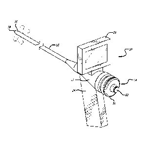
[0029] FIG. 1 is a perspective view showing an embodiment of an endoscope
made in accordance with the present invention having visualization components
along
with steering capability to control the angular positions of the steerable
distal end
of the bendable shaft of the device.
[0030] FIG. 2 is a
perspective view of the endoscope of FIG. 1 with a
endotracheal tube and a tongue retractor mounted on the bendable shaft of the
endoscope.
[0031] FIG. 3A is
a perspective view of the embodiment of the tongue retractor
shown in FIG. 2.
[0032] FIG. 3B is
a side perspective view of the handle of the tongue retractor
shown in FIG. 3A.
[0033] FIG. 3C is
a side perspective view of the upper shell which forms a portion
of the tongue retractor of FIG. 3A.
[0034] FIG. 3D is
a perspective view of the lower shell which forms a portion of
the tongue retractor of FIG. 3A.
[0035] FIG. 3E is a perspective view of another embodiment of a tongue
retractor made in accordance with the present invention.
[0036] FIG. 3F is
a view showing the mating surfaces of the upper shell and
lower shell making up the embodiment of FIG. 3E.
[0037] FIG. 3G is
a perspective view of the upper shell, lower shell and handle
portion which form the tongue retractor of FIG. 3E.
[0038] FIG. 3H is
a side view of the embodiment of the tongue retractor shown in
FIG. 3E.
- 14 -
CA 02906630 2015-09-14
WO 2014/151392
PCT/US2014/025642
[0039] FIG. 4 is a
perspective view (with top of outer casing removed) of an
embodiment of a stand-alone stylet made in accordance with the present
invention
having steering capability to control the angular position of the steerable
distal end of
the bendable shaft of the device.
[0040] FIG. 5 is a
side elevational view, partially in cross-section, showing the
tongue retractor of FIG. 2 being steered into the oral cavity of a patient.
100411 FIG. 6 is a
side elevational view, partially in cross-section, showing the
endoscope and endotracheal tube of FIG. 2 being steered into the trachea.
[0042] FIG. 7 is a
side elevational view, partially in cross section, showing the
endoscope of FIG. 2 being removed from the oral cavity of a patient, the
balloon cuff of the endotracheal tube being inflated and the handle portion of
the
tongue retractor being removed from the upper and lower shells.
100431 FIG. 8 is a
side elevational view, partially in cross section, showing the
upper shell and lower shell of the tongue retractor being split from each
other in
order to remove the tongue retractor from the patient.
[0044] FIG. 9 is a
side elevational view, partially in cross-section, showing the
endotracheal tube placed within the trachea of the patient.
[0045] FIG. 10 is
a perspective view showing an embodiment of a steering control
mechanism and its associated components for steering the distal end of the
bendable
shaft.
[0046] FIG. 11 is
a side elevational showing an embodiment of a center tube which
forms a apart of the device shown in FIG. 10.
[0047] FIG. 12 is
a cross-sectional view of an embodiment of a composite
bendable shaft made in accordance with the present invention.
[0048] FIG. 13 is
another cross-sectional view of an embodiment of a center tube
made in accordance with the present invention.
- 15 -
CA 02906630 2015-09-14
WO 2014/151392
PCT/US2014/025642
[0049] FIG. 14 is a cross-sectional view of an embodiment of a control
mounting
disc which forms a part of the steering control mechanism.
[0050] FIG. 15 is a perspective view of an embodiment of a control mounting
disc
which forms a part of the steering control mechanism.
[0051] FIG. 16 is a perspective view of an embodiment of a control case
which
forms a part of the steering control.
[0052] FIG. 17 is a cross-sectional view of the control case of FIG. 16.
[0053] FIG. 18 is a perspective view with a portion of the outer casing
removed
showing the endoscopic version of the present invention with its handle
portion and
the cables extending into the lumen of the center tube.
[0054] FIG. 19 is a side elevational of an embodiment of a stylet made in
accordance with the present invention with a endotracheal tube extending over
the
shaft of the instrument.
[0055] FIG. 20 is a side elevational along line 20 of FIG. 19 showing the
steering control mechanism in an unlocked position.
[0056] FIG. 21 is a cross-sectional view of the distal end of the bendable
shaft
taken along line 21 of FIG. 19.
[0057] FIG. 22 is a side elevational of the stylet of FIG. 19 with the
malleable
shaft shaped to a particular configuration.
[0058] FIG. 23 is a side elevational view along line 23 of FIG. 22 showing
the
proximal end of the stylet with the steering control mechanism in a locked
position.
[0059] FIG. 24 is a side elevation of the stylet of FIG. 22 with the
flexible
distal tip steered to another particular angulation.
[0060] FIG. 25 is a cross-sectional view along line 25 of FIG. 24 showing
the
proximal end of the stylet with the steering control mechanism in a locked
position.
- 16 -
CA 02906630 2015-09-14
WO 2014/151392
PCT/US2014/025642
DETAILED DESCRIPTION OF THE PREFERRED EMBODIMENTS
[0061] Embodiments
of devices made in accordance with the present invention
will now be described in detail with reference to the accompanying drawings.
Referring initially to FIGS. 1 and 2, a hermetically-sealed endoscope 10 made
in
accordance with the present invention includes an outer casing 12 which houses
a
steering control mechanism 14 that can be manually manipulated to steer the
distal
end 16 of a flexible or malleable shaft 18 to a number of different angular
positions. The steerable distal end 16 is shown in its neutral position (where
the
distal end is substantially straight) which maximizes the number of different
angular
positions that can be taken achieved. Just a few of the numerous angular
positions
that can be attained by the distal end 16 are represented by dotted lines
appearing in
FIG. 1. A removable endotracheal tube 20 (FIG. 2) with an inflatable balloon
cuff 22
located at its distal end and a t- piece fitting 23 at its proximal end can be
placed co-
axially over the flexible or malleable shaft 18 to allow the endotracheal tube
20 to be
placed into the opening of the desired body cavity. While the present
invention is
shown and described as an endoscopic instrument used in a tracheal intubation
procedure, it should be appreciated that the present invention can be used in
a number
of medical procedures and can be adapted in size and shape to fit other body
cavities
of the patient. Additionally, the present invention in endoscopic form can be
used in
non-medical applications as well.
[0062] The
endoscope 10 includes a handle 24 which enable the medical
practitioner to firmly grasp the instrument during the medical procedure. The
endoscope 10 includes a visualization system incorporated into the device
which
includes a video monitor or screen 26 mounted on the outer casing 12 just
above
the handle 24 to provide the practitioner with a clear view of images
appearing at
the distal end 16 of the flexible or malleable shaft 18. The endoscope 10
includes
image transmitting components and light transmitting components for providing
illumination at the distal end 16, which are described in greater detail below
As can
be seen in FIGS. 1 and 2, a wide angle lens 28 extends from the distal end 16
of the
bendable shaft 18. The visualization and illumination components which can be
- 17 -
CA 02906630 2015-09-14
WO 2014/151392
PCT/US2014/025642
incorporated into the endoscope 10 can extend from the outer casing through an
internal lumen of the shaft 18 to the distal end 16. A removable battery pack
(not
shown) can be placed with the handle 24 to power the visualization and
illumination components. Both the video screen 26 and battery pack can be
easily
removed from the outer casing 12 to allow the unit to be immersed in a
sterilizing
solution without compromising the steering control mechanism 14 or other
visualization components housed within the shaft 18 and outer casing 12. As
will be
described below in greater detail, the endoscope 10 can be hermetically sealed
to
protect the internal components from the sterilization solution.
[0063] The
endoscope 10 includes a number of control cables having segments
(shown in FIGS. 4, 10, 12 and 15) which extend from the steering control
mechanism 14 to the distal end 16 of the shaft 18. These control cables are
described in greater detail below. The steering control mechanism 14 is
designed to
apply tension to these control cables singly or in combination so that the
distal end 16
will move into any one of various angular positions. The practitioner can
manipulate
the steering control mechanism 14 by simply pushing the components forming the
steering control mechanism with her/his fingers to cause the control cables to
move
resulting in the distal end 16 being bent to the desired angular position.
[0064] Referring
specifically to FIG. 2, a tongue retractor 30 is shown positioned
with the endoscope 10 of FIG. 1. The tongue retractor 30 is designed to extend
co-
axially over the endotracheal tube 20 and the flexibly-configured shaft 18 of
the
endoscope 10. As its name implies, the tongue retractor 30 is used to move the
patient's tongue out of the way in order to better visualize the vocal cords
which
comprise the opening of the trachea and are therefore the target of
endotracheal
intubation. The upper portion of the tongue retractor 30 (the portion which
actually
contacts the tongue) is somewhat stiff to allow the practitioner to move the
tongue
during the procedure. The tongue
retractor 30 functions somewhat like a
laryngoscope in it is used to retract and control the position of the tongue
to create a
passage for visualizing the vocal cords and placing the tube. The conduit
formed by
- 18-
CA 02906630 2015-09-14
WO 2014/151392
PCT/US2014/025642
the current device also protects the visualization components from oral
secretions
that can obscure the view and require removal and cleaning of the device.
[0065] The
particular embodiment of the tongue retractor 30 of FIG. 2 is shown in
greater detail in FIGS. 3A-3D. As can be seen in FIGS. 3A-3D, the tongue
retractor
30 includes an upper shell 32 connected to a bottom shell 34. The upper shell
32
and lower shell 34 of the tongue retractor form a lumen (a conduit) through
which the
flexible shaft 18 of the endoscope 10 and the endotracheal tube 20 may pass
through in order to position the distal end of the shaft 18 within the
patient. The
upper shell 32 acts like the blade of a laryngoscope in that this upper shell
32 contacts
and moves the patient's tongue out of the visual field. Both the upper and
lower shells
32 and 34 include an outwardly projection finger tab 35 which can be grasped
by the
practitioner to split the upper and lower shells 32 and 34 from each other.
The
upper shell 32 includes a pair of grooves 36 which extend along the length of
the shell
and are adapted to receive a pair of flanged edges 37 formed along the outer
edges
of the lower shell 34. This structure allows the upper shell 32 to be split
from the
lower shell 34 once the retractor 30 is to be removed from the patient. A
handle 39
is attached to the upper and lower shells 32 and 34 to provide the
practitioner with
a structure to grasp when placing the tongue retractor into the oral cavity of
the
patient. Further, this handle 39 allows the operator to displace the tongue of
the
patient in order to place the distal end of the retractor 30 in the vicinity
of the
tracheal opening. The handle 39 is removable from the upper and lower shells
32
and 34 to allow the practitioner to tightly place an oxygen mask on the
patient, and
ventilate if needed during the procedure, while the upper and lower shells 32
and 34
remain in position within the patient's oral cavity.
[0066] The upper
shell 32 of the retractor can be made from a stiff plastic material
which provides sufficient stiffness when retracting the patient's tongue. The
upper
shell could alternatively be made from a malleable material, such as, but not
limited to
a malleable aluminum or copper, which would allow the medical practitioner to
bend
the shell to a desired configuration to conform with the anatomy of the
patient. The
lower shell 34 can be made from a material which is less stiff and more
flexible than
- 19 -
CA 02906630 2015-09-14
WO 2014/151392
PCT/US2014/025642
the material used to manufacture the upper shell 32. The softness of the lower
shell
34 allows the two shells 32 and 34 to be more easily split from each other.
Moreover, the groove 36 formed in the harder upper shell 32 would be stiffer
and
would provide a stronger structure for accepting the softer, mating edge of
the lower
shell 34. The materials used to form these shells 32 and 34 could also be
plastics
well known in the medical arts.
[0067] The upper
shell 32 of the tongue retractor 30 may include a distal
positioning member 29 designed to fit within the epiglottic vallecula, the
depression
formed between the tongue and epiglottis. The epiglottic vallecula is another
important reference location used during the intubation of the trachea. The
distal
positioning member 29 is to be placed as far as possible into the epiglottic
vallecula in order to retract the epiglottis and facilitate direct
visualization of the vocal
cords. The distal positioning member 29 will help to prevent the retractor
blade from
being pushed distally any further once engaged with the epiglottic vallecula.
In this
fashion, the tongue retractor will function very much like a laryngoscope.
Alternatively, the tongue retractor could be made without this distal
positioning
member 29. The tongue retractor 30 is shown having a pre-shaped curve which
will
help match the anatomy of the patient. It should be appreciated that the
tongue
retractor 30 could be made with any number of different curves and different
sizes to
match the different anatomies that may be encountered during the medical
procedure.
Additionally, as is noted above, the upper shell 32 or a portion of the upper
shell 32
can be made from a malleable material which provides the physician with the
ability
to bend the retractor 30 in order to change its curvature, if needed.
[0068] The upper
and lower shells 32 and 34 of the retractor blade also include
openings 31 formed at the proximal end of the shells which receive a pair of
arms 33
extending from the end of the handle 39 (see FIG. 3B). These arms 33 are
insertable
into the openings 31 to attach the handle 39 to the upper and lower shells 32
and
34. Each arm 33 includes an end designed to engage the edge of the lower shell
34
once the end extends out of the opening 31. These ends of the arms 33 are
designed
to bias outwardly to engage the edge of the lower shell 34 in order to lock
the handle
- 20 -
CA 02906630 2015-09-14
WO 2014/151392
PCT/US2014/025642
39 in place once the arms 33 are fully extended into the openings 31. The end
of each
arm can be easily moved inwardly to allow the arms 33 to be removed from the
openings 31. The arms 33 help to keep the upper shell 32 and lower shell 34 of
the
retractor blade together once the arms 33 are placed into the openings 31.
100691 FIGS. 3E-3H show another embodiment of a tongue retractor 30 which can
be used with the present invention. This particular retractor 30 similarly
includes
retractor blade including an upper shell 32 and lower shell 34. This
particular tongue
retractor 30 includes a removable handle 39 having a simple release mechanism
to
easily connect and disconnect the handle 39 to the upper and lower shells 32
and 34.
As can be seen in FIGS. 3E and 3G, the handle 39 is shaped with a pair of
slotted
openings 41 which receive a pin 43 (only one of which is shown in FIG. 3G)
formed
on the lower shell 34. The handle 39 is designed to extend over an upright
support
member 45 extending from the upper shell 32. In use, the handle 39 is placed
over
the upright support member 45 and pivoted to allow the pins 43 to engage the
slotted
openings 41 to connect the handle 39 to the shells 32 and 34.
100701 FIG. 3F
shows the profile of the mating surfaces of upper and lower shells
32 and 34 depicted in FIGS. 3E and 3G which allows the shells 32 and 34 to be
peeled
away from each other as is discussed above with respect to the embodiment of
FIG.
3A-3D. It should be appreciated that there are a number of ways to removably
join the upper shell 32 to the lower shell 34. For example, when a malleable
material is used to form a portion of the upper shell 32, a tongue and groove
joint could be used to help maintain the shells together. Other examples
include,
but are not limited to, dove tail joints which provide a bit more rigidity to
the joint.
In some instances, the joint would be more of a sliding type which would
require
the upper shell to slide relative to the lower shell and split the shells
apart. It should
again be appreciated that the lower shell 34 or a portion of the lower shell
34 also
could be made from a malleable material to allow the tongue retractor to be
pre-bent
by the operator to a desired shape.
- 21 -
CA 02906630 2015-09-14
WO 2014/151392
PCT/US2014/025642
100711 The
endoscope 10 made in accordance with the present invention can be
hermetically sealed to allow the instrument to be immersed in a sterilizing
solution. As can be seen in FIGS. 1 and 2, a flexible control case 38 extends
over a
portion of the outer casing 12 and is attached to a portion of the steering
control
mechanism 14. This flexible control case 38 can be made from an elastomeric
material which is stretchable to allow the steering control mechanism 14 to be
manipulated by the practitioner without impedance but yet provides a barrier
to
sterilizing solutions. The
remaining portions of the endoscope 10 can be
manufactured to be hermetically sealed to allow the entire instrument (minus
the video
screen and battery pack) to be immersed in a sterilizing solution. For
example, an
elastic sealant may be required to seal any small openings or gaps formed
between
the visualization and illumination components mounted at the distal end of the
shaft.
More detailed drawings of this particular embodiment of the control case 38
are
provided in FIGS. 5, 10, 16 and 17.
100721 FIGS. 5-9
depict how the endoscope 10, endotracheal tube 20 and tongue
retractor 30 can be used to perform a tracheal intubation. FIG. 5 shows the
tongue
retractor 30 being initially inserted into the patient's oral cavity. This
tongue
retractor 30 moves the patient's tongue and creates a conduit for receiving
the
remaining components of the system. An upward force can be applied against the
tongue by the practitioner so that the tongue will be retracted upwards or
sideways to
provide better visualization of the vocal cords. Since the tongue retractor is
made
from a relatively stiff material, it should easily move the tongue and
associated
tissue. The handle 39 provides a suitable structure which allows the
practitioner to
apply the necessary force to properly retract the patient's tongue. FIG. 6
shows the
composite system consisting of the endoscope 10 with the endotracheal tube 20
co-
axially disposed over the bendable shaft 18 of the endoscope being advanced
into the
patient's trachea. The endotracheal tube 20 is positioned within the lumen
formed
by the tongue retractor 30 and is steered into place towards the tracheal
opening. The
visualization and lighting instruments of the endoscope will allow the
practitioner to
clearly view the location of the vocal cords once the tongue has been
retracted.
- 22 -
CA 02906630 2015-09-14
WO 2014/151392
PCT/US2014/025642
Once the vocal cords are identified, the practitioner can then steer the
distal end
16 of the endoscope 10 utilizing the steering control mechanism 14 to allow
the
practitioner to maneuver the distal end 16 and endotracheal tube 20 into the
opening
of the trachea, as can be seen in FIG. 6. Alternatively, the practitioner can
steer the
distal end 16 of the flexible shaft 18 into alignment with the opening of the
trachea
allowing the endotracheal tube 20 to be positioned directly outside of the
trachea
opening. The distal end of the endotracheal tube 20 can then be carefully
pushed
into the trachea. This alternative approach eliminates the need to actually
position
the distal end 16 of the endo scope into the opening of the trachea. However,
absolute
confirmation of correct positioning in the trachea is afforded by
visualization of the
concentric tracheal rings, which lie distal to the vocal cords and are easily
distinguished from the interior of the esophagus.
[0073] The
benefits of the endoscope 10 of the present invention include the
ability to steer the distal end 16 of the shaft 18 to any advantageous angular
position, allowing the practitioner to simply push the endotracheal tube 20
into the
opening of the trachea. The present invention allows the practitioner to
utilize a
single instrument to advance the endotracheal tube 20 into the trachea thus
eliminating
the need to manipulate two separate components, such as a laryngoscope and a
stylet.
In a conventional endoscope both hands are needed to achieve true 360 angular
rotation, whereas this is here accomplished by the same hand. It should be
appreciated that the malleable shaft 18 can be pre- shaped by the practitioner
before
it is inverted into the patient's oral cavity or it can be reshaped if the
practitioner is
having trouble positioning the distal end 16 near the opening of the trachea.
The
malleability of the shaft 18 thus provides the practitioner with another means
by
which the device can be manipulated and bent to a desired configuration in
order to
conform with the particular anatomy of the patient.
[0074] FIG. 7
depicts the step of inflating the balloon cuff 22 of the endotracheal
tube 20 via syringe 41 which seals the the trachea around the tube. FIG. 7
also depicts
the endoscope 10 being withdrawn from the patient's oral cavity leaving only
the
endotracheal tube 20 and tongue retractor 30 in place. Ventilation of the
lungs
- 23 -
CA 02906630 2015-09-14
WO 2014/151392
PCT/US2014/025642
can be done now with the conduit in place or, after the endoscope 10 has been
removed, the handle 39 can be removed from the upper and lower shells 32 and
34, allowing the shells 32 and 34 to be split away from each other and removed
from the patient, as is shown in FIG. 8. In use, the finger flanges 35 formed
on each
of the upper and lower shells 32 and 34 could be grasped with an outward force
being applied to each flange 35 to start the splitting action between the
upper shell 32
and lower shell 34. Both the upper and lower shells 32 and 34 could be
retracted
simultaneously from the patient's oral cavity as the splitting action is being
applied to
the retractor 30 until both shell 32 and 34 are removed from the patient's
oral cavity.
[0075] In an
alternative method, the fitting 22 connected to the proximal end of the
endotracheal tube 20 could be removed to allow the tongue retractor 30 to be
co-
axially retracted from the tube 20. The fitting 22 could be reconnected to the
end of
the endotracheal tube 20 after the tongue retractor 30 has been removed. In
this
fashion of removing the tongue retractor 30, there would be no need for a
splittable
upper and lower shell 32 and 34. Rather, the tongue retractor 30 could be made
as
a solid piece since the retractor 30 could be simply slide over the
endotracheal
tube 20 in order to remove it from the patient's oral cavity
[0076] FIG. 9
shows the endotracheal tube 20 properly positioned in the patient's
trachea to maintain an open airway and to serve as a conduit through which
certain
controlled gases and/or drugs can be administered. If the proximal fitting 23
was
removed, it could be placed back on the tube 20. The proximal fitting 23 of
the
endotracheal tube can then be connected to an anesthesia breathing circuit,
bag
valve mask device, a mechanical ventilator or other instrument used in the
medical
procedure.
[0077] Another
particular embodiment of the present invention is shown in
FIG. 4. In this figure, the present invention is shown as a stand-alone stylet
40 which
includes many of the same components of the endoscope 10 disclosed in FIGS. 1
and
2. The main differences between the stand-alone stylet version and the
endoscopic
version of the present invention is the lack of visualization/illumination
instruments
- 24 -
CA 02906630 2015-09-14
WO 2014/151392
PCT/US2014/025642
and a handle in the stylet design. During use, the practitioner can hold the
outer
casing 12 in order to place the endotracheal tube 20 in place. The stylet
could be
grasped with the four fingers and palm of the hand and the disc depressed by
the
thumb in the appropriate location to achieve the desired angulation. Further
description of the steering control mechanism 14 appears below in conjunction
with
the stylet 40 depicted in FIG. 10. The particular stylet 40 shown in FIG. 4
can be
utilized, for example, in a conventional intubation procedure in which a
laryngoscope
is being used. This stylet 40 provides the practitioner with the ability to
steer the
distal end of the bendable shaft 18 while the stylet 40 is still in the
patient's oral
cavity. The bendable shaft 18 can also be pre-shaped, as needed, to work
around the
particular anatomy of the patient.
[0078] It should
be noted that the stylet 40 may include a side port (shown in
FIG. 10) formed in the outer casing 12 which is capable of receiving, for
example,
the video cable of a visualization system. The video cable and lens could be
placed into the internal lumen of the bendable shaft 18 to the distal end 16
of the
shaft 18. The video system could thus provide the image appearing at the
distal end
16 of the shaft 18 on a remote video screen or monitor.
[0079] One
particular embodiment of a steering control mechanism 14 which can
be implemented to steer the distal end of the bendable shaft is disclosed in
FIGS. 1,
4 and 10. While the steering control mechanism 14 is shown incorporated into
an
outer casing 12 used for the stand-alone stylet 40, as shown in FIGS. 4 and
10, this
same steering control mechanism 14 can be incorporated into the endoscopic
version of the present invention shown in FIGS. 1 and 2. The endoscope 10 of
FIGS.
1 and 2 will utilize a different outer casing 12 in order to form the handle
24 and a
mount for the video screen 26 but nonetheless can utilize the same steering
control
mechanism 14 and control cables shown in FIGS. 4 and 10 and disclosed below.
[0080] The
steering control mechanism 14 (FIGS. 10 and 14) includes a control
mounting disc 42 which is connected to the control cables 44 used to move the
distal
end 16 of the bendable shaft 18. The control mounting disc 42 is housed within
the
- 25 -
CA 02906630 2015-09-14
WO 2014/151392
PCT/US2014/025642
outer casing 12 and is pivotally mounted to a center tube 46 which extends
through
the outer casing 12. As can best be seen in FIG. 11, the center tube 46
includes a
distal portion 48 and a proximal portion 50. The proximal portion 50 is
utilized as a
pivoting mechanism which allows the control mounting disc 42 to move the
control
cables 44 to change the angular position of the distal end 16 of the bendable
shaft
18. A pivot member 52 (FIG. 14) is located at the end of the proximal portion
50
and is designed to come into contact with a surface formed on the control
mounting
disc 42. In the particular embodiment disclosed herein, the control mounting
disc 42
includes a conically-shaped recess 54 (FIG. 14) which is designed to pivotally
engage
the pivot member 52 of the center tube 46. In this fashion, the control
mounting
disc 42 can be pivoted/tilted to any one of a number of different positions to
move
the control cables 44 and move the distal end 16 to the desired angular
position. For
example, pressure on the disc at the 12 o'clock position will cause the disc
to tilt
outwards at the six o'clock position, exerting tension on the cable attached
there,
causing the distal tip to be deflected downwards. It should be appreciated
that the
pivot member/conical recess components which allow the control mounting disc
42 to
pivot is just one of a number of components which can be used to achieve a
pivoting/tilting action. Also, for example, the pivoting member 52 could be
formed on the control mounting disc 42 itself with the conical recess formed
on the
center tube 46. Additional pivoting joints, such as a universal joint, could
be used as
well to pivotally connect the components together. The pivot itself could be
eliminated entirely and the disc be allowed to "float" on the conical spring,
with
the spring itself forming the pivot. It should be appreciated that the
steering
control mechanism 14 can utilize any number of different moving mechanism
which
will allow the practitioner to manually move the steering control disc 42. A
pivoting/tilting mechanism is shown for purposes of disclosure.
[0081] The control
cables 44 are shown attached to the control mounting disc
42 in FIG. 10. As can be seen in FIG. 12, each control cable 44 extends
through
the internal lumen 53 of the center tube 46, the internal lumen 54 of the
bendable
shaft 18 all the way to the internal lumen 55 of the distal end 16. In this
particular
- 26 -
CA 02906630 2015-09-14
WO 2014/151392
PCT/US2014/025642
embodiment of the invention, each control cable 44 includes a first end 56 and
a
second end 58 which are both connected to the distal end 16 of the bendable
shaft 18.
Each of the first and second ends 56 and 58 are attached to a plate 59 located
at the
distal end 16 as is shown in FIG. 12. The ends 56 and 58 of each cable 44 are
spaced apart from the other on the plate 59 that the distal end 16 can be
moved to a
desired angular position whenever the cables 44 are pulled a certain amount.
It
should be appreciated that another cable configuration could consist of one
large
closed loop, anchored at the distal tip but allowed to slip through the
control disc as it
is adjusted and locked in position.
[0082] Since each
end of the control cable 44 is attached to the distal end 16 of
the shaft 18, a closed loop 60 is formed (FIG. 14) and is connected to the
control
mounting disc 42. The steering control mechanism 14 includes a locking
mechanism
which locks each of the loops 60 to the control mounting disc 42. A fastener,
such
as a compression screw 62, is a simple component which can be used to lock
each
cable loop 60 to the control mounting disc 42. As can be seen in FIGS. 10, 14
and
15, a mounting structure 64 which includes screw threads 66 can be used to
attach and
lock the cable loops 60 to the disc 42. The screw threads 66 allow the screw
62 to be
screwed downward to contact teach cable loop 60 and lock them in place. The
mounting structure 64 (FIG. 14) includes pairs of lateral openings 68 which
receive
each of the cable loops 60. Openings 70 in the face of the control mounting
disc 42
allow the cables 44 to extend through these openings. The control cables 44
then
extend though the openings 71 (FIG. 13) which are formed in the proximal
portion 48 of the center tube 46 which extend into the internal lumen 53 of
the
center tube 46 (FIG. 12). In this manner, the mounting structure 64 maintains
the
cable loops 60 disposed within the lateral openings 68 to center the loops and
prevent
them from moving in a lateral fashion relative to the control mounting disc
42. Each
loop 60, however, can move through the openings 70 until the loop 60 is locked
in
place by the screw 62. In use, the user merely screws the screw 62 down until
it
presses the loop 60 against a face of the control mounting disc 42. The loop
60 can
be unlocked by simply rotating the screw to releasing the force being exerted
on
- 27 -
CA 02906630 2015-09-14
WO 2014/151392
PCT/US2014/025642
the loop 60 by the screw 62. As can be seen in FIGS. 10 and 15, the loops 60
of
the control cables cross each other near the center of the control mounting
disc 42
to allow a single screw 62 to lock the loops 60 in place. It should be
appreciated
that each loop 60 of each control cable 44 could be individually locked by a
suitable locking mechanism as well without departing from the spirit and scope
of the
present invention.
[0083] The control
mounting disc 42 pivots/tilts about the pivot member 52
formed on the center tube 46 (see FIG. 25). A biasing member, such as a spring
72, (see FIG. 4) is mounted within the outer casing 12 and comes in contact
with
the bottom face of the control mounting disc 42. The other end of the spring
72
contacts a spring mount (FIG. 4) mounted within the interior of the outer
casing 12.
The spring 72 provides a biasing force on the control mounting disc 42 to move
the disc 42 to its neutral position, as is shown in FIG. 4, whenever there are
no forces
acting on the control disc 42. Accordingly, when the practitioner removes
his/her
fingers from the control mounting disc 42, the spring 72 will move the disc 42
back to its neutral position. Whenever the practitioner pushes the control
mounting disc 42 in a certain manner, the control cables 44 will either be
pulled or
relieved of tension to cause the distal end 16 to be moved to a particular
angular
position. The control cables 44 thus cooperate with each other to achieve the
desired
angular positioning of the distal end 16.
[0084] As can be
seen in FIGS. 1, 2 and 4, the steering control disc 42 is
encased by the control case 38 which helps to maintain a hermetic seal to the
outer
casing 12. This control case 38 can be made from an elastomeric material and
may
be bonded to the outer edges 76 of the casing 12. This cover 38 is designed to
come into contact with the control mounting disc 42 to allow the control
mounting
disc 42 to freely articulate on its pivot member 52 while still providing a
hermetic
seal. Further details relating to the structure of the particular control case
38 shown
in the drawings are disclosed in FIGS. 15 and 16 and are discussed below. The
control case 38 may include, for example, grooves 78 formed therein for
receiving and
holding the edges of the control mounting disc 42. This control case 38 is
designed to
- 28 -
CA 02906630 2015-09-14
WO 2014/151392
PCT/US2014/025642
bend and stretch as may be necessary in order to allow the control mounting
disc
42 to freely pivot within the interior of the outer casing 12.
[0085] FIG. 12
show a particular embodiment of the shaft 18 and its distal end
16. As can be seen in FIG. 12, the shaft 18 is attached to the distal portion
48 of
the center tube 46 and extends to a short length of flexible tubing which
forms the
distal end 16 of the shaft 18. The shaft 18 can be made from a length of
malleable
tubing 80 which possess sufficient bending strength to maintain a pre-shaped
configuration. The shaft can also be as flexible as a conventional endoscope,
and
conform passively to a conduit, whether it be a biological or artificial one.
For
example, the shaft 18 can be made from a malleable aluminum similar to the
malleable aluminum material used in intubating stylets such as those sold and
manufactured by Legend Medical Devices of South El Monte. CA. Other similar
malleable materials, such as copper, could also be used. The physical
properties of
the malleable material allow the practitioner to bend the shaft to a desired
configuration without the need to apply excessive force. The malleable
material
should be strong enough to maintain the configuration in the absence of a
strong
applied pressure. The proximal end 82 of the malleable tubing 80 is attached
to the
distal portion 48 of the center tube 46. The distal end 84 of the malleable
tubing
80 is, in turn, attached to the easily deformable tubing 86 which forms the
distal
end 16 of the shaft 18. A helical spring may be used to add flexibility and a
biasing
element to the distal tip. Suitable and known ways of bonding the tubing
together can
be implemented. The flexible tubing 86 has a mounting plate 59 which extends
within the lumen 55 of the tubing 86. As was mentioned above, this mounting
plate
59 is connected to the ends 56 and 58 of the control cables 44. Also, the
mounting
plate 59 includes openings for receiving optic fibers 90 which extend out of
the plate
59 in order to provide illumination at the distal end 16 of the shaft 18. A
lens 28,
which can be of the wide-angle or "fish-eye" type, extends from the mounting
plate
59 as well and is connected to an optic cable 92 which extends through the
lumen 54
of the shaft 18 into the outer casing 12 where it is connected to the video
monitor 26.
The optic fibers 90 and optical cable 92 also extend through the lumen 53 of
the
- 29 -
CA 02906630 2015-09-14
WO 2014/151392
PCT/US2014/025642
center tube 46 and are attached to the power source. The distal end of each
optic
fiber includes a lens 100 (see FIG. 4) which provides illumination at the
distal end 16
of the shaft 18.
[0086] FIG. 13
shows an embodiment of the present invention in which the
stand- alone stylet 40 utilizes a malleable rod 94 which extends through the
center
tube 46 and through the lumen of the bendable shaft. This malleable rod can be
connected to the center tube 46 and can extend at least partially along the
length of the
shaft. In this particular embodiment, a deformable tubing can be used to form
the shaft
with the malleable rod providing the bendability and strength needed to
maintain the
pre-shaped configuration. In this manner, the rod and deformable tubing
cooperate
to obtain the properties needed for the malleable shaft.
[0087] Referring
now to FIGS. 4, 10 and 14-18, various components forming the
embodiment of the stylet and endoscope are shown in greater detail and are
further
described. FIGS. 14 and 15 show further details of particular embodiment of
the
control mounting disc 42 shown in FIGS 4 and 9. As can best be seen in FIG.
15,
the cross openings 68 which extend into the face of the disc 42 create
abutments
which prevent each loop 60 from moving laterally on the disc 42. The mounting
structure 64 is shown in FIG. 15 as being cut into four separate sections but
is still
capable of receiving the screw 62 which tightens down on each of the loops 60
to
lock the loops 60 to the disc 42. The conically-shaped recess 54 which pivots
on
the pivot member 52 of the center tube 46 can best be seen in FIG. 14. The
face of the control mounting disc 42 includes openings 98 which are adapted to
receive projections 100 formed on the inner surface of the control case 38
(see
FIG. 17).
[0088] The control
case 38 is shown in greater detail in FIGS. 16 and 17.
FIG. 16 shows the grooves 76 which are designed to engage the structure on the
outer
edge of the casing 12 (see FIG. 10). The particular control case 38 of FIGS.
16 and
17 show a region where a bellows structure 102 is formed to allow the control
case 38
to flex and bend when the control mounting disc 42 is being manipulated by the
user.
- 30 -
CA 02906630 2015-09-14
WO 2014/151392
PCT/US2014/025642
The projections 100 formed on the inner surface of the control case 38 are
designed to
be placed within the openings 98 formed on the control mounting disc 42. These
projections 100 can be bonded within these openings 98 to ensure that the
control
case 38 remains connected to the control mounting disc 42. The connection of
these
projections 100 into the openings 98 on the disc 42 also helps to prevent the
disc 42
from possibly rotating on the pivot member 52. The remaining portions of the
control case 38 could likewise be bonded to the casing 12 to create and
maintain the
instrument hermetically sealed. A suitable elastomeric material can be used to
form
the control case 38.
[0089] FIG. 18
shows a view of the handle portion of the endoscope 10 of
FIGS. 1 and 2 with a portion of the handle removed to show how a battery pack
could
be placed within the handle 24 in order to provide a portable power source to
the
visualization and illumination components incorporated into the endoscope. The
view also shows a side opening 99 in the center tube 46 which leads to the
lumen
53 of the center tube 46 and the lumen 54 of the shaft 18. As can be seen in
FIG. 18,
a cable is shown entering the side opening 99 in the center tube 46.
[0090] The inner
lumens of the shaft 18 and center tube 46 may include spacers
(not shown) which are basically flat disks (like the mounting plate 59) having
openings for receiving the various cables and optic fibers used in conjunction
with
the endoscope/stylet of the present invention. Such spacers could be placed
along
the length of the shaft 18 and center tube 46 to help prevent the various
cables and
optical fibers from tangling with each other during usage.
[0091] FIGS. 19-25
depict the benefits of the present invention in utilizing a
mechanism which allows the control cables 44 to be "reset" after the flexible
or
malleable portion of the shaft 18 has been bent to a new configuration. One of
the
problems associated with prior art devices which utilize control cables or
wires to
move the distal end of the shaft is the fact that the control wires can have
unwanted tension applied to one or more of them whenever the shaft is bent to
a
new configuration. This occurs if the tension applied to the control cables is
- 31 -
CA 02906630 2015-09-14
WO 2014/151392
PCT/US2014/025642
changed passively by the bending of the shaft, causing the control cable to
move
the control disc or the distal tip to an undesired angular position. The
present
invention is designed to eliminate the possibility that the bending of the
bendable
shaft will have any impact on the control cables and disc.
[0092] The
steering control mechanism 14 of the present invention utilizes loops
60 formed by each of the control wire 44 which allows the attachment point of
the
loops 44 to the control mounting disc 42 be reset after the shaft 18 has been
bent
to a new configuration. FIG. 19 shows the bendable shaft 18 in a substantially
straight configuration (first configuration). The endotracheal tube 20 with
its
inflatable balloon cuff22 extends over a portion of the shaft and is shown in
these
figures covering the steerable distal end of the shaft 18. Since the
endotracheal tube
20 is flexible, the steerable distal end still is capable of changing angular
position
when the steering control mechanism is manipulated. Alternatively, the
endotracheal
tube 20 could be placed further up the shaft 18 so that the steerable distal
end would
be fully or partial exposed during usage. As can be seen in FIG. 20, the
locking
mechanism of the device, namely the screw 62, is in its unlocked position. In
this
position, the control cables 44 and the loops 60 formed by each of the control
cable
44 are free to move. The biasing member, namely the spring (not shown in FIG.
20),
provides a biasing force which keeps the control mounting disc in its neutral
position, as is depicted in FIG. 20. When the shaft 18 is in its first
configuration, the
angular position of the distal end 16 of the shaft is substantially straight
(its neutral
position). This substantially straight or neutral position of the distal end
maximizes
the number of angular positions to which the distal end can be steered by the
steering
control mechanism.
[0093] When the
shaft 18 is in its first configuration, as is shown in FIG. 19,
the lengths of each control cable (the length being defined as its point of
attachment to
the distal end to its point of attachment to the steering control mechanism)
are set.
These control cables have an initial length when the shaft is in its first
configuration.
- 32 -
CA 02906630 2015-09-14
WO 2014/151392
PCT/US2014/025642
100941 FIG. 22
shows the same stylet 40 of FIG. 19 except that the shaft 18 has
now been pre-bent to a curved configuration (a second configuration).
Accordingly,
if the loops 60 of the control cables 44 were initially in the locked position
on the
control mounting disc and the shaft 18 was then bent, there would be tension
draw on some of the control cables, which would then act on the distal end
resulting
in the distal end being moved from its substantially straight (neutral)
position to an
unwanted angular position. Generally, the control cables on the outside radius
of
the shaft bend will have additional tension placed on them as opposed to any
control cables located on the inside radius of shaft bend. Since the distal
end is
flexible, any tension acting on the control cable will be applied to the
distal end 16
since it is easily susceptible to such forces. Such tension will also be
applied to the
control disc 42 as well and displace it from its original neutral position. In
the
drawing of FIG. 23, however, the locking mechanism has remained in the
unlocked
position as the shaft 18 was bent to its second configuration. Since the loops
60 were
unlocked at the steering control mechanism, they are able to move freely,
eliminating
unwanted tension being applied to the control cables or the disc itself. What
in fact
happened is that one segment of the control cable has compensated for the
increase or
decrease in length of the other segment when it is bent from its straight or
neutral position. Accordingly, since the relative length of one or more of the
cable
segments has changed, there is a release of tension on that particular cable
that would
otherwise act on the distal end 16 of the shaft or the control disc.
Accordingly,
the change of the relative lengths of the control cable segments from their
initial
lengths allow the distal end 16 to remain in its desired substantially
straight (neutral)
position, as is depicted in FIG. 22. Likewise, the control disc 42 will now be
returned
to or remain in its initial, neutral position, allowing full functionality to
further
control the distal tip 16. One can easily appreciate that if there was no such
mechanism to return the device to a neutral position, functionality would be
lost. For
example, if the shaft were bent downward, towards six o'clock, tension would
be
applied to the cables on the outside of the curve, in this instance, the
cables on
the upward or twelve o'clock side of the shaft, causing the distal tip to bend
upward, towards twelve o'clock and the disc would passively react by tilting
-33 -
CA 02906630 2015-09-14
WO 2014/151392
PCT/US2014/025642
inward at twelve o'clock and outward at six o'clock. Full ability to tilt the
disc and
control the distal tip would then be impaired. But if the cable segments are
first freed
up at the disc, the shaft configured as desired, then the cable segments
locked at the
disc, full functionality can be maintained. It is only after the shaft 18 is
bent into
the desired second configuration that the loops 60 should be locked by the
locking
mechanism to the control mounting disc 42. The resulting structure allows the
shaft 18 to be bent to any one of a number of configurations in order to adapt
to
varying anatomical features of each patient without changing the substantial
straight
(neutral) position of the distal end.
100951 The angular
position of the distal end 16 of the device shown in FIG. 22
can now be easily changed. FIG. 24 shows how the shaft 18 remains in its bent
configuration, as is shown in FIG. 22, with the loops 60 remaining in the
locked position. The practitioner now simply has to manipulate the mounting
disc 42 to change the angular position of the distal end 16 in order to steer
the
device into the desired position. As can be seen in FIG. 25, movement of
the mounting disc 42 (inward at 12 o'clock and outward at 6 o'clock) will
cause a tensioning force to be applied to one or more of the control cables 44
which, in turn, causes those control cables to move the distal end 16 from its
initial
position shown in FIG. 22 (shown in dotted lines in FIG. 24) to its new
angular
position shown in solid lines in FIG. 24. The arrow in FIG. 24 shows the
direction
in which the distal tip 16 moves from its initial position shown in FIG. 22
(dotted
lines) to its new angular position epicted in FIG. 24 (solid lines.).
100961 If there
were no compensating device employed, i.e., if the cables were
fixed to the control disc, when the shaft itself is bent, added tension to the
control
cable 44 will force the control mounting disc 42 to move from its neutral
position to
a tilted position as is shown in FIG. 25 and result in the distal end 16 being
moved
to an undesired angular position. For example, if one was to take both hands,
grasp
the shaft at the midpoint and bend it to the position in FIG. 24, tension
would be
applied to the upper cables, the tip would rise to the position as in FIG. 22
and the
disc would tilt inwards at 12 o'clock. Further adjustment would be impaired.
- 34 -
CA 02906630 2015-09-14
WO 2014/151392
PCT/US2014/025642
However, if the cables are first unlocked from the disc and then the shaft
bent at the
midpoint, little tension will be applied to the disc or distal tip and so
should stay in
the "neutral " position. After the cables are locked, full functionality is
restored.
Using the unlocking and un-locking device herein described, pre-bending of the
shaft
does not limit further adjustments of the tip.
[0097] It should
be appreciated that the first configuration of the bendable
shaft 18 does not have to be substantially straight as is shown in FIG. 19.
The first
configuration of the shaft 18 can be, for example, a curved configuration as
is shown
in FIG 22. The second configuration could be another bent configuration, for
example, which is different from the first configuration. It could also be a
substantially straight configuration. Thus, the present invention is designed
to remove
tension from one or more control cables (by effectively lengthening that
cable) as the
bendable shaft is bent from one configuration to another.
[0098] The present
invention utilizes in one embodiment a loop formed on each
control wire to allow the relative initial length of the control cable to be
changed after
the shaft is to be bent to a new configuration. The loop structure provides a
simple
arrangement which allows the user to quickly change the length of the cables
whenever the shaft needs to be re-shaped to a new configuration. This re-
shaping can
occur in several ways. For example, with the malleably-configured embodiment,
the
shaft is configured outside the oral cavity. If utilizing a conduit, whether
natural or
artificial, the device is unlocked, inserted into the conduit, allowing the
shaft to return
to "neutral," and then locked, allowing full functionality to be restored.
Other
mechanisms could be utilized to allow the initial length of the cable to be
changed
whenever the shaft is to be re-bent to a new configuration without departing
from
the spirit and scope of the present invention.
[0099] Again,
while the present invention is shown and described as an endoscopic
instrument used in a tracheal intubation procedure, it should be appreciated
that the
present invention can be used in a number of medical procedures and can be
adapted
in size and shape to fit other body cavities of the patient. For example, the
-35 -
CA 02906630 2015-09-14
WO 2014/151392
PCT/US2014/025642
endoscopic version of the device could be used in nasal intubation in which
the
endotracheal tube is inserted through the nasal cavity, rather than the oral
cavity, in
order to intubate the patient. Nasal intubation is often used during oral
surgeries. In
such a case, the endotracheal tube is initially mounted to the shaft of the
endoscopic device and then the distal end of the shaft is inserted into the
nasal
cavity. If the shaft of the device has some rigidity, the shaft could be
initially
shaped to fit the anatomy of the patient. The endoscopic tube and shaft of the
device can then be moved simultaneously into the area adjacent to the opening
of
the trachea. The steerable distal end of the shaft can be used to steerthe
endotracheal
tube into the trachea. The visualization components of the endoscopic device
will
assist the practitioner in steering the distal end of the shaft and the
endotracheal tube
into proper position.
[0100] The device
described herein can be made from medical grade materials
which allow the device to be properly sterilized for each new use. The
material could
incorporate biocide chemistry which enhances the sterilization process. It
should be
appreciated that the shaft could have different portions having different
rigidities, as is
described above. The distal end, for example, could be made from substantially
softer materials than the flexible or malleable shafts, which would allow the
distal
end to react preferentially to tension being applied at the distal end. The
distal end
and tip would then flex more than the shaft, allowing fine control at that
portion.
The control cables can be made from plastic or suitable metals or metal
alloys.
The control case can be made from suitable elastomeric materials which allow
for
some stretching. The control mounting disc likewise can be made from suitable
medical grade materials. The visualization equipment (video screens, optical
cables/fibers, batteries) used with the device can be commercially available
equipment. The video screen can be removably mounted to the outer casing and
the
battery pack likewise be removable from the handle portion to allow these
components to be removed prior to placing the device in the sterilizing
solution.
Accordingly, the video screen and battery pack would utilizes quick release
electrical
- 36 -
CA 02906630 2015-09-14
WO 2014/151392
PCT/US2014/025642
connecters which would be incorporated into the outer casing of the device.
Such
connectors are well known in the art.
[0101] While
particular forms of the invention have been described and illustrated,
it will also be apparent to those skilled in the art that various
modifications can be
made without departing from the spirit and scope of the invention.
Accordingly, it is
not intended that the invention be limited except by the appended claims.
-37 -