Note: Descriptions are shown in the official language in which they were submitted.
CA 02907183 2015-09-15
WO 2014/151021 PCT/US2014/024779
ANTIBODY PROFILING, METHODS AND APPARATUS FOR IDENTIFYING AN
INDIVIDUAL OR SOURCE OF A BIOLOGICAL MATERIAL
GOVERNMENT RIGHTS
[0001] This invention was made with government support under Contract
Number
DE-AC07-05ID14517 awarded by the United States Department of Energy. The
government has certain rights in the invention.
FIELD
[0002] Embodiments of the present disclosure relate to analyzing biological
samples to
identify proteins useful in identifying individuals, and more particularly, to
methods and
an apparatus for identifying an individual using such proteins.
BACKGROUND
[0003] The importance of differentiating and identifying individuals based
on biological
samples with a high degree of efficiency and accuracy is presented in various
contexts.
For example, the need for accurate methods of identification is of increasing
importance
in law enforcement as it may be critical to link an individual to a forensic
sample, such as
blood, tissue, hair, saliva, or the like.
SUMMARY
[0004] A method for identifying a source of a biological material that
includes contacting
a sample of a biological material having individual-specific antibodies with
an array
including multiple proteins comprising less than about 200 proteins on a
support to bind
at least a portion of the individual-specific antibodies to the multiple
proteins of the array,
to form immune complexes; applying to the array at least one detection agent
that
includes at least one interacting protein conjugated to a marker, and
contacting the
detection agent with a plurality of control spots in the array to form control
complexes,
wherein each control spot of the plurality includes human Immunoglobulin G;
removing
non-immobilized individual-specific antibodies and unbound detection agent;
detecting
the immune complexes on the array to obtain an antibody profile; detecting an
intensity of
the control complexes to determine if results of the identifying are complete;
and
comparing the antibody profile to a known antibody profile obtained from an
individual.
[0005] A method for identifying a source of a biological material that
includes contacting
a sample of a biological material having individual-specific antibodies with
an array
1
CA 02907183 2015-09-15
WO 2014/151021 PCT/US2014/024779
including multiple proteins comprising less than about 200 proteins on a
support to bind
at least a portion of the individual-specific antibodies to the multiple
proteins of the array,
to form immune complexes; contacting the sample with a plurality of volume
assessment
spots in the array to form volume complexes, each volume assessment spot
including a
predetermined concentration of one or more volume determination proteins;
applying to
the array at least one detection agent comprising at least one interacting
protein
conjugated to a marker to detect the immune complexes and the volume
complexes;
removing non-immobilized individual-specific antibodies and unbound detection
agent;
detecting the immune complexes on the array to obtain an antibody profile;
detecting an
intensity of the volume complexes to determine if a volume of the sample is
sufficient for
an accurate result; and comparing the antibody profile to a known antibody
profile
obtained from an individual.
[0006] A protein array, for identifying an individual, that includes an
array of multiple
proteins having less than about 200 proteins immobilized on a support, wherein
each
protein is known, each protein is immobilized at a known predetermined
location on the
support, and the multiple proteins are configured to bind to at least a
portion of
individual-specific antibodies to form immune complexes; and a plurality of
control spots
as part of the array, wherein each control spot includes human Immunoglobulin
G
configured to form control complexes.
[0007] A protein array, for identifying an individual that includes an
array of multiple
proteins having less than about 200 proteins immobilized on a support, wherein
each
protein is known, each protein is immobilized at a known predetermined
location on the
support, and the multiple proteins are configured to bind to at least a
portion of
individual-specific antibodies to form immune complexes; and a plurality of
volume
assessment spots as part of the array, wherein each volume assessment spot
includes a
predetermined concentration of one or more volume determination proteins
configured to
bind to antibodies of a human to form volume complexes.
[0008] A protein array, for identifying an individual, that includes a
plurality of sub-
arrays, each sub-array each having an array of multiple proteins having less
than about
200 proteins immobilized on a support, wherein each protein is known, each
protein is
immobilized at a known predetermined location on the support, and the multiple
proteins
are configured to bind to at least a portion of individual-specific antibodies
to form
immune complexes; and a plurality of control spots as part of the array,
wherein each
control spot includes human Immunoglobulin G configured to form control
complexes.
2
CA 02907183 2015-09-15
WO 2014/151021 PCT/US2014/024779
[0009] A protein array, for identifying an individual that includes a
plurality of sub-
arrays, each sub-array has an array of multiple proteins that has less than
about 200
proteins immobilized on a support, wherein each protein is known, each protein
is
immobilized at a known predetermined location on the support, and the multiple
proteins
are configured to bind to at least a portion of individual-specific antibodies
to form
immune complexes; and a plurality of volume assessment spots as part of the
array,
wherein each volume assessment spot includes a predetermined concentration of
one or
more volume determination proteins configured to bind to antibodies of a human
to form
volume complexes.
[0010] While the invention is susceptible to various modifications and
implementation in
alternative forms, specific embodiments have been shown by way of non-limiting
example in the drawings and have been described in detail herein. However, it
should be
understood that the disclosure is not intended to be limited to the particular
forms
disclosed. Rather, the disclosure includes all modifications, equivalents, and
alternatives
falling within the scope of the disclosure as defined by the following
appended claims and
their legal equivalents.
BRIEF DESCRIPTION OF THE DRAWINGS
[0011] While the specification concludes with claims particularly pointing
out and
distinctly claiming that which is regarded as the present invention,
advantages of this
disclosure may be more readily ascertained from the following description of
the
disclosure when read in conjunction with the accompanying drawings in which:
[0012] FIG. 1 shows a protein array according to an embodiment of the
present
disclosure;
[0013] FIG. 2 shows a protein array including control spots and volume
assessment spots
according to one or more embodiments of the present disclosure; and
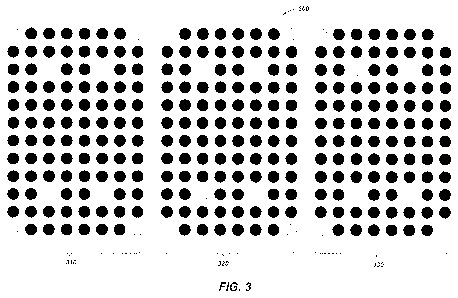
[0014] FIG. 3 shows a super array including three protein arrays according
to one or
more embodiments of the present disclosure.
DETAILED DESCRIPTION
[0015] Before embodiments of the present disclosure are described in
detail, it is to be
understood that this disclosure is not limited to the particular
configurations, process acts,
and materials disclosed herein as such configurations, process acts, and
materials may
vary somewhat. It is also to be understood that the terminology employed
herein is used
3
CA 02907183 2015-09-15
WO 2014/151021 PCT/US2014/024779
for the purpose of describing particular embodiments only and is not limiting
since the
scope of the present disclosure will be limited only by the appended claims
and
equivalents thereof
[0016] The publications and other reference materials referred to herein to
describe the
background of the disclosure and to provide additional detail regarding its
practice. The
references discussed herein are provided solely for their disclosure prior to
the filing date
of the present application. Nothing herein is to be construed as an admission
that such
documents constitute prior art, or that the inventors are not entitled to
antedate such
disclosure by virtue of prior invention.
[0017] While the known methods for using antibody profiling are generally
suitable for
their limited purposes, they possess certain inherent deficiencies that
detract from their
overall utility in analyzing, characterizing, and identifying biological
samples. For
example, the known methods rely on fractionation of antigens by
electrophoresis and then
transfer of the fractionated antigens to a membrane. Due to differences in
conditions
from one fractionation procedure to another, there are lot-to-lot differences
in the
positions of the antigens on the membrane such that results obtained using
membranes
from one lot cannot be compared with results obtained using membranes from
another lot.
Further, when colorimetric procedures are used for detecting immune complexes
on the
membrane, color determination may be subjective such that results may be
interpreted
differently by different observers.
[0018] It would be advantageous to provide a method identifying proteins
capable of
distinguishing an individual and methods for efficiently and accurately
determining
identity, distinguishing between individuals, as well as determining the
source of
biological fluids, especially those amenable to automation.
[0019] It must be noted that, as used in this specification and the
appended claims, the
singular forms "a," "an," and "the" include plural referents unless the
context clearly
dictates otherwise. Thus, for example, reference to a method for analyzing a
biological
sample from "an animal" includes reference to two or more of such animals,
reference to
"a support" includes reference to one or more of such supports, and reference
to "an
array" includes reference to two or more of such arrays.
[0020] As used herein, "blood" means and includes whole blood, plasma,
serum, or any
derivative of blood. A blood sample may be, for example, serum.
[0021] As used herein, "comprising," "including," "containing,"
"characterized by," and
grammatical equivalents thereof are inclusive or open-ended terms that do not
exclude
4
CA 02907183 2015-09-15
WO 2014/151021 PCT/US2014/024779
additional, unrecited elements or method acts. "Comprising" is to be
interpreted as
including the more restrictive terms "consisting of' and "consisting
essentially of"
[0022] As used herein, "consisting of' and grammatical equivalents thereof
exclude any
element, step, or ingredient not specified in the claim.
[0023] As used herein, "consisting essentially of' and grammatical
equivalents thereof
limit the scope of a claim to the specified materials or acts and those that
do not
materially affect the basic and novel characteristic or characteristics of the
claimed
invention.
[0024] As used herein, the terms "biological sample" and "sample" mean and
include a
sample comprising individual-specific antibodies obtained from an organism or
from
components (e.g., cells) of an organism. The sample may be of any biological
material.
Such samples include, but are not limited to, blood, blood fractions (e.g.,
serum, plasma),
blood cells (e.g., white cells), tissue or fine needle biopsy samples, urine,
saliva,
perspiration or semen. Biological samples may also include sections of tissues
such as
frozen sections taken for histological purposes.
[0025] As used herein, "color marker" refers to a substrate that produces a
colored
product in the visible light spectrum upon digestion with an appropriate
enzyme. Such
colored markers are distinguished from digestion that my produce fluorescent
and
luminescent products.
[0026] The term "discriminant analysis" means and includes a set of
statistical methods
used to select features that optimally discriminate between two or more
groups.
Application of discriminant analysis to a data set allows the user to focus on
the most
discriminating features for further analysis.
[0027] As used herein, the terms "immobilized" or "affixed" mean and
include an
association between a protein or antigen and a substrate at the molecular
level (i.e.,
through a covalent or non-covalent bond or interaction). For example, a
protein may be
immobilized to a support by covalent bonding directly to a surface of the
support which
may or may not be modified to enhance such covalent bonding. Also, the protein
may be
immobilized to the support by use of a linker molecule between the protein and
the
support. Proteins may further be immobilized on the support by steric
hindrance within a
polymerized gel or by covalent bonding within a polymerized gel. Proteins may
also be
immobilized on a support through hybridization between the protein and a
molecule
immobilized on the support.
CA 02907183 2015-09-15
WO 2014/151021 PCT/US2014/024779
[0028] The term "protein array" as used herein refers to a protein array, a
protein
macroarray, a protein microarray or a protein nanoarray. A protein array may
include, for
example, but is not limited to, ProtoArrayTM high density protein array, which
is
commercially available from Invitrogen (Carlsbad, California). The
ProtoArrayTM high
density protein array may be used to screen complex biological mixtures, such
as serum,
to assay for the presence of autoantibodies directed against human proteins.
Alternatively, a custom protein array that includes autoantigens, such as
those provided
herein, for the detection of autoantibody biomarkers, may be used to assay for
the
presence of autoantibodies directed against human proteins. In certain disease
states
including autoimmune diseases and cancer, autoantibodies are expressed at
altered levels
relative to those observed in healthy individuals.
[0029] As used herein, "support" means a generally or substantially planar
substrate onto
which an array of antigens is disposed. A support may comprise any material or
combination of materials suitable for carrying the array. Materials used to
construct these
supports need to meet several requirements, such as (1) the presence of
surface groups
that may be easily derivatized, (2) inertness to reagents used in the assay,
(3) stability
over time, and (4) compatibility with biological samples. For example,
suitable materials
include glass, silicon, silicon dioxide (i.e., silica), plastics, polymers,
hydrophilic
inorganic supports, and ceramic materials. Illustrative plastics and polymers
include
poly(tetrafluoroethylene), poly(vinylidenedifluoride), polystyrene,
polycarbonate,
polymethacrylate, and combinations thereof Illustrative hydrophilic inorganic
supports
include alumina, zirconia, titania, and nickel oxide. An example of a glass
substrate
would be a microscope slide. Silicon wafers used to make computer chips have
also been
used to make biochips. See, e.g., U.S. Pat. No. 5,605,662. The supports may
further
include a coating, such as, nitrocellulose, gelatin, a polymer (i.e.,
polyvinyl difluoride) or
an aldehyde.
[0030] As used herein, a "complex" refers to the binding of one molecule to
another
through a non-covalent interaction, such as the binding of an antibody to an
antigen.
[0031] In some embodiments, a method of determining proteins useful in
discriminating
one individual from 1 or more other individuals and/or positively identifying
an
individual is provided. Such proteins may be referred to herein as
"discriminant
proteins." The method may employ a protein array including a plurality of
proteins
immobilized on a support. As a non-limiting example, the protein array may be
a
ProtoArrayTM human protein microarray, which is commercially available from
6
CA 02907183 2015-09-15
WO 2014/151021 PCT/US2014/024779
Invitrogen Corporation (Carlsbad, California). The plurality of proteins
immobilized on
the support may include a plurality of antigens.
[0032] In a typical assay, a plurality of biological samples including
individual-specific
antibodies may each be physically contacted with a protein array, under
conditions that
permit high affinity binding, but that minimize non-specific interactions. In
one
embodiment, the biological samples are introduced to the protein array that
includes a
plurality of antigens immobilized in predetermined locations on a support. The
protein
array may be washed free of unbound material, and the presence of bound
antibodies may
be detected, and correlated with the cognate antigen.
[0033] The data collected from each of the plurality of biological samples
profiled on a
protein array may be used to determine an antibody profile for the individual.
The
antibody profiles may be analyzed using, for example, conventional
discriminant analysis
methods, to determine proteins relevant in discriminating and positively
identifying an
individual (i.e., discriminant proteins) from a population of one or more
other individuals.
The discriminant proteins may be used to generate a test panel for identifying
an
individual or determining a source of a biological sample. In some
embodiments, the test
panel may be, for example, a protein array 100, as shown in FIG. 1, including
a plurality
of the discriminant proteins arranged as spots 104 in predetermined locations
on a support
102.
Protein Array
[0034] The protein array may be prepared by attaching the antigens to the
surface of the
support 102 in a preselected pattern such that the locations of antigens in
the array are
known. As used herein, an antigen is a substance that is bound by an antibody.
Antigens
may include proteins, carbohydrates, nucleic acids, hormones, drugs,
receptors, tumor
markers, and the like, and mixtures thereof. An antigen may also be a group of
antigens,
such as a particular fraction of proteins eluted from a size exclusion
chromatography
column. Still further, an antigen may also be identified as a designated clone
from an
expression library or a random epitope library.
[0035] In one embodiment, antigens may be isolated from HeLa cells as
generally
described in A. M. Francoeur et al., Identification of Ki (Ku, p70/p80)
Autoantigens and
Analysis of Anti-Ki Autoantibody Reactivity, 136 J. Immunol. 1648 (1986).
Briefly, HeLa
cells may be grown in standard medium under standard tissue culture
conditions.
Confluent HeLa cell cultures may then be rinsed, preferably with phosphate-
buffered
7
CA 02907183 2015-09-15
WO 2014/151021 PCT/US2014/024779
saline (PBS), lysed with detergent, and centrifuged to remove insoluble
cellular debris.
The supernate contains approximately 10,000 immunologically distinct antigens
suitable
for generating an array.
[0036] There is no requirement that the antigens used to generate the array
be known. All
that is required is that the source of the antigens be consistent such that a
reproducible
array may be generated. For example, the HeLa cell supernate containing the
antigens
may be fractionated on a size exclusion column, electrophoretic gel, density
gradient, or
the like, as is well known in the art. Fractions may be collected, and each
fraction
collected could represent a unique set of antigens for the purpose of
generating the array.
Thus, even though the antigens may be unknown, a reproducible array may be
generated
if the HeLa cell antigens may be isolated and fractionated using the same
method and
conditions.
[0037] Other methods, such as preparation of random peptide libraries or
epitope libraries
are well known in the art and may be used to reproducibly produce antigens
(e.g., J.K.
Scott and G.P. Smith, Searching for Peptide Ligands with an Epitope Library,
249
Science 386 (1990); J.J. Devlin et al., Random Peptide Libraries: A Source of
Specific
Protein Binding Molecules, 249 Science 404-406 (1990); S.E. Cwirla et al.,
Peptides on
Phage: A Vast Library of Peptides for Identifj;ing Ligands, 87 Proc. Nat'l
Acad. Sci. USA
6378-6382 (1990); K.S. Lam et al., A New Type of Synthetic Peptide Library for
Identifting Ligand-binding Activity, 354 Nature 82-84 (1991); S. Cabilly,
Combinatorial
Peptide Library Protocols, Humana Press, 304 p.p., 129-154 1997; and U.S. Pat.
No.5,885,780). Such libraries may be constructed by ligating synthetic
oligonucleotides
into an appropriate fusion phage. Fusion phages may be filamentous
bacteriophage
vectors in which foreign sequences may be cloned into phage gene III and
displayed as
part of the gene III protein (pill) at one tip of the virion. Each phage
encodes a single
random sequence and expresses it as a fusion complex with pill, a minor coat
protein
present at about five molecules per phage. For example, in the fusion phage
techniques
of J.K. Scott and G.P. Smith, supra, a library was constructed of phage
containing a
variable cassette of six amino acid residues. The hexapeptide modules fused to
bacteriophage proteins provided a library for the screening methodology that
may
examine >1012 phages (or about 108-1010 different clones) at one time, each
with a test
sequence on the virion surface. The library obtained was used to screen
monoclonal
antibodies specific for particular hexapeptide sequences. The fusion phage
system has
also been used by other groups, and libraries containing longer peptide
inserts have been
8
CA 02907183 2015-09-15
WO 2014/151021 PCT/US2014/024779
constructed. Fusion phage prepared according to this methodology may be
selected
randomly or non-randomly for inclusion in the array of antigens. The fusion
phages
selected for inclusion in the array may be propagated by standard methods to
result in
what is virtually an endless supply of the selected antigens.
[0038] Other methods for producing antigens are also known in the art. For
example,
expression libraries may be prepared by random cloning of DNA fragments or
cDNA into
an expression vector (e.g., R.A. Young and R.W. Davis, Yeast RNA Polymerase II
Genes:
Isolation with Antibody Probes, 222 Science 778-782 (1983); G.M. Santangelo et
al.,
Cloning of Open Reading Frames and Promoters from the Saccharomyces cerevisiae
Genome: Construction of Genomic Libraries of Random Small Fragments, 46 Gene
181-186 (1986). Expression vectors that could be used for making such
libraries are
commercially available from a variety of sources. For example, random
fragments of
HeLa cell DNA or cDNA may be cloned into an expression vector, and then clones
expressing HeLa cell proteins may be selected. These clones may then be
propagated by
methods well known in the art. The expressed proteins may then be isolated or
purified
and may be used in the making of the array.
[0039] Alternatively, antigens may be synthesized using recombinant DNA
technology
well known in the art. Genes that code for many proteins from a gamut of
organisms
including viruses, bacteria, and mammals have been cloned, and thus large
quantities of
highly pure proteins may be synthesized quickly and inexpensively. For
example, the
genes that code for many eukaryotic and mammalian membrane-bound receptors,
growth
factors, cell adhesion molecules, and regulatory proteins have been cloned and
may be
useful as antigens. Many proteins produced by such recombinant techniques,
such as
transforming growth factor, acidic and basic fibroblast growth factors,
interferon,
insulin-like growth factor, and various interleukins from different species,
are
commercially available. In most instances, the entire polypeptide need not be
used as an
antigen. For example, any size or portion of the polypeptide that contains at
least one
epitope, i.e., antigenic determinant or portion of an antigen that
specifically interacts with
an antibody, will suffice for use in the array. In addition, a particular
antigen may be
purified or isolated from any natural or synthetic source of the antigen by
methods known
in the art.
[0040] The antigens, whether selected randomly or non-randomly, may be
disposed on
the support to result in the array. The pattern of the antigens on the support
should be
reproducible. In embodiments, the location and identity of each antigen on the
support
9
CA 02907183 2015-09-15
WO 2014/151021 PCT/US2014/024779
may be known. For example, in a 10 x 10 array one skilled in the art might
place
antigens 1-100 in locations 1-100, respectively, of the array. As a non-
limiting example,
each of the antigens of the array may be deposited on the support 102 as a
spot 104
having a diameter of from about 10 microns to about 500 microns and, more
particularly,
from about 50 microns to about 300 microns.
[0041] The proteins may placed in arrays on the surface of the support 102
using a
pipetting device or a machine or device configured for placing liquid samples
on the
support 102, for example, using a commercially available microarrayer, such as
those
from Arrayit Corporation (Sunnyvale, California); Genomic Solutions, Inc. (Ann
Arbor,
Michigan); Gene Machines (San Carlos, California); Genetic MicroSystems, Inc.
(Woburn, Massachusetts); GenePack DNA (Cambridge, UK); Genetix Ltd.
(Christchurch,
Dorset, UK); and Packard Instrument Company (Meriden, Connecticut).
[0042] Relevant methods to array a series of proteins onto a surface
include contact
printing processes, non-contact printing processes and in silico protein
synthesis arrayer
processes. Commercially available instruments are available for both methods.
In some
embodiments, conventional contact printing processes, such as contact pin
printing and
microstamping, in which the printing device may physically contact a surface
may be
used to apply the proteins to the surface of the support 102. For example, a
pin printing
device such as that commercially available from Arrayit Corporation may be
used to
deposit spots 104 having an average diameter of 65 microns or larger. As
another
non-limiting example, Genomic Solutions offers several nanoliter dispensing
instruments
that may dispense liquid volumes from 20 nL up to 250 IA from 96-, 384-, 1536-
, 3456-,
and 9600¨well microtiter plates and place them precisely on a surface with
densities up to
400 spots/cm2. The instruments will spot onto surfaces in a variety of
patterns. In
additional embodiments, the protein antigens may be applied to the surface
without
physical contact between the printing device and the surface using
conventional
non-contact printing processes including, but not limited to, photochemistry-
based
methods, laser writing, electrospray deposition, and inkjet. As the name
implies, inkjet
technology utilizes the same principles as those used in inkjet printers.
MicroFab
Technologies, Inc. (Plano, Texas), offers a ten-fluid print head that may
dispense picoliter
quantities of liquids onto a surface in a variety of patterns. An illustrative
pattern for the
present application would be a simple array ranging from 10 x 10 up to 100 x
100. The
protein antigens may be applied to the surface using a serial deposition
process or a
parallel deposition process.
CA 02907183 2015-09-15
WO 2014/151021 PCT/US2014/024779
[0043] There are a number of methods that may be used to attach proteins or
other
antigens to the surface of the support 102. The simplest of these is simple
adsorption
through hydrophobic, ionic, and van der Waals forces. As a non-limiting
example,
bifunctional organosilanes may be used in attachment of proteins to the
surface of the
support (e.g., Thompson and Maragos, Fiber-Optic Immunosensor for the
Detection of
Fumonisin B1 44 J. Agric. Food Chem. 1041-1046 (1996)). One end of the
organosilane
reacts with exposed -OH groups on the surface of the support to form a silanol
bond. The
other end of the organosilane contains a group that is reactive with various
groups on the
protein surface, such as -NH2 and -SH groups. This method of attaching
proteins to the
support results in the formation of a covalent linkage between the protein and
the support.
Other suitable methods that have been used for protein attachment to surfaces
include
arylazide, nitrobenzyl, and diazirine photochemistry methodologies. Exposure
of the
above chemicals to UV light causes the formation of reactive groups that may
react with
proteins to form a covalent bond. The arylazide chemistry forms a reactive
nitrene group
that may insert into C-H bonds, while the diazirine chemistry results in a
reactive carbene
group. The nitrobenzyl chemistry is referred to as caging chemistry whereby
the caging
group inactivates a reactive molecule. Exposure to UV light frees the molecule
and
makes it available for reaction. Still other methods for attaching proteins to
supports are
well known in the art, (e.g., S.S. Wong, Chemistry of Protein Conjugation and
Cross-Linking CRC Press, 340, 1991).
[0044] Following attachment of the antigens on the support 102 in the
selected array, the
support 102 may be washed. The wash solution may include, for example, one or
more
of a surfactant or a non-specific protein such as bovine serum albumin (BSA).
Appropriate liquids for washing include, but are not limited to, phosphate
buffered saline
(PBS) and the like, i.e., relatively low ionic strength, biocompatible salt
solutions
buffered at or near neutrality. Many of such appropriate wash liquids are
known in the art
or may be devised by a person skilled in the art without undue experimentation
(e.g., N.E.
Good and S. Izawa, Hydrogen Ion Buffers, 24 Methods Enzymology 53-68 (1972)).
[0045] The support 102 may be processed for blocking of nonspecific binding
of proteins
and other molecules to the support. This blocking step may prevent the binding
of
antigens, antibodies, and the like to the support wherein such antigens,
antibodies, or
other molecules are not intended to bind. Blocking may reduce the background
that
might swamp out the signal, thus increasing the signal-to-noise ratio. The
support 102
may be blocked by incubating the support 102 in a medium that contains inert
molecules
11
CA 02907183 2015-09-15
WO 2014/151021
PCT/US2014/024779
that bind to sites where nonspecific binding might otherwise occur. Examples
of suitable
blockers include, but are not limited to, bovine serum albumin, human albumin,
gelatin,
nonfat dry milk, polyvinyl alcohol, TWEENO 20, and various commercial blocking
buffers, such as SEABLOCKTM blocking buffer from EastCoast Bio, Inc., (West
Berwick, Maine) and SUPERBLOCKO blocking buffer from Pierce Chemical Co.,
(Rockford, Illinois). In some embodiments, one or more of the suitable
blockers may be
incorporated into the wash solution described above.
Antibody Profile
[0046] The array may be contacted with a sample of the biological material
to be tested.
For example, the biological sample may be obtained from various bodily fluids
and
solids, including blood, saliva, semen, serum, plasma, urine, amniotic fluid,
pleural fluid,
cerebrospinal fluid, and mixtures thereof. These biological samples may be
obtained
according to methods well known in the art. Depending on the detection method
used, it
may be required to manipulate the biological sample to attain optimal reaction
conditions.
For example, the ionic strength or hydrogen ion concentration or the
concentration of the
biological sample may be adjusted for optimal immune complex formation,
enzymatic
catalysis, and the like.
[0047] Antibodies (immunoglobulins) are a family of variable glycoproteins
that bind
specifically to foreign molecules (antigens). The binding strength between an
antigen
(epitope) and antigen-binding site in an antibody (paratope) is termed
affinity. Each
antibody has a minimum of two antigen-binding sites, and is thus multivalent
to its
antigen. The strength of a single antigen-antibody bond is termed the antibody
affinity
and it is produced by the number of bonds between the antigen and the
antibody. The
binding strength is greatly increased with more bonds because all of the
antigen-antibody
bonds must be broken simultaneously before the antigen and antibody can
dissociate.
Even when each antigen-binding site has a low affinity, antibodies can
function
effectively.
[0048] Antibodies have a variable region (FAB fragment) and a constant
region (FC
region) and both regions have antigen-binding sites that can be used for
detection.
[0049] As described in detail in U.S. Pat. No. 5,270,167 to Francoeur, when
ISAs are
allowed to react with a set of random antigens, a certain number of immune
complexes
form. For example, using a panel of about 1000 unique antigens, about 30
immune
complexes between ISAs in a biological sample that has been diluted 20-fold
may be
12
CA 02907183 2015-09-15
WO 2014/151021
PCT/US2014/024779
detected. If the biological sample is undiluted, the total number of possible
detectable
immune complexes that could form would be greater than 1023. The total number
of
possible immune complexes may also be increased by selecting "larger"
antigens, i.e.,
proteins instead of peptides) that have multiple epitopes. Therefore, it will
be appreciated
that depending on the antigens and number thereof used, the dilution of the
biological
sample, and the detection method, one skilled in the art may regulate the
number of
immune complexes that will form and be detected. As used herein, an "antibody
profile"
refers to the set of unique immune complexes that form and fail to form
between the ISAs
in the biological sample and the antigens in the array.
Detection and/or Quantification of Reactions
[0050] Methods for detecting antibody/antigen or immune complexes are well
known in
the art. The present disclosure may be modified by one skilled in the art to
accommodate
the various detection methods known in the art. The particular detection
method chosen
by one skilled in the art depends on several factors, including the amount of
biological
sample available, the type of biological sample, the stability of the
biological sample, the
stability of the antigen, and the affinity between the antibody and antigen.
Moreover, as
discussed above, depending on the detection methods chosen, it may be required
to
modify the biological sample. While these techniques are well known in the
art,
non-limiting examples of a few of the detection methods that may be used to
practice
embodiments of the present disclosure are briefly described below.
[0051] There are many types of immunoassays known in the art. The most
common
types of immunoassay are competitive and non-competitive heterogeneous assays,
such
as, for example, enzyme-linked immunosorbent assays (ELISAs). In a non-
competitive
ELISA, unlabeled antigen is bound to a support. A biological sample may be
combined
with antigens bound to the reaction vessel, and antibodies (primary
antibodies) in the
biological sample may be allowed to bind to the antigens, forming the immune
complexes. After the immune complexes have formed, excess biological sample
may be
removed and the array may be washed to remove nonspecifically bound
antibodies. The
immune complexes may then be reacted with an appropriate enzyme-labeled
anti-immunoglobulin (secondary antibody). The secondary antibody reacts with
antibodies in the immune complexes, not with other antigens bound to the
array.
Secondary antibodies specific for binding antibodies of different species,
including
humans, are well known in the art and are commercially available, such as from
Sigma
13
CA 02907183 2015-09-15
WO 2014/151021 PCT/US2014/024779
Chemical Co. (St. Louis, Missouri) and Santa Cruz Biotechnology, Inc. (Santa
Cruz,
California). After an optional further wash, the enzyme substrate may be
added. The
enzyme linked to the secondary antibody catalyzes a reaction that converts the
substrate
into a product. When excess antigen is present, the amount of product is
directly
proportional to the amount of primary antibody present in the biological
sample. By way
of non-limiting example, the product may be fluorescent or luminescent, which
may be
measured using technology and equipment well known in the art. It is also
possible to
use reaction schemes that result in a colored product, which may be measured
spectrophotometrically.
[0052] In other embodiments of the disclosure, the secondary antibody may
not be
labeled to facilitate detection. Additional antibodies may be layered (i. e.,
tertiary,
quaternary, etc.) such that each additional antibody specifically recognizes
the antibody
previously added to the immune complex. Any one of these additional (i.e.,
tertiary,
quaternary, etc.) may be labeled so as to allow detection of the immune
complex as
described herein.
[0053] Sandwich or capture assays may also be used to identify and quantify
immune
complexes. Sandwich assays are a mirror image of non-competitive ELISAs in
that
antibodies are bound to the solid phase and antigen in the biological sample
is measured.
These assays may be particularly useful in detecting antigens having multiple
epitopes
that are present at low concentrations. This technique requires excess
antibody to be
attached to a solid phase. The bound antibody is then incubated with the
biological
samples, and the antigens in the sample may be allowed to form immune
complexes with
the bound antibody. The immune complex is incubated with an enzyme-linked
secondary
antibody, which recognizes the same or a different epitope on the antigen as
the primary
antibody. Hence, enzyme activity is directly proportional to the amount of
antigen in the
biological sample. D.M. Kemeny and S.J. Challacombe, ELISA and Other Solid
Phase
Immunoassays, (John Wiley & Sons Ltd.) (1988).
[0054] Typical enzymes that may be linked to secondary antibodies include,
but are not
limited to, horseradish peroxidase, glucose oxidase, glucose-6-phosphate
dehydrogenase,
alkaline phosphatase, 13-galactosidase, and urease. Secondary antigen-specific
antibodies
linked to various enzymes are commercially available from, for example, Sigma
Chemical Co. and Amersham Life Sciences (Arlington Heights, Illinois).
[0055] Competitive ELISAs are similar to noncompetitive ELISAs except that
enzyme
linked antibodies compete with unlabeled antibodies in the biological sample
for limited
14
CA 02907183 2015-09-15
WO 2014/151021 PCT/US2014/024779
antigen binding sites. Briefly, a limited number of antigens may be bound to
the support.
Biological sample and enzyme-labeled antibodies may be added to the support
102.
Antigen-specific antibodies in the biological sample compete with enzyme-
labeled
antibodies for the limited number of antigens bound to the support 102. After
immune
complexes have formed, nonspecifically bound antibodies may be removed by
washing,
enzyme substrate is added, and the enzyme activity is measured. No secondary
antibody
is required. Because the assay is competitive, enzyme activity is inversely
proportional to
the amount of antibodies in the biological sample.
[0056] Another competitive ELISA may also be used within the scope of the
present
disclosure. In this embodiment, limited amounts of antibodies from the
biological sample
may be bound to the surface of the support as described herein. Labeled and
unlabeled
antigens may be then brought into contact with the support such that the
labeled and
unlabeled antigens compete with each other for binding to the antibodies on
the surface of
the support. After immune complexes have formed, nonspecifically bound
antigens may
be removed by washing. The immune complexes may be detected by incubation with
an
enzyme-linked secondary antibody, which recognizes the same or a different
epitope on
the antigen as the primary antibody, as described above. The activity of the
enzyme is
then assayed, which yields a signal that is inversely proportional to the
amount of antigen
present.
[0057] Homogeneous immunoassays may also be used when practicing the method
of the
present disclosure. Homogeneous immunoassays may be preferred for detection of
low
molecular weight compounds, such as hormones, therapeutic drugs, and illegal
drugs that
cannot be analyzed by other methods, or compounds found in high concentration.
Homogeneous assays may be particularly useful because no separation step is
necessary.
R.C. Boguslaski et al., Clinical Immunochemistry: Principles of Methods and
Applications, (1984).
[0058] In homogeneous techniques, bound or unbound antigens may be enzyme-
linked.
When antibodies in the biological sample bind to the enzyme-linked antigen,
steric
hindrances inactivate the enzyme. This results in a measurable loss in enzyme
activity.
Free antigens (i.e., not enzyme-linked) compete with the enzyme-linked antigen
for
limited antibody binding sites. Thus, enzyme activity is directly proportional
to the
concentration of antigen in the biological sample.
[0059] Enzymes useful in homogeneous immunoassays include, but are not
limited to,
lysozyme, neuraminidase, trypsin, papain, bromelain, glucose-6-phosphate
CA 02907183 2015-09-15
WO 2014/151021 PCT/US2014/024779
dehydrogenase, and 13-galactosidase. T. Persoon, "Immunochemical Assays in the
Clinical Laboratory," 5 Clinical Laboratory Science 31 (1992). Enzyme-linked
antigens
are commercially available or may be linked using various chemicals well known
in the
art, including glutaraldehyde and maleimide derivatives.
[0060] Prior antibody profiling technology involved an alkaline phosphatase
labeled
secondary antibody with 5-bromo-4-chloro-3'-indolylphosphate p-toluidine salt
(BCIP)
and nitro-blue tetrazolium chloride (NBT), both of which are commercially
available
from a variety of sources, such as from Pierce Chemical Co. (Rockford,
Illinois). The
enzymatic reaction forms an insoluble colored product that is deposited on the
surface of
membrane strips to form bands wherever antigen-antibody complexes occur. As a
non-limiting example, the array may be scanned to detect a colored product
using one of a
variety of conventional desktop scanners, which are commercially available
from a
variety of sources, such as from Canon U.S.A. (Lake Success, New York). The
intensity
of the colored product may be quantified by calculating the median feature
pixel intensity
minus median background pixel intensity.
[0061] As another non-limiting example, gold nanoparticle labeled
antibodies may be
employed and may be detected using a scanning, transmission electron
microscopy,
and/or dark-field zoom stereomicroscopy. Compared to conventional fluorescent
labels,
the gold nanoparticles scatter incident white light to generate monochromatic
light which
may be easily detected. The light intensity generated by the gold
nanoparticles may be up
to 100,000 times greater than that generated by fluorescent-labeled molecules.
For
example, the gold nanoparticles may be detected using a conventional desktop
scanner.
Han et al., Detection of Analyte Binding to Microarrays Using Gold
Nanoparticle Labels
and a Desktop Scanner, 3 Lab Chip 329; 329-332 (2003).
[0062] Fluorescent immunoassays may also be used when practicing the method
of the
present disclosure. Fluorescent immunoassays are similar to ELISAs except the
enzyme
is substituted for fluorescent compounds called fluorophores or fluorochromes.
These
compounds have the ability to absorb energy from incident light and emit the
energy as
light of a longer wavelength and lower energy. Fluorescein and rhodamine,
usually in the
form of isothiocyanates that may be readily coupled to antigens and
antibodies, are most
commonly used in the art. D.P. Stites et al., Basic and Clinical Immunology,
(1994).
Fluorescein absorbs light of 490 to 495 nm in wavelength and emits light at
520 nm in
wavelength. Tetramethylrhodamine absorbs light of 550 nm in wavelength and
emits
light at 580 nm in wavelength. Illustrative fluorescence-based detection
methods include
16
CA 02907183 2015-09-15
WO 2014/151021 PCT/US2014/024779
ELF-97 alkaline phosphatase substrate (Molecular Probes, Inc., Eugene,
Oregon);
PBXL-1 and PBXL-3 (phycobilisomes conjugated to streptavidin) (Martek
Biosciences
Corp., Columbia, Maryland); FITC (fluorescein isothiocyanate) and Texas Red
labeled
goat anti-human IgG (Jackson ImmunoResearch Laboratories, Inc., West Grove,
Pennsylvania); and B-Phycoerythrin and R-Phycoerythrin conjugated to
streptavidin
(Molecular Probes Inc.). ELF-97 is a nonfluorescent chemical that is digested
by alkaline
phosphatase to form a fluorescent molecule. Because of turnover of the
alkaline
phosphatase, use of the ELF-97 substrate results in signal amplification.
Fluorescent
molecules attached to secondary antibodies do not exhibit this amplification.
[0063] Phycobiliproteins isolated from algae, porphyrins, and chlorophylls,
which all
fluoresce at about 600 nm, are also being used in the art. I. Hemmila,
Fluoroimmunoassays and Immunofluorometric Assays, 31 Clin. Chem. 359 (1985);
U.S.
Pat. No. 4,542,104. Phycobiliproteins and derivatives thereof are commercially
available
under the names R-phycoerythrin (PE) and QUANTUM REDTM from Sigma Chemical
Co.
[0064] In addition, Cy-conjugated secondary antibodies and antigens may be
useful in
immunoassays and are commercially available. Cy3, for example, is maximally
excited
at 554 nm and emits light at between 568 and 574 nm. Cy3 is more hydrophilic
than
other fluorophores and thus has less of a tendency to bind nonspecifically or
aggregate.
Cy-conjugated compounds are commercially available from Amersham Life
Sciences.
[0065] Illustrative luminescence-based detection methods include CSPD 0 and
CDP star
alkaline phosphatase substrates from Roche Molecular Biochemicals,
(Indianapolis,
Indiana) and SUPERSIGNALO horseradish peroxidase substrate from Pierce
Chemical
Co., (Rockford, Illinois).
[0066] Chemiluminescence, electroluminescence, and electrochemiluminescence
(ECL)
detection methods may also be attractive means for quantifying antigens and
antibodies in
a biological sample. Luminescent compounds have the ability to absorb energy,
which is
released in the form of visible light upon excitation. In chemiluminescence,
the
excitation source is a chemical reaction; in electroluminescence the
excitation source is an
electric field; and in ECL an electric field induces a luminescent chemical
reaction.
[0067] Molecules used with ECL detection methods generally comprise an
organic ligand
and a transition metal. The organic ligand forms a chelate with one or more
transition
metal atoms forming an organometallic complex. Various organometallic and
transition
metal-organic ligand complexes have been used as ECL labels for detecting and
17
CA 02907183 2015-09-15
WO 2014/151021 PCT/US2014/024779
quantifying analytes in biological samples. Due to their thermal, chemical,
and
photochemical stability, their intense emissions and long emission lifetimes,
ruthenium,
osmium, rhenium, iridium, and rhodium transition metals are favored in the
art. The
types of organic ligands are numerous and include anthracene and polypyridyl
molecules
and heterocyclic organic compounds. For example, bipyridyl, bipyrazyl,
terpyridyl, and
phenanthrolyl, and derivatives thereof, are common organic ligands in the art.
A common
organometallic complex used in the art includes tris-bipyridine ruthenium
(II),
commercially available from IGEN, Inc. (Rockville, Maryland) and Sigma
Chemical Co.
[0068] ECL may be performed under aqueous conditions and under
physiological pH,
thus minimizing biological sample handling. J.K. Leland et al.,
Electrogenerated
Chemiluminescence: An Oxidative-Reduction Type ECL Reactions Sequence Using
Triprophyl Amine, 137 J. Electrochemical Soc. 3127-3131 (1990); WO 90/05296;
and
U.S. Pat. No. 5,541,113. Moreover, the luminescence of these compounds may be
enhanced by the addition of various cofactors, such as amines.
[0069] A tris-bipyridine ruthenium (II) complex, for example, may be
attached to a
secondary antibody using strategies well known in the art, including
attachment to lysine
amino groups, cysteine sulfhydryl groups, and histidine imidazole groups. In a
typical
ELISA immunoassay, secondary antibodies would recognize antibodies bound to
antigens, but not unbound antigens. After washing nonspecific binding
complexes, the
tris-bipyridine ruthenium (II) complex may be excited by chemical,
photochemical, and
electrochemical excitation means, such as by applying current to the array
(e.g., WO
86/02734). The excitation would result in a double oxidation reaction of the
tris-bipyridine ruthenium (II) complex, resulting in luminescence that could
be detected
by, for example, a photomultiplier tube. Instruments for detecting
luminescence are well
known in the art and are commercially available, for example, from IGEN, Inc.
(Rockville, Maryland).
[0070] Solid state color detection circuitry may also be used to monitor
the color
reactions on the array and, on command, compare the color patterns before and
after the
sample application. A color camera image may also be used and the pixel
information
analyzed to obtain the same information.
[0071] Still another method involves detection using a surface plasmon
resonance (SPR)
chip. The surface of the chip is scanned before and after sample application
and a
comparison is made. The SPR chip relies on the refraction of light when the
molecules of
interest may be exposed to a light source. Each molecule has its own
refraction index by
18
CA 02907183 2015-09-15
WO 2014/151021 PCT/US2014/024779
which it may be identified. This method requires precise positioning and
control circuitry
to scan the chip accurately.
[0072] In one embodiment, the detecting agents bind to specific portions of
the
antibodies. Antibodies have a variable region (FAB fragment) and a constant
region (FC
region) and both regions have antigen-binding sites that can be used for
detection. By
capitalizing on both regions of the antibody through multiple combinations,
the array can
essentially determine the quantity of antibodies present in the biological
sample and the
structural integrity (quality) of these antibodies by measuring the intensity
from each of
the bound spots.
[0073] Chosen antibodies can be conjugated to human IgG so that they can be
detected
directly in vitro by the anti-Human detection conjugate (AHG). AHG is used to
detect in
vitro (array) sensitization and detection of anti-red cell antibodies in serum
or plasma.
These monoclonal antibodies recognize an exposed surface determinant of intact
red
blood cells and will bind to the Fc (constant region) receptors. In addition,
other
monoclonal antibodies specific for an Fc portion of human IgG can be used on
the array
which recognizes an epitope common to all human IgG subclasses or can be
specific for a
FAB portion of human IgG, which in turn would be non-reactive with the Fc
portion of
human IgG, eliminating antigen-binding affinity competition. Polyclonal
antibodies
specific for the Fc or FAB portion may also be used, which is specific to
human IgG only
and will not bind to other Igs (immunoglobulins).
[0074] Utilizing all or a combination of the described antibodies to
determine the quantity
of antibodies present in the sample and the structural integrity of these
antibodies, the
colorigenic marker can directly detect the proportion of bound sample to serve
as sample
control at the time of assay. This eliminates the use of unnecessary sample
tests as well as
ensuring or discrediting the generated antibody profile in real time.
[0075] Yet another method involves a fluid rinse of the array with a
fluorescing reagent.
The antigens that combine with the biological sample will fluoresce and may be
detected
with a charge-coupled device (CCD) array. The output of such a CCD array is
analyzed
to determine the unique pattern associated with each sample. Speed is not a
factor with
any of the methods since the chemical combining of sample and reference takes
minutes
to occur.
[0076] Moreover, array scanners are commercially available, such as from
Genetic
Micro Systems, Inc. The GMS 418 Array Scanner uses laser optics to rapidly
move a
focused beam of light over the array. This system uses a dual-wavelength
system
19
CA 02907183 2015-09-15
WO 2014/151021 PCT/US2014/024779
including high-powered, solid-state lasers that generate high excitation
energy to allow
for reduced excitation time. At a scanning speed of 30 Hz, the GMS 418 may
scan a 22 x
75-mm slide with 10- m resolution in about four minutes.
[0077] Software for image analysis obtained with an array scanner is
readily available.
Available software packages include ImaGene (BioDiscovery, Los Angeles,
California);
ScanAlyze (available at no charge; developed by Mike Eisen, Stanford
University, Palo
Alto, California); De-Array (developed by Yidong Chen and Jeff Trent of the
National
Institutes of Health; used with IP Lab from Scanalytics, Inc., Fairfax,
Virginia); Pathways
(Research Genetics, Huntsville, Alabama); GEM Tools (Incyte Pharmaceuticals,
Inc.,
Palo Alto, California); and Imaging Research (Amersham Pharmacia Biotech,
Inc.,
Piscataway, New Jersey).
[0078] Once interactions between the antigens and antibodies have been
identified and
quantified, the signals may be digitized. The digitized antibody profile may
serve as a
signature that identifies the source of the biological sample. Depending on
the array used,
the digitized data may take numerous forms. For example, the array may include
10
columns and 10 rows for a total number of 100 spots, each including at least
one antigen.
After the biological sample including the antibodies is added to the array and
allowed to
incubate, interactions between antigens and antibodies in the biological
sample may be
identified and quantified. In each spot, an interaction between the antigen in
the spot and
the antibody in the biological sample will either result in or not result in a
quantifiable
signal. In one embodiment, the results of the antibody profile may be
digitized by, by
way of non limiting example, ascribing each one of the 100 spots a numerical
value of
either "0," if a quantifiable signal was not obtained, or "1," if a
quantifiable signal was
obtained. Using this method, the digitized antibody profile may comprise a
unique set of
zeroes and ones. It will be understood that the use of 1 and 0 is merely
exemplary and
that any set of values or indicators may be used to signify the absence,
presence, or
intensity of a particular signal.
[0079] The numerical values "0" or "1" may, of course, be normalized to
signals obtained
in internal control spots so that digitized antibody profiles obtained at a
later time may be
properly compared. For example, one or several of the spots may contain a
known
antigen, which will remain constant over time. Therefore, if a subsequent
biological
sample is more or less dilute than a previous biological sample, the signals
may be
normalized using the signals from the known antigen.
CA 02907183 2015-09-15
WO 2014/151021 PCT/US2014/024779
[0080] It will be appreciated by one skilled in the art that other methods
of digitizing the
antibody profile exist and may be used. For example, rather than ascribing
each spot with
a numerical value of "0" or "1," the numerical value may be incremental and
directly
proportional to the strength of the signal.
Statistical Analysis
[0081] The antibody profiles obtained from the plurality of individuals may
be analyzed
using conventional discriminant analysis methods to determine proteins useful
in
discriminating or identifying an individual from one or more other
individuals. For
example, discriminant proteins may be determined using forward selection,
backward
elimination, or stepwise selection to determine a subset of proteins that best
reveals
differences among the classes (i.e., the individuals). The STEPDISC procedure,
which is
available from SAS Institute, Inc. (Cary, North Carolina), may be used to
perform a
stepwise discriminant analysis to select a subset of the proteins useful in
discriminating
among individuals. Signals from a set of proteins that make up each class may
be
assumed to be multivariate normal with a common covariance matrix.
[0082] Using the STEPDISC procedure, variables (in particular, signals from
particular
proteins) may be chosen to enter or leave the model according to the
significance level of
an F-test from an analysis of covariance, where the variables already chosen
act as
covariates and the variable under consideration is the dependent variable. In
other
embodiments, a variable could be chosen to enter or leave the model according
to
whether the squared partial correlation for its prediction using the class
variable (and
controlling for the effects of the other variables already in the model) is
high.
[0083] In some embodiments, the discriminant proteins useful in
discriminating or
identifying an individual may be determined by calculating various
discriminant functions
for classifying observations using the protein signals. Linear or quadratic
discriminant
functions may be used for data with approximately multivariate normal within-
class
distributions. Nonparametric methods may be used without making any
assumptions
about these distributions.
[0084] One or more of the discriminant proteins may be used to identify an
individual, to
distinguish between individuals, or to establish or rule out the source of a
biological
sample. In some embodiments, one or more of the discriminant proteins may be
used as
part of a test panel. For example, discriminant proteins may be immobilized on
a support
in the form of an array as described above to form a protein array useful in
discriminating
21
CA 02907183 2015-09-15
WO 2014/151021 PCT/US2014/024779
among individuals and/or sources of a biological sample. However, other
methods of
detecting an interaction between a discriminant protein and an antibody
present in a
biological sample, such as conventional protein affinity chromatography
methods, affinity
blotting methods, immunoprecipitation methods, and cross-linking methods, may
also be
used. In embodiments, the array or test panel may be used to generate an
antibody profile
which may be used to distinguish between individuals in a population, or to
establish or
rule out the source of a biological sample within a population, wherein the
population
may comprise 1 million, 10 million, 100 million, 1 billion, 10 billion, 100
billion, or more
individuals.
[0085] The array may include several discriminant proteins, each of which
may be
immobilized on a support. The array may include less than about 200, 175, 170,
150,
125, 110, 100, 75, or 50 discriminant proteins. For example, the test panel
for
discriminating or identifying an individual may include from about 20 to about
90
discriminant proteins, and more particularly, from about 45 to about 80
discriminant
proteins, less than about 100 discriminant proteins, less than about 110
discriminant
proteins, or less than about 170 discriminant proteins. With "X" different
profiles that are
each independent, the probability that no two different people have the same
profile
among "m" people can be shown to be equal to exp[-m*m/(2X)]. As a non-limiting
example, greater than about 76 independent discriminant proteins may be used
to
distinguish an individual among a population of about 10 billion individuals,
the
probability of a match between two different individuals being less than about
0.0001. As
another non-limiting example, greater than about 86 independent discriminant
proteins
may be used to distinguish an individual among a population of about 100
billion
individuals, the probability of a match between two different individuals
being less than
about 0.0001. Examples of discriminant proteins include, but are not limited
to, those
proteins presented in Table 1.
[0086] In another embodiment, an array has sub-arrays and each sub-array
may include
less than about 200, 175, 170, 150, 125, 110, 100, 75, or 50 discriminant
proteins. For
example, each sub-array for the test panel for discriminating or identifying
an individual
may include from about 20 to about 90 discriminant proteins, and more
particularly, from
about 45 to about 80 discriminant proteins, less than about 100 discriminant
proteins, less
than about 110 discriminant proteins, or less than about 170 discriminant
proteins.
Comparing the detected immune complexes between each sub-array leads to
greater
confidence in identification.
22
CA 02907183 2015-09-15
WO 2014/151021
PCT/US2014/024779
[0087] Table 1.
SEQ ID NO Protein ID
SEQ ID NO:1 PM 2149
SEQ ID NO:2 PM 2151
SEQ ID NO:3 BC010125.1
SEQ ID NO:4 BC011414.1
SEQ ID NO:5 BC012945.1
SEQ ID NO:6 BC014409.1
SEQ ID NO:7 BC015219.1
SEQ ID NO:8 BC016470.2
SEQ ID NO:9 BC018206.1
SEQ ID NO:10 BC018404.1
SEQ ID NO:11 BC019039.2
SEQ ID NO:12 BC019315.1
SEQ ID NO:13 BCO21189.2
SEQ ID NO:14 BCO23152.1
SEQ ID NO:15 BCO26175.1
SEQ ID NO:16 BCO26346.1
SEQ ID NO:17 BC032825.2
SEQ ID NO:18 BC033711.1
SEQ ID NO:19 BC036123.1
SEQ ID NO:20 BC040949.1
SEQ ID NO:21 BC050377.1
SEQ ID NO:22 BC052805.1
SEQ ID NO:23 BC053602.1
SEQ ID NO:24 BC060824.1
SEQ ID NO:25 NM 015138.2
23
CA 02907183 2015-09-15
WO 2014/151021
PCT/US2014/024779
SEQ ID NO Protein ID
SEQ ID NO:26 NM 175887.2
SEQ ID NO:27 NM 000394.2
SEQ ID NO:28 NM 000723.3
SEQ ID NO:29 NM 001008220.1
SEQ ID NO:30 NM 001106.2
SEQ ID NO:31 NM 001312.2
SEQ ID NO:32 NM 001537.1
SEQ ID NO:33 NM 002737
SEQ ID NO:34 NM 002740
SEQ ID NO:35 NM 002744
SEQ ID NO:36 NM 003907.1
SEQ ID NO:37 NM 003910.2
SEQ ID NO:38 NM 004064.2
SEQ ID NO:39 NM 004394.1
SEQ ID NO:40 NM 004845.3
SEQ ID NO:41 NM 004965.3
SEQ ID NO:42 NM 005030
SEQ ID NO:43 NM 005246.1
SEQ ID NO:44 NM 006007.1
SEQ ID NO:45 NM 006218.2
SEQ ID NO:46 NM 006628.4
SEQ ID NO:47 NM 006819.1
SEQ ID NO:48 NM 012472.1
SEQ ID NO:49 NM 014240.1
SEQ ID NO:50 NM 014245.1
SEQ ID NO:51 NM 014460.2
24
CA 02907183 2015-09-15
WO 2014/151021
PCT/US2014/024779
SEQ ID NO Protein ID
SEQ ID NO:52 NMO14622.4
SEQ ID NO:53 NM 014891.1
SEQ ID NO:54 NMO14943.3
SEQ ID NO:55 NM 015149.2
SEQ ID NO:56 NM 015417.2
SEQ ID NO:57 NMO15509.2
SEQ ID NO:58 NM 016096.1
SEQ ID NO:59 NM 016520.1
SEQ ID NO:60 NM 017855.2
SEQ ID NO:61 NM 017949.1
SEQ ID NO:62 NM 018326.1
SEQ ID NO:63 NMO18584.4
SEQ ID NO:64 NM 024718.2
SEQ ID NO:65 NM 024826.1
SEQ ID NO:66 NM 025241.1
SEQ ID NO:67 NM 032345.1
SEQ ID NO:68 NM 032368.3
SEQ ID NO:69 NM 079420.1
SEQ ID NO:70 NM 080390.3
SEQ ID NO:71 NM 138623.2
SEQ ID NO:72 NM 145796.2
SEQ ID NO:73 NM 153757.1
SEQ ID NO:74 NM 177973.1
SEQ ID NO:75 NM 178010.1
SEQ ID NO:76 NM 199124.1
SEQ ID NO:77 NM 201262.1
CA 02907183 2015-09-15
WO 2014/151021 PCT/US2014/024779
SEQ ID NO Protein ID
SEQ ID NO:78 NM 203284.1
SEQ ID NO:79 NM 205853.1
SEQ ID NO:80 NM 212540.1
[0088] In embodiments of the disclosure, a protein array may comprise 1, 2,
3, 4, 5, 10,
15, 20, 25, 50, or more discriminant proteins selected from the group
consisting of SEQ
ID NOs: 1-80, SEQ ID NOs: 1-45, SEQ ID NOs: 1-3, 5, 6, 8, 9, 11, 12, 15-18, 22-
24, 26,
27, 29, 33, 38, 41, 44, 46-48, 51, 20, 54, 57-60, 62, 65, 68, 70, 72, 72-75,
77, and 79 and
SEQ ID NOs: 1-9, 11-13, 15-20, 22-24, 26-30, 33, 35, 36, 38-41, 44, 46-54, 57-
60, 62,
63, 66, 68, 70, and 72-80. In embodiments, a protein array may consist of SEQ
ID NOs:
1-80, SEQ ID NOs: 1-45, SEQ ID NOs: 1-3, 5, 6, 8, 9, 11, 12, 15-18, 22-24, 26,
27, 29,
33, 38, 41, 44, 46-48, 51, 20, 54, 57-60, 62, 65, 68, 70, 72, 72-75, 77, and
79 and SEQ ID
NOs: 1-9, 11-13, 15-20, 22-24, 26-30, 33, 35, 36, 38-41, 44, 46-54, 57-60, 62,
63, 66, 68,
70, and 72-80.
[0089] In embodiments of the disclosure, a protein sub-array may comprise
1, 2, 3, 4, 5,
10, 15, 20, 25, 50, or more discriminant proteins selected from the group
consisting of
SEQ ID NOs: 1-80, SEQ ID NOs: 1-45, SEQ ID NOs: 1-3, 5, 6, 8, 9, 11, 12, 15-
18,
22-24, 26, 27, 29, 33, 38, 41, 44, 46-48, 51, 20, 54, 57-60, 62, 65, 68, 70,
72, 72-75, 77,
and 79 and SEQ ID NOs: 1-9, 11-13, 15-20, 22-24, 26-30, 33, 35, 36, 38-41, 44,
46-54,
57-60, 62, 63, 66, 68, 70, and 72-80. In embodiments, a protein array may
consist of
SEQ ID NOs: 1-80, SEQ ID NOs: 1-45, SEQ ID NOs: 1-3, 5, 6, 8, 9, 11, 12, 15-
18,
22-24, 26, 27, 29, 33, 38, 41, 44, 46-48, 51, 20, 54, 57-60, 62, 65, 68, 70,
72, 72-75, 77,
and 79 and SEQ ID NOs: 1-9, 11-13, 15-20, 22-24, 26-30, 33, 35, 36, 38-41, 44,
46-54,
57-60, 62, 63, 66, 68, 70, and 72-80.
[0090] In an embodiment of the disclosure, a protein array including
discriminant
proteins may be used for forensic analysis for matching a biological sample to
an
individual such as, for example, a criminal suspect. Forensic samples obtained
from
crime scenes are often subject to drying of the samples, small sample sizes,
mixing with
samples from more than one individual, adulteration with chemicals, and the
like. The
present method provides the advantages of rapid analysis, simplicity, low
cost, and
accuracy for matching forensic samples with suspects. For example, the
forensic sample
and a sample from one or more suspects may be obtained according to methods
well
26
CA 02907183 2015-09-15
WO 2014/151021
PCT/US2014/024779
known in the art. The samples may be tested against the array and compared. If
the
discriminant proteins obtained from the samples match, it may be concluded
that the
forensic sample was obtained from the matching suspect. If no match of
discriminant
proteins is obtained, then none of the suspects was the source of the forensic
sample.
Example
[0091] Serum samples from ninety-four (94) individuals were profiled
against a high
throughput protein array with over 8000 proteins and the data from these chips
was
statistically analyzed to determine proteins useful for discriminating among
sets of
individuals in a population. The ninety-four (94) individuals included
nineteen (19)
Asian individuals, twenty (20) African American individuals, twenty (20)
Native
American individuals, and thirty-five (35) Caucasian individuals. For quality
assurance
(QA), the arrays contained the immobilized proteins in pairs on a support.
Thus, each
array provided two opportunities for antigen/antibody binding for each
protein.
[0092] The serum samples were diluted 1:150 and used to probe human
ProtoArrayTM.
The arrays were blocked for 1 hour and then incubated with the serum samples
for 90
minutes at about 4 C without shaking. The arrays were then transferred to ice
and
washed about three times by adding about 20 ml buffer (1X PBS, 5 mM MgC12, 0.5
mM
DTT, 0.05% Triton X-100, 5% Glycerol, 1% BSA) to the arrays, incubating the
arrays
with the buffer for 8 minutes at 4 C, and decanting the buffer from the arrays
by
inverting. The arrays were incubated with anti-human IgG antibody conjugated
to
AlexaFluor 647 for about 90 minutes, washed as above and dried. The arrays
were
scanned using a ScanArray Express 3.0 HT microarray scanner, which is
available
commercially from Perkin Elmer, Inc. (Waltham, Massachusetts). The images were
captured from the microarray scanner using a 633 nm laser with the scanner set
to 10 gm
resolution. Following scanning, data was acquired using ImaGene 8.0 microarray
analysis software from BioDiscovery (El Segundo, California). Background-
subtracted
signals from each population were normalized utilizing a quantile
normalization strategy.
Subjects were distinguished from one another using conventional discriminant
analysis.
The STEPDISC procedure from SAS Institute, Inc. was utilized to identify
discriminant
proteins based on the logarithms of the intensities detected. The discriminant
proteins of
interest were identified as significant in distinguishing between individuals.
A list of 80
discrimininating proteins from among the over 8,000 on the arrays was
determined. The
80 discriminating proteins are listed in Table 2.
27
CA 02907183 2015-09-15
WO 2014/151021
PCT/US2014/024779
[0093] Table 2.
SelOrd MinPSee maxCorr
SEQ ID NO Protein ID sRatio
All OrNot After
SEQ ID NO:1 PM 2149 16 0.45 22.1 0.683
SEQ ID NO:2 PM 2151 99 0.25 13.4 0.585
SEQ ID NO:3 BC010125.1 62 0.23 15.6 0.500
SEQ ID NO:4 BC011414.1 15 0.40 19.9 0.482
SEQ ID NO:5 BC012945.1 38 0.33 18.4 0.570
SEQ ID NO:6 BC014409.1. 0.32 10.7 0.448
SEQ ID NO:7 BC015219.1 76 0.29 15.6 0.652
SEQ ID NO:8 BC016470.2 74 0.19 14.6 0.579
SEQ ID NO:9 BC018206.1 31 0.38 16.1 0.551
SEQ ID NO:10 BC018404.1 93 0.27 19.0 0.754
SEQ ID NO:11 BC019039.2 33 0.41 17.2 0.544
SEQ ID NO:12 BC019315.1 27 0.48 17.8 0.846
SEQ ID NO:13 BCO21189.2 29 0.34 17.2 0.488
SEQ ID NO:14 BCO23152.1 6 0.10 25.3 0.752
SEQ ID NO:15 BCO26175.1 50 0.39 15.6 0.582
SEQ ID NO:16 BCO26346.1 78 0.48 16.4 0.360
SEQ ID NO:17 BC032825.2 13 0.10 18.9 0.491
SEQ ID NO:18 BC033711.1 72 0.29 14.6 0.567
SEQ ID NO:19 BC036123.1 101 0.35 15.0 0.649
SEQ ID NO:20 BC040949.1 45 0.37 17.9 0.523
SEQ ID NO:21 BC050377.1 70 0.14 11.0 0.310
SEQ ID NO:22 BC052805.1 56 0.29 16.6 0.501
SEQ ID NO:23 BC053602.1 42 0.32 16.1 0.621
SEQ ID NO:24 BC060824.1 12 0.28 19.4 0.421
28
CA 02907183 2015-09-15
WO 2014/151021
PCT/US2014/024779
SelOrd MinPSee maxCorr
SEQ ID NO Protein ID sRatio
All OrNot After
SEQ ID NO:25 NM 015138.2 91 0.33 13.3 0.607
SEQ ID NO:26 NM 175887.2 34 0.43 15.4 0.537
SEQ ID NO:27 NM 000394.2 44 0.38 20.2 0.737
SEQ ID NO:28 NM 000723.3 200 0.22 9.4 0.580
SEQ ID NO:29 NM 001008220.1 17 0.22 21.7 0.405
SEQ ID NO:30 NM 001106.2 22 0.41 20.3 0.303
SEQ ID NO:31 NM 001312.2 81 0.42 13.2 0.619
SEQ ID NO:32 NM 001537.1 84 0.49 23.5 0.733
SEQ ID NO:33 NM 002737 73 0.47 10.0 0.300
SEQ ID NO:34 NM 002740 79 0.28 12.4 0.620
SEQ ID NO:35 NM 002744 3 0.42 22.4 0.215
SEQ ID NO:36 NM 003907.1 57 0.37 14.8 0.440
SEQ ID NO:37 NM 003910.2 63 0.12 12.7 0.594
SEQ ID NO:38 NM 004064.2 54 0.20 13.8 0.422
SEQ ID NO:39 NM 004394.1 58 0.48 16.3 0.641
SEQ ID NO:40 NM 004845.3 30 0.25 18.0 0.432
SEQ ID NO:41 NM 004965.3 97 0.46 11.4 0.648
SEQ ID NO:42 NM 005030 95 0.41 14.2 0.683
SEQ ID NO:43 NM 005246.1 77 0.22 9.3 0.625
SEQ ID NO:44 NM 006007.1 80 0.24 13.3 0.417
SEQ ID NO:45 NM 006218.2 90 0.24 8.2 0.573
SEQ ID NO:46 NM 006628.4 66 0.29 15.0 0.538
SEQ ID NO:47 NM 006819.1 4 0.22 17.9 0.356
SEQ ID NO:48 NM 012472.1 11 0.49 23.0 0.578
SEQ ID NO:49 NM 014240.1 19 0.44 18.9 0.459
29
CA 02907183 2015-09-15
WO 2014/151021
PCT/US2014/024779
SelOrd MinPSee maxCorr
SEQ ID NO Protein ID sRatio
All OrNot After
SEQ ID NO:50 NM 014245.1 18 0.29 22.9 0.676
SEQ ID NO:51 NM 014460.2 21 0.32 19.7 0.414
SEQ ID NO:52 NM 014622.4 65 0.49 15.7 0.566
SEQ ID NO:53 NM 014891.1 32 0.23 19.1 0.343
SEQ ID NO:54 NM 014943.3 71 0.16 12.7 0.519
SEQ ID NO:55 NM 015149.2 96 0.18 11.4 0.665
SEQ ID NO:56 NM 015417.2 8 0.12 19.3 0.353
SEQ ID NO:57 NM 015509.2 43 0.23 12.8 0.554
SEQ ID NO:58 NM 016096.1 41 0.28 16.0 0.516
SEQ ID NO:59 NM 016520.1 60 0.38 13.3 0.471
SEQ ID NO:60 NM 017855.2 69 0.29 14.2 0.578
SEQ ID NO:61 NM 017949.1 49 0.16 16.2 0.630
SEQ ID NO:62 NMO18326.1 26 0.39 17.5 0.254
SEQ ID NO:63 NM 018584.4 7 0.37 21.7 0.448
SEQ ID NO:64 NM 024718.2 103 0.17 11.0 0.495
SEQ ID NO:65 NM 024826.1 20 0.41 17.8 0.328
SEQ ID NO:66 NM 025241.1 48 0.43 13.2 0.268
SEQ ID NO:67 NM 032345.1 85 0.16 13.4 0.765
SEQ ID NO:68 NM 032368.3 39 0.36 19.2 0.635
SEQ ID NO:69 NM 079420.1 51 0.45 14.0 0.643
SEQ ID NO:70 NM 080390.3 86 0.23 15.3 0.582
SEQ ID NO:71 NM 138623.2 67 0.12 14.4 0.538
SEQ ID NO:72 NM 145796.2 64 0.26 11.4 0.590
SEQ ID NO:73 NM 153757.1 46 0.46 16.8 0.402
SEQ ID NO:74 NM 177973.1 10 0.26 18.5 0.290
CA 02907183 2015-09-15
WO 2014/151021 PCT/US2014/024779
SelOrd MinPSee maxCorr
SEQ ID NO Protein ID sRatio
All OrNot After
SEQ ID NO:75 NM 178010.1 9 0.31 16.8 0.124
SEQ ID NO:76 NM 199124.1 28 0.38 14.0 0.252
SEQ ID NO:77 NM 201262.1 14 0.27 17.5 0.118
SEQ ID NO:78 NM 203284.1 5 0.31 26.9 0.277
SEQ ID NO:79 NM 205853.1 25 0.44 17.7 0.208
SEQ ID NO:80 NM 212540.1 75 0.17 12.4 .
[0094] The discriminant proteins of Table 2 were selected to discriminate
an individual
based on the primary criterion that the logarithms of the associated intensity
signals
appear as selected variables in a STEPDISC model. Several STEPDISC models were
tested. One used only data from the first QA sample associated with each
protein. A
second model used only data from the other QA sample. A third model used
average
values, and a fourth used all the data (a total of 198 sets of protein
intensity data from 99
non-blank arrays). The "SelOrdAll" column in Table 1 shows the order of
selection of
proteins from the fourth model. The values are ranked, so "1" corresponds to
the first
protein selected, "2" for the second, and so forth. The protein (SEQ ID NO: 6)
with no
value in this column was selected in a fifth STEPDISC model that used just
data from
subjects with replication (specifically, data from the two individuals with
more than one
array in the data set were used in this model). The fourth run identified a
total of 80
proteins.
[0095] The initial list was refined using three additional filters. First,
proteins retained on
the list had to have the between-subject standard deviation as the largest of
the estimated
standard deviations. The standard deviations for this filter were obtained
using a
conventional "components of variance" analysis for each protein that sought
variation
between subjects, arrays, spots on the array and the QA sampling variation.
The ratio of
the between-subject estimate divided by the QA sample standard deviation
estimate is
shown in the "sRatio" column of Table 1. This ratio was used as a further
criteria in
narrowing the selection (see further below).
[0096] The second criterion used in refining the list of discriminant
proteins to get just 80
was related to the probability of detection. For the example embodiment of the
31
CA 02907183 2015-09-15
WO 2014/151021 PCT/US2014/024779
disclosure, a median intensity of greater than 1500 was assumed to be required
in order to
observe the presence of antigen/antibody bonding for a protein. The fraction
of array data
exceeding 1500 was tabulated for each protein. In initial data screening, this
fraction was
required to be at least 0.1 and less than 0.9. If nearly all the sample
intensities are
invisible, or nearly all are visible, there is less potential for
discriminating between
people. The minimum of the probability of visibility, and 1-this probability,
was used
further as described below. This attribute of a protein is denoted as
"MinPSeeOrNot" in
Table 2.
[0097] To determine the subset of 45 discriminant proteins listed in Table
3 below,
pairwise correlation coefficients for all pairs among the 80 proteins were
evaluated. The
correlations were estimated using the data set of people with just one array
per person (92
arrays), so that complete independence in the results would be ideal. The
correlations
were estimated using JMPO statistical software from SAS Institute. For each of
the 80
proteins, a maximum correlation was identified. The pair of proteins in the
array with the
maximum correlation of all of these was identified. The protein in this pair
with other
relatively high correlations was identified as the worst protein from the
correlation
standpoint. This protein was recorded and then all correlations associated
with it were
removed from further consideration. This process was repeated using the
remaining data,
leading to identification of the second-worst protein and its highest
correlation,
conditioned on the first (worst) protein being omitted. This process was
repeated until
only two proteins remained in the set of data being considered. These are the
two most
"independent" proteins among the set of 80. The maximum correlation estimated
between a given protein and some other protein, given that the more highly-
correlated
proteins have been removed from the data set, is shown as "MaxCorrAfter" in
Table 2.
The most discriminating proteins have the lowest values for "MaxCorrAfter."
[0098] The 45 discriminant proteins in Table 2 were identified using the
following cutoff
values for the three filters discussed above: sRatio greater than or equal to
about 11, a
"MaxCorrAfter" less than about 0.6, and "MinPSeeOrNot" greater than about 0.2.
The
numbers in this filter were selected by trial and error to retain exactly 45
proteins.
[0099] Table 3. 45 proteins, sorted on sRatio.
32
CA 02907183 2015-09-15
WO 2014/151021
PCT/US2014/024779
MinPSee maxCorr
Protein ID SEQ ID NO selOrdAll sRatio
OrNot After
NM 203284.1 SEQ ID NO:78 5 0.3131 26.9 0.277
NM 012472.1 SEQ ID NO:48 11 0.4949 23.0 0.578
NM 002744 SEQ ID NO:35 3 0.4192 22.4 0.215
NM 018584.4 SEQ ID NO:63 7 0.3737 21.7 0.448
NM 001008220.1 SEQ ID NO:29 17 0.2172 21.7 0.405
NM 001106.2 SEQ ID NO:30 22 0.4091 20.3 0.303
BC011414.1 SEQ ID NO:4 15 0.4040 19.9 0.482
NM 014460.2 SEQ ID NO:51 21 0.3182 19.7 0.414
BC060824.1 SEQ ID NO:24 12 0.2828 19.4 0.421
NM 014891.1 SEQ ID NO:53 32 0.2323 19.1 0.343
NM 014240.1 SEQ ID NO:49 19 0.4444 18.9 0.459
NM 177973.1 SEQ ID NO:74 10 0.2576 18.5 0.290
BC012945.1 SEQ ID NO:5 38 0.3333 18.4 0.570
NM 004845.3 SEQ ID NO:40 30 0.2525 18.0 0.432
NM 006819.1 SEQ ID NO:47 4 0.2222 17.9 0.356
BC040949.1 SEQ ID NO:20 45 0.3737 17.9 0.523
NM 024826.1 SEQ ID NO:65 20 0.4141 17.8 0.328
NM 205853.1 SEQ ID NO:79 25 0.4394 17.7 0.208
NM 018326.1 SEQ ID NO:62 26 0.3939 17.5 0.254
NM 201262.1 SEQ ID NO:77 14 0.2727 17.5 0.118
BCO21189.2 SEQ ID NO:13 29 0.3434 17.2 0.488
BC019039.2 SEQ ID NO:11 33 0.4091 17.2 0.544
NM 178010.1 SEQ ID NO:75 9 0.3081 16.8 0.124
NM 153757.1 SEQ ID NO:73 46 0.4596 16.8 0.402
BC052805.1 SEQ ID NO:22 56 0.2879 16.6 0.501
33
CA 02907183 2015-09-15
WO 2014/151021 PCT/US2014/024779
MinPSee maxCorr
Protein ID SEQ ID NO selOrdAll sRatio
OrNot After
BCO26346.1 SEQ ID NO:16 78 0.4798 16.4 0.360
BC018206.1 SEQ ID NO:9 31 0.3838 16.1 0.551
NM 016096.1 SEQ ID NO:58 41 0.2828 16.0 0.516
NM 014622.4 SEQ ID NO:52 65 0.4899 15.7 0.566
BCO26175.1 SEQ ID NO:15 50 0.3889 15.6 0.582
BC010125.1 SEQ ID NO:3 62 0.2323 15.6 0.500
NM 175887.2 SEQ ID NO:26 34 0.4293 15.4 0.537
NM 080390.3 SEQ ID NO:70 86 0.2273 15.3 0.582
NM 006628.4 SEQ ID NO:46 66 0.2929 15.0 0.538
NM 003907.1 SEQ ID NO:36 57 0.3737 14.8 0.440
BC033711.1 SEQ ID NO:18 72 0.2929 14.6 0.567
NMO17855.2 SEQ ID NO:60 69 0.2879 14.2 0.578
NM 199124.1 SEQ ID NO:76 28 0.3788 14.0 0.252
NM 004064.2 SEQ ID NO:38 54 0.2020 13.8 0.422
PM 2151 SEQ ID NO:2 99 0.2475 13.4 0.585
NM 016520.1 SEQ ID NO:59 60 0.3838 13.3 0.471
NM 006007.1 SEQ ID NO:44 80 0.2424 13.3 0.417
NM 025241.1 SEQ ID NO:66 48 0.4343 13.2 0.268
NMO15509.2 SEQ ID NO:57 43 0.2273 12.8 0.554
NM 145796.2 SEQ ID NO:72 64 0.2576 11.4 0.590
[00100] FIG. 2 shows a protein array 200 including control spots 210 and
volume
assessment spots 220 according to one or more embodiments of the present
disclosure.
As with the embodiment of FIG. 1, a support 202 includes a plurality of spots
204
arranged in an array. These spots 204 may include any of the proteins as
described above
and be arranged in any of the arrangements described above.
34
CA 02907183 2015-09-15
WO 2014/151021 PCT/US2014/024779
[00101] Control spots 210 may be included in the embodiment of FIG. 2. The
control
spots 210 may be used during image capture and analysis of the protein array
200 as an
image registration tool to assist the image capture and analysis tools
determination of
where other spots 204 in the protein array 200 are relative to the control
spots 210. FIG.
2 illustrates the control spots 210 in the corners of the protein array 200.
However, the
control spots 210 may be positioned at any known locations within the protein
array 200
such that registration of other spots 204 relative to the control spots 210
can be
performed. Moreover, a different number of control spots 210 may be used in
the protein
array 200. As another non-limiting example, the control spots 210 may be
positioned to
minimize the distance between other spots 204 relative to a nearest control
spot 210.
[00102] The control spots 210 may also be used to indicate if the antibody
profile test is
working correctly when samples are analyzed. As a non-limiting example, the
control
spots 210 may be printed with human Immunoglobulin G (IgG) onto the protein
array
200. A detection agent may be used to bind with the human IgG of the control
spots 210
to form the control complexes. As a result, after completion of the AbP
process, if these
control spots 210 show a signal, regardless of which individual the sample is
from, the
identifying steps using the detection agent for the test were done correctly
and the test
results may be considered valid.
[00103] Volume assessment spots 220 also may be included in the embodiment of
FIG. 2.
Contacting the biological sample with the volume assessment spots 220 the
protein array
forms volume complexes. Each volume assessment spot 220 may include a
predetermined concentration of one or more volume determination proteins. It
may be
desirable to verify that enough of the biological sample was present in the
AbP test to
give an accurate result. The volume available from a biological sample can
have a huge
range. If enough of the biological sample is not utilized, the AbP test may
give an invalid
result. Volume assessment spots 220 including the volume determination
proteins may
be used to indicate that the biological sample has sufficient volume to give
an accurate
result. For this purpose, the volume determination proteins may include two
types of
protein printed onto the support 202, such as, for example, donkey anti-human
Immunoglobulin G and protein G. Both of these proteins will bind human IgG
antibodies. The two proteins may be titered with a concentration that will
produce a
signal when there is enough of the biological sample present.
[00104] In one embodiment, a detecting agent binds to the Fc portion of the
IgG antibodies
is used. And in another embodiment, a detecting agent binds to the FAB portion
of the
CA 02907183 2015-09-15
WO 2014/151021 PCT/US2014/024779
IgG antibodies is used. Or alternatively, a detecting agent binds to the Fc
portion of the
IgG and a detecting agent binds to the FAB portion of the IgG antibodies are
used.
[00105] For example, in analysis to determine a suitable concentration, an
analysis support
may include many different concentrations of the volume determination
proteins. Then,
different amounts of serum may be contacted with the volume determination
proteins.
Analysis can determine which concentrations would be suitable to indicate that
a
minimum amount of serum has been used to produce accurate results for an AbP
test.
This determined concentration for the volume assessment spots 220 may then be
used on
a protein array 200 and will indicate with a detectable signal if a sufficient
volume of
sample has been used in an AbP test.
[00106] The location of the volume assessment spots 220 in the protein array
200 of FIG.
2 are examples of one embodiment. Many different locations and number of
volume
assessment spots 220 may be used.
[00107] For the general spots 204 (i.e. not the control spots 210 or volume
assessment
spots 220), the amount of protein printed for each of the spots 204 may be
determined
empirically and varied for each spot 204. Some proteins may give a much
stronger signal
than others may. As a result, the spots 204 may be titered to a lower
concentration
relative to an average concentration to allow a response that is not
saturated. Conversely,
low response proteins may be printed at higher concentrations relative to an
average
concentration to give signals for these proteins that are above a background
and improve
signal-to-noise ratio.
[00108] The size of protein spots 204 on the protein array 200 may be
significant for the
optimal function of the AbP test. Large spots 204 (e.g., about 600 microns)
may give a
higher signal and better statistical analysis, but may also have a larger
variation in size
from print run to print run and within a print run. This larger variation may
create
inconsistencies between AbP tests and within the same AbP test. Small spots
204 (e.g.,
about 270 microns) may be more consistent between and within print runs, but
often have
signals that are too close to a background signal to produce accurate results.
Some
embodiments may use a spot size of about 340 microns as a balance between
sufficient
signal-to-noise ratio and sufficient repeatability between print runs.
[00109] The trend in the microarray community is to use smaller and smaller
spots so that
more proteins may be printed per slide. However, with AbP technology a
relatively large
spot size may produce more accurate and consistent results. With smaller spots
sizes, it
may be necessary to utilize fluorescent or luminescent detection, which may
necessitate
36
CA 02907183 2015-09-15
WO 2014/151021 PCT/US2014/024779
the use of expensive scanning systems for data analysis. Forensic laboratories
are
historically underfunded and may not be able to afford this type of equipment.
Thus, for
AbP tests it may be more cost effective to use a detection system based on
color that can
be captured by off the shelf desktop scanners that are readily available to
forensic
laboratories. Scanning for visible light colors on the protein array 200 may
produce more
accurate and consistent results with relatively larger spots 204 for use with
commercial
scanners with sufficient resolution to capture the signals of the larger spots
204.
[00110] Moreover, using color produces a more persistent (i.e., non-transient)
result that
will remain stable for a long time period relative to fluorescent or
luminescent type
detection systems. As a result, a protein array 200 using visible light colors
may be
rescanned at some future time if necessary. Fluorescent and luminescent
signals are
transient and are lost if not scanned within a short time window.
[00111] In some AbP processes, the rinsing protocols originally developed for
a strip
format may not produce acceptable results for a microarray format and may
result in high
levels of background signal. For example, during some acts in the process
fluid may
become trapped underneath the glass of a microarray slide and may not be
washed away
adequately. This trapped fluid may result in high background levels during
analysis.
[00112] For some embodiments, the slides may be removed from the tray after
certain
steps (e.g., the blood incubation step and the antibody detection step). With
the slides
removed, the trays may be quickly rinsed with a buffer to remove trapped
liquid and then
the slides may be returned to the trays. This change in protocol substantially
eliminates
the background signal levels due to trapped fluid.
[00113] FIG. 3 shows a super array 300 including three protein arrays 310,
320, and 330
according to one or more embodiments of the present disclosure. As an
alternate
description, the super array 300 may be referred to as a protein array 300 and
the protein
arrays 310, 320, and 330 may be referred to as sub-arrays. The forensic
science
community may place significant requirements that results from a given test be
statistically valid. Including multiple protein arrays 310, 320, 330 addresses
the statistical
validity issue by having three tests performed at the same time that should
produce near
identical (at least with statistical terms) results. Moreover, the results
from each sub-
array can then be averaged and utilized to perform various statistical
analyses. The
number of sub-arrays, and their relative positioning may vary greatly and be
adjusted
based on the type and accuracy of the statistical analysis desired.
37
CA 02907183 2015-09-15
WO 2014/151021 PCT/US2014/024779
[00114] It should be emphasized that the embodiments described herein are
merely
possible examples of implementations, merely set forth for a clear
understanding of the
principles of the present disclosure. Many variations and modifications may be
made to
the described embodiment(s) without departing substantially from the spirit
and principles
of the present disclosure. Further, the scope of the present disclosure is
intended to cover
any and all combinations and sub-combinations of all elements, features, and
aspects
discussed above. All such modifications and variations are intended to be
included herein
within the scope of the present disclosure, and all possible claims to
individual aspects or
combinations of elements or steps are intended to be supported by the present
disclosure.
[00115] One should note that conditional language, such as, among others,
"can," "could,"
"might," or "may," unless specifically stated otherwise, or otherwise
understood within
the context as used, is generally intended to convey that certain embodiments
include,
while alternative embodiments do not include, certain features, elements
and/or steps.
Thus, such conditional language is not generally intended to imply that
features, elements
and/or steps are in any way required for one or more particular embodiments or
that one
or more particular embodiments necessarily include logic for deciding, with or
without
user input or prompting, whether these features, elements and/or steps are
included or are
to be performed in any particular embodiment. Unless stated otherwise, it
should not be
assumed that multiple features, embodiments, solutions, or elements address
the same or
related problems or needs.
[00116] Various implementations described in the present disclosure may
include
additional systems, methods, features, and advantages, which may not
necessarily be
expressly disclosed herein but will be apparent to one of ordinary skill in
the art upon
examination of the following detailed description and accompanying drawings.
It is
intended that all such systems, methods, features, and advantages be included
within the
present disclosure and protected by the accompanying claims.
38