Note: Descriptions are shown in the official language in which they were submitted.
CA 02907384 2015-09-15
WO 2014/145355 PCT/US2014/030099
COMPOSITION COMPRISED OF ANTIGEN LINKED TO A TNF
SUPERFAMILY LI GAND
STATEMENT REGARDING FEDERALLY SPONSORED RESEARCH OR
DEVELOPMENT
[001] This invention was made with government support under Grant No. AI068489
awarded by
the National Institutes of Health (NIT-1) of the United States of America. The
government has
certain rights in this invention.
FIELD OF THE INVENTION
[002] The present invention generally relates to compositions useful for
generating or enhancing an
immune response against an antigen, to methods for using the compositions, and
to modified
immune cells useful in such methods.
BACKGROUND OF THE INVENTION
[003] Industrial applications of vaccines: Vaccines are considered to be among
the most cost-
effective and health-preserving medical inventions ever developed. The
rationale for vaccination is
that pre-exposure of the host to a vaccine against a given infectious agent
can ameliorate or prevent
disease should the vaccinated individual become exposed to that agent at a
later time. The gap in
time between vaccination and possible exposure requires "memory" on the part
of the immune
system. This memory is embodied in the persistence of immune cells for years
or even decades after
vaccination. Creating vaccines that induce strong and lasting protection is a
difficult task, given our
incomplete knowledge of the immune system. Nevertheless, continuing advances
in our
understanding make possible new approaches to vaccine design.
[004] Vaccines against infectious agents: For microbial agents, many vaccines
in use are comprised
of live attenuated or non-virulent strains of the disease-causing
microorganisms. Other vaccines are
comprised of killed or otherwise inactivated microorganisms. Yet other
vaccines utilize purified
components of pathogen ly-sates, such as surface carbohydrates or recombinant
pathogen-derived
proteins. Vaccines that utilize live attenuated or inactivated pathogens
typically yield a vigorous
1
CA 02907384 2015-09-15
WO 2014/145355 PCT/US2014/030099
immune response, but their use has limitations. For example, live vaccine
strains can sometimes
mutate back into disease-causing variants, especially when administered to
immunocompromised
recipients. Moreover, many pathogens, particularly viruses, undergo continuous
rapid mutations in
their genome, which allow them to escape immune responses to antigenically
distinct vaccine strains.
[005] Vaccines for the prevention or treatment of cancer: As the understanding
of immunity has
developed, it became clear that the immune system also controls or attempts to
control the
development of malignancies (Dunn et at., 2002;3(11):991-8). As a result,
immunotherapy is now
being used to eradicate or control certain human cancers. Some of the
technology and concepts of
vaccines against infectious agents also apply to using the immune system to
fight cancers, both solid
tumors and blood cancers such as leukemia. Patients at risk for cancer, such
as those infected by
cancer-associated viruses like human papilloma virus (HPV), can be protected
from developing the
particular cancer in question as exemplified by Gardasil vaccination against
human papilloma virus
(HPV), which causes cervical cancer. Patients who already have cancer, such as
prostate cancer, can
also be helped by vaccination, as exemplified by the Provenge vaccine which
is an immunotherapy
for prostate cancer.
[006] CD8+ T cells can recognize conserved antigens in many infectious agents
and prevent
disease: While these have been successful vaccines, there have been major
problems constructing
vaccines against antigens from rapidly mutating infectious agents such as
influenza, HIV, and
Plasmodium falciparum (a cause of malaria). In these cases and others, the
infectious agent has
surface protein(s) that can rapidly mutate to evade otherwise protective
antibodies. Nevertheless,
these agents also have relatively conserved and unchanging internal components
as exemplified by
nucleoprotein (NP) of influenza, Gag and Pol for HIV, and circumsporozoite
surface protein (CSP)
for Plasmodium falciparum. In these cases, antibodies (which can only bind to
the surface of
pathogens) are unable to bind to these more conserved and internal antigens.
Instead, there is a well-
established role for CD8+ T cells in controlling or clearing such infectious
agents ¨ provided that a
strong enough CD8+ T cell response can be generated. To cite just three
examples: (1) Protection
from disease caused by influenza can be achieved by high levels of CD8+ T
cells against the
conserved nucleoprotein (NP) viral protein (Webster et at., -Fur J Immunol.
1980;10(5):396-401;
Slutter et aZ,J linmunol. 2013;190(8):3854-8. PMCID: 3622175.). (2) Strong
CD8+ T cell responses
against the Gag and Pol proteins of simian immunodeficiency virus (SW, a non-
human primate
2
CA 02907384 2015-09-15
WO 2014/145355 PCT/US2014/030099
model for I IIV infection) can protect macaques from developing AIDS after
challenge with SW
(Hansen a al., Nature. 2011;473(7348):523-7. PMCID: 3102768). (3) CD8+ T cells
against
Plasmodium falciparum antigens can protect humans from malaria (Epstein et al,
Science.
2011;334(6055):475-80). Thus, there is an urgent and largely unmet need to
develop better ways of
eliciting strong CD8+ T cells to protect against infection.
[007] CD8+ T cells can recognize cancer antigens and cure malignancy: Similar
to the situation
with infectious agents, CD8+ T cells can also be generated against tumor cell
antigens. As
exemplified by Tumor-Infiltrating Lymphocytes (TILs), the passive
administration of anti-tumor
CD8+ T cells can be sufficient to cure patients of advanced cancers in a small
percentage of cases
(Restifo et al, Nature Reviews Immunology. 2012;12(4):269-81). These CD8+ T
cells recognize
peptides termed "tumor antigens" where the tumor contains antigens either not
found in normal
tissue or present at much lower levels. As noted above, some tumor antigens
are derived from
tumorigenic viruses such as the E6 and E7 antigens in HPV-related cervical
cancer. Other tumor
antigens are derived from mutations in germline proteins such as the V600E
mutation in the BRA17
protein. Yet other tumor antigens are normal proteins such as I IER-2/neu
which is overexpressed in
breast cancer, where the breast is a non-essential "disposable" tissue that
can be sacrificed by an
immune attack on breast-derived tissues. Here again, there is an urgent and
largely unmet need to
develop better ways of eliciting strong CD8+ T cell responses to protect
against cancer or treat
patients with already established malignant disease.
[008] Numerous licensed vaccines are live, attenuated viruses (LAV): As noted
above, there is a
major problem in the art which is that it has been difficult to develop
industrial applicable vaccines
that are able to generate antigen-specific CD8+ T cells. For viral infections,
one of the best ways is
to generate anti-viral CD8+ T cells is to vaccinate with a live, attenuated
virus (LAV) vaccine.
Familiar examples of LAY vaccines are the Measles/Mumps/Rubella (MMR) vaccine,
Sabin
poliovirus vaccine, FluMist influenza vaccine, Yellow Fever Virus 17D
vaccine, and Vaccinia
smallpox vaccine. But it has been difficult to produce LAV vaccines against
viral infections for a
variety of reasons that include inefficient manufacturing process, a need for
repeated vaccination
with follow-up "booster" vaccination many years later, and the generally poor
quality and low level
of the CD8+ T cell response to many vaccine candidates.
3
[009] CD8+ T cells can cure cancer in humans but are difficult to generate:
For cancers not
associated with viruses, there is no possibility of developing an LAV type
vaccine. Instead, tumor
antigens must be identified or otherwise isolated or predicted and used for
vaccination. To be
curative for cancer, a substantial CD8+ T cell response is needed. This has
been shown for regimens
that isolate and expand tumor-infiltrating lymphocytes (TIL) which are CD8- T
cells grown ex vivo
and then administered back to the patients. In these studies, a relatively
high number of TIL CD8+
T cells is required to successfully eradicate and cure metastatic melanoma
(Restifo et al., Nature
Reviews Immunology. 2012;12(4):269-81). Many seemingly auspicious cancer
vaccines and
immunotherapies turn out to be too weak to cure cancer when tested in vivo.
For example, simply
vaccinating with a tumor antigen peptide emulsified in MontanideTM lipid as an
immunostimulant fails
to cure cancer because the resulting CD8+ T cells do not enter the circulation
and go to the tumors
(Hailemichael et al., Nat Med. 2013;19(4):465-72. PMCID: 3618499).
[010] CD8+ T cells are stimulated by antigen peptides presented on MHC Class I
(MUG-I): In
order to understand the process for generating CD8+ T cells, it is helpful to
review how they arise
during a normal immune response. CD8+ T cells are named because they have the
CD8 protein on
their surface. CD8 works as a "co-receptor" along with the T cell receptor
(TCR) to recognize
peptide antigens (typically 7-11 amino acids in length) that are processed
inside of cells by the
cleavage of the intact proteins and then displayed on the surface of infected
cells by major
histocompatibility complex (MI-IC) Class I (MHC-I) molecules. These MI-IC-I
molecules hold the
peptide antigen in a "groove" and the CD8+ T cell then recognizes the peptide-
ME-IC-I (pMHC-I)
complex and becomes activated. CDS+ T cells that kill the infected cell are
termed "cytotoxic" but
they can also interfere with infectious agents by producing cytokines such as
interferon-gamma
(IFN-g).
[011] Considering the foregoing, it is highly desirable to find an
industrially applicable means for
producing vaccines that are highly effective for eliciting strong CD8+ T
cells, CD4+ T cells, and
antibody responses against infectious agents and tumor antigens.
[012] Need for antigen-presenting cells (APC) to generate antigen-specific CD8-
I- T cells: With this
as an introduction, it can be appreciated that a key event in the generation
of CD8+ T cells is to
develop a cell type called an "antigen-presenting cell" (APC) that can present
pMI IC-I to uneducated
or naive CD8+ T cell precursors to induce them to divide, expand in numbers,
and persist for
4
Date Recue/Date Received 2020-09-29
CA 02907384 2015-09-15
WO 2014/145355 PCT/US2014/030099
prolonged periods as highly active "memory" CD8+ T cells. To be effective at
generating CD8+ T
cells, an APC must both express peptide antigen on MUG-I (pMHC-I) that is
recognized by the TCR
(called "Signal 1") and also co-stimulate the responding cells through
additional receptor (called
"Signal 2") and even other receptors (called "Signal 3"). TCR stimulation by
pMHC-I provides Signal
1 and generally stimulation of the CD28 receptor on CD8+ T cells provides
Signal 2. Signal 3 can be
provided in a non-redundant fashion either by soluble proteins such as
interferon-alpha (Type I
interferon) and/or interleukin-12 (IL-12) and/or cell surface molecules such
as CD27 ligand
(CD27L, also called CD70 or TNFS1-7), 4-1BBL (also called CD137L or TNES149),
and/or OX4OL
(also called CD134L or TNFSF4) (Sanchez and Kedl, Vaccine. 2012;30(6):1154-61.
3269501). What is needed is a vaccine approach that can activate an APC to
provide all of these
signals. This requires a good dendritic cell stimulus, also called an "immune
adjuvant" or "adjuvant."
[0131 APC cross-presentation of extracellular antigens: The first requirement
for an APC is to
express peptide antigen on MUG-I (pMHC-I). The prototypic APC is the dendritic
cell which takes
up protein antigens from its environment, degrades these proteins into
peptides, loads the resulting
peptides onto MI IC-I, and then presents the pMI IC-I on their surface to
provide the TCR stimulus
that is Signal 1. This process is very different from cells infected by a
microbial pathogen or tumor
cells. In those cases, the protein antigen is produced within the cell itself
¨ not taken up from the
extracellular space ¨ and then protein degradation products (which are
peptides) are loaded onto
MUG-I and exported to the cell surface as pMHC-I to provide Signal I. What
makes dendritic cells
and other APCs special is that they can form pAILIC-I from proteins in their
environment, a
phenomenon termed "cross-presentation." For dendritic cells to do this, they
must take up the
protein antigen from their environment using one of a few very specialized
receptors, including
DEC205, CD11c, BDCA1, BDCA3, and/or CD40. After taking up protein antigen from
the
extracellular space, these receptors direct the delivery of the protein
antigen into membrane-limited
intracellular compartments ("endosomes") where the protein can be digested
into peptides and then
transferred into compartments where MHC-I is being assembled. Of special
important to the instant
invention is that the best receptor on dendritic cells for processing protein
antigen into pMHC-I (i.e.,
crosspresentation) is the CD40 receptor (Chatterjee et al., Blood.
2012;120(10):2011-20; Cohn et at., J
Exp Med. 2013210(5):1049-63. PMCID: 3646496). Therefore, it is highly
desirable for a vaccine to
include a protein antigen that is targeted toward the CD40 receptor on
dendritic cells.
CA 02907384 2015-09-15
WO 2014/145355 PCT/US2014/030099
[014] Activation of the APC stimulates crosspresentation: A second requirement
for an APC to
crosspresent an exogenous protein antigen is for the APC to be "activated."
For dendritic cells, such
activation is ideally provided by an effective stimulus through the CD40
receptor, which promotes
crosspresentation and the formation of the pMHC-I Signal 1 (Delamarre et al.,
J Exp Med.
2003;198(1):111-22). Similarly, B cells, which are another type of APC, can be
activated by a CD40
receptor stimulus to crosspresent soluble protein antigens (Ahmadi et al.,
Immunology.
2008;124(1):129-40).
[015] Crosspresentation of antigen by dendritic cells in the absence of CD40
stimulation leads to
CD8+ T cell tolerance: DEC-205 is a receptor on dendritic cells and B cells
recognized on mouse
cells by the NLDC-145 monoclonal antibody (Inaba et al, Cellular immunology.
1995;163(1):148-56).
Bonifaz et al showed that the binding portion of an anti-DEC205 antibody can
be genetically fused
to a model antigen, chicken ovalbumin (OVA). The injection of anti-DEC205/OVA
fusion protein
directs the OVA antigen to dendritic cells and leads to crosspresentation of
OVA peptide antigen on
MHC-I. However, while this treatment induces anti-OVA CD8+ T cells to divide
and proliferate,
these cells soon die off and are deleted. This results in specific tolerance
for OVA that cannot be
overcome by subsequent vaccination with OVA plus Complete Freund's Adjuvant
(CFA), which is
usually considered to be a gold standard for vaccination (although CFA is far
too inflammatory to be
used in humans). However, if anti-DEC205/OVA fusion protein is combined with a
stimulus for the
CD40 receptor, then very strong anti-OVA CD8+ T cell responses result (Bonifaz
et al,J Exp Med.
2002;196(12):1627-38. PMCID: 2196060). This indicates that simply targeting
antigens to dendritic
cells alone (e.g., using a fusion protein of anti-DEC205 and antigen) does not
succeed in eliciting
high levels of efficacious and persisting antigen-specific CD8+ T cells. In
fact, it shows that allowing
antigen to be taken up by unactivated dendritic cells should be avoided
because it will work against
the goal of creating strong antigen-specific CD8+ T cell responses.
[016] Generating CD8+ T cell responses is best when antigen is delivered to
dendritic cells in
conjunction with an adjuvant: Although they did not use a CD40 stimulus,
Kamath et al. CI Immunol.
2012;188(10):4828-37) developed a vaccine system for delivering an antigen
either directly attached
to an antigen or co-delivered with a separate, unattached antigen. When
antigen was delivered to
DCs in the absence of adjuvant, antigen-specific T cells were induced to
proliferate bur did not
subsequently differentiate into effector cells. Instead, effective immunity
was only induced when the
6
CA 02907384 2015-09-15
WO 2014/145355 PCT/US2014/030099
test vaccine provided antigen and adjuvant to the same individual DCs within a
short window of
time. These parameters are fulfilled when the antigen and adjuvant are linked
in time and space as
parts of the very same molecule, as provided by the instant invention.
[017] To fulfill the need for a vaccine that induces a strong CD8+ T cell
responses, the instant
invention provides for a composition that contains, for example, CD40 ligand
(CD4OL, TNFSF5,
which is an agonist of the CD40 receptor) physically linked to a
multimerization domain that
organizes it into a highly active many-timer structure in addition to being
physically linked to an
antigen. In this way, antigen can be targeted to dendritic cells via binding
to the CD40 receptor on
their surface and activates the dendritic cell simultaneously. This
arrangement can thus avoid delivery
of antigen to dendritic cells that do not become activated and which instead
would induce antigen-
specific CD8+ T cell tolerance. As a result, the compositions of the instant
invention provide for a
unusually high level of activity in inducing strong CD8+ T cell responses,
where the TCRs of elicited
CD8+ T cells show an exceptionally high level of avidity for pMHC-I and where
a vaccine of the
invention confers surprisingly profound protection from challenge by an
infectious agent (Vaccinia
encoding 11W-1 Gag as a model antigen). Variations on these compositions are
expected to elicit
very strong CD4+ T cells and B cell antibody responses in a similar fashion.
SUMMARY OF THE INVENTION
[018] The invention provides fusion proteins comprising antigens of infectious
disease agents and
cancer cells linked to many-timer forms of TNF SuperFamily (TNFSF) ligands.
The TNFSFs serve
as vaccine adjuvants for increasing the immune response to the antigens. In
particular, a fusion
polypeptide strand that self-assembles inside cells into a many-timer form of
CD40 ligand (CD40Iõ
TNFSF5) was shown to elicit surprisingly strong responses against an
infectious disease agent and a
tumor antigen. Other similar fusion proteins are contemplated and their
construction provided for in
the application. The fusion proteins can be delivered to a host either as
nucleic acids used directly in
DNA vaccination or carried and expressed by a viral vector such as adenoyirus.
It is contemplated
that isolated fusion proteins could be also be administered with good effect.
In addition to use as a
vaccine to prevent or ameliorate disease caused by an infectious agent,
compositions of the invention
may be used for the treatment of ongoing infection or for cancer
immunotherapy.
7
[019] To create a vaccine that effectively elicits strong CD8+ T cell
responses, highly active forms
of TNF Superfamily ligands (TNFSF5) were constructed as fusion proteins with
test antigens from
infectious disease agents and tumors. Using CD40 ligand (CD4OL, also called
TNFSF5) as an
exemplary TNFSF, the resulting fusion proteins were given to mice in the form
of a DNA vaccine (by
injection of plasmid DNA into muscle) as a means to deliver antigen to
dendritic cells and activate
these cells through their CD40 receptor at the same time. This approach
minimizes the separate delivery
of antigen to dendritic cells that have not been activated by adjuvant, which
would otherwise result in
CD8+ T cell tolerance as shown by Bonifaz et al. CI Exp Med.
2002;196(12):162738. PMCID: 2196060)
and Kamath et al. (J Immunol. 2012;188(10):4828-37). In the exemplary case,
this invention combines
one of the best vaccine adjuvants for dendritic cell activation (i.e., CD4OL)
along with targeting the
antigen to dendritic cells by virtue of the antigen being operatively linked
to CD4OL (the ligand for
the CD40 receptor) which binds to CD40 and delivers the antigen to dendritic
cells for cross-
presentation as pMFIC-I. Previous attempts to link CD4OL with antigen were
flawed by defective
molecular design and did not result in such a powerful vaccine. Instead, the
approach of the instant
invention provides a combination in such a way as to provide a surprisingly
strong CD8+ T cell
response that is highly protective. By selecting the appropriate antigen(s)
and TNFSFs and an
appropriate delivery method, applications include vaccines against infectious
agents and malignant cells.
Using fusion proteins directly or as their encoding nucleic acid sequences
delivered by a DNA or RNA
vaccine or by a viral vector such as adenovirus, the invention has substantial
industrial application.
[019a] There is provided a fusion protein comprising: (a) a multimerization
scaffold, composed of
two or more trimeric collectin-like arms consisting each of three individual
polypeptide strands, joined
at their N-terminus by a disulfide-linked hub region, wherein the
multimerization scaffold is selected
from the group consisting of SPD, Acrp30, C11, HIB27, SPA, conglutinin,
collectin-43, MBL1, and
MBL2, and (b) two or more complete Tumor Necrosis Factor SuperFamily (TNFSF)
receptor binders,
comprising three receptor binder protein chains, wherein the two or more
complete TNFSF receptor
binders bind a complete TNFSF receptor to activate a cell, and wherein the
TNFSF receptor binders
are selected from the group consisting of CD4OL (TNFSF5), CD27L (TNFSF7),
CD137L (TNFSF9),
OX4OL (TNFSF4), GITRL, 4-1BBL, RANKL, LIGHT, CD70, and BAFF, wherein each of
said
receptor binder protein chains is operatively linked by a peptide bond to the
C-terminus of one of said
individual polypeptide strands; and (c) one or more antigens recognized by the
immune system,
wherein the amino acid sequence of the one or more antigens is contained
within each of the individual
polypeptide strands within the multimerization scaffold.
8
Date Recue/Date Received 2022-05-16
BRIEF DESCRIPTION OF THE DRAWINGS
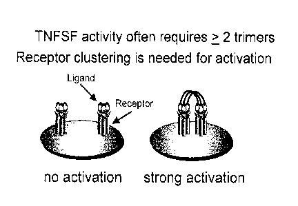
[020] FIG. 1: Schematic drawing illustrating the need to cluster a TNFSF
receptor such as the CD40
receptor on dendritic cells and other APCs in order to provide a strong cell
stimulus. This requirement
for clustering affects the design of an effective form of TNFSF ligand or an
anti-TNFSF receptor-
binding antibody.
[021] FIG. 2: Agonistic anti-CD40 antibodies can cluster CD40 receptors so
long as they bind to and
are "mounted" on a nearby cell that expresses receptors for the Fc tail of the
antibody molecule.
Abbreviations: FcyR ¨ the receptor for the Fc portion of immunoglobulin G
(IgG). Anti-CD40 MAb
- a monoclonal antibody that binds to CD40.
8a
Date Recue/Date Received 2022-05-16
CA 02907384 2015-09-15
WO 2014/145355 PCT/US2014/030099
[022] FIG. 3: Molecular design of fusion proteins that create many-trimer
forms of soluble
CD4OL. On the left is a schematic for a 1-turner form of CD4OL that cannot
cluster the CD40
receptor and as a result is inactive, as shown by Haswell eta! (Eur J Immunol.
2001;31 (10):3094-100)
and Holler et al (Mol Cell Biol. 2003;23(4):1428-40) and described in EP
1246925 Bl. As previously
described (Stone et al, J Virol. 2006;80(4):1762-72) and presented in US
7,300,774 B1 and US
7,332,298 B2, and also in EP 1246925 B1, the extracellular domain (ECD) of
CD40I, can be
genetically fused to scaffold-forming proteins such as Acrp30 (middle) or
surfactant protein D (SPD)
(right). The 2-trimer Acrp30-CD4OL protein is also called MegaCD40LTm or CD4OL
hexamer,
whereas the 4-trimer SP-D-CD4OL protein is also called UltraCD40LTM. These
many-trimer forms
of CD4OL can cluster the CD40 receptor and act as a vaccine adjuvant. This
occurs in part by
activating dendritic cells (Miconnet and Pantaleo, Vaccine. 2008;26(32):4006-
14).
[023] FIG. 4: Two- and four-trimer CD4OL fusion proteins are vaccine adjuvants
for CD8+ T cell
responses. Mice were vaccinated by injecting "naked" plasmid DNA into muscle
in order to test
different forms of CD4OL as an adjuvant for the HIV-1 Gag antigen. In Panel A,
CD8+ T cell
responses were detected as killing of P815 target cells pulsed with Gag
peptide. In Panel B, CD8+ T
cell responses were detected by measuring the number of individual interferon-
gamma secreting cells
in response to Gag peptide antigen using an ELISPOT assay. There was a
distinct improvement in
CD8+ T cell responses using a 2-trimer form of CD4OL (Acrp30-CD4OL) and more
preferably a 4-
trimer form of CD4OL (SPD-CD40L) (Stone et al.,J Virol. 2006;80(4):1762-72).
To show the general
applicability of this approach, a similar vaccine assay system was used to
show that other TNFSF
ligands could be multimerized as 4-trimer proteins and used as vaccine
adjuvants, including GITRL,
4-1BBL, OX4OL, RANKL, LIGHT, CD70, and BABE (Kanagavelu et al, Vaccine.
2012;30(4):691-
702. PMCID: 3253891).
[024] FIG. 5: Molecular design of multimeric CD4OL fusion proteins containing
an in-frame
insertion encoding HIV-1 Gag as a model antigen. Top: pSPD-Gag-CD4OL is a
plasmid containing
an antigen inserted into the protein strand that results in a 4-trimer form of
CD4OL. At the nucleic
acid level, the codons for a model antigen, HIV-1 Gag, were positioned into
the coding sequence of
the SPD-CD40I. construct. In the resulting translated protein, the N-terminus
is comprised of a
secretion signal peptide from SPD followed by an N-terminal sequence of SPD
termed the "hub"
which contains 2 cysteines in each strand, thereby producing disulfide bonds
that (a) covalently
9
CA 02907384 2015-09-15
WO 2014/145355 PCT/US2014/030099
couple three individual polypeptide strands together to form an "arm" and (b)
covalently couple 4
trimeric arms into the final 12-chain, 4-arm structure shown in the bottom
left of the figure (where
the inserted Gag antigen is shown as a solid bulge in each arm of the
protein). Note that the Gag
antigen sequence was positioned between the 105 and 106 amino acids of murine
SPD protein, while
retaining the previously constructed CD4OL domain at the C-terminal end. Like
the parent SPD-
CD4OL molecule, this protein strand of SPD-Gag-CD40I, spontaneously self-
assembles inside cells
into a multimeric, many-trimer form of CD4OL that is then secreted into the
extracellular space. 2nd
from Top: pTrimer-Gag-CD4OL (labeled plfr-Gag-CD4OL) is a plasmid constructed
by deleting
codons for amino acids 24-105 of murine SPD. This removes the hub region
containing the 2-
cysteines. Also included is the t-PA signal peptide sequence for secretion.
This results in the
production of a single-trimer, 1 "arm" form of the Gag antigen-CD4OL protein,
as shown in the
bottom right of the figure (where the Gag antigen is shown as a solid bulge in
this 1-trimer form of
CD4OL). 3rd from Top: pGag is the plasmid encoding amino acids for the p55 Gag
antigen preceded
by the t-PA signal sequence for secretion, as described by Qiu et al. (l
\Tirol. 1999;73(11):9145-52).
This is a control antigen construct that has no CD4OL adjuvant. 4th from Top:
pSPD-CD4OL is the
plasmid encoding a 4-trimer form of CD4OL previously described by Stone et al.
(J
2006;80(4):1762-72) and in US 7,300,774 B1 and US 7,332,298 B2. This is an
adjuvant-only protein
that does not contain an antigen. It can, however, be co-administered with an
antigen plasmid such
as pGag, as shown in FIG. 4.
[025] FIG. 6: p5PD-Gag-CD4OL encodes a secreted protein. Panel A shows a
Western blot of a
reducing SDS-PAGE gel analysis of the culture media of 293T cells were
transiently transfected with
DNA for the plasmids shown. An antibody for murine CD4OL was used to reveal
the protein bands.
As shown, pSPD-Gag-CD4OL encodes a single protein of the expected size of 105
kDa. A single 105
kDa band was also observed using antibody to the p24 portion of Gag (not
shown). Panel B shows a
similar analysis using non-denaturing PAGE in the absence of a reducing agent.
Multiple bands were
observed at >200 kDa molecular weight, demonstrating the formation of large
multimeric
complexes. As is commonly observed in such analyses of collagen-like proteins,
partial denaturation
during processing can result in an unwinding of some of the collagen triple
helix, which could thus
lead to a less compact protein that moves more slowly through the gel during
the electrophorctic
process.
CA 02907384 2015-09-15
WO 2014/145355 PCT/US2014/030099
[026] FIG. 7: Qualifying assay for the biological activity of SPD-Gag-CD4OL in
vitro. Panel A: In
vitro activity using a CD40 receptor NF-x13 indicator cell line. To produce
soluble protein, 293T cells
were transiently transfected with plasmids for pcDNA3.1 (empty vector
control), pSPD-CD4OL, or
pSPD-Gag-CD4OL and the protein-containing supernatants were collected 48 hours
later. To
determine the activity of the CD4OL in these proteins, the culture media as
added to cultures of 293
reporter cells containing an NF-zB-driven gene for secreted alkaline
phosphatase (SEAP) and
expressing the CD40 receptor (CD40-293-SEAP reporter cells). If the CD40
receptor is activated by
CD4OL, then NV-zB-driven SLAP production results in the secretion of SLAP
which can be
measured by a colorimetric enzyme assay at 0D650 (Maurais et al., Virology.
2009;385(4227-32). In
this assay, a single trimer of CD4OL (R&D Systems, Inc., Minneapolis, MN) was
entirely inactive and
did not induce SEAP production (not shown), indicating the strict requirement
for a many-trimer
form of CD4OL for activity in this assay. In contrast, both the pSPD-CD4OL
adjuvant protein and
the new SPD-Gag-CD40I, protein of the instant invention were active as CD40
receptor activators.
Panel B: Stimulating activity on mouse bone marrow-derived dendritic cells
(BMDDC). As in Panel
A, culture supernatants from 293T cells transfected with pcDNA3.1 or pSPD-Gag-
CD4OL were
incubated with BMDDC for 18 hours. Cells were washed, stained with
fluorochrome-conjugated
antibodies, and assayed by flow cytometry for the expression of activation and
maturation markers.
The SPD-Gag-CD4OL protein upregulated CD80 and especially CD86 and CCR7,
indicating that
this fusion protein was fully capable of activating normal dendritic cells. As
expected, the CD40
receptor was downregulated by exposure to SPD-Gag-CD4OL. A cytokine mix was
used as a
positive control ("Mimic," consisting of 10 ng/m1 of rhTNF-alpha, 10 ng/ml of
rhIL-lbeta, 1000
15/m1 of rhIL-6 and 1 Rg/m1 of PGE2; Sato et al., Cancer Sci. 2003;94(12):1091-
8). * p < 0.05, ** p
< 0.01, and *** p < 0.001 compared to pcDNA3.1 supernatant. Data represents
independent wells
in the same experiment.
[027] FIG. 8: DNA vaccination with pSPD-Gag-CD4OL demonstrates a surprisingly
high level of
CD8+ T cell responses. Panel A: DNA vaccination schedule. Mice were vaccinated
three times at
two-week intervals with an intramuscular injection of 100 ug of plasmid DNAs.
Panels B and C:
CD8+ ELISPOT assay. To measure the Gag-specific CD8+ T cell response, spleen
cells were
collected 14 days after the last vaccination and tested by ELISPOT assays.
Panel B shows cells
producing interferon-gamma and Panel C shows cells producing IL-2. The control
vaccination is
pGag + pcDNA where empty pcDNA3.1 (pcDNA) was used to keep the total amount of
DNA
11
CA 02907384 2015-09-15
WO 2014/145355 PCT/US2014/030099
constant. The previously reported mix of antigen and 4-trirner CD4OL adjuvant
plasmid is pGag +
pSPD-CD4OL which consists of separate plasmids for antigen and adjuvant, i.e.,
not present in the
same secreted molecule. Surprisingly, pSPD-Gag-CD4OL, the subject of the
instant invention,
resulted in a massive antigen-specific CD8+ T cell response (note that a
broken Y-axis is needed to
keep the results visible in the graph). In contrast, pGag + pIL-12 gave more
modest CD8+ T cell
responses, even though a pTI-12 plasmic] is currently being evaluated in human
vaccine trials. Panel
C shows the same analysis using IL-2 ELISPOT assay and showed the surprising
strength of pSPD-
Gag-CD4OL, the subject of the instant invention.
[028] FIG. 9: DNA vaccination with pSPD-Gag-CD4OL demonstrates a surprising
improvement
in CD8+ T cell quality. Panel A: T cell receptor avidity for peptide
antigen/MHC-I measured by
ELISPOT assay. Splenocytes were cultured with serial dilutions of CD8+ T cell
specific peptide
AMQ.MLKETI for 18 hours. Splenocytes from mice vaccinated with pSPD-Gag-CD4OL
induced a
significant increase in IFN-y ELISPOTs following stimulation with Gag peptide
AMQMLKETI at a
concentration of 1 ng/m1 and 10 penal whereas there was essentially no
activity at these doses using
splenocytes from mice vaccinated with pGag antigen alone or a mixture of
separate plasmids for
pGag and pSPD-CD4OL adjuvant. * p <0.05; ** p < 0.01; *** p <0.001 compared to
pGag alone
or pGag + SPD-CD4OL vaccination. Panel B: IgG antibody responses against Gag
antigen. Total IgG
specific for Gag was measured by ELISA assay from mouse serum collected on day
42. Consistent
with a previous study (Stone et aZ, J Virol. 2006;80(4):1762-72), CD4OL
adjuvant used in this format
is not an adjuvant for antibody responses.
[029] FIG. 10: The multi-trimer structure of SPD-Gag-CD4OL is necessary for
the improved
vaccine effect. In Panels A and B, pTrirner-Gag-CD4OL was used as 1-trimer
control for 4-trimer
pSPD-Gag-CD4OL. As shown, the many-trirner structure was necessary for the
strong adjuvant
effect.
[030] FIG. 11: Protective effects of pSPD-Gag-CD4OL vaccination measured by
vaccinia-Gag viral
challenge. BALB/c female mice were immunized intramuscularly with the plasmids
shown on days 0,
14, and 28. Two weeks following the final vaccination, the mice were
challenged intraperitoneally
with 10E7 plaque-forming units (PFU) of vaccinia-Gag.. Mice were sacrificed 5
days after viral
challenge and the ovaries were harvested and analyzed for PFU. Pane/ 4:
Intramuscular DNA
vaccination with pSPD-Gag-CD4OL resulted in significantly greater protection
from viral challenge.
12
CA 02907384 2015-09-15
WO 2014/145355 PCT/US2014/030099
In contrast, DNA vaccination with a mixture of pGag antigen plus pSPD-CD4OL
adjuvant as
separate plasmids only induced a modest reduction in viral loads that was not
significantly reduced
compared to pGag antigen alone. * p < 0.05; ** p < 0.01; *** p < 0.001. Panel
B: Evaluation of a
single trimer pTrimer-Gag-CD4OL construct. As shown before, the multi-trimer
structure of SPD-
Gag-CD4OL is necessary for the improved vaccine effect.
[031] FIG. 12: Adenoviral vector delivery of SPD-Gag-CD4OL is surprisingly
protective against
virus challenge. BALB/c female mice were immunized intramuscularly on days 0
and 14 with
adenovirus 5 (Ad5) expressing the I IIV-1 Gag antigen (Ad5-Gag) or the SPD-Gag-
CD4OL construct
(Ad5-SPD-Gag-CD4OL). Two weeks following the final vaccination, mice were
challenged
intraperitoneally with vaccinia-Gag virus (10E7 PFU). Mice were sacrificed 5
days later and ovaries
were harvested for vaccinia PFU determinations. Surprisingly, Ad5-SPD-Gag-
CD4OL vaccination
reduced viral load by ¨ 7 logs following vaccinia-Gag challenge. No detectable
virus could be found
in the mice that had received this vaccine, indicating complete protection
(sterilizing immunity).
[032] FIG. 13: Construction and Western blot of SPD-gp100-CD4OL. Panel A:
Model of SPD-
gp100-CD4OL fusion. Amino acids 25 to 596 (sequence KVPRNQD to EAGLGQV) of
human
gp100 was inserted between amino acids 105 and 106 of murine SPD within the
SPD-CD4OL fusion
construct. Panel B: Schematic diagram of expected SPD-gp100-CD4OL 4-trimer
structure. Panel C:
Western blot analysis. 293T cells were transfected with DNA plasmid encoding
gp100 or the SPD-
gp100-CD4OL fusion protein. After 48-hour culture, supernatant was collected
and run on an SDS-
PAGE gel in the presence of reducing agent. Western blot was performed using a
polyclonal
antibody to gp100.
[033] FIG. 14: Biological activity of SPD-gp100-CD4OL Pane/ A: In vitro
activity of SPD-CD40I,
and SPD-gp100-CD4OL was determined using a cell-based CD40 NE-kB enzymatic
reporter system.
An equivalent amount of 293T supernatant from pcDNA3.1, pSPD-CD4OL or pSPD-
gp100-CD4OL
transfcctcd cells was incubated with 293-CD4O-SEAP NF-kB reporter cells. Panel
B: In vitro activity
of SPD-gp100-CD4OL was evaluated on mouse bone marrow derived mouse DC and
compared to
empty vector or Mimic cytokine positive control. * p<0.05, p<0.01
by Student's t test compared
to pcDNA3.1 supernatant.
13
CA 02907384 2015-09-15
WO 2014/145355 PCT/US2014/030099
[034] FIG. 15: Immunotherapy of established B16F10 melanoma tumors. Panel A:
Immunization
schedule for B16-F10 tumor challenge and DNA/GVAX therapeutic vaccination, as
indicated by
arrows. B16F10 cells (50,000) were injected i.d. into the left flank of
C57BL/6 mice on day 0. Mice
were then immunized by i.m. injection of PBS or pSPD-gp100-CD4OL plasmid on
day 3, 10, and
17. GVAX, B16F10 tumor cells expressing GM-CSF, were irradiated at 5,000 rad
and 1 X 10E6 cells
injected subcutaneously on day 3, 6, and 9. Panel B: Tumor growth analysis.
Each point represents
the mean tumor volume in each group (n=5). We did not observe a statistical
difference in tumor
sizes between no treatment (PBS) and SPD-gp100-CD4OL vaccination groups. Panel
C Survival
analysis based on the date of death or when tumor size reached >1500 cm2. No
statistical differences
in survival were observed between groups.
[035] FIG. 16: Imnaunotherapy of established B16F10 melanoma tumors by DNA
vaccination
with a combination of pSPD-gp100-CD4OL, pIL-12p70 and pGM-CSF. Panel _A:
Immunization
schedule for B16F10 tumor challenge and DNA/GVAX vaccination, as indicated by
arrows.
B16F10 cells (50,000) were injected i.d. into the left flank of the mice on
day 0. Mice were
immunized i.m. with PBS, pSPD-gp100-CD4OL pIL-12, pSPD-gp100-CD4OL + pGM-CSF,
or
pSPD-gp100-CD4OL + pIL-12 + pGM-CSF on day 3, 10, and 17. For GVAX therapy B16-
F10
tumor cells expressing GM-CSF (GVAX), were irradiated at 5,000 rad and 1 X 10⁶
cells were
injected subcutaneously on day 3, 6, and 9. Panel B: Tumor growth analysis.
Each point represents
the mean tumor volume of animals in each group (n=5). There was a significant
reduction in tumor
growth kinetics for SPD-gp100-CD4OL + IL-12 + GM-CSF vaccinated mice compared
to other
groups. (** p<0.01; *** p<0.001 compared to PBS or SPD-gp100-CD4OL 4- IL-12 or
SPD-gp100-
CD4OL + GM-CSF vaccination groups). Panel C: Survival analysis of mice. We
observed a significant
increase in survival and tumor free survival (date of tumor appearance) for
pSPD-gp100-CD4OL +
pIL-12 + pGM-CSF vaccinated mice as compared to other groups (** p<0.01; ***
p<0.001
compared to PBS, pSPD-gp100-CD4OL + pIL-12, or pSPD-gp100-CD4OL pGM-CSF
vaccination
groups). Panel D: Tumor growth kinetics of individual mice from each treatment
group.
[036] FIG. 17: Separate expression of gp100 and SPD-CD4OL proteins fails to
induce anti-tumor
activity. As a control for pSPD-gp100-CD4OL, several other anti-tumor
treatment approaches were
tested and found to be inferior. Panel A: Immunization schedule for B16F10
tumor challenge and
DNA vaccination, as indicated by arrows. B16-1-'10 cells (50,000) were
injected into the left flank of
14
CA 02907384 2015-09-15
WO 2014/145355 PCT/US2014/030099
the mice on day 0. Mice were immunized i.m. with PBS, pgp100, pgp100 + pIL-12,
pgp100 + pGM-
CSF, pgp100 + pIL-12 + pGM-CSF, or pgp100-IRES-SPD-CD4OL + pIL-12 + pGM-CSF on
day
3, 10, and 17. Panel B: Tumor growth analysis. Each point represents the mean
tumor volume of
animals in each group (n=5). We did not observe any statistical difference in
tumor size between
vaccination groups. Panel C: Survival analysis. We did not observe any
statistical difference in survival
of mice between groups.
BRIEF DESCRIPTION OF THE SEQUENCES
[037] SEQ ID NO 1: DNA sequence for muSP-D-Gag-muSP-D-muCD40L. This is the DNA
sequence of a fusion protein using the murine sequences for SPD and CD4OL. Due
to minor
differences between species, it is preferable to use a murine sequence for
administration to mice, a
macaque sequence for administration in monkeys (Stone et al., Clin Vaccine
Immunol.
2006;13(11):1223-30), a human sequence for administration to humans, and so.
This minimizes the
possibility of antibodies forming against a xenogeneic protein, other than the
antigen contained in
the construct. In this example, what is shown is the nucleic acid sequence
used for the experiments
shown in FIGs. 6-12. (Note that surfactant protein D is variously abbreviated
as either `SPD' or 'SP-
D'. The location of the Gag antigen insert is shown in non-italicized type
face.
[038] SEQ ID NO 2: Protein sequence for muSP-D-Gag-muSP-D-muCD40L. This is the
translation of SEQ ID NO 1.
[039] SEQ ID NO 3: DNA sequence for tpa-muACRP30-gp120-muACRP30-muBAFF. This
is a
DNA sequence of a fusion protein using the previously described 2-trimer form
of Acrp30-BAFF
into which has been inserted a DNA sequence of HIV-1 gp120 envelope as an
antigen. It is
contemplated that the 2-trimer fusion protein encoded by this nucleic acid
sequence will activate the
Env gp120-binding B cell receptor (BCR) on B cells and simultaneously engage
receptors for BAFF
on these B cells that synergize with BCR engagement to stimulate the B cell to
produce anti-Env
antibodies.
[040] SEQ ID NO 4: Protein sequence for tpa-muACRP30-gp120-muACRP30-muBAFF.
This is
the translation of SEQ ID NO 3.
[041] SEQ ID NO 5: DNA sequence for muSP-D-gp100-muSP-D-muCD4OL. This is the
DNA
sequence of a fusion protein using the murinc sequences for SPD and CD4OL. The
inserted antigen
(non-italicized sequence) is encoded by the nucleotide sequence for human
gp100, a xenogenic
antigen that has been found to be useful in melanoma studies in mice (Gold et
aZ, J Immunol.
2003;170(10):5188-94).
[042] SEQ ID NO 6: Protein sequence for muSP-D-gp100-muSP-D-muCD40L. This is
the
translation of SEQ ID NO 5.
[043] SEQ ID NO 7: DNA sequence for tpa-huIgGlFc-gp120-GCN4-huAPRIL. This is a
DNA
sequence encoding a human t-PA signal sequence for protein secretion joined in-
frame with the
human IgG1 Fe region joined in-frame with HIV-1 Env gp120 joined in-frame with
the GCN4
trimerization motif joined in-frame with the extracellular domain of human
APRIL. It is
contemplated that the 2-trimer fusion protein encoded by this nucleic acid
sequence will activate the
Env gpl 20-binding B cell receptor (BCR) on B cells and simultaneously engage
receptors for APRIL
on these B cells that syncrgizc with BCR engagcmcnt to stimulate thc B ccll to
produce anti-Env
antibodies.
[044] SEQ ID NO 8: Protein sequence for tpa-huIgGlFc-gp120-GCN4-huAPRIL. This
is the
translation of SEQ ID NO 7.
[045] SEQ ID NO 9: DNA sequence for huSP-D-NP-huSP-D-huCD40L-NST. It was
previously
found that some embodiments of SPD-CD4OL can be equally or more active when
the extracellular
"stalk" region of CD4OL is deleted. This stalk links the CD4OL trimeric
extracellular domain (LCD)
with the transmembrane region that holds CD4OL in the membrane. The SPD-CD4OL-
NST
construct is disclosed in US 2009/0081157 Al (see especially FIG. 21, Examples
1, 11, and 13).
The instant sequence comprises an insertion of coding sequences
for the nucleoprotein (NP) antigen from influenza A. It is contemplated that
the 4-trimer fusion
protein encoded by this nucleic acid sequence will elicit strong CD8+ T
responses against this
conserved influenza antigen.
[046] SEQ ID NO 10: Protein sequence for huSP-D-NP-huSP-D-huCD40L-NST. This is
the
translation of SEQ ID NO 9.
16
Date Recue/Date Received 2020-09-29
CA 02907384 2015-09-15
WO 2014/145355 PCT/US2014/030099
[047] SEQ ID NO 11: DNA sequence for tpa-muACRP30-CSP1-muACRP30-muCD40L. This
is
a DNA sequence encoding a human t-PA signal sequence for protein secretion
joined in-frame with
a portion of the murine Acrp30 sequence joined in-frame with codons for the
circumsporozoite
protein-1 (CSP-1) of Plasmodium yoelii joined in-frame with a portion of the
murine Acrp30
sequence joined in-frame with the extracellular domain of murine CD4OL.
Plasmodium yoelii is used
for malaria vaccine studies because it causes a malaria-like disease in mice.
CD8+ T cells directed
against the CSP-1 antigen of this agent can provide immunity to malaria
(Sedegah et al., Proc Nad
Acad Sci U S A. 1998;95(13):7648-53). It is contemplated that mice vaccinated
with this construct
will be resistant to disease caused by intravenous challenge with Plasmodium
yoelii-infected red
blood cells.
[048] SEQ ID NO 12: Protein sequence for tpa-muACRP30-CSP1-muACRP30-muCD40L.
This
is the translation of SEQ ID NO 11.
[049] SEQ ID NO 13: DNA sequence for muSP-D-Gag-muSP-D-muRANKL. This is a DNA
scquence encoding a portion of the murinc SPD sequence joined in-frame with
codons for HIV-1
Gag antigen joined in-frame with a portion of the murine Acrp30 sequence
joined in-frame with the
extracellular domain of murine RANKL. Of special note is the difference of
position in placing the
antigen within the sequence of the SPD "arms," in this case shifted toward the
5' end (or N-terminal
end in the protein) the equivalent of 10 codons in the SPD sequence. It is
contemplated that this
construct used as a vaccine will elicit strong immune responses in mice.
[050] SEQ ID NO 14: Protein sequence for muSP-D-Gag-muSP-D-muRANKL. This is
the
translation of SEQ ID NO 13.
[051] SEQ ID NO 15: DNA sequence of huSP-D-WT1-huSP-D-huCD40L. This is a DNA
sequence encoding a portion of the human SPD sequence joined in-frame with
codons for the
human WTI protein joined in-frame with a portion of the human SPD sequence
joined in-frame
with the extracellular domain of human CD4OL. WTI is a tumor antigen present
in many types of
human cancer (Chaise et al., Blood. 2008;112(7):2956-64). It is contemplated
that this construct used
as a vaccine will elicit strong immune responses in humans against cancer
cells expressing the WT1
tumor antigen.
17
[052] SEQ ID NO 16: Protein sequence for huSP-D-WT1-huSP-D-huCD40L. This is
the
translation of SEQ ID NO 15.
[053] SEQ ID NO 17: DNA sequence of muSP-DAMGE-A3-muSP-D-muBAFE. This is a
contemplated DNA sequence encoding a portion of the murine SPD sequence joined
in-frame with
codons for the human MAGE-A3 tumor antigen (Groeper et al., Int j Cancer.
2007; 120(2):337-43)
joined in-frame with a portion of the murine SPD sequence joined in-frame with
the extracellular
domain of murine BAFF. Of note is that codons for 20 amino acids
(PPGLPGIPGPMGARASVLSG) in the N-terminal halt of the SPD arm have been
deleted. This
exemplifies how the SPD "arms" can be shortened N-terminal to the insertion
site of the antigen
sequence. Similar deletions in the C-terminal half of the SPD arm are also
contemplated, as are
deletions in both sides of the SPD arms that flank the antigen sequence
insertion site.
[054] SEQ ID NO 18: Protein sequence of muSP-D-ALAGE-A3-muSP-D-muBAFF. This is
the
translation of SEQ ID NO 17.
DEFINITIONS
[055] This disclosure uses art-recognized concepts and methods. The skilled
artisan will be familiar
with resources including the following: "Taneway's Immunology" by Kenneth
Murphy, Garland
Science Press, 2011; "Fundamental Immunology" by William E. Paul, Lippincott
Williams &
Wilkins, 2008; "Cellular and Molecular Immunology, 7th Edition" by Abul K.
Abbas, Andrew H. H.
Lichtman, and Shiv Pillai, Elsevier Press, 2011; "Current Protocols in
Immunology," Wiley Press,
2012; and "Current Protocols in Molecular Biology," Wiley Press, 2012. In
addition, the following
patents and applications are referred to: US 7,300,774B1; US 7,332,298 B2;
US 2009/0081157 Al.
[056] Unless otherwise explained, all technical and scientific terms used
herein have the same
meaning as commonly understood by one of ordinary skill in the art to which
this disclosure belongs.
Definitions of common terms in molecular biology can be found in Benjamin
Lewin, Genes V,
published by Oxford University Press, 1994 (ISBN 0-19-854287-9); Kendrew et
al. (eds.), The
Encyclopedia of Molecular Biology, published by Blackwell Science Ltd., 1994
(ISBN 0-632-02182-
9); and Robert A. Meyers (ed.), Molecular Biology and Biotechnology: a
Comprehensive Desk
Reference, published by VCR Publishers, Inc., 1995 (ISBN 1-56081-569-8).
18
Date Recue/Date Received 2020-09-29
[057] In order to facilitate review of the various embodiments of this
disclosure, the following
explanations of specific terms arc provided:
[058] The singular terms "a," "an," and "the" include plural referents unless
context clearly
indicates otherwise. Similarly, the word "or" is intended to include "and"
unless the context clearly
indicates otherwise. It is further to be understood that all base sizes or
amino acid sizes, and all
molecular weight or molecular mass values, given for nucleic acids or
polypeptides are approximate,
and are provided for description. Although methods and materials similar or
equivalent to those
described herein can be used in the practice or testing of this disclosure,
suitable methods and
materials are described below. The term "comprises" means "includes." The
abbreviation, "e.g." is
derived from the Latin exempli gratia, and is used herein to indicate a non-
limiting example. Thus,
the abbreviation "e.g." is synonymous with the term "for example."
[059] "Clq family protein" refers to a member of the Clq family. Exemplary Cl
q family proteins
include, but are not limited to, C11, Acrp30, and H1B27. Preference is given
to Acrp30. Like the
collcctins, C1q family members have 2 or morc trimeric, collagen-like "arms"
that provide the
multivalent structures of these molecules. The instant invention utilized Clq
family proteins as a
multimerization scaffold by replacing their normal C-terminal "Clq" domains
with a TNFSF
receptor binding such as the ECD of a TNFSF ligand.
[060] "Collectin" refers to a member of the collectin family.
They include
pulmonary surfactant A, pulmonary surfactant D, conglutinin, collectin-43,
mannose-hinding protein
MBL1 or MBL2, and others. Preference is given to surfactant protein D
(abbreviated alternatively as
SP-D or SPD). All collectins have two or more trimeric collagen-like "arms"
joined in the center at a
"hub" and radiating outward to display their C-terminal ends. Each collectin
has a C-terminal
domain that typically binds to carbohydrate. When used as a multimerization
scaffold in the instant
invention, each collectin is made without the natural C-terminal end and a
TNFSF ECD receptor
binding domain is placed there instead. Preference is given to surfactant
protein D which has four
irimeric arms ending C-ierminally.
[061] "Complete TNFSF receptor" is a term used herein in marked distinction to
a single
olypep tide chain often referred to as a TNFSF receptor protein
19
Date Recue/Date Received 2020-09-29
The nucleotide and peptide sequences of single TNFSF receptor
polypeptide chains are listed in GenBank, SwissProt, and other databases.
However, in actuality,
single TNFSF receptor polypeptide chains are not found in isolation on the
surface of cells. Instead,
two or more TNFSF receptor chains are co-localized or linked. As an example,
the Fos receptor
(CD95) for has ligand (FasL) is held together in the absence of FasL by their
N-terminal "pre-ligand
association domains" or PLAD (Siegel et al., Science. 2000288(5475):2351-4).
Similarly, there is a
domain in the extracellular region of CD40 that holds this receptor together
as 2 or more chains
(Smulski et al., J Biol Chem. 2013). Consequently, stimulation of TNFSF
receptors generally does not
involve simple bringing together of 2 or more receptor chains. When the ligand
does bind to the
receptor, computer modeling suggests that a ligand trimer engages three
receptor chains (Bajorath et
al., Biochemistry. 1995;34(31):9884-92). Thus, this application uses the term
"complete TNFSF
receptor" to indicate that binding to a TNFSF receptor involves binding to 2
or preferably 3 receptor
protein chains.
[062] "Immune system" refers to T cells, B cells, NK cells, dendritic cells,
monocytes, and
macrophages and the specialized tissues that contain them. The lymph nodes,
lymphatics, and spleen
are physical structures that housing many of the cells of the immune system.
In addition, other
immune system cells are found in non-lymphoid tissues and in blood. A
characteristic of the immune
system is that it responses to a first exposure to an antigen (primary
response) in a set fashion but
then responds more strongly and more quickly to a second exposure of an
antigen (secondary
response), which is a manifestation of immunological memory. The immune system
responds to
infectious agents and cancer by producing cells and effector molecules that
kill the offending
infectious agent or cancer cells. Among the cells that kill the attackers are
T cells including CD4+
and CD8+ T cells. B cells make antibodies that can neutralize the infectivity
of many infectious
agents. T cells, monocytes, macrophages, and dendritic cells can make
interferons that interfere with
the replication of certain viruses.
[063] "lVlultimerization scaffold" refers to a molecular structure that
confers upon the molecule
into which it is incorporated an overall structure that is operatively linked
to two or more TNFSF
receptor binding domains, such that contact with the multimerized molecule
leads to clustering of
the complete TNFSF receptor in the membrane of a responding cell and thereby
activates some or
Date Recue/Date Received 2020-09-29
all of the functional potential of the responding cell. A key concept of the
instant invention is that a
many-trimer form a TNFSF ligand is needed to stimulate a receptor-bearing
responding cell. For
example, structural studies of the GITRL/GITR interaction indicate that two
closely localized
trimers of GITRL are needed to bring together or "cluster" two complete GITR
receptor (3 chains
of GITR each) (Zhou et al., Proc Natl Acad Sci U S A. 2008;105(14):5465-70). A
multimerization
scaffold is a molecular structure that provides for this close localization of
2 or more TNFSF
receptor binding, typically 2 or more TNFSF ligand extracellular domains
(ECD). In the instant
invention, portions of collectins such as SPI) or portions of Cl q family
members such as Acrp30 are
used to make single polypeptide chains that self-assemble into multimerization
scaffolds. Preference
is shown for multimerization scaffolds that have "arms" capable of being
operatively linked to
TNFSF ECD trimers. Alternative embodiments are contemplated, such as
multimerization scaffold
that is operatively linked to single-chain antibodies that bind to a TNFSF
receptor.
[064] "Operatively linked" refers to a method for joining two molecules. For
polypeptides, this is
preferably by a peptide bond, typically achieved by constructing a DNA or RNA
template encoding
the operatively linked fusion protein and then expressing the DNA or RNA in a
cell Or by an in vitro
method. In some case, chemical crosslinkers can be used to construct
multimeric forms of TNFSF
receptor binding agents as described in US 6,482,411 B1 .
[065] "TNFSF" refers to a ligand in the Tumor Necrosis Factor (TNF)
SuperFamily.
The TNFSFs are produced as trimeric Type II membrane molecules meaning that
their N-terminus
points inside the cell and their C-terminal end is extracellular, which is the
reverse of most cell
surface proteins. This makes these proteins very challenging to engineer using
traditional fusion
protein strategies.
[066] "TNFSF receptor binder" refers to a molecular fragment that binds to a
TNFSF receptor.
Exemplary TNFSF receptor binders (or binding domains) include the
extracellular domain (ECD) of
TNFSF trimeric molecule or the receptor-binding portion of an antibody
recognizing a TNFSF
receptor. For a receptor-binding portion of an antibody, preference is give to
single-chain antibody
constructs (Ahmad et al., Clin Dev Immunol. 2012;2012:980250. 131\ICID:
3312285). Exemplary
TNFSF members whose extracellular domains can be used as TNFSF receptor
binders include
21
Date Recue/Date Received 2020-09-29
CA 02907384 2015-09-15
WO 2014/145355 PCT/US2014/030099
CD4OL (TNFSF5), CD27L (TNFSF7), CD137L (TNFSF9), OX4OL (TNFSF4), GITRL, 4-
1BBL,
RANKL, LIGHT, CD70, and BAFF.
[067] "Tumor antigens" refers to proteins, carbohydrates, or lipids found on
tumor cells against
which the immune system can launch an attack. For a discussion of tumor
antigens, see Kvistborg et
al (Curr. Opinion Immunol. 25:284-290, 2013) and Cheever et al. (Clin Cancer
Res 15, 5323-5337,
2009). Also contemplated as tumor antigens are antigenic peptides deduced from
next-generation
sequencing from the RNA or DNA of tumors, including exome sequencing (Segal et
al, Cancer Res.
2008;68(3):889-92; Castle et al, Cancer Res. 2012;72(5):1081-91).
DETAILED DESCRIPTION OF THE INVENTION
[068] This invention describes, inter alia, molecules comprising fusion
proteins and the nucleic acids
that encode them in which the following protein coding domains are operably
linked in the following
order: a scaffold comprised of a portion of a collectin or Clq family protein
or combinations of
dimerizing/trimerizing motifs, an antigen (either following the scaffold or
contained within the
scaffold), and the extracellular domain of a TNF superfamily ligand. An
exemplary fusion protein or
nucleic acid that encodes it comprises the antigen, surfactant protein D (SPD)
without its
carbohydrate receptor domain, and the extracellular domain of CD40 ligand.
Alternatives to
surfactant protein D can also be used, including using immunoglobin (Ig),
Acrp30, a GCN4
multimerization motif, or similar proteins as scaffolds for CD40 ligand, other
members of the TNF
superfamily ligands, or other ligands or receptors, including gp96 or 1\4HC
molecules. In one
embodiment, the molecules, compositions and/or fusion proteins of the
invention do not contain
portions of avidin or streptavidin.
[069] These fusion proteins are designed to allow the targeting of dendritic
cells, macrophages, B
cells or other antigen presenting cells with the antigen as well as providing
necessary activation
signals to induce maturation of the targeted dendritic cell, macrophage, B
cell or other antigen
presenting cell. This results in the optimal presentation of the antigen to
the immune system, and a
potent immune response in the treated individual, either T cell mediated or
antibody mediated.
[070] In more detail, the instant invention provides a solution for the
problem of vaccinating
against infectious agents and for cancer immunotherapy. It provides a way to
link an adjuvant in the
22
CA 02907384 2015-09-15
WO 2014/145355 PCT/US2014/030099
TNF SuperFamily (TNFSF) to an antigen such that the TNFSF adjuvant and antigen
arrive at the
same cell at the same time. In the case of CD8+ T cell responses, it is
important to provide antigen
to dendritic cells (DCs) and other antigen-presenting cells such that the
protein antigen is processed
by cleavage into peptides and loaded onto 1VIHC-I for cross-presentation on
the cell surface as
pl\IHC-I complexes which in turn stimulates the T cell receptor (Signal 1). It
is preferable to target
the antigen to the CD40 receptor on DCs since this results in superior cross-
presentation by a larger
number of DC subtypes (Chatterjee et al., Blood. 2012;120(10):2011-20). In
addition, it is important
to activate the DC that is presenting antigen in order that the DCs present
the antigen-specific T cell
with accessory signals (Signal 2 and Signal 3). If the DCs display only pl\IHC-
I and are not activated
to present other signals, then the resulting antigen-specific CD8+ T cell
becomes tolerant and lacks
protective effective functions (Bonifaz et al., J Exp Med. 2002;196(12):1627-
38. PMCID: 2196060).
Stimulation of the CD40 receptor on DCs activates the DCs to provide these
other signals and leads
to prof-build CD8+ T cell responses (Bonifaz et aZ, J Exp Med. 2004;199(6):815-
24). Thus, the
instant invention provides a strong vaccine for CD8+ T cells by fusing antigen
to previously
described multimeric forms of CD4OL comprised of the extracellular domain
(ECD) of CD4OL
fused to multimerization scaffolds employing portions of surfactant protein D
(SPD) or Acrp30.
[071] Activation of DCs and other APCs is best performed by a many-trimer form
of CD4OL
where 2 or more trimers are needed to cluster and thereby activate the CD40
receptors on DCs, as
depicted in FIG. 1.
[072] The new understanding of agonistic anti-TNFSF receptor antibodies is
shown in FIG. 2. In
this case, the antibody is first bound to an adjacent cell via its Fc portion
which binds to the Fc
receptors on the adjacent cell type (Li and Ravetch, Science.
2011.333(6045):1030-4.
3164589; Wilson et al., Cancer Cell. 2011;19(1):101-13; White et al.,J
Immunol. 2011;187(4):1754-63).
This leads to two problems: (1) DCs and other APCs that are not adjacent to an
FcR-bearing cell
cannot be stimulated; and (2) if the antibody binds to certain FcRs, then it
is possible that the
adjacent cell will kill the DC by antibody-dependent cellular cytotoxicity
(ADCC) or phagocytose the
DC and eliminate it (Bulliard el al., J Exp Med. 2013;210(9):1685-93. PMCID:
3754864). The later
phenomenon may explain the severe depletion of CD40 B cells when an antibody
against CD40 was
tested in humans with cancer (Vonderheide et aZ, J Clin Oncol. 2007;25(7):876-
83). These
23
CA 02907384 2015-09-15
WO 2014/145355 PCT/US2014/030099
considerations set the stage for a new and better way to provide both antigen
and CD40 stimulation
to DCs and other APCs.
[073] Another approach was taken by Xiang et al. (J Immunol. 2001;167(8):4560-
5) who made a
fusion protein of tumor antigen (CEA) joined to the C-terminal end of CD4OL
(US 7,279,464 B2;
US 6,923,958 B2). However, because the CD4OL moiety is not located on the end
of the protein, it
could conceivably have impaired binding of the ligand to the CD40 receptor. No
data were
presented to rule out this concern, hut the vaccine's effectiveness was
modest.
[074] In a related approach, Zhang et al. (Proc Nati Acad Sci U S A.
2003;100(25):15101-6) fused a
tumor antigen onto the N-terminus of the CD4OL extracellular domain and
delivered this construct
using an adcnovirus vector. In this case, the molecular design allowed for
CD40L to bind unimpaired
to its receptor. Even so, the effectiveness of this vaccine was relatively
modest. This is expected
when a 1-trimer form of CD4OL is used rather than a receptor-clustering multi-
trimer construct such
as SPD-Gag-CD4OL.
[075] Another approach was taken by Shirwan et al. who produced a fusion
protein between the
"core" region of bacterial streptavidin protein (CSA) and the extracellular
domain of CD4OL or
4-1BBIõ as disclosed in US 8,017,582 B2 and in Schabowsky et aZ, Exp Mol
Pathol. 86:198-207,
2009. In this case, the N-terminal half of the fusion proteins consisted of
CSA where streptavidin
naturally assembles into a 4-chain molecule. This multimerism pulls together
the covalen.tly linked
ECDs for CD4OL or 4-1BBL. Since streptavidin binds to biotin and since
proteins can be easily
biotinylated, it was possible to biotinylate antigens such as chicken
ovalbumin (OVA) or the tumor
antigens E7 from HPV which allows them to bind non-covalendy to CSA-CD4OL or
CSA-4-1BBL.
However, in order to be active, CD40I, must be used in a multi-trimer form
that clusters together
two or more CD40 receptors, as depicted in FIG. 1 of the instant application.
The relative inactivity
of a single trimer form of CD4OL was demonstrated by _Haswell et al. (Eur J
lmmunol. 31:3094-3100,
2001; see FIG. 3). In contrast, the CSA-CD4OL forms a single trimcr of CD4OL,
as depicted in 1-1G.
1B of Schabowsky et al., which is not desirable from the perspective of
efficient receptor stimulation.
Furthermore, the biotin-streptavidin interaction in the design of Shirwan et
al. is non-covalent. The
antigen has been biotinylated which then allows it to bind to the streptavidin
moiety in the CSA-
CD401, complex. However, in vivo, there is free biotin present in biological
fluids that can interfere
with the formation of the CSA-CD4OL/biotin-antigen complex or induce its
dissociation. In
24
contrast, the instant invention utilives antigen that has been covalently
joined to CD40I, by virtue of
the peptide bonds that make up the SPD-antigen-CD4OL fusion protein and thus
the protein is not
susceptible to dissociation in the presence of free biotin. Another important
difference is that CSA is
a xenogenic protein from bacteria that is highly antigenic in humans and other
vertebrates (Meyer et
al. Protein Science 2001 ;10(3):491 -503; Yumura et aL, Protein Science
2013222:2132i). In contrast,
the fusion proteins of the instant invention can be constructed with primarily
non-xenogenic
proteins sequences such that the only major foreign protein component is the
antigen selected for
immunization. Therefore, in one embodiment of the present invention, the
multimerization scaffold
and the plurality of TNFSF receptor binder do not contain any xenogenic
portions.
[076] Another system for producing many-trimer forms of OX4OL was described by
Weinberg et
aZ in US 7,959,925 B2. In this
system, fusion proteins are made
by using an N-terminal immunoglobulin Fc domain which naturally dimerizes via
interchain disulfide
bonds. When this is joined to a trimerizing domain which is then joined to a
TNFSF extracellular
domain, it results in what is described as a hexamer or "dimer of trimers". In
the instant invention,
SEQ ID NO:7 and SEQ ID NO:8 disclose a fusion protein that provides for a 2-
trimer form of
APRIL fused to the HIV-1 Env protein which is expected to elicit a strong
antibody response to
HIV-1. The skilled artisan will easily see how the extracellular domain of
APRIL could be replaced
by the extracellular domain of any other TNFSF ligand, and also how the 111V-1
Env antigen could
be replaced by other antigens of interest. Such antigen-multimeric TNFSF
fusion proteins are
claimed by the instant invention. In addition, the skilled artisan could
envision other dimerizing
domains (such as that from CD4 or CD8) or other trimerizing domains (such as
those from GCN4,
TRAF2, thrombospondin 1, 1VIatrilin-4, CMP (Matrilin-I), HSFI, or cubulin, as
described in US
7,959,925 B2) or the trimerizing domain from the SPD "neck" region in US
6,190,886
[077] As described in the instant application, a surprisingly active vaccine
can be made by
incorporating an antigen with the arms of SPD in the 4-trimer SPD-CD40I,
constmct that was
previously developed by the inventors and shown in FIGs. 3 and 4. For
demonstration purposes, the
HIV-I Gag antigen was inserted into the coding region for the SPD collagen-
like arm as shown in
SEQ Ill NO:1 and SEQ ID NO:2 and depicted in FIG. 5. This fusion protein uses
the natural SPD
"arm" which has been shown to be 46 nm long in shadow electronmicroscopic
studies. The
Date Recue/Date Received 2020-09-29
CA 02907384 2015-09-15
WO 2014/145355 PCT/US2014/030099
collagen-like triple helical structure and results from the class Gly-Xaa-Yaa
collagen-like repeats in
the protein which number 59 repeats in the arm. For the instant invention, the
length of this arm can
be varied in two ways: (1) Amino acid deletions can be introduced that
truncate one Gly-Xaa-Yaa
motif; and (2) the antigen can be inserted variably along the length of the
arm. Considering the 177
amino acids in the 59 collagen-like repeats, the antigen domain can be
positioned from 10 to 177
amino acids more C-terminal from the hub, or preferably from 20 to 140 amino
acids more C-
terminal from the hub, or more preferably from 40 to 120 amino acids more C-
terminal from the
hub. Likewise, the antigen domain can placed closer or further from the TNFSF
extracellular domain
(ECD). For example, the antigen domain could be from 0 to 167 amino acids more
N-terminal from
the TNFSF ECD, or more preferably from 40 to 120 amino acids more N-terminal
from the ECD.
As non-limiting examples, SEQ ID 13 and SEQ ID 14 show a fusion protein where
the antigen
domain was shifted by 10 amino acid positions within the arm of SPD. Likewise,
SEQ ID NO 17
and SEQ ID NO 18 show a fusion protein in which 20 amino acids have been
removed from the
SPD arm. With this as a guide, the skilled artisan will know that it is not
critical exactly where in the
SPD arm the antigen domain should be positioned.
[078] As previously described by the inventors, 2-trimer forms of TNFSF
ligands can be made
using Acrp30. FIGs. 3 and 4 show the design and vaccine adjuvant efficacy of
an Acrp30-CD4OL
fusion protein. This molecule has two collagen-like arms. Accordingly, it is
contemplated to place an
antigen domain within the arms of Acrp30 as shown in SEQ ID 3 and SEQ ID 4
which place the
HIV-1 Env antigen within the arms of an Acrp30-BAFF fusion protein. Analogous
fusion proteins
could be made from other collectin fusion proteins besides SPD-TNFSFs and from
other Clq family
molecules besides Acrp3-TNESFs.
[0791 A feature of these fusion proteins is that they can readily be made
using the natural collcctin
or C1q family sequences and TNFSF sequences from a variety of organisms. It is
preferable to use
the murine coding sequences for studies in mice, the macaque coding sequences
for studies in
macaques, the human coding sequences for use in humans, etc. As non-limiting
examples, the
sequences shown provide fusion protein made using either murine or human
sequences. Thus,
animal vaccine uses are specifically contemplated as one use of the instant
invention.
pm In these cases, antigen was introduced into many-trimer forms of TNFSFs by
standard
genetic engineering methods familiar to the skilled artisan. Such fusion
proteins can be made by
26
ligating together segments of genes or, more preferably, by ordering a custom
synthesis from a
commercial supplier (e.g DNA2.0, Genset, Gcnewiz, and other suppliers). In
other cases, it is
possible to prepare antigenic peptides and TNFSF trimcrs separately and then
link them together by
chemical methods. The linking reagents and synthesis strategies that can be
used are described in US
6,482,411 B1 .
[081] There is a wide choice of antigens from infectious disease antigens,
depending on the species
in need of vaccination. Without limitation, these can be selected from the
following list of disease-
causing pathogens:
[082] Viruses such as influenza A and B, parainfluenza, poxviruses, ebola
virus, hepadnavirus,
filoform viruses such as marburg virus, dengue fever virus, influenza A and B,
respiratory syncytial
virus, measles (rubeola virus), human immunodeficiency virus (HIV), human
papillomavirus (HPV) ,
varicella-zoster, herpes simplex I and 2, cytomegalovirus, Epstein-Barr virus,
JC virus, rhabdovirus,
rotavirus, rhinovirus, adcnovirus, orthomyxovirus, papillomavirus, parvovirus,
picornavirus,
poliovirus, mumps, rabies, rc virus, rub clla, togavirus, rc trovirus,
coxsackicvirusc3, equine
encephalitis, Japanese encephalitis, yellow fever, Rift Valley fever virus,
hepatitis A, B, C, D, and E
virus, hantavirus, coronavirus (including SARS and MFRS), and the like;
[083] Microbial agents such as Borrelia species, Bacillus anthracis, Borrelia
burgdorferi, Bordetella
pertussis, Camphylobacter jejuni, Chlamydia species, Chlamydial psittaci,
Chlamydial trachomatis,
Clostridium species, Go striclium tetani, Clostridium botulinum, Clostridium
perfringens,
Corynebacterium diphtheriae, Coxiella species, an Enterococcus species,
Erlichia speciesEscherichia
coil, Francisella tularensis, I Iaemophilus species, I Iaemophilus injiuenzae,
I Iaemophilus
parainjiucnzac, Lactobacillus species, a Legionella species, Legionclla
pncumophila, Lcptospirosis
interrogans, Listeria species, Listeria monocytogenes, Mycobacterium species,
Mycobacterium
tuberculosis, _Mycobacterium leprae, Mycoplasma species, Mycoplasma
pneumoniae, Neisseria
species, Neisseria meningitidis, Neisseria gonorrhoeae, Pneumococcus species,
Pseudomonas
species, Pseudomonas aeruginosa, Salmonella species, Salmonella typhi,
Salmonella enterica,
Rickettsia species, Rickettsia ricketsii, Rickettsia typhi, Shigella species,
Staphylococcus species,
Staphylococcus aureus, Streptococcus species, Streptococccus pnuemoniae,
Streptococcus
pyrogenes, Streptococcus mutans, Treponema species, Treponema pallidum, a
Vibrio species, Vibrio
cholerae, Yersinia pestis, and the like;
27
Date Recue/Date Received 2020-09-29
CA 02907384 2015-09-15
WO 2014/145355 PCT/US2014/030099
[084] Fungal, protozoan, and parasitic agents such as Aspergillus species,
Candida species, Candida
albicans, Candida tropicalis, Cryptococcus species, Cryptococcus neoformans,
Entamoeba
histolytica, Histoplasma cap sulatum, Coccidioides immitis, Leishmania
species, Nocardia asteroides,
Plasmodium falciparum, Plasmodium vivax, Toxoplasma gondii, Trichomonas
vaginalis, Toxoplasma
species, Trypanosoma brucei, Schistosonia mansoni, Pneurnocystis jiroveci, and
the like.
[085] There is a wide choice of rumor antigens, depending on the species in
need of cancer
immunotherapy. Without limitation, these can be selected from the following
list of cancer-
associated antigens:
[086] gpl 00; WT1; Melan-A; tyrosinase; PSMA; HER-2/neu; MUC-1; PRAME;
topoisomerase;
BRAE V600E; bcr-Abl; sialyl-Tn; carcinoembryonic antigen; ErbB-3-binding
protein-I; alpha-
fetoprotein; and the cancer testis antigens IVLAGE-AI, MAGEA4, and NY-ES0-1;
MART-1,
Dipeptidyl peptidase IV (DPPIV), adenosine deaminase binding protein (ADAbp),
cyclophilin b,
Colorectal associated antigen (CRC)-0017 -1 AlGA 733, Carcinoembryonic Antigen
(CEA) and its
immunogenic epitopes CAP-1 and CAP-2, etv6, amll, Prostate Specific Antigen
(PSA) and its
immunogenic epitopes PSA-I, PSA-2, and PSA-3, prostate specific membrane
antigen (PSMA),
MAGE-family of tumor antigens (e.g., MAGLAI, MAGE-A2, MAGE-A3, MAGE-A4, MAGL-
A5,
MAGE-A6, MAGE-A7, MACE-AS, MACE-A9, _MAGE, _MAGE-Xp2 (MAGE-B2), .MAGE-Xp3
(MAGE-B3), MAGE-Xp4 (MAGE-B4), MAGE-C1, MAGE-C2, 1VIAGE-C3, MAGE-C4,
MAGEC5), GAGE-family of tumor antigens (e.g., GAGE-I, GAGEIn 2, GAGE-3, GAGE-
4,
GAGE-5, GAGE-6, GAGE-7, GAGES, GAGE-9), BAGE, RAGE, LAGE-1, NAG, GnT, MUM-1,
CDK4, tyrosinase, p53, MUC family, HER2/neu, p2lras, RCAS1, a-fetoprotein, E-
caclherin, alpha-
catenin, beta-catenin and gamma-catenin, p 120ctn, brain glycogen
phosphorylase, SSX-1, SSX-2
(HOMAIEL), EGFRviii, SSX-1, SSX-4, SSX-5, SCP-1, CT-7, cdc27, adcnomatous
polyposis coil
protein (APC), fodrin, PI A, Counexin 37, Ig-idiotype, p15, gp75, GM2 and GD2
gangliosides, viral
products such as human papilloma virus proteins, Smad family of tumor
antigens, LMP-1, LMP-2,
EBV-encoded nuclear antigen (EBNA)-1, or c-erbB-2, and the like.
[087] There is a wide choice of delivery methods for the vaccines of the
instant invention. Where
the vaccine is comprised of a nucleic acid sequence, it can be delivered using
a DNA or RNA
vectors. Without limitation, these can be selected from the following list:
Adenovirus (as shown in
FIG. 12 for example), poxvinis including Modified Vaccinia Ankara,
Herpesvinises, retroviruses,
28
CA 02907384 2015-09-15
WO 2014/145355 PCT/US2014/030099
lentiviruses, Newcastle Disease Virus, Mumps Virus, Measles Virus, Vesicular
Stomatitis Virus,
rhabdovirus, Para-influenza Virus, Sendai virus, Influenza Virus, Reovirus,
and a Seneca Valley virus,
alphavirus, Sindbis virus, Venezuealan Equine Envephalitis (VEE), Coxsackie
virus, myxoma virus,
viral organisms include those that are dsDNA viruses, ssDNA viruses, dsRNA
viruses, (+ ) ssRNA
viruses (-) sRNA viruses, ssRNA-RT viruses, and dsDNA-RT viruses, and the
like.
[088] Vaccines of the present invention can also be delivered as plasmid DNAs
that include a
promoter (e.g., C]\/[\T promoter) and a transcription termination and
polvadenylation sequence. Such
plasmids also include genes needed for growth in bacteria, but fragments of
DNA can also be
prepared by in vitro enzymatic synthesis. An exemplary plasmid used in the
experiments in FIGa. 4
and 6-17 is pcDNA3.1 (Life Technologies, Inc., Carlsbad, CA) but other choices
are available. The
DNA can be delivered directly by injection into muscle ("naked" DNA
vaccination) as shown in
FIGs. 8-11 and 15-17. It can also be delivered by a number of means including
electroporation,
microinjection, gene gun delivery, lipofection, polymer-mediated delivery, and
the like. The same
methods can he used for RNA vaccination. In addition, for bacteria that enter
cells such as
Salmonella or Listeria, plasmid DNA can be introduced into these bacteria
which then carry that
DNA into the eukaryotic host cell, a process called "bactofection."
[089] As another use of the instant invention, fusion proteins comprised of an
antigen linked to a
many-trimer TNFSF can be administered to APCs like dendritic cells ex vivo, as
shown in FIGs. 7
and 14. Once the antigen has been delivered and the APCs activated, these DCs
can then be
delivered to a host as a cellular form of vaccination (Barth et al.,
2010;16(22):5548-56. PMCID:
2994719).
[090] Without limitation, the following examples of invention are disclosed:
EXAMPLE 1: Vaccination to elicit CD8+ T Cells against HIV-1 Gag antigen
[091] CD40 ligand (CD4OL, CD154) is a membrane protein that is important for
the activation of
dendritic cells (DCs) and DC-induced CD8+ T cell responses. To be active,
CD4OL must cluster
CD40 receptors on responding cells. To produce a soluble form of CD4OL that
clusters CD40
receptors necessitates the use of a multi-trimer construct. With this in mind,
a tripartite fusion
protein was made from surfactant protein D (SPD), HIV-1 Gag as a test antigen,
and CD4OL, where
SPD serves as a sea field for the multi-trimer protein complex. This SPD-Gag-
CD40I, protein
29
CA 02907384 2015-09-15
WO 2014/145355 PCT/US2014/030099
activated CD40-bearing cells and bone marrow-derived DCs in vitro. Compared to
a plasmid for
Gag antigen alone (pGag), DNA vaccination of mice with pSPD-Gag-CD4OL induced
an increased
number of Gag-specific CD8+ T cells with increased avidity for 1VIHC-I-
restricted Gag peptide and
improved vaccine-induced protection from challenge by vaccinia-Gag virus. The
importance of the
multi-trimeric nature of the complex was shown using a plasmid lacking the N-
terminus of SPD that
produced a single trimer fusion protein. This plasmid, pTrimer-Gag-CD4OL, was
only weakly active
on CD40-bearing cells and did not elicit strong CD8+ T cell responses or
improve protection from
vaccinia-Gag challenge. An adcnovirus-5 (Ad5) vaccine incorporating SPD-Gag-
CD4OL was much
stronger than Ad5 expressing Gag alone (Ad5-Gag) and induced complete
protection (i.e., sterilizing
immunity) from vaccinia-Gag challenge. Overall, these results show the
potential of a new vaccine
design in which antigen is introduced into a construct that expresses a multi-
trimer soluble form of
CD4OL, leading to strongly protective CD8 T cell responses.
[092] DNA vaccination induces both cellular and humoral responses against an
encoded antigen,
protecting animals against subsequent infection with a microbial pathogen. DNA
vaccines are potent
inducers of virus-specific T cell responses and studies have shown that
prophylactic DNA vaccines,
administered either alone or with recombinant viral vaccines as prime/boost
vaccine, can provide
protection against challenge with viral pathogens including SW. The HIV-1 Gag
antigen encoded
within DNA or viral vector vaccines is known to induce measurable immune
responses, providing a
method to vaccinate against HIV-1. One strategy to enhance the effectiveness
of DNA vaccines
encoding weakly immunogenic antigens is by co-delivering genes encoding
molecular adjuvants.
TNF superfamily ligands (TNFSFL) including CD4OL are costimulatory molecules
involved in
dendritic cell (DC) and T cell activation and have previously been tested as
adjuvants to enhance
immune responses in several vaccination studies.
[093] CD4OL acts on DCs to induce or "license" CD8+ T cell responses. CD4OL
also works on
DCs to diminish the immune suppression due to CD4+CD25+FoxP3+ regulatory T
cells (Tregs)
and prevents the premature disappearance vaccine-generated CD8+ T cells.
Consequently, we and
others have examined the potential of CD40 stimulation as an adjuvant for
vaccines designed to
generate CD8+ T cell responses.
[094] CD40-mediated activation requires clustering of this receptor leading to
the assembly of a
supramolecular signaling complex inside cells. When CD4OL is expressed on CD4+
T cells, the array
CA 02907384 2015-09-15
WO 2014/145355 PCT/US2014/030099
of membrane CD4OL molecules ligates receptors on DCs and other cells to create
a patch of
clustered CD40 receptors that activates downstream events. For soluble ligands
of CD40, some
other way must be found to induce CD40 receptor clustering. Most reports on
CD40 activation use
agonistic anti-CD40 antibodies. It is now recognized that these antibodies
only induce a CD40 signal
if they are mounted onto Pc receptors (FcRs), thereby creating an array of
anti-CD40 antibodies that
can cluster the receptors on an adjacent CD40 receptor-bearing cell. This
requirement restricts the
effectiveness of anti-CD40 antibodies to tissue microenvironments that contain
FcR-bearing cells.
Other drawbacks of using anti-CD40 antibodies arc their propensity to generate
host antibodies
against themselves, their toxicity for mice and humans, and their depleting
effect on CD40-bearing B
cells in the blood. These negative qualities argue against the routine use of
agonistic anti-CD40
antibody as an adjuvant for vaccines given to otherwise healthy people in
order to prevent infection
by pathogens such as HIV-1.
[095] The use of CD4OL presents an alternative to agonistic anti-CD40
antibodies as a vaccine
adjuvant. CD4OL is made as a Type II membrane protein but can be
proteolytically cleaved from the
cell surface and released as a soluble single trimer. By itself, a single
trimer of CD4OL is unable to
provide clustering of CD40 receptors sufficient to generate a cell signal.
Consequently, we devised
fusion proteins in which the extracellular domain of CD4OL is joined to a
scaffold protein such as
surfactant protein D (SPD). The resulting fusion protein, SPD-CD4OL, is
expected to form a plus
sign-shaped 4-trimer molecule held together at its N-terminal "hub" by
interchain cysteine bonds.
Each "arm" of the SPD portion is a collagen-like triple helix that presents
the CD4OL trimers on the
outside of the molecule for easy interaction with CD40 receptors. As expected,
we previously found
that S1D-CD40L activated DCs in vitro and was a strong vaccine adjuvant for
CD8+ T cell
responses against HIV-1 antigens.
[096] In the previous study, mice were vaccinated with plasmid DNAs for HIV
antigens such as
Gag (pGag) mixed in a single syringe with pSPD-CD4OL. In the present study, we
considered the
effects of introducing the HIV-1 Gag antigen into the SPD-CD4OL protein to
create SPD-Gag-
CD4OL, a single chain peptide that retains the ability to form a multi-trimer
structure capable of
clustering and thereby activating the CD40 receptor. This molecular design
resulted in a DNA
vaccine that elicited much stronger Gag-specific CD8+ T cell responses capable
of protecting mice
from challenge by vaccinia virus engineered to express Gag (vaccinia-Gag).
Since DNA vaccination
31
is relatively inefficient, viral delivery was also examined by introducing SPD-
Gag-CD4OL into an
adenovirus-5 (Ad5) vaccine vector. The resulting Ad5-SPD-Gag-CD4OL vaccine
provided essentially
total protection from vaccinia-Gag challenge, further attesting to the
remarkable effectiveness of
including the antigen inside the SPD-CD4OL construct rather than administering
SPD-CD4OL as a
separate adjuvant molecule.
Materials and Methods
Construction and preparation of DNA plasmids
[097] To construct a HIV-1 Gag DNA vaccine (pGag), the gag coding sequence was
fused with
the first 21 amino acids of human tissue plasminogen activator (t-PA) as a
signal peptide as described
previously (Stone et al., J Virol. 2006;80(4):1762-72). A DNA construct
encoding murine SPD-
CD4OL was also previously described (Stone et aZ,J Virol. 2006;80(4):1762-72).
To construct SPD-
Gag-CD4OL, the p55 gag sequence from pGag was inserted into the "arm" portion
of murine SPD
between amino acids 105 and 106 within the construct SPD-CD4OL (i.& between
peptide sequence
GERGLSG and PPGLPGI of murine SPD) (see HG. 5). To construct pTrimer-Gag-
CD4OL, the
ScGag coding sequence was fused with amino acid 106 of mouse SPD within
construct SPD-CD4OL
(i.e. fusing ScGag to a fragment of SPD-CD4OL starting at peptide sequence
PPGLPGI), thereby
deleting the N-terminal portion of SPD that contains the dicystine-containing
"hub" region needed
for self-assembly into a 4-armed molecule. As a result, this construct is
expected to form single
trimers of Gag-SPD-CD4OL (see FIG. 5). Plasmid pIL-12p70, encoding mouse
single chain IL-12,
was purchased from Invivogen Inc. All plasmids were propagated in Escherichia
cob strain TOP1 01.m.
Endotoxin-free DNA plasmid preparations were prepared using an Endofree GigaTM
plasmid kit
(QiagenTm). Plasmids were further purified to remove residual endotoxins with
additional Triton -X114Tm
extractions as previously described (Stone et al,J Virol. 2006;804 :1762-72).
Plasmid endotoxin level
was <0.2 EU/m1 for all constructs as confirmed by LAL endotoxin assay (Lonza
Inc.). Gag protein
secretion for all Gag-containing constructs was confirmed by p24 ELISA assay
on supernatants from
transfected 293T cells.
Transient transfection and Western blotting of protein constructs
[098] 293T cells were transiently transfected with plasmid constructs using
Genjet-plusTm
Transfection Reagent (Sigmagen Laboratories, Iamsville, MD). A control
transfection with GFP
32
Date Recue/Date Received 2020-09-29
plasmid was used to confirm transfection efficiency of each reaction. Forty-
eight hours later,
supernatants were centrifuged and filtered with a 0.45 linn filter to remove
debris. Filtered
supernatant was reduced with 2-mercaptoethanol, loaded onto sodium-dodccyl
sulfate-10%
polyacrylamide gels (10% SDS-PAGE) (BioRad), electrophoresed, and blotted onto
PVDF
membranes (Pierce). The membranes were blocked using 5% (w/v) dry milk and
then probed with
goat anti-mouse CD4OL antibody (R&D Systems), followed by incubation with anti-
goat horseradish
peroxidase-conjugated antibodies (ackson Immunoresearch). The protein bands
were developed
onto X-ray film using chemiluminescence. To further evaluate high molecular
weight complexes, a
non-denaturing PAGE was performed in the absence of SDS and reducing agent.
CD40 in vitro activity assay
[099] A CD40 receptor-bearing reporter cell line (CD40-293-SEAP) was used to
monitor CD4OL-
mediated activation. This 293-derived cell line constitutively expresses human
CD40 receptor along
with the gene for secreted alkaline phosphatase (SEAP) gene under control of
NE-xB (Maurais et al,
Virology. 2009;385(1):227-32). Briefly, 80,000 CD40-293-SEAP reporter cells,
grown in DME_Al
medium with 10% FBS, were plated in each well of a 96-well plate. A total of
100 1 of SPD-Gag-
CD4OL, SPD-CD4OL or pcDNA3.1 transfected 293T supernatant was added to the
reporter cells for
24 h in triplicate at various dilutions. On the following day, 10 1/well of
the supernatants was added
to the
wells of a 96-well assay plate together with 100 ill/well of QUANTI-Blue TM
Alkaline
Phosphatase substrate (InvivoGen). The plates were incubated for 20 min at 20
C and OD was read
at 650 nm.
DC activation and maturation assay
[100] Bone marrow-derived murine DCs were generated by standard methods (Inaba
et al., Cellular
immunology. 1995;163(1):148-56) with the following modifications: Bone marrow
cells were
obtained from C57BL/6 mice and washed in RPAII 1640 media. The cells were then
placed in tissue
culture treated T75 flasks at a concentration of 1 x 10⁶ cells per ml in 20 ml
complete RPMI (RPM'
1640 with 10% FBS, 20 ,g/mlgentamycin sulfate, 50 ]..iM 2-mercaptoethanol),
and 20 ng/ml murine
recombinant GM-CSF and 10 ng/ml murine recombinant IL-4 (Peprotech, Rocky
Hill, NJ)). Cells
were cultured at 37 C, 50/0 CO2 and on day 3, media was replaced with fresh
complete RPAIII
containing cytokines. On clay 5, cells were harvested and washed and
resuspended in complete RPAII
33
Date Recue/Date Received 2020-09-29
at 5 x 10E5 cells/ml. A total of 2 ml was added to each well of 6-well tissue
culture treated plates.
Subsequently, 300 v1 of supernatant containing SPD-Gag-CD4OL or DC activation
cytokinc mix
(containing TNL, IL-lbeta, IL-6, and PGE2) was added and cells were incubated
for 36 hours. Cells
were harvested and stained with hamster anti-mouse CD11c clone N418 PE-
Cyanine7 conjugate
(eBioscience, San Diego, CA) combined with one of the following antibodies:
anti-mouse CD80
clone 16-10A1, anti-mouse CD86 clone GL1, anti-mouse CD40 clone 1C10, anti-
mouse CD83 clone
Michel-17, anti-mouse MT-IC Class II (I-A/I-E) clone M5-114.15.2, and anti-
mouse CCR7 clone
4B12 (all from eBioscience). After flow cytometry analysis, the mean
fluorescence intensity for each
antibody was calculated for CD11c+ dendritic cells under each experimental
condition. FlowJoTM 7.6.4
flow cytometry analysis software (FlowJoTM, Ashland, OR) was used for
analysis. Three independent
wells were analyzed for each condition.
Production of recombinant adenovfrus containing Gag antigen or SPD-Gag-CD4OL
[101] The construction of replication-deficient adenovirus (pAdEasy-1)
containing codon-
optimized Gag with a t-PA signal peptide or SPD-Gag-CD4OL was performed as
described by the
manufacturer (AdEasyTM Adenoviral vector system, Agilent Technology, Inc.).
Briefly, gene constructs
were PCR amplified and cloned into the pAdenoVator-CMV5 (AdenoVatorTM) shuttle
vection (Qbiogene).
CMV5-shuttle vector clones were confirmed by sequencing and then
electroporated into BJ5183 cells
containing the pAdEasy-1 plasmid to induce homologous recombination. The
recombined pAdEasy-
1 vector was linearized and transfected into AD293 cells (Stratagene).
Following propagation in
AD293 cells, recombinant Ad5 viruses were purified and concentrated using the
AdenoXTM Mega
purification kit (Clontech). The concentration of Ad5 viral particles (vp) was
determined by
measuring the absorbance at 260 nm and 280 nm, and calculated using the
formula vp/m1=0D260 x
viral dilution x 1.1 x 10¹². To determine infectious units, viruses were
titered using the AdenoxTM
Rapid Titer kit (Clontech).
Mice and immunization schedule
[102] Female BALB/c mice (7-8 weeks old) were used in all vaccination
experiments. Animals
were housed at the University of Miami under the guidelines of the National
Institutes of Health
(NIH, Bethesda, MD). All animal experiments were performed in accordance with
national and
institutional guidance for animal care and were approved by the TAUT of the
University of Miami.
34
Date Recue/Date Received 2020-09-29
CA 02907384 2015-09-15
WO 2014/145355 PCT/US2014/030099
Different groups of mice were immunized with plasmid DNA or Ad5 viruses for
immunological and
vaccinia challenge experiments.
[103] DNA Immunization Schedule: DNA was injected intramuscularly into the
quadriceps muscle
of both hind limbs. Vaccinations were given three times at two-week intervals
with 100 jig of SPD-
Gag-CD4OL or 100ugGag plasmid mixed with either 20 jig of pcDNA3.1, pSPD-
CD4OL, or pIL-
12p70 plasmids. Doses were administered in a total volume of 100 p..1 PBS (50
ill per limb). Control
mice were injected with 100 p.g of pcDNA3.1 empty vector.
[104] Splenocyte preparation: Two weeks following the final DNA immunization,
mice were
euthataized and spleens were removed. Single cell splenocyte preparations were
obtained by passage
through a 40 !am nylon cell strainer (BD Falcon). Erythrocytes were depleted
with lysis buffer
(Sigma) and splenocytes washed thoroughly using R10 media (RP1VIII 1640
supplemented with 10%
fetal bovine serum (FBS), 50 aM 2-mercaptomethanol, 100 Ufml of penicillin,
100 g/m1
streptomycin, and 10 mM LIEPES).
[105] Adenovirus Immunization Schedule: Five mice per group were immunized by
intramuscular
injection with Ad5 constructs twice at a two-week interval. Viral vector was
injected in a total volume
of 100 al PBS (50 1 per limb) in the quadriceps muscles of both hind limbs.
Enzyme linked immunospot (ELISPOT) assay
[106] IFN-y and IL-2 ELISPOT assays were performed to determine antigen
specific cytokine
secretion from immunized mouse splenocytes. ELISPOT assays were carried out
per manufacturer's
protocol (R&D Systems) using 96-well IVE11P plates (Millipore). Freshly
prepared vaccinated mouse
splenocytes (1 X 10⁵ cells/well) were added to each well of the plate and
stimulated for 18 h at 37
C, 5% CO2 in the presence of HIV-1 Gag peptide AMQMLKFTI (10 Rg/m1 or as
described). A c-
myc peptide (negative control) and PMA/Ionomycin (positive control) were
evaluated to calculate
the minimum and maximum number of antigen-specific ELISPOTs respectively.
After 18 h, spots
were developed with AEC substrate kit (Vector Laboratories) according to
manufacturer's
instructions. Tile membrane was read by automated plate reader (CTL
Immunospot) for quantitative
analyses of the number of IFN-y or IL-2 spots forming counts (SFC) per million
cells plated,
subtracting negative control values.
T Cell Receptor Avidity ELISPOT Assay
[107] ELTSPOT was performed as described, stimulating the cells with 1 10-3
pg/ml, or
10-5 vg/m1 of Gag peptide (AMOMLKFTI) to evaluate the number of T cells able
to secrete IFN-
y at limiting peptide concentrations.
ELISA assay for anti-Gag IgG responses
[108] Anti-Gag antibody production was measured by ELISA assay. 1-11V-1 p55
Gag protein (10
ilg/mi.) was coated onto 96-well ELISA plates overnight at 4 'C. Mouse sera at
varying dilutions
(1:30, 1:120, 1:480 and 1:1,920) were added to Gag-coated wells and incubated
at room temperature
for 2 h with shaking. After the plates were washed, Gag antigen specific IgG
antibodies were
detected using alkaline phosphatase-conjugated goat anti-mouse IgG (Jackson
Immunoresearch
Inc.). Signal was developed using BluePhosi. substrate (KPL, Inc.). Plates
were analyzed using a 96-
well plate absorbance reader at 650 urn. Endpoint titers were calculated as
the highest dilution with
more than twice the background absorbance of control wells.
Vaccinia-Gag virus challenge
[109] Two weeks following DNA or Ad5 immunization, mice were challenged i.p
with 1 x 10⁷ vp
vaccinia-gag virus vP1287 as described (Qiu et al.,J Virol. 1999;73(11):9145-
52). Five days following
challenge, mice were sacrificed and ovaries were removed and homogenized in
500 Ill PBS. For
measurement of virus titers, samples were sonicated and evaluated in
triplicate by 10-fold serial
dilution on Vero cells plated in 24 well plates. Following 48-hour incubation,
the plates were stained
with 0.1% (w/v) crystal violet in 20% ethanol. Plaques were counted and
expressed as the plaque-
forming units (PFU) of virus in total lysate volume (PFU/mouse).
Statistical analysis
[110] All error bars represent standard error from the mean. Graph pad Prism
6.0 software was
used to calculate significance by one way ANOVA for multiple comparison or by
two-tailed
Student's t test, comparing mice vaccinated with SPD-Gag-CD4OL, Gag, or Gag
antigen + adjuvant
(SPD-CD4OL or IL-12p70). In all figures, p values are labeled by asterisks
denoting p <0.05 (*), p <
0.01 (**), and p <0.001 (***). Any unlabeled comparisons were not
statistically significant between
groups.
36
Date Recue/Date Received 2020-09-29
CA 02907384 2015-09-15
WO 2014/145355 PCT/US2014/030099
Results
Construction and expression of multi-tthner SPD-Gag-CD4OL
[111] CD4OL is naturally produced as a Type II membrane protein on the surface
of activated
CD4+ T cells and other cells. When an activated CD4+ T cell comes in contact
with a DC, an
immunological synapse forms that clusters CD40 receptors in the DC membrane,
which in turn
initiates downstream events in the DC. To mimic this situation using a soluble
CD4OL protein, a
many trimer form of CD4OL is needed since single trirners of CD4OL do not
provide an effective
stimulus (reviewed in Kornbluth et al., International Reviews of Immunology.
2012;31(4):279-88).
Consequently, multi-trimer soluble forms of CD4OL were developed by fusing SPD
with the CD4OL
extracellular domain, where SPD provides a self-assembling scaffold for
multimcrization. SPD-
CD4OL mimics the multivalent nature of membrane CD4OL and was previously shown
to activate B-
cells, macrophages and dendritic cells in vitro and enhance vaccine responses
in vivo. In the previous
vaccine studies, antigen and multi-trimer CD4OL adjuvant were used as separate
molecules and
mixed together for immunization (Stone et al., J Virol. 2006;80(4):1762-72).
To further improve this
vaccine design, an immunogen was developed that incorporated antigen
(exemplified by HIV-1 Gag)
and multi-trimer CD4OL into a single polypeptide, SPD-Gag-CD4OL. The p55
portion of Gag was
inserted into protein sequence for the collagen-like trimeric "arm" of SPD,
between amino acid 105
and 106 of mouse SPD within the SPD-CD4OL construct (FIG. 5A). To show that
SPD-Gag-
CD4OL has the expected structure, protein was produced by transfecting 293T
cells with pSPD-Gag-
CD4OL plasmid DNA. Using reducing conditions, SDS-PAGE, and western blotting
for CD4OL, the
resulting culture supernatant was found to contain a single protein of the
expected size of 105 kDa
(FIG. 6A). A single 105 kDa band was also observed using antibody to the p24
portion of Gag (data
not shown). To confirm that SPD-Gag-CD4OL forms a large protein complex, PAGE
and western
blotting were performed using a non-denaturing gel in the absence of reducing
agents. Multiple
bands were observed at >200 kDa molecular weight, demonstrating the formation
of large
multimeric complexes (FIG. 6B).
Biological activity of multi-trimer soluble SPD-Gag-CD4OL
[112] To assess the ability of SPD-Gag-CD4OL to stimulate the CD40 receptor, a
CD40-bearing
indicator cell line was used as described previously (Maurais et al, Virology.
2009;385(1):227-32). In
37
CA 02907384 2015-09-15
WO 2014/145355 PCT/US2014/030099
this cell line, CD40 stimulation activates the NF-zB pathway which in turn
activates the x13 promoter
driving the expression of secreted alkaline phosphatase (SEAP) that is
measured by a colorimetric
enzymatic assay. Supernatants from 293T cells transfected with pSPD-Gag-CD4OL
or parent pSPD-
CD4OL stimulated these CD40 receptor-bearing cells to produce SEAP (FIG. 7A).
In contrast,
supernatants from 293T cells transfected with pcDNA3.1 empty vector were
inactive. To evaluate
the biological activity of the soluble forms of CD4OL, bone marrow-derived
dendritic cells were
treated with supernatants from 293T cells transfected with either pSPD-Gag-
CD4OL or pcDNA3.1
empty vector. A cytokinc mix (LW IL-1 beta, IL-6, and PGE2) was used to
"mimic" an
inflammatory environment and used as a positive control. As shown in FIG. 7B,
CD80, CD86 and
CCR7 were significantly upregulated by SPD-Gag-CD4OL supernatant compared to
pcDNA3.1
control supernatant. In contrast, CD40 expression was significantly reduced,
consistent with
endocytosis of CD40 following SPD-Gag-CD4OL ligation.
As a DNA vaccine, multi-trirner soluble SPD-Gag-CD4OL was more
irnmunostimulatory
than separate plasmids for Gag antigen and SPD-CD4OL adjuvant
[113] Plasmid DNA for SPD-Gag-CD4OL (pSPD-Gag-CD40L) was evaluated For its
ability to
enhance immune responses as a DNA vaccine. Mice were vaccinated three times at
two-week
intervals with an intramuscular injection of 100 ng of pSPD-Gag-CD4OL plasmid
DNA. For
comparison, 100 ng of plasmid DNA encoding soluble secreted Gag antigen (pGag)
was mixed with
20 ng of separate plasmids encoding either SPD-CD4OL or IL-12p70 adjuvants or
pcDNA3.1 empty
control vector. The vaccination schedule is outlined in FIG. 8A. Two weeks
following the third
vaccination, T cell responses were analyzed by IFN-y and IL-2 ELISPOT assays
using the Kd-
restricted HIV-1 Gag peptide AMQMLKFTI to stimulate mouse splenocytes. As
shown in FIG. 8B,
there was a significant increase in Gag-specific CD8+ T cell responses
measured by IFN-y
ELISPOT in splenocytes from mice vaccinated with pSPD-Gag-CD4OL compared to
mice
vaccinated with pGag alone or a mixture of separate plasmids for pGag antigen
combined with either
pSPD-CD4OL or pIL-12p70 adjuvants. Comparing pSPD-Gag-CD4OL to unadjuvanted
pGag alone,
mean IFN-gamma ELISPOT responses increased >60-fold. In contrast, the
responses to separate
plasmids for pGag mixed with pSPD-CD4OL or pIL-12p70 adjuvants were much less.
Similarly, IL-2
ELISPOT responses were significantly increased for pSPD-Gag-CD4OL compared to
pGag alone or
38
CA 02907384 2015-09-15
WO 2014/145355 PCT/US2014/030099
separate plasmids for pGag antigen mixed with pSPD-CD4OL or pIL-12p70
adjuvants (FIG. 8C).
Comparing pSPD-Gag-CD4OL to pGag alone, mean IL-2 ELISPOT responses increased
>10-fold.
[114] To determine if high avidity CD8+ T cells were present, CD8+ T cell IFN-
y ELISPOT
responses were tested at limiting AMQMLKFTI peptide concentrations. As shown
in FIG. 9A,
pSPD-Gag-CD4OL significantly increased IFN-y ELISPOT responses compared to
other vaccine
groups at all peptide dilutions. At 10 pg/ml of AMQMLIKETI peptide, IFN-gamma
ELISPOT
responses were only detectable from the splenocytes of mice vaccinated with
pSPD-Gag-CD4OL.
Overall, these data show that pSPD-Gag-CD4OL markedly enhanced and-Gag CD8+ T
cell immune
responses and CD8+ T cell avidity levels compared to alternative vaccination
approaches.
[115] To evaluate humoral immune responses, Gag-specific IgG antibody titers
in mice scrum
were measured by ELISA assay two weeks following vaccination. As shown in FIG.
9B, all vaccine
groups induced similar Gag-specific IgG responses compared to Gag vaccination
alone and there
were no significant differences between groups.
Single-miner Gag-CD4OL fusion protein failed to enhance immune responses
compared to
multi-trimer SPD-Gag-CD4OL
[116] We next evaluated the role of multi-trirnerization by the SPD scaffold
on the immune
response. The N-terminus of SPD is involved in disulfide bonding and is
required to form 4-trimer
complexes (Crouch et al., J Biol Chem. 1994;269(25):17311-9). Deleting this N-
terminal portion of
SPD (amino acids 106-256 in murine SPD) results in a single-trimer form of Gag-
CD4OL (pTrimer-
Gag-CD4OL). A t-PA signal peptide was added at the N-terminus sequence to
direct protein
secretion, followed by HIV-1 Gag, amino acids 106-256 of murine SPD, and then
amino acids 47-
260 of murine CD4OL. Lacking the multimerizing "hub" of SPD, this construct is
expected to form
single trimer molecules containing Gag and C1)401-- To examine the biological
activity of pTrimer-
Gag-CD4OTõ protein was made by transfecting 293T cells with pTrimer-Gag-CD4OL
plasmid and
testing the resulting supernatant in the CD40 NF-xl3 SEAP indicator cell line
assay described above.
As expected, with only one trimer of CD4OL, the pTrimer-Gag-CD4OL-encoded
protein had little or
no activity in this assay (data not shown), confirming previous reports that
single trimers of CD4OL
are essentially unable to stimulate CD40 receptor-bearing cells (Holler et al,
Mol Cell Biol.
2003;23(4):1428-40; Haswell et al., Mol Cell Biol. 2003;23(4):1428-40). Mice
were then vaccinated
39
CA 02907384 2015-09-15
WO 2014/145355 PCT/US2014/030099
with DNA vaccines encoding pGag (unadjuvanted antigen alone), pTrimer-Gag-
CD4OL (single
trimer of Gag antigen fused to CD4OL) or pSPD-Gag-CD4OL (multi-trimer of Gag
antigen fused to
CD4OL). Mice vaccinated with pSPD-Gag-CD4OL showed a significant increase in
IFN-gamma
ELISPOT responses compared to unadjuvanted pGag alone or pTrimer-Gag-CD4OL
which
contains Gag and CD4OL but lacks the multi-trimer structure (FIG. 10A). Also
observed was a
significant increase in IL-2 ELISPOT responses For the pSPD-Gag-CD40I, group
vs. pTrimer-Gag-
CD4OL (FIG. 10B).
Vaccination with pSPD-Gag-CD4OL protected mice from virus challenge by
vaccinia-Gag
[117] To determine the protective efficacy of the CD8+ T cells induced by DNA
vaccination with
pSPD-Gag-CD4OL, vaccinated mice were challenged by vaccinia virus expressing
the HIV-1 Gag
antigen (vP1287 or vaccinia-Gag) (Qiu it il., j Virol. 1999;73(11):9145-52).
Two weeks following
final DNA vaccination, mice were challenged intraperitoneally with vaccinia-
gag (10E7 PFU). As
shown in FIG. 11A, mice vaccinated with pSPD-Gag-CD4OL had a significantly
less tissue virus in
ovaries compared with unvaccinated animals (p < 0.001) or animals vaccinated
with pGag DNA
vaccine alone (p < 0.05) when vaccinia PFUs were measured on day 5 following
vaccinia-Gag
challenge. Overall, 4 out of 13 mice vaccinated with pSPD-Gag-CD4OL had
undetectable viral titers
(less than 10 PFU in total ovary lysate).
[118] To determine the effect of CD4OL multi-trimerization on the protection
conferred by
vaccination, mice were vaccinated with pcDNA3.1 empty vector, pGag antigen
alone, pTrimer-Gag-
CD4OL, or pSPD-Gag-CD4OL (FIG 11B). There were no significant differences in
vaccinia-Gag
titers between pGag and pTrimer-Gag-CD4OL groups, with both groups reducing
viral load by ¨1
log compared to pcDNA3.1 treated mice. Tit contrast pSPD-Gag-CD401, reduced
mean viral load by
¨3 log in this experiment.
Mice vaccinated with an Ad5-SPD-Gag-CD4OL viral vector were completely
protected from
vaccinia-Gag challenge
[119] While DNA vaccination is effective in mice, its translation to humans
has proved difficult.
Instead, most currently tested HIV-1 vaccines have used viral vectors,
especially adenovirus-5 (Ad5).
Consequently, the nucleic acid sequences for Gag alone (Ad5-Gag) or SPD-Gag-
CD4OL (Ad5-SPD-
Gag-CD4OL) were cloned into replication defective Ad5 and used to vaccinate
mice twice at two-
CA 02907384 2015-09-15
WO 2014/145355 PCT/US2014/030099
week intervals with 1 x 10E9 viral particles (vp) i.m. Two weeks following the
final vaccination, mice
were challenged intraperitoneally with vaccinia-Gag (107 PFU). Remarkably, all
5 mice vaccinated
with Ad5-SPD-Gag-CD4OL had no detectable vaccinia virus in their ovaries (<10
PFU/mouse)
(FIG. 12), which was statistically significant compared with either the Ad5-
Gag or unvaccinated
groups (p <0.01). Overall there was a 7-log reduction in vaccinia virus titers
when Ad5-SPD-Gag-
CD4OL was compared to Ad5-Gag. A repeat experiment gave similar results (data
not shown). These
data support the strategy of introducing SPD-Gag-CD4OL into viral vector
vaccines such as Ad5.
Discussion
[120] Stimulation through the CD40 receptor is important for generating CD8+ T
cell responses
under non-inflammatory conditions. Numerous studies in mice have shown that
agonistic antibodies
to CD40 can activate strong responses to vaccination. However, the translation
of agonistic anti-
CD40 antibody to the clinic has proved challenging due to concerns about
toxicity, depletion of
CD40-positive cells such as B cells, and the relatively limited effectiveness
of agonistic anti-CD40
antibody in humans when compared to studies in mice.
[121] An important advance in the understanding of the CD4OL/CD40 system has
been the
recognition that DC activation requires clustering of the CD40 receptor in
order to stimulate the
formation of an intracytoplasmic signaling complex. For agonistic anti-CD40
antibodies, clustering
requires that the antibodies be mounted via FcRs on an adjacent cell. Under
conditions where an
adjacent FcR-bearing cell is absent, agonistic anti-CD40 antibodies are not
effective.
[122] Keeping in mind this requirement for CD40 receptor clustering, we and
others have
examined various multi-trimer forms of CD4OL as agonists for murine, macaque,
and human DCs.
These molecules were made as fusion proteins between a multimerization
scaffold such as SPD and
the extracellular domain of CD4OL. SPD is an ideal scaffold because CD4OL is a
Type TT membrane
protein in which the C-terminus faces outward and SPD forms a plus sign-shaped
structure where
the N-terminus is at the central "hub" and the C-terminus faces conveniently
outward. When used as
a DNA vaccine, multi-trimer SPD-CD4OL was an effective adjuvant when added to
plasmid DNA
encoding an antigen and led to significantly increased antigen-specific CD8+ T
cell responses.
However, we hypothesized that the vaccine response might be even stronger if
the antigen and
multi-trimer CD4OL protein sequences were physically linked rather than being
mixed together for
41
CA 02907384 2015-09-15
WO 2014/145355 PCT/US2014/030099
vaccination. Consequently, a tripartite fusion protein was constructed that
combined the SPD
multimerization scaffold, HIV-1 Gag as an antigen, and murine CD4OL as the
adjuvant (SPD-Gag-
CD4OL) (FIG. 5A and 5B).
[123] As a first step, non-denaturing PAGE was used to show that SPD-Gag-CD4OL
protein is
indeed a high molecular weight multimer complex (FIGs. 6C and 6D). In vitro,
this multi-trimer
CD4OL molecule could stimulate a CD40 receptor-bearing indicator cell line
that reports out NF-zB
activation by releasing secreted alkaline phosphatase (SEAP) (FIG. 7A). As a
control, a molecule was
made in which the N-terminal "hub" of SPD was deleted, leading to a 1-trimer
CD4OL molecule that
had little or no activating in this NF-xB activation assay (data not shown).
This control revealed the
critical importance of the multi-trimer structure in forming a highly actively
form of CD4OL, as
previously demonstrated by Haswell et al. (Fur I Immunol. 2001;31(10):3094-
100). As expected,
SPD-Gag-CD4OL stimulated murine bone marrow-derived DCs in vitro to express
cell surface
markers of activation (FIG. 7B). While these data do not present direct
evidence that the SPD-Gag-
CD4OL constructs folds into the structure outlined in Figure 5B, we consider
the ability of the
construct to form biologically active trimers to provide initial evidence that
functional trimers are
being generated. In preliminary experiments we have also observed biological
activity for SPD-
CD4OL fusions with alternative antigens including gpl 00 and HIV-1 Env gp120
(data not shown),
supporting the concept that SPD-CD4OL fusions with antigen is broadly
applicable as a vaccine
design strategy.
[124] In vivo, plasmid DNA (pSPD-Gag-CD4OL) was tested as a vaccine (FIG. 8A)
and compared
to vaccination with plasmid DNA for Gag alone (pGag) or an mixture of separate
pGag antigen
plasmid with pSPD-CD4OL adjuvant plasmid. Strikingly, pSPD-Gag-CD4OL elicited
the strongest
CD8+ T cell responses as judged by the number of 11-Ni, and IL-2 producing
cells in an ELISPOT
analysis (FIGs. 8B, BC, 10A and 10B). pSPD-Gag-CD4OL elicited CD8+ T cells
with remarkably
increased avidity for the Gag peptide antigen (FIG. 9A). However, as we and
others have previously
described, multi-trimer CD4OL is not a good adjuvant for antibody responses
(FIG. 9B), which
emphasizes the special effects of CD4OL on DCs and subsequent CD8+ T cell
responses. While
CD4OI, plays a role in promoting B-cell proliferation and irnmunoglobin class
switching, several
reports have shown that strong CD40 stimulation can also prevent the movement
of B cells into
germinal centers, block the development of memory B cells, and impair B-cell
differentiation into
42
CA 02907384 2015-09-15
WO 2014/145355 PCT/US2014/030099
antibody-secreting plasma cells. We have also observed similar responses by
SPD-CD4OL in
previous studies. We propose that SPD-Gag-CD4OL is unable to enhance antibody
responses
through one or more of these mechanisms.
[125] In addition, these CD8+ T cell responses were protective as judged by
the 2-3 log reduction
in tissue viral load after challenging the mice with vaccinia-Gag (FIGs. 11A
and 11B). However, we
note that viral titers following SPD-Gag-CD4OL vaccination were not
significantly different than
viral titers following vaccination with Gag plus SPD-CD4OL, despite a large
difference in interferon
gamma and IL-2 ELISPOT responses between the two groups. Partly this may
reflect the inherent
variability of DNA vaccine immune responses, given that 4/13 mice given SPD-
Gag-CD4OL were
able to clear virus while Gag plus SPD-CD4OL was unable to reduce titer below
104 pfu/mouse.
Overall, Gag plus SPD-CD4OL gave a similar mean viral titer to Gag plus empty
vector. Since DNA
vaccination is a relatively inefficient way to deliver a genetic construct, an
adenoviral vector (Ads)
was also used to vaccinate mice. Very remarkably, there was a ¨7 log reduction
in tissue viral load in
mice vaccinated with Ad5-SPD-Gag-CD4OL and no challenge virus could be
detected (FIG. 12).
[126] To account for the effectiveness of the SPD-Gag-CD40I, vaccine design,
three factors
should be considered: (1) Use of multi-trimer CD4OL to cluster the CD40
receptor and thereby
activate DCs; (2) Role of CD4OL in targeting antigen to CD40 receptor-bearing
DCs; and (3)
simultaneous delivery of both the Gag antigen and CD4OL adjuvant to the same
DC at the same
time.
[127] (1) Regarding the multi-trimer nature of CD4OL in SPD-Gag-CD4OL, it is
worth noting that
others have previously made antigen-CD4OL fusion proteins. Xiang et al. (J.
Immunol.
2001;167(8):4560-5) fused a tumor antigen to the C-terminal end of CD401, in a
position that could
conceivably impair binding of the ligand to the CD40 receptor. No data were
presented to rule out
this concern, but the vaccine's effectiveness was modest. Similarly, Zhang et
al. fused a tumor
antigen onto the N-terminus of the CD4OL extraccllular domain and delivered
this construct using
an adenovirus vector. In this case, the molecular design allowed for CD4OL to
bind unimpaired to its
receptor. Even so, the effectiveness of this vaccine was relatively modest
(Proc Nati Acad Sci U S A.
2003;100(25):15101-6). This is expected when a 1-trimer form of CD4OL is used
rather than a
receptor-clustering multi-trimer construct such as SPD-Gag-CD4OL.
43
CA 02907384 2015-09-15
WO 2014/145355 PCT/US2014/030099
[128] (2) Regarding the targeting of antigen to CD40 on DCs, this has emerged
as a very desirable
property for vaccine design. Barr et al. showed that antigen conjugated to
anti-CD40 antibody elicited
strong vaccine responses, although toxicity and anti-idiotypic antibody
development are drawbacks
to this approach (Barr et al., Immunology. 2003;109(1):87-92). In vitro,
Flamar et al. showed that anti-
CD40 antibody conjugated to five HIV antigenic peptides could be taken up by
human DCs in vitro
and the antigens were then presented to T cells from the blood of HIV-infected
subjects (Flamar et
al, AIDS. 2013 Aug 2427(13):2041-51). In vivo, Cohn et al. found that
conjugating antigen to anti-
CD40 antibody broadened the types of DCs that crossprescnt antigen to T cells
to include
BDCA1(+) DCs in addition to standard crosspresentation by BDCA3(+) DCs 0 Exp
Med.
2013;210(5):1049-63. PMCID: 3646496). However, DC crosspresentation alone does
not generate
CD8+ T cell responses. As shown by Bonifaz and Steinman, antigen conjugated to
anti-DEC205
antibody was targeted to DCs but the unactivated DCs lead to abortive T cell
responses and
subsequent tolerance. As they showed, the induction of CD8+ T cell responses
by the anti-DEC205
antibody/antigen vaccine also required the addition of a DC-activating CD40
stimulus ((Bonifaz et
al, J Exp Med. 2002;196(12):1627-38). Thus, targeting of antigen to CD40 is
helpful but not
sufficient for DC-mediated T cell activation and expansion. Indeed, targeting
a vaccine antigen to
unactivated DCs could be counterproductive and lead to tolerance rather than
augmented vaccine
responses.
[129] (3) Regarding the need for delivery of both antigen and adjuvant to the
same DC at the same
time, this issue was recently examined by Kainath et al ((j Immunol.
2012;188(10):4828-37). When
antigen was delivered to DCs in the absence of adjuvant, antigen-specific T
cells were induced to
proliferate but did not subsequently differentiate into effector cells.
Instead, effective immunity was
only induced when the test vaccine provided antigen and adjuvant to the same
individual DCs within
a short window of time. These parameters are fulfilled by the design of SPD-
Gag-CD4OL because
the antigen and adjuvant are linked in time and space as parts of the very
same molecule.
[130] In conclusion, a vaccine was developed that combines multi-trimer CD4OL
as an adjuvant
covalendy linked to HIV-1 Gag antigen. Extremely strong and highly protective
CD8+ T cell
responses were induced by this vaccine, especially when the constmct was
incorporated into an Ad5
vector. Since other antigens can be substituted for HIV-1 Gag in SPD-Gag-
CD4OL, this immunogen
44
CA 02907384 2015-09-15
WO 2014/145355 PCT/US2014/030099
design suggests a general method for constructing an effective preventative
and/or therapeutic
vaccine for infections and tumors for which a strong CD8+ T cell response is
required.
EXAMPLE 2: Cancer Immunotherapy
[131] Previous studies have shown that plasmid DNA vaccination using an
exogenous gene
encoding tumor associated antigens can induce cancer-specific CTLs with
antitumor activity. A
second-generation improvement on this approach is the targeting of antigen to
dendritic cells (pc)
by fusion to antibodies or natural ligands that bind dendritic cell receptors.
Recently it has been
shown that targeting of antigen to DC via CD40 is particularly effective at
inducing cross
presentation of targeted antigens.
[132] In this example we explored the use of CD40 ligand to target tumor
antigen to DC. A DNA
vaccine was generated encoding a single fusion protein composed of the
spontaneously
multimerizing gene Surfactant Protein D (SPD), gpl 00 tumor antigen, and the
extracellular domain
of CD4OL. This "third generation" antigen-CD4OL approach was developed to both
target antigen
to DC and optimally activate dendritic cells by clustering CD40 on the cell
membrane. SPD-gp100-
CD4OL was expressed as a single 110 kDa protein strand that self-assembles
inside cells into a
molecule with four trimcric arms containing 4 trimers of CD4OL. The protein
was biologically active
on dendritic cells and able to induce CD40-mediated signaling. SPD-gp100-CD4OL
was evaluated in
a B16-F10 melanoma DNA vaccine model either alone or in combination with
plasmids encoding
IL-12p70 and GM-CSF. Vaccination with SPD-g,p100-CD4OL + IL-12p70 + GM-CSF
significantly
increased survival and inhibited tumor growth compared to all other
treatments. Expression of
gp100 and SPD-CD4OL as separate molecules did not enhance survival, suggesting
incorporation of
gp100 within the SPD-CD4OL polymer is required for activity. These data
support a model where
gp100 antigen incorporated within SPD-CD4OL multi-trimers targets antigen to
DC in vivo, induces
activation of these DC, increases cross-presentation of gp100 antigen, and
generates a protective
anti-tumor T cell response when given in combination with IL-12p70 and GM-CSF
molecular
adjuvants.
[133] Cancer vaccination has attracted renewed attention as a therapy for the
treatment of tumor
growth and metastasis. The use of Tumor Associated Antigens (TAA) is
particularly promising.
Therapeutic effects specific to cancer cells can be generated through the
careful selection of TAA
CA 02907384 2015-09-15
WO 2014/145355 PCT/US2014/030099
preferentially expressed on tumor cells. In particular, it has been reported
that DNA vaccination
using an exogenous plasmid encoding a TAA can induce cancer-specific cytotoxic
T lymphocytes
(CTL) with antitumor activity. However, optimal CTL activity requires that the
antigen be selectively
and efficiently presented by antigen presenting cells (APC) such as dendritic
cells (DC), which play a
pivotal role in the initiation, programming and regulation of cancer-specific
immune responses. One
strategy to enhance the effectiveness of DNA vaccines encoding weakly
immunogenic antigens is by
co-delivering genes encoding molecular adjuvants that stimulate DC. TNT
superfamily ligands
CINEST) arc costimulatory molecules involved in DC and T cell activation and
have previously been
tested as adjuvants to enhance immune responses in several vaccination
studies, in particular the DC
activating molecule CD4OL, the cognate ligand for CD40.
[134] Melanoma-specific antigen gp100, encoded within DNA or viral vector
vaccines, is known to
induce measurable immune responses and suppress tumor growth. However,
molecular adjuvants
could enhance the overall immune response to this antigen, inducing an
effective immune response
able to prevent tumor growth. As important, targeting of tumor antigens
directly to DC using the
DC receptor DEC-205 has previously been shown to increase immune responses.
Similarly, it has
also been shown that delivery of antigens to DC via CD40 can enhance cross-
presentation of antigen
to CD8+ T cells via MHC I.
[135] CD4OL stimulation increases effector T cell differentiation and also
induces the production
of a variety of cytokines, such as IL-12p70. Based on previously published
data, a 4-trimer soluble
form of CD4OL has been shown to be particularly effective as a vaccine
adjuvant. This 4-trimer
soluble form was achieved using the scaffold protein Surfactant Protein D
(SPD), a collectin family
member that spontaneously forms a plus-sign-shaped molecule with four trimeric
arms, generating a
4-trimer soluble complex.
[136] In addition to CD4OL, other adjuvants previously tested in cancer
vaccine models include
GM-CSF and IL-12p70. Systemic co-administration of IL-12p70 or GM-CSF have
been shown to
induce antitumor immunity. Studies have also evaluated these cytokines as DNA-
encoded adjuvants
for DNA vaccines where they have shown modest efficacy.
[137] In the present study, the fusion protein SPD-gp100-CD4OL was generated
encoding murine
CD4OL extracellular domain fused to the collagen-like domain of marine SPD,
with gp100 antigen
46
inserted within the SPD coding region. We reasoned that these soluble CD4OL
multi-trimers would
deliver gp100 to DC while simultaneously activating the DC, thereby inducing
an enhanced CD8+ '1'
cell CIL response. As we report, SPD-gp100-CD4OL protein was stable, formed
large polymeric
complexes, and was biologically active on DC, suggesting proper assembly of
CD4OL trimers. Co-
delivery of SPD-gp100-CD4OL, GM-CSF, and IL-12p70 plasmids by intramuscular
injection
enhanced survival of mice challenged with B16-F10 and significantly suppressed
tumor growth. This
response was not observed with any other DNA vaccine combination, and was not
observed when
gpl 00 and SPD-CD4OL were delivered as separate molecules, either in presence
or absence of GM-
CSF and IL-12p70. Overall, these data support the hypothesis that SPD-gp100-
CD4OL, when
augmented with GM-CSF and 1L-12p70 cytokines, targets gp100 antigen to DC in
situ, activates
these DC via CD40 stimulation, and induces an immune response that controls
tumor growth and
enhances survival.
Materials and Methods
Construction and preparation of DNA plasmids
[138] Plasmid encoding human glycoprotein 100 (pgp100) was a gift of Dr.
Patrick Hwu. Plasmid
encoding the 4-trimer soluble form of murine SPD-CD4OL was generated as
previously described
(Stone et at., J \Tirol. 2006;80(4):1762-72). To construct pSPD-gp100-CD4OL,
DNA encoding amino
acids 25 to 596 (sequence KIVPRNQD to EAGLGQV) of human gp100, incorporating
the full
extracellular domain or gp100, was inserted between amino acids 105 and 106 of
mouse SPD within
construct SPD-CD4OL (i.e. between peptide sequences GERGLSG and PPGLPGI of
murine SPD).
:Vlurine IL-12p70 plasmid pIL-12 was purchased from Invivogen and encodes a
single chain dimer of
IL-12 p35 and p40 (InvivoG en). Murine GM-CSF plasmid was constructed using a
codon-optimized
gene encoding murine GM-CSF inserted into plasmid pcDNA3.1. Clone pgp100-IRES-
SPD-CD4OL
was generated by placing an IRES sequence between human gp100 (amino acids 1-
594) and murine
SPD-CD4OL (Zhou et al, Proc Natl Acad Sci U S A. 2008;105(14):5465-70). All
plasmids were
propagated in Escherichia coli strain TOP10Tm. Highly purified, endotoxin-free
DNA plasmid
preparations were produced using the QiagenTm endofree GIGATM plast lid. kit.
Plasiuids were further
purified using Triton-X114Tm purification method as previously described
(Stone et at, J ViroL
2006:80(4):1762-72). All plasmid endotoxin levels were <0.2 EU/m1 as confirmed
by LAL endotoxin
assay (Lonza Inc.).
47
Date Recue/Date Received 2020-09-29
Transient transfections and Western blotting of fusion protein constructs
[139] 293T cells were transiently transfected with plasmid constructs using
Genj et-plus TM transfection
reagent (Signagen Laboratories). Forty-eight hours later, supernatants were
centrifuged and filtered.
Supernatants were loaded onto a sodium-dodccyl sulfate-10% polyacrylamide gel
(13ioRad) in the
presence of DTT, electrophoresed, and blotted onto PVDF membrane (Pierce). The
membrane was
blocked using 5% (w/v) dry milk and then probed with goat anti-mouse CD4OL
antibody (R&D
Systems), followed by incubation with anti-goat horseradish peroxidase-
conjugated antibodies
Jackson Immunoresearch). The protein band was developed onto X-ray film using
chemiluminescence. For analytical light scattering analysis, 293T cells were
transiently transfected
with the pSPD-gp100-CD4OL construct and supernatant was collected and then
concentrated 10-
fold using an AmiconTM centrifugal filtration system with 100 k_Da cutoff
(Millipore).
CD40 SEAP in vitro activity assay
[140] The CD40 receptor hearing reporter cell line CD40-293-SEAP was used to
monitor CD4OL
mediated activation. This 293-derived cell line constitutively expresses human
CD40 receptor along
with the gene for secreted alkaline phosphatase (SEAP) under the control of NF-
E B [59]. Briefly,
80,000 CD40-293-SEAP reporter cells grown in DMEM medium with 10% FBS were
plated in each
well of a 96-well plate. A total of 100 vl of SPD-gp100-CD4OL, SPD-CD4OL or
pcDNA3.1
transfected 293T cell supernatant was added to the cells in triplicate at
various dilutions. After 18
hours, 10 il/well of the supernatant from each well was added to a 96-well
assay plate together with
100 gl/well of QUANTI-Bluen4 Alkaline Phosphatase substrate (InvivoGen). Wells
were incubated for
20 min at 20 C and read at 650 nm in a 96-well plate reader.
DC activation and maturation assay
[141] Bone marrow derived DC were generated by standard methods with the
following
modifications. Bone marrow cells were obtained from C57BL/6 mice and washed in
RPMI 1640
media. 'the cells were then placed in a non-tissue culture treated '175 flask
at a concentration of 1 x
106 cells per ml in 20 ml complete RPMI (RPMI 1640 with 10% FBS, 20
ilg/mlgentamycin sulfate,
30 i.iM Mercaptoethanol), 20 ng/ml murine recombinant GM-CSF and 10 ng/ml
murine
recombinant IL-4 (Peprotech, Rocky Hill, NJ)). Cells were cultured at 37 C, 5%
CO2 and on day 3,
media was replaced with fresh complete RPMI containing cytokines. On day 5,
cells were harvested,
48
Date Recue/Date Received 2020-09-29
CA 02907384 2015-09-15
WO 2014/145355 PCT/US2014/030099
washed and resuspended in complete RPMI at 5 x 10⁵ cells/ml. A total of 1 x
106 cells were added
to each well of 6-well non-tissue culture treated plates. Subsequently, 300 d
of supernatant
containing SPD-gp100-CD4OL, pcDNA3.1 control supernatant, or cytokine mix
positive control (15
ng/ml IL- theta, 5 ng/ml TNFalpha, and 1 p.,g/m1 PGE2 final concentration) was
added and cells
were incubated for 36 hours. Cells were harvested and stained with Hamster
anti-mouse CD11c
clone N418 PE-Cyanine7 conjugate (eBioscience, San Diego, CA) combined with
each of the
following antibodies: anti-mouse CD80 clone 16-10A1, anti-mouse CD86 clone
GL1, anti-mouse
CD40 clone 1C10, anti-mouse CD83 clone Michel-17, anti-mouse MI-IC Class II (I-
A/1-E)
cloneM5-114.15.2, and anti-mouse CCR7 clone 4B12 (all from eBioscience). After
flow cytometry
analysis, the mean fluorescence intensity was calculated for gated CD11e+
dendritic cells under each
experimental condition. FlowJo 7.6.4, flow cytometry analysis software,
(Flow=Jo, Ashland, OR) was
used for analysis. Three independent wells were analyzed for each condition.
Tumor immunotherapy studies
[142] Female C57BL/6 mice (7-8 weeks old) wcrc used in all experiments.
Animals were housed at
the University of Miami under the guidelines of the National Institutes of
Health (NIH, Bethesda,
MD). Animal experiments were performed in accordance with national and
institutional guidance for
animal care and were approved by the IACUC of the University of Miami. A total
of 50,000 B16-F10
cells were injected i.d. into the left flank. Mice were then injected i.m.
with plasmid DNA on day 3,
10, and 17 following tumor challenge into both hind quadriceps muscles. Mice
received a mixture of
from one to three plasmid constructs. Empty vector pcDNA3.1 was used as filler
to ensure all
groups received the same total micrograms of plasmid. Tumor volume was
measured 3 times per
week using a digital caliper, measuring the longest diameter (a) and shortest
width (b) of the tumor.
Tumor volume was calculated by the formula V (mm3) = 0.5 x ab2. Animals were
euthanized when
tumors reached >1500 mm3. For GVAX vaccination, B16-F10 tumor cells expressing
GM-CSF,
kindly provided by Dr. Glenn Dranoff, were irradiated (5,000 rad) and 1 x 10⁶
cells were injected
subcutaneously on the right flank on day 3, 6, and 9.
49
CA 02907384 2015-09-15
WO 2014/145355 PCT/US2014/030099
Histology
[143] Tumors were harvested for histological analysis on day 15-20, fixing the
tissue overnight at
4 C in 10% formalin prior to embedding in paraffin. Serial 4 gm sections were
then stained with
hematoxylin and eosin (1-I&E) to evaluate for the presence of lymphocyte
infiltration.
Statistical analysis
[144] Graph pad Prism 6.0 software was used to calculate significance by two-
tailed Student's t test.
In all figures, p values were labeled by asterisks for p < 0.05 (*), p < 0.01
(**), and p <0.001
Results
Construction and expression of muld-uimedc soluble SPD-gp100-CD4OL
[145] Previous studies have shown that CD4OL-mediated signaling is required
for functional CTL
memory development against tumors. Similarly, we have previously shown that
injection of plasmid
DNA expressing SPD-CD40I, into B16-F10 tumors can slow tumor growth when
combined with
TI,R agonists. CD4011, mediates the co-stimulation, activation, and maturation
of dendritic cells
(DC), and this function is critical for the induction of an effective T cell
mediated immune response.
Previous research has shown that monoclonal antibodies targeting DC surface
protein DEC-205 can
target cancer antigens to DC in vivo, inducing a protective immune response.
We surmised that
SPD-CD4OL could similarly be used as a carrier to transport tumor associated
antigens (TAA) to DC
in vivo by incorporating the antigen within the SPD collagen-like domain of
SPD-CD4OL. We
constructed the plasmid pSPD-gp100-CD4OL, where human gp100 is fused between
amino acids
105 and 106 of the collagen-like domain of murine SPD-CD4OL (FIG. 13A) and SEQ
ID NO 5 and
SEQ Ill NO 6. A model of the expected 4-trimer complex is shown in HG. 13B.
Following
transfection of pSPD-gp100-CD4OL into 293T cells, secreted SPD-gp100-CD4OL was
detected at
the expected size of 110 KDa by SDS-PAGE Western blot in the presence of DTT
(FIG. 13C).
Biological activity of SPD-gp100-CD4OL
[146] To confirm that SPD-gp100-CD4OL retains biological activity, an SE,AP
cell line reporter
assay was performed as described previously. We monitored the ability of SPD-
gp100-CD4OL
supernatant to drive NF-kappaB-mediated expression of the SEAP reporter
enzyme. Empty vector
pcDNA3.1 transfected 293T cell supernatant was used as a negative control. As
shown in FIG. 14A,
CA 02907384 2015-09-15
WO 2014/145355 PCT/US2014/030099
both SPD-CD4OL and SPD-gp100-CD4OL induced SEAP activity at a similar level in
a dose-
dependent manner when compared to empty vector.
[147] Next, we evaluated the ability of SPD-gp100-CD4OL to activate bone
marrow derived DCs.
DCs were cultured with supernatant from 293T cells transfected with either
empty vector pcDNA3.1
or pSPD-gp100-CD4OL. A cytokine mix containing recombinant IL-lbeta, TNFalpha,
and PGE2
(Mimic) was used as a positive control. We observed a significant increase in
CD80, CD86 and
CD83 MFI (comparing pcDNA3.1 to pSPD-gp100-CD40I, supernatant). SPD-gp100-
CD40I, was
moderately active compared to the Mimic positive control.
SPD-gp100-CD4OL DNA alone did not inhibit B16-F10 tumor growth in mice
[148] We next investigated the anti-tumor efficacy of pSPD-gp100-CD4OL
plasmid, using a B16-
F10 melanoma therapeutic vaccination model (FIG. 15). Mice were divided into
three vaccination
groups: (i) PBS, (ii) pSPD-gp100-CD4OL, and (iii) GVAX therapy. Group (ii)
received 100 ug of
pSPD-g-p100-CD4OL i.m. per vaccination. We did not observe a statistical
difference in tumor sizes
and survival between groups (FIG. 15B and 15C), suggesting that pSPD-gp100-
CD4OL alone is
insufficient to induce an anti-tumor activity.
The combination of pSPD-gp100-CD4OL, pGM-CSF, and pIL-12p70 inhibited tumor
growth
and enhanced survival following B16-F10 tumor challenge
[149] Next, we investigated whether SPD-gp100-CD40I, activity could be
enhanced using the
molecular adjuvants GM-CSF and IL-12p70. We hypothesized that DC
chemoattraction induced by
GM-CSF and '1 cell costimulation induced by IL-12p70 would syncrgize with the
CD4OL-mediated
DC activation induced by SPD-gp100-CD4OL, increasing the overall anti-tumor
immune response.
Mice were divided into 5 vaccination groups: (i) PBS, (11) pSPD-gp100-CD4OL +
pGM-CSF, (iii)
pSPD-g,p100-CD4OL + pIL-12, (iv) pSPD-gp100-CD4OL + pGM-CSF + pIL-12, and (v)
GVAX.
Empty vector pcDNA3.1 was used as filler to ensure all DNA vaccine groups
received the same
quantity of total plasrnid (120 ig). All DNA vaccinations contained 80 ug of
pSPD-gp100-CD40I,
and 20 ng each of pGM-CSF, pIL-12, and/or pcDNA3.1. The mean tumor size for
group (iv) (SPD-
gp100-CD4OL + GM-CSF + IL-12) was significantly lower compared to groups (i),
(ii), and (iii) on
days 15, 17, and 20 (FIG. 16B). We observed a statistically significant
difference in survival between
group (iv) and groups (i), (ii) and (iii) (P <0.05) (FIG. 16C), and a
statistically significant difference
51
CA 02907384 2015-09-15
WO 2014/145355 PCT/US2014/030099
in tumor-free survival between group (iv) and groups (i), (ii), and (iii)
(p<0.01). As shown in FIG.
16D, five out of five mice in group (iv) were free of palpable tumors on day
11 while five out of five
mice in groups (i) (ii) and (iii) had palpable tumors on day 11. GVAX "gold
standard" vaccination
slowed tumor growth compared to untreated animals, however neither tumor
growth nor survival
reached statistical significance when comparing GVAX to other groups (FIG. 16B
and 16C).
Alternative combinations of gp100, SPD-CD4OL, IL-12, and GM-CSF fail to
control of B16-
F10 tumor growth
[150] The previous experiments did not evaluate all possible combinations of
gp100, SPD-CD4OL,
GM-CSF, and IL-12. We therefore wished to confirm that physically linking
gp100 and SPD-CD4OL
was required for activity. Six groups were evaluated: (i) PBS, (ii) pgp100,
(iii) pgp100 + pGM-CSF,
(iv)pgp100 + pIL-12, (v) pgp100 + pGM-CSF + pIL-12, and (vi) pgp100-IRES-SPD-
CD4OL (gp100
and SPD-CD4OL expressed as separate molecules) + pIL-12 + pGM-CSF. Empty
vector pcDNA3.1
was used as filler to ensure all DNA vaccine groups received the same quantity
of plasmid (120 g
total, including 80 lag of the gp100-containing plasmid and 20 !..tg each of
pGM-CSF, pIL-12, and/or
pcDNA3.1). We observed no statistical difference in mean tumor sizes between
any of the six groups
(FIG. 17B). We also failed to observe a statistical difference in survival
between groups (FIG. 17C).
Discussion
[151] Recent advances in cancer immunotherapy support the concept that the
immune system can
induce effective antitumor responses. In this context it has been reported
that DNA vaccination is
effective for the prevention of metastasis and relapse. In particular, the
application of DNA
vaccination against melanoma has shown promise following the identification of
tumor associated
antigens (TAA) including gp100, MART-1 and TRP2. For the most part, melanoma
DNA
therapeutic vaccines are based on the expression of full length antigen
following intramuscular
injection or electroporation of plasmid DNA. The antigen is secreted from the
vaccination site and
taken up by APC at the vaccine site or the local draining lymph node. However,
it is becoming
recognized in the field that targeting cancer antigens directly to APC (in
particular dendritic cells)
induces a more effective immune response compared to untargeted tumor
antigens. We
hypothesized that fusing melanoma antigen gp100 within the SPD collagen-like
domain of SPD-
CD4OL multi-trimeric clusters would: 1) target g,p100 to DC expressing CD40 in
situ, 2) induce cross
52
CA 02907384 2015-09-15
WO 2014/145355 PCT/US2014/030099
presentation of gp100 by these DC, possibly via delivery of gp100 to the early
endosome, and 3)
activate and mature the DC via CD40 crosslinking with CD4OL multi-trimers on
the DC membrane
surface. The SPD-gp100-CD4OL fusion protein is a single gene 3.1 kb in size
that can be easily
encoded within DNA, RNA, or viral vector cancer vaccines. Initially, we
determined that SPD-
gp100-CD4OL was efficiently secreted from transfected cells and formed large
niultimeric
complexes. Western blotting showed that SPD-gp100-CD40I, was expressed and
secreted into the
culture supernatant at the expected molecular weight of 110 kDa. We also
confirmed the biological
activity of SPD-gp100-CD4OL protein using an NF-xB reporter system and DC
activation assay.
Together these data suggest that SPD-gp100-CD4OL is forming a biologically
active trimeric CD4OL
headgroup, in a manner similar to the previously characterized SPD-CD4OL
protein, and these
trimers are forming spontaneous 4-trimer complexes, consistent with the native
SPD protein.
[1521 In a cancer model, therapeutic immunization with SPD-gp100-CD4OL DNA
vaccine failed
to control tumor growth or improve survival of B16-F10 melanoma (FIG. 16).
This is not surprising,
given the aggressive nature of established B16-F10 tumor. One possibility is
that secretion of
immunosuppressive cytokines such as VEGF, IL-10 and TGF-B by B16-F10 prevents
activated
cytotoxic T lymphocytes (CTL) induced by SPD-gp100-CD4OL from entering into
the tumor bed.
Alternately, these and other immunosuppressive cytokines suppress cytotoxic
activity once the CTL
enters the tumor tissue. Previous studies have evaluated cytokines IL-12 and
GM-CSF for their
ability to enhance T cell mediated immune responses. We hypothesized that SPD-
gp100-CD4OL
combined with cytokines IL-12 and GM-CSF would enhance antigen cross-
presentation (via SPD-
gp100-CD4OL) and immune activation (via GM-CSF and IL-12), overcoming tumor-
mediated
immune suppression. Consistent with this hypothesis, we observed that
vaccination with all 3 genes
significantly slowed tumor growth, delayed tumor onset, and improved mouse
survival (FIG. 17).
Only the triple combination was effective, and all other combinations failed
to significantly suppress
tumor growth or enhance survival (FIG. 16), including separate expression of
gp100 and SPD-
CD4OL (together with IL-12 and GM-CSF). All animals received the same amount
of plasmid (120
lig), allowing us to control for immune stimulation provided by plasmid DNA
itself. Based on the
literature and our data we propose a model where the effectiveness of SPD-
gp100-CD4OL is due to
the targeting of gp100 to DC, enhanced cross-presentation through CD40-
mediated delivery to the
early endo some, and the capacity of CD4OL multi-trimers to enhance DC
activation and maturation.
In this model, SPD-gp100-CD4OL-mediated DC cross-presentation and activation,
coupled with IL-
53
CA 02907384 2015-09-15
WO 2014/145355 PCT/US2014/030099
12-p70-mediated T cell stimulation and GM-CSF-mediated chemoattraction of DC,
generated an
enhanced CD8+ T cell response that was able to overcome immune tolerance at
the tumor site. Our
results also suggest that CD4OL stimulation is a critical component of this
vaccine. We did not
observe any reduction in tumor growth kinetics when gp100 alone was combined
with IL-12 and
GM-CSF, despite higher levels of gp100 protein expression in pgp100
transfected cells compared to
pSPD-gp100-CD4OL transfected cells (FIG. 13C). In addition, the separate
delivery of gp100 and
SPD-CD4OL molecules (using an IRES construct) was unable to replicate the
effect of SPD-gp100-
CD4OL (FIG. 17), consistent with the requirement that gp100 be physically
linked to the CD4OL
multi-trimers for optimal activity. Additional research will be required to
determine whether multi-
trimerization of CD4OL plays a role in the activity of this construct. Of
interest, recent studies have
shown that delivery of antigen via CD40 can enhance cross presentation to DC.
Both enhanced
cross-presentation and the simultaneous antigen delivery and DC activation to
the same cell may
explain the ability of SPD-gp100-CD4OL to induce a robust anti-tumor immune
response.
[153] In conclusion, this study demonstrates that the fusion of gp100 within
SPD-CD4OL multi-
trimers induces a response against B16-F10 melanoma when combined with IL-
12p70 and GM-CSF
molecular adjuvants. Overall, SPD-gp100-CD4OL is a novel cancer DNA vaccine
reagent that
provides CD40-mediated APC activation in the context of efficient targeting
and cross-presentation
of cancer antigen. Future studies will explore alternative SPD-TAA-CD4OL
fusion proteins using
tumor-associated antigens other than gp100. This will allow us to determine
whether this strategy
can be expanded to a wider range of cancers and TAA. In summary, this study
presents a novel
reagent for use in cancer therapeutic vaccines, exploiting the unique
properties of CD4OL on the
activation of DC and using CD4OL for the targeting and enhanced cross
presentation of antigen on
APC.
54