Note: Descriptions are shown in the official language in which they were submitted.
CA 02908051 2015-09-24
WO 2014/163568
PCT/SE2014/050399
1
PROBIOTIC STRAINS FOR USE IN TREATMENT OR PREVENTION OF
OSTEOPOROSIS
Technical field of the invention
The present invention relates to at least one probiotic strain chosen
from Lactobacillus paracasei, or at least one probiotic strain chosen from
Lactobacillus paracasei in combination with at least one probiotic strain
chosen from Lactobacillus plantarum, for use in the treatment or prevention of
osteoporosis or for use in increasing the absorption of Ca2+ ions, in a
mammal, preferably in a human.
Background Art
Osteoporosis is a disease in which bones become fragile and more
likely to fracture. Usually the bone loses density, which measures the amount
of calcium and minerals in the bone. Osteoporosis is the most common type
of bone disease. About half of all women over the age of 50 will have a
fracture of the hip, wrist, or vertebra (bone of the spine) during their
lifetime.
Bone is living tissue. Existing bone is constantly being replaced by new bone.
Osteoporosis occurs when the body fails to form enough new bone, when too
much existing bone is reabsorbed by the body, or both. Calcium is one of the
important minerals needed for bones to form. If you do not get enough
calcium and vitamin D, or your body does not absorb enough calcium
from your diet, your bones may become brittle and more likely to fracture.
A drop in estrogen in women at the time of menopause and a drop in
testosterone in men is a leading cause of bone loss.
Fractures caused by osteoporosis constitute a major health concern
and result in a huge economic burden on health care systems. The lifetime
risk of any osteoporotic fracture is high in the western world (around 50% for
women and 20% for men) and fractures are associated with significant
mortality and morbidity. Cortical bone constitutes approximately 80% of the
bone in the body and several studies have shown that cortical bone is the
major determinant of bone strength and thereby fracture susceptibility. Bone
loss after the age of 65 is mainly due to loss in cortical bone and not
trabecular bone (Lancet, 2010, May 15; 375(9727): 1729-36).
CA 02908051 2015-09-24
WO 2014/163568
PCT/SE2014/050399
2
The skeleton is remodeled by bone forming osteoblasts (OBs) and
bone resorbing osteoclasts (OCLs). Macrophage colony stimulating factor (M-
CSF) increases proliferation and survival of OCLs precursor cells as well as
up-regulates expression of receptor activator of nuclear factor-KB (RANK) in
OCL. This allows RANK ligand (RANKL) to bind and start the signalling
cascade that leads to OCL formation. The effect of RANKL can be inhibited
by Osteoprotegerin (OPG), which is a decoy receptor for RANKL.
The association between inflammation and bone loss is well
established and in auto-immune diseases osteoclastic bone resorption is
driven by inflammatory cytokines produced by activated T-cells. In addition,
several studies demonstrate that low-grade systemic inflammation, indicated
by moderately elevated serum levels of high sensitivity C-reactive protein
(hsCRP), associate with low BMD, elevated bone resorption and increased
fracture risk. The estrogen deficiency that occurs after menopause results in
increased formation and prolonged survival of osteoclasts. This is suggested
to be due to a number of factors including loss of the immunosuppressive
effects of estrogen, resulting in increased production of cytokines promoting
osteoclastogenesis, and direct effects of estrogen on OCLs. In line with these
data, blockade of the inflammatory cytokines TNFa and IL-1 leads to a
decrease in bone resorption markers in early postmenopausal women.
In recent years, the importance of the gut microbiota (GM) for both
health and disease has been intensively studied. The GM consists of trillions
of bacteria which collectively contain 150-fold more genes than our human
genome. It is acquired at birth and, although a distinct entity, it has
clearly
coevolved with the human genome and can be considered a multicellular
organ that communicates with and affects its host in numerous ways. The
composition of the GM is modulated by a number of environmental factors
such as diet and antibiotic treatments. Molecules produced by the gut
bacteria can be both beneficial and harmful and are known to affect endocrine
cells in the gut, the enteric nervous system, gut permeability and the immune
system. Perturbed microbial composition has been postulated to be involved
in a range of inflammatory conditions, within and outside the gut including
Crohn's disease, ulcerative colitis, rheumatoid arthritis, multiple sclerosis,
CA 02908051 2015-09-24
WO 2014/163568
PCT/SE2014/050399
3
diabetes, food allergies, eczema and asthma as well as obesity and the
metabolic syndrome.
Probiotic bacteria are defined as live microorganisms which when
administered in adequate amounts confer a health benefit on the host and are
believed to alter the composition of the gut microbiota. The suggested
underlying mechanisms are manifold including increased solubility and
absorption of minerals, enhanced barrier function and modulation of the
immune system.
In Gilman et al, The effect of Probiotic Bacteria on Transepithelial
Calcium Transport and Calcium uptake in Human Intestinal-like Caco-2 cells,
Curr. Issues Intestinal Microbial. 7: 1-6, a strain of Lactobacillus
salivarius
(UCC 118) and a strain of Bifidobacterium infantis (UCC 35624) was tested
on calcium uptake and transepithelial calcium transport in human intestinal-
like Caco-2 cells in culture. Said strains had no effect on transepithelial
calcium transport in fully differentiated 16-d old Caco-2 cells. Calcium
uptake
into the Caco-2 cell monolayers after 24 h was significantly higher in the
cells
exposed to Lactobacillus saliva rius.
W099/02170 describes the use of lactobacilli in the preparation of non-
fermented enteral compositions for facilitating or increasing the absorption
of
minerals from the diet such as calcium, zinc, iron and magnesium. The
experiments performed therein, in supporting said claimed absorption, are an
in vitro model of calcium transportation using Caco-2 intestinal lines (a
carcinogenic cell line).
KR101279852 discloses compositions for prevention or treatment of
osteoporosis containing calcium and magnesium in addition to specific lactic
acid bacterial strains such as Streptococcus thermophilus with deposition
number KCTC11870BP, Lactobacillus rhamnosus with deposition number
KCTC 11868BP, and Lactobacillus paracasei with deposition number
KCTC11866BP.
There is still a need within the art to find effective preventive and
therapeutic methods against osteoporosis in humans.
Summary of the invention
CA 02908051 2015-09-24
WO 2014/163568
PCT/SE2014/050399
4
The present invention relates in one aspect to at least one probiotic
strain chosen from Lactobacillus paracasei, or at least one probiotic strain
chosen from Lactobacillus paracasei in combination with at least one probiotic
strain chosen from Lactobacillus plantarum, for use in the treatment or
prevention of osteoporosis or for use in increasing the absorption of Ca2+
ions, in a mammal, preferably in a human.
Brief description of the figures
Fig. 1 discloses Ca2+ transport tested with the different bacterial strains
as disclosed in experiment 1 and 2.
Fig. 2 discloses the remaining intracellular Ca2+ in the cells after 2 h as
described in the experiments 1 and 2.
Fig. 3 discloses the experiment design and body weight of experiment
3. Outline of experiment design (A). Eight-week-old mice were treated with
either vehicle (veh), a single Lactobacillus (L) strain (L. para) or a mixture
of
three strains (L. mix) during 6 weeks, starting two weeks before ovx or sham
surgery. The L. strains were given in the drinking water at a concentration of
109 colony-forming units (cfu)/m1 while control mice received tap water with
vehicle. Mice were 14-week-old at the end of the study, when tissues were
collected for later analysis. Ovx resulted in an expected increased body
weight compared to sham mice that was not different after probiotic treatment
(B). Results are given as mean SEM (n=9-10), **
0.01. Student's t test
ovx vs. sham.
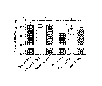
Fig. 4 discloses that probiotics protect mice from ovx induced cortical
bone-loss. Eight-week-old mice were treated with either vehicle (veh), a
single Lactobacillus (L) strain (L. para) or a mixture of three strains (L.
mix)
during 6 weeks, starting two weeks before ovx or sham surgery to study the
preventive effect of probiotic treatment on ovx induced bone-loss. At the end
of the experiment, dissected femurs were analysed with high-resolution pCT
and peripheral quantitative computed tomography (pQCT). Representative
pCT images of one cortical section from the veh and L. mix treated sham and
ovx groups (A). Cortical bone mineral content (BMC) (B) and cortical area (C)
were measured by pQCT in the mid-diaphyseal region of femur. Values are
given as mean SEM, (n=9-10). ** p).01, * p).05. Student's t test ovx vs.
CA 02908051 2015-09-24
WO 2014/163568
PCT/SE2014/050399
sham. # p).05, ANOVA followed by Dunnett's post hoc test within the
groups, ovx L. Para and L. mix vs. ovx veh.
Fig. 5 discloses that probiotics reduces expression of inflammatory
cytokines and the RANKL/OPG ratio in cortical bone. QRT-PCR analysis of
5 the expression of genes known to promote bone resorption; (A) Tumor
Necrosis Factor alpha (TNFa), (B) Interleukin-113 (IL-113), (C) Interleukin-6
(IL-
6), (D) Ratio of Receptor activator of nuclear factor kappa-B ligand (RANKL)
and Osteoprotegerin (OPG), and individual graphs for (E) OPG, (F) RANKL
and genes known to promote bone formation; (G) Osterix, (H) Collagen, type
I, al (Coll al) and (I) osteocalcin in cortical bone from 14-week-old
ovariectomized (ovx) mice treated with either vehicle (veh) or a mixture of
three probiotic Lactobacillus strains (L. mix) during 6 weeks, starting two
weeks before ovx or sham surgery to study the preventive effect of probiotic
treatment on ovx-induced bone-loss. Values are given as mean SEM, n=9-
10. *P).05 versus veh treatment, Student's t-test.
Fig. 6 discloses that the fractional excretion of Ca was increased by
ovx in the veh treated but not in the L. para or the L. mix treated mice.
Ca and creatinine were measured in serum and urine from 14-week-old mice
that had been treated with vehicle (veh), a single Lactobacillus (L) strain
(L.
para) or a mixture of three strains (L. mix) during 6 weeks, starting two
weeks
before ovx or sham surgery. Urinary fractional Ca excretion was calculated
with the formula FECa = (urine Ca x serum creatinine)/(serum Ca x urine
creatinine). Values are given as mean SEM, n=5-10 in each group. * p).05.
Student's t test ovx vs. sham. # p).05, ANOVA followed by Dunnett's post
hoc test within the groups, ovx L. Para and L. mix vs. ovx veh.
Description of the invention
The present invention relates, in an embodiment, to at least one
probiotic strain chosen from Lactobacillus paracasei, or at least one
probiotic
strain chosen from Lactobacillus paracasei in combination with at least one
probiotic strain chosen from Lactobacillus plantarum, for use in the treatment
or prevention of osteoporosis or for use in increasing the absorption of Ca2+
ions, in a mammal, preferably in a human.
CA 02908051 2015-09-24
WO 2014/163568
PCT/SE2014/050399
6
The present invention relates, in an embodiment of the invention, to at
least one probiotic strain for use in the treatment or prevention of
osteoporosis by preventing cortical bone loss, by preventing bone mineral
content loss, and by preventing bone-resorption.
Cortical bone constitutes approximately 80% of the bone in the body
and several studies have shown that cortical bone is the major determinant of
bone strength and thereby fracture susceptibility. It has been shown in the
experiments of the present invention that a probiotic strain of the species
Lactobacillus paracasei either alone or in combination with strains of the
species Lactobacillus plantarum prevents cortical bone loss. It is also
indicated in the experiments of the invention that probiotic treatment alters
the
immune status in bone resulting in attenuated bone resorption. In addition, it
is also shown in the experiments of the invention that bone mineral content
was not reduced in the probiotic group compared to vehicle group (Figure 4a-
c). Bone mineral content in cortical bone was higher in both probiotic groups
compared to vehicle group (p<0.05, Figure 4b).
The present invention relates to at least one probiotic strain chosen
from Lactobacillus paracasei, or at least one Lactobacillus paracasei in
combination with at least one probiotic strain chosen from Lactobacillus
plantarum, for use in the treatment or prevention of osteoporosis, for
preventing bone mineral content loss, for preventing bone-loss in a mammal,
preferably in a human.
The present invention relates to at least one probiotic strain chosen
from Lactobacillus paracasei, or at least one Lactobacillus paracasei in
combination with at least one probiotic strain chosen from Lactobacillus
plantarum, for use in preventing bone mineral content loss, for preventing
bone-loss in a mammal, preferably in a human.
In an embodiment of the invention at least two or more Lactobacillus
plantarum strain are used together in a combination with at least one
Lactobacillus paracasei strain. In another embodiment at least two or more,
for example three or more, Lactobacillus paracasei strains are used together
in a combination with at least one Lactobacillus plantarum strain.
CA 02908051 2015-09-24
WO 2014/163568
PCT/SE2014/050399
7
In an embodiment of the invention the probiotic strain is viable, inactivated
or dead. In an embodiment of the invention said strains are present in a
composition comprising additionally at least one carrier. The carrier could be
any carrier conventionally used in for instance a dietary supplement. The
carrier may be any cereal based carrier such as an oatmeal carrier or barley
carrier that can be used in a functional food or any other kind of food. The
carrier may be water or any other aqueous solvent in which the probiotic
strain is mixed before intake.
In an embodiment of the invention the composition is supplemented with
additional Ca2+ in the form of for instance a salt, e.g. calcium carbonate,
calcium chloride, calcium salts of citric acid, calcium gluconate, calcium
glycerophosphate, calcium lactate, calcium oxide, calcium sulphate. The
recommended daily intake (RDI) of Ca2+ is 800 mg. The amount of Ca2+ in the
composition may be in the range of 10-40% of the RDI, preferably in the
range of 15-30% of the RDI. Thus, the amount of Ca2+ e.g. in the form of a
salt in the composition may be in the range 80¨ 320 mg, preferably 120 ¨
240 mg. The amount of Ca2+ added to the composition could be adjusted to
any amount within the above range so that the composition is still stable and
provides its beneficial effects.
The composition may be a dry, non-fermented composition or a fermented
composition. In the case of a dry, non-fermented composition, the fermen-
tation takes place after intake of the composition by an individual, i.e. in
the
gastrointestinal tract. In addition, the strains may be present in the
composition as freeze-dried strains.
The probiotic strain of Lactobacillus paracasei may be chosen from
Lactobacillus paracasei 8700:2, DSM 13434, and Lactobacillus paracasei
02:A, DSM 13432 and the probiotic strain of Lactobacillus plantarum may be
chosen from Lactobacillus plantarum 299, DSM 6595, Lactobacillus
plantarum 299v, DSM 9843, Lactobacillus plantarum HEAL 9, DSM 15312,
Lactobacillus plantarum HEAL 19, DSM 15313, and Lactobacillus plantarum
HEAL 99, DSM 15316.
CA 02908051 2015-09-24
WO 2014/163568
PCT/SE2014/050399
8
Lactobacillus paracasei 8700:2, DSM 13434, and Lactobacillus paracasei
02:A, DSM 13432, were both deposited on 10 April 2000 at the Deutsche
Sammlung von Mikroorganimsen und Zellkulturen GmbH.
Lactobacillus plantarum HEAL 9, DSM 15312, Lactobacillus plantarum
HEAL 19, DSM 15313, and Lactobacillus plantarum HEAL 99, DSM 15316
were deposited at the Deutsche Samm lung von Mikroorganimsen und
Zellkulturen GmbH on 28 November 2002.
Lactobacillus plantarum 299v, DSM 9843, was deposited on 21 March
1995 and Lactobacillus plantarum 299, DSM 6595, was deposited on 5 July
1991 at the Deutsche Sammlung von Mikroorganimsen und Zellkulturen
GmbH.
In an embodiment of the invention, the composition including the at
least one strain may be chosen from the group consisting of a food product, a
dietary supplement, a medical food, a functional food and a nutritional
product.
In the case where said composition is a food product, it may be chosen
from the group comprising beverages, yoghurts, juices, ice creams, breads,
biscuits, cereals, health bars, and spreads.
When any of the above mentioned strains are used in a composition
such as a dietary supplement the carrier(s) to be added are known to a skilled
person. Any other ingredients that are normally used in dietary supplements
are known to a skilled person and may also be added conventionally together
with the strains.
In an embodiment of the invention, the above mentioned probiotic
strain(s) are present in a composition in an amount from about 1x10⁶ to about
1x10¹⁴ CFU, preferably 1x10⁸ to 1x10¹², and more preferably 1x10⁹ to 1x10¹¹.
The strains may be also be used alone in the above amount in water or any
other aqueous vehicle in which the strains are added or mixed before intake.
The invention is suitable to be used by mammals, preferably any
humans, such as elderly people, postmenopausal women and
premenopausal women, in which bone loss, bone mineral content loss and
increased bone-loss or bone resorption are or may become a problem.
CA 02908051 2015-09-24
WO 2014/163568 PCT/SE2014/050399
9
Healthy people may naturally also benefit from the invention in order to stay
healthy and prevent getting sick by osteoporosis.
Experimental
Experiment 1
Materials and methods
"Transport Solutions"; total volume 6 ml
The transport Solutions contained Hank's Balanced Salt solution (HBSS) with
Ca and Mg, Hepes (2%), Glutamine (4 mM), D-Glc (3, 5 g/1) and CaC12. 2 H20
(1.47 g/1). Analysis of the solution gave a [Ca21 on10,65 mM.
"Basal solution"
Similar to the transport solution, but without the addition of external
calcium.
Analysis of this solution gave a [Ca21 1,22 mM.
Experiment 1:
1. Control of the transport solution only
2. Lyophilised Lactobacillus plantarum 299 v. 0,788 mg corresponding to
4,02 x 10⁸ bacteria/6 ml in transport solution
3. Lactobacillus plantarum 299v. 4.02 x 10⁸ bacteria/6 ml in transport
solution
4. Lactobacillus plantarum 299. 4.95 x 10⁸ bacteria/6 ml in transport
solution
5. Lactobacillus plantarum HEAL 19, 4.95 x 10⁸ bacteria/6 ml in transport
solution
6. AMJ 1277. 4.95 x 10⁸ bacteria/6 ml in transport solution. AMJ 1277 is a
mutated form of Lactobacillus plantarum 299v.
Experiment 2:
1. CNCM 1-2332, Lactobacillus acidophilus (La10); + 4.95 x 10⁸ bacteria/6
ml in transport solution
CA 02908051 2015-09-24
WO 2014/163568
PCT/SE2014/050399
2. Lactobacillus plantarum 299 v. 4,02 x 10⁸ bacteria/6 ml in transport
solution
3. Control - transport solution only
All strains except strain La10 were cultured aerobically in 30 ml of MRS.
5 (30 C, 210 rpm). La10 was cultured in 37 C and covered with sterile
filtrated nitrogen before sealing. Number of inoculated cells was 3 x 10⁸
/flask.
The bacteria were precultivated over night and were in exponential phase at
inoculation. The cells were harvested at 0D600= 0.1-0.5 and for each strain a
volume corresponding to 4,02 x 10⁸ cells was collected. These samples were
10 centrifuged at 5000 rpm, 3 min, the supernatant was poured and the cells
re-
suspended in NaCI (0.9%). The cells were centrifuged again and the
supernatant was decanted. The now washed pellet was then suspended in
the transport solution.
Caco-2 cells were washed with PBS (2 times) before the test. 45Ca Cl2
(74kBq/m I) were added to the transport solutions. The mixture was then
added, in a volume of 0.5 ml, apically to Caco-2 cells growing on inserts.
Each well has then obtained 6.7 x 10⁷ bacteria/ml. Only the basal solution
(1.5 ml) containing only the endogenous [Ca21 in HBSS (1,22 mM) was
added the basal Chamber. Suspensions and controls were allowed to
function on the Caco-2 cells for 2 h. They were then aspired and the cells
were washed with ice-cold washing-buffer 3 times in accordance with the
method described in W099/02170. The Caco-2 cells were thereafter lysated
in 0,5 ml of NaOH (0.5 M). The solutions in the basal chambers were
collected for measurement of transport with the help of scintilator (Tri-carb
2800TR, Perkin Elmer). The lysates were analyzed for 45Ca, but also for
protein content. All measured values for Ca2+ transport/uptake were
normalized against the protein content in the respective cells. All wells were
checked before and after incubation with the test solutions regarding
transepitelial resistance (TEER). No differences in TEER, before and after the
trials, could be seen. TEER is measured to ensure that the epithelium is not
leeking. A difference in resistance after a trial may lead to the suspicion
that
the intercellular bonds may have been damaged by the solutions used.
CA 02908051 2015-09-24
WO 2014/163568
PCT/SE2014/050399
11
Results
To enable comparison of the results from experiments 1 and 2, transport and
uptake data from the studied strains have been normalised against the control
solution without bacteria. All results are therefore presented as a percentage
of the control (similarly to as done in W099/02170). It should also be noted
that the experiments 1 and 2 were done on days 16 and 21 of cellular culture,
respectively. In general, it is observed that the standard deviations of the
obtained results are relatively large. This could be due to some form of
aggregation of bacteria and Ca2+ , leading to a variation in the availability
of
Ca2+. This is not observed for the freeze-dried bacteria where the standard
deviations were relatively small compared with the remaining samples.
Transport of Ca2+
The results of experiment 1 showed a significant improvement of the Ca2+
transport when the strain AMJ1277 was present (134,7 18,9%, p=0,002),
see fig. 1. None of the other strains (Lp299v freeze-dried and viable, Lp299,
Lp HEAL 19) gave significant differences in transport of Ca2+, se table 1. In
experiment 2 where La10 and Lp299v were tested no significant differences
in the transport of Ca2+ were detected when comparing the samples with
Lp299v vs control without bacteria (p=0,2). For La10 a slight decrease of the
Ca2+ transport in the presence of the same bacteria was observed (79,5
19,3%) and this change was significant (p=0,049).
Uptake of Ca2+
Uptake data presented here show the amount of Ca2+ present in the cells
after 2 hrs. For an estimate of the actual uptake data, the intracellular
amounts of Ca2+ should be added to the amount of Ca2+ detected in the
lateral compartments. The sum of these data describes the total amount of
Ca2+ that has been transported through the apical membrane. The results of
these experiments generally shows a reduction in intracellular [Ca2 in the
presence of bacteria, compared to the bacteria-free control solutions, see
Figure 2. However, only the differences of the lyophilized and viable Lp299v,
were significant (p = 0.04 and p = 0.02). Note, that the intermediate
intracellular Ca2+ levels are only a few percent in comparison to the
transported amount of Ca2+. Due to this, the demonstrated effect of the
CA 02908051 2015-09-24
WO 2014/163568
PCT/SE2014/050399
12
uptake of Ca2+ do not have the same impact as the effects on transport. This
can be observed for lyophilized and viable Lp 299v, which has a higher
intracellular [Ca2] but do not demonstrate an improvement in transport.
Conclusion
In the presence of the strain AMJ1277, a mutant Lactobacillus plantarum
299v, an increased transport and even a total uptake of calcium is observed
compared to both the control and the remaining strains. Thus, there is
variation between the strains looked at.
Experiment 3
Ovariectomized mouse-model and probiotic treatment
Ovariectomy (ovx) results in bone loss associated with altered immune status.
The purpose of this experiment was to determine if probiotic treatment
protects mice from ovx induced bone loss. Mice were treated with either a
single Lactobacillus (L) strain, L. paracasei DSM13434 (L. para) or a mixture
of three strains, L. paracasei DSM13434, L. plantarum DSM 15312 and DSM
15313 (L. mix) given in the drinking water during 6 weeks, starting two weeks
before ovx.
Six-week old C57BL/6N female mice were purchased from Charles River
(Germany). The mice were housed in a standard animal facility under
controlled temperature (22 C) and photoperiod (12-h light, 12-h dark) and
had free access to fresh water and soy-free food pellets R70 (Lactam in AB,
Stockholm, Sweden). The ovariectomized (ovx) model for osteoporosis is
included in the FDA guidelines for preclinical and clinical evaluation for
agents
used for the treatment of postmenopausal osteoporosis. Probiotic treatment
started two weeks before ovx to study the preventive effect of probiotic
treatment on ovx induced bone-loss. Mice were treated with either a single
Lactobacillus (L) strain, L. paracasei DSM13434 (L. para) or a mixture of
three strains, L. paracasei D5M13434, L. plantarum DSM 15312 and DSM
15313 referred to as L. mix during 6 weeks. The probiotic strains were
selected based on their anti-inflammatory properties. The L. strains were
given in the drinking water at a concentration of i09 colony-formingunits
(cfu)/m1while control mice received tap water with vehicle. Water bottles were
changed every afternoon. The survival of the L. strains in the water bottles
CA 02908051 2015-09-24
WO 2014/163568
PCT/SE2014/050399
13
was checked regularly and after 24 h the concentration dropped one log unit
to approximately 108 cfu/ml. Each mouse drank on average 4.5 ml water/day.
After two weeks of probiotic treatment, the mice were either sham-operated or
ovx under inhalation anesthesia with isoflurane (Forene; Abbot Scandinavia,
Solna, Sweden). Four weeks after surgery, blood was collected from the
axillary vein under anesthesia with Ketalar/Domitor vet, and the mice were
subsequently killed by cervical dislocation. Tissues for RNA preparation were
immediately removed and snap-frozen in liquid nitrogen for later analysis.
Bones were excised and fixed in 4% paraformaldehyde. All animal
experiments had been approved by the local Ethical Committees for Animal
Research at the University of Gothenburg.
Peripheral quantitative computed tomography (pQCT)
Computed tomographic scans were performed with the pQCT XCT
RESEARCH M (version 4.5B, Norland, Fort Atkinson, WI, USA) operating at a
resolution of 70pm, as. Cortical bone parameters were analyzed ex vivo in the
mid-diaphyseal region of the femur.
High-resolution pCT
High-resolution pCT analyses were performed on the distal femur by using an
1172 model pCT (Bruker micro-CT, Aartselaar, Belgium). The femurs were
imaged with an X-ray tube voltage of 50 kV and current of 201 pA, with a 0.5-
mm aluminium filter. The scanning angular rotation was 180 and the angular
increment 0.70 . The voxel size was 4.48 pm isotropically. The NRecon
(version 1.6.9) was employed to perform the reconstruction following the
scans. In the femur, the trabecular bone proximal to the distal growth plate
was selected for analyses within a conforming volume of interest (cortical
bone excluded) commencing at a distance of 538.5 pm from the growth plate,
and extending a further longitudinal distance of 134.5 pm in the proximal
direction. Cortical measurements were performed in the diaphyseal region of
femur starting at a distance of 3.59 mm from the growth plate and extending a
further longitudinal distance of 134.5 pm in the proximal direction. For BMD
analysis, the equipment was calibrated with ceramic standard samples.
RNA isolation and Real Time PCR
CA 02908051 2015-09-24
WO 2014/163568
PCT/SE2014/050399
14
Total RNA was prepared from cortical bone (femur with the ends removed
and bone marrow flushed out with PBS before freezing) and bone marrow
using TriZol Reagent (Invitrogen, Lidingb, Sweden). The RNA was reverse
transcribed into cDNA using High-Capacity cDNA Reverse Transcription Kit
(#4368814, Applied Biosystems, Stockholm, Sweden). RT-PCR analyses
were performed using the ABI Prism 7000 Sequence Detection System (PE
Applied Biosystems). We used predesigned RT-PCR assays from Applied
Biosystems (Sweden) for the analysis of IL-6 (Mm00446190_m1), IL-1p
(Mm00434228_m1), TNFa (Mm00443258_m1), RANKL (Mm00441908_m1),
OPG (Mm00435452_m1), Runx2 (Mm00501580_m1), Coll a1
(Mm00801666_g1), osteocalcin (Mm01741771_g1) and TGFp1
(Mm03024053_m1) mRNA levels. The mRNA abundance of each gene was
calculated using the "standard curve method" (User Bulletin 2; PE Applied
Biosystems) and adjusted for the expression of 18S (4308329) ribosomal
RNA.
Blood Analysis
Analyses were performed according to the manufacturer's instructions for
serum and urine calcium (QuantiChromTmCalcium Assay Kit (DICA-500),
Bioassays systems, Hayward, CA, USA), serum and urine creatinine (Mouse
Creatinine Kit, Crystal Chem, Downers Grove, IL, USA). As a marker of bone
resorption, serum levels of type I collagen fragments were assessed using a
RatLaps ELISA kit (Nordic Bioscience Diagnostics, Herlev, Denmark). Serum
levels of osteocalcin, a marker of bone formation, were determined with a
mouse osteocalcin immunoradiometric assay kit (Immutopics, San Clemente,
CA).
Flow Cytometry
Bone marrow cells were harvested by flushing 5 ml PBS through the bone
cavity of one femur using a syringe. After centrifugation at 515 g for 5 min,
pelleted cells were resuspended in Tris-buffered 0.83% NH4CI solution (pH
7.29) for 5 min to lyse erythrocytes and then washed in PBS. Bone marrow
cells were resuspended in RPM! culture medium (PAA Laboratories,
Pasching, Austria) before use. The total number of leucocytes in bone
marrow was calculated using an automated cell counter (Sysmex, Hamburg,
CA 02908051 2015-09-24
WO 2014/163568
PCT/SE2014/050399
Germany). For flow cytometry analyses, cells were stained with
allophycocyanin (APC)-conjugated antibodies to CD4 for detection of T helper
cells (Beckton-Dickinson) and fluorescein isothiocyanate (FITC)-conjugated
antibodies to CD8 cytotoxic T cells (Beckton-Dickinson) or Peridinin-
5 chlorophyll proteins (PerCP)-conjugated antibodies to Gr-1/Ly-6G
(BioLegend) to eliminate granulocytes and FITC-conjugated antibodies to
CD11b for detection of OCL precursor cells (Beckton-Dickinson). The cells
were then subjected to fluorescence activated cell sorter analysis (FACS) on
a FACSCalibur (BD Pharmingen, Franklin Lakes, NJ USA) and analyzed
10 using FlowJo software. Results are expressed as cell frequency (%).
Statistical analyses
All the statistical results are presented as the means SEM. Between-group
differences were calculated using unpaired t tests. Comparisons between
multiple groups were calculated using a one-way analysis of variance
15 (ANOVA) followed by Dunnett's test to correct for multiple comparisons.
A
two-tailed p 0.05 was considered significant.
Results - Probiotic treatment protects mice from ovx-induced cortical bone
loss and increased bone resorption
To determine the preventive effect of probiotic treatment on ovx-
induced bone-loss, eight-week-old mice were treated with vehicle (veh), a
single Lactobacillus (L) strain (L. para) or a mixture of three strains (L.
mix)
during 6 weeks, starting two weeks before ovx or sham surgery (Figure 3A).
Uterus weight can be used as an indicator of estrogen status and ovx resulted
in an expected decrease in uterus weight that was similar for all treatments
(Table 2). In addition, ovx increased body weight, fat mass and thymus weight
in all treatment groups (Figure 3B, Table 2).
In the vehicle treated mice, ovx decreased the cortical bone mineral
content and cortical cross sectional bone area in the mid-diaphyseal region of
femur (p<0.01, Figure 4a-c). Importantly, ovx did not reduce cortical bone
mineral content or cortical cross sectional bone area in the L. para or the L.
mix treated mice (Figure 4a-c). Cortical bone mineral content was higher in
both L. para and L. mix treated ovx mice compared to veh treated ovx mice
(p<0.05, Figure 4b). To determine if the preventive effect of probiotics on
CA 02908051 2015-09-24
WO 2014/163568
PCT/SE2014/050399
16
cortical bone is caused by affected bone resorption, serum levels of C-
term inal telopeptides (RatLaps) were analyzed. Ovx increased levels of
RatLaps in veh-treated mice (+45 11 A, p<0.05 over sham) but not in L. para
treated (20 9 A, non-significant) or L. mix treated (23 9 A, non-
significant)
mice. Bone formation, as indicated by serum osteocalcin, was not significantly
affected by probiotic treatment (data not shown). Trabecular bone parameters
(BV/TV and trabecular BMD) in the distal metaphyseal region of femur were
significantly reduced by ovx in all treatment groups (p<0.05 Table 1). These
findings demonstrate that probiotic treatment protects mice from ovx-induced
cortical bone loss and increased bone resorption.
Probiotics reduces expression of inflammatory cytokines and the
RANKL/OPG ration in cortical bone
To investigate the mechanism for the effect of probiotic treatment on ovx-
induced cortical bone loss, we measured bone related mRNA transcripts in
cortical bone (Figure 5). The m RNA levels of TNFa, an inflammatory cytokine
produced by myeloid cells that promotes osteoclastogenesis, and IL-113, a
downstream regulator of the effects of TNFa on bone, were significantly
decreased by probiotic treatment compared to vehicle treatment in ovx mice
(TNFa -46%, p<0.05; IL-113 -61 A, p<0.05, Figure 5a and 5b). The expression
of, IL-6 did not differ between treatments although there was a tendency to
decreased expression in the probiotic treatment group (-20%, p=0.12, Figure
Sc).
The RANKL/osteoprotegerin (OPG) ratio is a major determinant of
osteoclastogeneisis and, thereby, bone resorption. Importantly, probiotic
treatment decreased the RANKL/ OPG ratio (-45%, p<0.05 compared with
veh, Figure 5d) and this was caused by an increased OPG expression (OPG;
+28%, p<0.05 and RANKL; +1 A, non-significant, Figure 5e, f). In contrast, the
m RNA levels of three osteoblast-associated genes, Osterix, Coll a1 and
osteocalcin, were not affected by probiotic treatment (Figure 5g-h).
Immune Status in Bone Marrow
Some of the anti-inflammatory effects exerted by probiotic bacteria are
thought to be mediated via the induction of regulatory T (Tõg) cells. FACS
analysis of bone marrow showed that the frequency of Tõg (CD4+ CD25+
CA 02908051 2015-09-24
WO 2014/163568 PCT/SE2014/050399
17
Foxp3+) cells was decreased by ovx in veh treated but not in probiotic-treated
mice (Table 2). Tõg cells are dependent on TGF8 for their induction and
maintenance and the expression of TGF81 was increased in bone marrow
from ovx probiotic-treated compared with ovx vehicle treated mice (+77 19%,
p<0.01).
It was also examined if probiotic treatment modulated the frequency of
osteoclast precursor cells (pre0CLs) in bone marrow. The frequency of
pre0CLs (CD11b+ Gr1-) in bone marrow was not affected by ovx in any of the
treatment groups (Table 2).
Mineral Metabolism
The urinary fractional excretion of Calcium (FECa = (urine Ca x plasma
creatinine)/(plasma Ca x urine creatinine)) was increased by ovx in veh
treated mice (+86%, p<0.05, Figure 6). The ovx induced increase in FECa
was completely prevented by probiotic treatment suggesting enhanced
accretion of Ca (Figure 6). There was a tendency of increased serum levels of
Ca after ovx in veh but not probiotic treated mice (+13%, p=0.05, Table 3).
The urine Ca/creatinine ratio was not affected by ovx in any of the treatment
groups (Table 3).
Table 1.
Strain Transport
intracellular Ca (2h)
(pmol /mg prot) % of control (pmol /mg prot) % of control
Average sdev ( ) Average sdev ( ) *p=
Average sdev ( ) average sdev ( ) *p=
control without bact. 0,14822 0,03254 100 x 1
0,010295 0,002456 100 x 1
Freeze-dried Lp 299v 0,131773
0,0217772 88,903807 5,771665 0,246025 0,008195 0,001638 79,60091
15,90269 0,048654
Lp 299v (1) 0,161124
0,0439384 108,70637 29,68811 0,489095 0,00777 0,001767 75,47339
17,15187 0,023494
Lp 299 0,159745 0,0517202 107,77569 34,9461
0,579356 0,008296 0,001452 80,58207
14,09352 0,063082
co
Lp heal 19 0,161361 0,0291023 108,866
19,66369 0,497942 0,008736 0,001901 84,85697
18,45752 0,151598
AMJ1277 0,199657 0,0279951 134,70331 18,91562 0,002
0,008744 0,000942 84,9374 9,141623 0,096014
La 10 0,182937 0,0443204 79,537982 19,26973 0,049
0,015901 0,003197 97,43651 19,61249 0,826775
Lp 299v (2) 0,210131 0,0289741 91,361137 12,59742 0,2
0,014491 0,002629 88,79696 16,12947 0,309532
control without bact 0,229676 0,0111063 100 x 1 0,016319
0,003254 100 x 1
*if p<0,05 the change is seen as significant
(:)
CA 02908051 2015-09-24
WO 2014/163568
PCT/SE2014/050399
19
Table 2
Sham Ovx
Veh L. Para L. Mix Veh L. Para L.
Mix
Estrogen Responsive
Organ Weights
Uterus weight (mg) 45.9 4.8 65.9 12.5 65.2 10.4 8.950.7**
11.0 3.2** 6.90.3**
Gonadal Fat (mg) 371 41 296 40 326 40 597 68* 630 28**
577 58**
Thymus weight (mg) 55.5 3.2 53.6 5.1 47.7 2.6 93.9 4.2**
90.0 5.1** 73.7 5.2**#
Trabecular Bone
BV/TV (%) 16.20.7 16.80.8 17.40.8 13.20.7**
14.40.6* 13.80.5**
BMD (mg/cm3) 322 9 331 12 344 8 285 9* 302 7* 298
7**
Immune Cells in Bone
marrow
Tõg (CD4+ Foxp3+ 0.1170.023 0.1090.017 0.0900.017 0.0540.004
0.0690.008 0.0700.014
CD25+) (%)
pre0CLs 7.77 1.01 6.75 1.20 8.94 1.17 6.700.94
7.17 1.32 8.57 1.50
(CD11b+ Grip(%)
Table 1. Estrogen Responsive Organ Weights, Trabecular Bone and Immune
Cells in Bone Marrow Eight-week-old mice. Trabecular bone parameters were
analysed by high resolution pCT in the distal metaphyseal region of femur;
Trabecular bone volume as a percentage of tissue volume (BV/TV);
Trabecular bone mineral density (BMD). Femur bone marrow cells were
stained with antibodies recognizing CD4, Foxp3, CD25, CD11b and Gr1.
Values represent the percentage of Treg (CD4+ Foxp3+ CD25+) or pre0CLs
(CD11b+ Gr1-) in the total bone marrow population. Results are given as
mean SEM, n=6-10 in each group. ** p< 0.01, * p< 0.05, Student's t test ovx
vs. sham, # p< 0.05, ANOVA-Dunnets within groups.
Table 3
Sham Ovx
Veh L. Para L. Mix Veh L. Para
L. Mix
Serum Ca 9.1 0.4 9.2 0.4 8.5 0.3 10.3 0.4(*) 9.3 0.4
8.7 0.3#
(mg/dI)
Urine 6.7 0.7 6.3 0.4 5.4 0.6 8.5 1.3 5.9 0.5 5.6 0.7
Ca/Cre
atinine
Ratio
Table 2. Mineral Metabolism Calcium and creatinine were measured in
serum and urine from 14-week old mice. Results are given as
mean SEM, n=5-10 in each group. ** p< 0.01, * p< 0.05, (*) p=0.05.
Student's t test ovx vs. sham, # p< 0.05, ANOVA-Dunnets within
groups.
In conclusion, the data of the present invention show that probiotics in
the drinking water reduces ovx induced cortical bone-loss suggesting a
therapeutic potential for probiotics in the treatment of postmenopausal
osteoporosis. In addition, the results support a role of the gut microbiota
for
the regulation of bone mass.
CA 02908051 2015-09-24
WO 2014/163568
PCT/SE2014/050399
Both the L. para and the L. mix treatment protected mice from ovx-
induced cortical bone loss and increased bone resorption. Cortical bone
mineral content was higher in both L. para and L. mix treated ovx mice
compared to vehicle (veh) treated ovx mice. The urinary fractional excretion
5 of calcium and the resorption marker RatLaps were increased by ovx in the
veh treated but not in the L. para or the L. mix treated mice. Thus,
probiotics
inhibit ovx induced excretion of calcium in urine. Probiotic treatment reduced
the expression of the two inflammatory cytokines, TNFa and IL-113, and
increased the expression of OPG in cortical bone of ovx mice. In addition, ovx
10 decreased the frequency of regulatory T-cells (CD4+ CD25+ Foxp3+) in
bone
marrow of veh treated but not probiotic treated mice. Thus, probiotics inhibit
the ovx induced decrease in frequency of regulatory T-cells in bone marrow.
In addition, probiotics increased the expression of TGFb1 in bone marrow and
probiotics inhibit the ovx induced increase of the resorption marker Rat Lap.
15 In conclusion, treatment with L. para or the L. mix prevents ovx induced
cortical bone loss. The findings of the invention indicate that these
probiotic
treatments alter the immune status in bone as demonstrated by reduced
expression of inflammatory cytokines and increased expression of OPG,
resulting in attenuated bone resorption in ovx mice.
20 Experiment 4
In this experiment it will be tested if the same probiotics as mentioned
above will affect the same parameters as above (i.e. cortical bone mass,
bone mineral content, bone resorption) in female mice that already have been
ovariectomized and thus already have lost bone-mass. Ovariectomized
female mice is a well established model of postmenopausal bone loss in
women. The time line of the experiments will be as indicated below.
Ovariectomize mice => 4 weeks => Probiotic treatment for 6 weeks => End
Probiotics will be given in drinking water and will begin first after 4 weeks
after
ovariectomization. Dose will be 109 cfu/ml/day and ovariectomization will take
place at 9-10 weeks of age. Analyses after ended experiment will be (CT) of
the bone to measure the density and thickness, as well as serum analysis
and bone markers.