Note: Descriptions are shown in the official language in which they were submitted.
Title: METHODS AND COMPOSITIONS FOR GENERATING CHONDROCYTE LINEAGE CELLS
AND/OR CARTILAGE LIKE TISSUE
[0001] This is a Patent Cooperation Treaty Application which claims the
benefit of 35 U.S.C. 119
based on the priority of U.S. Provisional Patent Application No. 61/809,050,
filed April 5, 201 3.
Field
[0002] The disclosure relates to methods for producing chondrocytes and
cartilage and particularly
articular chondrocytes and articular cartilage like tissue as well as
hypertrophic chondrocytes and
growth plate cartilage resembling tissue from human pluripotent stem cells.
Background
[0003] The ability to efficiently and reproducibly generate
differentiated cell types from
pluripotent stem cells in vitro has opened the door for the development of
cell-based therapies for the
treatment of a broad range of degenerative and debilitating diseases.
Osteoarthritis (OA) is a
candidate for such therapy as it affects at least one in ten adults (Lawrence,
Felson et al. 2008),
leaving patients with a poor quality of life due to pain associated with joint
movement. Pathogenic
hallmarks of OA include the degradation of the extracellular matrix (ECM) of
articular cartilage that
lines the joints together with thickening of the underlying subchondral bone
and the formation of
osteophytes (bone spurs). Articular cartilage is generated by a distinct
subpopulation of chondrocytes
known as articular chondrocytes (ACs) that are specified early in development
and persist throughout
adult life. While ACs function to maintain integrity of the articular
cartilage under normal
circumstances, they display little capacity to repair cartilage damaged by
injury or disease.
Consequently, with disease progression, damage to the cartilage is so
extensive that surgical
intervention such as joint replacement is often required to improve the
quality of life for the patient.
ACs differ from growth plate chondrocytes (GPCs), whose primary function is to
form bone through
the process of endochondral ossification (Colnot, 2005). Interestingly, with
the onset of OA, ACs
appear to acquire some characteristics of GPCs, including hypertrophy, which
may contribute to the
pathogenesis of this disease.
[0004] Chondrocyte and cartilage replacement represent a potential new
therapy for OA that
could, at some point dramatically reduce the need for mechanical devices. This
type of therapy,
however, is dependent on access to appropriate tissue and sufficient numbers
of highly enriched ACs.
It is well established that adult mesenchymal stem cells (MSCs) are able to
differentiate to
chondrocytes in vitro, however, it is unclear if they are able to give rise to
ACs as the cartilage-like
tissue generated from them prematurely undergoes hypertrophy (Pelttari, Winter
et al. 2006, Steinert,
Ghivizzani et al. 2007, Pelttari, Steck et al. 2008) Alternatively, ACs have
been harvested directly from
Date recue/date received 2021-10-21
CA 02908369 2015-09-29
WO 2014/161075 PCT/CA2014/000312
patients and used for tissue generation ex vivo, despite their limited
capacity to proliferate. Tissue
generated by passaged chondrocytes exhibits fibrocartilage characteristics,
which can improve the
quality of life for the patient in the short term but ultimately undergoes
degradation as it lacks sufficient
weight bearing capacity (Tins, McCall et al. 2005, LaPrade, Bursch et al.
2008). Pluripotent stem cells
(PSCs) such as embryonic and induced pluripotent stem cells (ESCs, iPSCs) may
represent a novel
and potentially unlimited source of chondrocytes and tissues for therapeutic
applications as these
cells are able to generate a broad spectrum of cell types under appropriate
conditions in vitro.
[0005]
Chondrocytes develop from paraxial mesoderm that is induced in the early
embryo in
an ordered temporal pattern following the generation of lateral plate mesoderm
(LPM) fated to give
rise to hematopoietic and cardiovascular lineages (Lawson, Meneses et al.
1991, Kinder, Tsang et al.
1999). Following induction, strips of paraxial mesoderm are segmented into
somites (Tam and Tan
1992, Kulesa and Fraser 2002). Somite development is regulated, in part, by
the transcription factors
paraxis (TCF15) and TBX18, whose expression coincides with induction of
paraxial mesoderm
(Burgess, Rawls et al. 1996, Bussen, Petry et al. 2004, Singh, Petry et al.
2005). Individual somites
are then patterned into the ventral sclerotome, which forms the axial
skeleton, including cartilage and
the vertebral column, and the dorsal dermomyotome which develops into skeletal
muscles and the
dermis of the back (Hirsinger, Jouve et al. 2000). Specification of the
sclerotome is marked by the
expression of two transcription factors, Meoxl (Mankoo, Skuntz et al. 2003)
and Nkx3.2 (Bapx1). A
population of collagen 2 (Col2a1) positive mesenchymal cells with chondrogenic
potential develops
from sclerotome-derived cells at E12.5 of mouse development (Akiyama,
Chaboissier et al. 2002,
Dao, Jonason et al. 2012).
[0006] While
methods for differentiating progenitor cells to the chondrogenic lineage are
established, the ability to specify ACs, and ultimately stable cartilage
tissue containing non-
hypertrophic chondrocytes, remains poorly understood. ACs are derived from
interzone cells, a fibrotic
population of cells that forms at future sites of synovial joints, marked by
the upregulation of Wnt9a/14
and growth and differentiation factor 5 (GDF5/BMP14), a member of the TGF13
superfamily (Archer,
Dowthwaite et al. 2003, Pacifici, Koyama et al. 2006). Lineage tracing studies
have shown that GDF5-
expressing interzone cells give rise to several joint tissues including ACs,
but do not contribute to the
GPC population (Koyama, Shibukawa et al. 2008). GPCs, by contrast, develop
from the condensing
chondrogenic mesenchyme and express BMP 2, 4 and 7, as well as hypertrophy
related genes
including collagen 10. Distinct regions of ACs and GPCs are observed as early
as postnatal day 7-8
when the secondary ossification center begins to form (Murakami, Balmes et al.
2004, Blumer,
Longato et al. 2007). These observations suggest that ACs and GPCs are
generated from separate
progenitor populations during development and as such, may represent distinct
lineages.
[0007] A number
of studies have demonstrated that it is possible to derive chondrocytes
from mouse (m) and human ESCs and iPSCs in vitro. Most, however, used serum-
based media to
support the early stages of differentiation resulting in the generation of
mixed lineage end stage
cultures (Kramer, Hegert et al. 2000, zur Nieden, Kempka et al. 2005, Hwang,
Kim et al. 2006,
2
CA 02908369 2015-09-29
WO 2014/161075 PCT/CA2014/000312
Hwang, Varghese et al. 2008, Jukes, Both et al. 2008, Yamashita, Krawetz et
al. 2008). Recent
studies have reported the use of defined culture media with specific pathway
agonists and
antagonists to direct differentiation (Nakayama, Duryea et al. 2003, Darabi,
Gehlbach et al. 2008,
Tanaka, Jokubaitis et al. 2009). In mESCs, Tanaka et al (2009) showed that the
combination of Wnt
signaling with BMP inhibition resulted in the generation of paraxial mesoderm
with chondrogenic
potential, identified by the expression of PDGFRalpha and a lack of expression
of Elk-i. This
mesoderm also displayed some cardiac potential but showed no capacity to
generate hematopoietic
cells indicating that dependency on BMP signaling distinguishes different
types of mesoderm.
[0008] Oldershaw
et al. (Oldershaw, Baxter et al. 2010) used a serum free protocol. No
tissues were obtained in vitro or in vivo with the method of Oldershaw.
[0009] Umeda et al
(Umeda, Zhao et al. 2012) used a method using PDGF stimulation that
produced nodules comprising Runx2 expressing cells.
[0010]
Osteoarthritis is a degenerative disease that mainly affects the joint-lining
articular
cartilage of the joint. Articular cartilage has very limited capacity to
regenerate itself upon injury, thus
cell and tissue replacement strategies are the only means of replacing this
tissue effectively. Methods
of producing human cartilage from pluripotent stem cells are currently
lacking, despite great need for
such tissues for drug discovery and cartilage replacement strategies in
patients with joint diseases
such as osteoarthritis.
Summary
[0011] An aspect of the
application provides a method for generating chondrocyte lineage
cells and/or cartilage like tissue, optionally articular like non-hypertrophic
chondrocyte cells and/or
cartilage like tissue and/or hypertrophic chondrocyte like cells and/or
cartilage like tissue, the method
comprising:
(a) culturing
a primitive streak-like mesoderm cell population (e.g. stage 2), optionally a
CD56+, PDGFRalpha+ primitive streak-like mesoderm cell population, with a
paraxial
mesoderm specifying cocktail comprising:
(i) a FGF agonist;
(ii) a BMP inhibitor, optionally Noggin, LDN-193189, and/or Dorsomorphin,
and
(iii) optionally one or more of a TGFbeta inhibitor, optionally SB431542;
and a
VVnt inhibitor, optionally IWP2 (N-(6-Methyl-2-benzothiazoly1)-24(3,4,6,7-
tetrahydro-4-oxo-3-phenylthieno[3,2-d]pyrimidin-2-y1)thioj-acetamide; Sigma);
Dickkopf-related protein 1 (DKK1; R & D Systems), and/or XAV939 (3,5,7,8-
Tetrahydro-244-(trifluoromethyl)pheny1]-4H-thiopyrano[4,3-d]pyrimidin-4-one;
Sigma);
to specify a paraxial mesoderm cell population expressing cell surface CD73,
CD105
and/or PDGFR-beta;
3
CA 02908369 2015-09-29
WO 2014/161075
PCT/CA2014/000312
(b) generating a chondrocyte precursor population from the paraxial
mesoderm cell
population expressing cell surface CD73, CD105 and/or PDGFR-beta, the
generating
the chondrocyte precursor population comprising:
(i) culturing the paraxial mesoderm cell population expressing cell surface
CD73, CD105 and/or PDGFR-beta at a high cell density in serum free or
serum containing media;
(ii) culturing the high cell density CD73+, CD105+ and/or F'DGFRbeta+
paraxial
mesoderm cell population with a TGFbeta agonist, optionally TGFB1, TGFB2
and/or TGFB3 in serum free media to produce a high cell density Sox9+,
collagen 2+ chondrocyte precursor population (e.g. Stage 3); and
(c) either:
culturing the high cell density Sox9+, collagen 2+ chondrocyte precursor
population with a TGFbeta agonist (optionally TGFBeta1, TGFbeta2 and/or
TGFBeta3) for an extended period of time to produce articular like non-
hypertrophic chondrocyte like cells and/or cartilage like tissue; or
(ii) culturing the high cell density Sox9+ collagen2+ chondrocyte precursor
population with a BMP4 agonist for an extended period of time to produce
hypertrophic chondrocyte like cells and/or cartilage like tissue (e.g. stage
4).
[0012] Another aspect includes a method for generating chondrocyte like
cells and/or
cartilage like tissue, optionally articular like non-hypertrophic chondrocyte
like cells and/or cartilage
like tissue and/or hypertrophic chondrocyte like cells and/or cartilage like
tissue, the method
comprising:
(a) culturing a starting population of pluripotent stem cells with a
primitive streak inducing
cocktail to induce a primitive streak-like mesoderm cell population expressing
CD56
and/or PDGFR-alpha (e.g. stage 1);
(b) culturing the primitive streak-like mesoderm cell population expressing
CD56 and
PDGFR-alpha with a paraxial mesoderm specifying cocktail comprising:
(I) a FGF agonist;
(ii) a BMP inhibitor, optionally Noggin, LDN-193189, and/or
Dorsomorphin; and
(iii) one or more of a TGFbeta inhibitor, optionally SB431524; and a Wnt
inhibitor,
optionally DKK1, IWP2 and/or XAV939;
to specify a paraxial mesoderm cell population expressing cell surface CD73,
CD105
and PDGFR-beta;
4
CA 02908369 2015-09-29
WO 2014/161075
PCT/CA2014/000312
(c) generating a chondrocyte precursor population from the paraxial
mesoderm cell
population expressing cell surface CD73, CD105 and/or PDGFR-beta, the
generating
the chondrocyte precursor population comprising:
(I) culturing the paraxial mesoderm cell population expressing
CD73, CD105
and/or PDGFR-beta at a high cell density in serum free or serum containing
media;
(ii) culturing the high cell density CD73+, CD105+ and
PDGFRbeta+ paraxial
mesoderm cell population with a TGFbeta agonist in serum free media to
produce a high cell density Sox9+, collagen 2+ chondrocyte precursor
population; and
(d) either
culturing the high cell density Sox9+, collagen 2+ chondrocyte precursor
population with a TGFbeta agonist for an extended period of time to produce
articular like non-hypertrophic chondrocyte like cells and/or cartilage like
tissue; or
(ii) culturing the high cell density Sox9+ collagen2+ chondrocyte precursor
population with a BMP4 agonist for an extended period of time to produce
hypertrophic chondrocyte like cells and/or cartilage like tissue.
[0013] In an embodiment, the method is for generating articular like non-
hypertrophic
chondrocyle like cells and/or cartilage like tissue. In another embodiment,
the method is for
generating hypertrophic chondrocyte like cells and/or cartilage like tissue.
[0014] In an embodiment, the extended period of time the high cell
density Sox9+
collagen2+ chondrocyte precursor population is cultured with the TGF beta
agonist is at least 3
weeks, at least 4 weeks, at least 5 weeks, at least 6 weeks, at least 7 weeks,
at least 8 weeks, at
least 9 weeks, at least 10 weeks, at least 11 weeks, at least 12 weeks or more
to generate a non-
hypertrophic chondrocyte like and/or cartilage like tissue that expresses for
example lubricin and
C I LP2.
[0015] In an embodiment, the extended period of time the high cell
density Sox9+
collagen2+ chondrocyte precursor population is cultured with the BMP4 agonist
is at least 3 weeks, at
least 4 weeks, at least 5 weeks, at least 6 weeks, at least 7 weeks, at least
8 weeks, at least 9 weeks,
at least 10 weeks, at least 11 weeks, at least 12 weeks or more to generate a
cartilage like tissue that
expresses collagen 2 or hypertrophic chondrocyte cells that express collagen
10.
[0016] A method of generating chondrocyte like cells comprising:
5
CA 02908369 2015-09-29
WO 2014/161075 PCT/CA2014/000312
(a) culturing chondrocyte precursor cells at a high cell density in serum
free or serum
containing media;
(b) culturing the high cell density chondrocyte precursor cells with a
TGFbeta agonist in
serum free media; and
(c) either
(i) culturing the chondrocyte precursor cells with a TGFbeta agonist for an
extended period of time to produce articular cartilage like chondrocyte cells;
Or
(ii) culturing the chondrocyte precursor cells with a BMP4
agonist for an
extended period of time to produce hypertrophic chondrocyte lineage cells
and/or cartilage like tissue.
[0017] In an embodiment, the chondrocyte precursor cells are primary
fetal chondrocytes or
passaged fetal chondrocytes.
[0018] In an embodiment, the generated cells and/or tissues are
administered to a subject.
[0019] Also provided in another embodiment is an isolated population of
articular like non-
hypertrophic chondrocyte like cells and/or cartilage like tissue and/or
hypertrophic chondrocyte like
cells and/or cartilage like tissue generated according to a method described
herein.
[0020] A further aspect includes composition comprising the population
of articular like non-
hypertrophic chondrocyte like cells and/or cartilage like tissue and/or
hypertrophic chondrocyte like
cells and/or cartilage like tissue, and a carrier optionally PEG, hydrogel,
bone scaffolding, bone
substitute scaffolding and/or matrigel. In an embodiment, the carrier is
pharmaceutical grade.
[0021] In an embodiment, the isolated population is comprised in a
composition comprising a
diluent or carrier, optionally a pharmaceutical diluent. In an embodiment, the
diluent is culture media,
optionally comprising a cryopreservation agent such as glycerol and/or DMSO,
serum and albumin,
such as human serum albumin.
[0022] A further aspect includes a cartilage and/or bone tissue product
comprising the cells
or composition described herein, and a scaffold.
[0023] Another aspect includes method for ameliorating symptoms and/or
treating a subject
in need thereof comprising administering cells and/or tissue generated using a
method described
herein and/or transplanting a cartilage and/or bone tissue product described
herein.
[0024] Also provided in another aspect is use of cells and/or tissue
generated using a
method described herein and/or a cartilage and/or bone tissue product
comprising said cells and/or
tissue for ameliorating symptoms and/or treating a subject in need thereof.
[0025] A further aspect includes a method of generating a paraxial
mesoderm cell population
comprising:
6
(a) culturing a starting population of pluripotent stem cells with a
primitive streak inducing
cocktail to induce a primitive streak-like mesoderm cell population expressing
CD56 and
PDGFR-alpha (e.g. stage 0);
(b) culturing a primitive streak-like mesoderm cell population expressing
CD56 and PDGFR-
alpha with a paraxial mesoderm specifying cocktail comprising:
(I) a FGF agonist; and
(ii) a BMP inhibitor, optionally Noggin, LDN-193189, Dorsomorphin; and
(iii) one or more of a TGFbeta inhibitor, optionally SB431524; and a Wnt
inhibitor,
optionally DKK1, IWP2, and/or XAV939;
to specify a paraxial mesoderm cell population expressing cell surface CD73,
CD105 and PDGFR-beta.
[0026] Methods of isolating cells and screening assays are also provided.
[0026a] The invention provides a method for generating chondrocytes,
and/or cartilage, and/or
cartilage-like tissue, and/or hypertrophic chondrocyte-like cells, and/or
cartilage-like tissue, the method
comprising: a. culturing a CD56+, PDGFa+, Brachyury+ primitive streak-like
mesoderm population, with
a paraxial mesoderm specifying cocktail comprising: i. a molecule that binds
and activates a fibroblast
growth factor receptor agonist; and ii. a bone morphogenetic protein signaling
inhibitor, to generate a
CD73+CD105+, CD73+PDGFRB+, and/or CD73+CD105+PDGFRB+ paraxial mesoderm
population; b.
culturing the CD73+CD105+, CD73+PDGFRB+, and/or CD73+CD105+PDGFRB+ paraxial
mesoderm
population at a high cell density; c. further culturing the high cell density
CD73+CD105+, CD73+PDGFRB+,
and/or CD73+CD105+PDG FRB+ paraxial mesoderm population with a tumor growth
factor beta receptor
agonist in serum free media to produce a high cell density Sox9+, collagen 2+
chondrocyte precursor
population; and d. either: i. further culturing the high cell density Sox9+,
collagen 2+ chondrocyte
precursor population with the tumor growth factor beta receptor agonist for an
extended period to
produce articular like non-hypertrophic chondrocyte cells and/or cartilage-
like tissue; or ii. further
culturing the high cell density Sox9+, collagen 2+ chondrocyte precursor
population with a molecule that
activates the receptor for bone morphogenetic protein 4 receptor agonist for
an extended period greater
than 3 weeks to produce a hypertrophic chondrocyte-like cells and/or cartilage-
like tissue.
[0027] Other features and advantages of the present disclosure will
become apparent from the
following detailed description. It should be understood, however, that the
detailed description and the
specific examples while indicating preferred embodiments of the disclosure are
given by way of
illustration only, since various changes and modifications within the spirit
and scope of the disclosure will
become apparent to those skilled in the art from this detailed description.
7
Date recue/date received 2021-10-21
Brief description of the drawings
[0028] An embodiment of the present disclosure will now be described in
relation to the
drawings in which:
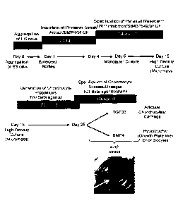
[0029] Figure 1. Serum-free differentiation of paraxial mesoderm,
chondrocyte progenitors,
and cartilage tissues from human pluripotent stem cells (hPSCs). (A) hPSCs are
differentiated in 4 stages
including the induction of a primitive streak-like mesoderm population (stage
1) using Activin A, BMP4
and basic (b) FGF from days 1 to 4 of differentiation as embryoid bodies. On
day 4 (T4), mesoderm
populations are monitored by the expression of CD56 and PDGFRa on the cell
surface by flow cytometry
(B). Day 4 mesoderm cells are specified to a paraxial mesoderm fate in
monolayer culture by treatment
of Dorsomorphin, a BMP inhibitor, a TGFbeta inhibitor 56431542, and bFGF, from
days 4 to 6 and bFGF
from days 4 to 15 (stage 2). Day 15 paraxial mesoderm cells can generate
chondrocyte progenitors by
plating in a high density 'spot' termed micromass, or by plating onto collagen
coated membrane filters
(not shown) in the presence of TGFB3 for approximately 10 days (stage 3).
Chondrocyte progenitors can
be specified to articular chondrocytes or growth plate-like chondrocytes in a
cartilage tissue format
during stage 4 of differentiation by extended stimulation with TGFB3
(articular) or BMP3 (growth plate-
like) for example for 12 weeks. Tissues have been kept in culture for at least
7 months. Efficient induction
of a primitive streak-like population from hESCs (C) and hIPSCs (D,E) was
confirmed by the expression of
CD56 and PDGFRa by flow cytometry on day 3 of differentiation. hESCs were
induced to generate a
primitive streak population with the following
7a
Date recue/date received 2021-10-21
CA 02908369 2015-09-29
WO 2014/161075
PCT/CA2014/000312
cytokines: Activin A (2 ng/ml), BMP4 (3 ng/ml) and basic (b)FGF (5 ng/ml).
hiPSCs were induced to
generate a primitive streak-like mesoderm population (Stage 1) using Activin A
(3 ng/ml), BMP4 (1
ng/ml), bFGF (5 ng/ml) in the presence (C) or absence (D) of the Wnt pathway
agonist CHIR99061 (1
pM) from days 1 to 3 of differentiation as embryoid bodies.
[0030] Figure 2. Characterization of paraxial mesoderm derived from
hPSCs. Flow
cytometric analysis of day 5 mesoderm treated with no additional factors (0
DM, no FGF), FGF, 4 pM
DM, or 4 pM DM+FGF. Day 5 profiles depict KDR and PDGFRa expression, a double-
positive
population (gated) indicates mesoderm that has cardiac potential (20).
Treatment with FGF results in
less PDGFRa expression on day 5. (B) Expression of cell surface markers CD73,
CD105, and
PDGFR-beta on mesoderm populations on day 15 of differentiation. (C) Wnt
inhibition can also
improve the efficiency of CD73 and CD105 expression. Experimental cell
treatments during day 4 to
day 6 include the combination of Dorsomorphin, bFGF, TGFbeta inhibitor
(SB431542) in the presence
or absence of the wnt pathway inhibitor IWP2. Flow cytometric analyses of CD73
and CD105, on day
15 mesoderm populations derived as indicated. (D) Gene expression analyses of
day 15 mesoderm
populations derived in indicated factors. Nkx2.5 is a cardiac transcription
factor, Meox1 and Nkx3.2
are paraxial mesoderm and somite transcription factors. (E) Micrographs
depicting 1 day old
micromasses and 1 week old micromasses derived from day 15 mesoderm
populations as indicated.
(F) Flow cytometric analysis of cardiac troponin T (cTnT) expression in 1 week
old micromasses
derived from day 15 mesoderms as indicated. (G) 4 week old micromasses derived
from DM+FGF-
treated paraxial mesoderm generates cartilage tissues, but mesoderm specified
with FGF alone do
not generate cartilage-like tissues (see non-adherent aggregates).
[0031] Figure 3. CD73+CD105+PBeta+ cells contain chondrocyte potential
and the potential
to generate cartilage-like tissues in-vitro. (A) Flow cytometric analysis of
DM+FGF-treated paraxial
mesoderm on day 12 and day 15. Double-positive (C073+CD105+ and CD73+PBeta+)
populations
were isolated from the double-negative populations by cell sorting and plated
in micromass culture.
(B) Micromass cultures after 10 days of culture. (C) Micromass cultures after
2 weeks of culture. (D)
Photographs of cartilage tissues derived from sorted populations after 5 weeks
of culture.
[0032] Figure 4. TGFB3 and BMP4 specify chondrocytes and cartilage-like
tissues with
articular cartilage and growth plate cartilage phenotypes. (A) Micrographs of
5 week old micromasses
derived with TGFB3 or BMP4, 20x magnification. (B) Tissue histology (stained
with toluidine blue) of
13 week cartilage tissues derived with TGFB3 or BMP4. Toluidine blue stains
cartilage tissues
metachromatically, and these tissue sections are pink/purple in color
indicating that cartilage tissue is
present. (C) Flow cytometric analysis of forward and side cell scatter
parameters of 3 week and 5
week old micromasses. Side scatter indicates cell granularity and forward cell
scatter indicates cell
size. (D) Comparison of hPSC-derived micromass tissues to fetal primary
chondrocyte derived
micromass tissues and the developing human fetal femur cartilage. Articular
cartilage regions appear
to have smaller cells in size compared to growth plate like regions, which
contain cells which appear
enlarged (hypertrophic). Cartilage tissues in micromass as well as in the
fetal femur stain uniformly
8
CA 02908369 2015-09-29
WO 2014/161075
PCT/CA2014/000312
with the toluidine blue stain (images are pink/purple in color and indicate
the presence of cartilage
proteins). The BMP4-treated micromass tissues contain a large number of
enlarged cells which is
similar to the bottom panel of the fetal cartilage which represents a growth
plate cartilage. TGFB3
treated micromass cultures contain fewer, if any, enlarged chondrocytes, and
appear similar to the
upper panel of the fetal cartilage which is the site of articular cartilage.
(E) Micrograph using GDF5
instead of BMP4 to generate hypertrophic chondrocytes. (F) Histological
analyses of cartilage tissues
derived from hiPSCs stained with Toluidine blue after 12 weeks. (G,H)
lmmunohistochemical staining
of hESC-derived cartilage tissues for type II Collagen (G, 8 week tissues),
and lubricin (H, 12 week
tissues).
[0033] Figure 5. Gene expression analyses of chondrocyte specification
in the presence of
TGFB3 or BMP4 during stages 3 and 4 of differentiation respectively. General
chondrocyte genes
Sox9 (A) and collagen 2 (B), hypertrophic genes collagen 10 (C), Runx2 (D),
osterix (E) and alkaline
phosphatase (F), articular cartilage associated genes lubricin (G) and
cartilage intermediate layer
protein 2 (CILP2) (H), interzone-related (joint progenitor) genes GDF5 (I),
ERG (J) and Wnt9a (K).
Expression is copy number relative to TBP (n = 3 to 8 biological replicates)
and is compared to
primary fetal chondrocytes (aged 16 to 19 weeks, n = 4), primary healthy adult
articular chondrocytes
(n = 2), and growth plate-like chondrocytes isolated from the iliac crest of
an adult (n = 1). T15
Mesoderm indicates day 15 hESC-derived paraxial mesoderm (DM+FGF-treated).
Error bars indicate
s.e.m.
[0034] Figure 6. CD73 is expressed by articular chondrocytes. Flow
cytometric analyses of
primary chondrocytes (A) Healthy adult articular chondrocytes and iliac crest
GPC-like chondrocytes,
(B) Primary fetal chondrocytes, primary (C) or passaged (passage (P)2, D)
fetal chondrocytes after 9
to 10 weeks of micromass culture in the presence of TGFB3 or BMP4, and (E,F)
hPSC-derived
chondrocytes after 11 weeks derived in the presence of TGFB3 or BMP4. (G) Time
course of CD73
and PDGFR-beta cells surface expression on T12 and T15 paraxial mesoderm
populations, and
micromass cultures treated with TGFB3 after 3 days, 10 days, and 2 weeks. (H)
Time course of CD73
expression on TGFB3-treated micromasses after 3 days, 7 days, 10 days, and 2
to 5 weeks.
[0035] Figure 7. hPSC-derived chondrocytes maintain respective articular
or hypertrophic
chondrocyte phenotypes in vivo. Micromass tissues (aged 8-12 weeks) treated
with TGF63 or BMP4
were dissociated by collagenase treatment and chondrocytes were injected
subcutaneously into
immunodeficient mice for 12 weeks. Grafts were harvested and analyzed
histologically after 12
weeks. Sections were stained with Toluidine blue (A, C) to indicate the
presence of proteoglycans and
von Kossa (B) to identify areas of mineralization. Type 11(D) and type X
collagen (E) was detected
immunohistochemically. After 12 weeks in vivo, TGF63-treated chondrocyte-
derived grafts stained
positive for type II collagen (D) and stained metachromatically with toluidine
blue (A, C), and no areas
of von Kossa (B) or type X collagen positivity (E) were found. Areas of
mineralization (B), von Kossa
positive, black) were identified in grafts derived from BMP4-treated
chondrocytes after 12 weeks, but
9
CA 02908369 2015-09-29
WO 2014/161075
PCT/CA2014/000312
these areas contained little proteoglycan (A, C) and stained positively for
type II (D) and type X
collagen (E), indicating the development of calcified cartilage.
[0036] Figure 8. TGF61, TGF62, and TGF63 generated articular
chondrocytes from hPSC-
derived paraxial mesoderm. COL2A1, lubricin, and CILP2 gene expression after
12 weeks of
micromass culture in the presence of TGFp agonists as indicated (10 ng/ml).
Values represent copy
number mRNA relative to TBP. Error bars indicate s.e.m.
[0037] Figure 9. hPSC-derived articular-like cartilage respond
appropriately to the pro-
inflammatory molecule IL16. (A) The experimental plan is depicted. Articular
cartilage tissues were
derived from hPSCs for 10 weeks in the presence of TGF63. Cartilage tissues
(micromasses) were
treated for two weeks (from week 10-12) with TGF63 or IUD (10 ng/ml), as
indicated. Cartilage
tissues were analyzed histologically or dissociated for gene expression
analyses. hPSC-derived
articular chondrocytes significantly upregulated the expression of MMP13 (B),
MMP2 (C), ADAMTS4
(D) and ADAMTS5 (E) in response to exogenous IL113. (F, G) Genes encoding
extracellular matrix
components, COL2A1 and ACAN, are significantly downregulated in response to
IL1p. (H, I)
Expression of superficial zone chondrocyte genes PRG4 (lubricin) and CILP2
were downregulated in
presence of 1L113. (J) VEGF was upregulated in the presence of IL16. Values
represent copy number
mRNA relative to TBP (n=7). Error bars indicate s.e.m. (K) Histological
analysis of 12-week tissues
after treatments as indicated. Metachromatic toluidine blue staining indicates
proteoglycans.
Detailed description of the Disclosure
1. Definitions
[0038] The term "primitive streak-like mesoderm cell population" as used
herein means a population
of mesoderm cells expressing Brachyury and the cell surface markers CD56 and
PDGFRalpha. For
example, the primitive streak-like mesoderm cell population can comprise at
least 50%, at least 60%,
at least 70%, at least 80% or about 90% cells expressing CD56 and PDGFRalpha
Cartilage
differentiation has been obtained with the disclosed methods using for example
50%
CD56/RDGFRalpha+ cells.
[0039]
[0040] The term "paraxial mesoderm cell population expressing cell surface
CD73, CD105 and/or
PDGFR-beta" as used herein means mesoderm cells expressing CD73, CD105 and/or
PDGFR-beta
and the paraxial mesoderm transcription factor Meox1. For example, the
paraxial mesoderm cell
population comprises at least 70% cells expressing Meox1, CD73, CD105 and/or
PDGFR-beta As
shown in Fig. 2D. Meoxl expression is increased in FGF and Dorsomorphin
treated cells compared to
non FGF and Dorsomorphin treated cells.
[0041] As used herein, the term "express" refers to the transcription of a
polynucleotide or
translation of a polypeptide in a cell, such that levels of the molecule are
measurably higher in a cell
that expresses the molecule than they are in a cell that does not express the
molecule. Methods to
CA 02908369 2015-09-29
WO 2014/161075 PCT/CA2014/000312
measure the expression of a molecule are well known to those of ordinary skill
in the art, and include
without limitation, Northern blotting, RT-PCR, in situ hybridization, Western
blotting, and
immunostaining such as FACS.
[0042] The term "expressing" also represented as "+" as used herein means,
with respect to a cell
protein level, detectable protein expression compared to a cell that is not
expressing the protein, for
example as measured by FACS analysis. Using FACS analysis, a cell is
considered positively
expressing the protein on the cell surface if the mean fluorescence of the
signal is brighter than a cell
that was not stained with the antibody (unstained control) or cells that were
stained with the antibody
but do not express the protein on the cell surface. With respect to a cell
population, "expressing" as
used herein means at least 50% of the cells in the cell population express the
marker. In an
embodiment, the cells expressing for example cells expressing CD73 or the
other markers are sorted
such that for example 70%, 80, 90% or more of the cells are positive and
express the marker.
[0043] The term "lacking expression" also represented as "-" as used herein
means with respect to
a cell protein level, undetectable protein expression compared to a cell that
is expressing the protein,
for example as measured by FACS analysis. With respect to a cell population,
'lacking expression" as
used herein means less than 25%, less than 20%, less than 15%, less than 10%,
less than 5% or less
than 1% of the cells in the cell population express the marker.
[0044] The term "culturing" as used herein incubating and/or passaging cells
in an adherent,
suspension or 3D culture. As used herein, the term "adherent culture" refers
to a cell culture system
whereby cells are cultured on a solid surface, which may in turn be coated
with an insoluble substrate
that may in turn be coated with another surface coat of a substrate, such as
those listed below, or any
other chemical or biological material that allows the cells to proliferate or
be stabilized in culture. The
cells may or may not tightly adhere to the solid surface or to the substrate.
The substrate for the
adherent culture may comprise any one or combination of tissue culture treated
plastic, polyornithine,
laminin, poly-lysine, purified collagen, gelatin, fibronectin, tenascin,
vitronectin, entactin, heparin
sulfate proteoglycans, poly glycolytic acid (PGA), poly lactic acid (PLA), and
poly lactic-glycolic acid
(PLGA). In one embodiment, the cells are plated on MATRIGELO-coated plates. In
another
embodiment, the cells are plated on fibronectin-coated plates. Cells can be
cultured in filter cultures
and micromass cultures. In an embodiment, cells are plated onto membrane
filters, optionally those
that are placed into tissue cultures dishes as part of a transwell system
(Millipore, alvatex are two
brands). The substrate could also be a bone scaffold substitute such as CPP
(calcium polyphosphate)
or other pharmaceutically available scaffolds available. Micromass culture is
comprised of a high
density suspension of cells is permitted to adhere to a small area of the
substrate (e.g. 200,000-
500,000 cells adhere to a 0.2-1 cm diameter circular area of the substrate).
Any shape or size of
substrate can be used, prepared for example by 3D printing. The term
"suspension" as used in the
context of cell culturing is used as it is in the art. Namely, cell culture
suspensions are cell culture
environments where the cells do not adhere to a surface. One of skill in the
art will be familiar with
suspension culture techniques, including, but not limited to, the use of
equipment such as flow hoods,
11
CA 02908369 2015-09-29
WO 2014/161075
PCT/CA2014/000312
incubators and/or equipment used to keep the cells in constant motion, e.g.,
rotator platforms,
shakers, etc, if necessary.
[0045] The term "contacting" or "culturing ... with" is intended to include
incubating the
component(s) and the cell/tissue together in vitro (e.g., adding the compound
to cells in culture) and
the step of "contacting" or "culturing.., with" can be conducted in any
suitable manner. For example
the cells may be treated in adherent culture, or in suspension culture, or in
3D culture; the
components can be added temporally substantially simultaneously (e.g. together
in a cocktail) or
sequentially (e.g. within 1 hour, 1 day or more from an addition of a first
component). The cells can
also be contacted with another agent such as a growth factor or other
differentiation agent or
environments to stabilize the cells, or to differentiate the cells further and
include culturing the cells
under conditions known in the art. Stage 1 for example is typically practiced
in suspension culture.
Stag 2 is in an embodiment carried outin suspension. Stage 3and/or4 can for
example be carried
out in suspension culture for example if the cells are aggregated in a pellet
format instead of a
micromass or filter format. Pellet cultures are a cluster of cells at high
density that can float in
suspension in a tube. In an embodiment, part of a stage is carried out in
suspension or mixed
suspension and adherent, optionally 3D culture. For example, some tissues
become non-adherent
over time and are thus in suspension for some of the culture period of stage
4.
[0046] The term "high cell density" as used herein means about 200,000 cells -
about1,000,000
cells per about 0.2 cm ¨ about 2cm diameter surface area (2D), or with respect
to micromass is at
least about 100,000 cells per about 20 microliters of media, or for example
upt to about 2,000,000
cells per about 20 microliters of media to allow for cells to adhere to the
small surface area permitted
for a micromass 'spot'. For membrane filters, the area is dependent on the
commerically available
membrane that is purchased, for example approximately 400,000 cells- about
2,000,000 cells can be
plated in about 200 microliters ¨ about 500 microliters of media in for
example a about 1cm - about
2cm cylinder shaped membrane filter-containing insert to allow cells to
adhere. In both mircomass
and membrane filter culture, cells adhere in about a 1-5 cell layer and tissue
is permitted to grow
'thicker 'after adherence. A similar cell density could be used to seed onto a
bone substitute scaffold
such as the CPP.
[0047] As used herein, "serum free" refers to the absence of serum in the
solutions e.g. medias
used to culture the given cell population. For example, serum free medium or
environment can
contain less than 4, 3, 2, or 1% serum. In a preferred embodiment, the serum
free composition does
not contain serum, or only contains trace amounts of serum from the isolation
of components that are
added to the defined media (e.g. contains 0% added serum).
[0048] The term "BMP inhibitor" as used herein means any inhibitor of BMP
signaling and includes
for example a type 1 BMP receptor inhibitor, BMP ligands and/or soluble BMP
receptors, optionally
selected from dorsomorphin (DM), noggin, Chordin, LDN-193189, soluble BMPR1a,
and/or soluble
BMPR1b.
12
CA 02908369 2015-09-29
WO 2014/161075
PCT/CA2014/000312
[0049] The term "FGF agonist" as used herein means a molecule such as a
cytokine, including for
example FGF, or a small molecule, that activates a FGF signalling pathway, e.g
binds and activates a
FGF receptor.
[0050] The term "FGF" as used herein refers to any fibroblast growth factor,
and optionally bFGF,
FGF2, FGF4, FGF9 and/or optionally FGF 19, 21, 3, 5, 6, 8a, 16-18, 20 and/or
23, for example human
FGF1 (Gene ID: 2246), FGF2 (also known as bFGF; Gene ID: 2247), FGF3 (Gene ID:
2248), FGF4
(Gene ID: 2249), FGF5 (Gene ID: 2250), FGF6 (Gene ID: 2251), FGF7 (Gene ID:
2252), FGF8 (Gene
ID: 2253), FGF9 (Gene ID: 2254) and FGF10 (Gene ID: 2255) optionally including
active conjugates
and fragments thereof, including naturally occuring active conjugates and
fragments. In certain
embodiments, FGF is bFGF, FGF2, FGF4, and/or FGF9. As used herein, "active
conjugates and
fragments of FGF" include conjugates and fragments of a fibroblast growth
factor that bind and
activate a FGF receptor and optionally activate FGF signalling.
[0051] The term "TGFbeta agonist" or TGFb agonist as used herein any molecule
that promotes
TGFbeta signaling and includes for example TGFb1, TGFb2 and/or TGFb3.
[0052] The term "TGFbeta inhibitor" as used herein means any molecule that
inhibits receptors
ALK4 and ALK7 and/or TGF-13R1, for example SB431542 (Sigma Aldrich) A83-01
(Tocris, 2929), D
4476, GW 788388, LY 364947, RepSox, SB 505124, SB 525334 (Sigma Aldrich), and
SD 208.
[0053] The term "BMP4 agonist" as used herein means any molecule optionally
any BMP or GDF
that activates the receptor for BMP4, including for example GDF5, GDF6, GDF7,
BMP4, BMP2,
BMP6, BMP7 and/or, BMP10.
[0054] The term "BMP4" (for example Gene ID: 652) as used herein refers to
Bone Morphogenetic
Protein 4, for example human BMP4, as well as active conjugates and fragments
thereof, optionally
including naturally occuring active conjugates and fragments, that can for
example activate BMP4
receptor signlaing.
[0055] The term "nodal agonist" as used herein means any molecule that
activates nodal signal
transduction such as "nodal" (for example human nodal such as Gene ID: 4338)
or "activin" in a
hepatocyte lineage cell.
[0056] The term "activin" or "ActA" as used herein refers to "Activin A" ( for
example Gene ID:
3624), for example human activin, as well as active conjugates and fragments
thereof, optionally
including naturally occuring active conjugates and fragments, that can for
example activate nodal
signal transduction as well as active conjugates and fragments thereof,
including naturally occuring
active conjugates and fragments.
[0057] The term "a wnt agonist" as used herein means any molecule that
activates wnt/beta-catenin
receptor signaling in a chondrocyte lineage cell and incldues for example
Wnt3a and as well as GSK3
selective inhibitors such as CHIR99021 (StemoleculeTM CHI R99021 Stemgent), 6-
Bromolndirubin-3'-
Oxime (B10) (Cayman Chemical (cat:13123)), or StemoleculeTM BIO from Stemgent
(cat:04003).
13
CA 02908369 2015-09-29
WO 2014/161075
PCT/CA2014/000312
CHIR99021 is a selective inhibitor of GSK3. The GSK3 selective inhibitors
contemplated are for
example selective inhibitors for GSK-3a/6 in the Wnt signaling pathway.
[0058] The term "Wnt3a" as used herein refers to wingless-type MMTV
integration site family,
member 3A factor (e.g. Gene ID: 89780), for example human Wnt3a, as well as
active conjugates and
fragments thereof, including naturally occuring active conjugates and
fragments.
[0059] The term "Wnt antagonist" or "wnt inhibitor" as used herein means any
molecule that inhibits
wnt/beta cantenin receptor signaling in a chondrocyte lineage cell, including
for example IWP2 (N-(6-
Methy1-2-benzothiazoly1)-2-[(3,4,6,7-tetrahydro-4-oxo-3-phenylthieno[3,2-
clipyrimidin-2-ypthio}-
acetamide; Sigma); Dickkopf-related protein 1 (DKK1; R & D Systems), and/or
XAV939 (3,5,7,8-
Tetrahydro-244-(trifluoromethyl)pheny1]-4H-thiopyrano[4,3-d]pyrimidin-4-one;
Sigma).
[0060] The term "agonist" as used herein means an activator, for example, of a
pathway or
signaling molecule. An agonist of a molecule can retain substantially the
same, or a subset, of the
biological activities of the molecule (e.g. nodal). For example, a nodal
agonist means a molecule that
selectively activates nodal signaling.
[0061] The term "inhibitor' as used herein means a selective inhibitor, for
example of a pathway or
signaling molecule. An inhibitor or antagonist of a molecule (e.g. BMP4
inhibitor) can inhibit one or
more of the activities of the naturally occurring form of the molecule. For
example, a BMP4 inhibitor is
a molecule that selectively inhibits BMP4 signaling.
[0062] The term "selective inhibitor" as used herein means the inhibitor
inhibits the selective entity
or pathway at least 1.5X, 2X, 3X, 4X or 10X more efficiently than a related
molecule.
[0063] The term "specifying" as used herein means a process of committing a
cell toward a specific
cell fate, prior to which the cell type is not yet determined and any bias the
cell has toward a certain
fate can be reversed or transformed to another fate. Specification induces a
state where the cell's fate
cannot be changed under typical conditions. Specification is a first step of
differentiation.
[0064] The term "stem cell" as used herein, refers to an undifferentiated
cell which is capable of
proliferation, self-renewal and giving rise to more progenitor or precursor
cells having the ability to
generate a large number of mother cells that can in turn give rise to
differentiated, or differentiable,
daughter cells. The daughter cells can for example be induced to proliferate
and produce progeny
cells that subsequently differentiate into one or more mature cell types,
while also retaining one or
more cells with parental developmental potential. The term "stem cell"
includes embryonic stem cell
and pluripotent stem cell.
[0065] The term "embryonic stem cell" is used to refer to the pluripotent stem
cells of the inner cell
mass of the embryonic blastocyst (see, for example, U.S. Pat. Nos. 5,843,780,
6,200,806). Such cells
can also be obtained from the inner cell mass of blastocysts derived from
somatic cell nuclear transfer
(see, for example, U.S. Pat. Nos. 5,945,577, 5,994,619, 6,235,970).
14
CA 02908369 2015-09-29
WO 2014/161075
PCT/CA2014/000312
[0066] The term "pluripotent stem cell" as used herein refers to a cell
with the capacity, under
different conditions, to differentiate to more than one differentiated cell
type, and for example the
capacity to differentiate to cell types characteristic of the three germ cell
layers. Pluripotent cells are
characterized by their ability to differentiate to more than one cell type
using, for example, a nude
mouse teratoma formation assay. Pluripotency is also evidenced by the
expression of embryonic stem
(ES) cell markers. Pluripotent stem cells include induced pluripotent stem
cells (iPSC) and embryonic
stem cells. In an embodiment, the pluripotent stem cell is derived from a
somatic cell. In an
embodiment, the pluripotent stem cell is derived from a human somatic cell.
[0067] As used herein, the terms "iPSC" and "induced pluripotent stem cell"
are used
interchangeably and refers to a pluripotent stem cell artificially derived
(e.g., induced or by complete
reversal) from a non-pluripotent cell, typically an adult somatic cell, for
example, by inducing
expression of one or more genes including POU4F1/OCT4 (Gene ID; 5460) in
combination with, but
not restricted to, SOX2 (Gene ID; 6657), KLF4 (Gene ID; 9314), cMYC (Gene ID;
4609), NANOG
(Gene ID; 79923), LIN28/ LIN28A (Gene ID; 79727)). The expression can be
induced for example by
forced gene expression or using small molecules, small RNAs, non-integrating
gene expression
vectors, or proteins.
[0068] The term "chondrocyte like cells" as used herein means chondrocyte
cells and cells that are
cytochemically similar and express chondrocyte markers, including for example
Sox9 and Collagen 2,
and behave as chondrocyte cells. The chondrocyte cells can be articular
cartilage like chondrocytes
or precursors or chondrocytes that are capable of hypertrophy (optionally
referred to as GPC like
cells) or precursors thereof.
[0069] The term "cartilage like tissue" as used herein means cartilage tissue
and tissue that is
histologically similar and expresses cartilage markers, for example collagen 2
and aggrecan, and
behaves as cartilage, including articular cartilage tissue and/or growth plate
cartilage like tissue.
[0070] The term "articular chondrocyte like cells and/or cartilage tissue" as
used herein means a
population, optionally enriched or mixed, comprising articular chondrocyte
cells and/or articular
chondrocyte like cells including for example, cartilage like tissue comprising
articular chondrocyte like
cells.
[0071] The term "hypertrophic chondrocyte like cells and/or cartilage tissue"
or "GPC like cells
and/or cartilage tissue" as used herein means a population, optionally
enriched or mixed, comprising
hypertrophic chondrocyte cells and/or hypertrophic chondrocyte like cells
(e.g. iliac crest
chondrocytes) including for example, cartilage like tissue comprising
hypertrophic chondrocyte like
cells.
[0072] The term "articular cartilage like tissue" or "cartilage containing non
hypertrophic
chondrocyte-like cells" is histologically similar and expresses articular
cartilage markers such as
lubricin and/or CILP2 and behaves as articular cartilage. For example,
articular cartilage is
maintained as stable cartilage in vivo.
CA 02908369 2015-09-29
WO 2014/161075
PCT/CA2014/000312
[0073] The tern "growth plate cartilage like tissue" as used herein means
cartilage tissue that is
histologically similar and expresses cartilage markers that are found in
growth plate cartilage tissue
including collagen X, RUNX2, SP7 and/or alkaline phosphates and behaves like
growth plate cartilage
For example, growth plate cartilage functions in vivo to provide a scaffold
onto which new bone will
form.
[0074] The term "isolated population" with respect to an isolated population
of cells as used herein
refers to a population of cells that has been removed and separated from a
mixed or heterogeneous
population of cells. In some embodiments, an isolated population is a
substantially pure population of
cells as compared to the heterogeneous population from which the cells were
isolated or enriched
from.
[0075] The term "substantially pure", with respect to a particular cell
population, refers to a
population of cells that is at least about 65%, preferably at least about 75%,
at least about 85%, more
preferably at least about 90%, and most preferably at least about 95% pure,
with respect to the cells
making up a total cell population.
[0076] The terms "enriching" or "enriched" are used interchangeably herein and
mean that the yield
(fraction) of cells of one type is increased by at least about 10%, at least
about 20%, at least about
30%, at least about 40%, at least about 50% or at least about 60% over the
fraction of cells of that
type in the starting culture or preparation. Enriching and partially purifying
can be used
interchangeably.
[0077] The population of cells can be enriched using different methods such as
methods based on
markers such as cell surface markers (e.g. FACS sorting etc).
[0078] The term "subject" as used herein includes all members of the animal
kingdom including
mammals such as and including a primate such as human, monkey or ape, a dog,
cat, cow, horse,
goat, pig, rabbit, sheep or a rodent such as a rat, or mouse, and suitably
refers to a human.
[0079] The terms "treat", "treating", "treatment", etc., as applied to an
isolated cell, include
subjecting the cell to any kind of process or condition or performing any kind
of manipulation or
procedure on the cell. As applied to a subject, the terms refer to providing
medical or surgical
attention, care, or management to a subject.
[0080] The term "treatment" as used herein as applied to a subject, refers to
an approach aimed at
obtaining beneficial or desired results, including clinical results and
includes medical procedures and
applications including for example pharmaceutical interventions, surgery,
radiotherapy and
naturopathic interventions as well as test treatments for treating joint/bone
disorders. Beneficial or
desired clinical results can include, but are not limited to, alleviation or
amelioration of one or more
symptoms or conditions, diminishment of extent of disease, stabilized (i.e.
not worsening) state of
disease, preventing spread of disease, delay or slowing of disease
progression, amelioration or
palliation of the disease state, and remission (whether partial or total),
whether detectable or
undetectable.
16
CA 02908369 2015-09-29
WO 2014/161075
PCT/CA2014/000312
[0081] As used herein, the terms "administering", "implanting" and
"transplanting" are used
interchangeably in the context of delivering cells tissues and/or products
described herein into a
subject, by a method or route which results in at least partial localization
of the introduced cells at a
desired site. The cells can be implanted directly to a joint, or alternatively
be administered by any
appropriate route which results in delivery to a desired location in the
subject where at least a portion
of the implanted cells or components of the cells remain viable.
[0082] In understanding the scope of the present disclosure, the term
"comprising" and its
derivatives, as used herein, are intended to be open ended terms that specify
the presence of the
stated features, elements, components, groups, integers, and/or steps, but do
not exclude the
presence of other unstated features, elements, components, groups, integers
and/or steps. The
foregoing also applies to words having similar meanings such as the terms,
"including", "having" and
their derivatives. Finally, terms of degree such as "substantially", "about"
and "approximately" as used
herein mean a reasonable amount of deviation of the modified term such that
the end result is not
significantly changed. These terms of degree should be construed as including
a deviation of at least
5% of the modified term if this deviation would not negate the meaning of the
word it modifies.
[0083] In understanding the scope of the present disclosure, the term
"consisting" and its
derivatives, as used herein, are intended to be close ended terms that specify
the presence of stated
features, elements, components, groups, integers, and/or steps, and also
exclude the presence of
other unstated features, elements, components, groups, integers and/or steps.
[0084] The recitation of numerical ranges by endpoints herein includes all
numbers and fractions
subsumed within that range (e.g. 1 to 5 includes 1, 1.5, 2, 2.75, 3, 3.90, 4,
and 5). It is also to be
understood that all numbers and fractions thereof are presumed to be modified
by the term "about."
Further, it is to be understood that "a," "an," and "the" include plural
referents unless the content
clearly dictates otherwise. The term "about" means plus or minus 0.1 to 50%, 5-
50%, or 10-40%,
preferably 10-20%, more preferably 10% or 15%, of the number to which
reference is being made.
[0085] Further, the definitions and embodiments described in particular
sections are intended to be
applicable to other embodiments herein described for which they are suitable
as would be understood
by a person skilled in the art. For example, in the following passages,
different aspects of the
invention are defined in more detail. Each aspect so defined may be combined
with any other aspect
or aspects unless clearly indicated to the contrary. In particular, any
feature indicated as being
preferred or advantageous may be combined with any other feature or features
indicated as being
preferred or advantageous.
2. Methods and products
[0086] Described here are methods of producing paraxial/chondrogenic
mesoderm cells
from human pluripotent stem cells (PSCs); of generating articular cartilage-
like tissue in-vitro that
expresses the articular cartilage marker lubricin and histologically cannot be
distinguished for example
from human cartilage tissue of the knee joint; as well as methods of making a
cartilage-like tissue with
17
CA 02908369 2015-09-29
WO 2014/161075
PCT/CA2014/000312
growth plate-like properties, which is the second type of cartilage found in
humans, and is the
cartilage that is responsible for the growth of long bones due to its
propensity to undergo hypertrophy
and express collagen 10. Chondrocyte cells prepared using methods described
herein are further
demonstrated to be stable and maintain their articular cartilage like or
growth plate cartilage like
properties after transplant In addition, CD73 cell surface marker was found to
be expressed by
articular chondrocytes.
[0087] It is demonstrated herein that CD73 is expressed by primary
adult and fetal healthy
chondrocytes as well as hESC-derived articular-like chondrocytes but is not
expressed on growth
plate-like chondrocytes derived from hESCs.
[0088] The methods described use, in an embodiment, serum free methods
to generate
paraxial/chondrogenic mesoderm (CD73+CD105+PDGFRbeta+), as well as organized
cartilage-like
tissue that resembles human cartilage for example of the knee. Serum free
methods disclosed herein
are useful for cell and tissue based engineering strategies and may be used
for example for articular
cartilage replacement. These cells are also useful for identifying molecules
that may be involved in
degradation of cartilage in patients with osteoarthritis, drug discovery
applications which identify
molecules that can permit expansion of these chondrocytes in-vitro for
potential application to
autologous chondrocyte transplantation surgeries, or drugs that may attentuate
osteoarthritis. Further,
access to both pluripotent stem cell-derived articular and growth plate-like
cartilage tissues will allow
for the development of cell and tissue based therapies for treatment of
osteoarthritis as well as other
joint and bone disorders.
[0089] It is demonstrated herein for example that, chondrocyte
specification can be
accomplished in a high-density culture of the paraxial mesoderm population in
serum free media
containing TGFb3, TGFb2 or TGFb1 for a brief period (e.g. 10 days). Continued
or extended TGFb
agonist stimulation generates cartilage tissue with articular cartilage
characteristics (histology and
gene expression), while stimulation with BMP4 induces a growth plate-like
cartilage tissue containing
hypertrophic chondrocytes.
[0090] Extended culture has been performed, for example for over a 12
week, or longer
optionally 14 week period, during which maturation of tissue to lubricin+ or
collagen 10+ cartilage
tissue was demonstrated.
[0091] Using the methods described herein co-culture-with other cells
is not required, nor is
conditioned media or a scaffold, although these can be used in some
embodiments.
[0092] CD73 expression, a cell surface marker, is demonstrated to mark
healthy primary
adult and fetal articular chondrocytes but is not expressed in adult growth
plate chondrocytes of the
iliac crest. Similar to primary healthy articular chondrocytes, hESC-derived
articular-like chondrocytes
(TGFB3-treated) express 0D73. Conversely, hESC-derived growth plate-like
chondrocytes (BMP4-
treated) express significantly less CD73. This marker can be used to
distinguish these two
18
CA 02908369 2015-09-29
WO 2014/161075
PCT/CA2014/000312
chondrocyte sub-populations, where CD73 is expressed by both primary and hESC-
derived articular
chondrocytes but is not expressed (substantially) on growth plate like
chondrocytes.
[0093]
Accordingly, an aspect disclosed includes a method for generating chondrocytes
and/or cartilage, optionally articular like non-hypertrophic chondrocyte cells
and/or cartilage like tissue
and/or hypertrophic chondrocyte like cells and/or cartilage like tissue, the
method comprising:
(a) culturing a
primitive streak-like mesoderm cell population, optionally a CD56+ and/or
PDGFRalpha+ primitive streak-like mesoderm population, with a paraxial
mesoderm
specifying cocktail comprising:
(i) a FGF agonist;
(ii) a BMP inhibitor, optionally Noggin, LDN-193189, Dorsomorphin; and
(iii) optionally one or more
of a TGFbeta inhibitor, optionally SB431542; and a
wnt inhibitor;
to specify a paraxial mesoderm population expressing cell surface CD73, 00105
and/or PDGFR-beta;
(b) generating a chondrocyte precursor population comprising:
(I) culturing the paraxial
mesoderm population expressing CD73, CD105 and/or
PDGFR-beta at a high cell density in a serum free or serum containing
media;
(ii) culturing the high cell density CD73+, CD105+ and/or PDGFRbeta+
paraxial
mesoderm population with a TGFbeta agonist in serum free media to
produce a high cell density Sox9+, collagen 2+ chondrocyte precursor
population; and
(c) either:
(i) culturing the high cell density Sox9+, collagen 2+ chondrocyte
precursor
population with the TGFbeta agonist for an extended period of time to
produce articular like non-hypertrophic chondrocyte cells and/or cartilage
like
tissue; or
(ii) culturing the high cell density Sox9+ c011agen2+ chondrocyte precursor
population with a BMP4 agonist for an extended period of time to produce
hypertrophic chondrocyte like cells and/or cartilage like tissue.
[0094] In an
embodiment, the TGFbeta agonist is selected from TGFb1 , TGFb2, TGFb3
and/or combinations thereof. In an embodiment, the TGFbeta agonist is TGFb1.
[0095] In methods
described herein, the agonist, inhibitor or component can be added on
day 1 of a time period for a specific time period or added repeatedly during a
time period for example
with media changes. For example, FGF is required for example at day 4 and is
added with culture
media replacement until day 15.
19
CA 02908369 2015-09-29
WO 2014/161075
PCT/CA2014/000312
[0096] In an embodiment, the media used in one or more or all steps is
serum free. It is
demonstrated that a wnt antagonist (e.g. a wnt pathway inhibitor) can increase
CD73 and CD105
expression when inducing a primitive streak mesoderm population derived from
induced PSCs.
[0097] In an embodiment the method is for generating articular like non-
hypertrophic
chondrocyte like cells and/or cartilage like tissue, and step c) comprises
culturing the high cell density
Sox9+, collagen 2+ chondrocyte precursor population with the TGFbeta agonist
for an extended
period of time to produce an articular like non-hypertrophic chondrocyte cells
and/or cartilage like
tissue.
[0098] In another embodiment, the BMP4 agonist is BMP4.
[0099] In another embodiment the method is for generating hypertrophic
chondrocyte like
cells and/or cartilage like tissue, and step c) comprises culturing the high
cell density Sox9+
collagen2+ chondrocyte precursor population with a BMP4 agonist for an
extended period of time to
produce a hypertrophic chondrocyte like cells and/or cartilage like tissue.
[00100] In an embodiment, the TGFbeta inhibitor is selected from SB431542
A 83-01, D
4476, GW 788388, LY 364947, RepSox, SB 431542, SB 505124, SB 525334, SD 208
(e.g. any
inhibitor of receptors ALK4 and ALK7 and/or TGF-6RI).
[00101] The primitive streak like mesoderm that is contacted with
mesoderm specifying
cocktail is for example CD56+ and PDGFRalpha+ but does not express
cardiomyocyte specific
precursor differentiation markers.
[00102] In an embodiment, of the mesoderm specifying cocktail comprises a
TGFbeta
inhibitor, optionally SB431524.
[00103] In an embodiment, the primitive streak-like mesoderm cell
population is cultured with
the TGFbeta inhibitor for at least 2 days (optionally T3-5), 3 days or 4 days.
[00104] In an embodiment, the mesoderm specifying cocktail further
comprises a Wnt
inhibitor, optionally DKK1, IWP2, or XAV939. In an embodiment, a Wnt inhibitor
is added if for
example the percentage of cells expressing CD73 and CD105 or PDGFRbeta is less
than 70%, less
than 60%, 50%, less than 40%, less than 30% or less than 20%.
[00105] The percentage of cells expressing CD73 and CD105 or PDGFRbeta
can increase if
a Wnt antagonist is used for example for about two days during stage 2 of
differentiation. In an
embodiment, the mesoderm specifying cocktail comprises a wnt inhibitor,
optionally for 2 days, 3 days
or 4 days.
[00106] In an embodiment, the starting primitive streak like mesoderm
population is induced
by about day 4 (e.g. KDR+/PDGFRalpha+ cells appear for example at day 5),
which for example
induces the CD73, CD105 and PDGFR-beta markers to be upregulated in response
to BMP inhibition
and FGF during the paraxial mesoderm specification phase.
CA 02908369 2015-09-29
WO 2014/161075
PCT/CA2014/000312
[00107] In an embodiment, the paraxial mesoderm population is comprised in
embryoid
bodies, monolayer culture and/or a combination thereof.
[00108] The paraxial mesoderm population can be isolated from any culture,
including from
an inefficient differentiation, using cell sorting methods based on the
expression of the cell surface
markers, including for example C073 and CD105 and/or PDGFR-beta. For example,
by enriching for
CD73, CD105 and PDGFRbeta cells. The paraxial mesoderm population can also be
produced from
induced pluripotent stem cells (iPSCs) obtained from a subject.
[00109] Accordingly, a further aspect includes a method for generating
chondrocytes and/or
cartilage, optionally articular like non-hypertrophic chondrocyte cells and/or
cartilage like tissue and/or
hypertrophic chondrocyte like cells and/or cartilage like tissue, the method
comprising:
(a) culturing a starting population of pluripotent stem cells with a
primitive streak inducing
cocktail to induce a primitive streak-like mesoderm population expressing CD56
and
PDGFR-alpha;
(b) culturing a primitive streak-like mesoderm population with a
paraxial mesoderm
specifying cocktail comprising:
(i) a FGF agonist;
(ii) a BMP inhibitor; optionally Noggin, LDN-193189, Dorsomorphin; and
(iii) optionally one or more of a TGFbeta inhibitor, optionally SB431524;
and a wnt inhibitor;
to specify a paraxial mesoderm population expressing cell surface CD73, CD105
and/or PDGFR-beta;
(c) generating a chondrocyte precursor population comprising:
(I) culturing the paraxial mesoderm population expressing cell
surface CD73,
CD105 and/or PDGFR-beta at a high cell density, optionally in serum free or
serum containing media;
(ii) culturing the high cell density CD73+, CD105+ and/or PDGFRbeta+
paraxial
mesoderm population with a TGFbeta agonist in serum free media to
produce a high cell density Sox9+, collagen 2+ chondrocyte precursor
population; and
(d) either
(I) culturing the high cell density Sox9+, collagen 2+ chondrocyte
precursor
population with a TGFbeta agonist for an extended period of time to produce
an articular like non-hypertrophic chondrocyte cell and/or cartilage like
tissue;
or
(ii) culturing the high cell density Sox9+ c011agen2+
chondrocyte precursor
population with a BMP4 agonist for an extended period of time to produce a
hypertrophic chondrocyte like cell and/or cartilage like tissue.
21
CA 02908369 2015-09-29
WO 2014/161075
PCT/CA2014/000312
[00110] In an embodiment, the method is for generating articular like non-
hypertrophic
chondrocyte like cells and/or cartilage like tissue and step d) comprises
culturing the high cell density
Sox9+, collagen 2+ chondrocyte precursor population with a TGFbeta agonist,
optionally TGFbeta1, 2
and/or 3, for an extended period of time to produce an articular like non-
hypertrophic chondrocyte like
cell and/or cartilage like tissue. TGFbeta agonist used in different steps can
be the same or different.
In an embodiment, the TGFbeta agonist used to generate a chondrocyte precursor
population is the
same TGFbeta agonist used for an extended period of time to produce an
articular like non-
hypertrophic chondrocyte cell and/or cartilage like tissue. In another
embodiment, the TGFbeta
agonist used to generate a chondrocyte precursor population is a different
TGFbeta agonist than that
used for an extended period of time to produce an articular like non-
hypertrophic chondrocyte cell
and/or cartilage like tissue.
[00111] In another embodiment, the method is for generating hypertrophic
chondrocyte like
cells and/or cartilage like tissue, and comprises culturing the high cell
density Sox9+ collagen2+
chondrocyte precursor population with a BMP4 agonist for an extended period of
time to produce
hypertrophic chondrocyte like cells and/or cartilage like tissue.
[00112] In an embodiment, the primitive streak inducing cocktail comprises
a nodal agonist,
such as activin A, a BMP4 agonist, a FGF agonist and a wnt agonist.
[00113] According to the methods disclosed herein there are typically up
to 4 "stages" for
generating chondrocytes and/or articular like cartilage and hypertrophic
cartilage, depending on the
stage of the starting population, and include 1. Primitive streak induction;
2. Paraxial mesoderm
specification; 3. Generation of chondrocyte like cells and 4. Generation of
cartilage like tissue.
Depending on the starting population, the method can also include a stage 0
which comprises
generation of induced pluripotent stem cells from a somatic cell, generating
aggregations of PSCs
either by making embryoid bodies from hPSCs in culture or by generating a
single cell suspension
from hPSCs in culture in the presence or absence of self-renewing culture
media,
[00114] The methods described herein are in an embodiment for generating
chondrodcytes
and cartilage tissues from human ESC and tissues. An embodiment comprising
these stages is
described in further detail below.
Stage 1 ¨ primitive streak induction
[00115] Human primitive streak mesoderm is induced by contacting the
pluripotent cells with
primitive streak inducing cocktail for example with activin, BMP4 and basic
FGF, for example on
and/or between days 1 and 4 of differentiation In some embodiments, the
contacting is during days 1-
3 for example if the CD56+/PDGFRa+ population is generated sooner. In cell
lines and starting
populations where endogenous Wnt signaling is absent or low Adding a Wnt
agonist can improve the
efficiency of primitive streak formation from PSCs, and blocking Wnt signaling
with an antagonist
inhibit primitive streak formation. Endogenous Wnt signaling, is for example
sufficient in cell lines
22
CA 02908369 2015-09-29
WO 2014/161075
PCT/CA2014/000312
described in the Examples (e.g. HES2). It was found using an iPSC line that a
Wnt agonist improved
development of a CD56+PDGFRa+ primitive streak-like population when added from
day Ito day 3.
[00116] In an embodiment, the wnt agonist is Wnt3a or a GSK-3 selective
inhibitor such as
CHIR-99021 (StemoteculeTM CHIR99021 Stemgent), 6-Bromolndirubin-3'-Oxime (B10)
(Cayman
Chemical (cat:13123)), or StemoleculeTM BIO from Stemgent (cat:04003).
[00117] Brachyury expression is also induced during this time, as monitored
by gene
expression on approximately day 2-3, and the expression of cell surface
markers PDGFRa and 0D56
by day 4. In human PSCs, PS-like mesoderm induction relies on activin and wnt
signaling (see for
example, 19 20), and is monitored by Brachyury and PDGFRa expression. 0D56 can
for example be
used to monitor for example human primitive streak cell formation.
Stage 2 ¨ paraxial mesoderm
[00118] The next stage is the generation of paraxial mesoderm,
characterized by the
expression of transcription factors Meoxl and Nkx3.2. The primitive streak
(PS)-like cells can for
example be specified to a paraxial fate in monolayer culture, and during this
stage (e.g. day 4-6),
BMP signaling can be inhibited using a small molecule such as Dorsomorphin,
and TGFb signaling
can be inhibited using a small molecule such as SB431542. Human paraxial
mesoderm requires the
addition of FGF (such as bFGF) and it is added to culture media for example,
between days 4 and 15
of monolayer culture. A wnt antagonist is also added in some embodiments. The
emergence of
human paraxial mesoderm is marked by the expression of cell surface markers
including CD73,
CD105, and PDGFRbeta.
[00119] Human paraxial mesoderm can be specified with the BMP inhibitor
Dorsomorphin
(e.g. days 4-6) and bFGF during for example a monolayer culture between days 4
and 15 of
differentiation. Day 15 human paraxial mesoderm is characterized by the
expression of cell surface
markers CD73, CD105, PDGFRbeta and Meox1 and Nkx3.2 gene expression on day 15.
Expression
of these markers begins for example at day 12 and is maximal for example
around day 15.
Stage 3 ¨ generation of chondrocytes and Stage 4- generation of tissues
[00120] Paraxial mesoderm for example from day 15, can be plated directly
into a high cell
density cartilage tissue formation assay such as A micromass or filter
culture. Chondrogenesis is
induced in one embodiment with TGFb agonist, for example by culturing with
TGFb3 for about 10
days to about 2 weeks, and is characterized by the expression of Sox9 and
Collagen 2. A switch to
BMP4 agonist such as BMP4 or GDF containing media induces a hypertrophic
chondrocyte
phenotype. Extended TGFb agonist, optionally with TGFb1 or TGFb3, treatment
induces an articular
chondrocyte like phenotype in hESC-derived chondrocytes and cartilage tissues,
and, GDF5 also
induces a hypertrophic phenotype.
[00121] Chondrocytes from human paraxial mesoderm are generated by the
plating of day 15
0D73+/CD105+ or 0D73+/PDGFRbeta+ cells at a high cell density directly in
micromass or filter
23
CA 02908369 2015-09-29
WO 2014/161075 PCT/CA2014/000312
cultures in serum free media containing a TGF agonist such as TGFb1 or TGFb3.
Cartilage tissues
are generated during this high cell density culture phase by the extended
treatment with TGFb agonist
or BMP4 agonist.
[00122] Any human embryonic stem cell population can be used as the
starting population
including induced pluripotent stem cell populations. In an embodiment, the
starting population is a
human embryonic stem cell population (hESC) or an induced pluripotent stem
cell population (iPSCs),
optionally primary hESC and/or primary iPSC. Many human ESC lines are
commercially available and
listed for example on the NIH HESC registry. In an embodiment, the human ESC
population is a cell
line optionally selected from a HES2, H1, H9, or any NIH ESC Registry
available hESC cell line; or
any human iPS cell line, such as any commercially available iPS cell lines for
example as available
from System Biosciences.
[00123] In an embodiment, the starting population is aggregated into
embryoid bodies. In
another embodiment, the starting population is cultured in a monolayer.
[00124] In an embodiment, the starting population is contacted with the
primitive streak
inducing cocktail for about 1 to about 5 days and prior to cardiomyocyte
specification. In an
embodiment, the primitive streak inducing cocktail comprises an activin
agonist, optionally activin A or
nodal, a BMP4 agonist, optionally BMP4, BMP2, BMP6, BMP7 and/or, BMP10, and a
FGF agonist,
optionally bFGF, FGF2, FGF4, FGF9 and/or optionally FGF 19, 21, 3, 5, 6, 8a,
16-18, 20 and/or 23. In
an embodiment, the primitive streak inducing cocktail further comprises a wnt
agonist, optionally
selected from Wnt3a and a GSK3b inhibitor such as such as CHIR-99021
(Stemolecule TM CHIR99021
Stemgent), 6-Bromolndirubin-3'-Oxime (B10) (Cayman Chemical (cat:13123)),
and/or StemoleculeTM
BIO from Stemgent (cat:04003).
[00125] The primitive streak-like mesoderm population expresses both C056
and
PDGFRalpha, as measured for example by flow cytometry (Fig. 1B). In some cell
lines, the induction
takes from T1 (day 1) to T4 (as in the case of the HES2 hESC line used in the
Examples). In other cell
lines, such as iPSCs for example, this induction may only require two days
(from T1-T3). The
appearance of cell surface markers such as CD56 and PDGFRalpha indicates that
stage 1 is
complete and stage 2 can begin.
[00126] hiPSCs were differentiated with the following modifications of
the protocol shown in
Figure 1A; the Wnt pathway agonist CHIR99061 (1 micromolar) was added to the
stage 1 cultures
and stage 1 was shortened from 3 to 2 days. The paraxial mesoderm fate was
specified in the
monolayer cultures by treatment with Dorsomorphin (DM) and SB431542 from day 3
to day 5, and
FGF from day 3 to day 14 (Stage 2).
[00127] In an embodiment, the iPSCs receive a 3 day induction, and in
another embodiment,
an iPSC population receives a two day induction. In an embodiment an hESC
population receives a 2
day induction and in another embodiment, the hESC population receives a 3 day
induction.
24
CA 02908369 2015-09-29
WO 2014/161075
PCT/CA2014/000312
[00128] Stage 2 can be considered two steps, which results in the
generation of a population
marked by the expression of CD73, CD105, and/or PDGFR-beta. The cells can be
plated in a
monolayer culture in the presence of a BMP inhibitor (e.g. such as
Dorsomorphin) and a FGF agonist
such as basic FGF. Dorsomorphin is effective in inhibiting cardiomyocyte
specification, for example,
between the window of day 4 to day 6 (T4-T6), and treatment with Dorsomorphin
can be limited to this
two day period. FGF agonist, optionally basic FGF, is required for example,
for the duration of the
monolayer culture to specify the mesoderm population to a paraxial mesoderm
fate.
[00129] Accordingly, in an embodiment, the paraxial mesoderm is
specified in a monolayer
culture.
[00130] In another embodiment, the BMP inhibitor is a type 1 BMP
receptor inhibitor and/or
soluble BMP receptors, optionally selected from dorsomorphin (DM), noggin,
Chordin, LDN-193189,
soluble BMPR1a, and/or soluble BMPR1b.
[00131] In another embodiment, the primitive streak-like mesoderm
population is contacted
with the BMP inhibitor for about 1, 2, 3 or 4 days to inhibit cardiomyocyte
specification.
[00132] In an embodiment, the FGF agonist for specifying the primitive
streak-like mesoderm
population and/or the paraxial mesoderm population is selected from FGF2,
bFGF, FGF4 and/or
FGF9.
[00133] As mentioned, the emergence of this paraxial mesoderm can be
monitored by
detecting the expression of CD73, CD105 and/or PDGFR-beta on the cell surface
(observed by flow
cytometry for example as shown in Fig. 2B, 3A). When a population of mesoderm
that expresses
these 3 markers emerges (by day 15/T15), this stage ends. Fig. 3A shows the
upregulation of these
markers between T12 and T15. These markers are not expressed on a significant
portion of cells
between T4 and T10. Over the course of monolayer differentiation (e.g. T4-T15)
an upregulation of
Meox1 and Nkx3.2, two transcription factors expressed in paraxial mesoderm and
somites are
detected. Expression of these markers in cultures for example by day 15
indicates that paraxial
mesoderm has been generated.
[00134] In an embodiment, the primitive streak-like mesoderm population
is contacted with
the FGF agonist for at least 5 days, 6 days, 7 days, 8 days, 9 days, 10, days,
11 days, 12 days or
more (for example from T3-T14) to increase the proportion of cells expressing
CD73 and/or CD105
for example, by at least 20%, 25%, 30%, 35%, 40%, 45%, 50%, 55%, 60% or 65%
compared to FGF
agonist untreated cells.
[00135] In an embodiment, the paraxial mesoderm population also
expresses transcription
factors Meox1 and Nkx3.2 and is negative for Nkx2.5.
[00136] The paraxial mesoderm population can be plated in any high cell
density format,
including for example in a micromass culture, pellet culture or filter
culture. For example to generate
micromass tissues typically between about 200,000 to about 500,000 cells are
plated in one 20
CA 02908369 2015-09-29
WO 2014/161075
PCT/CA2014/000312
microliter 'spot' to start the tissue formation. Any more cells and they will
not adhere due to lack of
area available in the spot/tissue culture plastic area, less cells and the
'spot' will not be confluent with
cells. As another example, in membrane filter cultures the minimum plating is
about 500,000 cells,
and the maximum is about 2 million cells per 12mm diameter filter.
[00137] In an embodiment, the paraxial mesoderm population is plated at a
cell density
between 10 million cells/ml and 50 million cells/ml, optionally at least 10
million cells per 1 ml, 20
million cells/ml, 30 million cells/ml, 40 million cells/ml or 50 million
cells/ml for example in a micromass
culture. In an embodiment, between about 500,000 and 2 million cells,
optionally about 500,000,
about 750,000, about 1 million, about 1.25 million, about 1.5 million, about
1.75 million, about 2 million
cells are plated in a 12mm diameter membrane filter culture.
[00138] In certain embodiments, serum free methods are used for example to
generate
CD73+CD105+PDGFRBeta+ paraxial mesoderm from a primitive streak-like mesoderm
population
using for example bFGF and BMP inhibition.
[00139] In an embodiment, optionally during stages 3 and/or 4 the media
is serum free and
comprises a base media optionally high glucose DMEM + dexamethasone, ascorbic
acid, insulin,
transferrin, selenium, and proline. An example of a base media is provided for
example in reference
18.
[00140] As used herein, a base media refers to a mixture of salts that
provide cells with water
and certain bulk inorganic ions essential for normal cell metabolism, maintain
intra- and extra-cellular
osmotic balance, provide a carbohydrate as an energy source, and provide a
buffering system to
maintain the medium within the physiological pH range. Examples of base medias
include, but are not
limited to, Dulbecco's Modified Eagle's Medium (DMEM), Minimal Essential
Medium (MEM), Basal
Medium Eagle (BME), RPM1 1640, Ham's F-10, Ham's F-12, alpha-Minimal Essential
Medium
(aMEM), Glasgow's Minimal Essential Medium (0-MEM), and Isc,ove's Modified
Dulbecco's
Medium(IMDM), Stem Pro and mixtures thereof. In one particular embodiment, the
basal salt nutrient
solution is an approximately 50:50 mixture of DMEM and Ham's F12. In an
embodiment, the base
media is high glucose DMEM .
[00141] In another embodiment, the media comprises a base media
comprising insulin,
transferrin and optionally selenium in combination with DMEM, Stem Pro ,
Mesofate (Stemgent),
RMPI 1640 or IMDM.
[00142] It is contemplated that the media and/or compositions can further
comprise trace
elements. Trace elements can be purchased commercially, for example, from
Mediatech. Non-limiting
examples of trace elements include but are not limited to compounds
comprising, aluminum, chlorine,
sulfate, iron, cadmium, cobalt, chromium, germanium, sodium, potassium,
calcium, phosphate and
magnesium. Specific example of compounds containing trace elements include but
are not limited to,
AlC13, AgNO3, Ba(02H302)2, CdC12, CdSO4, CoCl2, CrCI3, Cr2(SO4)3, CuSO4,
ferric citrate, Ge02, KI,
KBr, LI, molybdic acid, MnSO4, MnCl2, NaF, Na2SiO3, NaV03, NH4V03,
(NH4)6Mo7024, NiSO4, RbCI,
26
CA 02908369 2015-09-29
WO 2014/161075
PCT/CA2014/000312
selenium, Na2Se03, H2Se03, seleniteNa, selenomethionone, SnCl2, ZnSO4, ZrOC12,
and mixtures and
salts thereof.
[00143] It is contemplated that amino acids can be added to the defined
media. Non-limiting
examples of such amino acids are Glycine, L-Alanine, L-Alanyl-L-Glutamine, L-
Glutamine/Glutamax,
L-Arginine hydrochloride, L-Asparagine, L-Aspartic acid, L-Cysteine, L-
Glutamic Acid, L-Histidine, L-
Isoleucine, L-Leucine, L-Lysine hydrochloride, L-Methionine, L-Phenylalanine,
L-Proline, L-
Hydroxyproline, L-Serine, L-Threonine, L-Tryptophan, L-Tyrosine and L-Valine.
In certain
embodiments, the amino acid is L-Isoleucine, L-Phenylalanine, L-Proline, L-
Hydroxyproline, L-Valine,
and mixtures thereof.
[00144] It is also contemplated that the base media can comprise ascorbic
acid.
[00145] In addition, the compositions and methods may also comprise other
components
such as albumin, transferrin, L-glutamine, lipids, antibiotics,
betaMercaptoethanol, vitamins, minerals,
ATP and similar components may be present. In another specific embodiment, the
compositions and
methods comprise vitamin D3 and ATP.
[00146] In an embodiment, the high cell density spot is maintained for
about 0 to 4 days, for
example the paraxial mesoderm population is cultured at high cell density for
about 0 to about 4 days,
optionally 0, 1, 2, 3, or 4 days before addition of TGFbeta3 agonist.
[00147] In an embodiment, the CD73+, CD105+ and/or PDGFRbeta+ paraxial
mesoderm
population is cultured with the TGFbeta agonist in serum free media for at
least 3 days, or for about 3
days to about 14 days, optionally at least a week, to produce a Sox9+,
collagen 2+ chondrocyte
precursor population.
[00148] As demonstrated herein, extended TGFbeta signaling can result in
generation of an
articular like cartilage tissue. In an embodiment, the extended period of time
the Sox9+, collagen 2+
chondrocyte precursor population is cultured with a TGFbeta agonist is at
least 4 weeks, 5 weeks, 6
weeks, 7 weeks, 8 weeks, 9 weeks, 10 weeks, 11 weeks, 12 weeks, 13 weeks, 14
weeks or more to
produce an articular cartilage like tissue. In an embodiment, the chondrocyte
precursor population is
cultured with the TGFb agonist until lubricin and/or cartilage intermediate
layer protein 2 (0ILP2) is
expressed. In an embodiment, the paraxial mesoderm population and/or the
Sox9+, collagen 2+
chondrocyte precursor population is cultured with a TGFb agonist selected from
TGFb3, TGFb2
and/or TGFb1.
[00149] Switching the culture from comprising TGFb agonist to BMP agonist,
induces a
hypertrophic chondrocyte population that is growth plate like. In an
embodiment, the hypertrophic
chondrocyte like cells and/or cartilage like tissue, is cultured with the BMP4
agonist to produce a
collagen 10+ and/or Runx2+ hypertrophic chondrocyte like cells and/orcartilage
like tissue. In an
embodiment, the extended period of time the high cell density Sox9+ co11agen2+
chondrocyte
precursor population is cultured with the BMP4 agonist, optionally BMP4 or
GDF5, is at least 2 weeks,
3 weeks, 4 weeks, 5 weeks, 6 weeks, 7 weeks, 8 weeks, 9 weeks, 10 weeks, 11
weeks, 12 weeks, 13
27
CA 02908369 2015-09-29
WO 2014/161075
PCT/CA2014/000312
weeks, 14 weeks or more to generate a cartilage tissue that expresses collagen
2 and/or a
hypertrophic chondrocyte population that expresses collagen 10.
[00150] For example, stage 3 can comprise a 2-3 day 'spotting' phase
which can be done in
2% serum containing media, or serum free. This allows a high cell density of
cells to adhere to a small
area for example of a tissue culture dish or membrane filter. At the end of
this stage of optionally 3
days (+3 days), the majority and/or substantially all cells in the micromass
express CD73, CD105, and
PDGFRbeta. In an embodiment, this step is followed by TGFB agonist treatment
in serum free media
for a period of time that is optionally at minimum about 1 week (e.g. at +10
days, the culture is at day
25). By about two weeks (from about 10 to about 14 days), early chondrocyte
genes are expressed
such as Sox9 and Collagen 2. The cultures can either be maintained in TGFb
agonist such as TGFb3
for example over a period of several weeks to several months to generate an
articular cartilage (AC)
like tissue (e.g. stage 4). Upregulation of the AC gene lubricin is detected
for example after about 5-
10 weeks of micromass culture.
[00151] Histological analyses over this extended period of time shows the
generation of higher
quality tissue with longer culture, for example after 12 weeks compared to 6
weeks.
[00152] The generation of hypertrophic growth plate-like cartilage tissue
is achieved by the
switching of TGFb agonist containing media to a media containing BMP4 agonist
instead. The BMP4
agonist switch typically takes place at about day 25 after the cells have been
stimulated with TGFb
agonist such as TGFb3 for at least 1 week. This switch causes the micromass
cells to convert to a
hypertrophic chondrocyte phenotype, which is an enlarged cell (e.g. see Fig.
4C) that expresses
genes associated with growth plate differentiation (e.g. collagen 10 and
Runx2).
[00153] It has been found that immediate stimulation with BMP4 at about 3
days of micromass
(when TGFb3 agonist is usually added) results in the micromasses balling up,
becoming non-
adherent, and/or not surviving and/or making tissue. A switch to BMP4 anytime
for example between
about 10 days and 6 weeks of stage 3 culture is able to induce this
hypertrophic response in TGFb
agonist treated micromasses. In an embodiment, the switch to BMP4 agonist,
optionally BMP4 is on
about day 25 to generate growth plate like cartilage.
[00154] Collagen 10 expression, indicative of growth plate hypertrophic
chondrocytes is
expressed for example after several weeks (e.g. 9-12 weeks in the cell line in
Example 1); similar
timing wise to lubricin in TGFb agonist treated micromasses. Thus, extended
BMP4 agonist treatment
results in the generation of collagen 10 expressing growth plate-like
cartilage tissue from hPSCs.
[00155] Accordingly, in an embodiment, the chondrocyte precursors are
cultured in TGFbeta
agonist or BMP4 agonist for cartilage tissue formation.
[00156] The desired populations at one or more stages can be enriched.
For example, the
CD73+ CD105+ cells and/or CD73+ PDGFR-beta+ can be isolated, optionally by
flow cytometry, from
the paraxial mesoderm population expressing cell surface CD73, CD105 and/or
PDGFR-beta prior to
high cell density culture.
28
CA 02908369 2015-09-29
WO 2014/161075 PCT/CA2014/000312
[00157] The methods described can also be used for example on chondrocyte
precursor cells
that have been generated using other methods and/or that are isolated from a
subject.
[00158]
Accordingly, in another aspect, the disclosure includes a method of generating
chondrocyte like cells comprising:
(a) culturing chondrocyte precursor cells at a high cell density, in serum
free or serum
containing media;
(b) culturing the high cell density the chondrocyte precursor cells with a
TGFbeta agonist
in serum free media; and
(c) either
(i) culturing the chondrocyte precursor cell with a TGFbeta agonist for an
extended period of time to produce an articular cartilage like chondrocyte
population; or
(ii) culturing the chondrocyte precursor cell with a BMP4 agonist for an
extended
period of time to produce a hypertrophic chondrocyte population of cells
and/or cartilage like tissue.
[00159] In an embodiment, the chondrocyte precursor cells are primary fetal
chondrocytes or
passaged fetal chondrocytes. In another embodiment, the chondrocyte precursor
cells are primary
cells obtained from a subject with a cartilage or bone condition or disease.
Cells obtained from a
subject can be subjected to methods to induce pluripotency prior to high cell
density culture. For
example, primary chondrocytes can be isolated from patients and tested
directly using the micromass
methods, or any somatic cell from a patient can be used to make patient
specific IPS cells which
would then be differentiated using the 4 stages of the method described herein
to generate cartilage
tissues. Cells obtained from a subject for example from a disease site can be
used to test for
ameliorating drugs and/or cultured for example where the subject has
osteoarthritis, with synovial fluid
components or other test substances to try to identify components that
propagate and/or ameliorate
one or more symptoms. Cells or fluid components can also be obtained from a
subject, for example
from a non-disease site and used to generate cells and/or tissue for
autologous chondrocyte
implantation for example wherein the generated cells and/or tissues are
administered to a subject. In
an embodiment, the cells are used for allograft transplantation.
[00160] The steps
can be performed in vitro. Alternatively, cells and/or compositions
comprising the cells or tissue can be administered for example prior to full
cartilage like tissue
formation to a subject and monitoring for cartilage formation in vivo. For
example, cells prepared using
a method described herein and optionally dissociated prior to administration.
29
CA 02908369 2015-09-29
WO 2014/161075
PCT/CA2014/000312
[00161] The methods can also be employed to generate a paraxial mesoderm
population of
cells. In an embodiment, the method comprises:
(a) culturing a starting population of pluripotent stem cells with a
primitive streak inducing
cocktail to induce a primitive streak-like mesoderm population expressing CD56
and
PDGFR-alpha;
(b) culturing a primitive streak-like mesoderm population with a paraxial
mesoderm
specifying cocktail comprising:
(i) a FGF agonist;
(ii) a BMP inhibitor; optionally Noggin, LDN-193159, or Dorsomorphin; and
(iii) optionally one or more of a TGFbeta inhibitor, optionally S8431524;
and/or
a Wnt inhibitor, optionally DKK1, IWP2, or XAV939;
to specify a paraxial mesoderm population expressing cell surface CD73, CD105
and
PDGFR-beta.
[00162] In an embodiment the method further comprises enriching CD73,
CD105 and/or
PDGFRbeta expressing cells.
[00163] In an embodiment, the concentration of a component (e.g. agonist,
inhibitor, etc.) used
is an effective amount, for example effective to induce the expression of a
marker indicative of the
desired cell type.
[00164] In an embodiment, the FGF agonist is a FGF.
[00165] In an embodiment, the concentration of FGF is any concentration
between about 2
ng/ml to about 100 ng/ml, optionally about 20 ng/ml.
[00166] In an embodiment, the BMP inhibitor is Dorsomorphin (DM).
[00167] In an embodiment, the concentration of DM is any concentration
between about 0.5
uM and about 5uM, optionally about 4 uM (e.g.micromolar).
[00168] In an embodiment, the TGFb agonist is TGFb1, 2 and/or 3.
[00169] In an embodiment, the concentration of TGFb1, 2 and/or 3 is any
concentration
between about 1 ng/ml and about 50 ng/ml, optionally about 10 ng/ml.
[00170] Any number between a specified range includes for example every
0.1 or every 0.5
unit increment
[00171] In an embodiment, the concentration of TGFbeta3 is any
concentration between
about 1 ng/ml and about 50 ng/ml, optionally about 10 ng/ml.
[00172] In an embodiment, the BMP4 agonist is BMP4.
[00173] In an embodiment, the concentration of the BMP4 is any
concentration between
about 10 ng/ml and about 10Ong/ml, optionally about 50 ng/ml.
CA 02908369 2015-09-29
WO 2014/161075
PCT/CA2014/000312
[00174] Methods can also include a step of monitoring, optionally in vitro
or in vivo, for
proteoglycan production, and/or calcification and/or mineralization, for
example by Von Kossa
staining. For example von Kossa staining can be used to confirm mineralization
and indicates the
development of growth plate like cartilage.
[00175] A further
aspect of the disclosure includes a population of cells or tissue generated
using a method described herein, optionally for use for a utility described
herein.
[00176]
Accordingly provided is an isolated population of chondrocyte like cells,
optionally
articular like non-hypertrophic chondrocyte like cells and/or cartilage like
tissue and/or hypertrophic
chondrocyte like cells and/or cartilage like tissue, or precursor population
generated according to a
method described herein.
[00177] In an
embodiment, the isolated population is a paraxial mesoderm population
expressing cell surface CD73, CD105 and PDGFR-beta.
[00178] In an embodiment, the isolated population of chondrocyte like cells
comprise cells
expressing one or more chondrocyte markers and/or genes, for example GDF5,
WNT9A, and/or ERG
similar to joint interzone cells, lubricin Meox1 and/or CIP2 similar to
articular chondrocytes, or RUNX2,
SP7, alkaline phosphatase (ALP/ALPL), and/or COL10A1 similar to hypertrophic
chondrocyte cells.
[00179] A further
aspect is a composition comprising the population of articular like non-
hypertrophic chondrocyte like cells and/or cartilage like tissue and/or
hypertrophic chondrocyte like
cells and/or cartilage like tissue, and/or precursor cells generated according
to a method described
herein; and a carrier. Depending on the use, the carrier can be optionally
Polyethylene glycol (PEG),
hydrogel, bone scaffolding, bone substitute scaffolding and/or matrigel. Other
carriers include for
example carrier comprises one or more of a group consisting of sodium
hyaluronate, hyaluronic acid
and its derivatives, gelatin, collagen, chitosan, alginate, buffered PBS,
Dextran and polymers. For
example, the carrier can be a carrier that is suitable for use in
transplantation applications e.g.
pharmaceutical grade carriers. The carrier can also be suitable for
stabilizing the cells for transport
and/or storage. Cells can for example be cryofrozen and/or tissues can be
shipped at room
temperature and/or any between room temperature and about 4 C.
[00180] The
composition can for example be in a slurry comprising dissociated cells for
example for administration to a subject. In an embodiment, the composition can
comprise other cells
for example endothelial cells and/or fibroblasts for example for growth plate
cell/cartilage
transplantation.
[00181] A further
aspect includes a cartilage or bone tissue product comprising cells and/or
tissue described herein and a scaffold or membrane. For example, during
transplantation
applications, chondrocytes can be administered to a damaged area in
combination with a membrane
(e.g. tibial periosteum or biomembrane) or pre-seeded in a scaffold matrix. In
an embodiment, the
scaffold is a bone substitute.
31
CA 02908369 2015-09-29
WO 2014/161075 PCT/CA2014/000312
[00182] Cells and tissues generated according to the methods disclosed
herein can for
example be used for ameliorating symptoms in a subject afflicted with a joint
or bone disorder.
[00183] Another aspect accordingly includes a method for ameliorating
symptoms and/or
treating a subject in need thereof comprising administering the population of
cells and/or tissues
described herein and/or inserting/implanting a product comprising said cells.
[00184] Uses of the cells, tissues and products are also provided in
another aspect. In an
embodiment, the disclosure provides use of the population of cells and/or
tissues or composition or
product described herein for ameliorating symptoms and/or treating a subject
in need thereof.
[00185] In an embodiment, the population of cells for a use or method
described herein for
example that are to be administered to the subject are induced from autologous
cells.
[00186] In an embodiment, the population of cells is enriched for articular
like non-hypertrophic
chondrocyte like cells and/or cartilage like tissue.
[00187] In an embodiment, the subject has a joint condition such as
osteoarthritis,
osteochondritis dissecans, polychondritis, and other chondropathies, or joint
injuries affecting the
cartilage.
[00188] In a further embodiment, the population of cells is enriched for
hypertrophic
chondrocyte like cells and/or cartilage like tissue.
[00189] In yet another embodiment, the enriched hypertrophic chondrocyte
like cells and/or
cartilage like tissue, is adhered to a scaffold or membrane.
[00190] In another embodiment, the subject has a bone condition such as a
bone fracture,
bone break or is in need of a bone replacement for example due to a malignancy
or trauma,
achondroplasia, osteogenesis imperfect, osteoporosis or other osteopathies.
[00191] The generated cells can be optionally immortalized and/or
modified for example to
stably express a reporter gene operably linked to a promoter of a gene
typically expressed in articular
chondrocyte like cells such as lubricin promoter and/or typically expressed in
hypertrophic
chondrocytes such as collagen 10 promoter (e.g. reporter system) to provide a
model cell, that can for
example be used for testing for candidate substances for their ability to
promote, inhibit, maintain or
are active in articular chondrocytes or hypertrophic chondrocytes. A large
number of reporter genes
are known in the art including for example fluorescent proteins such as GFP,
RFP, dsRed etc,
luciferase. Reporter gene assays are versatile and sensitive methods and can
be used to assay
numerous candidate substances in high-throughput drug-screening programs.
[00192] Also provided herein are kits comprising one or more of a cell or
tissue generated
according to a method described herein, a product or composition comprising a
cell or tissue
generated, optionally comprising a reporter system or other modification,
according to a method
described herein, a combination of at least two selected from an agonist,
inhibitor, media, apparatus
32
CA 02908369 2015-09-29
WO 2014/161075
PCT/CA2014/000312
or other component that can be used in a method described herein and
instructions for use for
example instructions on how to generate the cells, perform an assay or
administer the cell, tissue,
composition, or product, and a vial or other container for housing one of
these aforementioned cells,
tissues, compositions, products, agonists, inhibitors, medias etc.
[00193] The cells
and tissues generated according to a method described herein can be used
for various applications. For example the cells and tissues can be used for
predictive drug toxicology
and drug discovery. For example, a population of enriched hPSC-derived
chondrocytes, articular or
growth-plate like, can be used in predictive drug toxicology screens as well
as for screens aimed at
identifying novel compounds that impact chondrocyte biology and physiology.
Drugs which promote
the proliferation, but also maintenance, of articular chondrocytes, optionally
in the presence of one or
more disease mediators, will be of interest as expansion of primary articular
chondrocytes from
patients in the past has led to the dedifferentiation of the chondrocytes to a
mesenchymal like
phenotype, and results in less than ideal cartilage replacement.
[00194]
Accordingly, an embodiment includes a method of testing a candidate
chondrogenic
modulating substance, the method comprising:
a) contacting a test substance with a chondrocyte precursor lineage cell
population, the test
substance contacted with the chondrocyte precursor lineage cell population at
any step in the
method described herein;
b) assessing the effect of the test substance on chondrocyte proliferation,
maintenance and/or
differentiation compared to a control population generated in the absence of
test substance;
and
C) identifying the test substance as a candidate chondrogenic modulating
substance if the test
substance increases or decreases proliferation, and/or affects chondrocyte
maintenance or
differentiation compared to the control.
[00195] The
modulating substance can for example be a disease mediator or a component
with protective activity. Disease mediators can also be used in the presence
or absence of test
substances to screen for agents that inhibit and/or reduce the disease
inducing effect of the disease
mediators.
[00196] The cells
and tissues are optionally used for assessing cell transplantation protocols
ad may be used for cell transplantation. For example, these methods will allow
for example
comparison of: a) the effects and efficiency of transplanting articular or
growth plate like hPSC-derived
chondrocytes or cartilage tissues versus autologous chondrocyte
transplantation or adult
mesenchymal stem cell-derived chondrocytes or cartilage tissues, b) the
effects of transplanting
articular like cartilage tissues and/or a chondrocyte-cell-slurry to treat
various articular cartilage
defects in animal models or patients with varying levels of joint disease
including osteoarthritis, c) the
ability of hESC-derived growth plate-like chondrocytes or cartilage like
tissues to be used for bone
regeneration (via a cartilage template intermediate).
33
CA 02908369 2015-09-29
WO 2014/161075
PCT/CA2014/000312
[00197] The cells and tissues can for example be used in tissue engineering
applications. For
example enriched populations of chondrocytes from hPSC cultures can be
generated and used in
engineered constructs for example with defined proportions of chondrocytes and
other cell types or
scaffolding. The hPSC-derived chondrocytes can be seeded for example onto a
bone substitute, for
example, may allow for cartilage/bone interfaces in vitro, or in vivo. Such
products can be
transplanted into patients or animals with damage to an osteo-chondral
junction.
[00198] The methods can be used to establish patient-specific disease
models (by generating
iPS cell lines from patients for example for diseases comprising a genetic
component). Chondrocyte
like cell and tissue populations can be established from human patients using
methods described
herein. In order to analyze the differentiation as well as the phenotypes of
these diseased cells, a
paraxial mesoderm population can be generated and those cells can be specified
to a chondrocyte
fate, and finally articular or growth plate like cartilage tissues using
protocols described in this
disclosure.
[00199] The methods described herein can also be used to establish
general models of
cartilage disease (e.g. hypertrophy) including those associated with
osteoarthritis. Without wishing to
be bound by theory, BMP4 may be inducing a hypertrophic fate in articular-like
chondrocytes and
cartilage tissues, which is a pathway which is often upregulated in articular
cartilage at the onset of
osteoarthritis. As another example, the addition of factors isolated from
osteoarthritis patients to
cultures described herein can be used to determine whether metabolically
active compounds (such as
those found in the fat pad in the knee of OA patients) can affect the quality
of the tissue in vitro.
[00200] Also, as CD73 has been found to identify articular chondrocyte like
cells, the use of
CD73 by flow cytometry, and the quantitative measure of cell size indicating
hypertrophy by flow
cytometry, can facilitate high throughput screening of factors which promote
articular or growth plate
like fates, as well as histology of the tissue itself through marker based
assessment methods.
Hypertrophy is associated with osteoarthritis for example in mouse models, and
is thought to be
similarly causative in patients. These applications could for example be used
to identify modulators of
hypertrophy.
[00201] In an embodiment, the candidate chondrogenic modulating
substance is a factor
isolated from a subject with diseased cartilage or bone. In an embodiment the
factor is isolated from a
fat pad in a joint, optionally a knee joint, of a subject with arthritis
and/or obese or from healthy
subjects as controls. In another embodiment, the test substance is added with
the BMP4 agonist and
the test substance is assessed for its ability to inhibit hypertrophy compared
to controls treated in the
absence of the test substance. In yet another embodiment, hypertrophy is
assessed using flow
cytometry, optionally by assessing forward and side scatter.
[00202] In another embodiment, the method comprises a method of
assessing a candidate
articular chondrocyte proliferation inducer comprising:
34
CA 02908369 2015-09-29
WO 2014/161075 PCT/CA2014/000312
(a) obtaining articular like non-hypertrophic chondrocyte cells and/or
cartilage like tissue
generated according to the method of described herein,
(b) culturing the articular like non-hypertrophic chondrocyte cells
and/or cartilage like
tissue with a test substance;
(c) measuring the articular like non-hypertrophic chondrocyte like
cell proliferation;
(d) detecting an increase in proliferation compared to articular like non-
hypertrophic
chondrocyte cells and/or cartilage like tissue cultured in the absence of the
test
substance indicating that the test substance is a candidate articular
chondrocyte
proliferation inducer.
[00203] A further embodiment includes a method of assessing a candidate
hypertrophic
.. chondrocyte proliferation inducer comprising:
(a) obtaining hypertrophic chondrocyte like cells and/or cartilage like
tissue, generated
according to the method described herein,
(b) culturing the hypertrophic chondrocyte like cells and/or cartilage like
tissue, with a test
substance;
(c) measuring hypertrophic chondrocyte, cell proliferation;
(d) detecting an increase in proliferation compared to hypertrophic
chondrocyte like cells
and/or cartilage like tissue cultured in the absence of the test substance
indicating
that the test substance is a candidate hypertrophic chondrocyte proliferation
inducer.
[00204] In an embodiment, CD73 articular like non-hypertrophic
chondrocyte cells and/or
cartilage like tissue are isolated prior to culture with the test substance,
optionally isolated by flow
cytometry.
[00205] In another embodiment, the articular like non-hypertrophic
chondrocyte cell and/or
hypertrophic chondrocyte like cell comprise a reporter gene functionally
coupled to an articular
chondrocyte specific promoter (i.e. articular chondrocyte reporter system),
optionally a lubricin
promoter element and/or a reporter gene functionally coupled to a hypertrophic
chondrocyte specific
promoter, optionally a collagen 10 promoter element (i.e. hypertrophic
chondrocyte reporter system);
and a compound that induces articular chondrocyte differentiation (identified
by measuring the
articular chondrocyte reporter system activity) and/or a compound that induces
hypertrophic
chondrocyte differentiation (identified for example by measuring hypertrophic
chondrocyte reporter
.. system activity).
[00206] In an embodiment, an increase in proliferation is measured using
one or more of the
following methods: a 3H Thymidine incorporation assay; a 5-bromo-2'-
deoxyuridine (BrdU)
incorporation assay; and a propidium iodine assay.
[00207] A further embodiment includes a method of assessing AC cell
and/or GPC cell
toxicity or protective activity of a test compound, comprising:
CA 02908369 2015-09-29
WO 2014/161075
PCT/CA2014/000312
(a) generating
articular like non-hypertrophic chondrocyte cells and/or cartilage like
tissue andlor GPC like cells and/or growth plate cartilage according to the
method
described herein;
(b) culturing the articular like non-hypertrophic chondrocyte cells
and/or cartilage like
tissue and/or GPC cells with the test substance;
(c) measuring cell toxicity and/or cell protective activity of the test
substance;
(d) detecting an increase in cell toxicity compared to articular like
non-hypertrophic
chondrocyte cells and/or GPC cells and/or tissue cultured in the absence of
the test
substance indicating that the test substance is toxic to articular chondrocyte
and/or
GPC cells or detecting an increase in protective activity (e.g. a decrease in
cell
toxicity) compared to articular like non-hypertrophic chondrocyte cells and/or
GPC
cells and/or tissue cultured in the absence of the test substance indicating
the test
substance is protective.
[00208] In an
embodiment, cell toxicity is measured using one of the following assays: a
Trypan blue dye assay; a luciferase assay; a tetrazolim salt conversion assay
such as a MIT assay
and a WST-1 assay.
[00209] As
demonstrated herein, IL-1 beta can induce osteoarthritis like changes in a
cartilage
cell population. II-lb and other mediators, for example other cytokines and
knee fat pad components
(e.g. disease mediators) as well as mechanical disruption can be used in
screening methods
described herein. For example, cells produced using a method described herein
can be contacted
with such disease mediators or mechanical disruption in the presence or
absence of a test substance
(e.g. either added prior to the addition of the disease mediators or
mechanical disruption or after the
cells have been contacted and/or mechanically disrupted to assess the test
substances ability to
inhibit or reverse the disease mediator function.
[00210] In an
embodiment, the AC like chondrocytes and/or cartilage like tissue or the
hypertrophic like chondrocytes and/or cartilage like tissue is/are contacted
with a disease mediator
optionally prior to culture with the test substance.
[00211] In an
embodiment, the disease mediator is a cytokine, optionally IL-1 beta. In
another
embodiment, the disease mediator is a joint fat pad component, optionally a
knee fat pad component
[00212] In an
embodiment, the screening assay comprises one or more of the following
analyses or assays: histological analysis, biochemical assays such as those
that quantify the
production of glycosaminoglycans and proteoglycans, gene expression analyses,
gain/loss of a
fluorescent reporter such as lubricin or collagen 10 by microscopy or flow
cytometry, gain or loss of
CD73 cell surface receptor expression, assays for cell death, and flow
cytometry for cell size which
can indicate chondrocyte hypertrophy.
[00213] CD73 can be used as a positive selection marker for cell sorting
experiments to
enrich for articular-like chondrocytes. These markers could facilitate the
isolation of articular
36
CA 02908369 2015-09-29
WO 2014/161075
PCT/CA2014/000312
chondrocyles from primary sources of tissue, to be used in conjunction with
allogenic or autologous
cartilage repair strategies currently in use.
[00214] Accordingly a further aspect includes a method of isolating
articular chondrocytes
comprising: contacting a mixed population of cells comprising chondrocytes
with an antibody (or other
binding molecule) that binds CD73 under conditions that allow for the
formation of an antibody(or
other binding molecule):CD73 cell complexes; and isolating the antibody 0D73
cell complexes. This
can be done by a number of known immunological methods known in the art
including cell sorting
based methods.
[00215] In an embodiment the mixed population of cells comprises non
chondrocyte cells,
non-articular chondrocyte like cells, and/or hypertrophic chondrocyte like
cells.
[00216] In an embodiment, the antibody is coupled to a tag such as a bead
such as a
sepharose bead or magnetic bead that for example facilitates isolation.
[00217] The combination of CD73, CD105, and PDGFR-beta can be used as
positive
selection markers for isolating paraxial/chondrogenic mesoderm from other
lineages or progenitors of
other lineages. As other lineages such as the cardiac lineage are often
induced using similar
protocols to those used to generate paraxial mesoderm, isolation of paraxial
mesoderm by cell sorting
using these cell surface markers provides a means to enrich for this
population. For example, the cells
are enriched using flow cytometry.
[00218] Accordingly an embodiment includes a method of isolating paraxial
chondrogenic
mesoderm population of cells comprising: contacting a population of cells
comprising paraxial
chondrogenic mesoderm cells with a cocktail comprising a 0D73 specific binding
agent, CD105
specific binding agent, and a PDGFRbeta specific binding agent; and enriching
for CD73+, CD105+
and PDGFRbeta+ cells. In an embodiment, the binding agent is an antibody. In
another embodiment,
the cells are enriched using flow cytometry. Also provided is an isolated
paraxial chondrogenic
mesoderm population of cells prepared according to the method described herein
and which can be
comprised in a composition or in a product as described herein. These cells
can also be used in
screening assays described herein. The paraxial mesoderm population can be
used for example for
generating other cell types for example for generating skeletal muscle
progenitors, adipocyte
progenitors (fat cells) and potentially bone progenitors (osteoblasts).
[00219] Further, the definitions and embodiments described in particular
sections are
intended to be applicable to other embodiments herein described for which they
are suitable as would
be understood by a person skilled in the art. Selections and combinations of
different agonists,
inhibitors and/or other components including for example agonists, inhibitors,
etc. recited in definitions
are also contemplated. For example, in the following passages, different
aspects of the invention are
defined in more detail. Each aspect so defined may be combined with any other
aspect or aspects
unless clearly indicated to the contrary. In particular, any feature indicated
as being preferred or
37
CA 02908369 2015-09-29
WO 2014/161075
PCT/CA2014/000312
advantageous may be combined with any other feature or features indicated as
being preferred or
advantageous.
[00220] The above disclosure generally describes the present application.
A more complete
understanding can be obtained by reference to the following specific examples.
These examples are
described solely for the purpose of illustration and are not intended to limit
the scope of the
application. Changes in form and substitution of equivalents are contemplated
as circumstances
might suggest or render expedient. Although specific terms have been employed
herein, such terms
are intended in a descriptive sense and not for purposes of limitation.
[00221] The following non-limiting examples are illustrative of the
present disclosure:
Examples
Example 1
Results
[00222] Chondrocyte formation in vitro includes the induction of a
primitive-streak-like (PS)
population as embryoid bodies (stage 1), the specification of paraxial
mesoderm in a monolayer
culture (stage 2), the generation of chondrocyte progenitors in high cell
density nnicromass culture or
on collagen-coated membrane filters (stage 3), and the specification of
articular and growth plate
chondrocytes and cartilage tissues in micromass or filter cultures (stage 4)
(Fig. 1A).
[00223] A first step in differentiation from a pluripotent stem cell
(PSC) state is the formation
of a PS population which, in the embryo, occurs during gastrulation when the
three germ layers
(endoderm, mesoderm, and ectoderm) are formed. The PS population and endoderm
and mesoderm
subsets can be induced from PSCs using a combination of Activin A (activin, a
surrogate for Nodal),
Wnt, and BMP signaling molecules (Nostro, Cheng et al. 2008, Kattnnan, Witty
et al. 2011, Craft,
Ahmed et al. 2013). A PS population induced with activin, BMP4 and bFGF can be
observed by the
expression of cell surface markers CD56 and PDGF receptor alpha (PDGFRa) (Fig.
1B).
[00224] hESC derived primitive streak (PS) mesoderm expresses 0D56,
PDGFRalpha and
KDR, as shown in Figure 1C. hIPSCs were induced to a PS mesoderm population
using Activin A (3
ng/ml), BMP4 (1 ng/ml), basic FGF (5 ng/ml) and CHIR99061 ( 1 micromolar), a
small molecule Wnt
agonist during days 1 and 3 of induction. hIPSCs were induced for a two day
period (day 1 to day 3)
instead of a three day period (day 1 to day 4) as in the case for the HES2 hES
cell line (Figure 1C).
hIPSC-derived PS mesoderm also expresses CD56, PDGFRalpha and KDR (Figure 1D),
however
these cell surface markers are not expressed if hiPSCs are induced in the
absence of CHIR99061
(Figure 1E).
[00225] With no additional factors beyond day 4 of culture, progenitors
contained within this
population, can be specified to a cardiogenic fate as observed by co-
expression of the cell surface
markers KDR and PDGFRa at 15 (Fig. 2A, 0 DM, 0 FGF), and the subsequent
expression of the
cardiac transcription factor Nlo<2.5 on day 15 (T15) (Kaltman, Witty et al.
2011), Fig. 2C).
38
CA 02908369 2015-09-29
WO 2014/161075
PCT/CA2014/000312
[00226] To generate chondrocytes, it is essential to first specify this PS
population to a
paraxial mesoderm fate. Previous studies have shown that mesoderm populations
that express cell
surface markers often found on mesenchymal stem cells, CD73 and CD105, have
chondrogenic
potential (Hwang, Kim et al. 2006). The expression of these two cell surface
markers, as well as the
expression of transcription factors Meox1 and Nkx3.2 combined with the lack of
expression of Nkx2.5
.. to monitor the emergence of a paraxial mesoderm population was used.
Previous experiments in the
mouse ESC model system have shown the importance of inhibiting the BMP
signaling pathway in the
context of FGF signaling to prevent the emergence of cardiac mesoderm and
promote the
development of paraxial mesoderm (Craft, Ahmed et al. 2013). The addition of
the inhibitor of type I
BMP receptors Dorsomorphin (DM) for a period of two days from T4 to T6 (days 4
to 6 of
differentiation) changed the expression patterns of PDGFRa and KDR by as early
as T5, as the cells
expressed less PDGFRa than untreated mesoderm (Fig. 2A). The addition of bFGF
did not change
the PDGFRa/KDR population drastically. Very few cells in untreated mesoderm
conditions expressed
CD73 and CD105 (7.46%, Fig. 2B). Treatment with DM increased this population
only slightly,
however, treatment with FGF from day 4 until day 15 increased the proportion
of cells expressing
CD73 and CD105 dramatically (52%, Fig. 2B). The changes in expression of these
surface markers
were accompanied by the upregulation of paraxial genes Meox1 and Nkx3.2 (Fig.
2C). Treatment with
both DM from 14-T6 and bFGF from 74-T15 resulted in a more robust CD73/CD105
population (67%)
and even higher expression of Meox1 and Nkx3.2 (Fig. 2C), suggesting that both
BMP inhibition and
FGF treatment are required for paraxial mesoderm specification from the PS
population. In addition to
CD73/CD105 expression, 0D73-positive cells derived from FGF- or DM+FGF-treated
monolayers
also expressed the PDGF receptor beta (PDGFR-beta, PBeta), suggesting that
paraxial mesoderm
also expresses this cell surface marker (Fig. 2B).
[00227] During paraxial mesoderm specification in the monolayer culture,
it was observed that
Wnt pathway inhibition (during the two day period after PS mesoderm induction
(days 4 to 6 for hESC,
days 5 to 7 for hIPSCs)) resulted in an increase in the percentage of cells
expressing the cell surface
markers CD73 and CD105 on day 15 of differentiation (Figure 2C). Thus, in some
cell lines, the
efficiency of paraxial mesoderm specification may be improved in the presence
of a Wnt pathway
antagonist immediately following the PS mesoderm induction phase.
[00228] Previous studies have shown that TGFBeta/BMP signaling is
required to generate
chondrocyte progenitors from paraxial mesoderm. It has been found that TGFB3
(as well as
TGFbetal or TGFbeta2) stimulation is required for stage 3 (e.g. starting at
day 15), as direct plating
of this mesoderm into BMP4 containing media resulted in the development of non-
adherent cell
aggregates that did not form cartilage tissue. Following 10 days of TGFb3
treatment (day 25 total),
cultures could be switched to media containing BMP4 or maintained in TGFb3
(stage 4) in order to
specify subsets of chondrocytes (articular/non-hypertrophic or growth
plate/hypertrophic). To
determine the chondrocyte potential of the day 15 mesoderm populations, the
cells were plated at
high cell density in a 20 microliter volume on tissue culture treated Petri
dishes for an hour
(micromass) and then the 'spot' was submerged/covered with media. In our
initial studies, the cells
39
CA 02908369 2015-09-29
WO 2014/161075 PCT/CA2014/000312
were 'spotted' in a media containing 2% fetal bovine serum. However, this
short serum exposure can
be omitted with no effects on cartilage formation at later stages, making this
protocol optionally serum-
free. After 2 to 3 days, serum-free media containing TGFb3 is added to the
culture to generate
chondrocyle progenitors. Populations derived from the 4 different mesoderm (0
DM +/- FGF; 4 pM DM
+/- FGF) were tested in this micromass assay. While all four mesoderm
populations adhered to the
dish (Fig. 2D) within 1 day of culture, different phenotypes were observed
after 1 week. Contracting
(beating) cardiomyocytes, quantified by the expression of cardiac troponin T
(cTnT) by flow cytometry
(Fig. 2E) were observed in micromasses derived from untreated mesoderm (0 DM,
0 FGF).
Mesoderm treated with DM alone did not remain adherent to form chondrocytes,
but instead
aggregated in long strands that were washed away with media changes. FGF-
treated, as well as
DM+FGF-treated cells survived the first week of micromass culture as an
adherent layer of cells, and
did not generate any cardiomyocytes. After 4 weeks of micromass culture, cells
treated in the
monolayer phase with DM+FGF generated cartilage-like tissues that could be
observed by eye
(approximately 1 cm in diameter). Cells that were treated in monolayer phase
with FGF alone did not
maintain a cartilage tissue phenotype, and detached from the culture dish
(Fig. 2F). While both FGF-
treated and DM+FGF-treated monolayer cultures expressed similar levels of
CD73/CD105/PDGFR-
Beta, the paraxial gene expression at day 15, as well as the overall survival
of the cartilage tissue,
suggests that both BMP inhibition (here in the form of DM treatment) and FGF
stimulation is required
to specify a paraxial mesoderm fate that has the potential to form cartilage-
like tissues in-vitro.
[00229] To determine if CD73, CD105 and PBeta are expressed on the
populations with
chondrocytes potential in the hESC differentiation cultures, CD73+CD105+ and
C073-CD105- and
CD73+PBeta+ and CD73-PBeta- fractions were isolated from DM+FGF-treated
monolayers on day 15
and assayed in the micromass cultures (Fig. 3A). After 10 days in micromass
the CD73+CD105+
cells and the CD73+PBeta+ cells adhered and survived. The double-negative
cells in both
experiments failed to survive under these conditions (Fig. 3B). After two
weeks, cartilage tissues
developed in the cultures derived from the CD73+CD105+ and CD73+PBeta+ sorted
cells (either
maintained in TGFB3 or switched to BMP4-containing media). In contrast, the
double-negative cells in
both cases failed to form any cartilage tissues (Fig. 3C). The differences in
the potential to generate
cartilage tissue are shown in Fig. 3D. These data demonstrate that the
CD73+CD105+PBeta+ cells
have the potential to generate chondrocytes and cartilage-like tissues, while
cells lacking the
expression of these markers do not.
[00230] Access to the appropriate paraxial mesoderm population derived at
the end of stage
2, and the generation of chondrocyte progenitors at the end of stage 3,
provided an opportunity to
study the development of two subtypes of chondrocytes, articular chondrocytes
(ACs) and growth
plate chondrocytes (GPCs). After the paraxial mesoderm cells are stimulated
with TGFb3 for about 10
days, these chondrocyte/cartilage cultures can either be maintained in TGFb3
containing media or
switched to BMP4-containing media for several months (stage 4). TGFb3 or BMP4-
treated cultures
form cartilage-like tissues over the course 12 weeks that can be analyzed
histologically using stains
that indicate the presence of a cartilage-specific extracellular matrix
(toluidine blue) and by the
CA 02908369 2015-09-29
WO 2014/161075
PCT/CA2014/000312
expression of genes associated with either articular cartilage or growth plate
cartilage.
Morphologically, chondrocytes found in TGFb3-treated cartilage tissues have a
small fibroblastic-like
phenotype, while BMP4-treated chondrocytes are round with cobblestone-like
appearance (Fig. 4A).
TGFb3-treated cartilage stain uniformly with toluidine blue contains small
cells (chondrocytes)
dispersed evenly throughout the tissue (13 week old tissues, Fig. 4B). In
contrast, the BMP4-treated
tissue contains enlarged hypertrophic chondrocytes. The remarkable increase in
chondrocyte size in
BMP4-treated cartilage tissues may be indicative of chondrocyte hypertrophy, a
normal process
involved with GPC differentiation. Hypertrophic chondrocytes in the growth
plate in-vivo are marked
by collagen 10 expression. The growth plate differentiation program ultimately
leads to cell death,
leaving a calcified matrix on which new bone can be formed. Hypertrophy was
also observed in
BMP4-treated cartilage tissues by flow cytometric analysis using forward and
side scatter plots of live
cells (Fig. 4C). TGFb3-treated chondrocytes form a tight population of
smaller, less granular cells,
while BMP4-treated cells display a larger forward scatter (FSC), signifying
larger cell size, as well as a
larger side scatter (SSC), signifying higher cell granularity at all time
points assessed (3 week and 5
week micromasses are shown).
[00231] Histologically,
TGFb3-treated cartilage tissue appears to have many of the same
characteristics of the future site of articular cartilage of fetal femurs aged
19 weeks (upper panel, Fig.
40), while hypertrophic chondrocytes found in BMP4-treated cartilages resemble
the appearance of
the growth plate chondrocytes found near the subchondral bone area of the
fetal femur (lower panel).
These phenotypes suggest that cartilage tissues with characteristics of the
two unique subtypes of
cartilage have been generated in-vitro from human PSCs. Primary fetal
chondrocytes isolated from
the knee joint were also cultured in a micromass assay, identical to the
protocol used for day 15
hPSC-derived paraxial mesoderm, and also generated cartilage tissue in-vitro.
Histologically, TGFB3-
treated and BMP4-treated fetal chondrocyte derived cartilage tissues also look
very similar to cartilage
tissues derived from hPSCs. Fig. 4E shows that replacing BMP-4 with GDF5
generates hypertrophic
chondrocytes similar to those in Fig. 4A.
[00232] Similar
differences in cell size and morphology were observed in 12-week-old
cartilage tissues generated from hiPSCs with TGFbeta3 and BMP4. Tissues
stained
metachromatically with Toluidine blue which indicates the presence of
proteoglycans (Figure 4F). As
expected, type II collagen protein was present in hPSC-derived tissues
generated under both
conditions (Figure 4G). Type X collagen was not detected in either tissue at
this time point. Lubricin
protein was present in TGFbeta3- but not in the BMP4-treated micromass tissue
(Figure 4H), and was
found preferentially in the flattened cells that line the top of the tissue
structure. Taken together, these
findings provide strong support for the interpretation that sustained TGFbeta3
signaling promotes the
development of articular chondrocytes that can generate articular cartilage-
like tissue, whereas BMP4
signaling induces the differentiation of hypertrophic (enlarged) chondrocytes
that form cartilage with
growth plate characteristics.
41
CA 02908369 2015-09-29
WO 2014/161075 PCT/CA2014/000312
[00233] The tissue
generated under the two conditions was next analyzed for gene
expression patterns by qRT-PCR (Figure 5) and for the presence of specific
proteins associated with
cartilage development by immunohistochemistry and immunostaining (Figure 4).
Expression of
SOX9 and COL2A1, genes expressed by both articular and hypertrophic
chondrocytes, was
upregulated by 2 weeks of culture in both TGFbeta3- and BMP4-treated tissues
(Figure 5A-B). The
levels of expression were similar to those found in primary human fetal ACs,
healthy adult ACs, and
iliac crest (hypertrophic) chondrocytes. Expression
of genes associated with hypertrophic
chondrocyles including RUNX2, SP7, alkaline phosphatase (ALP/ALPL), and
COL10A1 was
significantly higher in the 8- to 12-week old BMP4-treated tissue than in the
tissue maintained in
TGFb3 (Figure 5C-F). The reverse pattern was observed for genes known to be
expressed by
superficial zone articular chondrocytes including lubricin (PRG4) and
cartilage intermediate layer
protein 2 (CILP2) (Figure 5G-H), as well as for those expressed in joint
interzone cells, the progenitor
population of ACs, such as GDF5, WNT9A, and ERG (Figure 1-K).
[00234] Together,
the histology and gene expression analyses of hPSC-derived cartilages
derived from TGFB3-treated cells and BMP4-treated cells suggest that two
unique chondrocyte
populations and cartilage-like tissues have been generated in-vitro. The
maturation of TGFB3-treated
cartilage tissues for an extended period of time (up to 12 weeks) allows for
the expression of mature
AC genes, such as lubricin and CILP2. Treatment of the TGFB3 cultures with
BMP4 induces a
hypertrophic response that is easily observed by histology and by the
upregulation of genes
associated with hypertrophic chondrocytes found in the growth plate, collagen
10 and Runx2. Thus,
hPSC-derived cartilage tissue derived with TGFB3 treatment represents
articular-like cartilage, while
BMP4-treated cartilage tissue represents growth plate-like hypertrophic
cartilage.
[00235] In an
effort to identify cell surface markers that can be exploited for the
enrichment of
ACs either from hPSC differentiation cultures, or from cartilage tissue
isolated from patients for the
purpose of autologous chondrocyte transplantation, we performed a flow
cytometry based antibody
screen comprised of 350 antibodies. Several sources of primary chondrocytes
from the knee joint
were screened, including at least two samples of human healthy adult articular
cartilage that were
used for allogeneic transplantations, at least 4 human fetal chondrocytes aged
16-19 weeks of age,
and 1 sample of human adult chondrocytes isolated from the iliac crest in the
hip area, which have
growth plate characteristics. From these screens, we identified 0D73 as a
marker of articular
chondrocytes (Fig. 6). C073 is expressed by virtually all (>96%) of healthy
adult articular
chondrocytes isolated from the knee, but it is expressed by only about 22% of
iliac crest GPC-like
chondrocytes (Fig. 6A). CD73 is expressed by approximately half of fetal
chondrocytes (Fig. 6B),
which may represent an impure population of articular chondrocytes as it is
difficult to isolate just
these cells from the growth plate chondrocytes if the secondary ossification
centre has not yet been
ossified, which happens during adolescence.
[00236] Primary
(PO) and passaged (P2) fetal chondrocytes can also be cultured in
micromass with TGFB3 or BMP4, similar to how hPSC-derived cartilage tissues
are formed. TGFB3-
42
CA 02908369 2015-09-29
WO 2014/161075
PCT/CA2014/000312
treated fetal PO-derived or fetal P2-derived cartilage tissues contain over
93% CD73 positive cells,
while BMP4 treatment reduces the percentage of CD73+ cells to 70% and 61%
respectively (Fig.
6C,D). CD73 expression is also expressed by over 98% of hPSC-derived cartilage
tissues derived
with TGFB3-treatment (Fig. 6E). The percentage of CD73+ cells is reduced to
only about 57% of cells
after BMP4-treatment. Thus, CD73 marks primary healthy adult ACs, a proportion
of healthy fetal
chondrocytes, fetal chondrocytes or passaged fetal chondrocytes cultured as
micromasses in TGFB3,
and TGFB3-treated articular chondrocyte-like cells derived from human PSCs.
[00237] It is
interesting that a mesenchymal cell surface marker such as CD73 marks the
hPSC-derived paraxial mesoderm early in the differentiation as well as the end-
stage articular-like
chondrocytes derived from that mesoderm. Paraxial mesoderm on day 12 (T12) and
day 15 (T15)
expresses both CD73 and PDGFR-Beta, and after the three day 'spotting' phase
(day 18 total), all
cells are CD73+PDGFR-Beta+ (Fig. 6F). Interestingly, both of these cell
surface receptors are
downregulated after 10 days to 2 weeks of micromass culture. After 4 to 5
weeks, CD73 becomes re-
expressed in TGFB3-treated micromass cultures (Fig. 6G), suggesting that CD73
may become
expressed when the chondrocyte progenitors are differentiating toward the
articular chondrocyte fate.
[00238] To further
characterize the potential of the two types of chondrocytes, cells from
dissociated 8-12-week-old tissue were injected subcutaneously into NSG
immunodeficient mice. Both
populations generated proteoglycan-rich cartilage tissue that expressed type
II collagen with no
evidence of mineralization by 4 weeks following transplantation. Distinct
differences were observed in
the grafts after 12 weeks of transplantation. Tissues derived from BMP4-
treated chondrocytes
retained little proteoglycan (Figure 7A, C) and contained areas of
calcification/mineralization and
hypertrophy as revealed by positive von Kossa (Figure 7B) and type X collagen
staining (Figure 7E),
respectively.
Interestingly, grafts from the TGFbeta3-treated chondrocytes maintained a
proteoglycan- (Figure 7A, C) and type ll collagen-rich ECM (Figure 7D) with no
evidence of
calcification/mineralization or hypertrophy (Figure 7 B, E). The findings from
these transplantation
studies demonstrate that the two chondrocyte populations are functionally
distinct and provide
additional evidence that the TGFbeta3-treated cells represent articular
chondrocytes as they generate
and maintain stable cartilage for over 12 weeks in vivo.
Chondrocytes that developed in the
presence of BMP4, by contrast, display characteristics of those found in the
growth plate, as they
gave rise to tissue that initiated endochondral ossification in vivo.
[00239] COL2A1, PRG4
(lubricin) and CILP2 were also upregulated in micromass tissues
treated with TGFbeta1 and TGFbeta2 for 12 weeks (Figure 8), indicating that
the generation of
articular chondrocytes from hPSC-derived paraxial mesoderm response was not
ligand specific.
[00240] Access to
an unlimited supply of hPSC-derived articular-like cartilage provides an
opportunity to establish platforms to analyze the effects of pro-inflammatory
cytokines such as
interleukin-1a (IL1beta), known to play a role in the early stages of
osteoarthritis (OA). Treatment of
10-week-old hPSC-derived ACs with IL1beta in the absence of TGFb3 for two
weeks (Figure 9A)
resulted in the upregulation of expression of catabolic enzymes including
matrix metallopeptidases 13
43
CA 02908369 2015-09-29
WO 2014/161075
PCT/CA2014/000312
(MMP13) (Figure 9B) and MMP2 (Figure 9C), and ADAMTS4 & S5 (Figure 9D,E).
These enzymes
cleave proteins found in cartilage extracellular matrix (ECM), such as
collagens and aggrecans, which
leads to the degradation of cartilage tissue. Significant upregulation of
MMP13 and ADAMTS4 was
observed only in the absence of TGFbeta3. ADAMTS5 was induced with or without
TGFbeta3
present. The expression of lubricin (PRG4) and CILP2 was also downregulated
when IL1beta was
added to tissues in the absence (Figure 9H-I) or presence of TGFbeta3. The
addition of ILA beta also
led to a reduction in COL2A1 and ACAN expression (Figure 9F-G), an increase in
vascular
endothelial growth factor (VEGF) expression (Figure 9J), and a noticeable loss
of proteoglycans in the
tissue (Figure 9K). Together, these findings suggest that IL1beta signaling
can initiate a transition
from an anabolic environment to a catabolic state in the hPSC-derived ACs,
similar to that observed in
native cartilage during early OA pathogenesis.
Example 2
[00241] CD73+ cells represent articular non-hypertrophic chondrocytes,
and the lack of CD73
positivity could identify growth plate-like hypertrophic chondrocytes.
[00242] Chondrocytes and cartilage-like tissues can be generated using a
method described
herein for the example the method described in Example 1. Articular
chondrocyte cells can be
isolated and/or separated from precursors or growth plate-like chondrocyte
cells, using the C073 cell
marker. As described herein, when AC-like cells are stimulated with BMP4 they
become hypertrophic
and lose the expression of CD73 on their cell surface. A method of monitoring
expression of the CD73
cell surface markers that can be used is the fluorescent-activated cell
sorting (FAGS) analysis.
Example 3
Use of hESC-derived chondrocytes or cartilage for drug toxicology screenings
[00243] HESC-derived chondrocytes obtained using a method described in
herein for
example in Example 1 could be used for predictive drug toxicology screenings
as well as drug
discovery. For example, the cartilage tissue and/or hypertrophic chondrocyte
cells typical of growth-
plate-like cartilage tissue lineages as well as their precursors can be
contacted with a test substance
and one or more biological endpoints measured such as cell death. For example,
cell death can be
measured using for example a vital cell dye exclusion assay, such as the
Trypan Blue assay. For
instance, AC-like chondrocytes which are exposed to a test substance (drug)
can be monitored for
cell toxicity after desired time-points by counting the cells that are
permeable to Trypan Blue dye.
Other assays include tetrazolim salt conversion assay. Examples of such assay
include the MTT
assay as well as the WST-1 assay. The assay can be automated for high
throughput screening.
Example 4
Use of hESC-derived chondrocytes or cartilage in testing cell proliferation as
induced by test
substances
44
CA 02908369 2015-09-29
WO 2014/161075
PCT/CA2014/000312
[00244] Another example of the use of hESC-derived chondrocytes is the
testing of drugs that
have an effect on cell differentiation or proliferation. Of particular
interest would be the testing of drugs
that can influence the proliferation of primary articular chondrocytes into
articular cartilage tissue, as
monitored by the use of the CD73 marker which indicates an AC-like fate. A
variety of cell proliferation
assays are available and can monitor response to a test substance of interest.
For instance the 3H
Thymidine incorporation assay monitors the proliferation of cells after
treatment with a test substance
or growth factor. Following such treatment, cells are incubated with 3H-
thymidine for 16-24 hours. An
alternative assay is a 5-bromo-2'-deoxyuridine (BrdU) incorporation assay. Yet
another alternative is
the use of the use of the propidium iodine test. The fluorescence is directly
proportional to the DNA
content in the samples. This method of monitoring cell proliferation can be
particularly useful in the
context of hESC-derived chondrocytes as it can be used with cells that are
grown on a monolayer.
Example 5
Use of size and granularity of chondrocytes as a way to distinguish cell
lineage
[00245] The disclosure describes methods for generating different lineages
of chondrocytes,
more specifically the AC-like chondrocyte as well the growth-plate like
chondrocytes. These lineages
can for example be distinguished based on size. For example, using the forward
scatter (FSC-A) and
side scatter (SSC-A) of hESC-derived chondrocytes, in the absence of an
antibody based staining
observed during flow cytometric analysis of live cells could be used. TGFB3
treated AC like cell
population displayed as a tight population of cells with generally uniformly
size and granularity, while
BMP4-treated hypertrophic chondrocytes display a heterogeneous cell population
that generally have
larger FSC-A and SSC-A attributes. Thus, FSC-A and SSC-A attributes of live
cells can be a useful
readout in experiments where factors that induce or prevent chondrocyte
hypertrophy are being
tested.
Example 6
Monitoring of cell development and cell lineage using reporter gene assays
[00246] A reporter gene assay can be used with the methods described
herein to identify
factors and substances that maintain or alter the expression of chondrocyte
specific genes including
mature cartilage genes such as lubricin or collagen 10. For example, a
lubricin promoter- RFP (red
fluorescent protein) targeted hESC line could be used for screening test
substances that induce
lubricin promoter activity and the expression of REP detectable by fluorescent
microscopy. An
increase in lubricin promoter RFP+ expression, fluorescence, or an increase in
the percentage of cells
that express lubricin promoter RFP+ would indicate an increase in the
percentage of cells that
displayed non-hypertrophic articular like chondrocyte characteristics. In
contrast, a loss of lubricin
promoter RFP+ may indicate a loss of these cells and/or articular chondrocyte
like characteristics.
[00247] Likewise, a collagen 10 reporter (such as collagen 10 promoter-
Green Fluorescent
Protein - GFP) would be useful in detecting an increase or decrease in the
level of collagen 10
expression in cartilage cells or tissues. An increase in collagen 10-GFP+
expression, fluorescence, or
an increase in the percentage of cells that express collagen 10-GFP would
indicate an increase in the
percentage of cells that displayed hypertrophic chondrocyte characteristics.
In contrast, a loss of
collagen 10-GFP may indicate a loss of hypertrophy.
[00248] The reporter lines can also be used in flow cytometry based
screens.
[00249] Alternatively, a non-hypertrophic chondrocyte cell and/or
hypertrophic chondrocyte
cell can be transiently or stably transfected with a reporter gene system
where the reporter gene is
functionally coupled to an articular chondrocyte specific promoter (i.e.
articular chondrocyte reporter
system), optionally a lubricin promoter element and/or a reporter gene
functionally coupled to a
hypertrophic chondrocyte specific promoter, optionally a collagen 10 promoter
element (i.e.
hypertrophic chondrocyte reporter system). The cells can be selected and then
contacted with a test
substance. Test substances that induce articular chondrocyte differentiation
can be identified by
measuring the articular chondrocyte reporter system activity (e.g. relative to
a control) and test
substances that induce hypertrophic chondrocyte differentiation can be
identified by measuring
hypertrophic chondrocyte reporter system activity (e.g. relative to a
control).
[00250] While the present application has been described with reference
to what are
presently considered to be the preferred examples, it is to be understood that
the application is not
limited to the disclosed examples. To the contrary, the application is
intended to cover various
.. modifications and equivalent arrangements included within the spirit and
scope of the appended
claims.
46
Date recue/date received 2021-10-21
CA 02908369 2015-09-29
WO 2014/161075 PCT/CA2014/000312
CITATIONS FOR REFERENCES REFERRED TO IN THE SPECIFICATION
Akiyama, H., M. C. Chaboissier, J. F. Martin, A. Schedl and B. de Crombrugghe
(2002). "The
transcription factor Sox9 has essential roles in successive steps of the
chondrocyte differentiation
pathway and is required for expression of Sox5 and Sox6." Genes Dev 16(21):
2813-2828.
Archer, C. W., G. P. Dowthwaite and P. Francis-West (2003). "Development of
synovial joints." Birth
Defects Res C Embryo Today 69(2): 144-155.
Blumer, M. J., S. Longato, C. Schwarzer and H. Fritsch (2007). "Bone
development in the
femoral epiphysis of mice: the role of cartilage canals and the fate of
resting chondrocytes." Dev Dvn
236(8): 2077-2088.
Burgess, R., A. Rawls, D. Brown, A. Bradley and E. N. Olson (1996).
"Requirement of the
paraxis gene for somite formation and musculoskeletal patterning." Nature
384(6609): 570-573.
Bussen, M., M. Petry, K. Schuster-Gossler, M. Leitges, A. Gossler and A.
Kispert (2004). "The T-box
transcription factor Tbx18 maintains the separation of anterior and posterior
somite compartments."
Genes Dev 18(10): 1209-1221.
Craft, A. M., N. Ahmed, J. S. Rockel, G. S. Baht, B. A. Alman, R. A. Kandel,
A. E. Grigoriadis
and G. M. Keller (2013). "Specification of chondrocytes and cartilage tissues
from embryonic stem
cells." Development 140(12): 2597-2610.
Dao, D. Y., J. H. Jonason, Y. Zhang, W. Hsu, D. Chen, M. J. Hilton and R. J.
O'Keefe (2012).
"Cartilage-specific ss-CATENIN signaling regulates chondrocyte maturation,
generation of ossification
centers, and perichondrial bone formation during skeletal development." J Bone
Miner Res.
Darabi, R., K. Gehlbach, R. M. Bachoo, S. Kamath, M. Osawa, K. E. Kamm, M.
Kyba and R.
C. Perlingeiro (2008). "Functional skeletal muscle regeneration from
differentiating embryonic stem
cells." Nat Med 14(2): 134-143.
Hirsinger, E., C. Jouve, J. Dubrulle and 0. Pourquie (2000). "Somite formation
and
patterning.'' Int Rev Cytol 198: 1-65.
Hwang, N. S., M. S. Kim, S. Sampattavanich, J. H. Baek, Z. Zhang and J.
Elisseeff (2006).
"Effects of three-dimensional culture and growth factors on the chondrogenic
differentiation of murine
embryonic stem cells." Stem Cells 24(2): 284-291.
Hwang, N. S., S. Varghese, H. J. Lee, Z. Zhang, Z. Ye, J. Bae, L. Cheng and J.
Elisseeff
(2008). "In vivo commitment and functional tissue regeneration using human
embryonic stem cell-
derived mesenchymal cells." Proc Natl Acad Sci U S A 105(52): 20641-20646.
Jukes, J. M., S. K. Both, A. Leusink, L. M. Sterk, C. A. van Blitterswijk and
J. de Boer (2008).
"Endochondral bone tissue engineering using embryonic stem cells." Proc Natl
Acad Sci U S A
105(19): 6840-6845.
Kattman, S. J., A. D. Witty, M. Gagliardi, N. C. Dubois, M. Niapour, A. Hotta,
J. Ellis and G.
Keller (2011). "Stage-specific optimization of activin/nodal and BMP signaling
promotes cardiac
differentiation of mouse and human pluripotent stem cell lines." Cell Stem
Cell 8(2): 228-240.
Kinder, S. J., T. E. Tsang, G. A. Quinlan, A. K. Hadjantonakis, A. Nagy and P.
P. Tam (1999).
"The orderly allocation of mesodermal cells to the extraembryonic structures
and the anteroposterior
axis during gastrulation of the mouse embryo." Development 126(21): 4691-4701.
Koyama, E., Y. Shibukawa, M. Nagayama, H. Sugito, B. Young, T. Yuasa, T.
Okabe, T.
Ochiai, N. Kamiya, R. B. Rountree, D. M. Kingsley, M. Iwamoto, M. Enomoto-
lwamoto and M. Pacifici
(2008). "A distinct cohort of progenitor cells participates in synovial joint
and articular cartilage
formation during mouse limb skeletogenesis." Dev Biol 316(1): 62-73.
Kramer, J., C. Hegert, K. Guan, A. M. Wobus, P. K. Muller and J. Rohwedel
(2000).
"Embryonic stem cell-derived chondrogenic differentiation in vitro: activation
by BMP-2 and BMP-4."
Mech Dev 92(2): 193-205.
Kulesa, P. M. and S. E. Fraser (2002). "Cell dynamics during somite boundary
formation
revealed by time-lapse analysis." Science 298(5595): 991-995.
LaPrade, R. F., L. S. Bursch, E. J. Olson, V. Havlas and C. S. Carlson (2008).
"Histologic and
immunohistochemical characteristics of failed articular cartilage resurfacing
procedures for
osteochondritis of the knee: a case series." Am J Sports Med 36(2): 360-368.
Lawrence, R. C., D. T. Felson, C. G. Helmick, L. M. Arnold, H. Choi, R. A.
Deyo, S. Gabriel,
R. Hirsch, M. C. Hochberg, G. G. Hunder, J. M. Jordan, J. N. Katz, H. M.
Kremers and F. Wolfe
(2008). "Estimates of the prevalence of arthritis and other rheumatic
conditions in the United States.
Part II." Arthritis Rheum 58(1): 26-35.
47
CA 02908369 2015-09-29
WO 2014/161075
PCT/CA2014/000312
Lawson, K. A., J. J. Meneses and R. A. Pedersen (1991). "Clonal analysis of
epiblast fate
during germ layer formation in the mouse embryo." Development 113(3): 891-911.
Mankoo, B. S., S. Skuntz, I. Harrigan, E. Grigorieva, A. Candia, C. V. Wright,
H. Arnheiter and
V. Pachnis (2003). "The concerted action of Meox homeobox genes is required
upstream of genetic
pathways essential for the formation, patterning and differentiation of
somites." Development 130(19):
4655-4664.
Murakami, S., G. BaImes, S. McKinney, Z. Zhang, D. Givol and B. de Crombrugghe
(2004).
''Constitutive activation of MEK1 in chondrocytes causes Statl -independent
achondroplasia-like
dwarfism and rescues the Fgfr3-deficient mouse phenotype." Genes Dev 18(3):
290-305.
Nakayama, N., D. Duryea, R. Manoukian, G. Chow and C. Y. Han (2003).
"Macroscopic
cartilage formation with embryonic stem-cell-derived mesodermal progenitor
cells." J Cell Sci 116(Pt
10): 2015-2028.
Nostro, M. C., X. Cheng, G. M. Keller and P. Gadue (2008). "Wnt, activin, and
BMP signaling
regulate distinct stages in the developmental pathway from embryonic stem
cells to blood." Cell Stem
Cell 2(1): 60-71.
Oldershaw, R. A., M. A. Baxter, E. T. Lowe, N. Bates, L. M. Grady, F. Soncin,
D. R. Brison, T.
E. Hardingham and S. J. Kimber (2010). "Directed differentiation of human
embryonic stem cells
toward chondrocytes." Nat Biotechnol 28(11): 1187-1194.
Pacifici, M., E. Koyama, Y. Shibukawa, C. Wu, Y. Tamamura, M. Enomoto-Iwamoto
and M.
Iwamoto (2006). "Cellular and molecular mechanisms of synovial joint and
articular cartilage
formation." Ann N Y Acad Sci 1068: 74-86.
Pelttari, K., E. Steck and W. Richter (2008). "The use of mesenchymal stem
cells for
chondrogenesis." Injury 39 Suppl 1: S58-65.
Pelttari, K., A. Winter, E. Steck, K. Goetzke, T. Hennig, B. G. Ochs, T.
Aigner and W. Richter
(2006). "Premature induction of hypertrophy during in vitro chondrogenesis of
human mesenchymal
stem cells correlates with calcification and vascular invasion after ectopic
transplantation in SCID
mice." Arthritis Rheum 54(10): 3254-3266.
Singh, M. K., M. Petry, B. Haenig, B. Lescher, M. Leitges and A. Kispert
(2005). "The T-box
transcription factor Tbx15 is required for skeletal development." Mech Dev
122(2): 131-144.
Steineh, A. F., S. C. Ghivizzani, A. Rethwilm, R. S. Tuan, C. H. Evans and U.
Noth (2007).
"Major biological obstacles for persistent cell-based regeneration of
articular cartilage." Arthritis Res
Ther 9(3): 213.
Tam, P. P. and S. S. Tan (1992). "The somitogenetic potential of cells in the
primitive streak
and the tail bud of the organogenesis-stage mouse embryo." Development 115(3):
703-715.
Tanaka, M., V. Jokubaitis, C. Wood, Y. Wang, N. Brouard, M. Pera, M. Hearn, P.
Simmons
and N. Nakayama (2009). "BMP inhibition stimulates WNT-dependent generation of
chondrogenic
mesoderm from embryonic stem cells." Stem Cell Res 3(2-3): 126-141.
Tins, B. J., I. W. McCall, T. Takahashi, V. Cassar-Fullicino, S. Roberts, B.
Ashton and J.
Richardson (2005). "Autologous chondrocyte implantation in knee joint: MR
imaging and histologic
features at 1-year follow-up." Radiology 234(2): 501-508.
Umeda, K., J. Zhao, P. Simmons, E. Stanley, A. Elefanty and N. Nakayama
(2012). "Human
chondrogenic paraxial mesoderm, directed specification and prospective
isolation from pluripotent
stem cells." Sci Rep 2: 455.
Yamashita, A., R. Krawetz and D. E. Rancourt (2008). "Loss of discordant cells
during micro-
mass differentiation of embryonic stem cells into the chondrocyte lineage."
Cell Death Differ.
zur Nieden, N. I., G. Kempka, D. E. Rancourt and H. J. Ahr (2005). "Induction
of chondro-, osteo- and
adipogenesis in embryonic stem cells by bone morphogenetic protein-2: effect
of cofactors on
differentiating lineages." BMC Dev Biol 5:1.
48