Note: Descriptions are shown in the official language in which they were submitted.
MEDICAL UNIT DOSE CONTAINER
[0001]
BACKGROUND
[0002] The central nervous system (CNS) includes the brain, the brain stem,
and the spinal
cord. The CNS is isolated from the external world by several membranes that
both cushion and
protect the brain, the brain stem, and the spinal cord. For example, the
membranes that form
the blood-brain barrier (BBB) protect the brain from certain contents of the
blood. The blood-
cerebrospinal fluid barrier (BCSFB) protects other portions of the CNS from
many chemicals
and microbes.
[0003] A majority of studies investigating the nose-to-brain delivery
route have been
performed in rodents. Evidence supports the nose-to-brain delivery route also
exits in man.
One of the challenges of translating these results into a useful clinical and
commercial brain
and CNS product is the successful deposition of drug on the olfactory region
of the nasal cavity.
Delivering drug so that it is deposited on the olfactory region of the nasal
cavity is difficult and
challenging to accomplish. The complex architecture of the nasal cavity and
the turbinate
guided air path for inhaled breath through the nose act as natural obstacles
to prevent materials
from depositing on the olfactory region as a way to protect this entry way
into the Central
Nervous System.
1
Date Recue/Date Received 2020-04-22
CA 02909954 2015-10-20
WO 2014/179228
PCT/US2014/035711
[0004] Traditional methods for delivering compounds to the CNS are
typically
invasive. For example, a pump implanted in the skull, such as an
intracerebroventricular pump, can deliver a variety of compounds to the brain.
However, implanting such a pump requires brain surgery, which can entail a
variety
of serious complications. Certain compounds, for example epidural painkillers,
can
be injected directly through the protective membrane into the CNS. However,
such
injection is impractical for most compounds.
[0005] Current nasal drop or spray devices are designed to saturate the
lower nasal
cavity. Drug deposited on the nasal mucosa of the lower nasal cavity is
absorbed into
the blood stream instead of the CNS. Deposition to the lower nasal cavity
eliminates
the advantage of using the nasal route for CNS delivery.
[0006] Intranasal administration has traditionally focused on the
distribution of
drug solutions as a mist for topical delivery to the nasal epithelium. Because
of the
nasal cavity's easily accessed vascular bed, nasal administration of
medications has
.. focused the delivery of medications either locally to the nasal cavity or
directly to the
blood stream.
[0007] Much of the current brain research is focused on the enhancement of the
drug being delivered to the brain by various formulations. The traditional
approaches
to improve uptake of compounds to the brain by formulation enhancement include
(1)
MuCoadhesive formulations; 2) penetration enhancers; 3) liposomes; 4)
vasoconstrictors; and 5) nanopartieles. Examples of various compounds with
have
enhanced formulations include various cytokines, for example, tumor necrosis
factors,
interleukins, and interferons discussed in US Patent 6,991,785 and growth and
differentiation factor-5 (GDF-5) and related proteins discussed in US
Publication No.
.. 20100074959.
2
CA 02909954 2015-10-20
WO 2014/179228
PCT/US2014/035711
[0008] Targeting of drugs to the central nervous system (CNS) is a
challenging
task. A great number of drugs, including biotechnology products, are
candidates for
treatment of CNS diseases, but drug delivery is a problem for brain targeting.
A
limitation in the treatment of brain tumors is that less than 1% of most
therapeutic
agents administered systemically are able to cross the BBB. The transport of
small
molecules across the BBB is the exception rather than the rule, and 98% of all
small
molecules do not cross the BBB (Partridge, NeuroRx. 2005 January; 2(1): 1-
2.2005);
approximately 100% of large-molecule drugs or genes do not cross the BBB
(Partridge, NeuroRx. 2005 January; 2(1): 1-2.2005). The BBB allows small
(about
less than 500 Da), lipophilic molecules from the bloodstream to enter the CNS
(Partridge, Arch Neurol. 2002; 59:35-40). Many larger therapeutic agents are
prevented from reaching the brain for treating CNS disorders such as but not
limited
to Parkinson's disease, Alzheimer's disease, depression, stroke, and epilepsy
(Partridge, NeuroRx. 2005 January; 2(1): 3-14). Disorders including autism,
lysosomal storage disorders, fragile X syndrome, ataxis, and blindness, are
serious
disorders where there is little effective treatment. In many of these cases,
the gene
underlying the disease is known, but BBB delivery is the rate-limiting problem
in
gene therapy or enzyme replacement therapy, and no therapeutics have been
developed. Drug delivery of therapeutic compounds, for example proteins, faces
several challenges because of their instability, high enzymatic metabolism,
low
gastrointestinal absorption, rapid renal elimination, and potential
immunogenicity.
[0009] There is a need for devices that can deliver compounds to the upper
nasal
cavity for direct nose-to-brain delivery. Certain existing nasal drug delivery
devices
do not adequately propel the drug from the device. Inconsistent propulsion of
drug
due to inconsistent user actuation is also far from optimal. Still further,
the plume
generated by such existing devices is too wide. Even further, some drug
products do
3
not readily mix and/or stay suspended with propellants in a MDI type device.
Certain existing
nasal drug devices rely on circumferential velocity to propel medicaments to
the olfactory
epithelium. Traditional circumferential devices result in a lower percentage
of compound
deposited on the olfactory epithelium. A circumferential component in the
aerosol plume
tends to result in a wider spray plume with a portion of the aerosol particles
targeted to the
sides of the nasal cavity in the lower part of the nasal cavity.
[0010] Better mechanisms for administering desired agents to the CNS (CNS;
brain, brain
stem, and/or spinal cord) are needed.
SUMMARY
[0011] Accordingly, there is provided a device for delivering a compound to
an olfactory
region of a nasal cavity comprising: a porous diffuser for converting liquid
propellant into
gaseous propellant, a canister for containing a liquid propellant, the
canister in communication
with a proximal end of the porous diffuser such that liquid propellant exiting
the canister comes
into contact with the porous diffuser, a container cavity, a unit dose
container accepted by the
container cavity, the unit dose container being adapted to contain a compound
and in
communication with a distal end of the diffuser such that, following contact
with the porous
diffuser, the gaseous propellant and compound come into contact with each
other in the in the
compound chamber as the gaseous propellant propels the compound, a front
puncture member
and a rear puncture member wherein the front puncture member serves to
puncture a proximal
end of the unit dose container, wherein the rear puncture member serves to
puncture a distal
end of the unit dose container and a nozzle in communication with the unit
dose container,
wherein the propellant propels the compound to be expelled via the nozzle,
wherein the device
delivers the compound to the olfactory region of the nasal cavity.
[0012]
[0013]
[0014]
[0015] In one aspect, the rear puncture member has an angle of puncture of 90
degrees, 60
degrees, 45 degrees, 30 degrees or 15 degrees or combinations thereof.
4
Date Recue/Date Received 2020-04-22
[0016] In one aspect, the rear puncture member further comprises a side
orifice.
[0017] In one aspect, the compound is an intranasal formulation.
[0018] In one aspect, the unit dose container further includes a rubber
stopper or a foil seal
or combinations thereof.
[0019] In one aspect, the end of the unit dose container includes a
puncture area.
[0020] In one aspect, the puncture area is a dimple.
[0021] In one aspect, the rear puncture member is metal, a polymer,
Teflon or combinations
thereof.
[0022] In one aspect, the unit dose container is made of a polymer or
glass.
[0023] In one aspect, the polymer is polyethylene, ethyl vinyl alcohol
copolymer, low-
density polyethylene, high-density polyethylene, or polypropylene.
[0024] In one aspect, the unit dose container is substantially cylinder-
shaped, cone-shaped,
tube-shaped, rectangular-shape, polygonal, hexagonal, or oval-shaped.
[0025] In one aspect, the unit dose container is formed by injection
molding, blow molding,
injection blow molding, or a blow-fill-seal process.
[0026] In one aspect, the diffuser is a frit.
[0027] The invention will best be understood by reference to the
following detailed
description, taken in conjunction with the accompanying drawings. The
discussion below is
descriptive, illustrative and exemplary and is not to be taken as limiting the
scope defined by
any appended claims.
DESCRIPTION OF THE DRAWINGS
[0028] The foregoing aspects and many of the advantages will be more readily
appreciated
as the same become better understood by reference to the following detailed
description, when
taken in conjunction with the accompanying drawings, wherein:
[0029] FIG. 1 is a schematic drawing of one embodiment of the invention.
5
Date Recue/Date Received 2020-04-22
CA 02909954 2015-10-20
WO 2014/179228
PCT/US2014/035711
[0030] FIG. 2 shows another illustration.
[0031] FIG. 3 shows another illustration.
[0032] FIG. 4 shows another illustration.
[0033] FIG. 5 shows another illustration.
[0034] FIG. 6 shows another illustration.
[0035] FIG. 7 shows another illustration.
[0036] FIG. 8 shows another illustration with a nasal guide attached.
[0037] FIG. 9 shows an illustration of a diffuser and compound chamber,
whereby
the diffuser is cylindrical and homogeneously porous.
[0038] FIG. 10 shows an illustration of a diffuser and compound chamber,
whereby the diffuser is cylindrical and homogeneously porous with a non-porous
open tipped cone extending into the drug product.
[0039] FIG. 11 shows an illustration of a diffuser and compound chamber,
whereby the diffuser is cylindrical with an open tipped cone extending into
the drug
.. product and is homogeneously porous.
[0040] FIG. 12 shows an illustration of a diffuser and compound chamber,
whereby the diffuser is cylindrical with many open tipped cones extending from
it
which allow gaseous propellant to enter the compound chamber.
[0041] FIG. 13 shows an illustration of a diffuser and compound chamber,
whereby the diffuser is cylindrical with many cones extending from it which
allow
gaseous propellant to enter the drug chamber. It also includes a tube which
allows
propellant to enter the compound chamber ahead of the drug to assist in
aerosolization.
[0042] FIG. 14 shows an embodiment of a diffuser and compound chamber,
whereby the diffuser is cylindrical and homogeneously porous. It also includes
a tube
6
CA 02909954 2015-10-20
WO 2014/179228
PCT/US2014/035711
which allows propellant to enter the compound chamber ahead of the drug to
assist in
aerosolization.
[0043] FIG. 15 shows an illustration of the invention where the propellant
is
created by manual air compression.
[0044] FIG. 16 A shows an illustration of the device which has a compound
chamber within the device body which allows for propellant flow through and
around
the compound chamber. FIG. 16 B shows a cross section of the device of FIG. 16
A.
[0045] FIG. 17 shows a schematic drawing of the device used to administer
2-
PAM drug to rats in Example 1.
[0046] FIG. 18 demonstrates deposition testing of the POD device in the rat
nasal
cavity of 2-PAM (dark shading) being deposited on the olfactory region (light
circle).
Little drug was deposited on either the respiratory region of the nasal cavity
and none
was found in the trachea or esophagus.
[0047] FIG. 19 is a graph demonstrating POD administration of a 2.5 mg
dose of
2-PAM that resulted in significantly lower plasma values at every point in the
first 60
minutes and overall lower plasma AUC. *=p<0.05
[0048] FIG. 20 is a graph demonstrating POD administration of a 2.5 mg dose of
2-PAM that resulted in significantly higher brain values at 5 and 120 minutes
and an
overall higher brain AUC. *=p<0.05
[0049] FIG. 21 shows the human nasal cavity model which was used in the
deposition testing of the model drug fluorescein described in Example 3.
[0050] FIG. 22 shows a processed image of human nasal cavity deposition as
described in Example 3. Five separate parts, vestibule, turbinates, olfactory
base, and
esophagus, were analyzed for deposition after a spray of the device. FIG. 22
shows a
majority of the spray to be in the olfactory region.
7
CA 02909954 2015-10-20
WO 2014/179228
PCT/US2014/035711
[0051] FIG. 23 is a schematic showing the experimental setup for the
impaction
testing described in Example 4.
[0052] FIG. 24 is a schematic of the experimental setup for estimating any
temperature changes on a surface that the device is targeting, which is
described in
Example 5. A laser thermometer was used to measure the surface temperature of
a
target. The device sprayed either only HFA gas or HFA gas mixed with a liquid
dose
and any temperature fluctuations were noted.
[0053] FIGURE 25 illustrates a POD device for a unit dose container.
[0054] FIGURE 26 illustrates front and rear puncture members and the unit dose
container.
[0055] FIGURE 27 illustrates a cross section of the unit dose container
with the
front puncture unit and the rear puncture unit inserted.
[0056] Figure 28 illustrates the unit dose container made of glass or a
polymer with
rubber stoppers
[0057] FIGURE 29 illustrates a side angled view of the rear puncture
member.
[0058] FIGURE 30 illustrates a cross section of the rear puncture member
showing
the distal opening along the side angle view of Fig. 29.
[0059] FIGURE 31a illustrates a side angle view of the rear puncture member
showing an orifice in the rear puncture member.
[0060] FIGURE 3 lb illustrates a cross section of the rear puncture unit
along the
side angle view of Fig. 31a.
[0061] FIGURE 32 illustrates a cross section of a side angle view of a
porous rear
puncture member.
[0062] FIGURE 33 illustrates a cross section of the front puncture member
integral
with a nozzle.
[0063] FIGURE 34 illustrates a nozzle and front puncture member.
8
CA 02909954 2015-10-20
WO 2014/179228
PCT/US2014/035711
[0064] FIGURE 35 illustrates a centering dimple on a unit dose container.
[0065] FIGURE 36 illustrates a unit dose container with an overmolded
nozzle.
[0066] FIGURE 37 illustrates an end view of a unit dose capsule including
ribs
contacting with the POD device.
[0067] FIGURE 38 illustrates a cross section of the POD device having a
translational tip, partially operatively assembled, showing the rear puncture
unit
engaged with the unit dose container and a front puncture member.
[0068] FIGURE 39 illustrates a cross section of the POD device having a
translational tip fully operatively assembled showing both the rear puncture
member
and the front puncture member engaged with the unit dose container.
[0069] FIGURE 40 illustrates an exploded view of a POD device having a
translational tip with a front puncture member, a rear puncture member and a
unit
dose container.
[0070] FIGURE 41 illustrates an exploded view of a POD device having a
rotational threaded tip with a front puncture member, a rear puncture member
and a
unit dose container.
[0071] FIGURE 42 illustrates the angle of puncture for the rear puncture
member
and the front puncture member.
[0072] FIGURE 43 shows graphs of punctures at various angles described in
Example 7.
[0073] FIGURE 44 shows graphs of punctures at various angles described in
Example 7.
[0074] FIGURE 45 shows a graph correlating the maximum force per specimen
described in Example 7.
[0075] FIGURE 46 shows a graph of residual drug from Example 8.
9
CA 02909954 2015-10-20
WO 2014/179228
PCT/US2014/035711
DETAILED DESCRIPTION
[0076] Unless defined otherwise, all technical and scientific terms used
herein
have the same meaning as commonly understood by one of ordinary skill in the
art
pertinent to the methods and compositions described. As used herein, the
following
terms and phrases have the meanings ascribed to them unless specified
otherwise:
[0077] As used herein the specification, "a" or "an" may mean one or more.
[0078] A "diagnostic agent" refers to and encompasses an atom, molecule,
or
compound that is useful in diagnosing a disease. Diagnostic agents include,
but are
not limited to, radioisotopes, dyes, contrast agents, fluorescent compounds or
molecules and enhancing agents (e.g., paramagnetic ions). A non-radioactive
diagnostic agent is a contrast agent suitable for magnetic resonance imaging,
computed tomography or ultrasound. The diagnostic agent can be used to perform
positron emission tomography (PET), MRI, X-ray, CT, ultrasound, operative,
intravascular, laparoscopic, or endoscopic procedure.
[0079] A "diffuser" refers to and encompasses a device for dispersing or
deflecting
a compound in various directions.
[0080] A "frit" shall refer to and encompass a porous member or filter.
[0081] An "imaging agent" refers to and encompasses an atom, molecule or
compound that is useful in detecting physical changes or produces images of
internal
body tissues. In some aspects, the imaging agent may be a diagnostic agent.
[0082] A "propellant" shall refer to and encompass a compound that acts as
a
vehicle for creating propulsion or thrust.
[0083] As used herein, the term "puncture" or "puncturing" refers to any
form of
opening, including piercing, perforating and tearing.
[0084] The term "therapeutically effective amount" or "effective dose"
refers to
and encompasses an amount of a drug effective to treat a disease or disorder
in a
CA 02909954 2015-10-20
WO 2014/179228
PCT/US2014/035711
mammal. In one aspect, the therapeutically effective amount or effective dose
refers
to a target CNS concentration that has been shown to be effective in, for
example,
slowing disease progression. Efficacy can be measured in conventional ways,
depending on the condition to be treated.
[0085] The term "treatment" and "treat", and the like, refers to and
encompasses
therapeutic or suppressive measures for a disease or disorder leading to any
clinically
desirable or beneficial effect, including, but not limited to, alleviation or
relief of one
or more symptoms, regression, slowing or cessation of progression of the
disease or
disorder. Treatment can be evidenced as a decrease in the severity of a
symptom, the
number of symptoms, or frequency of relapse.
[0086] A "user" or "subject" shall refer to and encompass a human or other
animal. For example, the animal may be a primate or a non primate and may
include
a rabbit, bovine, equine, pig, rat, mouse, dog or cat.
[0087] The device may be used in treatment, prevention, palliative care
for humans
.. and veterinary purposes. The device may be used in research and industrial
uses. For
example, the device may be used to deposit compound in agricultural settings.
[0088] When trade names are used herein, applicants intend to
independently
include the trade name product formulation, the generic drug, and the active
pharmaceutical ingredient(s) of the trade name product.
[0089] For clarity of disclosure, and not by way of limitation, the
detailed
description is divided into the subsections which follow.
[0090] Nasally administered compounds contact the upper olfactory region and
molecular transport occurs directly across this tissue and into compartments
of the
central nervous system. (Henry, R.J., et al., Pediatr Dent, 1998. 20(5): p.
321-6;
Sakane, T., et al., J Pharm Pharmacol, 1991. 43(6): p. 449-51; Banks, W.A., et
al., J
Pharmacol Exp Ther, 2004. 309(2): p. 469-75; Westin, et al., Pharm Res, 2006.
23(3):
11
CA 02909954 2015-10-20
WO 2014/179228
PCT/US2014/035711
p. 565-72). The olfactory mucosa is located in the upper nasal cavity, just
below the
cribriform plate of the skull. It contains olfactory cells which traverse the
cribriform
plate and extend up into the cranial cavity. When compounds come in contact
with
this specialized mucosa, they are rapidly transported directly into the brain,
they
bypass the BBB, and are rapidly transported directly into the central nervous
system,
often faster than if the compound is given intravenously.
[0091] The olfactory mucosa includes the olfactory epithelium. The
olfactory
epithelium is located at the top of the nose between the superior turbinate
and the roof
of the nasal cavity, just beneath the cribriform plate of the ethmoid bone. In
humans,
it covers about 10 to about 20 cm2, or about 8% of the total nasal surface
area, and is
composed of four main cell types: epithelial cells, olfactory receptor
neurons,
supporting cells, and basal cells. (Mathison S. et al., (1998) Journal of Drug
Targeting 5: 415-441). Although 3% of the nasal cavity is occupied by
olfactory
epithelium (Morrison and Costanzo, Morphology of the human olfactory
epithelium, J
Comp Neurol. 1990 Jul I; 297(1):1-13), this route is direct, since the
olfactory
neurons do not have a synapse between the receptive element and the afferent
path
(Ding X, Dahl AR. Olfactory mucosa: composition, enzymatic localization and
metabolism. In: Doty R, editor. Handbook of Olfaction and Gustation. New York:
Marcek Dekker; 2003). The olfactory epithelium is more than twice the depth of
the
respiratory epithelium, with the olfactory nerve cell bodies typically located
in the
middle and deeper regions of the epithelium while nuclei of the supporting
cells are
organized in a single layer closer to the mucosal surface. Tight junctions
exist
between the supporting cells and between the supporting cells and olfactory
nerve
cells. Morrison E.E, et al. (1992) Journal of Comparative Neurology 297(1): 1-
13.
[0092] When a nasal drug formulation is delivered deep and high enough into
the
nasal cavity, the olfactory mucosa is reached and drug transport into the
brain and/or
12
CA 02909954 2015-10-20
WO 2014/179228
PCT/US2014/035711
CSF via the olfactory receptor neurons occurs. The transfer of compounds from
the
nose to the brain is referred to as the nose-brain pathway. The nose-brain
pathway
has implications when centrally acting medications such as but not limited to
sedatives, anti-seizure drugs and opiates are delivered nasally. The present
device
allows for delivery via the nose-brain pathway allowing for nearly immediate
delivery
of nasal medications to the central nervous system and brain, by-passing the
blood
brain barrier.
[0093] The current challenge in nose-to-brain drug delivery is also due to
the
complex architecture of the nose, which is naturally designed to channel drugs
into
the lower nasal airway toward the lungs making it difficult for drugs to reach
the
olfactory region. Most of the drug dispensed from traditional nasal devices
such as
sprayers or pumps is subjected to the natural air movement in the nasal cavity
towards
the esophagus. The majority of the spray dispensed from traditional devices
encounters the natural downward airflow displacement within the nasal cavity.
The
remaining fraction from traditional devices is found in the respiratory
epithelium and
cleared by the mucocilliary clearance mechanism or absorbed into the blood
stream.
While nasal catheter instillation and nose drops are less impacted by this
natural
downward air movement, it requires subjects to be in a supine position, is
often
associated with user discomfort, and is not optimal for frequent clinical
administration.
[0094] Moreover, a reservoir of residual air exists at the top of the
nasal cavity that
is not removed during normal respiration; thus remaining in the olfactory
region and
acting as a barrier to deposition. This residual air must be displaced in
order to
deliver aerosolized drug to the olfactory epithelium in the upper nasal cavity
in a
consistent manner. The device described herein delivers a majority of the
aerosolized
drug to the upper part of the nasal cavity to increase exposure of the drug at
the
13
CA 02909954 2015-10-20
WO 2014/179228
PCT/US2014/035711
olfactory epithelium, a site of nose-to-brain pathway , by both avoiding the
natural
downward air movement and displacing the residual air of the upper nasal
cavity.
[0095] The device
herein advantageously and consistently deposits a large fraction
of dose into the more distal parts of the nasal cavity such as the olfactory
region. A
drug product (also referred to herein as drug formulation or nasal dosage
form) is
propelled from the device with a velocity into the nasal cavity.
[0096] Figure 1 shows one embodiment of the device where a container 10
contains a propellant. The propellant may be pressurized. The propellant is a
fluid,
for example, a liquid or gas. In one aspect, the propellant is a liquid. In
another
aspect, the propellant is a gas. Propellants include pharmaceutically suitable
propellants. Some examples
of pharmaceutically suitable propellants include
hydrofluoroalkane (HFA) including but not limited to HFA, HFA 227, HFA 134a,
HFA-FP, HFA-BP and the like HFA's. In one aspect, the propellant is liquid
HFA.
In another aspect, the propellant is gaseous HFA. Additional examples of
suitable
propellants include nitrogen or choloroflourocarbons (CFC). Additionally,
propellants may be pressurized air (e.g. ambient air). The container 10 may be
a
conventional metered dose inhaler (MDI) device that includes a pressurized
canister,
metering valve (including stem) to meter the propellant upon actuation. In
certain
aspects, the propellant is not metered upon actuation. In one aspect, the
container 10
does not contain drug. In another aspect, the container includes a propellant
and a
drug.
[0097] The container 10 is in communication with a diffuser. For example, when
the diffuser is in communication with the container 10, "communication" shall
refer
to and encompass congruousness or fluid communication. The propellant from the
container 10 is diffused via the diffuser. In one aspect, a majority of the
propellant is
diffused via the diffuser. In another aspect, a minority of the propellant is
diffused via
14
CA 02909954 2015-10-20
WO 2014/179228
PCT/US2014/035711
the diffuser. Majority refers to and encompasses at least 50 percent. Minority
refers
to and encompasses less than 50 percent. In another aspect, at least about
10%, 15%,
20%, 25%, 30%, 35%, 40%, 45%, 50%, 55%, 60%, 65%, 70%, 75%, 80%, 85%,
90%, 95%, 99% or about 100%, inclusive of endpoints, of the propellant is
diffused
via the diffuser. The diffuser is in communication with the compound chamber
14.
The compound chamber 14 is capable of holding a compound, such as but not
limited
to a drug or/and a diagnostic agent. In one aspect, the diagnostic agent is an
imaging
agent. In an example, the imaging agent is fluorodeoxyglucose (FDG) or
fluorothymidine (FLT). In another aspect, the compound is a drug. In another
aspect, the compound is not an imaging agent. In one aspect, the compound is a
liquid. In another aspect, the compound is a powder. In yet another aspect,
the
compound is an intranasal formulation of a drug in a liquid or powdered state.
The
intranasal formulation may contain suitable intranasal carriers and excipicnts
known
in the art.
[0098] The propellant in the container 10 acts as a vehicle to deliver
propulsion or
thrust to expel from the compound chamber 14 the compound. The compound
chamber 14 is in communication with a nozzle 16. The propulsion or thrust from
the
propellant is capable of expelling the compound from the compound chamber 14
and
nozzle 16 when in communication with the compound chamber 14.
[0099] In one aspect, when the MDI device is actuated, a discrete amount of
pressurized HFA fluid is released. The MDI may contain between about 30 to
about
300 actuations, inclusive of endpoints, of HFA propellant. The amount of fluid
propellant released upon actuation may be between about 20 and about 200 1,
inclusive of endpoints, of liquid propellant.
[00100] FIG. 2 shows one embodiment of the device. The actuator body 20 houses
a container 10, in one aspect the container 10 is a metered dose inhaler that
includes a
CA 02909954 2015-10-20
WO 2014/179228
PCT/US2014/035711
propellant canister 18 having a neck 19 and a metering valve assembly 21. A
valve
stem 23 is in communication with a connection channel 22. The propellant
exiting
the valve stem 23 is a fluid. The fluid may be liquid, gas, or a combination.
A
diffuser 28 is in communication with the propellant exiting the container 10
and the
compound chamber 14.
[00101] Propellant exiting the container 10 comes into contact with the
diffuser 28.
The diffuser 28 is capable of converting liquid propellant exiting the
container 10 into
gaseous propellant. In one aspect, the diffuser 28 is capable of converting
all or a
majority of the liquid propellant into gaseous propellant. In another aspect,
the
diffuser is capable of converting a minority of the liquid propellant into
gaseous
propellant. Majority refers to and encompasses at least 50 percent.
[00102] Minority refers to and encompasses less than 50 percent. In another
aspect,
at least about 10%, 15%, 20%, 25%, 30%, 35%, 40%, 45%, 50%, 55%, 60%, 65%,
70%, 75%, 80%, 85%, 90%, 95%, 99% or about 100%, inclusive of endpoints, of
the
liquid propellant is converted into gaseous propellant. Following contact with
the
diffuser 28, the diffused propellant comes into contact with the compound in
the
compound chamber 14. The diffused propellant and the compound come into
contact
with each other as the propellant propels the compound in the compound chamber
114. The nozzle 16 is in fluid communication with the compound chamber 14. The
compound is propelled by the diffused propellant into communication with the
nozzle
16. The propellant propels the compound to be expelled via the distal end of
the
nozzle 16. Exiting from the nozzle 16 is compound, propellant, or a
combination
thereof
[00103] In some aspects, the diffuser 28 functions to convert propellant from
a
liquid to a gas. In other aspects, the diffuser 28 functions to prevent the
compound
contained in the compound chamber 14 from coming in contact with the container
10.
16
CA 02909954 2015-10-20
WO 2014/179228
PCT/US2014/035711
In another aspect, the diffuser acts as a one way check valve. In other
aspects, the
diffuser 28 functions to convert propellant from a liquid to a gas and to
prevent the
compound contained in the compound chamber 14 from coming into contact with
the
container 10. In yet another aspect, the diffuser functions to increase the
temperature
of the propellant.
[00104] An example of a diffuser 28 includes a frit, a plurality of frits, or
a diffuser
member or combinations thereof. In one aspect, the diffuser is a frit. In
another
aspect, the diffuser is a plurality of frits. In another aspect, the diffuser
is a diffuser
member.
[00105] In one aspect, the frit(s) are of any suitable size and shape and are
formed
using any suitable porous material of any suitable density. In one aspect, the
frit is
made of a hydrophobic material. In one aspect, the frit is made of an inert
material to
avoid chemically reacting with any of the compounds. The inert material may be
metal or non metal. In one aspect, the frit is composed of metal. In another
aspect,
the frit is composed of a non-metal. In one aspect, the inert material is
sintered
nickel. As one example, a frit formed using a porous stainless steel having a
pore size
in the range of approximately 1 micron to approximately 100 microns can be
used.
In another aspect the pore sizes is in the range of about 1 to about 10, about
10 to
about 20, about 20 to about 30, about 30 to about 40, about 40 to about 50,
about 50
to about 60, about 60 to about 70, about 70 to about 80, about 80 to about 90,
about
90 to about 100 microns, inclusive of endpoints. In another aspect, the frit
can be
formed using aluminum foam. The number and size of the pores and the overall
dimensions (e.g., diameter and thickness) of the frit are set to maximize
surface area
for vaporization while limiting pressure drops accompanying passage of
vaporized
propellant through the frit. In certain aspects, the frit may be constructed
of Teflon,
glass, metal mesh, screen, porous metal, polyether ether ketone or another
plastic
17
CA 02909954 2015-10-20
WO 2014/179228
PCT/US2014/035711
material. In one aspect, the passage of liquid propellant through the
increased surface
area of the frit transitions the liquid to gas and increases the temperature
of the
resulting gas. In another aspect, the passage of gas propellant through the
increased
surface area of the fit increases the temperature of the gas.
[00106] As shown in FIG. 2, in one aspect, the diffuser 28 is disposed on the
connection channel 22. In another aspect, the diffuser 28 is disposed within a
drug
chamber 24 whereby an intranasal dosage form is disposed in the drug chamber
24. A
nozzle 26 is in communication with the drug chamber 24. The diffuser 28, drug
chamber 24 and nozzle 26 are housed by a drug capsule 30 adjacent the actuator
body
20.
[00107] The drug capsule body 30 may be of any suitable material to house the
components. In one aspect, the drug capsule body 30 may be constructed from
plastic. In one aspect, the drug capsule body 30 may taper at the distal end
to allow
the nozzle 26 to be brought closer to the septum. The taper functions to
improve the
positioning of the device at a suitable horizontal angle relative to the upper
nasal
cavity.
[00108] Shown in FIG. 3 is another embodiment of the device. The actuator body
32 (or, housing) houses the propellant canister 34 having a neck 33 and a
metering
valve assembly 35. A valve stem 37 is disposed within a connection channel 36.
The
propellant exiting the valve stem 37 is in a liquid form or a mixture of
liquid and
gaseous form. A diffuser 44 is disposed on the channel 36 and is adapted to
convert a
majority or all of the liquid propellant into gaseous propellant. The diffuser
44 is
disposed within a drug chamber 42, whereby the intranasal dosage form is
disposed in
the drug chamber 42. A nozzle 40 is in communication with the drug chamber 42.
The diffuser 44, drug chamber 42 and nozzle 40 are disposed within a drug
capsule 46
adjacent the actuator body 32.
18
CA 02909954 2015-10-20
WO 2014/179228
PCT/US2014/035711
[00109] An insertion port 38 is provided for the insertion of a compound into
the
drug chamber 42. The insertion port 38 may be constructed from silicone or
plastic.
In one aspect, the needle of a syringe may be inserted through the insertion
port 38 so
as to inject the compound into the drug chamber 42. In one aspect, the
compound is a
drug. In another aspect, the compound is a diagnostic agent. In yet another
aspect,
the compound is not an imaging agent. The drug may be a liquid or a powder.
[00110] Shown in FIG. 4 is another embodiment of the device. A housing body 48
houses a pressurized propellant container 50, a connection channel 52, a
release valve
assembly 51, a diffuser 54, a drug chamber 56 and a nozzle 58. The pressurized
propellant container 50 contains a liquid propellant and has a release valve
assembly
51. A connection channel 52 is congruous with the release valve assembly 51 of
the
container 50 and a diffuser 54. The diffuser 54 is in communication with a
drug
chamber 56. In one aspect, the drug chamber contains a drug-containing
intranasal
dosage form. A nozzle 58 is in communication with the drug chamber 56.
[00111] Shown in FIG. 5 is another embodiment of the device. An actuator body
60
houses a propellant container 62 having a neck 61, a metering valve assembly
63 and
valve stem 65. A valve stem 65 is disposed within a connection channel 72. The
propellant exiting the valve stem 65 is in a liquid form, gaseous form, or a
mixture of
liquid and gaseous form. A diffuser 70 is disposed on the channel 72 and is
adapted
to convert the liquid propellant into gaseous propellant. The diffuser 70 is
in
communication within a drug chamber 68. In one aspect, the drug chamber 68
contains an intranasal dosage form. A nozzle 66 is in communication with the
drug
chamber 68. The diffuser 70, drug chamber 68 and nozzle 66 are disposed within
a
drug capsule 69 adjacent to the actuator body 60. The actuator body 60 is
shaped
allowing or accommodating for an aiming guide. The aiming guide includes one,
a
19
CA 02909954 2015-10-20
WO 2014/179228
PCT/US2014/035711
plurality, or all of the nose-aiming guide 64, the septum-aiming guide 74, an
upper lip
aiming guide 76, and a visual indicator 71.
[00112] In one aspect, a nose-aiming guide 64 is provided on the actuator body
60.
The nose-aiming guide 64 functions to accommodate the user's nose. In another
aspect, the nose-aiming guide 64 functions to aim the nozzle 66 at the user's
olfactory
region.
[00113] In another aspect, a septum-aiming guide 74 is provided on the
actuator
body 60. In one aspect, the septum-aiming guide 74 functions to accommodate
contacting the user's septum.
[00114] In yet another aspect, an upper lip aiming guide 76 is provided on the
actuator body 60. The upper lip aiming guide 76 functions to accommodate
contacting the user's upper lip. In one aspect, a visual indicator 71 is
provided to alert
the user to the length or amount of the capsule's 70 insertion into the user's
nasal
cavity. In one aspect, the visual indicator 71 is inserted to a specified
amount or
length into the user's nasal cavity.
[00115] Shown in FIG. 6 is another embodiment of the device. A housing body 80
houses a pressurized propellant container 94, a release valve assembly 91, and
a
connection channel 92. The pressurized propellant container 94 contains the
liquid
propellant and has a release valve assembly 91. A connection channel 92 is in
communication with the release valve assembly 91 and a diffuser 84. The
diffuser 84
is in communication with the drug chamber 82. In one aspect, the drug chamber
82
contains an intranasal dosage. A nozzle 78 is in communication with the drug
chamber 82.
[00116] In one aspect, a guide function is provided. The guide function
includes a
guide post 86. The guide post 86 is adjacent to a guide post arm 88. The guide
post
arm 88 is integral to a rotation arm 90. The rotation arm 90 may be affixed or
CA 02909954 2015-10-20
WO 2014/179228
PCT/US2014/035711
rotatably connected to the housing body 80 so as to accommodate right or left-
handed
users. The guide post 86 guides aiming of the nozzle 78 within the user's
nasal cavity
by entering the opposing naris of the user and by limiting the angle of
administration.
In one aspect, the guide post arm 88 and rotation arm 90 is constructed of
plastic. In
yet another aspect, the guide post arm and rotation arm is constructed of
structural
foam.
[00117] Shown in FIG. 7 is another embodiment of the device. A housing body 98
is provided to assist in placement and to house the various component
structures
shown. A pressurized propellant container 108 contains propellant and has a
release
valve assembly 107. A connection channel 104 is disposed between the release
valve
assembly 107 and a diffuser 102. The diffuser 102 is disposed within a drug
chamber
100, whereby the drug-containing intranasal dosage form is disposed within the
chamber 100. A nozzle 96 is disposed on the chamber 100.
[00118] Shown in FIG. 8 is a nasal guide 112 which could be added to the drug
chamber 118. The guide would not obstruct the nozzle 116 or the nozzle
orifices 114
and would serve to limit the placement/insertion of the device within the
nasal cavity
to the desired angle of administration.
[00119] FIG. 9 shows one embodiment of a diffuser 122 and its relationship
with
the drug chamber 130. Propellant comes into to contact with the diffuser 122.
The
diffuser 122 converts the liquid propellant to gaseous propellant. In one
aspect, it
converts a majority of the liquid propellant into a gaseous propellant. In
another
aspect, it converts a minority of the liquid propellant into a gaseous
propellant. In yet
another aspect, it converts all of the liquid propellant into a gaseous
propellant. In
one aspect, the diffuser 122 is cylindrical in shape. In yet another aspect,
the diffuser
122 is congruous in shape with the compound chamber 130.
21
CA 02909954 2015-10-20
WO 2014/179228
PCT/US2014/035711
1001201 The diffuser 122 is porous. The pores may be homogenous in size and
shape. In another aspect, the pores of the diffuser 122 are heterogeneous in
size and
shape. In yet a further aspect, the diffuser 122 is homogenously porous. In
yet a
further aspect, the diffuser 122 is heterogeneously porous. As shown in FIG.
9, the
diffuser 122 is cylindrical in shape and is homogenously porous, whereby the
gas may
pass through the pores, but the pores are impervious to the drug product 124.
The
gaseous propellant then contacts a drug product 124 propelling the drug
product 124
through a nozzle 128 and out of the device.
1001211 FIG. 10 shows is another embodiment of the diffuser 134 and its
relationship with the drug chamber 138. A propellant comes into contact with
the
diffuser 134, propelling the drug product 142 through a nozzle 146. A portion
of the
gaseous propellant exiting the diffuser 134 is propelled through a diffuser
extension
140, which aids in aerosolization of the drug product 142. As shown in FIG.
10, the
diffuser 134 is heterogeneously porous via the diffuser extension 140.
1001221 FIG. 11 shows another embodiment of the diffuser 150 and its
relationship
with the drug chamber 154. Propellant comes into contact with the diffuser
150. The
diffuser 150 is an extended shape or elongated shape. In one aspect, the
diffuser 150
is an extended cylindrical shape. The function of the extended cylindrical
shape is to
increase the area of diffuser 150 in the drug chamber 154 and contact with any
drug
product 156 contained therein. A portion of the gaseous propellant contacts
drug
product 156 propelling the drug product 156 into a nozzle 160. Another portion
of the
gaseous propellant passes through the extended or elongated shape, aiding in
aerosolization of the drug product 156. As shown in FIG. 11, the diffuser 150
is
cylindrical in shape and is homogenously porous, whereby the gas may pass
through
the pores, but the pores are impervious to the drug product 156.
22
CA 02909954 2015-10-20
WO 2014/179228
PCT/US2014/035711
[00123] FIG. 12 shows another embodiment of the diffuser 164 and its
relationship
with the drug chamber 166. The propellant contacts the diffuser 164. The
diffuser
164 has a plurality of conical points each with a distal hole at the tip,
whereby the tips
permit flow primarily of the gaseous propellant in the drug product 168. The
propellant contacts the drug product 168 propelling it through the nozzle 172.
[00124] FIG. 13 shows another embodiment of the diffuser and its relationship
with
the drug chamber 178. The propellant contacts the diffuser member 176. The
diffuser member 176 has a plurality of conical points each with a distal hole
at the tip,
whereby the tips permit flow of the primarily gaseous propellant in the drug
product
180. A diffusion tube 182 allows propellant mixture to bypass the drug product
180
into the void space 184. The gaseous propellant exiting the diffuser member
176
contacts the drug product 180 propelling it into the void space 184 and
through a
nozzle 186.
[00125] The diffusion tube 182 allows for respiration to occur concurrent with
use
of the device. As a user uses the device, the diffusion tube 182 allows for
inhalation
by the user to bypass inhalation of the drug product 180 contained in the drug
chamber 178. Further, the diffusion tube 182 allows for propellant to
aerosolize the
drug product 180 as it comes into contact with the drug product 180 in the
drug
chamber 178. The drug product 180 exits the device aerosolized. In another
aspect
absent the diffusion tube 182, the drug product 180 exits the nozzle as a
liquid or
partial aerosol or a combination. In one aspect, a frit or a plurality of
frits (not shown)
is in communication with the diffusion tube 182 and/or diffusion member 176 so
as to
act as a check valve.
[00126] FIG. 14 shows another embodiment of the diffuser 190 and its
relationship
with the drug chamber 194. The propellant contacts the diffuser 190 that is
homogenously porous whereby the gas may pass through the pores, but the pores
are
23
CA 02909954 2015-10-20
WO 2014/179228
PCT/US2014/035711
impervious to the drug product. A diffusion tube 196 allows propellant mixture
to
bypass the drug product 192 into the void space 197. The gaseous propellant
exiting
the diffuser 190 contacts the drug product drug 192 propelling it into the
void space
197 and through a nozzle 198.
1001271 The diffusion tube 196 allows for respiration to occur concurrent with
use
of the device. As a user uses the device, the diffusion tube 196 allows for
inhalation
by the user to bypass inhalation of the drug product 192 contained in the drug
chamber 194. Further, the diffusion tube 196 allows for propellant to
aerosolize the
drug product 192 as it comes into contact with the drug product 192 in the
drug
chamber 194. The drug product 192 exits the device aerosolized. In another
aspect
absent the diffusion tube 196, the drug product 192 exits the nozzle 198 as a
liquid or
partial aerosol or a combination. In one aspect, a frit or a plurality of
frits (not shown)
is in communication with the diffusion tube 196 so as to act as a check valve.
1001281 FIG. 15 shows another embodiment of the device. The manual pressure
actuator allows the user to administer the device without the need of a
prefilled
pressurized canister or HFA canister. This device has a piston 200 which is
depressed
into the air compression chamber 202 resulting in a quantity of compressed air
held
within the air compression chamber 202. The trapped air is thus raised from
ambient
pressure to several times that of ambient air pressure. In one aspect, the
manual
pressure actuator is a syringe or syrette. The device contains a lock pin 204
that is
inserted to hold the piston in the high pressure position. In addition the
device
contains a trigger valve 206. In an aspect, the trigger valve 206 is similar
to a
stopcock valve. There is a diffuser 208 in communication with the trigger
valve 206
and the compound holding chamber 210. The compound is placed in the compound
holding chamber 210 which is in communication with a nozzle 212. While the
device
is put in the high pressure state, the trigger valve 206 is placed in the load
position,
24
CA 02909954 2015-10-20
WO 2014/179228
PCT/US2014/035711
which blocks the high pressure air in the air compression chamber 202. When
the
trigger valve 206 is moved into the open position by the user, the compressed
air in
the air compression chamber 202 travels through the diffuser and into the
compound
holding chamber where it mixes with the compound. A mixture of compressed air
and compound then exits the device through the nozzle 212 with a positive
velocity.
[00129] FIG. 16A shows another embodiment of the device which is suitable to
deliver a compound into the nasal cavity of an animal or human. A pressurized
propellant container 214 is in communication with a diffuser 216. The diffuser
216 is
in communication with the interior of the housing body 218 and with the
compound
chamber 220. The interior of the housing body 218 is in communication with a
nozzle
222. FIG 16B is a cross section of FIG 16A at the dashed line. FIG 16B shows
that
the compound chamber 220 is connected to the housing body 218 by flanges 224.
The
propellant is diffused by the diffuser 216 and the flanges 224 allow the
diffused
propellant to travel both through the compound chamber 220 and also around the
compound chamber 220. When the pressurized propellant container 214 is
actuated to
release an amount of propellant, the propellant travels through the diffuser
216. The
diffuser disperses the propellant into the interior of the housing body 218
and into the
compound chamber 220 where the propellant mixes with the compound. The
propellant also travels on the outside of the compound chamber 220 and then
mixes
with the compound exiting the compound chamber 220. The mixture of
pharmaceutical compound and propellant then exits the nozzle 222. As a user
uses
the device, the relationship of the compound chamber 220 with the housing 218
allows for inhalation by the user to bypass inhalation of the drug product
contained in
the compound chamber 220.
[00130] The device may be for pediatric or adult use. One of skill in the art
can
envision modifications of the device to accommodate for pediatric or adult
use.
CA 02909954 2015-10-20
WO 2014/179228
PCT/US2014/035711
[00131] In another embodiment, the device delivers a compound through the
mucosa or epithelium of the tongue, mouth, skin, or conjunctiva. In another
embodiment, the method includes administering a composition of the compound on
or
to the tongue, on or to the skin, or on or to the conjunctiva of the subject.
[00132] In yet another embodiment, the device delivers the compound to the
turbinate regions of the nasal cavity. In one aspect, the device delivers the
compound
primarily to the turbinate regions of the nasal cavity.
[00133] In additional embodiments, the device may be used for treatment,
prevention, or palliative care. The device may be used in research or
industrial
purposes. The device can be used to disperse a compound which has been
propelled
by a propellant having been in communication with a diffuser. For example, the
device may be used in agriculture to dispense an agricultural compound.
[00134] An intranasal formulation of an oxime is provided. Additionally, a
method
of intranasal administration of an oxime to the olfactory region is described.
[00135] Oximes can be delivered to the central nervous system (CNS) for the
prevention, treatment, and palliative care of exposure to organophosphate (OP)
compounds such as chemical warfare nerve agents (e.g. sarin, tabun, soman,
Russian
VX, etc.) or pesticides (e.g. diisopropylfluorophosphate). Oximes had
traditionally
been delivered, for example, intravenously. Intranasal administration of an
oxime to
the olfactory region allows for transport across the BBB.
[00136] Nerve agents containing organophosphorous compounds are a significant
threat to the warfighter, who may be exposed in battlefield settings on land,
sea, air
and space. Civilian populations also face health risks associated with nerve
agents
during the use of commercially available pesticides, as do first responders to
a
terrorist attack. The current treatment regimen for nerve agent exposure
includes the
use of a cholinergic reactivator (pralidoxime, 2-PAM), muscarinic receptor
antagonist
26
CA 02909954 2015-10-20
WO 2014/179228
PCT/US2014/035711
(atropine) and an anticonvulsant (diazepam). While 2-PAM and atropine are
available
in multiple injection formats, (e.g. IV infusion or IM autoinjector),
injection presents
significant and practical challenges in the battlefields, such as the need to
remove
body armor, and have correct training in the use of autoinjectors. Moreover,
newer
oximes such as MMB4 and HI6 are difficult to formulate in current autoinjector
formats. There is great need to develop practical, more effective and rapid
onset
systems capable of distributing anti nerve gas agents, such as oximes, capable
of
penetrating into the central nervous system (CNS) of subjects in battlefield
and
emergency situations.
to [00137] The method for delivering an oxime across the blood brain
barrier to a
subject in need thereof includes administering to the subject a
therapeutically
effective dosage of an oxime, where the dosage is delivered to the upper
olfactory
region of the nasal cavity.
[00138] In one aspect of the method, the therapeutically effective amount of
an
oxime administered to the user is within the range of about 0.001 mg/kg to
about 100
mg/kg.
[00139] In another aspect of the method, the therapeutically effective amount
of an
oxime administered to the user is within the range of about 0.01 mg/kg to
about 10
mg/kg.
[00140] In yet another aspect of the method, the therapeutically effective
amount of
an oxime administered to the user is within the range of about 0.1 mg/kg to
about 1
mg/kg. In one aspect, the mg/kg is mg of compound per kilogram of body weight.
In
another aspect, the dosage is a flat dosage independent of weight.
[00141] In performance of the method of delivery of an oxime intranasally to
the
olfactory region includes providing the device described herein for insertion
into the
user's nasal cavity. The device is inserted into the user's nasal cavity. At
least one
27
CA 02909954 2015-10-20
WO 2014/179228
PCT/US2014/035711
therapeutically effective dose of an oxime is delivered via the device. At
least one
therapeutically effective dose of the oxime is delivered to the olfactory
region.
Delivery of the oxime to the olfactory region allows for delivery of the oxime
across
the BBB.
[00142] Oximes such as but not limited to 2-PAM (2-pyridine aldoxime methyl
chloride), MMB4, H16, TMB4, H1o7 are currently used to treat OP exposure but
they
poorly penetrate the blood-brain-barrier. Thus, the oximes, in their current
form of
administration, do little to treat or prevent the CNS damage caused by these
compounds.
[00143] By using the using the device described herein for the method, the
compound, such as the oxime, can be self-administered, or administered by a
battle-
buddy or civilian, with or a user without prior medical training. The device
delivers
compound without requiring a specific breathing pattern by the user and can be
administered to an unconscious user.
[00144] Direct transport percentage (DTP%) to the brain was calculated using
an
oxime to determine the amount of drug in the brain that was distributed
directly from
the nasal cavity to the CNS. In one embodiment, the DTP was 62.6 +/- 9.6%. In
one
aspect, the DTP was greater than 64.2%. In another aspect, the DTP was at
least
64.3%. In another aspect, the DTP was at least 53%. In another aspect, the DTP
was
greater than 53%. In another aspect, the DTP was greater than 55%. In another
aspect the DTP was at least about 55%, 60%, 65%, 70%, 75%, 80%, 85%, 90%, 95%,
99%, or 100%, inclusive of endpoints. In another aspect, the DTP was at least
about
40%, 45%, 505, 55%, 60%, 65%, 70%, 75%, 80%, 85%, 90%, 95%,
vv% or 100%,
inclusive of endpoints.
[00145] The device deposits a compound on the olfactory region. In one
embodiment, the percent deposition of the compound is at least 64.2 %. In one
28
CA 02909954 2015-10-20
WO 2014/179228
PCT/US2014/035711
aspect, the percent deposition of the compound was greater than 64.2%. In
another
aspect, the percent deposition of the compound was at least 64.3%. In another
aspect,
the percent deposition of the compound was greater than 50%. In another
aspect, the
percent deposition of the compound was greater than 55%. In another aspect the
percent deposition of the compound was at least about 55%, 60%, 65%, 70%, 75%,
80%, 85%, 90%, 95%, 990
/0 or 100%, inclusive of endpoints. In another aspect, the
percent deposition of the compound was at least about 40%, 45%, 505, 55%, 60%,
65%, 70%, 75%, 80%, 85%, 90%, 95%, 99% or 100%, inclusive of endpoints.
[00146] Compounds which can be delivered by the device described include but
are
not limited to those for the palliative, prevention or treatment of infectious
diseases,
inflammatory diseases, and oncology. Compounds which can be delivered by the
device include but are not limited to those for the palliative, prevention or
treatment
of Parkinson's disease, Alzheimer's disease, depression, stroke, epilepsy,
autism,
lysosomal storage disorders, fragile X syndrome, ataxis, insulin deficiency,
and
blindness. Compounds which can be delivered include but arc not limited to
deferoxamine (DFO), glucagon-like peptide-1 antagonist, cephalexin, midazolam,
morphine, insulin-like growth factor-1, nerve growth factor, insulin, oximes,
imaging
agents including but not limited to FDL and FLT, GDP-5, and cytokines
including
but not limited to interleukins (i.e., IL-1, IL-2, IL-3, IL-4, IL-5, IL-6, IL-
7, IL-8, IL-9
and IL-10), interferons, and tumor necrosis factor (i.e., TNF-a and TNF-fl).
[00147] To overcome the deficient brain penetration associated with many
orally or
intravenously administered drugs, the intranasal route is a means to achieve
direct
drug access to the CNS. The upper region of the nasal cavity provides
immediate
access to the olfactory epithelium, which, by virtue of being a leaky barrier
between
the nose and the brain, presents a unique opportunity to deliver drugs into
the brain.
Drug deposited in this olfactory region results in rapid access to the brain
with
29
CA 02909954 2015-10-20
WO 2014/179228
PCT/US2014/035711
minimal absorption into the blood. Preclinical studies indicate a rapid
transport from
the nasal cavity to many regions of the brain and spinal cord at greatly
enhanced
concentrations compared to systemic drug delivery methods.
[00148] Intranasal administration of compounds offers several advantages over
traditional surgical, intravenous or oral routes for administration across the
blood
brain barrier (BBB). Intranasal administration to the olfactory region
avoids
gastrointestinal destruction and hepatic first pass metabolism, such as
destruction of
drugs by liver enzymes. Intranasal administration provides ease, convenience
and
safety. Intranasal drug administration is generally painless and does not
require
sterile technique, intravenous catheters or other invasive devices, and is
generally
immediately and readily available for all patients.
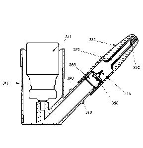
[00149] In one embodiment of the POD device 310, the POD device 310 has a rear
puncture member 315 and a front puncture member 320 and is capable of
accepting a
unit dose container 330. In another aspect, the front puncture member 320 may
be
integral with the nozzle 360. In yet another aspect as show in Fig. 36, the
POD device
310 does not have a front puncture member 320. In this aspect, the POD device
310
has a nozzle 360 integral with the unit dose container 330 as shown in Fig.
36. In
another aspect, the angles of the front puncture member 320 and/or the rear
puncture
member 315 may vary. In yet another aspect, the unit dose container 330 may be
sealed by, for example but not limited to a rubber or foil seal. In an aspect,
the POD
device 310 may be a translational device or a rotational device. In yet
another aspect,
the puncture members may have one or more orifices. In yet another aspect, the
POD
device 310 may have a combination of one or more of the various aspects
described
herein.
[00150] The unit dose container 330 described herein may be manufactured using
a
variety of manufacturing processes including but not limited to injection
molding,
CA 02909954 2015-10-20
WO 2014/179228
PCT/US2014/035711
blow molding, or a blow-fill-seal process. Blow fill seal technology involves
forming, filling, and sealing a dosage form in a continuous process in a
sterile
enclosed area inside a machine. Depending on the product to be contained and
the
manufacturing process used, the unit dose container 330 may be made of a
polymer,
such as polyethylene, ethyl vinyl alcohol copolymer, low-density polyethylene
(LDPE), high-density polyethylene (HDPE), polypropylene (PP) or any other
suitable
polymer, mixture or the like that is suitable for forming the unit dose
container 330.
[00151] Furthermore, while unit dose container 330 is illustrated as being
substantially cylinder-shaped, the unit dose container 330 may comprise any
other
shape suitable for selectively dispensing a unit-dose of a compound or
product. For
example, the unit dose container 330 may be substantially cone-shaped, tube-
shaped,
rectangular-shape, polygonal, oval-shaped, or combinations of any of these.
Moreover, while the end of the unit dose container 330 is shown as being
substantially flat, the end may alternatively be crimped (e.g., in the case
where the
dispenser is formed by a blow-fill-seal process).
[00152] Figure 25 illustrates one aspect of the POD device 310 having a rear
puncture member 315 and a front puncture member 320 and having a container
holding area capable of accepting a unit dose container 330. In one aspect,
the
container holding area is a hollow or a container cavity 325. These components
are
.. housed in the POD device tip 335. The POD device tip 335 is an umbrella
term for
the assembly of the tip body, rear puncture member 315, diffuser 350, unit
dose
container 330, front puncture member 320 and nozzle 360.
[00153] Fig. 26 illustrates a unit dose container 330. The rear puncture
member 315
is located at the distal end of the POD device 310 as the POD device 310 is
located
when in use and inserted into a user nasal cavity; e.g. farther from the nasal
cavity of
the user. The front puncture member 320 is located at the proximal end of the
POD
31
CA 02909954 2015-10-20
WO 2014/179228
PCT/US2014/035711
device 310 as the POD device 310 is located when in use and inserted into a
user
nasal cavity; e.g. nearer to the nasal cavity of the user.
[00154] As illustrated in Fig. 26, in one aspect, the unit dose container 330
is
cylindrically shaped with closed ends. Moreover, while the end of the unit
dose
container 330 is shown as being closed and substantially flat, the end may
alternatively be crimped or dimpled.
[00155] Fig. 26 shows the rear puncture member 315 and the front puncture
member 320 not operationally engaged with the unit dose container 330. The
rear
puncture member 315 and the front puncture member 320 both allow for transport
or
conveyance of the propellant and/or propellant compound mixture. In further
illustrations of this aspect, as illustrated in Fig. 30 and 31 b, the rear
puncture member
315 has a hollow portion for delivery of the propellant or, as illustrated in
Fig. 32, is
constructed of a porous material. Whereas, in the aspect with a front puncture
member 320, the front puncture member 320 has a hollow portion for delivery of
at
least the compound contained in the unit dose container 330.
[00156] Figures 27 shows the rear puncture member 315 and the front puncture
member 320 operatively engaged with the unit dose container 330, with the rear
puncture member 315 and the front puncture member 320 inserted into the unit
dose
container 30.
[00157] Figure 28 illustrates the unit dose container 330. The unit dose
container
330 may be manufactured from glass or a polymer, such as polyethylene, ethyl
vinyl
alcohol copolymer, low-density polyethylene (LDPE), high-density polyethylene
(HDPE), polypropylene (PP) or any other suitable polymer, mixture or the like
that is
suitable for forming the unit dose container 330. In one illustration of this
aspect, the
unit dose container 330 is manufactured from polyethylene. In another
illustration of
this aspect, the unit dose container 330 is manufactured from ethyl vinyl
alcohol
32
CA 02909954 2015-10-20
WO 2014/179228
PCT/US2014/035711
copolymer. In yet another illustration of this aspect, the unit dose container
330 is
manufactured from low-density polyethylene (LDPE). In yet another illustration
of
this aspect, the unit dose container 330 is manufactured from high-density
polyethylene (HDPE). In another illustration of this aspect, the unit dose
container
.. 330 is manufactured from polypropylene (PP). In yet another illustration of
this
aspect, the unit dose container 330 is manufactured from any other suitable
polymer,
mixture or the like that is suitable for forming the unit dose container 330.
[00158] The unit dose container 330 described herein may be manufactured using
a
variety of manufacturing processes, such as injection molding, blow molding,
or a
blow-fill-seal process.
[00159] Figure 28 illustrates one aspect of the unit dose container 330 where
the
unit dose container 330 is sealed with polymeric stoppers 331, such as but not
limited
to rubber. In further examples of this aspect, the unit dose container 330 may
be foil
sealed. The stoppers can be a combination of the above options.
[00160] As shown in Figure 28, the unit dose container 330 has aspects of the
POD
device 310 compound chamber. The unit dose container 330 is capable of holding
a
compound 332. The unit dose container 330 is designed to be prefilled to a
specific
volume. The unit dose container 330 can release the entirety of the dose
(single
dosing).
[00161] As shown in Figures 29-32, the puncture materials can be rigid, semi-
rigid,
or porous. The puncture designs can be either through hole, side orifice,
porous flow,
or a combination.
[00162] Figure 29 illustrates a rear puncture member 315. In an example of
this
aspect, the rear puncture member 315 may or may not have a side orifice 340 or
a
plurality thereof. As shown in Figs. 30 and 31b, the side orifice 340
assists in
reducing residuals of the compound which may remain in the unit dose container
330
33
CA 02909954 2015-10-20
WO 2014/179228
PCT/US2014/035711
after actuation. The side orifice(s) 340 allow for the propellant released
from the
canister 311 to scour the sides of the unit dose container 330. In one example
of the
side orifice 340, the side orifice 340 may be substantially oval, circular,
square,
triangular, or rectangular in shape or combinations thereof.
[00163] As illustrated in Fig. 30, the rear puncture member 315 provides a
distal
opening 345. The distal opening 345 allows for a path through which the
propellant
journeys or is conveyed from the canister 311 as it is introduced into the
unit dose
container 330. As illustrated by the arrow showing the direction of travel of
the
propellant in Fig. 30, the propellant travels across the diffuser 350 of the
POD device
to 310. As illustrated by the arrows showing the direction of travel of the
propellant in
Fig. 3 1 b, the propellant travels across the diffuser 350 of the POD device
310 and out
the side orifice(s) 340.
[00164] Fig. 32 illustrates one aspect of the rear puncture member 315 in
which the
rear puncture member 315 is of porous material. The arrows in Fig. 32
illustrate the
direction of flow of the propellant from the canister 311 across the porous
rear
puncture member 315. In further illustrations of this aspect, the porous rear
puncture
member 315 is seated inside a solid non-porous puncture member housing 355.
[00165] Figs. 33 and 34 show the front puncture member 320. Figs. 33 and 34
show the front puncture member 320 engaged within the unit dose container 330.
Fig.
33 shows the front puncture member 320 with an integrally molded nozzle 360.
Whereas, Fig. 34 shows the front puncture member 320 with a separately molded
nozzle 360. The front puncture member 320 sits within a puncture member
housing
355. The front puncture member 320 provides a proximal opening 365.
[00166] In operation of the POD device 310 with a non porous rear puncture
member 315 and no side orifice(s) 340, the propellant from the canister 311 is
conveyed or travels from the canister 311, across the diffuser 350, follows
the path of
34
CA 02909954 2015-10-20
WO 2014/179228
PCT/US2014/035711
the arrow shown in Fig. 30, exits the distal opening 345 of the rear puncture
member
315, enters the unit dose container 330, the compound and/or propellant
travels along
the path of the arrow shown in Fig. 33 and 34, and exits the proximal opening
365 of
the front puncture member 320.
[00167] In operation of the POD device 310 with a porous rear puncture member
315 and no side orifice(s) 340, the propellant from the canister 311 is
conveyed or
travels from the canister 311, across the diffuser 350, follows the path of
the arrow
shown in Fig. 32, exits the distal opening 345 of the rear puncture member
315, enters
the unit dose container 330, the compound and/or propellant travels along the
path of
the arrow shown in Fig. 33 and 34, and exits the proximal opening 365 of the
front
puncture member 320.
[00168] In operation of the POD device 310 with a non porous rear puncture
member 315 and a side orifice(s) 340, the propellant from the canister 311 is
conveyed or travels from the canister 311, across the diffuser 350, follows
the path of
the arrow shown in Fig. 3 lb, exits the side orifice 340 of the rear puncture
member
315, enters the unit dose container 330, the compound and/or propellant
travels along
the path of the arrow shown in Fig. 33 and 34, and exits the proximal opening
365 of
the front puncture member 320.
[00169] As shown in Figure 35, the geometry can be changed from straight
cylinder
designs. In blow fill seal in particular, the geometry is very customizable.
Some of the
geometry changes include a dimple 395 to center the puncture.
[00170] As shown in Figure 36, a blow fill seal has the potential to over mold
the
nozzle 360 and eliminate the need for a double puncture of the unit dose
container
330. It would allow for a rear puncture member 315 and removal of a front tab
370.
Removal of the tab 370 provides access to the nozzle 360 through with the dose
travels to be released.
CA 02909954 2015-10-20
WO 2014/179228
PCT/US2014/035711
[00171] As shown in Figure 37, to center and stabilize the unit dose container
330, a
rib 375, or plurality of ribs, can be added along the length of the unit dose
container
330 in order to well fit the unit dose container 330 in the container cavity
325. In Fig.
37, the ribs 375 are shown in engagement with the container cavity wall 380.
The rib
375 would form an interference fit and prevent sliding or movement prior to
piercing
or puncturing of the unit dose container 330.
[00172] Figs. 25 and 41 illustrate two aspects of POD device 310 having a rear
puncture member 315 and a front puncture member 320 and having a container
cavity
325 capable of accepting a unit dose container 330. The container cavity 325
and the
unit dose container 330 are cooperatively sized. Figure 25 shows one aspect of
the
POD device 310 where the tip 335 engages the actuator body 385 in a
translational
manner. Fig. 41 shows another aspect of the POD device 310 where the tip 335
engages the actuator body 385 in a threaded manner 386.
[00173] As shown in Fig. 25, on the actuator body 385 of the POD device 310
which engages the tip 335 in a translational manner is a stop 390. The tip 335
slides
onto the actuator body 385 and comes to a fixed position as it journeys over
the stop
390. In Fig. 41, the tip 335 is associated with the actuator body 385 via male
and
female threads. In this aspect, the male threads or female threads may be
cooperatively arranged to be either on the tip 335 or the actuator body 385.
The tip
.. 335 is threaded onto the actuator body 385. In both illustrations shown in
Fig. 25 and
41, the POD device 310 is not in its operational use when the tip 335 is not
engaged
with the actuator body 385.
[00174] Fig. 25 shows an aspect of the embodiment where the container cavity
325
does not contain a unit dose container 330. Fig. 38 shows the POD device 310
in
partial engagement with the unit dose container 330. Fig. 38 shows the rear
puncture
member 315 in engagement with the unit dose container 330. Figure 39 shows the
36
CA 02909954 2015-10-20
WO 2014/179228
PCT/US2014/035711
rear puncture member 315 and the front puncture member 320 in engagement with
the
unit dose container 330.
[00175] Figure 40 shows an exploded view of the of the translational POD
device
shown in Fig. 25.
.. [00176] Fig. 42 illustrates the various angles of puncture of the rear
puncture
member 315. Fig. 42a shows a 90 degree rear angle of puncture on the rear
puncture
member 315. Figure 42b shows a 60 degree angle of puncture on the rear
puncture
member 315. Figure 42c shows a 45 degree angle of puncture on the rear
puncture
member 315. Figure 42d shows a 30 degree angle of puncture on the rear
puncture
member 315. Figure 42e shows a 15 degree angle of puncture on the rear
puncture
member 315. In one aspect, the puncture angle is from about 15 degrees to
about 90
degrees, from about 30 degrees to about 90 degrees, from about 45 degrees to
about
90 degrees, from about 60 degrees to about 90 degrees, or combinations thereof
[00177] In further illustrations of the angles of puncture shown in Fig. 42a-
e, the
angles of puncture may also apply to the front puncture member 320. Although
the
front puncture member 320 is shown as a beveled member in the figures, the
front
puncture member may be shaped with angles of puncture as illustrated in Fig.
42 with
regards to the rear puncture member 315.
[00178] The invention is further described in the following examples, which
are in
not intended to limit the scope of the invention.
Example 1
[00179] An oxime drug, 2-PAM, was administered into the olfactory nasal region
in
rats with the device, (e.g. a Pressurized Olfactory Delivery (POD) device).
The brain
and plasma concentrations of 2-PAM was measured at certain time points after
drug
37
CA 02909954 2015-10-20
WO 2014/179228
PCT/US2014/035711
administration. The device enabled delivery of 2-PAM resulted in higher brain
exposure and lower plasma exposure compared to intravenous injection.
[00180] Animal use. Rats were used for deposition, tolerability and
distribution
experiments. Adult male Sprague-Dawley rats (200-300 g; Harlan, Indianapolis,
IN)
were housed under a 12 hour light/dark cycle with food and water provided ad
libitum. Animals were cared for in accordance with institutional guidelines,
and all
experiments were performed with an approved protocol from the Pacific
Northwest
Diabetes Institute Institutional Animal Care and Use Committee under protocol
number 12610.
[00181] Statistical analysis. In most cases where two values were compared a t-
test
was used. When more than two groups were compared, such as comparing the
powder
2-PAM POD formulation with the aqueous 2-PAM POD formulation and the IV 2-
PAM, a two-way ANOVA was used with a bonferroni post test. When comparing the
AUC plasma and brain values which were derived from different animals at each
time
point the method described in Westin et al., 2006 was used. In all cases
statistical
significance was defined as p <0.05.
[00182] Aqueous formulations of 2-PAM were made by dissolving 2-PAM in
deionized water. 2-PAM was dissolved into 500 I of water at 10mg/ml, 100
mg/ml,
250 mg/ml, and 500 mg/ml and left in a closed microcentrifuge tube at ambient
temperature (25 ). These water based formulations were then visually observed
at 1
hour, 24 hours, and 48 hours for any cloudiness or precipitant.
[00183] Dry powder formulation of 2-PAM was prepared by placing the 2-PAM
free drug in a microcentrifuge tube and grinding the drug with a motorized
pestle
(Kontes, Vineland, NJ). The 2-PAM powder was then observed under a microscope
to
ensure the homogeneity of the powder formulation. The 2-PAM was ground with a
pestle to ensure that there were no agglomerations of 2-PAM greater than 100
um in
38
CA 02909954 2015-10-20
WO 2014/179228
PCT/US2014/035711
diameter. Such larger agglomerates could clog the 810 gm diameter POD nozzle
used
in the rat experiments.
[00184] The construction of the rat use POD nasal aerosol device is
illustrated in
Figure 17. A meter dose inhaler (MDI) can dispensing 25 gl hydrofluoroalkane
227 is
attached to the plastic actuator. The actuator is in gas communication with a
polytetrafluoroethylene frit which had a 50 gm pore size. The frit is in
communication
with the dose holding cylinder which is placed inside the body of the POD in
order to
create an aerosolized flow. On actuation the HFA propellant is converted to a
gas by
passing through the frit material and then it mixes with the dose and the dose
and
propellant mixture exits from the 23 gauge stainless steel tubing nozzle which
is
covered with a fluorinated ethylenepropylene liner was placed over the outside
of the
metal tip in order to protect the nasal epithelia from being damaged by the
nozzle
during use. The construction of the rat use POD device was successful and
consistently delivered powder 2-PAM formulations with no measurable residual
drug
left in the device.
[00185] The basic operation of either POD device in rats was as follows. The
animal was anesthetized with 5% isoflurane for 2 minutes to enable consistent
administration. The rat was removed from the isoflurane chamber and placed in
a
supine position. The dose was loaded into the device and the nozzle was
carefully
placed 8.0 mm into the rat nasal cavity and pointed in the direction of the
cribriform
plate. Then the MDI can was pressed to discharge the dose into the rat nasal
cavity. In
addition, the dry powder dose chamber was weighed on a scale with a
sensitivity of
0.1mg (Mettler Toledo, Columbus, OH) before loading the dose, after the dose
was
placed in the dose loading chamber, and after firing to ensure that the
correct dose
was loaded into the device and that the complete dose was released into the
rat nasal
cavity.
39
CA 02909954 2015-10-20
WO 2014/179228
PCT/US2014/035711
[00186] The 2-PAM formulations were made with 0.1% coomassie blue dye in
order to test nasal cavity deposition in rats. The animals were dosed using
the dry
power POD device as described above with a single dose of 2.5 mg dose of 2-PAM
with coomassie blue. Shortly after administration was complete (< 5 minutes),
the
animals were overdosed with 250 mg/kg pentobarbital. The nasal cavity was then
bisected at the septum, the septum was removed, and the tissues were examined
for
dye localization. In addition the trachea and esophagus were dissected from
the back
of the mouth to the lungs to determine if the POD spray deposited any 2-PAM
beyond
the nasal cavity. This deposition study was performed with N=4 rats. The
typical
result of the deposition testing is shown in Figure 18. In Figure 18 the
olfactory region
of the rat nasal cavity in the upper panel is circled in white. The dark dye
can be seen
as being deposited primarily within this olfactory region.
[00187] A sensitive LC/MS method was established in order to determine the
distribution of POD administered 2-PAM in both the plasma and the brain of
rats. A
fixed volume (20 I) of 2Chlorolmethylpyridinium iodide d6 (Cerilliant, Palo
Alto,
CA) was added into each tissue and plasma sample to act as an internal
standard.
Tissue samples were homogenized in 3 mls of water. 60 1 of acetonitrile was
added
to the samples to cause protein precipitation. The samples were centrifuged
for 10
minutes at 1000g. An Agilent HPLC/MS series 1100 series B with autosampler
(Agilent, Technologies, Inc., Santa Clara, CA) was used for quantification.
The
injection volume was 5 1. The morphine samples were passed over a Phenomenex
Synergi 4u PolarRP 80A (Agilent, Technologies, Inc., Santa Clara, CA) with a
flow
rate of 0.3 ml/min.
[00188] A standard curve was created on the day of analysis according to the
same
process described for the samples. Each standard curve was linear with a
coefficient
of linear regression R2 > 0.99. In addition, two quality control samples with
a known
CA 02909954 2015-10-20
WO 2014/179228
PCT/US2014/035711
amount of drug were processed on the day of analysis in order to ensure day to
day
consistency of the analytical assay.
[00189] This LC/MS method was successful and resulted in reproducible
quantification of both tissue and brain samples. The 2-PAM detectable peaks
were
much higher than background in most cases. The sensitivity of this detection
method
was 0.05 g/ml in plasma and 1.0 ng in brain tissue. This method could be used
in
future studies with primates or in clinical studies.
[00190] In the tissue distribution experiments, the animals were anesthetized
with
5% isoflurane for two minutes. Then the animals were removed from the
isoflurane
induction box and placed in a supine position. The animals were then dosed
with
either the POD device (2.5 mg in a single 10 ul dose) or via intravenous
injection (2.5
mg in 500 ill). Animals that were sacrificed 5 minutes after dosing remained
under
2% isoflurane anesthesia until they were sacrificed. The animals sacrificed at
the
remaining time points were allowed to wake up from isoflurane anesthesia and
placed
back into housing. At 3 minutes before the sacrifice time the animals were
again
exposed to 5% isoflurane and then quickly overdosed with Beuthanasia-D
(Schering--
Plough Animal Health Corp, North Chicago, IL). Using IV 2-PAM and the aqueous
POD formulation of 2-PAM, animals were sacrificed at 5, 15, 30, 60, and 120
minutes
(N=6). Animals dosed with the dry powder 2-PAM POD formulation were sacrificed
at 5 and 15 minutes (N=6).
[00191] Immediately after death, the animal was decapitated. Blood was
collected
from the trunk and placed in a microcentrifuge tube with 10 .1 of 40 mM EDTA.
The
plasma was separated from the blood by centrifuging at 6,000g for 10 minutes.
Then
the plasma was frozen until it was analyzed for 2-PAM concentration with the
LC/MS
method previously described. The base of the skull and the parietal bones were
quickly removed from the head. The brain was removed within 2 minutes of
sacrifice.
41
CA 02909954 2015-10-20
WO 2014/179228
PCT/US2014/035711
The brain was placed in a microcentrifuge tube and frozen until it was
analyzed for 2-
PAM concentration with LC/MS.
[00192] A direct transport percentage (DTP%) to the brain was calculated in
order
to determine the amount of drug in the brain that was distributed directly
from the
nasal cavity to the CNS. The DTP% is used to estimate the amount of drug in
the
brain that cannot be accounted for by systemic distribution. The DTP as
defined was
calculated as follows:
[00193] Administration of the aqueous formulation of 2-PAM with POD resulted
in
lower systemic exposure and greater CNS exposure compared to an equivalent IV
dose. The IV dose resulted in a typical plasma curve with the highest point at
5
minutes (Figure 19). The POD administered 2-PAM resulted in plasma
concentrations
that were lower than the IV values, which is not expected given 2-PAM's
limited
absorption across the nasal respiratory epithelium into the blood stream. The
total
plasma AUC was significantly lower after POD administration compared to IV
AtICb,,a,n(x) Bx
________________________________ _
AUC plas-mtt(W) Cplas (nasal)
C brain(nasal) X
DTP% = X 100%
AU Cbrain (nasal)
administration.
[00194] In contrast to the plasma values, the brain concentrations of 2-PAM
after
POD administration were significantly higher than after IV administration at
both 5
and 120 minutes (Figure 20). In addition, the total brain concentration AUC
was
significantly greater after POD administration compared to IV. Of interest for
the
application of 2-PAM as a nerve gas exposure treatment is the fact that at 5
minutes
after administration, POD 2-PAM resulted in 3.5X the brain concentration
compared
to IV administration.
42
CA 02909954 2015-10-20
WO 2014/179228
PCT/US2014/035711
[00195] The brain-to-plasma ratios were significantly higher after POD 2-PAM
compared to IV at every time point except for 30 minutes (Table 1). These
increased
ratios point to the fact that a portion of the drug was directly delivered to
the brain
from the nasal cavity, effectively bypassing the blood brain barrier. When the
direct
transport percentage (%DTP) was calculated it was found to be 80.9%. This %DTP
can primarily be accounted for by the large brain values found 5 minutes after
POD 2-
PAM administration. Table 2 shows brain to plasma concentration ratios. At
each
time point except for 30 minutes, POD administration resulted in significantly
greater
brain to plasma ratios with a 15.25 fold increased brain to plasma ration
after 5
minutes.
Table 1
Time (min.) POD
5 132 74' 8?
58.5* 13.1
30 411 16
60 6L4*
120 126.7 6.7
15 [00196] The powder formulation of 2-PAM administered via the POD device
led to
even greater 2-PAM concentrations in the brain (Table 2). The powder 2-PAM POD
study was more limited than the aqueous formulation, but at 5 and 15 minutes
after
administration the powder formulation resulted in similar blood levels
compared to
the aqueous 2-PAM POD, but significantly higher brain concentrations.
43
CA 02909954 2015-10-20
WO 2014/179228 PCT/US2014/035711
Table 2
standard
Pg asrn a 2 P .601.1 ,c.c,n,c snit orti.ra geNtli( q tLssne tim es attic, n
ro.rtto.f r
torn. (31-1Nn) V p3,0,vd,.r -1LM3 P03L V P t_31_)
5 0.44 1.$2 0.46 5 0 -- 0.4 -- 027
15 033 0T3 038 15 03 0.2 -- 8.11
standard
03ran 2 PAM ,...rvcsntrar3,an ing( q sstss d.vEathara
p mewl, r
nrn. nnr ) F`tr.N VW` poadar FeND F 9V P043
41 6 11 100.39, 5 390 2.0 13.75
304 9.0 293,32 15 6.4 3.0 22 o 27
5
[00197] Table 2 shows distribution of the powder formulation of 2-PAM
administered via POD. The powder formulation of POD resulted in plasma values
at 5
and 15 minutes that were not significantly different than the liquid
formulation of
POD. However, the 2-PAM concentrations after POD administration of the powder
10 formulation were significantly greater than either the aqueous POD 2-PAM
or the IV
2-PAM. *=p<0.05
[00198] The phan-nacokinetic and distribution experiments resulted in data
supporting the potential of POD administered 2-PAM as a treatment for nerve
gas
exposure. The POD administration in both the aqueous formulation and the
powder
15 formulation resulted in high brain exposure within the first 5 minutes of
administration.
Example 2
[00199] The device used in Example 2 is described in Figure 3. The device in
this
example is referred to as a pressurized olfactory delivery (POD) device. In
order to
determine the amount of compound being delivered from the device to the
olfactory
region of the nasal cavity a method was developed for determining the
percentage of
dose deposited within key regions of a human nasal cavity model. This method
relies
on a quantitation by image analysis and is able to detect and quantitate
deposition
44
CA 02909954 2015-10-20
WO 2014/179228
PCT/US2014/035711
within 5 specified regions that describe the whole nasal model, including the
upper
olfactory region.
[00200] Materials: A human nasal cavity model was constructed from clear heat
moldable plastic sheeting. (Figure 21) This mold is thin-walled and is
transparent to
a blue light source that allows for the excitation of the indicator dye
fluorescein used
in the experimental doses. This human nasal cavity model was based on a
computer
model generated from MR1 scans from multiple subjects (Liu, J Appl Physiol,
2009
Mar;106(3):784-95). The model therefore represents an "average" human nasal
cavity.
[00201] A stage for positioning the nasal models and aiming the POD device
during
targeting and actuation was designed and constructed. This stage was flexible
enough
in operation to allow for a wide set of aiming angles, both horizontal and
vertical. By
aiming the device at various angles with respect to the nasal cavity, the
robustness of
the device administration could be tested.
[00202] A thin walled transparent nasal model was prepared by coating the
inside
with a very thin layer of imitation mucus, which was simply a store bought
hand
sanitizer solution. The prepared model was then photographed in a custom made
transilluminator/photo box as a blank reference for that particular
experimental point.
The model was then mounted onto the stage along with the POD device that has
been
loaded with a dose of 0.1mg/mL Fluorescein/water. Immediately after POD
actuation, the model was removed from the stage and held horizontally to
prevent
dose migrating. As soon as possible, the dosed model was placed in the
transilluminator/photo box and photographed. The model was then washed under a
stream of tap water and dried by shaking or forced air to be readied for
another test.
The two camera images were then digitally analyzed as described below to
reveal
deposition within the model.
CA 02909954 2015-10-20
WO 2014/179228
PCT/US2014/035711
[00203] Data processing of the blank and experimental images obtained was
carried
out with ImageJ software. For ImageJ to repeatedly compare images and perform
background subtraction accurately, the digital photographs were taken with the
model
carefully held in the same register within the transilluminator/photo box.
ImageJ
performs three key functions: 1) the image was color processed with the RGB
channel splitter. This function eliminates red and blue signals from the
image,
leaving primarily signal generated by the fluorescent signal from the
fluorescein in the
dose.
[00204] The ImageJ ROT manager allowed us to define five regions of interest;
olfactory, turbinate, esophagus, base and vestibule which were quantitatively
analyzed
with each device administration. The regions are defined by the lines seen in
Figure
and these regions contain a specific area, in pixels that can be quantitated
based on
the signal intensity of the fluorescein. Figure 22 also shows a typical spray
pattern
after a POD administration. The fluorescein administered into the model by the
POD
15 device can be seen as the light intensity on the dark background. It can
be noted from
Figure 20 that a majority of the administered dose resides within the
olfactory region
of the human nasal model. Each pixel within these photos can possess a value
of 0 to
255. The Measure function of ImageJ calculates the mean pixel value over each
defined region of interest. The total signal recorded within a particular
region of
20 interest is therefore the product of the mean pixel value by the number
of pixels
measured. Of additional interest is the reported Max value. Because the photo
cannot
record more than 256 levels of signal, we conclude that the assay is not valid
if we
receive values of 255 in that column, because we cannot be sure if the actual
signal is
not significantly greater than 255 if it could be measured. Such a situation
would
have the effect of underreporting signal in that ROT because the signal is
effectively
clipped. For this reason, the camera exposure settings are critical to ensure
that the
46
CA 02909954 2015-10-20
WO 2014/179228
PCT/US2014/035711
signals recorded fall within the sensitivity range of the method yet allow for
the
maximal sensitivity of the method as well.
[00205] In addition, our calculations involved the subtraction of values
obtained
from a blank recording. This is because there is some stray light leakage and
always
therefore the potential for background fluorescence involving the model and
the
imitation mucus. Because these elements are not perfect in application, we do
a
background photo record each time and do a subtraction for each data point.
This
method offers the advantage of providing fractional deposition on more than
one
region of the nasal model. It also offers clear qualitative photo/visual
confirmation of
the quantitative results.
[00206] The results of a deposition study are shown in Table 3. Two different
POD
devices were used and are referred to as Tip#1 and Tip #2. Each Tip was
administered
into the nasal model N=3 times at either 0 degrees horizontal angle with
respect to the
septum or 5 degrees horizontally towards the septum. All POD administrations
were
administered at a vertical angle of 55 degrees with respect to the base of the
nasal
cavity.
Table 3
-19 Fa- -4-1,
st.elgr-00.5 S cite.g.1^-00,5.
tiov. 60,
raht0lEck,r, - - .3_
3 _ a_5s.
- 3
rEØger0,0s S cte.ees aturs:t0rikurr
Zone' A CO O,s2yzt. S'tc$.
f.D.tac-tcary 3. 9' +ff.2.
30.
"Fn., r-k=Flr,.,:_,2 .
lE42.01,=00:urs.
- 3.4
Example 3
47
CA 02909954 2015-10-20
WO 2014/179228
PCT/US2014/035711
[00207] Impaction force testing was used to compare several nozzle/dose
chamber
configurations with MDI drivers to several commercial nasal spray products.
Impact
impaction force is an ideal method to characterize plume characteristics that
are
important for dose delivery consistency, dose localization and dosing comfort
and
safety. A schematic of the experimental setup used in this example is shown in
FIG.
23.
[00208] Impaction force measurements were carried out on a Mettler Toledo XS
64
with data output set at 10 per second coupled to an Apple MacBook Pro 2.2 GHz
Intel
Core 2 Duo processor, 4 GB 667 MHz DDR2 SDRAM via a ft. RS232 (Mettler
Toledo) to USB cable (Gigaware) with supporting driver software. Data
acquisition
was carried out using Windmill Logger version 4.07, release 7 (Windmill
Software
Ltd.) in a Windows Vista virtual machine environment using Parallels Desktop 5
for
Mac on the MacBook Pro. Data collected via Windmill Logger was imported
directly
into Microsoft Excel for graphical processing and analysis.
.. [00209] An impaction force stage was constructed to perform the
measurements.
This stage included means for accurate level and distance controls along with
customized holders for the individual devices tested. Actuation was carried
out
manually. POD or commercial devices were aligned to impact the direct center
of a
16.9 gram aluminum pan, 74mm X 80mm. The pan was cleaned of dose/debris
.. between each data shot. The distance from nozzle aperture to pan was 4cm,
consistent
with the conclusions of Guo, et al. 2009 (Guo, J Pharm Sci., 2009,
Aug;98(8):2799-
806.) as being within the 3cm to 6cm window of distances that generate the
highest
impaction forces and also consistent with our target distances in human nasal
models.
MDI triggered values obtained via valve actuation as tested was broadly
insensitive
from shot to shot when used as directed. The only effects seen were lower
values if
actuated very slowly.
48
CA 02909954 2015-10-20
WO 2014/179228
PCT/US2014/035711
[00210] Three commercial nasal spray products were tested in this Example:
Rite
Aid Pump Mist Nasal relief, oxymetasoline HCL 0.05%; NeilMed NasoGel For Dry
Noses, Saline gel spray; and Rite Aid NoDrip Nasal Spray, pump, oxymetazoline,
0.05%.
[00211] The device used in this study is shown in Figure 3 and is referred to
as a
pressurized olfactory delivery (POD) device in this Example. The POD nozzle
was
compared to the commercial spray pumps tested above. In this Example we tested
the
POD device under the same parameters as the commercial sprays using MDI
canisters
loaded with a 5% Ethanol, fluorescein mixed with either HFA 134a or HFA 227.
The
MDI valves were set to deliver a fixed volume of 50uL.
[00212] The impaction forces measured for three commercial pump style nasal
sprays were found to generate peak forces generally below 0.8 grams. These
products
are noted for either generating very broad spray patterns or slow moving
streams of
gelatinous material. The forces generated from these tested products fall well
below
the forces quoted by Guo et al., 2009 of 3.0 to 4.9 grams. The POD device
generated
impaction force measurements with peaks near 4 grams with an average of just
below
3 grams of force when the more highly volatile HFA 134a was used. This force
dropped to below 2 grams when HFA 227 was used instead. In either case, the
impaction forces for the POD device also fell well within the range of
impaction
forces measured for commercial MDI device by Guo et al., 2009, which showed a
maximum value of 6.5 grams.
[00213] It was found that the impaction forces measured are affected by the
HFA
type used and the volume of HFA dispensed by the MDI canister. Also the dose
chamber and nozzle configuration have impacts on impaction forces. In no case
have
we measured forces greater than that measured for the one commercial product
referenced in the Guo et al. paper.
49
CA 02909954 2015-10-20
WO 2014/179228
PCT/US2014/035711
Example 4
[00214] In this example the device, referred to as a pressurized olfactory
delivery
(POD) device, was tested to determine if the device would release a cold
temperature
spray. This testing involved the measurement of surface temperature changes on
the
target region caused by HFA POD. A schematic of the experimental setup used in
this
example is shown in FIG. 24.
[00215] The hydrofluoroalkane (HFA) used as a propellant in the POD device is
released from the metering can as a liquid. Very quickly after release the HFA
vaporizes and expands to form the pressure impulse that drives the dose
through the
POD nozzle. It is also a characteristic of the HFA POD that the HFA gas is
expelled
toward the target along with and after the dose is delivered. The expansion of
the
HFA causes a marked drop in temperature of the propellant gas during the
tiring
process. In order to establish whether this temperature drop is transferred to
target
tissues and to what extent, we designed and performed experiments to detect
and
measure the surface temperature of targets during and immediately after they
were
impacted by the device while only releasing HFA or while releasing a mixture
of
HFA and liquid compound (as it would be used for administering a liquid drug
product).
[00216] Materials: Kintrex infrared thermometer, model IRT0421, capable of
measuring surface temperature without actually contacting the surface being
tested.
Temperatures are reported in degrees Fahrenheit. An actuator fitted with a HFA
134a
canister designed to deliver 50uL of propellant, Kimwipe paper wipes, petri
dish, 1%
agaroselwater 3 tips, including a high impedance, low impedance nozzle and
open
configuration/absent frit.
[00217] Figure 24 illustrates the experimental setup for measuring temperature
changes during the firing of the POD device under different conditions. The
CA 02909954 2015-10-20
WO 2014/179228
PCT/US2014/035711
thermometer was positioned 4cm from the target. At that distance the
thermometer
"sees" and reads from a circular spot of 0.33cm diameter (target circle in
Figure 24).
[00218] Three tip configurations were tested. 1. A tip with a high impedance
nozzle
fitted. A high impedance nozzle is sufficiently restrictive to flow of HFA gas
that the
nozzle is the limiting feature of the POD system. It releases gas over a
longer
duration. 2. A tip with a low impedance nozzle fitted. In this tip, the frit,
near the
actuator end of the tip is actually the limiting feature of the device. It
releases gas
faster than the high impedance nozzle. 3. A tip that contains neither a nozzle
nor
frit. This tip offers essentially no restriction to HFA gas or liquid flow
through the
device. With these three configurations, we expected to understand how
restrictions
on gas flow affects the temperature of target upon firing and also define the
distinct
role that the Teflon frit plays in diffusing and facilitating the transition
of HFA from
the liquid state to the gaseous state.
[00219] We also tested the effect of target proximity to the nozzle with
respect to
temperature changes experienced by the target. We fired from a distance of 4cm
and
2cm.
[00220] In addition, we fired the device at three different targets. 1) We
used a very
low mass target. This target was constructed of a Kimwipe tissue paper. We
anticipated that a low mass target would have a very low thermal inertia and
therefore
would display much more change in temperature upon firing. 2) We created a
mock
epithelium (epithelium mimic #1) by overlaying a Kimwipe tissue paper wipe
onto
1% agaroseiwater. This was designed so that the thermometer would react to a
similar color and texture surface as the low mass target. 3) Another mock
epithelium
(epithelium mimic #2) made from 1% agaroselwater with Kimwipe paper embedded
just below the surface (less than 0.5mm) of the agarose. This target was
designed in
51
CA 02909954 2015-10-20
WO 2014/179228
PCT/US2014/035711
case the thermometer would react to the paper layer just below the essentially
clear
agarose to see if the temperature effects were mostly superficial.
[00221] In addition, some temperature measurements were done on the epithelium
mimics when a 50 L water dose was added to the setup. Table 4 summarizes the
temperature changes detected upon the firing of only hydrofluoroalkane
propellant.
The temperature change in degrees Fahrenheit is represented by the symbol A.
We
believed and confirmed that this would create the conditions for the most
dramatic
temperature changes. With the low mass, low thermal inertia paper target, the
greatest temperature change was when no frit or nozzle was installed in the
tip. The
data for this condition was closely clustered near -25 F. Indeed, with this
setup
particulate or mist can be seen ejecting from the end of the tip, suggesting
that a
certain fraction of the HFA remains liquid through its transit through the
actuator
body and tip. Any liquid HFA that were to reach the target would then ablate
on the
target and could explain the dramatic temperature drops seen.
[00222]
Table 4
a }>204.10%
: =-,f1 =-xl-MaX ---A2asx
Low ...... 2.4 ....... ............. os
27.7
,i3,47227.6Pc:),7==661ScMl 1I.5.1
,p125w51:,,rx ostmk<nttl? ¨ - - ---17F;1 -1:F31
s=TIK.Ae
ds.,tw-get t.vet
A==63..fax 1.4 -A
= 3.71. 2.4 [ ......
cnia-rtc #3. 434 ..
..............
s-exen4, 2.61 .1.5t 1. 4-21
0
[00223] In contrast, all other experimental conditions resulted in far smaller
temperature drops at the target. Modest drops of 3-4 F were seen with the
52
CA 02909954 2015-10-20
WO 2014/179228
PCT/US2014/035711
unobstructed tip on the epithelium mimics. It is clear the thermal capacity of
the
target is critical in this analysis.
[00224] Inclusion of the Teflon frit and nozzle into the tip resulted in even
smaller
temperature drops. Against the low mass tissue target, the low impedance
nozzle
resulted in the greatest temperature drop, with a maximum value of 5.6 F at a
distance of 2cm. The high impedance nozzle resulted in slightly lower
temperature
drops. Typical values were 3 F or less.
[00225] There is a slight trend depending on tip distance to target. As would
be
expected, shots at closer range can result in lower temperatures at the
target.
[00226] When a dose load of 504 water was added to the tip that included a
Teflon
frit and low impedance nozzle very small temperature effects were seen. The
data
ranged from a 0.5 F drop to a 0.2 F increase. It was determined that with the
small
changes seen and the difficulty of handling the liquid doses in the
experimental setup
that we would not be able to get reliable data with liquid doses. However we
believe
the data collected with the liquid doses in consistent with predicted
outcomes.
[00227] The hydrofluoroalkane propellant used in the POD device will have very
minimal effects on the temperature of impacted tissues. The data show the
Teflon
frit' s function in the POD and the decrease in the temperature of the
impacted site
when only HFA is delivered. In addition, a typical load of 500_, will itself
likely
reduce any temperature effects.
Example 5
[00228] In assaying the targeting of the human olfactory region with a drug
product,
2 formulations of 2-PAM were delivered from the device into a human nasal
cavity
model and analyzed for olfactory deposition.
53
CA 02909954 2015-10-20
WO 2014/179228
PCT/US2014/035711
[00229] A silicon rubber human nasal cavity model was purchased from Koken
Inc.
(Tokyo, Japan). A trace amount (0.1%) of Coomassie blue (Sigma Aldrich, St.
Louis,
MO) was mixed into the dry powder 2-PAM. The dry powder 2-PAM and Coomassie
blue were crushed to a homogenous powder with a mortar and pestle. 0.1%
rhodamine B was added into the aqueous formulation (250 mg/ml) for
visualization
within the nasal cavity model. The dry powder formulation was sprayed into the
model nasal cavity (N = 10) with the device and pictures were taken to get a
qualitative measure of deposition in the olfactory region. The pictures were
judged as
to whether a majority of the powder 2-PAM was deposited in the olfactory
region.
[00230] The same was done with the aqueous formulation, and the deposition in
the
olfactory region was also quantified by weight for this formulation (N = 10).
The
olfactory region of the nasal cavity model was cut from the model so that it
was
removable. The olfactory region was weighed before the POD spray and after the
spray and the percent of dose administered to the olfactory region was
calculated by
weight.
[00231] The dry powder 2-PAM formulation administered into the human nasal
cavity was effective in depositing of drug in the olfactory region.
Qualitative
examination of 10 administration attempts into the model consistently was
judged to
show a majority of drug (about 50% or greater) in the olfactory region. In
addition to
depositing drug on the olfactory region, the dry powder POD device deposited a
substantial amount of the 2-PAM dose at the interface with the cribriform
plate area
of the model which separates the olfactory region of the nasal cavity from the
brain.
[00232] The aqueous 2-PAM formulation displayed similar patterns of deposition
in
the human nasal cavity model as the dry powder formulation. In addition to the
qualitative photos of the human nasal cavity, 62.6 9.6 % of the dose was
determined
to deposit in the olfactory region of the nasal cavity.
54
CA 02909954 2015-10-20
WO 2014/179228 PCT/US2014/035711
[00233] Example 6: Comparison of a POD Device for a unit dose container and a
clinical POD device with injection port.
[00234] To construct a POD device for a unit dose container, a clinical POD
device
was bored out to accept a tube the size of the unit dose container. The tube
was a
.. proxy for example, commercially, for a blow fill sealed unit dose
container. The tube
for the unit dose container was sealed at the distal end by the diffuser; in
this
construction the diffuser was a frit. Into the hollow of the tube for the unit
dose
container was inserted a propellant injector in the form of a cannula, the
cannula had
an outer diameter smaller than the inner diameter of the hollow of the tube of
the unit
dose container. The cannula sealed the distal end of the tube for the unit
dose
container. The cannula was inserted a depth into the tube hollow so that side
orifices
on the cannula were located inside the tube hollow. The cannula conveyed or
delivered a propellant into the tube via the side orifices. The proximal
end of the
tube for the unit dose container was left open to receive a nozzle. The unit
dose
container was not actively punctured by the propellant injector and the
propellant
injector cannula was inserted into the tube for the unit dose container.
[00235] The unit dose container tube in this example was filled so that: 1)
dose was
filled so it sat at the distal portion of the tube (e.g. dose sat near the
frit), 2) dose was
filled so it sat near the nozzle or proximal portion of the unit dose
container and 3)
dose was filled so it sat evenly spread along the entirety of the unit dose
container.
The POD device was actuated and residual and high speed video was collected of
each deposition. Residual data was obtained by mass measurements.
[00236]
?4 155? 035 1Jc 1009
1 7 19a4.91 1.7}4., 1957 17.7 17%
KTOWIAt: ii:*:2(graW
7 7 19Ø9.3 2040.5. 1953.5. Fl% 101.2
a 2 1-945.0 2047.3 1S0.0 1.4 10% 100.4
CA 02909954 2015-10-20
WO 2014/179228
PCT/US2014/035711
[00237] Example 7. Puncture design.
[00238] Puncture members were tested for puncturing the distal, proximal, or
both
ends of the unit dose container. In this example, the puncture member is
shaped like a
cylinder. In this example where the puncture member is shaped like a cylinder,
the
outer diameter of the cylinder is .0625 cm at the distal base, non puncturing
end of the
puncture member. The proximal end of the puncture member is located immediate
to
the unit dose container when assembled with the POD device. From a center line
thru
the center plane of each cylinder, at the proximal end, five different angles
of taper at
the proximal end of the device were tested. The angles of taper, with respect
to a
center plane, are: 90 degrees, 60 degrees, 45 degrees, 30 degrees and 15
degrees as
shown in Figure 42(a), (b), (c), (d), and (e). In this example, the puncture
members
are made of aluminum, however, any suitable material such but not limited to
as PVC,
PEP, PP, ABS and COC may be utilized.
[00239] To test the puncture members, capsules containing a liquid or a powder
was
used in this example. The capsules used were a dry powder inhaler (DPI)
capsule
and a gel capsule. The capsules were held in a capsule holder and an Instron
Puncture
Tester, 5900 series model, from Instron, Norwood, MA, USA was used to
puncture.
[00240] Gel Capsules
[00241] Gel capsules were punctured at the rate of 1 ipm, 10 ipm, and 20 ipm
(inch
per minute). Of the punctures performed, the 15 degree puncture provided for
the
lowest maximum force to puncture the capsule.
[00242]
Specimen Puncture Rate Fracture Characteristic
1 90 1 No fracture, compression
2 90 10 No fracture, compression
3 90 20 No fracture, compression
4 60 1 No fracture, compression
56
CA 02909954 2015-10-20
WO 2014/179228 PCT/US2014/035711
60 10 No fracture, compression
6 60 20 No fracture, compression
7 45 1 No fracture, compression
8 45 10 No fracture, compression
9 45 20 No fracture, compression
30 1 Small puncture, little compression
11 30 10 Small puncture, lots of compression
12 30 20 Small puncture, little compression
13 15 1 Very small puncture, no
compression
14 15 10 Fractional puncture, no compression
15 20 Fractional puncture, no compression
[00243] Figure 43(a), (b), (c), (d) and (e) show a progression of decrease in
extension from a puncture with an angle of 90 degrees to a puncture with an
angle of
15 degrees. The puncture with an angle of 15 degrees showed consistently the
least
extension; see Figure 43(a), (b), (c), (d) and (e).
5 [00244] DPI capsule
[00245] The experiment described immediately above in this Example 7 was
conducted also with DPI capsules.
[00246]
Specimen Puncture Rate Fracture Characteristic
16 90 1 Significant compression
17 90 10 Significant compression
18 90 20 Significant compression
19 60 1 Significant de formation
60 10 Significant deformation
21 60 20 Significant deformation
22 45 1 Some compression then clean puncture
23 45 10 Small indents, then puncture (fracture)
24 45 20 Small indents, then puncture (fracture)
30 1 Small indents, then puncture
26 , 30 , 10 , Small indents, then puncture (some fracture)
27 30 20 Little indent, functional puncture
28 15 1 Very little indent, slight fracturing
29 15 10 Very little indent, slight fracturing
15 20 Very little indent, slight fracturing
[00247] With the dry powder inhaler capsule, from the table immediately above
it is
to .. apparent that a 30 degree angle of the puncture provided puncture
without fracturing
of the dry powder inhaler capsule. Figure 44 (a), (b), (c), (d), and (e) show
the
extension over the dry powder inhaler. At 30 degree angle with a DPI capsule
results
57
CA 02909954 2015-10-20
WO 2014/179228 PCT/US2014/035711
in acceptable puncture with the least force to puncture and punctured with the
least
extensimidisplacement.
[00248] We determined in both the gel and dry powder capsule each can
accommodate user variations in the amount of force per square inch that the
user can
apply. Angle of puncture has an impact. The sharper the puncture angle, the
less
maximum puncture force and the smallest required displacement to puncture the
capsules.
[00249] BFS film
[00250] BFS film of .6mm thickness was tested using an Instron Puncture
Tester,
5900 series model, from Instron, Norwood, MA, USA using the five puncture
devices
shown in Figure 42 (a), (b), (c), (d), and (c) and described in this example.
Specimen number Puncture Rate Characteristic
1 1 90 1 Despite the blunt puncture, the deformation
region
was limited to the cross sectional area
2 2 90 1
3 3 90 1
4 1 90 10 Identical to the 1 ipm rate puncture
5 2 90 10
6 3 90 10
7 1 90 20 Identical to the 1 ipm and 10 ipm puncture
rates
8 ? 90 20
9 3 90 20
10 1 60 1 Slower initial ramp than the 90 degree
11 2 60 1
1`) 3 60 1
13 1 60 10 Similar initial ramp to 1 ipm but longer tail
14 2 60 10
3 60 10
16 1 60 20 Very similar puncture to 10 ipm profile
17 ", 60 20
18 3 60 20
19 1 45 1 Lower force but longer tail than 60 degree
puncture
2 45 1
21 3 45 1
22 1 45 10 Identical to 1 ipm
23 2 45 10
24 3 45 10
1 45 20 Very similar to 1 ipm and 10 ipm
26 ? 45 20
27 3 45 20
28 1 30 1 Significantly less tail, lower force, and
less abrupt
, puncture than 45 degree
29 ? 30 1
3 30 1
58
CA 02909954 2015-10-20
WO 2014/179228 PCT/US2014/035711
31 1 30 10 Very similar to 1 ipm
32 2 30 10
33 3 30 10
34 1 30 20
35 2 30 20
36 3 30 20
37 1 15 1 Much lower force profile
38 2 15 1
39 3 15 1
40 1 15 10 Similar M 1 ipm
41 2 15 10
42 3 15 10
43 1 15 20 Similar to 1 and 10 ipm
44 2 15 20
45 3 15 20
[00251] Fig. 45 shows a correlation that the steeper angle puncture reduced
the
maximum puncture force.
[00252] Example 8: Determination of puncture and residuals of various puncture
devices.
[00253] Distal Puncture
[00254] Various puncture members were constructed and tested for their ability
to
puncture a unit dose container and to measure the amount of residual drug
remaining
in the container. Pass criteria for the puncture member was no more than 10%
residual.
[00255] The following rear puncture members were made; each puncture member is
essentially cylindrically shaped and has an opening its proximal end, where
proximal
end is the end closest to the nozzle, except in one illustration of this
Example where
the rear puncture member is sealed or closed at the proximal end:
[00256] A puncture member having an outer diameter of 1.651 mm; an inner
diameter of 1.194 mm; and a wall thickness of .229 mm (comparable to 16 gauge
on
the Birmingham Wire Gauge); (A-16)
[00257] A puncture member having an outer diameter of .8192 mm; an inner
diameter of .514 mm; and a wall thickness of .1524 mm (comparable to a 21
gauge on
the Birmingham Wire Gauge); (B-21)
59
CA 02909954 2015-10-20
WO 2014/179228
PCT/US2014/035711
[00258] A puncture member having an outer diameter of .8192 mm; an inner
diameter of .514 mm; and a wall thickness of .1524 mm (comparable to a 21
gauge on
the Birmingham Wire Gauge); closed end to the puncture member and a single
orifice
located on a lateral of the puncture member; (C-21)
[00259] A puncture member having an outer diameter of .5144 mm; an inner
diameter of .260; and a wall thickness of .1270 mm (comparable to a 25 gauge
on the
Birmingham Wire Gauge); (D-25)
[00260] A puncture member made of three cylinders each having an outer
diameter
of .3112 mm; an inner diameter of .159 mm; and a wall thickness of .0726 mm
(comparable to a 30 gauge on the Birmingham Wire Gauge). The three cylinders
for
the puncture member were arranged so that the three channels were parallel to
each
other on the long axis and each at the point of an isosceles triangle in the
plane of
puncture; (E-30) and
[00261] A puncture member having an outer diameter of .8192 mm; an inner
diameter of .514 mm; and a wall thickness of .1524 mm (comparable to a 21
gauge on
the Birmingham Wire Gauge); closed end to the puncture member and a two
orifice
located on a lateral of the puncture member; the two orifices located 180
degrees apart
(F-21).
[00262] A unit dose container was made of flexible tubing (e.g. Tygon tubing)
of
approximately 4 mm outer diameter and 27 mm length. The distal end of the
tubing
was sealed (by, for example, using a rubber stopper). The surrogate unit dose
is filled
with liquid; in this instance with 20 microgram/nil fluorescein. The unit dose
container was punctured by one of the puncture members, video was taken of the
dose
release, and measurements were made of residual drug. The following table
shows
the results of the residuals remaining.
CA 02909954 2015-10-20
WO 2014/179228 PCT/US2014/035711
,',,,,,,,,-, v,,,:i.=;;i: . =-.., '`:=INYZ,''.:"Z: in ',=:',:.:::,_:µ,
:,....-_._..._. .. nii.i.
'4( ,./.
61/44':µ,:," '..'= '== = - ..'''=, e=,*.:''' `.'''S' '.']i
,.,.',':K'''';'s,,:i fs,,s '''..::N.i:.': ,,s,.: ;:i ,',,,,k,r3
,,,,,....,..; iz.:3 .:,-..i,::¶; ;:::
:: .3,3.=?; 3 3:>:3 ::µ.,,,
. * ;:'= 3;.:.,i*.ii . '4,'.:3 ,': Mir:.. 4 SNS =i..?:
S ::;:,,
,:. ::: .3:.,i:3.4,:Z *.;:f:,; i,.;i:.: A SR.: '.';
:i=.:' :.:'.:
::-=::.µ:' '3 :::.i , s i
< = .' ,: 1.:A .; 3;.3'.; .': ..z '..Zi.:S%
.Z.t:,,V ..i, :!.?.*S i "i,'3% '3' :.,,,:3 .i. .n,
.f= f:.µ!ii'l
,:: ::::: :::i.:.:iv) :3 i .,V ===:;!;.:,, i:.::.= :3 ii
===:.).,:1.4
i3..:`::, =i ;:i'..? :: ": ?,, .', 3, ,3 3..3
.3.7:,'õ:.'
3,
:=., ::: :: .3.rM , .?,:;.>. A 1.M3ii
=: ic,,,-;:,i,' , .::, i z::, :=,;.::3s-z.:i
,i: =::: .E.::,.i..i.
,: i.:: ) tt, 3.s. '..: ..'.?=::. ;. ,,,:. . ::.,. s ,?
.: '.. 3 .:::::.;
;-2a 160 S. I:i.i..i,..:' :>.4.3. ,:, .i,ii,i 6.i
.',4=KI
I'i tv=3. amii xissi! i; s.e.ft
[00263] Minimal drug residuals were obtained with the C-21 and F-21 puncture
devices, as shown in Figure 46. Interestingly, B-21, a puncture member without
a
lateral orifice, had similar residual drug as C-21 and F-21, puncture members
with
lateral orifices. The lateral orifices seem to correlate with reduced drug
residual.
Whereas, decrease in the size of the puncture mechanism does not correlate
with a
decrease in residual drug. C-21 seems to provide for the least residual drug
with
residual drug being consistently under 5 microliters.
[00264] Proximal Puncture
[00265] Following the testing of the distal puncture member for residual drug,
a
POD device was constructed for acceptance of a distal C-21 puncture member and
a
proximal puncture member of two varieties:
[00266] An outer diameter of 1.270 mm; an inner diameter of .838 mm; and a
wall
thickness of .216 mm; ((comparable to a 18 gauge on the Birmingham Wire
Gauge);
T-18A) and
[00267] An outer diameter of 1.270 mm; an inner diameter of .838 mm; and a
wall
thickness of .216 mm; closed tip to the puncture device and a single orifice
located on
a lateral of the puncture device (T-18B).
61
CA 02909954 2015-10-20
WO 2014/179228
PCT/US2014/035711
[00268] The end of the proximal puncture member that would come into contact
with the unit dose container to puncture it was beveled.
[00269] The same method as in Example 8, Distal Puncture, above was used, with
C-21 puncturing the distal end and either T-18A or T-18B puncturing the
proximal
end of the unit dose container. Results are shown in the table below:
Results Average Residual Standard deviation
microliter
T-18A 3.23 0.99
T-18B 6.43 2.01
[00270] T-18A, without an orifice located on the lateral of the unit dose,
decreased
the residual.
[00271] Overall, for the distal and proximal puncture, having the gas exit
perpendicular to the central axis of the dose holding chamber led to decreased
residuals.
[00272] The present invention is not to be limited in scope by the specific
embodiments described herein. Indeed, various modifications of the invention
in
addition to those described herein will become apparent to those skilled in
the art
from the foregoing description and accompanying figures. Such modifications
are
intended to fall within the scope of the appended claims.
62