Note: Descriptions are shown in the official language in which they were submitted.
A MULTI-COMPONENT NON-BIODEGRADABLE IMPLANT, A METHOD OF
NudaNG AND METHOD OF IMPLANTATION
RELATED APPLICATION =
The present application claims priority to U.S. patent application serial No.
61/816,209, filed April 26, 2013.
FIELD OF rirg INVENTION
The present invention relates to a multi-component implant comprising a solid
hydrogel, a porous hydrogel, and a porous rigid base suitable for implantation
into a
mammal, to treat, repair or replace defects and/or injury to musculoskeletal
tissue, a method
of manufacturing the multi-component implant, and a method of implantation.
BACKGROUND OF TJAZ INVENTION
Articular cartilage defects in joints are a significant source of pain, have a
limited
ability to heal, and can lead to the development of osteoarthritis (Buckwalter
and Mtmkin,
1998; Shelbourne et aL, 2003). Surgical options for symptomatic ,cartilage
defeats include
palliative, reparative, and restorative methods (Cole and Lee, 2003). However
the treatment
algorithm and surgical indications for each of these procedures continues to
evolve
(Magnussen et al., 2008; Bekkers et at., 2009). Alternative treatments have
been developed
using biodegradable Implants intended to encourage the formation of articular
=Wage
within the 'defect site. However, these implants have mechanical properties
that are
continually changing and often inferior to that of the native tissue during
the regeneration
process (Mauck et al., 2002). Furthermore, these implants rely on a controlled
and robust
cellular response in order to recreate an organized tissue that looks and
mechanically
functions ike the native articular cartilage, a goal that has thus far proven
elusive in the
biological environment of the defective joint.
Another method to treat this clinical problem is to use well characterized,
non-
biodegradable Implants capable of resisting in vivo mechanical loads
immediately after
implantation and for the duration of the regeneration process. Non-degradable
constructs
should ideally: (i) integrate with adjacent tissue; (ii) transmit loads much
in the way, of the
native tissue that the implant is intended to replace; (iii) transfer load to
the underlying bone
(to avoid bony resorption); (iv) resist wear; (v) not cause abrasion to
opposing cartilage
1
CA 2910167 2020-01-13
CA 02910167 2015-10-22
WO 2014/176493
PCT/US2014/035442
surfaces; and (vi) allow for easy implantation and fixation to the surrounding
tissues.
However, to date, such an implant has not been developed that fulfills all of
these criteria.
SUMMARY OF THE INVENTION
The present invention overcomes the problems in the art by providing a novel
implant
for treating, repairing, and/or replacing a defect and/or injury in biological
tissue or the
biological tissue as a whole, more specifically musculoskeletal tissue, that
meets the six
requirements set forth above. It also provides a method to manufacture the
novel implant, a
method to treat, repair and/or replace a defect and/or injury in biological
tissue with the
implant, and a method to implant or insert the implant.
Thus, one embodiment of the present invention is an implant comprising at
least three
components: a solid hydrogel or polymer, a porous hydrogel or polymer that can
surround the
solid hydrogel or polymer (together considered "the hydrogel", "hydrogel
layer" or "hydrogel
portion"); and a porous rigid base. Other embodiments of the implant can
comprise of
multiple solid hydrogel or polymer sections within the porous hydrogel or
polymer, or layers
of solid hydrogel or polymer and porous hydrogel or polymer. In every
embodiment of the
current invention, the porous hydrogel or polymer is adjacent to the solid
hydrogel or
polymer. The hydrogel portion of the implant can be integrated with one, or
two or more
porous rigid bases. The solid hydrogel(s) and porous rigid base(s) resist
joint load, and the
.. porous hydrogel(s) and the porous rigid base(s) allow for cellular
migration into and around
the implant.
The implant of the present invention can also comprise an interface that
maximizes
integration between the two very different layers ¨ the hydrogel and the
porous rigid base.
This interface can comprise a hydrogel or polymer layer of high or low
viscosity that
interdigitates into the micro- and macropores and other features of the porous
rigid base.
These geometric features such as micro- and maeropores, as well as holes,
tapers, and steps
are either part of, or added to the porous rigid base, and facilitate the
integration.
The implant of the present invention also has features allowing for ease of
implantation. One such feature is that the hydrogel portion of the implant can
be dehydrated
prior to implantation such that the hydrogel decreases in size and/or changes
shape, and upon
implantation and rehydration, the hydrogel increases in size and/or regains
its shape. Another
feature of the hydrogel portion of the implant is that upon dehydration, the
hydrogel stiffens
such that the implant can be inserted into the defect by pressing the hydrogel
without the
hydrogel changing shape.
2
CA 02910167 2015-10-22
WO 2014/176493
PCT/US2014/035442
Another feature of the implant that facilitates implantation is that the
porous rigid
base is tapered at the bottom to provide self-alignment of the implant with
the defect or
injury.
The implant can also comprise other agents that facilitate migration,
integration,
regeneration, proliferation, and growth of cells into and around the implant
or patch
composition, and/or the injury or defect, and/or promote healing of the injury
or defect,
and/or are chondrogenic and osteogenic, i.e., build, grow and produce
cartilage and bone,
respectively.
These agents, include but are not limited to, cytokines, chemokines,
chemoattractants,
anti-microbials, anti-virals, anti-inflammatories, pro-inflammatories, bone or
cartilage
regenerator molecules, cells, blood components (e.g., whole blood and
platelets), and
combinations thereof.
Agents that increase strength and facilitate attachment can also be included
in the
implant.
A further embodiment of the present invention is a method of manufacturing or
producing an implant suitable for implantation into a mammal for the
treatment, repair or
replacement of defects or injury in biological tissue, more specifically,
musculoskeletal
tissue, comprising:
a. creating a porous rigid base with macropores and other features in the
surface;
b. adding a hydrogel or polymer of low or high viscosity to the macropores and
other features and the surface of the porous rigid base, such that the
macropores and features are filled and the surface covered, to create a porous
rigid base-polymer construct;
c. placing a hydrogel on the porous rigid base-polymer construct to create the
implant; and
d. freezing and thawing the implant;
wherein the freeze/thaw process is performed 1 to 5 times.
In a further embodiment, the hydrogel in step (c) comprises one or more solid
hydrogel(s) and porous hydrogel layer(s) and can be made by a method
comprising the steps:
a, soaking a degradable polymer sponge in deionized water for a period of
about
1 hour to 5 days;
b. centrifuging the sponge during the soaking;
c. substituting the water with a non-biodegradable polymer in steps of
increasing
concentration up to a desired final concentration;
3
CA 02910167 2015-10-22
WO 2014/176493
PCT/US2014/035442
d. cross-linking the non-biodegradable polymer;
e. removing a center section from the sponge after performing steps a.- d;
f. adding additional non-biodegradable polymer to the center section; and
g. performing additional cross-linking processes.
The cross-linking process can include but is not limited to methods such as
freeze/thaw cycles. A preferred freeze/thaw cycle comprises freezing the
sponge to about -
20 C for about 4 to 24 hours and subsequently thawing the sponge at about 25 C
for about 4
to 12 hours, and is performed 1 to 8 times. The method of manufacture can
further comprise
digesting away the degradable polymer in the implant and/or dehydrating the
implant prior to
implantation. Enzymatic digestion is preferred.
The present invention also comprises a method of implanting or inserting the
implant
into a mammal for the treatment, repair or replacement of a defect or injury
in
musculoskeletal tissue, comprising:
a. dehydrating the implant so that the solid hydrogel and porous hydrogel
of the
implant changes shape and is smaller than the size of defect or injury;
b. placing a wire perpendicular to the surface of the musculoskeletal tissue
surrounding the defect or injury;
c. cutting the edges of the musculoskeletal tissue surrounding the defect or
injury to create a clean circular edge around the defect or injury and to
measure the thickness of the surrounding musculoskeletal tissue;
d. drilling the defect or injury;
e. measuring the final depth of the defect or injury;
f. choosing an implant size based upon the final depth of the defect or injury
and/or the depth of the musculoskeletal tissue, and optionally partially re-
hydrating the implant using a supplemental agent;
g. inserting the implant into a delivery tube and inserting a rod into the
delivery
tube;
h. placing the delivery tube over the defect or injury; and
I. inserting the implant into the defect or injury by using the rod in the
delivery
tube.
The implant will then rehydrate with surrounding bodily fluids, and the
hydrogel
portion of the implant will expand to fill the defect. Using this method can
insure that the
implant is inserted contiguous or proud to the adjacent tissue.
4
CA 02910167 2015-10-22
WO 2014/176493
PCT/US2014/035442
Yet a further embodiment of the present invention is a novel method to treat,
repair
and/or replace defects and/or injuries to biological tissue, more specifically
musculoskeletal
tissue, by implanting the novel implant into a subject in need thereof.
A further embodiment of the present invention is a kit comprising the implant,
various
tools for implantation, supplemental agents, and instructions.
BRIEF DESCRIPTION OF THE DRAWINGS
For the purpose of illustrating the invention, there are depicted in drawings
certain
embodiments of the invention. However, the invention is not limited to the
precise
arrangements and instrumentalities of the embodiments depicted in the
drawings.
Figure IA is a schematic illustrating the device after implantation into a
defect, where
the hydrogel is dehydrated. Figure 18 is a schematic picture of the implant
used to treat,
repair or replace cartilage and bone defects or injury, i.e., osteochondral
defect, which
includes a solid hydrogel to resist load, a porous hydrogel layer for
cartilage integration and a
porous rigid base for tissue integration and transmission of loads to the
underlying bone.
Figure 1C is a schematic illustrating the use of the implant to treat, repair
or replace cartilage
defects or injury using a smaller porous rigid base. Figure 1D is a schematic
that illustrates
the use of the implant to treat, repair, or replace ligaments (left) and
tendons (right). Figure
lE is a schematic illustrating the use of the implant to treat, repair or
replace meniscus tissue.
Figure IF is a schematic illustrating the use of the implant to treat, repair
or replace spinal
discs.
Figure 2A illustrates an implant with a dehydrated hydrogel portion and Figure
2B
illustrates the implant with a rehydrated hydrogel portion. The dehydrated
hydrogel attached
to the rigid porous base is a trapezoidal shape. The rehydrated hydrogel is
larger than the
defect or injury to create a press-fit with the edges of the injury or defect.
Figures 3A and 3B are graphs of the quantification of the changes in the
hydrogel
portion of an implant (total hydrogel, the solid hydrogel and porous hydrogel
edge) in
diameter (Figure 3A) and thickness (Figure 3B) after dehydration represented
as the mean
and standard deviation.
Figures 4A and 4B are graphs showing the quantification of the changes in the
hydrogel portion of an implant, the total and the solid hydrogel, in diameter
(Figure 4A) and
thickness (Figure 4B) after dehydration and 15 minutes, I hour, 2 hour, 6 hour
and 4 days
after rehydration. The differences in the rate of rehydration between the
solid hydrogel and
porous hydrogel periphery or layer can be visualized in the hatched region of
Figure 4A, with
5
CA 02910167 2015-10-22
WO 2014/176493
PCT/US2014/035442
a larger gap between the total (solid hydrogel and periphery hydrogel) and
solid hydrogel
indicating a faster rate of rehydration of the porous periphery. Little
difference can be seen in
the thickness of the hydrogel during rehydration.
Figure 5 is a view post implantation of the device in a defect created in a
rabbit
trochlea. The porous hydrogel in the periphery filled with blood and marrow
from
subehondral bleeding.
Figure 6A depicts drawings of samples of configurations of the porous rigid
base.
Figure 613 is an image of a porous rigid base showing the step, the macropores
added to the
base, and the micropores throughout the base.
Figures 7A and 7B depict the porous rigid base with a taper at the bottom of
3.80 taper
to facilitate alignment during implantation. Figure 7A is the entire base and
Figure 7B is
close-up showing the location of the taper.
Figure 8A is a schematic of a test to determine the ultimate interfacial shear
stress
between the porous rigid base and the hydrogel. Figure 8B is a schematic of a
test to
.. determine the ultimate interfacial tensile stress between the porous rigid
base and the
hydrogel.
Figure 9 is a schematic of one process for manufacturing the hydrogel portion
of the
implant.
Figures 10A and 10B illustrate methods of making the hydrogel portion of one
embodiment of the implant such that the porous layer is uniform around the
solid hydrogel.
Figure 10A shows a centering jig used in present invention to maintain
consistent positioning
of the center cored region of the hydrogel. Figure 10B shows the use of a
concentric cutting
die.
Figure 11 is a schematic of one method for assembling the completed hydrogel
layer
with the porous rigid base.
Figure 12 show stress sensor readings from the tibial plateaus of cadaver
knees.
Reading were taken when they were intact, with a defect, and repaired with the
device of the
present invention.
Figure 13A is a side view of a K-wire alignment tool. Figures 13B shows that
the
alignment tool has a surface curvature that matches that of the cartilage
surface and Figure
13B shows that the tool has a cannula in which a K-wire can be passed through
and placed
perpendicular to the articular cartilage surface as shown in Figures 13C and
13D.
Figures 14A and B illustrate a system to cut and measure the thickness of the
cartilage. Figure 14A shows the cartilage scoring instrument which is
cannulated to fit over
6
CA 02910167 2015-10-22
WO 2014/176493
PCT/US2014/035442
the k-wire. Figure 14B and C are representative arthroscopic views of the
cartilage scoring
instrument in use.
Figures 15A, 15B, 15C, and 15D show representative views of drilling the
defect in
an arthroscopic horse implantation. Figure 15A shows a cannulated 9 mm half-
moon
diameter reamer is placed over the K-wire on the surface of the cartilage and
material is
removed. Figure 15B illustrates that the K-wire is then removed and Figure 15C
shows that
material remaining in the defect is cleared. Figure 15D shows a 9 mm diameter
measuring
instrument used to more accurately measure the depth of the defect.
Figures 16A-E show a tool for implanting the device. Figure 16A is a view of
an
implant delivery tube. Figure I 6B shows an insertion rod and Figure 16C shows
the delivery
tube with the insertion rod placed inside of the delivery tube. Figure 16D is
a view of the
delivery system disposing the implant into the defect and Figure 16E is a view
of the implant
after being placed into the defect.
DETAILED DESCRIPTION OF INVENTION
Definitions
The terms used in this specification generally have their ordinary meanings in
the art,
within the context of this invention and the specific context where each term
is used. Certain
terms are discussed below, or elsewhere in the specification, to provide
additional guidance
to the practitioner in describing the methods of the invention and how to use
them.
Moreover, it will be appreciated that the same thing can be said in more than
one way.
Consequently, alternative language and synonyms may be used for any one or
more of the
terms discussed herein, nor is any special significance to be placed upon
whether or not a
term is elaborated or discussed herein. Synonyms for certain terms are
provided. A recital of
one or more synonyms does not exclude the use of the other synonyms. The use
of examples
anywhere in the specification, including examples of any terms discussed
herein, is
illustrative only, and in no way limits the scope and meaning of the invention
or any
exemplified term. Likewise, the invention is not limited to its preferred
embodiments.
The terms "about" or "approximately" means within an acceptable error range
for the
particular value as determined by one of ordinary skill in the art, which will
depend in part on
how the value is measured or determined, i.e., the limitations of the
measurement system, i.e.,
the degree of precision required for a particular purpose, such as a
pharmaceutical
formulation. For example, "about" can mean within 1 or more than 1 standard
deviations, per
7
CA 02910167 2015-10-22
WO 2014/176493
PCT/US2014/035442
the practice in the art. Alternatively, "about" can mean a range of up to 20%,
preferably up
to 10%, more preferably up to 5%, and more preferably still up to 1% of a
given value.
Alternatively, particularly with respect to biological systems or processes,
the term can mean
within an order of magnitude, preferably within 5-fold, and more preferably
within 2-fold, of
a value. Where particular values are described in the application and claims,
unless otherwise
stated, the term "about' meaning within an acceptable error range for the
particular value
should be assumed.
The terms "implant", "device", and "construct", are used interchangeably
throughout
this application and means any material inserted or grafted into the body that
maintains
support and tissue contour.
The term "porous" as used in the application means having pores, which are
defined
as a minute opening.
The term "micropores" as used in the application means pores with a diameter
of less
than about 1 mm, and the term "microporous" means having micropores or pores
with a
diameter less than about 1 mm.
The term "macropores" as used in the application means pores with a diameter
greater
than about 1 mm, and the term "macroporous" means having macropores or pores
with a
diameter greater than about 1 mm.
The term "interconnected" as used in the application means having internal
connections or continuity between parts or elements.
The term "rigid" as used in the application means a porous material that has
an elastic
modulus that is about at least 20 times greater than the hydrogel or polymer
it is interfaced
with. This minimum fold difference was determined from the previously measured
elastic
moduli for cartilage (ranges from 7.01 MPa to 40 MPa) (Deneweth et al., 2012;
Radin et al.,
1970) and bone (785 to 1,115 MPa) (Radin et al., 1970; Choi et al., 1990). In
some
embodiments, the porous rigid base can have an elastic modulus greater than
bone.
The term "subject" as used in this application means an animal with an immune
system such as avians and mammals. Mammals include canines, felines, rodents,
bovine,
equines, porcines, vines, and primates. Avians include, but are not limited
to, fowls,
songbirds, and raptors. Thus, the invention can be used in veterinary
medicine, e.g., to treat
companion animals, farm animals, laboratory animals in zoological parks, and
animals in the
wild. The invention is particularly desirable for human medical applications
The term "in need thereof' would be a subject known or suspected of having an
injury
to or defect in biological tissue including, but not limited to
musculoskeletal tissues, arteries
8
CA 02910167 2015-10-22
WO 2014/176493
PCT/US2014/035442
and blood vessels, and organs. Musculoskeletal tissue includes but is not
limited to, cartilage,
bone, tendon, ligaments, meniscus, ternporomandibular joint, and the discs of
the spine but
can be adapted to any tissue that is comprised of two tissue types, e.g., bone
and cartilage.
The current invention is particularly suited for humans with osteochondral
defects or injuries.
The terms "treat", "treatment", and the like refer to a means to slow down,
relieve,
ameliorate or alleviate at least one of the symptoms of the defect or injury
or reverse the
defect or injury after its onset.
The term "repair" and the like refer to any correction, reinforcement,
reconditioning,
remedy, making up for, making sound, renewal, mending, patching, or the like
that restores
.. function. Accordingly, the term "repair" can also mean to correct, to
reinforce, to recondition,
to remedy, to make up for, to make sound, to renew, to mend, to patch or to
otherwise restore
function.
The term "replace", "replacement", and the like refer to a means to substitute
or take
the place of defective or injured tissue.
The term "defect" and the like refer to a flaw or a physical problem in a
structure, or
system, especially one that prevents it from functioning correctly, or a
medical abnormality.
Defects can include, but arc not limited to, wounds, ulcers, burns, natural
defects, such as
birth defects, and any other defects of biological tissue, including skin,
bone, cartilage,
muscle, tendon, ligament, meniscus, temporomandibular joint, arteries and
blood vessels, and
organs.
The term "injury" and the like refer to wound or trauma; harm or hurt; usually
applied
to damage inflicted on the body by an external force.
The term "proud" as used in the application means less than or equal to about
1 mm
above the adjacent tissue, with about 0.5 mm above the adjacent tissue being
preferred, and
about 0.3 mm above the adjacent tissue being most preferred.
The term "polymer" means a large molecule composed of repeating structural
units
often connected by covalent chemical bonds. Polymers can be natural or
synthetic.
"Biodegradable polymers" are those that can be degraded by living organisms or
molecules
produced by living organisms such as enzymes and other proteins, and "non-
biodegradable
polymers" cannot be degraded by such enzymes or proteins. The non-
biodegradable polymer
as used herein means any polymer that has mechanical properties that can be
controlled
separately by varying the polymer concentration and/or the method of
polymerization such as
freeze/thawing.
9
CA 02910167 2015-10-22
WO 2014/176493
PCT/US2014/035442
"Degradable polymers" include biodegradable polymers as well as polymers that
can
be degraded using other methods such as but not limited to acid/base erosion,
solubilization
and melting.
"Non-degradable polymers" cannot be degraded by anything.
The term "hydrogel" means a degradable or non-degradable natural or synthetic
polymer network which is hydrophilic and can absorb a high amount of water.
The hydrogel
as used herein means any hydrogel that has mechanical properties that can be
controlled
separately by varying the polymer and water concentrations and/or the method
of gelation
such as freeze/thawing.
The terms "polymerization" and "gelation" and the like refer to a means to
polymerize, solidify, gel, interconnect, integrate, and the like to form
polymer or hydrogel
three-dimensional networks.
The term "biocompatible" as used in the application means capable of
coexistence
with living tissues or organisms without causing harm.
The term "extracelfular matrix" as used in the application means the substance
of a
tissue outside and between cells.
The term "moiety" as used in the application means part of a composition that
exhibits a particular set of chemical and pharmacologic characteristics.
"Biological
moieties" are those which derive from living organisms or through protein
engineering.
.. "Chemical moieties" do not derive from living organisms.
The term "agent" as used herein means a substance that produces or is capable
of
producing an effect and would include, but is not limited to, chemicals,
pharmaceuticals,
biologics, small organic molecules, cells, blood products, antibodies, nucleic
acids, peptides,
and proteins.
The term "supplemental agent" as used herein would mean an agent that is added
to
the implant to impart beneficial properties to the implant.
The Multi-Component Implant
A novel multi-component implant 100 of one exemplary embodiment of the present
invention comprises a solid hydrogel 110 to resist load, a porous hydrogel
layer 120 to enable
cellular infiltration and implant-tissue integration, and a porous rigid base
130 to which the
solid and porous hydrogels 110, 120 are both attached. As set forth below, in
certain
embodiments, only the solid hydrogel 110 is attached to the porous rigid case
130. As shown
in the figures, the porous hydrogel layer 120 is disposed over at least a
portion of the solid
CA 02910167 2015-10-22
WO 2014/176493
PCT/US2014/035442
hydrogel 110 and therefore, in some embodiments, the layer 120 can be thought
of as
surrounding at least a portion of the solid hydrogel 110. It will be
appreciated that the
illustrated layer 120 is not applied to all of the surfaces of solid hydrogel
110 in at least some
embodiments and in particular, when the solid hydrogel 110 is formed to have a
top surface, a
bottom surface and a side surface, the layer 120 can be applied so as to be
disposed about the
side of the solid hydrogel 110, thereby leaving the top and bottom uncovered
as shown in
Figures lA and 1B. However, it will be understood that the porous layer 120
can be applied
to more than one surface (e.g., across the top as well) of the solid hydrogel
110. In some
embodiments, the solid hydrogel 110 can resemble a core and the layer 120 can
have an
annular shape. However, these are merely exemplary shapes and not limiting of
the present
invention.
There are many advantages to the implant 100 of the present invention.
Integration
between the implant 100, and cartilage 20 and bone tissue 10 simultaneously
occur. Loads
acting on the hydrogel surface are transmitted through the hydrogel solid 110
to the porous
rigid base 130 and underlying bone 10. In addition, the implant 100 or
construct is provided
to the surgeon as a dehydrated entity, allowing it to be more easily implanted
into the defect
or injury site at the time of surgery. Once the implant 100 is in place, the
hydrogel portion of
the implant rehydrates with surrounding joint fluid and swells to fill the
site of implantation.
A schematic of the implant design 100 is illustrated in Figure IA. Note the
shape of
the dehydrated hydrogel/polymer 110, 120 upon implantation. Hydration of the
device 100
with joint fluids will cause an expansion of the hydrogel/polymer 110, 120 to
fill the defect as
illustrated in Figure 1B.
Figure 1B shows device 100 for use in an osteochondral defect, where a larger
portion
of the bone 10 would need to be repaired or replaced with the porous rigid
base 130. The
schematic also illustrates the functional requirements of the device 100 for
the defect: the
solid hydrogel/polymer 110 to carry load; the porous hydrogel/polymer layer
120 for
cartilage integration; and the porous rigid base 130 for bony integration and
transmission of
loads to the underlying bone 10.
Figure 1C shows a schematic of a device 100 for use in an osteochondral defect
where
less or no bone needs to be repaired or replaced. The porous rigid base 130 in
this
embodiment is smaller, as compared to the hydrogel/polymer 110, 120, and acts
as an anchor
to fix the implant into the defect. In this embodiment, the rigid base 130
does not interface
with the porous hydrogel 120.
11
CA 02910167 2015-10-22
WO 2014/176493
PCT/US2014/035442
Figure 1D is a schematic of an embodiment of the implant 100 for use in to
repair or
replace a ligament (left figure showing attaching bone 10 to bone 10) and
tendon (right figure
showing attaching bone 10 to muscle 15). In the embodiment on the left where
the implant is
used to repair or replace a ligament that is attached to two bones 10, the
hydrogel/polymer
110, 120 is interfaced with two porous rigid bases 130, one for each bone 10.
In both of
these embodiments, there are also more than one solid hydrogel/polymer 110 and
more than
one porous hydrogel/polymer 120. The
solid hydrogel/polymer 110 and porous
hydrogel/polymer 120 are layered in this particular embodiment. The purpose of
this is to
allow for cellular ingrowth into the porous hydrogel/polymer while the solid
hydrogel/polymer provides the necessary tensile mechanical forces. This
type of
configuration can be used for other musculoskeletal tissue.
This embodiment of the implant can be manufactured by either alternating
layers of
porous hydrogel/polymer and solid hydrogel/polymer and then crosslinking the
layers by
freeze/thaw or other methods, or by inserting the solid hydrogel/polymer into
the porous
hydrogel/polymer impregnated degradable sponge prior to the digestion of the
sponge.
Figure 1E is a schematic of a further embodiment of the implant 100 for use in
the
meniscus. In this embodiment, there is one porous hydrogcl/polymcr layer 120
surrounding a
solid hydrogel/polymer 110 attached on either end to a porous rigid base 130.
Figure IF depicts the use of the implant to treat, repair or replace spinal
discs. In this
embodiment, one relatively large solid hydrogel/polymer 110 has two porous
hydrogel/polymcr layers 120 and is interfaced with two relatively small porous
rigid bases
130, which mimics spinal discs in structure and function.
As can be seen from the exemplified embodiments, there are many types of
musculoskeletal tissue in which the multi-component implant can be implanted.
While
Figures 113-IF show specific embodiments of the implant 100 for specific
musculoskeletal
tissue, one of skill in the art can determine the size and number and
configuration of the
various components (solid hydrogel/polymer 110, porous hydrogel/polymer 120,
and porous
rigid base 130) of the implant 100 based upon the structure and function of
the
musculoskeletal tissue to be treated, repaired or replaced. In addition, as
shown in Figure 1D,
the implant of the current invention can be used to replace musculoskeletal
tissue in its
entirety and not just to treat, repair or replace a defect or injury.
While it has been previously suggested that a non-porous hydrogel layer
combined
with a porous base (U.S. Patent No. 5,314.478) would make for a suitable
osteochondral
12
CA 02910167 2015-10-22
WO 2014/176493
PCT/US2014/035442
implant, there are specific and unique aspects of the present implant design
that are not
present in the prior art and include:
a. The porous rigid base 130 is interfaced with the solid hydrogel 110 and the
porous hydrogel 120. The solid hydrogel 110 (e.g. a solid core) resists
deformation and transmits the load to the porous rigid base 130, while the
porous layer 120 of the implant 100 as well as the porous rigid base 130
enables cellular migration from the surrounding tissue into the implant 100
and matrix generation within the pores, thereby enabling simultaneously
integration from both the cartilage and bone (which is not possible using U.S.
Patent No. 5,314,478 clue to the non-porous design of the hydrogel layer).
b. The interface between the hydrogel 110, 120 and the porous rigid base 130
is
designed to maximize integraiion between these very different layers; specific
geometric features (macro- and micro- porous holes) combined with use of an
intermediate layer of low viscosity polymer solution, at the time of
manufacture, are required to prevent hydrogel-porous rigid base separation.
c. Both the porous and non-porous hydrogels 110 120 are dehydrated prior to
implantation, then rehycirated when in the biological environment of the site
of
the defect or injury. The initial dehydration reduces the size of and stiffens
the
hydrogels and enables the device 100 to be pushed into the defect site at the
time of implantation. After the hydrogel rehydrates within the site of
implantation, the hydrogel 110, 120 expands to ensure that the implant 100
fills the defect site.
d. The dehydration-rehydration process can allow for the inclusion of
supplemental agents in the implant at the time of surgery.
e. The porous rigid base 130 has a unique gradual taper to enable ease of
implantation into the defect site.
f. The solid hydrogel 110 and porous rigid base 130 "carry" joint loads by
ensuring that the surface of the implant 110 is contiguous and proud to the
articular surface of the adjacent tissue.
For a preferred embodiment of the implant of the present invention (designed
for use
to treat, repair or replace an osteochondral defect), the ultimate shear
stress at the hydrogel
110 120 and porous rigid base 130 was determined to be 0.4 MPa and the tensile
stress
required to separate the hydrogel 110 120 and porous rigid base 130 was found
to be 0.22
MPa. See Example 7 and Figures 8A and 88. One of skill in the art will
understand that the
13
CA 02910167 2015-10-22
WO 2014/176493
PCT/US2014/035442
value for the ultimate shear stress and ultimate tensile stress of the
interface will vary
depending on the tissue type to be repaired as well as the forces that the
interface must resist,
but this data show that the implant of the current invention can withstand the
forces necessary
to be used in treatment, repair and replacement of musculoskeletal tissue.
Moreover, the implant 100 of the present invention comprising the solid
hydrogel/polymer 110, the porous hydrogel/polymer 120 and the porous rigid
base 130 were
found to restore normal joint loading. See Example 8 and Figure 12.
As stated, the implant of the present invention comprises three components:
the solid
hydrogel 110, the porous hydrogen layer 120, and the porous rigid base 130.
The Solid Hydrogel/Polymer
The solid hydrogel/polymer 110 can be made from any non-biodegradable polymer.
While polyvinyl alcohol or PVA is preferred, any non-biodegradable polymer
which has
mechanical properties that can be controlled separately by varying the polymer
concentration
and/or the method of polymerization can be used including but not limited to,
polyvinyl
pyrrolidone, polyacrylamide, polyethylene glycol, polyurethane, and
combinations thereof.
It will be understood by those in the art that the solid hydrogel 110 will
have little to
no porosity and be able to resist deformation and transmit the load to the
porous rigid base.
In its dehydrated form, the solid hydrogel 110 can change in shape, size, and
stiffness
providing support during insertion of the implant. In addition, the solid
hydrogel 110 upon
rehydration will swell with joint fluid providing lubrication with any
opposing surfaces.
The Porous Hydrogel/Polymer
The porous hydrogel/polymer 120 also can be made from any non-biodegradable
polymer, in such a way that the material contains pores.
While polyvinyl alcohol or PVA is preferred, any non-biodegradable polymer
which
has mechanical properties that can be controlled separately by varying the
polymer
concentration and/or the method of polymerization can be used including but
not limited to,
polyvinyl pyrrolidone, polyacrylamide, polyethylene glycol, polyurethane, and
combinations
thereof. In some embodiments, the porous hydrogel layer 120 surrounds the
solid hydrogel
110 (see Figures 1A, B, C, E, and F). In other embodiments, the solid hydrogel
and the
porous hydrogel are in layers (see Figure ID). In every embodiment, the porous
hydrogel/polymer is adjacent to the solid hydrogcl/polymer.
14
CA 02910167 2015-10-22
WO 2014/176493
PCT/US2014/035442
Another important aspect of the present invention is that the implant 100 must
have
the ability to be integrated into the tissue. This is achieved by surrounding
cells integrating
into the construct upon implantation into the body. This is achieved in part
by the porous
hydrogel layer 120 which is porous and has a pore size large enough to allow
cells to
infiltrate the porous hydrogel. Allowing cells to integrate with the porous
hydrogen/polymer
creates an environment with more uniform loading at the tissue-implant
interface preventing
cell death.
A chondrocyte is 10 to 30 p.m in diameter. Thus, a construct with a pore size
larger
than 10 urn would allow for migration and infiltration of these cells. In
order for
fibrochondrocytes to move into and through a material, pore sizes of about 100
to about 300
p.m are required. The optimal porosity for museuloskeletal tissue repair is
50% to 90%
porosity however, porosity for the construct can range from 0% to 99% porous
depending on
the application. The porosity of the porous hydrogel 120 is determined by site
of the injury
and can be easily modified by the person of skill in the art in order to
obtain optimum
porosity.
A further unique feature of the hydrogel/polymer portions 110, 120 of the
implant 100
(i.e., the solid hydrogel 110 and the porous hydrogel 120) is that they can be
dehydrated and
reduced in size prior to implantation. Dehydration of the implant 100 creates
a unique
geometry for easy implantation with shrinkage of both the solid hydrogel and
porous
hydrogel (top layer diameter) 110, 120, and no change in the dimensions of the
porous rigid
base 130, together which can form a trapezoidal shape (Figure 2A). After
implantation, the
hydrogel layer 110, 120 will expand to fill the defect (Figure 2B).
These geometric changes facilitate; (i) implantation of the device 100; (ii)
addition of
supplemental agents at the time of implantation; and (iii) expansion of the
solid and porous
hydrogel 110, 120 to fully fill the defect sealing off the margins of the
defect from fluid flow
that may cause cysts in the bone or other tissue.
In one quantification of the changes in hydrogel diameter after dehydration
show
about a 44% decrease in diameter at the top surface and about a 31% decrease
in diameter at
the bottom surface from the initial size of the hydrogel (Figure 3A). There
was a smaller
change in the height of the hydrogel 110, 120 with about a 22% decrease in
thickness from
the pre-dehydrated thickness (Figure 3B). See Example 4. Optimal changes in
the hydrogel
size is about 10% to 50% of its original size however, the decrease in the
length, width and
thickness of the hydrogel can be altered by changing the porosity of the
hydrogel,
crosslinking of the polymer chains, and/or rate of evaporation of the aqueous
phase.
CA 02910167 2015-10-22
WO 2014/176493
PCT/US2014/035442
Since the hydrogel portion or the device 100 was designed to create a press-
fit with
the surrounding native tissue, dehydration facilitates implantation of the
device by making
the diameter of the hydrogel smaller than the size of the defect into which it
will be
implanted. Initial fixation of the device 100 is through the porous rigid base-
bone interface.
However, hours after implantation, the hydrogel portion 110, 120 of the device
100 will have
fully rehydrated, so that cartilage-hydrogel integration can occur.
Once dehydrated, the hydrogel surface layer is stiffer than in the hydrated
state, thus
allowing the top hydrogel surface to be pressed into the defect site at the
time of surgery.
The rehydration times for the implants were characterized as shown in Example
5 and
.. Figures 4A and 4B. The solid hydrogel 110 and porous hydrogel layer 120 of
the implant
100 rehydrated at different rates with the solid hydrogel rehydrating in about
2 hours and the
porous hydrogel layer 120 fully rehydrating in about 1 hour.
Based upon work performed by Ng et al. 2012, the optimal press fit is between
about
8% and about 40% interface interference, The percent interface interference is
dependent on
the stiffness of the hydrogel material and can be determined by those of skill
in the art. For
the hydrogel portion 110, 120 of the implant 100 made in Example 1, about a
15%
interference between the defect and the hydrogel was found to give the best
implant /
cartilage edge integration. Characterization of the hydrogels from Example 1
from their
initial hydrated, dehydrated and rehydrated states showed an approximately 46%
decrease in
the size of the hydrogels from initial hydrated to dehydrated states, and an
approximate 8%
decrease in the size of the hydrogels from initial hydrated to fully
rehydrated state. This
information was used to create hydrogels that were initially 8% larger than
the desired 15%
interference fit (e.g., initial hydrated hydrogel size of 10 mm in diameter
with a final
rehydrated size of 9.20 mm in diameter that will be placed into an 8 mm
defect). However,
using the 10 mm diameter hydrogel, the average rehydrated diameter of the PVA
hydrogel
was 9.10 mm giving an interference fit of about 13.75%. Using these
guidelines, parameters,
and the size of defect, a person of skill in the art can determine the initial
size to make the
final hydrogel portion of the implant based upon the final desired size of the
implant and the
change in size when the hydrogel is dehydrated and rehydrated.
In addition, since the rate of rehydration of the solid hydrogel 110 differs
from that of
the porous hydrogel 120, this allows time for the porous hydrogel 120 to
rehydrate with the
patient's own fluids or pre-hydrated with agents such as blood, platelet rich
plasma, or
proteins, which can contain growth factors that may facilitate cell migration
into the porous
16
hydrogel. As shown in Figure 5, the porous hydrogel periphery fills with blood
as it
rehydrates and expands, while the solid hydrogel remains dehydrated.
The hydrogel portion of the implant (the solid hydrogel (e.g,, a core) and
porous
hydrogel) can manufactured by the novel method set forth below and in co-owned
U.S.
Patent No. 8,557,270, or by any method known in the art.
The Porous Rigid Base
The porous rigid base 130 of the current implant 100 has three functions, it
carries
load, provides initial fixation for the hydrogel layer in the tissue, and
enables cellular
migration from the surrounding tissue Into the implant 100 for matrix
generation within the
pores, thereby enabling simultaneously integration from both the cartilage and
bone.
The porous rigid base 130 may be made from any material that is strong enough
to
carry load in the site of the injury or defect, and is porous. Preferred
material for the porous
rigid base 130 includes but is not limited to bone, metal,
polyetherketoneketone (PEKK),
polyetheretherketone (PEEK), and bioactive glass (e.g., silicone oxide, sodium
oxide). This
porous rigid base 130 should have walls which contain micropores ranging from
about 150 to
500 Om in diameter. =
The porous rigid base 130 can also have many different features, including but
not
limited to, a step at the hydrogel-base Interface and macroporous structures
to improve
mechanical interlock between the two layers, and a taper on the bottom of the
porous rigid
base 130 to allow alignment of the device 100 with the defect.
The first unique aspect of the present invention is the interface between the
hydrogel
and the porous rigid base designed to maximize integration between these very
different
layers. This interface uses specific geometric features (e.g., macro- and
micro- porous holes
and steps) combined with use of an intermediate layer of polymer, such as
poly(vinyl)
alcohol, poly(vinyl) pyrrolidone, or other liquid polymer solutions.
Figures 6A and 6B show sample configurations. Figure 6A Example II shows the
porous rigid base 130 with an extension 132 to the surface area of the base
with the inclusion
of macroporous structures. Example IV demonstrates the addition of a step 134
in the porous
rigid base 130, and Examples 111, V, and VI show possible variations in the
design of the
macroporous structures. Macropores can range in size from about 1% to 90% of
the surface
of the porous rigid base in diameter and from about 10% to 50% of the porous
rigid 'base
depth. For one embodiment of the implant used for treatment and repair of
osteochondral
defects, a porous rigid base with a single macropore with dimensions of about
2 to 4 mm in
17
CA 2910167 2020-01-13
CA 02910167 2015-10-22
WO 2014/176493
PCT/US2014/035442
diameter and about 1 to 5 mm in depth in the center is used. Porous rigid
bases with more
than one macropore can be used with the maeropores ranging in size from 1 to 2
mm in
diameter and 1 to 3 mm in depth.
Another unique aspect of the porous rigid base 130 is that it is shaped in a
slight, but
long taper (shown at 135 in Figure 7A) to facilitate insertion. The base of
the porous rigid
base 130 can be tapered from about 1 to about 100 with about 3.840 being
most preferred.
This is done to facilitate insertion of the implant (Figures 7A and 7B). This
taper 135 allows
for self-alignment of the implant 100 with the edges of the defect thereby
preventing
misaligned implantation of the device 100.
For use in bone, the porous rigid base must be osteoinductive, meaning it has
an
affinity for bone ingrowth.
Supplemental Agents
Other agents can be optionally added to the implant 100, either externally or
internally. Any agent that facilitates migration, integration, regeneration,
proliferation, and
growth of cells into and around the implant, and/or the injury or defect,
and/or promotes
healing of the injury or defect, and/or are chondrogcnie and osteogenic, i.e.,
build bone and
cartilage, can be added to the implant.
These agents include, but are not limited to, cytokines, chemokines,
chemoattractants,
anti-microbials, anti-virals, anti-inflammatories, pro-infiammatories, bone or
cartilage
regenerator molecules, blood, blood components, platelet rich plasma, and as
combinations
thereof, specific for the injury or defect being treated, repaired, and/or
replaced. Addition of
these components can be performed by soaking the dehydrated hydrogel in the
agent for
about 15 minutes prior to implantation to allow the porous hydrogel to
rehydrate with the
agent. The implant can then be delivered as described below into the defect
with the agent in
the porous hydrogel.
Cytokines for use in the invention include, but are not limited to,
interleukins (e.g.,
IL-13), interferons, transforming growth factor (TGF), epidermal growth factor
(EGF),
insulin growth factor (IGF), fibroblast growth factor (FGF), vascular
endothelial growth
factor (VEGF), dermal growth factor, stem cell factor (SCF), granulocyte-
colony stimulating
factor (G-CSF), granulocyte-macrophage stimulating factor (GM-CSF), stromal
cell-derived
factor-1, steel factor, platelet derived growth factor (PDGF), angiopoeitins
(Ang), hepatocyte
growth factor, insulin-like growth factor (IGF-1), colony-stimulating factors,
thrombopoietin,
18
CA 02910167 2015-10-22
WO 2014/176493
PCT/US2014/035442
elythropoietin, fit3-ligand, and tumor necrosis factor a (TNI7a) as well as
combinations
thereof.
Chernokines include, but are not limited to, CC, CXC, C, and CX3C chemokines,
Chernoattractants include, but are not limited to, bone morphogenic protein
(BMP),
fibroblast growth factor (FGF), and transforming growth factor (TGF).
These chemokines, cytokines, and chemoattractants will have the ability to
stimulate
cell migration, proliferation, and regeneration around and into the defect or
injury, as well as
promote adhesion, and synthesis of the extracellular matrix.
Anti-microbial agents include, but are not limited to, 3-lactam antibiotics,
such as
cefoxitin, n-formamidoyl thienamyein and other thienamycin derivatives,
tetracyclines,
ehloramphenicol, neomycin, gramicidin, bacitracin, sulfonamides,
aminoglycoside antibiotics
such as gentamycin, kanarnyein, arnikacin, sisomicin and tobramycin, nalidixic
acids and
analogs such as norfloxican, the antimicrobial combination of
fluoroalanine/pentizidone, and
nitrofurazones,
Anti-inflammatory agents are agents that inhibit or prevent an immune response
in
vivo. Exemplary anti-inflammatory agents include: agents which inhibit
leukocyte migration
into the area of injury ("leukocyte migration preventing agents"); and
antihistamines.
Representative leukocyte migration preventing agents include, but are not
limited to, silver
sulfadiazine, acetylsalicylic acid, indomethacin, and Nafazatrom.
Representative anti-
histamines include, but are not limited to, pyrilarnine, chlorpheniramine,
tetrahydrozoline,
antazoline, and other anti-inflammatories such as cortisone, hydrocortisone,
beta-methasone,
dexamethasone, fluocortolone, prednisolone, triamcinolone, indomethaein,
sulirtdae, its salts
and its corresponding sulfide.
Pro-inflammatory agents would be added to an implant or patch when the
generation
of scar tissue is desired to increase the stability of the implant, such as
when the implant is
being implanted into a fascia defect or the annulus to allow healing of scar
tissue in a
controlled manner.
Additional agents that can be included or added to the patch or implant could
include,
for example: aminoxyls, furoxans, nitrosothiols, nitrates and anthocyanins;
nucleosides, such
as adenosine; and nucleotides, such as adenosine diphosphate (ADP) and
adenosine
triphosphate (ATP); neurotransmitter/neuromodulators, such as acetylcholine
and 5-
hydroxytryptamine (serotonin/5-HT); histamine and catecholamines, such as
adrenalin and
noradrenalin; lipid molecules, such as sphingosine-1 -phosphate and
lysophosphatidic acid;
amino acids, such as arginine and lysine; peptides such as the bradykinins,
substance P and
19
calcium gene-related peptide (CORP), and proteins, such as insulin, vascular
endothelial
growth factor (VEGF), and thrombin.
Other agents can include pharmaceutically active compounds, hormones, enzymes,
DNA, plasmid DNA, RNA, siRNA, viruses, proteins, lipids, pro-inflammatory
molecules,
antibodies, anti-sense nucleotides, and transforming nucleic acids or
combinations thereof.
Adhesives may also be added to the implant. One particular preferred adhesive
are
those disclosed in commonly-owned U.S. Patent No. 8,440,618.
Such adhesives are chemical and biological moieties having the
ability to bind to a component of the extracellular matrix of the host tissue
upon Implantation.
Upon implantation, the moiety of the composition would allow the implant to
integrate with
the extra-cellular matrix components of the host tissue in a short period of
time. In a
preferred embodiment, the moiety would bond with collagen, thus, any tissue
that contains
collagen in its extracellular matrix is a candidate for implantation of the
composition.
In a preferred embodiment, the moiety is chemical, and in a most preferred
embodiment, Contains a chemically reactive group, such as a carbonate ("open
carbonate" or
"OC").
In another preferred embodiment, the moiety is biological. Biological moieties
would
be derived from living organisms or through protein engineering, and could
include, but are
not limited to, proteins, protein sub-domains, and mutated proteins with
altered affinity for a
ligand, in particular, collagen. One source for biological moieties would be
bacteria,
including but not limited to Staphylococcus aureus, Enterococcus faecal's, and
Streptococcus
mums. Other sources would be mammalian collagen binding proteins, such as
deoorin. A
preferred biological moiety is a protein derived from Staphylococcus aureus,
encoded by the
collagen adhesion gene, CNA.
The implant can also comprise agents that increase the strength of the solid
hydrogel
including but not limited to, polymer fibers, carbon nanofibers, five radicals
(to enhance
crosslinicing), and hydrogel chemistry modification agents.
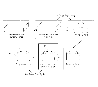
Axemolary Illethod of Manufacture
To obtain the implant meeting the criteria set forth above, the method of
manufacture
can comprise at least the following steps:
I.
preparation of the hydrogel/polymer portion 110, 120 of the implant 100, both
the solid portion 110 and the porous portion 120, preferably from a
CA 2910167 2020-01-13
CA 02910167 2015-10-22
WO 2014/176493
PCT/US2014/035442
interconnected sponge made of or containing a degradable or biodegradable
polymer;
2. preparation of the porous rigid base 130 by creating geometric features
such as
macropores and steps, and filling the geometric features, e,g., macropores,
with a non-biodegradable polymer;
3. assembling the implant 100 by placing the hydrogel portion 110, 120 onto
the
top surface of the porous rigid base portion 130 and cross-linking the
hydrogel
110, 120 to the polymer in the porous rigid base 130;
4. removal of the biodegradable or degradable polymer from the sponge in
the
hydrogel portion 110, 120 of the implant 100 to form macroporous network in
at least a portion of the hydrogel portion 120 of the implant 100; and
5. dehydration of the hydrogel portion 110, 120 of the implant 100.
Preparation of the Hydro gel Portion of the Implant
The preparation of the hydrogel/polymer portion 110, 120 of the implant 100
can be
manufactured by the method disclosed and claimed in co-owned U.S. Patent No.
U.S. Patent
No. 8,557,270.
The hydrogel portion 110, 120 of the implant 100 is preferably prepared using
an
interconnected sponge which is made of or contains a biodegradable polymer.
The sponge is
first hydrated and then the water is replaced with non-biodegradable polymer
solutions,
cross-linking the non-biodegradable polymer, coring the sponge, filling the
sponge with a
non-biodegradable polymer solution, and cross-linking the non-biodegradable
polymer in the
solid hydrogel (core). This process is generally shown in Figure 9.
Gelatin sponges, which are the preferred starting material, are sterile
absorbable
gelatin products used to control bleeding. They are available commercially,
from Ethicon-
Johnson & Johnson, Pfizer, Baxter, and Medtronic. The sponge can also be made
of or
contain other biodegradable polymers including, but not limited to, collagen,
poly(lactic
acid), poly(glycolic acid), chitosan, and alginate or degradable substance
such as salts and
polyethylene glycol.
Moreover the sponge's size, porosity and wall thickness can be varied
depending on
the needs of the final implant.
The sponge is hydrated by soaking it in deionized water for I hour to 5 days,
with
about 12 hours being preferred. A person of skill in the art would easily be
able to determine
a sufficient amount of time wherein the sponge is saturated with water.
21
CA 02910167 2015-10-22
WO 2014/176493
PCT/US2014/035442
The sponge is then centrifuged to remove the trapped air bubbles. The
preferred
method is at 3000 g for 1 hour at a time, 3-5 times, with gentle agitation
between the
centrifugations to restore, the original shape. However, a person of skill
could easily
determine the extent of centrifugation necessary to remove air bubbles from
the sponge.
Another technique is the intermittent application of a vacuum for 30 minutes
on and 30
minutes off, with agitation between the vacuum steps, for 3-5 times.
The next step in the method of the invention is replacement of the water in
the sponge
with poly(vinyl) alcohol or PVA. While PVA is preferred, any non-biodegradable
polymer
which has mechanical properties that can be controlled separately by varying
the polymer
concentration and/or the method of polymerization such as freeze/thawing can
be used.
The mechanical properties of the final device are determined by the final
concentration of the PVA in the device. Generally, the higher the final
concentration of PVA
in the device, the stiffer the device. A device with a higher concentration of
PVA can
generally withstand a higher load.
The PVA is substituted into the sponge under gentle agitation in steps of
increasing
concentration up to the desired concentration. PVA solutions of varying
concentration are
made and the sponges soaked until the desired concentration is obtained. The
PVA solutions
range from 1% to 40% weight/volume solutions, up to the desired final
concentration, with
the preferred final concentration of PVA scaffolds ranging from 10% to 40%.
The preferred
final concentration will depend upon the final use of the scaffold, as
determined by the person
of skill. The preferred method is to soak the sponge from about 1% to about 5%
PVA up to a
final concentration of 10% PVA.
The PVA hydrogels are then subject to a series of freeze/thaw cycles. PVA
offers the
advantage of being physically cross-linked using freeze/thaw cycles, without
the need for use
of potentially toxic cross-linking agents. During freezing, water freezes and
cause regions of
high PVA cross-links to form. As the PVA chains come in close contact with one
another,
crystallite formation and hydrogen bonding can occur between the chains. These
interactions
remain intact following thawing, and thereby create a three-dimensional
network. Thus, the
mechanical properties of the hydrogcl can be controlled by increasing the
number of
freeze/thaw cycles such that the amount of hydrogen bonding and crystallite
formation can be
increased. The increase in freeze/thaw cycles increases the strength of the
construct. The
mechanical properties can also be controlled by the duration and rate of
freezing and thawing.
22
CA 02910167 2015-10-22
WO 2014/176493
PCT/US2014/035442
The preferred method involves freezing the construct at about -20 C for about
20
hours and then thawing the construct at about 25 C for about 4 hours. However,
this part of
the process can be easily varied by the person of skill in order to vary the
mechanical
properties of the construct as desired. Both the number of hours of freezing
and/or thawing
can be varied as well as the number of cycles. For example, the total number
of freeze/thaw
cycles can range from 1 to 8. The construct can be frozen at each interval for
a time ranging
from 4 to 24 hours, with 20 hours being preferred. The thaw time can range
from 4 to 12
hours, with 4 hours being preferred.
While PVA is the preferred non-biodegradable polymer, and freeze/thawing the
preferred method for cross-linking the PVA, other non-biodegradable polymers,
and methods
known in the art to cross-link such polymers, can be used.
To obtain an implant with a solid hydrogel 110 in the center (a core), the
center of the
porous hydrogel is removed by any method known in the art. It is preferred
that a customized
centering jig as shown in Figure 10A and Example 1 is used. However, a
concentric cutting
die shown in Figure 10B can also be used. After the hydrogel material in the
center is
removed, it is filled with a liquid polymer and subjected to further cross-
linking, preferably
by additional freeze/thaw cycles. Again the number of freeze/thaw cycles can
range from 1
to 8, with 6 being preferred. The liquid polymer that can be used is chosen
from the group
comprising polyvinyl pyrrolidone, polyacrylamide, polyethylene glycol,
polyurethane, with
polyvinyl alcohol being preferred.
After the freeze/thaw cycles are performed, the hydrogel portion can be
trimmed to a
desired size depending on the size of the defect or injury being replaced,
repaired or treated.
The preferable thickness of the final hydrogel portion ranges from about 0.5
mm to about 7
mm thick.
To obtain an implant with alternating layers of porous and solid hydrogel, the
porous
hydrogel is made using the method set forth above, and cut into sections.
Additional polymer
is added to some of the strips and additional crosslinking is performed, to
obtain sections
comprising a solid hydrogel. Then sections or strips of porous hydrogel and
solid hydrogel
can be alternated and crosslinked together using 3 to 8 freeze/thaw cycles.
The alternating
porous and solid hydrogel can then be trimmed to the desired thickness and
length, with the
thickness preferably ranging from about 0.5 mm to 0.7 mm thick and the length
preferably
ranging from about 1 mm to 5 mm long.
23
CA 02910167 2015-10-22
WO 2014/176493
PCT/US2014/035442
Preparation of the Porous Rigid Base
The porous rigid base 130 can be manufactured to contain many different
features,
including but not limited to, a step at the hydrogel-base interface and
macroporous structures
to improve mechanical interlock between the two layers, and a taper on the
bottom of the
porous rigid base to allow alignment of the device with the defect. Figures 6A-
B and 7A-B
show these features discussed above.
Preferred material for the porous rigid base includes but is not limited to,
bone, metal,
polyetherketoneketone (PEKK), polyetheretherketone (PEEK), and bioactive glass
(e.g.,
silicone oxide, sodium oxide). This porous rigid base should contain
mieropores ranging from
about 150 to 500 in size
Macropores ranging from about 1% to 90% of the porous rigid base in diameter
and
from about 10% to 50% of the porous rigid base depth are created in the
surface of the porous
rigid base, which contains micropores, to further increase interdigitation
between the
hydrogel and the porous rigid base (Figure 6B). For a preferred embodiment of
the implant
for osteochondral defects, a single macropore with dimensions of 2 to 4 mm in
diameter and
1 to 5 ram in depth in the center of the implant can be used. Porous rigid
bases with multiple
macropores can also be created in the range of 1 to 2 mm in diameter and 1 to
3 min in depth.
Assembly of Implant,Itemoving the Collago Sponge and Dehydration
To create a robust interface that includes both the porous and non-porous
components
requires specific manufacturing and design specifications at that interface.
The macropores in the porous rigid base 130 are tilled with a liquid polymer
solution
ranging from about 5% to about 20% polymer in deionized water, Polymers that
can be used,
include but are not limited to, polyvinyl pyrollidone, polyacrylamide,
polyethylene glycol,
and polyurethane, with polyvinyl alcohol being preferred. The thin layer of
liquid polymer
used to fill the macropores is then injected across the entire porous base
using a syringe or
other device. The liquid polymer is then infiltrated into the pores by
pressurization.
Pressurization can be accomplished by either displacing a known volume of
polymer,
applying positive pressure (e.g., a known weight to force the polymer into the
porous rigid
base), or by using negative pressure (e.g., a vacuum). This improves the
interdigitation of the
hydrogel with the porous rigid base.
Next the solid-porous hydrogel is placed onto the top surface of the porous
rigid base
with the liquid polymer. The assembled implant was then subject to any method
that allows
the hydrogel portion and liquid polymer interface to cross-link. A preferred
method is
physical crosslinking such as freeze/ thaw cycling. See Figure 11.
24
CA 02910167 2015-10-22
WO 2014/176493
PCT/US2014/035442
The collagen sponge can then be removed from the hydrogel portion of the
implant by
any technique including but not limited to, enzymatic digestion, and the
entire implant is
dehydrated prior to sterilization and implantation, which results in unique
geometric changes
in the hydrogel layer discussed above. This process allows the stiffer,
dehydrated construct to
be securely inserted into the defect at the time of surgery, while also
ensuring that when the
implant rehydrates it will expand to fill the site of the defect.
Exemplary Method of Implantation
As discussed above, the mechanical function of the implant 100 is enhanced by
the
surface of the hydrogel being contiguous or slightly proud with the surface of
the adjacent
tissue where the implant is implanted. With this in mind, a method of
implantation was
devised to ensure that the surface of the hydrogel is properly aligned to the
surface of the
adjacent tissue. This method is as follows:
1. An alignment tool 200 (Figures 13A-D shows alignment tool 200 with inner
cannula 210 formed therein) is placed on the surface of the tissue surrounding
the
defect or injury. Such alignment tool 200 is preferably curved to match the
surface curvature of the tissue and is cannulatcd to allow a Kirschner wire
201 (K-
wire) to pass through the cannula 210 of the tool 200 and be inserted into the
tissue perpendicular to the tissue surrounding the defect or injury. Any
method
known in the art such as CT scans and MRI can be used to determine the surface
topography of the tissue to obtain the alignment tool 200 with the proper
curvature
to match the tissue.
2. The edges of the defect or injury are scored. Preferably a tool (see tool
300 of
Figure 14A) is made that can be shuttled over the K-wire in such a way that it
is
concentric to the K-wire. The tool is then used to score the tissue
surrounding the
defect or injury to create a circular clean edge. The cutting can also be used
to
determine the thickness of the tissue, such as cartilage and thus, determine
the
appropriate thickness of the hydrogel portion of the implant to be used in the
patient. Figures 14B and C show the scoring of the tissue using tool 300.
3. The tissue surrounding the defect or injury is drilled and the final depth
of the
defect or injury is measured (see Figures 15A-D showing the use of a reamer).
4. Based upon the two measurements, the size of the implant is chosen. The
implant
can optionally be partially rehydrated with an agent approximately 15 minutes
before implantation.
CA 02910167 2015-10-22
WO 2014/176493
PCT/US2014/035442
5. The implant 100 is inserted into a tool with a delivery tube 400 and the
delivery
tube 400 is place over or around the defect or injury, or vice versa. A rod
420 is
inserted into the delivery tube 400 and used to insert the implant 100 into
the
defect or injury by depressing the rod 420 into the tube 400. See Figures 16A-
E.
In the case of an osteochondral defect, not only is the final depth of the
defect
measure (i.e., the bone and the cartilage), the thickness of the cartilage is
also measured and
matched to the thickness of the hydrogel portion of the implant keeping in
mind the interface
interference and the change in size of the dehydrated versus rehydrated
implant as discussed
above.
Tissue Treatment, Repair and Replacement
The implant 100 of the present invention can be used to treat, replace or
repair defects
and/or injuries in various musculoskeletal tissues, in a subject in need
thereof, preferably a
mammal, and most preferably a human. Musculoskeletal tissue contemplated to be
treated,
replaced or repaired includes bone, tendon, ligaments, cartilage, meniscus,
and the discs of
the spine. Those of skill in the art would appreciate that the implants of the
present invention
may be implanted into a subject using operative techniques and procedures,
utilizing such
techniques as magnetic resonance imaging and computer guided technology.
The implant 100 of the present invention can also be used to treat, replace or
repair
defects and/or injuries in other biological tissue, including but not limited
to, arteries and
blood vessels, and organs.
Kits
The present invention also includes kits, which could include the device 100
of the
present invention, a tool for aligning (tool 200), a tool for cutting or
scoring (tool 300), a tool
(delivery tube 400) for insertion of the device 100 into the tissue,
additional agents that can
be added prior to implantation, and instructions for use, including
determining the correct
size of the implant and proper insertion.
For example, the device 100 of the present invention could be packaged in the
kit by
total defect depth and contain devices with different hydrogel heights ranging
from 0.5 mm to
5.0 mm hydrogel height in increments of 0.5 mm. Preferably the hydrogel
portion 110, 120
of the device 100 in the kit is dehydrated. The height of the porous rigid
base 130 can be
adjusted such that the total implant height remains constant for all devices
included in the kit.
The kit can include instructions for determining the correct size of the
hydrogel 110, 120
26
CA 02910167 2015-10-22
WO 2014/176493
PCT/US2014/035442
based upon the general parameters of the change in size when the hydrogel 110,
120 in
rehydrated.
The various tools to be included in the kit, e.g., alignment tool 200, cutting
or scoring
tool 300, and insertion tool 400, can be modeled after the ones used in
Example 7.
The agents that can included to add to the implant prior to insertion or
implantation
are discussed in detail above and include but are not limited to cytokines,
ehemokines,
chemoattractants, anti-microbials, anti-virals, anti-inflammatories, pro-
inflammatories, bone
or cartilage regenerator molecules, blood components, platelet rich plasma,
and as
combinations thereof, specific for the injury or defect being treated,
repaired, and/or replaced.
Examples
The present invention may be better understood by reference to the following
non-
limiting examples, which are presented in order to more fully illustrate the
preferred
embodiments of the invention. They should in no way be construed to limit the
broad scope
of the invention.
Example 1- Manufacture of the IIydrogel Portion of the Implant
All handling and fabrication techniques were performed aseptically to minimize
contamination with bacteria and other infectious agents.
A collagen sponge (Ethicon Surgifoam, Ref 1974) was soaked in deionized water
overnight until the entire sponge was saturated with water via capillary
action. The sponges
were transferred to 50 mL conical tubes and repeatedly centrifuged at 3000g
for I hour at a
time, with gentle agitation of the tube between centrifugations to restore its
original shape,
until all remaining air bubbles had been removed.
The sponge was then impregnated through increasing gradients of liquid
polyvinyl
alcohol (PVA) from 1% to 5%, up to the desired final concentration of 10% PVA.
The
impregnated collagen sponge was then subjected to one freeze/thaw cycle (20
hours at -
200C/4 hours at 25 C).
Next the frozen impregnated sponge was cored using a cutting die, and the
center of
each core was removed using a cutting die and discarded. To ensure that the
removed core is
concentric with the outside walls, a customized centering jig was used as
shown Figure 10A.
The center of the cylinder was filled with a liquid polymer (20% PVA) and
subjected to
another 6 freeze/thaw cycles.
27
CA 02910167 2015-10-22
WO 2014/176493
PCT/US2014/035442
After the freeze/thaw cycles the hydrogel portion of the implant was trimmed
using a
freezing-stage sledge microtome to the desired thickness.
Example 2- Manufacture of the Porous Rigid Base Portion of the Implant
A titanium (Ti6A14V or Ti6A14VELI) cylinder with a diameter of 9 mm and pores
of
about 150 to 500 m in size and a 3.8% taper at the bottom, was drilled with
one additional
hole (1.3 mm diameter and 4.5 mm deep) at the top surface of the base. A 0.5
mm step was
also created. These bases were designed using computer aided design and
created using
techniques such as electron beam melting or by laser metal sintering.
Example 3- Assembly of the Implant
As illustrated in Figure 11, the maeropores in the porous metal base from
Example 2
were filled with a low viscosity PVA solution (10% or 1.33 molar PVA in
deionized water),
and a volume of 50 EL of 20% (or 2.67 molar) PVA was injected across the
entire surface of
the porous metal using a syringe. Positive pressure was applied using a known
weight of 500
grams over 30 seconds to drive a 20% liquid PVA solution into the pores of the
porous rigid
base. The weight was removed and the solid-porous hydrogel from Example 1 was
placed
onto the top surface of the porous metal. The assembled implant was then
subjected to 3
freeze/thaw cycles (-20 C for 20 hours/40 C for 4 hours) to crosslink the
hydrogel portion
and the liquid polymer interface. The collagen sponge in the porous periphery
was removed
by digestion using bacterial collagenase (Collagenase Type 2, Worthington
Biochemical
Corporation) for 16 hours to create the interconnected porous hydrogel
structure in the
hydrogel periphery portion of the implant.
After collagenase digestion, the completed implants were washed at least 5
times (10
minutes each) on a rocker with deionized water and then placed in 100% ethyl
alcohol for at
least 1 hour in order to dehydrate the hydrogel portion. The implants were
then removed
from the alcohol solution and allowed to air dry for at least 4 hours at room
temperature
under laminar air flow.
.. Example 4- Changes in Elvdrogel Size after Dehydration
Hydrogels prepared as set forth in Example 1 were dehydrated as set forth in
Example
3. The total hydrogel, the solid hydrogel (core) and the porous periphery or
edge was
measured both in diameter and in thickness. The change in hydrogel diameter
after
dehydration showed about a 44% decrease in diameter at the top surface and
about a 31%
28
CA 02910167 2015-10-22
WO 2014/176493
PCT/US2014/035442
decrease in diameter at the bottom surface from the initial size of the
hydrogel (Figure 3A).
There was a smaller change in the height of the hydrogel with about a 22%
decrease in
thickness from the pre-dehydrated thickness (Figure 3B).
Example 5- Rehydration Times of Solid Hydrogel and Porous Periphery of the
Hydrogel
Implants made according to Example 1 and dehydrated as in Example 3 were
rehydrated by soaking the implants in phosphate buffered saline solution at
room
temperature. The diameter and the thickness of the entire hydrogel and the
solid hydrogel
(core) were measured initially, after dehydration, 15 minutes, 1 hour, 2
hours, 6 hours, and 4
days post rchydration. As shown in Figure 4A, the solid hydrogel (core) and
porous hydrogel
periphery of the implants rehydrated at different rates with the solid
hydrogel rehydrating in
about 2 hours and the porous hydrogel periphery fully rehydrating in about 1
hour. Little
difference is seen in the thickness of the hydrogel during rehydration (Figure
4B).
Example 6- Implantation of the Implant
The implant manufactured using Examples 1-3 was implanted into the trochlear
groove of a horse, using the following method:
The alignment tool 200 shown in Figure 13A was used to place a Kirschner wire
201
(K-wire) perpendicular to the surface of the cartilage surrounding the defect
(see Figure 13C).
The surface of the alignment tool was curved to match the surface curvature of
the cartilage.
The alignment tool was cannulated to allow the K-wire to pass through the
guide and be
driven perpendicular to the surface of the cartilage (see Figure 13B). The
tool 200 thus
contains a central cannula (lumen) 210 formed therein. CT scans were used to
determine the
surface topography of the trochlear groove of the horse and matched the
surface of the tool to
the curvature of the cartilage in the groove.
A cartilage scoring tool 300 (Figure 14A) was used to score edges of defect.
The
cartilage scoring tool was cannulated to maintain perpendicular alignment with
the cartilage
surface by fitting concentrically over the k-wire. The cartilage scoring tool
300 was used to
score the edges of the cartilage to create clean edges around the defect
(Figures 14B and C).
Also by scoring the cartilage to the bone surface, the thickness of the
cartilage was effectively
measured and the appropriate thickness of the hydrogel region of the implant
that should be
used in the patient was determined. A 9 mm diameter half-moon reamer 305
(Arthrex,
Catalogue #: 031247) was placed over the K-wire and drilled to the desired
depth (Figure
15A). Figure I5A shows an arcuate line that depicts the previous scored
surface (see
29
CA 02910167 2015-10-22
WO 2014/176493
PCT/US2014/035442
discussion above with respect to Figures 14A-C). For the large animal study
done in this
example, the depth was maintained at approximately 10 mm from the surface of
the cartilage.
The K-wire was removed and the defect was cleared of any debris (Figures 15B
and C). A 9
mm diameter measuring instrument 307 was used to measure the final depth of
the defect
(Figure 15D).
Based on the depth of the defect, an implant with the same height as the
defect depth
was chosen. A delivery tube 400 (see Figures 16C-D) was placed over the
defect, and
visually aligned using the cutout windows 410 at the distal tip. The implant
100 was then
inserted into the end of the delivery tube 400 and an insertion rod 420 was
then used to insert
the implant 100 into the defect until the hydrogel surface 110, 120 was flush
with the
cartilage surface (Figure 16E).
Example 7- The Integration between the nydrogel and Porous Rigid Base Resist
Forces
The integration between the hydrogel and the porous rigid base of the
preferred
implant design for osteochondral defects manufactured as set forth in Examples
1-3 was
tested in both shear and tension. For testing the integration in shear, the
porous rigid base and
hydrogel were fixed as shown in Figure 8A. The shear head was moved at a rate
of 0.03
mm/sec to induce shear at the interface of the porous rigid base and the
hydrogel. Using this
method, the ultimate shear stress at the hydrogel and porous rigid base was
determined to be
0.4 MPa.
To determine the integration strength of the implant in tension, the implant
was fixed
as shown in Figure 8B. The loading platen was displaced at a rate of 0.03
mm/sec and the
tensile stress required to separate the hydrogel and porous rigid base was
found to be 0.22
MPa.
The implant withstood forces that would be expected for use in treatment,
repair and
replacement of osteoehondral defects.
Example 8- Implants can Restore Normal Mechanical Function
The implants made according to Examples 1-3 were tested in human cadaver
knees.
The implant was inserted using the instrumentation designed and using the
technique for
implantation described in Example 6. The implants were placed in the defect
flush to about
0.5 mm proud to the surface of the adjacent articular cartilage in the
cadaveric knees.
Electronic stress sensors (Tekscan, Inc, South Boston MA) were placed under
the
meniscus of the cadaveric knees on top of the tibial plateau to measure the
stress on the
CA 02910167 2015-10-22
WO 2014/176493
PCT/US2014/035442
surface of the tibial cartilage. The cadaveric knee joints were placed on a
Stanmore Knee
simulator and subjected to simulate level walking while intact and with a
created defect. The
defect was then filled with completed implants as described in Examples 1-3
containing solid
PVA cores with elastic modulus ranging between 50 kPa to 500 kPa and 0.5 mm
proud from
.. the surface of the cartilage. Using this embodiment of the implant
manufactured to treat,
repair or replace osteochondral defects, the contact stress on the cartilage
surface showed that
the devices were able to restore normal joint loading within 10% of the intact
condition
having an elastic modulus of 100 kPa. See Figure 12.
REFERENCES
1. Bekkers, J. E., et al. (2009). "Treatment selection in articular cartilage
lesions of
the knee: a systematic review." Am J Sports Med 37 Suppl 1: 148S-155S.
2. Buckwalter, J. A. and H. J. Mankin (1998). "Articular cartilage: tissue
design and
chondrocyte-matrix interactions." 1nstr Course Lect 47: 477-486.
3. Choi, K., et al. (1990) "The elastic moduli of human subchondral,
trabecular, and
cortical bone tissue and the size-dependency of cortical bone modulus." J.
Biomech 23(11):1103-13.
4. Cole, B. J. and S. J. Lee (2003). "Complex knee reconstruction: articular
cartilage
treatment options." Arthroscopy 19 Suppl I: 1-10.
5. Deneweth, J.M., etal. (2013) "Heterogeneity of tibial plateau cartilage
in response
to a physiological compressive strain rate." J Orthop Res 31(3):370-5.
6. Magnussen, R. A., et al. (2008). ''Treatment of focal articular cartilage
defects in
the knee: a systematic review." Clin Orthop Relat Res 466(4): 952-962.
7. Mauck, R. L., et al. (2002). "Influence of seeding density and dynamic
deformational loading on the developing structure/function relationships of
chondrocyte-seeded agarose hydrogels." Ann Biomed Eng 30(8): 1046-1056.
31
CA 02910167 2015-10-22
WO 2014/176493
PCT/US2014/035442
8. Ng, K.W., et al. (2012) "A novel macroporous polyvinyl alcohol scaffold
promotes chondrocyte migration and interface formation in an in vitro
cartilage
defect model." Tissue Eng Part A 18(11-12): 1273-81.
9. Raclin, EL., et al. (1970) "A comparison of the dynamic force transmitting
properties of subchondral bone and articular cartilage." J Bone Joint Surg Am
52(3):444-56.
10. Shelbourne, K. D., et al. (2003). "Outcome of untreated traumatic
articular
cartilage defects of the knee: a natural history study." J Bone Joint Surg Am
85-A
Suppl 2: 8-16.
32