Note: Descriptions are shown in the official language in which they were submitted.
STABILIZATION APPARATUSES AND METHODS FOR MEDICAL PROCEDURES
FIELD OF THE INVENTION
The present invention generally relates to apparatuses for medical procedures,
and
methods of use thereof.
BACKGROUND
The following description includes information that may be useful in
understanding the
present invention. It is not an admission that any of the information provided
herein is prior art
or relevant to the presently claimed invention, or that any publication
specifically or implicitly
referenced is prior art.
When physicians are performing procedures on or around certain areas of the
body such
as the spinal cord, brain, and joints, very precise, controlled, and stable
manipulations are often
required to avoid patient injury and to optimize outcome. There is a need in
the art for
apparatuses and methods that will improve the safety and accuracy of
performing certain medical
procedures in those areas.
More specifically, certain medical procedures performed by physicians are
associated
with especially high risks of accidental patient injury and/or treatment
failure, due to a
combination of the nature of the tissues involved in the procedure, the high
degree of accuracy
demanded by the procedure, limitations of existing surgical instruments
(including stabilizing
apparatuses), limitations associated with the field of view, and human error.
In order to increase
the likelihood of a favorable outcome, a number of attempts have been made to
improve upon
the stabilizing apparatuses used in conjunction with a number of medical
instruments for a
1
Date Recue/Date Received 2020-08-17
CA 02910268 2015-10-23
WO 2014/179458 PCMJS2014/036161
variety of different surgical procedures, including those involving the
introduction of a substance
into or removal of a substance from a delicate area of a patient's body.
Exemplary stabilizing
apparatuses known in the art include the Spinal Derrick, the Warner Device,
and the Brundobler
Device. Unfortunately, these devices are all either difficult to use
(requiring a large amount of
physician training), have an excessive part count (thereby carrying a
relatively high risk of
equipment failure or patient injury), or have significant problems related to
positioning. For
example the Spinal Derrick device used for spinal surgery comprises over 50
parts, making its
assembly long and difficult, and leading to an increased risk of one of its
parts falling into the
incision and causing spinal cord trauma. Additionally, this device lacks
accurate scales, and
requires the use of four percutaneous posts that are placed "blindly," further
increasing the risk
of spinal cord injury, infection, and bleeding (partly due to the four
additional incisions
required).
SUMMARY OF THE INVENTION
In various embodiments, the invention teaches an apparatus that includes a
securing arm
that includes a first end, a second end, a long axis, and a short axis; a
connecting arm that
includes a first end, a second end, a long axis, and a short axis; a
positioning arm that includes a
first end, a second end, a long axis, and a short axis; and a guiding aim that
includes a first end, a
second end, a long axis, and a short axis; wherein (1) the first end of the
connecting arm is
attached to the second end of the securing arm, (2) the second end of the
connecting arm is
attached to the first end of the positioning aim, (3) the long axis of the
connecting arm is
perpendicular to the long axis of each of the securing arm and positioning
arm, (4) the first end
of the securing arm and the second end of the positioning arm can be
positioned to extend in
substantially the same direction away from the connecting arm, (5) the
positioning arm is
attached at its second end to the second end of the guiding arm, such that the
positioning arm and
guiding arm are perpendicular to one another, and (6) the guiding arm can be
positioned such
that the axis along which its long axis is situated is perpendicular to but
does not intersect with
the axes along which the long axis of the securing arm and the long axis of
the connecting arm
are respectively situated. In some embodiments, the securing arm further
includes one or more
2
CA 02910268 2015-10-23
WO 2014/179458 PCMJS2014/036161
clamps on its first end, and the one or more clamps are configured to attach
to an arm of a tissue
retractor. In some embodiments, the guiding arm further includes an instrument
attaching
component configured to slide along the long axis of the guiding arm. In some
embodiments, the
instrument attaching component includes one or more clamps configured to clamp
a medical
instrument. In certain embodiments, the sliding motion of the instrument
attaching component is
controlled by a dial situated at the first end of the guiding arm. In some
embodiments, the
connecting arm includes elongated nesting elements that allow for telescoping
motion in the
direction of its long axis, such that the length of the connecting arm can be
increased or
decreased. In certain embodiments, the positioning arm includes elongated
nesting elements that
allow for telescoping motion in the direction of its long axis, such that the
length of the
positioning arm can be increased or decreased. In some embodiments, the
telescoping motion of
the connecting arm is controlled by rotation of a dial situated at its second
end. In certain
embodiments, the telescoping motion of the positioning arm is controlled by
rotation of a dial
situated at its first end. In certain embodiments, the medical instrument is
selected from the
group consisting of: a cannula, a biopsy needle, a needle, a tube, a
cauterization device, a laser, a
drill, an endoscope, a guidewire, a fiberoptic device, an electrode, a saw, an
ultrasonic device, a
spectroscopic device, a camera, an electrical sensor, a thermal sensor, a
catheter, a draining tube,
and combinations thereof. In some embodiments, the apparatus further includes
a side clamp
attached to the securing arm, wherein the side clamp is configured to attach
to an elongated
object. In some embodiments, the securing arm is removably attached to the
connecting arm. In
various embodiments, the positioning arm is removably attached to the
connecting arm and/or
the guiding arm. In some embodiments, the side clamp is removably attached to
the securing
arm. In certain embodiments, the elongated object is a device selected from
the group consisting
of: a liquid reservoir, a gas reservoir, a pump, an imaging device, and
combinations thereof.
In various embodiments, the invention teaches a system. In some embodiments,
the
system includes any apparatus described above and a tissue retractor attached
to the securing atm
of the apparatus by one or more clamps of the securing arm. In some
embodiments, the system
further includes an instrument attached to the instrument attaching component,
wherein the
instrument is selected from the group consisting of: a cannula, a biopsy
needle, a needle, a tube,
3
CA 02910268 2015-10-23
WO 2014/179458 PCMJS2014/036161
a cauterization device, a laser, a drill, an endoscope, a guidevvire, a
fiberoptic device, an
electrode, a saw, an ultrasonic device, a spectroscopic device, a camera, an
electrical sensor, a
thermal sensor, a catheter, a draining tube, and combinations thereof In some
embodiments, the
instrument includes a cannula with a needle situated at the end thereof In
some embodiments,
the cannula and needle are configured to inject cells into a region of
interest in a subject's body.
In various embodiments, the cannula contains a quantity of neural progenitor
cells. In some
embodiments, the neural progenitor cells express glial cell line derived
neurotrophic factor. In
certain embodiments, the region of interest is the subject's spine. In some
embodiments, the
system further includes a liquid reservoir and a pump connected thereto,
wherein the liquid
reservoir and pump are attached to the side clamp.
In various embodiments, the invention teaches a method for performing a
surgical
procedure on a subject. In some embodiments, the method includes attaching any
apparatus
described herein above to an arm of a tissue retractor that is engaged in an
incision in the
subject's body, and guiding a medical instrument attached to the guiding arm
of the apparatus
through the incision in the subject's body. In certain embodiments, the
medical instrument is a
cannula with a needle situated at the end thereof. In some embodiments, the
cannula and needle
are configured to inject cells into a region of interest in the subject's
body. In some
embodiments, the region of interest is the subject's spine. In some
embodiments, the cells are
neural progenitor cells. In some embodiments, the subject has been diagnosed
with amyotrophic
lateral sclerosis (ALS). In various embodiments, the method further includes
performing
imaging of the region of interest in the subject's body. In some embodiments,
the imaging
performed is selected from the group consisting of computed tomography (CT),
magnetic
resonance imaging (MRI), ultrasound, and combinations thereof. In some
embodiments, the
method further includes injecting neural progenitor cells expressing glial
cell line derived
neurotrophic factor into the subject's spine.
BRIEF DESCRIPTION OF THE DRAWINGS
Exemplary embodiments are illustrated in the referenced figures. It is
intended that the
embodiments and figures disclosed herein are to be considered illustrative
rather than restrictive.
4
CA 02910268 2015-10-23
WO 2014/179458 PCMJS2014/036161
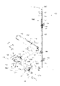
Figure 1A depicts, in accordance with an embodiment of the invention,
stereotactic
apparatus 100. Stcreotactic apparatus 100 is clamped to arm 301 of tissue
retractor 300.
Cylindrical object 400 is fastened to stereotactic apparatus 100 by side clamp
6000. Figure 1B
depicts stcrcotactic apparatus 100 without attachment to a tissue retractor.
Figure 1C depicts
stereotactic apparatus 200. Figure ID depicts stereotactic apparatus 100
attached to cylindrical
object 400 and tissue retractor 300. Instrument 7000 is shown attached to
guiding arm 1000 of
stereotactic apparatus 100, and extending downward along the z-axis between
the arms of tissue
retractor 300.
Figure 2A depicts, in accordance with an embodiment of the invention,
stereotactic
apparatus 100. Tissue retractor 300 and cylindrical object 400 are shown.
Figure 2B depicts an
alternate view of stereotactic apparatus 100. Figure 2C depicts an alternate
view of stereotactic
apparatus 200.
Figure 3 depicts, in accordance with an embodiment of the invention, a
partially exploded
view of stereotactic apparatus 100.
Figure 4 depicts, in accordance with an embodiment of the invention, a
partially exploded
view of stereotactic apparatus 100.
Figure 5 depicts, in accordance with an embodiment of the invention, loosening
knob 114
allows for adjustment of the position of positioning aim 2000 along the x-
axis.
Figure 6 depicts, in accordance with an embodiment of the invention, loosening
screw
135 allows for adjustment of the position of positioning arm 2000 along the y-
axis.
Figure 7 depicts, in accordance with an embodiment of the invention, loosening
knob 130
allows for adjustment of the position of cylindrical object 400 along the x-
axis.
Figure 8 depicts, in accordance with an embodiment of the invention, loosening
of knob
114 allows for rotation of positioning arm 2000 around the x-axis and
associated motion of
guiding arm 1000 along the y-z plane.
Figure 9 depicts, in accordance with an embodiment of the invention, loosening
screw
135 allows for rotation of cross clamp 132 around the y-axis, and associated
motion of guiding
arm 1000 along the x-z plane.
5
CA 02910268 2015-10-23
WO 2014/179458 PCMJS2014/036161
Figure 10 depicts, in accordance with an embodiment of the invention, rotating
dial 116
causes telescoping of inner nesting element 112 of positioning arm 2000.
Figure 10 also shows
rotating dial 101 causes motion of instrument attachment component 107 along
the z-axis.
Figure 11 depicts, in accordance with an embodiment of the invention, rotating
dial 131
.. causes telescoping motion of inner nesting element 119 of connecting arm
3000.
Figure 12 depicts, in accordance with an embodiment of the invention, a
partially
exploded view of connecting arm 3000. Arrows labeled "14A" indicate the cross
section
represented in Figure I 4A.
Figure 13 depicts, in accordance with an embodiment of the invention, an
exploded view
.. of a portion of connecting arm 3000.
Figure 14A depicts, in accordance with an embodiment of the invention, a cross-
sectional
view of the long axis of connecting arm 3000. Figure 14B depicts a cross-
sectional view of the
short axis of connecting arm 3000.
Figure 15 depicts, in accordance with an embodiment of the invention, a
partially
.. exploded view of positioning arm 2000. Arrows labeled "17A" indicate the
cross section
represented in Figure 17A.
Figure 16 depicts, in accordance with an embodiment of the invention, a
partially
exploded view of a portion of positioning arm 2000.
Figure 17A depicts, in accordance with an embodiment of the invention, a cross-
sectional
.. view of the long axis of positioning arm 2000. Figure 17B depicts, in
accordance with an
embodiment of the invention, a cross sectional view of the short axis of
positioning arm 2000.
Figure 18 depicts, in accordance with an embodiment of the invention, an
exploded view
of guiding min 1000. Arrows labeled "19- indicate the cross section
represented in Figure 19.
Figure 19 depicts, in accordance with an embodiment of the invention, a cross-
sectional
.. view of the long axis of guiding am 1000.
Figure 20 depicts, in accordance with an embodiment of the invention, an
exploded view
of side clamp 6000, and it's attachment to securing arm 4000.
Figure 21 depicts, in accordance with an embodiment of the invention, an
alternate
exploded view of securing arm 4000.
6
Figure 22 depicts, in accordance with an embodiment of the invention, side
clamp 6000.
Figure 23 depicts, in accordance with an embodiment of the invention, scales
381, 382,
and 383 on device 300. Device 300 is identical to device 100 with respect to
all other features.
DETAILED DESCRIPTION OF THE INVENTION
Unless defined otherwise, technical and scientific terms used herein have the
same
meaning as commonly understood by one of ordinary skill in the art to which
this invention
belongs. Szycher's Dictionary of Medical Devices CRC Press, 1995, may provide
useful
guidance to many of the terms and phrases used herein. One skilled in the art
will recognize
many methods and materials similar or equivalent to those described herein,
which could be used
in the practice of the present invention. Indeed, the present invention is in
no way limited to the
methods and materials specifically described.
In some embodiments, properties such as dimensions, shapes, relative
positions, and so
forth, used to describe and claim certain embodiments of the invention are to
be understood as
.. being modified by the term "about."
With the aforementioned shortcomings of previously existing technologies in
mind, the
inventors developed novel stabilizing apparatuses and methods of use thereof.
While one of skill
in the art would readily appreciate that there are many possible applications
of the apparatuses
described herein, certain embodiments are especially useful for procedures
performed on or
.. around the spinal cord, including delivery of cutting edge cellular and
molecular therapies
thereto. Importantly, all versions of the devices described herein also render
the use of
percutaneous posts unnecessary and therefore allow for a minimally invasive
surgical approach.
Although numerous embodiments of stereotactic apparatuses are described
herein, there
are certain features common to all of them. First, each apparatus includes one
or more
components that make up a "securing section" capable of stably connecting to
an arm of a tissue
retracting device. The second feature common to each of the apparatuses
described herein is a
"positioning section," which includes one or more components capable of
positioning an
instrument over a desired location in a subject's body. The third common
feature is a
7
Date Recue/Date Received 2020-08-17
CA 02910268 2015-10-23
WO 2014/179458 PCMJS2014/036161
"connecting section," which serves to operably connect the positioning section
and the securing
section. A fourth common feature is a "guiding section," which can be used to
guide an
instrument into or remove an instrument from a subject's body.
Provided below are descriptions of various components, combinations of
components,
and configurations of components relative to one another that can be used to
arrive at each of the
common sections described above. Additional features that can be added to the
stereotactic
apparatus are also described.
Securing Section
In some embodiments, the securing section of the stereotactic apparatus is
configured to
removably attach to an arm of a tissue retractor. Removable attachment can be
accomplished in
any of a number of ways, using a wide range of components and combinations
thereof. Merely
by way of non-limiting examples, the securing section could attach to the arm
of a tissue
retractor by using one or more clasps, one or more clamps, one or more
magnets, one or more
screws, one or more pins, one or more slot and groove arrangements, one or
more straps,
combinations thereof and the like. Therefore, each of these components, and
modified versions
thereof, are within the scope of the invention. It is further contemplated
that the attaching
portion of the apparatus could be configured to attach to any of a variety of
types of equipment
that might be found in a setting in which a medical procedure is perfoinied,
including, but in no
way limited to a table, a lamp, a brace. a tray, imaging equipment, and the
like. It is also
contemplated that the device could be configured for use in a non-surgical
setting, in which it
may be used to perform any objective that requires the use of precision
guidance. It is also
contemplated that the device could be scaled appropriately for such
objectives.
In some embodiments, a clamping mechanism is incorporated on the securing arm,
and
used to attach the stereotactic apparatus to the arm of a tissue retractor.
One of skill in the art
would readily appreciate that numerous types of clamping mechanisms are
suitable to
accomplish this function. One non-limiting example is depicted in Figure 3,
which shows
clamping mechanism 5000 of securing arm 4000 can be used to clamp arm 301 of
tissue retractor
300 (partially shown). A more detailed view of the clamping components of this
particular
8
CA 02910268 2015-10-23
WO 2014/179458 PCMJS2014/036161
embodiment is shown in Figure 21, and the individual components (and their
functions) are
thoroughly described in the examples section.
Importantly, the clamping mechanism shown in Figure 21 can he used to securely
and
removably attach a stereotactic apparatus (including stereotactic apparatus
100) to the arm of a
number of different types of tissue retractors. Non-limiting examples of
retractors to which the
clamping mechanism can attach include the Mast Quadrant Retractor System
(Medtronic), the
MARS Retractor System (Globus Medical), the Spyder Retractor System
(Aesculap), the Ravine
Retractor System (K2M), the Synframe Retractor System (DePuy Synthes), and the
Luxor
Retractor System (Stryker). One of skill in the art would readily appreciate
that any retractor
with one or more arms similar to those retractors described above could also
be used in
conjunction with the inventive stereotactic apparatuses described herein. One
of skill in the art
would further appreciate that the alternative attaching mechanisms described
above would allow
for the attachment of the securing section of an apparatus to one or more arms
of alternative
retractor devices that are not specifically listed above.
Positioning Section
The purpose of the positioning section is to allow for stable positioning of
an instrument
over a desired anatomical location, by positioning a guiding arm to which the
instrument is
attached. One of skill in the art would readily appreciate that there are many
possible
components and configurations thereof that could make up a positioning section
of the
stereotactic apparatus. In certain embodiments the positioning section
includes components that
allow for telescoping motion, which permits fine adjustment of the position of
the instrument
attached to the guiding arm. In some embodiments, a positioning arm is used.
In various
embodiments, the positioning arm includes two or more nested elements that are
operably
connected to one another as well as an input component (e.g., a dial) in a
manner that allows for
telescoping motion. In a non-limiting example, the telescoping motion is
accomplished by the
components depicted in Figures 15-17. The interaction between and operation of
the
components of Figures 15-17 are thoroughly described in the examples section.
9
CA 02910268 2015-10-23
WO 2014/179458
PCT11JS2014/036161
One of skill in the art would readily appreciate that there are numerous
possible ways of
stabilizing and controlling the telescoping motion of the positioning arm.
Merely by way of non-
limiting example, if a mechanism with a threaded shaft is used, as depicted in
Figures 15-17, the
number of threadings on the shaft and the pitch of the threadings can he used
to dictate the
degree to which the positioning arm telescopes in response to associated input
(e.g. rotation of a
dial). In certain embodiments, the positioning arm is stabilized through the
use of components
that limit its range of motion in all hut the axis along which it is advanced
or retracted. Merely
by way of non-limiting example, Figure 16 shows the configuration of guiding
set screws 176a
and 176b and supporting elements 178a and 178b is used to apply pressure on L-
shaped tracks
179a and 17911 of inner nested element 112 of positioning arm 2000. Figure 16
also shows that
screw 175 is positioned on the opposite side of set screws 176a and 176b, in
order to add to the
stability of inner nested component 112, especially while it is being extended
or retracted.
One of skill in the art would readily appreciate that there are many possible
ways of
attaching the positioning arm to the guiding arm. As shown in Figure 3, one
way positioning
arm 2000 can he connected to guiding arm 1000 is through the use of screw 133
that traverses
the short axis of guiding arm 1000 and connects to grooved receiving socket
134.
Connecting Section
The long axis of the connecting section of the stereotactic apparatus can be
configured to
be perpendicular to the long axis of the securing section and the positioning
section. In some
embodiments, the connecting section, like the positioning section, is a
telescoping arm. In some
embodiments, the telescoping connecting arm can be stabilized and controlled
by any of the
aforementioned components associated with the positioning section. Merely by
way of non-
limiting example, telescoping of the connecting arm can be accomplished
through the use of the
components shown in Figures 12-14, the interaction between which and function
of which are
thoroughly described in the examples section.
Guiding Section
The guiding section can be configured to allow for the attachment of one or
more
instruments that can be extended into and retracted from a subject's body. In
some
embodiments, the guiding section includes a guiding arm. There are many
possible ways by
which an instrument can be attached to a guiding arm. One of skill in the art
would readily
appreciate that the possible components that could be used to attach an
instrument to a guiding
arm would vary depending upon the dimensions and nature of the instrument to
be attached.
Merely by way of non-limiting examples, attachment of various instruments to
the guiding arm
can be accomplished by using one or more straps, clamps, clasps, magnets, and
combinations
thereof.
Examples of instruments that could be attached to the guiding arm include, but
are in no
way limited to a cannula, a biopsy needle, a needle, a tube, a cauterization
device, a laser, a drill,
an endoscope, a guidewire, a fiberoptic device, an electrode, a saw, an
ultrasonic device, a
spectroscopic device, a camera, an electrical sensor, a thermal sensor, a
catheter, a draining tube,
an imaging device (such as any of those listed and/or described herein) and
the like. In certain
embodiments, the instrument guided by the inventive apparatuses described
herein includes a
guide needle and an injection needle configured to be concentrically housed
therein. In some
embodiments, the concentric arrangement of the guide needle and the injection
needle allows the
injection needle to be advanced through the guide needle, once the guide
needle is properly
positioned in a subject during a medical procedure, so that the injection
needle can deliver a
payload of biological or chemical material to an appropriate site in the
subject. In some
embodiments, the instrument guided and/or stabilized by the inventive
apparatus is the spinal
multisegmental cell and drug delivery device described in U.S. Patent
Application No.
12/598,667.
One of skill the art would also readily appreciate that there are numerous
possible ways
by which the apparatus can be configured to allow for an instrument to be
extended into and
retract from a subject while connected to the guiding arm. Figure 18 depicts
one non-limiting
example of a mechanism that can be used for that purpose. The association
between the
components shown in Figure 18 and the function of those components are
thoroughly described
in the examples section.
11
Date Recue/Date Received 2020-08-17
CA 02910268 2015-10-23
WO 2014/179458
PCT/1JS2014/036161
Orientation of Individual Sections
The securing section, connecting section, positioning section and guiding
section can be
connected to one another by any of a variety of ways depending upon the
desired range of
motion of each section. In some embodiments, a perpendicular orientation of
the positioning
arm and connecting arm, relative to one another, is established through the
use of a component
with perpendicularly situated clamping collars. In an embodiment, cross clamp
132 (depicted in
Figure 1A) can be used. As shown in Figure 5, when cross clamp 132 is used to
secure
positioning arm 2000, knob 114 can be rotated to loosen collar 115, thereby
allowing for
adjustment of the position of positioning arm 2000 along the x-axis. As shown
in Figure 8,
loosening of collar 115 by rotating knob 114 also allows for rotation of
positioning arm 2000
along the x-axis, which translates into motion of guiding arm 1000 along the y-
z plane.
As shown in Figure 6, when cross clamp 132 is used to secure connecting arm
3000,
rotation of screw 135 loosens lower collar 117, which allows for adjustment of
the position of
positioning arm 2000 along the y-axis. As shown in Figure 9, loosening collar
117 also allows
for rotation of cross clamp 132 along the y-axis, which in turn translates
into motion of guiding
arm 1000 along the x-z plane.
Additional Features
The main sections of the stereotactic apparatuses described above can be
configured to
allow for incorporating additional features on the apparatuses. For example,
the stereotactic
apparatus can include clamps (or any other means of attachment described
herein) situated on
one or more of the main sections of the apparatus (i.e. guiding section,
positioning section,
connecting section, and attaching section) for attaching additional useful
instruments or devices.
In certain embodiments, the stereotactic apparatus includes a side clamp
attached to the
securing section, which allows for attaching a useful instrument or device.
For example, as
demonstrated in Figure 3, side clamp 6000 can be used to hold cylindrical
device 400. The
components of side clamp 6000 are clearly shown in Figure 22, and thoroughly
described in the
examples section. One of skill in the art would readily appreciate that a side
clamp such as side
12
CA 02910268 2015-10-23
WO 2014/179458 PCMJS2014/036161
clamp 6000 can be used to attach any of a number of devices with appropriate
dimensions to the
stereotactic apparatus.
Devices that can be attached to the stereotactic apparatuses described herein
can include,
but are in no way limited to, a pump, a reservoir for containing a substance
to be injected into a
subject's body, a reservoir for receiving a substance removed from a subject's
body, a small
motor, a control panel, an imaging device or portion thereof (including any
appropriately sized
imaging device described herein) and the like. In some embodiments, the device
attached is a
fiber optic camera that can be positioned to view an opening in a patient's
body in which a tissue
retractor is engaged. In some embodiments, a reservoir attached to the
apparatus can be
configured to hold any of a variety of useful substances, including but in no
way limited to cells,
gasses, liquids, medications, contrast agents, radioactive materials,
combinations thereof, and the
like.
An additional category of devices that could be attached to one or more
sections of the
inventive apparatuses described herein is a light source. In various
embodiments, the inventive
.. apparatuses may include one or more light sources configured to project
light onto a region of
interest on or in a subject's body during a medical procedure. In some
embodiments, one or
more of the light sources is attached to the guiding arm. In some embodiments,
the light source
is a laser. In some embodiments, the light source is a relatively high energy
laser that can be
used for cauterizing or cutting. In some embodiments, the light source is a
relatively low energy
laser that can be used for visually targeting a region on or in a subject's
body for incision or
other medical intervention. In other embodiments, the light source provides
relatively low
energy light for aiding in visualizing a region of interest. In still other
embodiments, the light
source provides light of a wavelength that causes fluorescence of a
fluorophore. In various
embodiments, the fluorophore is introduced into a subject's body directly,
present in cells
residing in a subject's body, or naturally occurring. Merely by way of non-
limiting examples,
the wavelength of the light projected by the light source can be in the
visible, IR, or UV range.
Another category of devices that can be incorporated onto the stereotactic
apparatuses
described herein is an imaging modality. In some embodiments, the imaging
modality is
attached to the guiding arm. However, one of skill in the art would recognize
that all or a portion
13
CA 02910268 2015-10-23
WO 2014/179458 PCMJS2014/036161
of an imaging modality (or any other device described herein, or similar
thereto) of an
appropriate size could be attached to any arm of the apparatuses described
herein, by any form of
attachment described herein. In some embodiments, the imaging modality
includes a device
used to perform MRI, CT, or ultrasound imaging. In some embodiments, an
endoscope is
attached to the guiding arm. In some embodiments, one or more components of a
microscope or
other magnifying instrument are attached to the guiding arm. One of skill in
the art would
readily appreciate that any of a number of other useful instruments of a size
suitable for attaching
to the guiding arm could be used in conjunction with the inventive apparatuses
described herein,
and attached thereto by any means for attachment described herein.
As indicated above, in some embodiments, the apparatus is configured so that
the
positions of the various sections described above can be manipulated manually.
However, one of
skill in the art would readily appreciate that the apparatus could also be
configured with one or
more motors, gears, pulleys, and electronic controls, so that one or more
sections of the
apparatus could be electronically controlled.
In some embodiments, the apparatuses described herein are made of stainless
steel. In
some embodiments, the apparatuses are made of titanium, austenitic steel,
martensitic steel,
brass, carbon fiber, plastic, combinations thereof, and the like. In preferred
embodiments, the
material or materials used are biocompatible.
In some embodiments, the invention teaches a method that includes using any of
the
stereotactic apparatuses described herein for the purposes of facilitating one
or more of the
processes of (1) introducing a substance into a subject, (2) removing a
substance from a subject,
and (3) manipulating a portion of a subject's body. One of skill in the art
would readily
appreciate that the device could be used to introduce a substance into and/or
remove a substance
from any portion of subject's body, including, but in no way limited to an
organ, joint (shoulder,
hip, knee, etc.), ligament, tendon, muscle, eye, cavity, or any other tissue.
In some embodiments,
the substances introduced into the subject's body can include but are in no
way limited to
biological and/or synthetic substances. Biological substances can include, but
are in no way
limited to stem cells, neural progenitor cells, tissues, blood, hormones,
clotting factors, vectors
(including but not limited to viral vectors, plasmids and the like), DNA, RNA,
proteins, growth
14
CA 02910268 2015-10-23
WO 2014/179458 PCMJS2014/036161
factors, inhibitory substances, matrices, combinations thereof, and the like.
Synthetic substances
that can be introduced into a subject's body can include but are in no way
limited to
pharmaceutical agents, markers (including but not limited to biomarkers or any
other type of
marker that could be visualized with or without the use of imaging equipment),
implantable
medical devices, electrical sensors, electrical stimulators, glue, sutures,
chemotherapeutics,
radioactive substances, hyperpolarized substances, combinations thereof, and
the like.
Substances that can be removed from a subject's body utilizing the inventive
apparatuses
and methods include, but are in no way limited to, any of the above-named
substances that can
be introduced into a subject, in addition to tissues, organs, cancer cells and
pre-cancer cells, bone
marrow, fluid, foreign bodies, combinations thereof, and the like.
In some embodiments, the inventive method includes using any of the inventive
apparatuses described herein to position any of the instruments described
herein such that they
can be introduced between the spreading elements of a retractor device
described herein and then
the adjacent sections of tissue associated therewith. In an embodiment, the
inventive method
includes using guiding arm 1000 of inventive apparatus 100 to introduce a
needle associated with
a cannula into any portion of a subject's spinal cord (including the section
specifically described
in the non-limiting examples herein). A payload of neural progenitor cells is
then advanced
through the cannula and needle and into the subject's spinal cord.
In some embodiments, the invention teaches a method that includes (1)
attaching any
apparatus described herein to the arm of a retractor, (2) attaching any
instrument described
herein to the guiding arm of the apparatus (by any means described above), and
(3) advancing
the instrument through the separating elements of the retractor and into a
subject's body through
an incision in the subject's body. Figure 1D shows a non-limiting example of
how the
components of an apparatus can be situated to perform this method.
15
CA 02910268 2015-10-23
WO 2014/179458 PCMJS2014/036161
EXAMPLES
Example I
Stereatactic Apparatus with Side Clamp
Figure IA depicts exemplary stereotactie apparatus 100. Stereotactic apparatus
100
includes guiding arm 1000, which includes an elongated channel 103 situated
along its long axis
(Figure 1A). Guiding arm 1000 includes a dial 101 and an elongated cylindrical
body 102
(Figure 1A). Guiding arm 1000 also includes instrument attachment component
107, and clamps
105 and 110 which are tightened and loosened by screws 104 and 109,
respectively (Figure 1A).
The guiding arm 1000 further includes instrument attachment component guide
108. Figure 18
depicts an exploded view of guiding arm 1000, in which the assembly of
threaded shaft 148,
bushing 147, curved spring washer 146, radial ring 145, set screw 144, and
dial 101 is shown.
Figure 18 also depicts the assembly of screws 153a and 153b, instrument
attachment component
guide 108 (with screw receiving holes 152a and 152b), cylindrical receiving
stopper 151, and
screw 133. Figure 18 shows instrument attachment component 107 is attached to
sliding
carriage 149 through hole 150, Figures 10 and 18 show that as dial 101 is
turned, intermediate
components 145-148 (shown in Figure 18) cause carriage component 149 to glide
along
elongated channel 103 (along the z-xis), together with instrument attachment
component 107. It
follows that any instrument attached to instrument attachment component 107
would also travel
along the z-axis when the position of instrument attachment component 107 is
adjusted by
rotating dial 101.
Figure 3 shows an exploded view of stereotactic apparatus 100, in which the
attachment
of guiding arm 1000 to positioning arm 2000 is shown to be accomplished by
securing screw 133
of guiding arm 1000 to receiving socket 134 of positioning arm 2000. Figure 3
also shows that
positioning arm 2000 traverses a cylindrical opening through upper collar 115
of cross clamp
132. Figure 15 shows a partially exploded view of positioning aim 2000, in
which the assembly
of collar 174, threaded shaft 173, bushing 172, curved spring washer 171,
radial ring 170, set
screw 169, and dial 116 is shown. Figure 15 also shows outer nested component
113 and inner
nested component 112 of positioning arm 2000. Figure 16 shows the assembly of
inner 112 and
outer 113 nesting components of positioning arm 2000. Specifically, screw 175
and set screws
16
CA 02910268 2015-10-23
WO 2014/179458 PCMJS2014/036161
176a and 176b traverse outer nested component 113 and inner stabilizing collar
177. The set
screws 176a and 176b then contact supporting elements 178a and 1 7811,
respectively, which in
turn rest on the flat portions of elongated L-shaped grooves 179a and I 79b,
respectively. This
arrangement allows supporting elements 178a and 178b (and screw 1 75) to
constrain motion of
inner nesting component 112 of positioning arm 2000, and adds to the stability
and control of its
telescoping motion. Cross-sectional views of positioning arm 2000 are depicted
in Figure 17A
and B.
In addition to guiding arm 1000 and positioning arm 2000, Figure 3 also shows
connecting arm 3000 of stereotactic apparatus 100 with outer nested element
118 and inner
.. nested element 119. Figure 3 shows connecting arm 3000 traverses the
cylindrical opening of
lower collar 117 of cross clamp 132. Figure 3 also shows that connecting arm
3000 traverses a
cylindrical opening in clamp 121, and is fastened to end screw 136. An
alternate view of these
components is demonstrated in Figure 4. Figure 4 also depicts knob 120 and
screw 135, which
can each be tightened to secure connecting arm 3000 in clamp 121 and lower
collar 117 (of cross
clamp 132), respectively. Figure 13 shows the assembly of inner 119 and outer
118 nesting
components of connecting aim 3000. Screw 168 and set screws 167a and 167b
traverse outer
nested component 118 and inner stabilizing collar 164. Set screws 167a and
167b then contact
supporting elements 166a and 166b, respectively, which in turn rest on the
flat portion of
elongated L-shaped grooves 165a and 165b, respectively. This arrangement
allows supporting
elements 166a and 166b (and screw 168) to constrain motion of inner nesting
element 119, and
adds to the stability and control of its telescoping motion. Cross-sectional
views of attaching
arm 3000 are depicted in Figure 14A and B.
Figure 3 also shows a view of securing arm 4000, which includes clamp 121,
body 122,
and retractor attaching clamp 5000. Retractor attaching clamp 5000 is formed
by knob 123,
stabilizing screw 126 (which passes through upper lip 124 of clamp 5000),
upper stabilizing
arms 125a and 125b, and lower stabilizing arms 127a and 127b. An exploded view
of securing
arm 4000 is shown in Figure 21. In this view, incorporation of set screw 162
and rod 161 in the
context of the other components of the clamp can be seen.
17
CA 02910268 2015-10-23
WO 2014/179458 PCMJS2014/036161
Figure 3 further shows side clamp 6000 of stereotactic apparatus 100. Side
clamp 6000
includes tray arms 128a and 128b, and hinged top 129. Hinged top 129 includes
an opening
through which a portion of an object clamped by side clamp 6000 (such as
elongated object 400
shown in Figure 1) can be viewed.
Turning now to the various possible adjustments and orientations of the arms
(and
components thereof) of stereotactic apparatus 100 shown in Figures 5-11.
Figure 5 shows
rotation of knob 114 loosens upper collar 115 of cross clamp 132, thereby
allowing adjustment
of the position of positioning arm 2000 along the x-axis. Figure 8 shows that
rotation of knob
114 (and associated loosing of upper collar 115 of cross clamp 132) allows for
rotation of
positioning arm 2000 along the x-axis, which translates into motion of guiding
arm 1000 along
the y-z plane. Figure 6 shows that rotation of screw 135 results in loosening
lower collar 117 of
cross clamp 132, which allows for adjustment of the position of positioning
arm 2000 along the
y-axis. Figure 9 shows that rotation of screw 135 (and associated loosening of
lower collar 117
of cross clamp 132) allows for rotation of cross clamp 132 along the y-axis,
which translates into
motion of guiding arm 1000 alone the x-z plane. Figure 7 demonstrates that
rotation of knob 130
(and associated loosening of side clamp component 129) allows for adjustment
of the position of
cylindrical object 400 along the x-axis. Figure 10 shows that rotation of dial
116 is associated
with telescoping of positioning arm 2000 along the x-axis. Figure 10 also
shows that rotation of
dial 101 is associated with motion of instrument attachment component 107 of
guiding arm 1000
along the z-axis. Figure 11 shows that rotation of dial 131 is associated with
telescoping of
connecting arm 3000 along the y-axis.
Example 2
Stereotactic Apparatus without Side Clamp
Figures 1C and 2C depict stereotactic apparatus 200, which includes the same
components as stereotactic apparatus 100, with the exception of the side clamp
128 depicted in
stereotactic apparatus 100. Stereotactic apparatus 200 also functions in the
same way as
stereotactic apparatus 100, with the exception of the functions that relate to
side clamp 128.
18
CA 02910268 2015-10-23
WO 2014/179458 PCMJS2014/036161
Example 3
Surgical Procedure
A single level laminectomy can be performed on the L4 vertebral segment.
Standard
anesthetie/preoperatory techniques are used and the patient is positioned
prone. A 4 cm incision
is made at the midline above the L4 spinous process. Cutting electrocautery is
used to cut the
fascia and extend the incision to the spinous process, as well as achieving
hemostasis of any
small hemorrhages from the incision site. At this point a Weitlancr retractor
can be used to keep
the incision open. A bilateral sub-periosteal dissection is performed
carefully by elevating the
muscles and periosteum off of the lamina. Cutting electrocautery is used to
facilitate the
dissection. The spinous process is then removed using a Leksell rongcur. A
high-speed drill is
used to thin the lamina laterally. The lamina is then lifted and the
ligamentous attachment is cut
to release the lamina. Kerrison rongeurs are then be used to extend the
laminectomy or clean up
any left over bone fragments. In this case, the Medtronic Mast Quadrant
retractor system is
used. The Weitlaner retractor is removed, and the Mast Quadrant retractor
blades are inserted
into the incision and attached to the retractor system flex arms. The
retractor is opened
rostrocaudally to achieve maximum tissue spread. The mediolateral retractor is
used in order to
keep muscle out of the field. A ¨2.5 cm dura incision is made using an #11
blade and a dural
guide to prevent spinal cord injury. Using 4-0 Neurolon the dura is then
tacked at the four
corners of the opening to be able to visualize the nerve roots and facilitate
injections. At this
point, inventive device 100 is attached to the Mast Quadrant using clamp 5000.
Corona] and
saggital angles can be adjusted on the device depending on the spinal cord
target using the
adjustment mechanisms described above. In this case, the ventral horn is
targeted, so a 90-
degree (orthogonal) angle of the surgical instrument (needle, cannula, etc) to
the spinal cord is
established. The surgical instrument (needle, cannula) can now be attached to
the device. Using
the dials of the device, rostrocaudal and mediolateral movement can be
achieved to find accurate
placement to the target. The surgical instrument is then positioned into the
spinal cord using the
ventral rostral movement provided by dial 101 to the appropriate depth.
Imaging (CT, MRI,
Ultrasound, and the like) can be used to help position the device in all
planes (coronal and
saggital angle, rostrocaudal, mediolateral and dorsoventral positioning). When
the surgical
19
CA 02910268 2015-10-23
WO 2014/179458 PCMJS2014/036161
instrument (needle) is in position, the therapeutic agent (neural progenitor
cells) can be infused
into the spinal cord target. The surgical instrument is then returned to the
starting position and
can then be repositioned for subsequent injections. Once all of the
injections/infusions are
completed, the surgical instrument can be removed, followed by the device. The
dura tacks can
then be cut and the retractor system removed. The incision can then be closed
in four layers.
The dura is closed with a running stitch using a 4-0 neurolon. Once its
closed, a valsalva
maneuver can be performed to ensure it's watertight and there's no
cerebrospinal fluid leakage.
The deep muscle layer is closed with a 0 Vycril suture as well as the Muscle
fascia. The dermal
layer is closed using a 3-0 vycril and finally the skin is closed using a
locked running stitch with
2-0 nylon.
The various methods and techniques described above provide a number of ways to
carry
out the invention. Of course, it is to be understood that not necessarily all
objectives or
advantages described can be achieved in accordance with any particular
embodiment described
herein. Thus, for example, those skilled in the art will recognize that the
methods can be
performed in a manner that achieves or optimizes one advantage or group of
advantages as
taught herein without necessarily achieving other objectives or advantages as
taught or suggested
herein. A variety of alternatives are mentioned herein. It is to be understood
that some
embodiments specifically include one, another, or several features, while
others specifically
exclude one, another, or several features, while still others mitigate a
particular feature by
.. inclusion of one, another, or several advantageous features.
Furthermore, the skilled artisan will recognize the applicability of various
features from
different embodiments. Similarly, the various elements, features and steps
discussed above, as
well as other known equivalents for each such element, feature or step, can be
employed in
various combinations by one of ordinary skill in this art to perform methods
in accordance with
.. the principles described herein. Among the various elements, features, and
steps some will be
specifically included and others specifically excluded in diverse embodiments.
Although the application has been disclosed in the context of certain
embodiments and
examples, it will be understood by those skilled in the art that the
embodiments of the application
extend beyond the specifically disclosed embodiments to other alternative
embodiments and/or
uses and modifications and equivalents thereof.
In some embodiments, the terms "a" and "an" and "the" and similar references
used in
the context of describing a particular embodiment of the application
(especially in the context of
certain of the following claims) can be construed to cover both the singular
and the plural. The
recitation of ranges of values herein is merely intended to serve as a
shorthand method of
referring individually to each separate value falling within the range. Unless
otherwise indicated
herein, each individual value is incorporated into the specification as if it
were individually
recited herein. All methods described herein can be performed in any suitable
order unless
otherwise indicated herein or otherwise clearly contradicted by context. The
use of any and all
examples, or exemplary language (for example, "such as") provided with respect
to certain
embodiments herein is intended merely to better illuminate the application and
does not pose a
limitation on the scope of the application otherwise claimed. No language in
the specification
should be construed as indicating any non-claimed element essential to the
practice of the
application.
Certain embodiments of this application are described herein, including the
best mode
known to the inventors for carrying out the application. Variations on those
embodiments will
become apparent to those of ordinary skill in the art upon reading the
foregoing description. It is
contemplated that skilled artisans can employ such variations as appropriate,
and the application
can be practiced otherwise than specifically described herein. Accordingly,
many embodiments
of this application include all modifications and equivalents of the subject
matter recited in the
claims appended hereto as permitted by applicable law. Moreover, any
combination of the
above-described elements in all possible variations thereof is encompassed by
the application
unless otherwise indicated herein or otherwise clearly contradicted by
context.
In closing, it is to be understood that the embodiments of the application
disclosed herein
are illustrative of the principles of the embodiments of the application.
Other modifications that
can be employed can be within the scope of the application. Thus, by way of
example, but not of
limitation, alternative configurations of the embodiments of the application
can be utilized in
21
Date Recue/Date Received 2020-08-17
accordance with the teachings herein. Accordingly, embodiments of the present
application are
not limited to that precisely as shown and described.
22
Date Recue/Date Received 2020-08-17