Note: Descriptions are shown in the official language in which they were submitted.
CA 02912209 2015-11-10
WO 2014/186596 PCT/US2014/038231
CORRELATES OF EFFICACY RELATING TO TUMOR VACCINES
FIELD OF THE INVENTION
100011 The present invention relates to methods and compositions for treating
cancer by
stimulating humoral and cellular immune responses against tumor cells. In
particular, this
invention is directed to toward methods of producing improved whole cell tumor
vaccines and
identifying markers which correlate with improved patient outcome.
BACKGROUND OF THE INVENTION
[00021 The basic rationale for immune therapy against tumors is the induction
of an effective
immune response against tumor-associated antigens (TAA), which in turn results
in immune-
mediated destruction of proliferating tumor cells expressing these antigens.
For an immune
response to be effective against TAAs comprising protein, these antigens must
first be
endocytosed by antigen presenting cells (APC) such as macrophages, dendritic
cells and B cells.
Within APCs, TAAs are degraded in the lysosomal compartment and the resulting
peptides are
expressed on the surface of the macrophage cell membrane mostly in association
with MHC
Class II molecules but also in association with MHC class I molecules. This
expression mediates
recognition by specific CD4+ helper T cells and subsequent activation of these
cells to effect the
immune response (Stevenson, 1991, FASEB J. 5:2250; Lanzavecchia, 1993, Science
260:937;
PardoII, 1993, Immunol. Today 14:310). The majority of human TAA molecules
have not been
defined in molecular terms, preventing these for use as targets for drug
therapy or as anti-tumor
vaccines.
100031 As described in U.S. 2011/0250233, (herein incorporated by reference in
its entirety)
autologous and allogeneic tumor cells may be engineered to express an aGal
epitope to induce an
immune response which selectively targets and kills tumor cells. The
engineered tumor cells are
killed and/or attenuated (by gamma or ultraviolet irradiation, heat,
formaldehyde and the like)
and administered to a patient. The aGal epitope causes opsonization of the
tumor cell which
enhances tumor specific antigen presentation of antigens present in the entire
tumor cell. The
aGal epitope expressed on the surface of the modified cancer cell is important
for processing of
tumor associated antigens present within the entire tumor cell regardless of
whether those
proteins have been affected by the addition of aGal epitopes or not. Since
aGal modifications
CA 02912209 2015-11-10
WO 2014/186596 PCT/US2014/038231
affect multiple glycoproteins and glycolipids on the cell-surface, the
patient's immune system
will have an increased opportunity to detect, process, and generate antibodies
to induce a cellular
immune response to tumor specific antigens. The patient's immune system thus
is stimulated to
produce tumor specific antibodies and immune cells, which will attack and kill
aGal negative
tumor cells present in the animal that bear these tumor associated antigens.
SUMMARY OF THE INVENTION
[00041 The present inventors have identified certain cell-surface markers
expressed on the cell-
surface of a tumor cell population modified to express aGal. After
administration of these
modified tumor cell populations to a patient, these cell-surface markers
induce the production of
antibodies, the levels of which correlate with an. increased overall survival
in patients. The
present invention provides a tumor cell population modified to express aGal
that also expresses
m.esothelin and carcinoembryonic antigen (CEA) on the cell-surface. After
administration of
these cells to cancer patients, the increased expression of antibodies
directed to these markers
correlates with an improved overall survival. The present invention provides a
method of
altering the immunotherapy dosage or adding other anti-cancer treatments to
the treatment
regimen depending on the antibody titers produced by the patient after
administration of the
compositions of the invention.
[00051 The present invention provides a method to produce a pancreatic
antitumor composition
effective in a patient comprising the steps of introducing into an isolated,
non-tumorigenic cancer
cell population a polynucleotide expression cassette having a functional a
(1,3)-
galactosyltransferase (aGT) protein, isolating and enriching for a transduced
cancer cell
population which expresses aGal, mesothelin and/or carcinoembryonic antigen on
the cell-
surface irradiating such cells. The present invention also provides the
antitumor composition
produced by this method.
100061 The present invention provides a method to produce a pancreatic
antitumor composition
effective in a patient comprising the steps of introducing into an isolated,
non-tumorigenic cancer
cell population a polynucleotide expression cassette having a functional a
(1,3)-
galactosyltransferase (aGT) sequence, introducing into the modified cancer
cell population one
or more polynucleotide expression cassettes having a mesothelin and/or
carcinoembryonic
polynucleotide sequences, or fragments thereof, isolating a transduced cancer
cell population
2
CA 02912209 2015-11-10
WO 2014/186596 PCT/US2014/038231
which expresses aGal, mesothelin, and/or carcinoembryonic antigen on the cell-
surface
irradiating such cells. The present invention also provides the antitumor
composition produced
by this method.
[0007] In one embodiment, the invention provides an isolated, non-tumorigenic
cancer cell
population modified to express aGal, which also express mesothelin,
calreticulin, and/or
carcinoembryonic antigen (CEA) on the cell-surface, wherein after
administration to a cancer
patient, the production of antibodies to aGal, mesothelin, calreticulin,
and/or carcinoembryonic
antigen in said patient correlates with an improved overall survival. In
another embodiment, the
aGal expressed on the cell-surface is a trisaccharide of formula Gala1-3Gala1-
4G1c, or Gala1-
3Galal-4G1cNAc. In another embodiment, the cancer cell is a pancreatic cancer
cell. In a further
embodiment, a least a 10-fold increase in anti-aGal antibodies compared to
baseline correlates
with. improved overall survival. In a further embodiment, an increase in the
levels of anti-
mesothelin antibodies compared to baseline correlates with improved overall
survival. In yet a
further embodiment, an increase of about 25% or more of anti-mesothelin
antibodies compared
to baseline correlates with improved overall survival. In yet another
embodiment, an increase in
the levels of anti-carcinoembryonic antigen antibodies compared to baseline,
correlates with
improved overall survival. In yet another embodiment, an increase in the
levels of anti-
calreticulin antibodies compared to baseline correlates with improved overall
survival. In yet a
further embodiment, an increase of about 20% or more of anti-calreticulin
antibodies compared
with baseline correlates with improved overall survival.
[0008] in one embodiment, an increase in antibodies to one or more of aGal,
mesothelin,
calreticulin, and/or carcinoembryonic antigen in said patient correlates with
an improved overall
survival compared to that of patients exhibiting no increase in antibodies to
any of these markers.
In another embodiment, an increase in antibodies to two or more of aGal,
mesothelin,
calreticulin, and/or carcinoembryonic antigen in said patient correlates with
an improved overall
survival compared to that of patients exhibiting an increase in antibodies to
one or two of these
markers. In a further embodiment, an increase in antibodies to aGal,
mesothelin, calreticulin, and
carcinoembryonic antigen in said patient correlates with an improved overall
survival compared
to that of patients exhibiting an increase in antibodies to two or three of
these markers.
100091 In one embodiment, the compositions of the invention are administered
in conjunction
with one or more chemotherapeutic agents. In a further embodiment, the
chemotherapeutic
3
CA 02912209 2015-11-10
WO 2014/186596 PCT/US2014/038231
agent is gemcitabine. In another embodiment, the compositions of the invention
are
administered in conjunction with radiation therapy. In a further embodiment,
the radiation
therapy is 5FU chemo-radiation therapy. In another embodiment, the
compositions of the
invention are administered in conjunction with one or more chemotherapeutic
agents and
radiotherapy. In a further embodiment, the chemotherapeutic agent is
gemcitabine and the
radiation therapy is 5-FU chemo-radiation therapy.
DESCRIPTION OF THE FIGURES
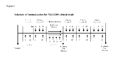
[001.01 Figure 1 shows the Schedule of Immunization for the NLGO205 pancreatic
cancer
clinical trials testing Algenpantucel-L (HyperA.cute.t Pancreas
Immunotherapy). Patients
enrolled in this Phase II pancreatic clinical trial received two immunizations
of Algenpantucel-L
before the first chemotherapy cycle after surgery. Subsequently, the patients
received
immunizations while receiving radiation therapy and/or chemotherapy. Serum
samples were
collected immediately before the first immunization to determine baseline
levels. Serum
samples were then obtained on Day 1 of cycle #2, Days 1 and 43 of
chemoradiation, Day I of
cycle #3, Day 1 of cycle #4, Day 1 of cycle #5, and at every follow-up visit.
[00111 Figure 2 shows the accuracy and precision of the ELISA method used in
these studies.
Two operators performed this study using 4 reference normal pool sera (NPS)
samples (NPS7-
NPS8, NFS9 and NFS10) and the reference standard. The percent coefficient of
variation (CV)
or Relative Standard Deviation (RSD) within and between experiments is
expected to be < 20%
and accuracy is suggested to be in the range of 80¨ 120%.
100121 Figure 3 shows the determination by ELISA of anti-aGal antibody values
in reference
samples by two operators in multiple experiments.
100131 Figure 4 shows the anti-aGal antibody titers of patients before
receiving Algenpantucel-L
immunotherapy. The baseline value of anti-aGal antibodies varies significantly
among patients.
Patients tested in this trial had a mean titer of 24 g/mL with a range of 2
to 149 jig/ml.
[00141 Figure 5 is a graph comparing the levels of a-Gal antibodies found in
patient sera before
immunization with Algenpantucel-L and overall survival. There is no apparent
correlation with
baseline antibody values and survival (p= 0.1074) indicating that there is no
apparent predictive
value for the amount of anti-aGal antibodies produced in a patient before
immunization and a
better prognosis or outcome.
4
CA 02912209 2015-11-10
WO 2014/186596 PCT/US2014/038231
[00151 Figures 6A-6G show the levels of anti-a-Gal antibodies detected in all
tested patients
after immunization with Algenpantucel-L. After immunization the vast majority
of tested
patients responded by increasing the levels anti-aGal antibodies produced. Of
50 patients tested,
46 (92%) responded with at least 2-fold increased anti-aGal antibody levels
compared to pre-
immunization values. The level of the response varied significantly among
patients.
[00161 Figure 7 shows the increase in anti-a-Gal antibody levels in patients
following
immunization. The mean fold-response (test/baseline) for the entire NLGO205
trial showed a 16
fold increase in anti-aGal antibody levels (range 2 to 128) compared to
baseline. Patients
receiving 300M cells tend to exhibit a higher anti-aGal antibody response
compared to patients
receiving 100M dose cells. Patients in the 300M dose cohort have a mean fold-
increase of 23
compared to 13 in the 100M dose cohort. These data suggest a dose-response in
the induction of
anti-aGal antibodies in these patients.
(0017) Figure 8 shows that there is a statistically significant correlation
between the
development of high titers of anti-aGal antibodies and better outcome (Overall
Survival) in 50
tested patients.
[00181 Figure 9 shows that the correlation of increased anti-a-Gal antibody
production in
patients with better outcome (Overall Survival) is observed only in the group
of patients
receiving high doses (300M) of Algenpantucel-L.
[00191 Figure 10 shows there is no correlation between overall survival and
anti-aGal antibody
levels observed in patients in a clinical trial testing Tenrgenpumatucel-L
(HyperAcute Lung
Immunotherapy) in lung cancer patients. Consequently the data observed on the
pancreatic trials
is unique to the pancreatic trial.
[00201 Figure 11 shows the performance characteristic of the control sample
NPS10. NPS10 was
tested in each experiment on each plate as a control. Testing of NPS10 was
consistent with less
than 10% CV for the determination of both the slope and the y-intercept,
demonstrating that the
values obtained during this study have acceptable quality with variation among
experiments
within acceptable range.
[00211 Figure 12 shows the obtained upper limit of quantification (ULOQ)
values obtained for
the anti-aGal antibody standard and the acceptable range of expected values
(dotted lines). The
ULOQ values observed were within acceptable range and the %CV observed was
less than 10%
indicating acceptable degree of variability.
CA 02912209 2015-11-10
WO 2014/186596 PCT/US2014/038231
[00221 Figure 13 shows the accuracy and precision of the ELISA method used in
these studies.
The values obtained for all the experiments and the summary for the
performance of the anti-
aGal antibody standard. The variability and coefficient determinates are
within the expected and
acceptable range.
[00231 Figure 14 shows that several of the cell lines tested that are
components of
HyperAcute-Pancreas and Lung vaccines express high levels of CEA tumor
associated antigen.
CEA RNA can be detected in HALL HAL3, HAPal , BxPC3 and Capan 2 cells.
[0024] Figure 15 shows that 17 out of 63 patients enrolled in NL00205 showed a
statistically
significant increase in the anti-CEA. antibody levels post-immunization with
Al.genpantucel-L.
This clustering of an anti-CEA antibody response is characterized by a
threshold of 20% increase
in the response after immunization compared to baseline. This clustering of
response was
statistically significant and potentially clinically meaningful (p<0.0001).
(0025) Figure 16 is a graph showing the survival of patients producing or not
producing
increased anti-CEA antibody levels after immunization with Algenpantucel-L.
Patients that
seroconverted to higher levels of anti-CEA antibody levels after immunizations
showed
improved overall survival compared to patients with no increase in anti-CEA
antibody levels.
100261 Figure 17 shows that there is a not a statistically significant
correlation between the titer
of anti-CEA antibodies produced in the patient after immunization and better
outcome,
indicating that the response itself (sero-conversion) and not the magnitude of
the response is
associated with better outcome.
100271 Figure 18 shows that the administered dose of Algenpantucel-L does not
affect the
percent change in the levels of anti-CEA antibodies produced in the patient
following
immunization. There is no difference in the change in anti-CEA antibody levels
observed in
patients receiving who received 300M of Algenpantucel-L compared with those
who received
the 100M of Algenpantucel-L, suggesting that at least concerning the anti-
tumor immune
response measured by the change in the levels of this antibody, both dose
regimes seem similar.
[0028] Figure 19 shows the preliminary analysis of the overall survival of
patients analyzed in a
lung cancer study. The production of anti-CEA antibodies in patients after
administration of
Tenrgenpumatucel-L (HyperAcutet Lung Immunotherapy) does not positively
correlate with
increased overall survival of patients.
CA 02912209 2015-11-10
WO 2014/186596 PCT/US2014/038231
[0029] Figure 20 shows the performance characteristic of the control sample
NPS10. NPS10
was tested in each experiment on each plate as a control. Testing of NPS10 was
consistent with
less than 10% CV for the determination of both the slope and the y-intercept,
demonstrating that
the values obtained during this study have acceptable quality with variation
among experiments
within acceptable range.
[00301 Figure 21 shows the detection by RT-PCR of mesothelin RNA in pancreatic
cell lines.
One of the cell line components of Algenpantucel-L immunotherapy, HAPal,
expresses
detectable levels of mesothelin antigen.
[00311 Figure 22 shows that membrane-bound mesothelin can be detected by FACS
analysis in
pancreatic cell lines. HAPal cells possess membrane-bound mesothelin on the
cell-surface. An
ovarian cancer cell line (CaoV3) that shows high expression of mesothelin was
used as a positive
control.
(0032) Figure 23 shows the levels of anti-mesothelin antibody (anti-MSLN)
produced in patients
after immunization. A clustering of anti-mesothelin antibody response is
characterized by a
threshold of a 25% increase in the anti-MSLN antibody titers after
immunization compared to
baseline. Of the 64 patients evaluated, 20 (31%) patients showed an increased
anti-MSLN
antibody response after immunization.
[0033] Figure 24 shows the sub-group analysis of patients producing or not
producing elevated
anti-MSLN antibody levels following immunization. Patients who seroconverted
to anti-MSLN
antibodies had a better outcome, with a median overall survival of 42 months
compared to
patients who had no anti-MSLN antibody response after immunization and had a
median overall
survival of 20 months.
[00341 Figure 25 shows the correlation of elevated anti-MSLN antibody levels
and overall
survival in immunized patients. There is a statistically significant
correlation between the
development of anti-MSLN antibodies and better outcome.
[0035} Figure 26 shows the performance characteristic of the control sample
NPS10 for these
studies. NPS10 was tested in each experiment on each plate as a control.
Testing of NPS10 was
consistent with less than 10% CV for the determination of both the slope and
the y-intercept,
demonstrating that the values obtained during this study have acceptable
quality with variation
among experiments within acceptable range.
7
CA 02912209 2015-11-10
WO 2014/186596 PCT/US2014/038231
[00361 Figure 27 shows the survival analysis of pancreatic cancer patients
after receiving
Algenpantucel-L. There are three groups of patients: those who exhibited no
increase in
antibody response, those who exhibited an increase in one type of antibody
production, and those
who exhibited an increase in the production of two or more antibodies. An
increased antibody
titer in a patient after inununiz.ation correlates with a better overall
survival (p=0.012). Patients
responding with one type of antibody studied (n=26) had a significantly better
outcome
compared to patients with no antibody response (n=27) after immunization (26
months vs. 17
months, p=0.047). Patients responding with two or more types of antibodies had
an even better
outcome ¨ as of January 23, 2013, the median level of survival has not yet
been reached.
[00371 Figure 28 shows the comparison of median survival including the
confidence intervals of
the three groups of patients. The likelihood of a patient's responding to
therapy is significantly
greater if an increase in antibody titer is observed after immunization with
Algenpantucel-L.
Those patients who showed no antibody response (n=27) showed a survival rate
of 19%, while
those showing an increased level of one antibody after immunization (n=26)
have a survival rate
of 42% (p=0.0476). Those patients showing an increased level of two or more
antibodies studied
(n=13) have a survival rate of 69% (p=0.0073).
[0381 Figure 29 shows an increase in eosinophil levels after immunization with
Algenpantucel-
L. Patients that show an increase in eosinophil levels at least three times
during the course of
immunization have a median survival of 27 months compared to a median survival
of 21 months
in those patients who did not exhibit an increase in eosinophil levels.
100391 Figure 30 shows skin biopsies which indicate presence of eosinophils at
the injection
sites might be unique to Algenpantucel-L.
100401 Figure 31A-D shows different receptors present on dying cells. Panel A
represents
lytienecrotic death, panel B represents apoptotic death, panel C represents
apoptotic cells
expressing clareticulin on their surface, and panel D represents cell markers
that stimulate
phagocytosis.
100411 Figure 32 shows the expression of calreticulin on both HAPal and HAPa2
cells.
[00421 Figure 33 shows the clustering of anti-calreticulin antibody response
post-immunization
with Algenpantucel-L.
[00431 Figure 34 shows the Kaplan-Meir graph of the sub-group analysis of
patients responding
or not with elevated anti-CALR antibodies after immunization with
Algenpantucel-L.
8
CA 02912209 2015-11-10
WO 2014/186596 PCT/US2014/038231
[00441 Figure 35 shows the reactivity of NPS10 anti-CALR, anti-CEA, and anti-
Mesothelin in a
qulaffied normal pool ser sample (NPS 10) detected by Western blot.
[00451 Figure 36 shows the variability in the detection of NPS10 reactivity
against calreticulin
intra-experiment The variability for the uppler limit of detection of anti-
CALR antibodies
present in NPSIO is below 10% in all experiments except EXF03, where the
variability observed
was 17.66%.
[00461 Figure 37 shows the variability observed inter-experiment. Triangles
denote the mean
value expected plus or minus 1.75 standard deviations (SD); squares denote the
mean value of all
experiments; circles denot the values obtained.
[0047] Figure 38 shows the serial dilution curve for NPS10 and their
corresponding OD value.
[00481 Figure 39 shows a linear regression of the inter-experiment
variability. This figure shows
the average valued for each point with error bars as SD. The solid line with
no circles represents
the fitted curve.
DETAILED DESCRIPTION OF THE INVENTION
(0049) Cancer immunotherapy is an emerging form of cancer treatment in which
the patient is
administered with an engineered tumor cell to induce an immune response
against the cancer
cells, thereby targeting the pre-existing tumor for destruction. Some forms of
immunotherapy use
allogeneic tumor cells genetically engineered to express aGal epitopes on the
cell-surface. These
cells are estimated to contain at least one to two million aGal epitopes (U.S.
2011/0250233,
herein incorporated by reference in its entirety). This large number of
binding sites for naturally
pre-existing anti-aGal antibody results in a high density of opsonization
followed by
complement destruction which sets off a variety of processes that activate
both the humoral and
cellular branches of the immune system. The presence of such a high density of
aGal residues on
the surface of allogeneic tumor cells induces a hyperirnmune response
analogous to xenograft
hyperacute rejection at the site of the modified tumor cell injection.
Furthermore, these cancer
vaccines are polyvalent meaning that they present multiple tumor antigen
targets to the immune
system. This will result in a more efficient treatment in that several TAAs
will be presented and
in a more widely effective treatment as with the increased number of TAAs
presented it is more
likely that there will be overlap in epitopes from different individual
tumors. Opsonized cells are
readily ingested by phagocytes providing a mechanism whereby most of the tumor
antigens can
9
CA 02912209 2015-11-10
WO 2014/186596 PCT/US2014/038231
be simultaneously presented to the adaptive immune system. Within these cells,
proteins from
the cancer vaccine cells will be digested and given class II MHC presentation
thereby exposing
the mutant proteins epitopes in the cancer cell to 1-cell surveillance. In
addition, the uptake of
opsonized cells by antigen presenting cells (APCs) via Fe receptor mediated
endocytosis may
facilitate the activation of WIC class I restricted responses by CD84 cells
through a cross
presentation pathway. The immune system cascade set in motion by this process
provides the
stimulus to induce a specific 1-cell response to destroy native tumor cells
from an established
hum.an malignancy. Furthermore, the inflammatory environment induced by the
prim.ary immune
response results in an amplification effect mediated by cytokines, histamines
and other up-
regulated molecules that boost the 1-cell response. 1-cells activated in this
manner are directly
capable of killing cancer cells. The addition of aGal epitopes to
glycoproteins and glycolipids
present in the tumor vaccine will not restrict the development of an immune
response only to
those antigens that become glycosylated but to any antigen present within the
tumor cell whether
it is affected by glycosylation or not.
[00501 Natural anti-aGal antibodies are of polyclonal nature and synthesized
by I% of
circulating B cells. They are present in serum and human secretions and are
represented by IgM,
IgG and IgA classes. The main epitope recognized by these antibodies is the
aGal epitope
(Gala1-3Galli 1 -4NAcGlc-R) but they can also recognize other carbohydrates of
similar
structures such as Gala1-3Ga1131-4G1c-R, Gala1-3Ga 101-4NAcGlcil 1 -3Galil 1 -
4G1c13.-R, Gala1-3G1c (melibiose), a-methyl galactoside, Gala1-6Gala1-6G10 (1-
2)Fru (stachyose), Gala1-
3(Fucal -2)Gal-R (Blood B type epitope), Gala1-3Gal and Gala1-3Gal-R (Galili
et al. 1987;
Galili et al. 1985; Galili et al. 1984). Similarly, non-natural synthetic
analogs of the aGal epitope
have been described to bind anti-aGal antibodies and their use has been
proposed to deplete
natural anti-aGal antibodies from human sera in order to prevent rejection of
xenogeneic
transplants (lanczuk et al. 2002; Wang et al. 1999). Therefore, glycomimetic
analogs of the aGal
epitope could also be used to promote the in vivo formation of immunocomplexes
for
vaccination purposes. Other carbohydrates such as rhamnose and Forssman
antigen may also be
used (U.S. Application No. 13/463,420 herein incorporated by reference in its
entirety).
100511 Applicants' invention provides the identification of cell-surface
markers which, when
enriched on a population of engineered tumor cells that express aGal epitopes
or other suitable
CA 02912209 2015-11-10
WO 2014/186596 PCT/US2014/038231
carbohydrates, induce the production of antibodies in the patient that
positively correlate with an
increased overall survival.
[00521 The compositions of the invention comprise tumor cells that are
engineered to express a
aGal epitopes (or other suitable carbohydrate epitopes). Such epitopes may be
added by
expressing in the cells a nucleic acid encoding an alpha galactosyltransferase
(aGT) or other
suitable enzyme, for example a viral or non-viral vector. Alternatively, such
epitopes may be
inserted directly into the cell membrane or conjugated to proteins on the cell
surface. These
modified cells are enriched for the presence of certain cell-surface markers,
including, but not
limited to, mesothelin, calreticulin, and/or carcinoembryonic antigen (CEA),
and are then
lethally irradiated or otherwise killed and administered to a patient. The
binding of aGal epitopes
by naturally pre-existing anti-aGal antibodies causes opsonization of the
tumor cells and
enhances tumor specific antigen presentation. The invention contemplates the
use of whole cells,
and a mixture of a plurality of transduced cells in the pharmaceutical
compositions of the
invention. Since aGal modifications affect multiple glycoproteins on the cell-
surface, the
patient's immune system will have an increased opportunity to detect, process,
and generate
antibodies to tumor specific antigens.
(0053) One embodiment of the invention comprises transfection of tumor cells
with a nucleotide
sequence which encodes upon expression, the enzyme a-(1,3)-galactosyl
transferase (aGT). The
aGT cDNA has been cloned from bovine and murine cDNA libraries. Larson, R. D.
et al. (1989)
"Isolation of a cDNA Encoding Murine UDP galactose; 13-D-galactosy1-1, 4-N
Acetol-D-
Glucosamine al -3. Galactosyl Transferase: Expression Cloning by Gene
Transfer", PNAS, USA
86:8227; and Joziasse, D. H. et al., (1989) "Bovine al -3 Galactosyl
Transferase: isolation and
Characterization of a cDNA Clone, Identification of Homologous Sequences in
Human Genomic
DNA", J. Biol Chem 264:14290. Any other nucleotide sequence which similarly
will result in the
tumor cells expressing an aGal epitope on the cell-surface may be used
according to the
invention, for example other enzymes that catalyze this reaction or perhaps
event the engineering
of the cells to have additional glycoproteins present on the cell-surface
hence the artificial
creation of a TAA which can be presented to the immune system.
10054] The tumor cells of the present invention may be syngeneic, allogeneic,
or autologous.
The transformed cells and the tumor cells to be treated must have at least one
epitope in
common, but will preferable have many. To the extent that universal, or
overlapping epitopes or
11
CA 02912209 2015-11-10
WO 2014/186596 PCT/US2014/038231
TAA exist between different cancers, the pharmaceutical compositions may be
quite widely
applicable.
[00551 Applicants have surprisingly found that after immunization with
compositions of the
invention, the production of antibodies in a patient to certain cell-surface
markers positively
correlates with an increased overall survival. The data described herein
demonstrate that the
levels of antibodies to aGal, mesothelin, calreticulin, and/or CEA produced by
the patient after
immunization with a composition of the invention correlate with an increased
overall survival for
the patient. In one embodiment, at least a 10-fold increase in anti-aGal
antibodies after
immunization with a composition of the invention correlates with an increased
overall survival
for the patient.
[00561 Overall survival for a patient has also been found to correlate with
the number of cell-
surface markers to which the patient demonstrates an increase in antibody
titers. Applicants
have found that patients who produce elevated antibody titers to aGal,
mesothelin, calreticulin,
or CEA after administration of immunotherapy have a better overall survival
than patients who
do not produce elevated antibody titers to any of these antigens.
Additionally, those patients
who produce elevated titers to two or more of these antigens after
administration of
immunotherapy have a better overall survival than those who produce antibodies
to only one or
two of these antigens.
100571 One aspect of the present invention provides for an isolated, non-
tumorigenic tumor cell
population which has been modified to express aGal and also expresses
mesothelin, calreticulin,
and/or CEA. The expression of mesothelin, calreticulin, and/or CEA on the
surface of these
modified cells may be achieved through any standard means in the art,
including, but not limited
to enrichment of the cell population by selecting for those cells which
already express one or
both of these antigens, or by engineering a cell through recombinant means to
express one or
both of these antigens.
[0058} Use of traditional techniques for cell sorting, such as by
immtmoselection (including, but
not limited to, FACS), permits identification, isolation, and/or enrichment
for aGal(+) cells that
express mesothelin, calreticulin, and/or CEA. The reagent can be an anti-
mesothelin antibody,
an anti-CEA antibody, an anti-calreticulin antibody, an anti-aGal antibody or
a combination
thereof. The modified tumor cells expressing aGal are grown in culture and in
one embodiment,
FACS is used to select those aGal(+) cells from the population expressing
mesothelin,
12
CA 02912209 2015-11-10
WO 2014/186596 PCT/US2014/038231
calreticulin, anclior CEA. The selection step can further entail the use of
magnetically responsive
particles as retrievable supports for target cell capture and/or background
removal. A variety of
FACS systems are known in the art and can be used in the methods of the
invention (see e.g.,
W099/54494, filed Apr. 16, 1999; U.S. Ser. No. 20010006787, filed Jul. 5,
2001, each expressly
incorporated herein by reference in all its entirety). The aGal expressing
tumor cells that are
found to express mesothelin and/or CEA are then cultured further and expanded.
[00591 Alternatively, the modified aGal-expressing tumor cell can be
recombinantly engineered
to express mesothelin, calreticulin, and/or CEA. Using standard techniques
known in the art,
polynucleotides encoding these antigens or fragments thereof can be inserted
into the modified
tumor cell for expression of one or more of these antigens on the cell
surface. In one
embodiment, the modified aGal expressing tumor cell is transduced with an
expression vector
comprising a mesothelin polynucleotide or fragment thereof for expression of
the mesothelin
antigen on the cell. In another embodiment, the modified aGal-expressing tumor
cell is
transduced with an expression vector comprising a CEA polynucleotide or
fragment thereof for
expression of the CEA antigen on the cell. In another embodiment, the modified
aGal-
expressing tumor cell is transduced with an expression vector comprising a
calreticulin
polynucleotide or fragment thereof for expression of the calreticulin antigen
on the cell. In
another embodiment, the modified aGal-expressing tumor cell is transduced with
an expression
vector comprising a mesothelin polynucleotide or fragment thereof for
expression of the
mesothelin antigen on the cell, and an expression vector comprising a CEA
polynucleotide or
fragment thereof for expression of mesothelin and CEA on the cell-surface, in
another
embodiment, the modified aGal-expressing tumor cell is transduced with an
expression vector
comprising polynucleotide sequences of both mesothelin and CEA or fragments
thereof for
expression of these antigens on the cell. In another embodiment, the modified
aGal-expressing
tumor cell is transduced with an expression vector comprising a calreticulin
polynucleotide or
fragment thereof for expression of calreticulin on the cell surface and an
expression vector
comprising a mesothelin polynucleotide or fragment thereof and/or an
expression vector
comprising a CEA polynucleotide or fragment thereof for expression of
calreticulin and
mesothelin and/or CEA on the cell-surface. In a further embodiment, the
modified aGal-
expressing tumor cell is transduced with an expression vector comprising
polynucleotide
13
CA 02912209 2015-11-10
WO 2014/186596 PCT/US2014/038231
sequences of calreticulin and mesothelin and/or CEA or fragments thereof for
expression of
these antigens on the cell.
[00601 The aGalactosyltransferase, mesothelin, calreticulin, and/or CEA
nucleic acid sequences
or fragments thereof, can be contained in an appropriate expression vehicle
which fransduces
tumor cells. Such expression vehicles include, but are not limited to,
eukaryotic vectors,
prokaryotic vectors (for example, bacterial vectors), and viral vectors. In
one embodiment, the
expression vector is a viral vector. Viral vectors that may be employed
include, but are not
limited to, retroviral vectors, adenov irus vectors, herpes virus vectors, and
adeno-associated
virus vectors, or DNA conjugates. Examples of retroviral vectors which may be
employed.
include, but are not limited to, Moloney Murine Leukemia Virus, spleen
necrosis virus, and
vectors derived from. retroviruses such as Rous Sarcoma Virus, Harvey Sarcoma
Virus, avian
leukosis virus, human immunodeficiency virus, myeloprol.iferative sarcoma
virus, and mammary
tumor virus.
[0061.] The vector includes one or more promoters. Suitable promoters which
may be employed
include, but are not limited to, the retroviral LTR., the SV40 promoter, and
the human
cytomegalovirus (CMV) promoter described in Miller, et al. Biotechniques, Vol.
7, No. 9, 980-
990 (1989) (incorporated herein by reference in its entirety), or any other
promoter (e.g. cellular
promoters such as eukaryotic cellular promoters including, but not limited to,
the histone, poi
and 13-actin promoters). Other viral promoters which may be employed include,
but are not
limited to, adenovinis promoters, TK promoters, and 819 parvovirus promoters.
100621 in another embodiment the invention comprises an inducible promoter.
One such
promoter is the tetracycline-controlled transactivator (tTA)-responsive
promoter (tet system), a
prokaryotic inducible promoter system which has been adapted for use in
mammalian cells. The
tet system was organized within a retroviral vector so that high levels of
constitutively-produced
tTA mRNA function not only for production of t-17A protein but also the
decreased basal
expression of the response unit by antisense inhibition. See, Paulus, W. et
al., "Self-Contained,
Tetracycline-Reeulated Retroviral Vector System for Gene Delivery to Mammalian
Cells", J of
Virology, January. 1996, Vol. 70, No. 1, pp. 62-67. The selection of a
suitable promoter will, be
apparent to those skilled in the art from the teachings contained herein.
[00631 The vector then is employed to transduce a packaging cell, line to form
a producer ce1.1
line. Examples of packaging cells which may be transfected include, but are
not limited to the
14
CA 02912209 2015-11-10
WO 2014/186596 PCT/US2014/038231
PE501., PA317, 2, 4-AM, PA12, T1914X., VF-19-1.7-H2, V-CRE, 4-CR1P, GP+E-
86,
GP+env.AM12, DAN and AM12 cell lines. The vector containing the nucleic acid
sequence
encoding the agent which is capable of providing for the destruction of the
tumor cells upon
expression of the nucleic acid sequence encoding the agent, and activation of
the complement
cascade may transduce the packaging cells through any means known in the art.
Such means
include, but are not limited to, electoporation, the use of Liposomes, and
Ca.PO4 precipitation. In
one embodiment, the invention comprises a viral vector which commonly infects
humans and a.
packaging cell line which is human based. For example vectors derived from.
viruses which
commonly infect humans such as Herpes Virus, Epstein Barr Virus, may be used.
[0064] After administration of the compounds of the invention to patients, the
antibody levels to
aGal, mesothelin, calreticulin, and/or CEA in the patient samples may be
measured by
immunoassays commonly used in the art (see, e.g., Harlow & Lane, Antibodies,
A. Laboratory
Manual (1988), hereby incorporated by reference in its entirety, for a
description of
immunoassay formats and conditions that can be used to determine specific
im.munoreactivity).
Non-limiting examples of such assays include, but are not limited to,
radioimm.unoassay, indirect
immunofluorescence assays (1FA), and ELISA. Suitable immunoassay methods
typically
include: receiving or obtaining (e.g., from a patient) a sample of body fluid
or tissue likely to
contain antibodies; contacting (e.g., incubating or reacting) a sample to be
assayed with an
antigen, under conditions effective for the formation of a specific antigen-
antibody complex
(e.g., for specific binding of the antigen to the antibody); and assaying the
contacted (reacted)
sample for the presence of an antibody-antigen reaction (e.g., determining the
amount of an
antibody-antigen complex). The presence of an elevated amount of the antibody-
antigen
complex indicates that the subject has produced antibodies to the marker on
the cell-surface. An
antigen, including a modified form thereof, which "binds specifically" to
(e.g., "is specific for" or
binds "preferentially" to) an antibody against a cell surface marker interacts
with the antibody,
or forms or undergoes a physical association with it, in an amount and for a
sufficient time to
allow detection of the antibody. By "specifically" or "preferentially," it is
meant that the antigen
has a higher affinity (e.g., a higher degree of selectivity) for such an
antibody than for other
antibodies in a sample. For example, the antigen can have an affinity for the
antibody of at least
about 1.5-fold, 2-fold, 2.5-fold, 3-fold, or higher than for other antibodies
in the sample. Such
affinity or degree of specificity can be determined by a variety of routine
procedures, including,
CA 02912209 2015-11-10
WO 2014/186596 PCT/US2014/038231
e.g., competitive binding studies. In an ELISA assay, a positive response is
defined as a value 2
or 3 standard deviations greater than the mean value of a group of
unirnm.unized controls.
Phrases such as "sample containing an antibody" or "detecting an antibody in a
sample" are not
meant to exclude samples or determinations (e.g., detection attempts) where no
antibody is
contained or detected. In a general sense, this invention involves assays to
determine whether an
antibody produced in response to immunization with compositions of the
invention is present in
a sample, irrespective of whether or not it is detected.
[00651 In one embodiment, the measurement of antibody levels in the patient
samples is
accomplished by ELISA. In one embodiment, the reagents for evaluating antibody
expression
are polypeptide antigens. In another embodiment, the antigen is aGal. In a
further embodiment,
the antigen is CEA. In yet a further embodiment, the antigen is mesothelin.
The levels of one or
more of these antibodies in the patient sample may be measured using one or
more of these
reagents.
[0066] Patient samples that may be tested for the levels of antibodies to
aCial, mesothelin,
calreticulin, and/or CEA produced by patients after administration of the
compositions of the
invention include, but are not limited to, blood, plasma, and/or serum. In one
embodiment, the
patient sample is serum.
[0067] The measurement of antibody titers to aGal, mesothelin, calreticulin,
and/or CEA may be
useful for the early identification of patient populations who will or will
not benefit from
treatment with the compositions of the invention. The measurement of the
levels of antibody
titers to certain cell-surface markers may be used to maintain current
treatment, change the
course or dosage of treatment, or add alternate therapies. Patients may
respond to
imrnunotherapy by producing increased antibodies to zero, one, two, or all
three of these
antigens. In one embodiment, patients who produce increased antibodies to none
or one of these
antigens are given an increased dosage of the compositions of the invention or
put on additional
forms of cancer therapy, including but not limited to, 1D0 inhibitors,
chemotherapy, alternate
immunotherapy, radiation, and/or a combination thereof.
[00681 The antibodies produced by the patient to the cell-surface molecules
may be measured
after one, two, three, four, five, six, seven, eight, nine, ten, or more
immunizations with the
compounds of the invention. In one embodiment, the antibodies produced by the
patient to the
cell-surface molecules are measured after two immunizations with the compounds
of the
16
CA 02912209 2015-11-10
WO 2014/186596 PCT/US2014/038231
invention. In a further embodiment, the antibodies produced by the patient to
the cell-surface
molecules are measured after five immunizations with the compounds of the
invention. In yet a
further embodiment, the antibodies produced by the patient to the cell-surface
molecules are
measured after ten immunizations with the compounds of the invention.
[00691 In one embodiment, the invention provides a method of treating cancer
or an uncontrolled
cellular growth comprising administering the compounds of the invention.
Tumors which may be
treated in accordance with the present invention include malignant and non-
malignant tumors.
Cells from malignant (including primary and metastatic) tumors include, but
are not limited to,
those occurring in the adrenal glands; bladder; bone; breast; cervix;
endocrine glands (including
thyroid glands, the pituitary gland, and the pancreas); colon; rectum; heart;
hematopoietic tissue;
kidney; liver; lung; muscle; nervous system; brain; eye; oral cavity; pharynx;
larynx; ovaries;
penis; prostate; skin (including melanoma); testicles; thymus; and uterus.
Examples of such
tumors include apudoma, choristoma, branchioma, malignant carcinoid syndrome,
carcinoid
heart disease, carcinoma (e.g., Walker, basal cell, basosquamous, Brown-
Pearce, ductal, Ehrlich
tumor, in situ, Krebs 2, Merkel cell, mucinous, non-small cell lung, oat cell,
papillary, scirrhous,
bronchiolar, bronchogenic, squamous cell, and transitional cell),
plasmacytoma, melanoma,
chondroblastoma, chondroma, chondrosarcoma, fibroma, fibrosarcoma, giant cell
tumors,
hi stiocytoma, lipoma, I iposarcoma, mesothel ioma, myxoma, myxosarcoma,
osteom a,
osteosarcoma, Ewing's sarcoma, synovioma, adenofibroma, adenolymphoma,
carcinosarcoma,
chordoma, mesenchymoma, mesonephroma, myosarcoma, ameloblastoma, cementoma,
odontoma, teratoma, thymoma, trophoblastic tumor, adenocarcinoma, adenoma,
cholangioma,
cholesteatoma, cylindroma, cystadenocarcinoma, cystadenoma, granulosa cell
tumor,
gynandroblastoma, hepatoma, hidradenoma, islet cell tumor, Leydig cell tumor,
papilloma,
Sertoli cell tumor, theca cell tumor, leiomyoma, leiomyosarcoma, myoblastoma,
myoma,
myosarcoma, rhabdomyoma, rhabdomyosarcoma, ependymoma, ganglioncuroma, glioma,
mcdulloblastoma, meningioma, neurilemnnoma, neuroblastoma, neuroepithelioma,
neurofibroma, neuroma, paraganglioma, paraganglioma nonchromaffin,
angiokeratoma,
angiolymphoid hyperplasia with eosinophilia, angioma sclerosing, angiomatosis,
glomangioma,
hemangioendothelioma, hemangioma, hemangiopericytoma, hemangiosarcoma,
lymphangioma,
lymphangiomyoma, lymphangiosarcoma, pinealoma, carcinosarcoma, chondrosarcoma,
cystosarcoma phyllodes, fibrosarcoma, hemangiosarcoma, leiomyosarcoma,
leukosarcoma,
17
CA 02912209 2015-11-10
WO 2014/186596 PCT/US2014/038231
liposarcoma, lymphangiosarcoma, myosarcoma, myxosarcoma, ovarian carcinoma,
rhabdomyosarcoma, sarcoma (e.g., Ewing's experimental, Kaposi's, and mast-
cell), neoplasms
and for other such cells.
[0070] In one embodiment, a patient may demonstrate an increase in eosinophil
levels after
administration with the compounds of the invention which correlates with an
increased overall
survival. In one embodiment, an increase of eosinophil levels at least three
times after
administration of the compounds of the invention correlates with an increased
overall survival.
[0071.] In one embodiment, both increased production of eosinophils and
antibodies to aGal,
mesothelin, calreticulin, and/or CEA in a patient are measured after
administration with the
compounds of the invention. In another embodiment, a patient who demonstrates
a lack of an
increase in eosinophils and/or antibody titer to one or more of these antigens
is given a higher
dose of the compounds of the invention or put on additional forms of cancer
therapy, including
but not limited to, IDO inhibitors, chemotherapy, alternate immunotherapy,
radiation, and/or a
combination thereof.
100721 According to the invention, attenuated aGal expressing tumor cells
enriched for the
expression of mesothelin, calreticulin, and/or CEA are used as either
prophylactic or therapeutic
vaccines to treat tumors. Thus the invention also includes pharmaceutical
preparations for
humans and animals involving these transgenic tumor cells. Those skilled in
the medical arts will
readily appreciate that the doses and schedules of pharmaceutical composition
will vary
depending on the age, health, sex, size and weight of the human and animal.
These parameters
can be determined for each system by well-established procedures and analysis
e.g., in phase I, II
and III clinical trials and by review of the examples provided herein.
[0073] The compositions of the invention are generally administered in
therapeutically effective
amounts. The term "therapeutically effective amount" is meant an amount of
treatment
composition sufficient to elicit a measurable decrease in the number, quality
or replication of
previously existing tumor cells as measurable by techniques including but not
limited to those
described herein. These compositions may be administered in a single dose or
in multiple doses.
Standard dose-response studies, first in animal models and then in clinical
testing, reveal optimal
dosages for particular disease states and patient populations. In some
embodiments, an effective
dosage of the vaccine of the invention will contain at least 100 million or
more cells. In another
18
CA 02912209 2015-11-10
WO 2014/186596 PCT/US2014/038231
embodiment, an effective dosage will comprise at least about 300 million or
more cells. In
another embodiment, an effective dosage will comprise at least about 500
million or more cells.
[00741 For administration, the compositions of the invention can be combined
with a
pharmaceutically acceptable carrier such as a suitable liquid vehicle or
excipient and an optional
auxiliary additive or additives. The liquid vehicles and ex.cipients are
conventional and are
commercially available. Illustrative thereof are distilled water,
physiological saline, aqueous
solutions of dextrose, and the like.
[00751 Suitable formulations for parenteral, subcutaneous, intradermal,
intramuscular, oral, or
intraperitoneal administration include aqueous solutions of active compounds
in water-soluble or
water-dispersible form. In addition, suspensions of the active compounds as
appropriate oily
injection suspensions may be administered. Suitable lipophil.ic solvents or
vehicles include fatty
oils for example, sesame oil, or synthetic fatty acid esters, for example
ethyl oleate or
triglycerides. Aqueous injection suspensions may contain substances which
increase the
viscosity of the suspension, include for example, sodium carboxymethyl
cellulose, sorbitol,
and/or dextran. Optionally, the suspensions may also contain stabilizers.
Also, compositions
can be mixed with immune adjuvants well known in the art such as Freund's
complete adjuvant,
inorganic salts such as zinc chloride, calcium phosphate, aluminum hydroxide,
aluminum
phosphate, saponins, polymers, lipids or lipid fractions (Lipid A,
monophosphoryl lipid A),
modified oligonucleotides, etc.
[0076} In addition to administration with conventional carriers, active
ingredients may be
administered by a variety of specialized delivery drug techniques which are
known to those of
skill in the art.
100771 For administration, the modified tumor cells can be combined with a
pharmaceutically
acceptable carrier such as a suitable liquid vehicle or excipient and an
optional auxiliary additive
or additives. The liquid vehicles and excipients are conventional and are
commercially available.
Illustrative thereof are distilled water, physiological saline, aqueous
solutions of dextrose and the
like.
[00781 The compositions of the invention can be administered alone or in
conjunction with other
cancer treatments. In one embodiment, the compositions of the invention may be
administered
in conjunction with chemotherapeutic agents. In another embodiment, the
compositions of the
invention may be administered in conjunction with radiation therapy. In yet a
further
19
CA 02912209 2015-11-10
WO 2014/186596 PCT/US2014/038231
embodiment of the invention, the compositions of the invention may be
administered in
conjunction with one or more chemotherapeutic agents and radiation therapy.
[00791 Examples of chemotherapeutic agents which may be administered in
conjunction with the
compositions of the invention include, but are not limited to, alkylating
agents, such as nitrogen
mustards (e.g., mechlorethamine, cyclophosphamide, ifosfamide, melphalan, and
chloram.bucil.);
nitrosoureas (e.g., carmustine (BCNU), lomustine (CCNU), and semustine (methyl-
CCNU));
ethyleneimines and methyl-melamines (e.g., triethylenem.elamine (TEM),
triethylene
thiophosphoramide (thiotepa), and hexamethylmelamine (TIMM, altretamine));
alkyl sulfonates
(e.g., buslfan); and triazines (e.g., dacabazine (DTIC)); antimetabol.ites,
such as folic acid
analogues (e.g., methotrexate, trimetrex ate, and pemetrexed (multi-targeted
antifolate));
pyrimidine analogues (such as 5-fluorouracil (5-FU), fluorodeoxyuridine,
gemcitabine, cytosine
arabinoside (AraC, cytarabine), 5-azacytidine, and 2,2'-
difluorodeoxycytidine); and purine
analogues (e. g , 6-mercaptopurine, 6- thiogua.n ine, azathioprine, 2'-
deoxycoformycin
(pentostatin), erythrohydroxynonyladenine (EHNA), fludarabine phosphate, 2-
chlorodeoxyadenosine (cladribine, 2-CdA)); Type I topoisomerase inhibitors
such as
camptothecin (CPT), topotecan, and irinotecan; natural products, such as
epipodophylotoxins
(e.g., etoposide and teniposide); and vinca alkaloids (e.g., vinblastine,
vincristine, and
vinorelbine); anti-tumor antibiotics such as actinomycin D, doxorubicin, and
bleomycin;
radiosensitizers such as 5- bromodeozyuridine, 5-iododeoxyuridine, and
bromodeoxycytidine;
platinum coordination complexes such as cisplatin, carboplatin, and
oxaliplatin; substituted
ureas, such as hydroxyurea; and methylhydrazine derivatives such as N-
methylhydrazine (MIH)
and procarbazine; and inhibitors of microtubule function such as docetaxel and
paclitaxel. In one
embodiment, the chemotherapeutic agent is gemcitabine. The chemotherapeutic
agent
administered in combination with the compositions of the invention is
administered as
determined by the treating physician, and at doses typically given to patients
being treated for
cancer.
100801 Examples of radiation therapy that may be administered in conjunction
with
compositions of the invention include, but are not limited to, radiation
emitters such as alpha-
particle emitting radionuclides (e.g., actinium and thorium radionuclides),
low linear energy
transfer (LET) radiation emitters (i.e. beta emitters), conversion electron
emitters (e.g. strontium-
89 and samarium- I53-EDTMP, or high-energy radiation, including without
limitation x-rays,
CA 02912209 2015-11-10
WO 2014/186596 PCT/US2014/038231
gamma rays, and neutrons. The radiation therapy may be performed with a
sensitizer, including
but not limited to, 5R.J. The radiation therapy administered in combination
with the
compositions of the invention is administered as determined by the treating
physician, and at
doses typically given to patients being treated for cancer.
[00811 The compositions of the invention and the further therapeutic agent may
be given
simultaneously in the same formulation. Alternatively, the agents are
administered in a separate
formulation but concurrently, with concurrently referring to agents given, for
example, within
minutes, hours or days of each other. In some embodiments, the compositions of
the invention
comprise a plurality of autologous tumor cells which may be the same or
different. The
autologous tumor cells may be administered separately or together.
[0082] In another aspect, the further therapeutic agent is administered prior
to administration of
the compositions of the invention. Prior administration refers to
administration of the further
therapeutic agent within the range of minutes, hours, or one week prior to
treatment with the
compositions of the invention. It is further contemplated that the further
therapeutic agent is
administered subsequent to administration of the compositions of the
invention. Subsequent
administration is meant to describe administration more than minutes, hours,
or weeks after
administration of the compositions of the invention.
100831 The present invention also provides a kit for the detection of
antibodies produced to the
aGal, mesothelin, calreticulin, andlor CEA in a patient receiving
immunotherapy. In a non-
limiting example, one or more reagents for evaluating antibody expression can
be provided in a
kit. In one embodiment, the kit contains one reagent to measure the expression
levels of one
antibody in the patient sample. In another embodiment, the kit contains the
reagents to measure
the expression of two antibodies the patient sample. In yet a further
embodiment, the kit
contains the reagents to measure the levels of three antibodies in the patient
sample. In a further
embodiment, the kit contains the reagents to measure the levels of more than
three antibodies in
a patient sample. The kits may thus comprise, in suitable container means,
nucleic acids,
antibodies, polypeptides, or other regents that can be used to determine
antibody titers in a
sample. In one embodiment, the reagents are attached or fixed to a support,
such as a plate, chip
or other non-reactive substance. For example, a reagent can be fixed to a
microtiter well, and the
sample placed in the well to determine the expression level of antibodies to
the cell-surface
markers expressed on the compounds of the invention. In one embodiment, the
reagents for
21
CA 02912209 2015-11-10
WO 2014/186596 PCT/US2014/038231
evaluating antibody expression are polypeptide antigens. In another
embodiment, the antigen is
aGal. In a further embodiment, the antigen is CEA. In yet a further
embodiment, the antigen is
mesothelin. In yet a further embodiment, the antigen is calreticulin.
100841 The kits may comprise a suitably aliquoted nucleic acids that can be
used as probes or
primers; alternatively, it may comprise a suitably aliquoted antibody that
can. be used in
immunohistochemical detection methods or any other method discussed herein or
known to
those of skill in the art.
[00851 The components of the kits may be packaged either in aqueous media or
in lyophilized
form. The container means of the kits will generally include at least one
vial, test tube, flask,
bottle, syringe or other container means, into which a component may be
placed, and preferably,
suitably aliquoted. Where there is more than one component in the kit, the kit
also will generally
contain a second, third or other additional container into which the
additional components may
be separately placed. However, various combinations of components may be
comprised in a vial.
The kits of the present invention also will typically include a means for
containing the containers
in close confinement for commercial sale. Such means may include injection or
blow-molded
plastic containers into which the desired vials are retained.
100861 To confidently and immediately measure the levels of antibody
production in patients
receiving immunotherapy with the compounds of the invention, assays to measure
the antibody
titers may be performed at the point of care using transportable, portable,
and handheld
instruments and test kits. Small bench analyzers or fixed equipment can also
be used when a
handheld device is not available. In one embodiment of the invention, a kit is
provided to the
point-of-care to allow immediate testing of the levels of anti-cell-surface
markers produced in
patients receiving immunotherapy.
EXAMPLES
[0087} The following examples are provided to further illustrate the
advantages and features of
the invention, but are not intended to limit the scope of this disclosure. All
citations to patents
and journal articles are hereby expressly incorporated by reference in their
entireties.
Example 1
22
CA 02912209 2015-11-10
WO 2014/186596 PCT/US2014/038231
Humoral immunologic response before and after Immunization with Algenpantucel-
L
(HyperAcute-Pancreas NLGO205).
[00881 We evaluated whether in patients enrolled in Phase II clinical trials
vaccination with
Alegenpantucel-L HyperAcute-Pancreas immunotherapy induced antibodies against
aGAL
epitopes, the cell-surface marker Carcinoembryonic antigen (CEA.) and the
recombinant
membrane bound mesothelin (MSLN).
[00891 Patients enrolled in the Phase II pancreatic clinical trials NL00205
received two
immunizations before the first chemotherapy cycle after surgery. Subsequently,
they received
immunizations while receiving radiation therapy and/or chemotherapy.
[00901 Serum samples for the study of the humoral immune response (anti-aGal
antibody, anti-
CEA antibody and anti-MSLN antibody) were collected immediately before the
first
immunization to determine the baseline values. Serum samples were obtained on
Day I of cycle
#2, days I and 43 of chemoradiation, Day 1 of cycle #3, Day I of cycle #4, Day
1 of cycle #5,
and at every follow-up visit (Figure 1).
Assay Qualification/ Validation fbr the Detection of anti-aGal Antibodies
belbre and after
immunization in serum samples by ELISA.
(0091) Measurements of immune response provide important potential surrogate
endpoints for
monitoring the efficacy of anti-cancer immunotherapy trials. Anti-aGalactosyl
antibody
Enzyme-Linked Immunosorbent Assay (anti-aGal antibody ELISA) is an endpoint
assay used in
immunological studies on human serum samples obtained from clinical trials of
NewLink
Genetics cancer immunotherapy (HyperActute). in this study we improved and
characterized the
performance quality and consistency of the assay to demonstrate that it is a
suitable and reliable
method for quantifying the anti-aGal antibodies in serum samples from clinical
trial. This
summary report provides a description of the anti-aGal antibody ELISA method
validation and
the performance characteristics of the assay that were established. The study
was based mainly
on method validation guidelines published by the US Food and Drug
Administration (FDA),
2001 and the ICH Harmonized Tripartite Guidelines, 2005.
Brief assay description
[0092] The detection of anti-aGal antibodies was performed by ELISA. Briefly A
96-well
microliter plate is coated with a-Gal-HSA (antigen) overnight, washed and
blocked with HSA at
37 C. Samples (Primary Antibodies) are dispensed on the plate, allowed to
react with antigen
23
CA 02912209 2015-11-10
WO 2014/186596 PCT/US2014/038231
and washed. Enzyme conjugated secondary antibodies are dispensed on the plate
and allowed to
react with primary antibodies. A chromogenic detection substrate is dispensed
on plate and
allowed to react with conjugate yielding a product with blue color. Reaction
is stopped with 2M
Sulphuric acid and optical density (OD) of samples is detected with a plate
reader at a
wavelength of 450 nm. Analysis of data is performed using Microsoft Excel
and/or GraphPad.
Prism software. In each plate the purified standard is tested in duplicates
wells, a qualified
normal pooled serum. sample (NPS10) is also tested in each experiment as an
additional quality
control reagent. All patients samples are tested in each plate.
Optimized anti-aGal ELISA parameters
[00931 Accurate validation of a bioanalytical procedure is only possible when
the operational
parameters or conditions of the method are optimized. In this regard, several
experiments were
performed to determine the optimal conditions for the anti-aGal ELBA. method.
The validation
parameters of the method and procedural efficiency within and between
experiments and
operators established herein presumes optim.ized assay conditions and stable
assay reagents and
samples. Only under such conditions can the performance characteristics of the
method
presented in this report be expected to be reproducible within limits of
random experimental
error.
Results and conclusions
The established parameters for optimal anti-aGal antibody ELISA method are as
presented in
Table 1. These assay conditions constitute the elements of the Standard
Operation Procedure
(SOP) for application and validation of the anti-aGal antibodies ELISA method
as presented
herein. The validation parameters established herein are therefore only
applicable under the
optimized conditions presented below, changes to which may necessitate partial
re-validation of
the method or otherwise:
Table 1. Optimized assay conditions for anti-aGal ELISA protocol.
Reagent Concentration/time
Antigen (aGal-HSA) in carbonate-bicarbonate buffer 5 Ag/m1
Antigen (aGal-HSA) coating per well (50111) 250 ng
Coating buffer (HSA) in carbonate-bicarbonate buffer 1%
Secondary antibody (Goat anti-Human IgG-HRP conjugated Ab) 1/24000
24
CA 02912209 2015-11-10
WO 2014/186596 PCT/US2014/038231
TMB substrate concentration 0.4 mg/ml
Hydrogen peroxide concentration (in citric acid buffer) 0.02 %
TMB substrate-Hydrogen peroxidase mix 100 p.1/well
Stopping reagent (100 ml/well) 2M 112504
Substrate incubation time 20 minutes
calibration of Standard curve
100941 Calibration of the anti-aGal antibody standard curve is the empirical
determination of the
relationship between measured absorbance (OD) values for the Standards and the
true or known
concentration of the standards. A chromatographically purified total human IgG
was further
affinity purified to obtain the anti-aGal IgG standard used in this study The
Limit of Detection
(LOD), Lower Limit of Quantification (LLOQ), and Upper Limit of Quantification
(ULOQ) are
essential values that define or delimit the dynamic range of the standard
curve. The range of the
calibrated standard curve include a blank (matrix sample processed without
internal standard),
zero-sample (matrix sample processed with internal standard), and six non-zero
sample points
including the LLOQ and ULOQ.
[0095] Table 2 summarizes the findings described above.
Table 2. Performance characteristics of the anti-aCial antibody ELISA standard
Performance characteristic Actual/set values
Limit of Detection (LOD)/Sensitivity 1 ng/ml
Lower Limit of Quantification (LLOQ) 2 ng/ml
Upper Limit of Quantification (ULOQ) 20 ng/ml
Dynamic range of standard curve 0 ¨ 20 ng/ml
Standards (ng) 0, .05, 2, 4, 8, 12, 16, 20
Matrix effect None
Selectivity/Recovery in serum Acceptable: 90-110%
Dilution linearity Acceptable: 90-110%
Stability of Assay Reagents
[00961 Consistency in experimental results is also greatly influenced by
stability of reagents used
in an assay. The stability of samples and reagents used in anti-aGal ELISA
method were tested
CA 02912209 2015-11-10
WO 2014/186596 PCT/US2014/038231
under conditions at which they are stored or processed in order to determine
the time frame for
stability of the samples and reagents. The storage conditions investigated are
refrigeration at
4 C, and freezing at -20 C or -80 C for both samples and reagents. Exposure of
reagents and
samples to room temperature (RT) as well freeze-thaw of reagents or samples
was investigated.
Stability of the Standard Conclusions
[00971 The standard is stable on storage at 4 C for at least up to 100 as
indicated by a consistent
or stable range of the OD values demonstrated for the standard with acceptable
range of
variability (ULOQ , %CV: 7.5 and LLOQ %CV 16%).
[00981 Freeze-thaw cycles affect the performance of the Standard. Data
indicates a high
coefficient of variation (CV:35%) in experiments performed. Consequently,
freezing and
thawing is not recommended. A paired t-test comparison between 4 C and RT
exposure data
indicate that the reactivity of the standard is not affected by exposure to
room temperature for up
to 4 hours.
Stability of the secondary antibody Conclusions
[00991 Results show no significant change in performance of secondary antibody
on exposure to
room temperature for up to 72 hours (P=0.7868). A significant difference was
observed when all
data was compiled together indicating a slight trend for reduced performance
of the secondary
antibodies when exposed at RT for 192 hours. Consequently the secondary
antibody is
considered stable for at least 72 hours when stored at room temperature. The
results also indicate
that the secondary antibody is stable when stored at 4C for up to 18 months.
Stability of the samples
[001001 Patient's samples with low, intermediate and high titer of anti-
aGal antibodies
were tested for stability on storage at 4 C, exposure to room temperature for
up to 4 hours, and
freeze-thaw cycles.
[001011 Results show that freshly thawed and continuously monitored samples
stored at
4 C remain stable for up to 6 months. Results of exposure of sample to room
temperature for up
to 4 hours show no significant effect on estimate of analyte, and freeze-thaw
cycles of samples
stored at -20 C did not show a specific trend indicating an adverse effect of
freeze-thaw cycles
on the samples. The results suggest that the samples are stable on freeze-thaw
for at least up to
cycles.
Procedural efficiency, precision and accuracy.
26
CA 02912209 2015-11-10
WO 2014/186596 PCT/US2014/038231
[001021 The procedural efficiency is a measure of the application of the
anti-aGal
antibody ELISA methods in terms of accuracy and precision of data within and
between
experiments and operators. Under the optimized assay conditions, the percent
coefficient of
variation (CV) or Relative Standard Deviation (RSD) within and between
experiments is
expected to be < 20% and accuracy is suggested to be in the range of 80 ¨
120%. The results for
procedural efficiency obtained from analysis of reference NPS samples are
presented below.
100103] The procedure for testing anti-aGal antibodies was repeated
multiple times to
determine the precision and the accuracy of the method. Two operators
performed this study
using 4 reference normal pool sera (NPS) samples (NPS7-NPS8, NPS9 and NPS10)
and the
reference standard. .A summary of the standard performance by two operators is
shown in Figure
2.
[001.04] The estimates of anti-aGAL antibody values for reference samples
obtained in
these experiments are shown in Figure 3.
[001.05) The summary of the precision and accuracy is depicted in Table 3.
Table 3. Precision and Accuracy over experiments
Statistics
Standard 1007.42 31.43 3.12 100.74209 33
NPS07 5.43 0.83 15.3 31
NPS08 5.29 0.80 15.1 33
NPS09 3.38 0.57 16.9 33
NIPS 1 0 21.21 2.75 13.0 59
Robustness
[00106) The robustness of an analytical procedure is the property that
indicates
insensitivity against changes made to known operational parameters on the
results of the method
which provide an indication of its suitability or reliability for its defined
purpose. Insensitivity of
a method to inadvertent changes made to known operational parameters between
operators or
laboratories defines ruggedness of the procedure.This report presents data on
empirical
investigation on the robustness of anti-aGal ELISA method for seven assay
parameters that were
27
CA 02912209 2015-11-10
WO 2014/186596 PCT/US2014/038231
selected and considered to be the most important in the procedure. The
parameters in question
are quantity of antigen (aGal-HSA) coated on plate, incubation time for
primary antibody,
incubation time for secondary antibody, wash cycles, substrate incubation
time, TMB
temperature, and time lapse before reading of plates. . A Plackeft-Burman
design for screening of
many variables or factors for their main effect (Plackett and Burman, 1964) is
chosen for the
study on the robustness of anti-aGal ELISA method. Table 4 shows the
parameters evaluated
during this study and results.
Table 4. Experimental factors and levels
Factor Factor description - I +
XI Coating with aGal-HSA per well 235 ng 265 ng
X2 Incubation time with primary antibody 45 min 75 min
X3 Incubation time with secondary antibody 45 min 75 min
X4 Wash cycles 3 7 cycles
cycles
X5 Substrate incubation time 15 min 25 min
X6 Temperature of substrate (TMB) 4 C 25 C
Time lapse to reading of plate 1 min 10 mm n
Conclusions of the Validation/Qualffication study
1001071 This study demonstrates that the anti-aGal ELISA method is robust,
shows
acceptable precision and accuracy, and therefore suitable for quantification
of anti-aGal antibody
in patient serum samples.
Example 2
Detection of anti-aGal Antibodies Wore and after immunization in serum samples
by EL EA.
[00108] The detection of anti-aGal antibodies was performed by ELISA using
standard
techniques. Each experiment was considered valid if the following criteria for
valid test were
demonstrated.
Criteria for valid test
28
CA 02912209 2015-11-10
WO 2014/186596 PCT/US2014/038231
[001091 Anti-aGal concentration of 21 5 pg/ird for NPS10 as analyte
control.
1001101 OD value range for standard: Upper limit from 0.65 to 0.91.
[001111 Standard curve coefficient of determination (R2) ranging from
0.9800 to 0.9999.
Results
[001121 All patients tested in this trial had detectable anti-aGal
antibodies before receiving
Algenpantucel-L immunotherapy (Figure 4).
[001131 As demonstrated in other clinical trials using HyperAcute
technology the baseline
values of anti-aGal antibodies varies significantly among patients. Patients
tested in this trial had
a mean antibody titer of 24 i.tg/mL with a range of 2 to 149 ftg/ml.
1001141 We performed a Pearson correlation study to determine if the
baseline values of
anti-aGal antibodies have any predictive value to a favorable survival. Data
below shows that
there is no apparent correlation with baseline values and survival (p= 0.1074)
indicating no
apparent predictive value for the amount of anti-aGal antibodies before
immunization and better
prognosis or outcome (Figure 5).
1001151 After immunization the vast majority of tested patients responded
by increasing
their anti-aGal antibody levels. In this study we tested 50 patients and 46
(92%) responded with
at least 2-fold increase in the levels of anti-aGal antibodies compared to pre-
immunization
values. The level of the response varied significantly among patients. Figures
6A-6G show the
levels of anti-aGal antibodies detected in all tested patients.
100116) The magnitude of the response after vaccination was calculated by
the fold-
increase in the anti- aGal antibody response after immunization. The fold-
increase is calculated
by the ratio of the peak response divided by the baseline value. As shown in
Figure 7, the mean
fold-response (test/baseline) for the entire NLGO205 trial was a 16 fold
increase in anti-aGal
antibody levels (range 2 to 128) compared to baseline. We performed the
analysis comparing
both dose cohort for the anti-aGal antibody response after immunization. As
shown in Figure 7,
patients receiving 300M cells tend to have a higher anti-aGal antibody
response compared to
those patients receiving 100M dose cells. Patients in the 300M dose cohort
have a mean fold-
increase of 23 compared to 13 in the 100M dose cohort. These data suggest a
dose-response in
the induction of anti-aGal antibody response in these tested patients.
[001171 To determine if the magnitude of the response in anti-aGal antibody
had a
correlation with better outcome, we performed a Pearson correlation
calculation. Figure 8
29
CA 02912209 2015-11-10
WO 2014/186596 PCT/US2014/038231
demonstrates that there is a statistically significant correlation between the
development of high
titers of anti-aGal antibodies and better overall survival in 50 tested
patients.
[001181 The correlation among favorable overall survival (OS) and increased
anti-aGal
antibody titers was performed for patients receiving 100M dose and 300M dose.
As shown in
Figure 9, the correlation with better outcome is observed only in the group of
patients receiving
high doses on Algenpantucel-L suggesting again a dose-response. Furthermore,
in trials of other
cancers treated with different HyperAcute immunotherapy, the presence of
increased titers of
anti-aGal antibodies does not correlate with an improved overall survival.
Figure 10 shows there
is no correlation between overall survival and anti-aGal antibody levels
observed in patients in a
clinical trial testing Tenrgenpum.atucel-L (HyperA.cutert Lung Immunotherapy)
in lung cancer
patients. While patients responded to therapy with increased anti-aGal
antibody levels in the
lung cancer study, the fold increase in anti-aGal antibody levels does not
correlate with an
increase in overall survival in patients. The correlation between the fold
increase in anti-aGal
antibody levels and overall survival is unique to the pancreatic trial.
Conclusions
100 1 191 All patients had detectable pre-existing naturally acquired anti-
aCial antibodies
(mean 24 ps/mL, range 2 to 149 jig/m1). The vast majority of patients
receiving Algenpantucel-L
immunotherapy responded by increasing the anti-aGal antibody after
immunization. In this study
46 out of 50 patients (92%) had increased anti-aGal antibody titer by at least
2 fold. The
development of high titters of anti-aGal antibody correlated with better
outcome for the entire
clinical trail (p=0.01). Subgroup analysis indicated that in the 300M dose
cohort correlation
between anti-aGAL antibody response and better outcome was still observed in
comparison to
the 100M dose cohort where statistically significant correlation was lost.
This result indicates
that 300M dose cohort induced higher titers of anti-aGAL antibodies that
correlated with better
overall survival suggesting a dose-response effect.
[00120} Example 3
1001211 Performance characteristic qf the anti-aGal antibody ELISA assay
during the
testing qfpatients enrolled in NLGO205
1001221 The following section describes the assay performance during the
testing of anti-
aGal antibody values by ELISA.
CA 02912209 2015-11-10
WO 2014/186596 PCT/US2014/038231
[001231 As explained above, the criteria for valid test for this assay were
developed during
the qualification/validation process of this assay. It is expected that the
value obtained for the
NPS10 control sample will be 21 5 p.g/ml, the upper limit of OD value for
the standard curve
should be with 0.65 to 0.91 OD units and the coefficient of determination for
the standard curve
is expected to be 0.9800 to 0.9999.
Testing of NPSIO
[001241 As explained above, each patient sample was analyzed in a single
plate, In
addition to patient's samples, we added a qualified commercially available
reagent from normal
pooled sera (NPS10). This reagent was tested extensively and it was
established that the
expected concentration of anti-aGal antibodies was 21 5 gg/ml.
[001251 Figure 11 shows the summary of values obtained during the course of
this study.
The expected variability is shown (dotted lines). As shown in Figure 11, the
percent coefficient
of variability (%CV) in less than 15% indicating that variability observed in
this study is within
acceptable range.
Purified anti-aGal Antibody Standard performance
1001261 In order to quantify the amount of anti-aGal antibody values in
patient's samples
we utilized an affinity purified anti-aGal antibody standard. This reagent was
fully characterized
during the qualification/validation portion of this study. In the patients
testing phase the anti-
aGal antibody standard was included in each plate for each patient tested.
Figure 12 shows the
obtained Upper limit of quantification (ULOQ) values obtained for the anti-
aGal antibody
standard and the acceptable range of expected values (dotted lines). As shown
in Figure 12, the
ULOQ values observed were within acceptable range and the %CV Observed was
less than 10%
indicating acceptable degree of variability.
[00127} Each experiment conducted utilized the standard to determine the
anti-aGal
antibody values present in patient samples. Figure 13 shows the values
obtained for all the
experiments and the summary for the performance of the anti-aGal antibody
standard. As shown
in Figure 13, the variability and the coefficient of determination are within
expected and
acceptable range.
Conclusion
[001281 The anti-aGal antibody ELISA for the testing of patient's samples
was conducted
according to guidelines published by the US Food and Drug Administration
(FDA), 2001 and the
31
CA 02912209 2015-11-10
WO 2014/186596 PCT/US2014/038231
ICH Harmonized Tripartite Guidelines, 2005. Results indicate that the
performance of all quality
controls utilized in this study have acceptable range of variability intra and
inter assay. This
study showed that the assay has high level of consistency, consequently it is
considered adequate
to support conclusions presented in this report.
Example 4
Detection of anti-CEA antibodies in patients enrolled in AIG0205 and NI,G0305
clinical trials
Introduction
[001291 CEA (Carcinoembryonic antigen) is a well-studied member of the
immunoglobulin superfamily. CEA is a complex, highly glycosylated
macromolecule containing
approximately 50% carbohydrate, with a molecular weight of approximately 200
kDa. CEA was
first discovered in human colon cancer tissue extracts. It is a useful marker
for monitoring colon
cancer after surgery and for monitoring treatment progression of lung and
pancreatic cancer
patients.
[00130] The HyperAcute immunotherapy is manufactured using cell lines
genetically
engineered to express aGal epitopes. Several of the cell line tested that are
components of
HyperAcute-Pancreas and Lung vaccines expresses high levels of CEA tumor
antigen (Figure
14).
[00131} We tested the development of anti-CEA antibodies in patients
receiving HAPa
inununotherapy before and after immunization as a mean of performing the
immunological
monitoring of evaluable patients and to determine if this biomarker could be
correlated with
better outcome.
Brie' assay description
1001321 The detection of anti-CEA antibodies was performed by ELISA.
Briefly A 96-
well microliter plate is coated with commercially available CEA antigen
overnight, washed and
blocked with buffer at 37 C. Samples (Primary Antibody) are dispensed on the
plate, allowed to
react with antigen and washed. Enzyme conjugated secondary antibody is
dispensed on the plate
and allowed to react with primary antibody. A chromogenic detection substrate
is dispensed on
plate and allowed to react with conjugate yielding a product with blue color.
Reaction is stopped
with 2M Sulphuric acid and optical density (OD) of samples is detected with a
plate reader at a
wavelength of 450 nm. Analysis of data is performed using Microsoft Excel
and/or GraphPad
32
CA 02912209 2015-11-10
WO 2014/186596 PCT/US2014/038231
Prism software. In each plate a qualified normal pooled serum sample (NPS10)
is also tested as
quality control reagent. All patients' samples are tested in each plate.
[00133] We tested the immunological response to CEA of patients enrolled in
NLGO205.
For analysis of the anti-CEA antibody, we selected samples collected before
immunization, a
sample after patients received 3 vaccinations, and a sample after they
received all
immunizations. Samples from the same patient were analyzed at the same time.
All patients with
available samples were evaluated.
[00134] We calculated the percent of change compared to baseline values
according to the
formula: Formula= Percent of change compare to baseline values (initial):
N final - N initial
__________ = (100)¨%change
N
tiai
Results
[00135] In this study we analyzed 63 patients with available samples before
and after
immunization. We observed a clustering of response that was characterized by a
threshold of
20% increase in the response after immunization compared to baseline. This
clustering of
response was statistically significant and potentially clinically meaningful
(p<0.0001) Figure 15
shows the statistically significant clustering of the response post-
immunization of evaluated
patients.
[001361 In this study, 17 out of 63 patients showed a statistically
significant increase in the
anti-CEA antibody values post immunization. Table 5 below shows the summary of
the
response.
Table 5. Anti-CEA antibody response in patients receiving Algenpantucel-L
immunotherapy
t. t%.# Ab
.............. ............................ No increased L Increase Total
.....
Counts 46 17 63
µpercent 73 27
OS (Months) 20.5 39.5
Survival Rate (0/01 3 i 470/
33
CA 02912209 2015-11-10
WO 2014/186596 PCT/US2014/038231
[001371 Patients that seroconverted to higher levels of anti-CEA antibodies
after
immunizations showed improved overall survival compared to patients with no
increase in the
anti-CEA antibody response (Table 5 and Figure 16). Figure 16 shows the
survival proportions
in a Kaplan-Meir plot of patients with and without increased anti-CEA
antibodies after
immunization.
[001381 To determine if the magnitude of the response anti-CEA had a
correlation with
better outcome we performed a Pearson correlation calculation. Figure 17
demonstrates that
there is not a statistically significant correlation between the development
of higher levels of
anti-CEA. antibody and better outcome indicating that the response (sero-
conversion) and not the
magnitude of the response is associated with better outcome.
[001391 We compared the percentage of change in the anti-CE.A antibody
levels in
patients receiving 300M cell vaccines or 100M cell vaccine (Figure 18). As
shown in Figure 18,
there is no difference in the type of response observed in patients receiving
either dose
suggesting that at least concerning the anti-tumor immune response measured by
the change in
the levels of this antibody, both dose regimes seem similar.
[001401 In a separate trial testing the efficacy Tenrgenpumatucel-L
(HyperAcute Lung
Immunotherapy) in lung cancer, 32 patients have been tested for the presence
of anti-CEA
antibodies before and after immunization. The patients analyzed have received
more than two
immunizations. Of the 32 patients analyzed, 20 patients responded with
significant increased
anti-CEA antibody values after immunization, although three did exhibit a
slight decrease in
anti-CEA antibodies during immunization (considered not changed in the
analysis) . Preliminary
analysis of the overall survival of patients analyzed in this study suggest no
predictive or
favorable correlation in patients responding with increased anti-CEA antibody
titer compared to
patients with no change in the anti-CEA antibody levels (Figure 19).
Anti-SEA Antibody EL1SA assay performance
[001411 To monitor the quality of the assay, a qualified normal pool sera
sample (NPS10)
was tested in each experiment in each plate. Data below shows the performance
of the NPS 10
control sample during the course of this study (Figure 20). As shown in Figure
20, testing of
NPS10 was consistent with less than 10% CV for the determination of both the
slope and the y-
intercept, demonstrating that the values obtained during this study have
acceptable quality with
variation among experiments within acceptable range.
34
CA 02912209 2015-11-10
WO 2014/186596 PCT/US2014/038231
Conclusions
1001421 Production of anti-CEA antibodies was elevated in 17 out of 63
(27%) patients
evaluated after immunotherapy. The elevation of anti-CEA antibody was
associated with better
outcome. Patients responding with anti-CEA antibody had median overall
survival (OS) of 25.2
months comparing favorably to patients without sero-conversion which had a
median OS of only
21.4 months. In addition, it is feasible to perform the immunological
monitoring of
Algenpantucel-L immunotherapy trials detecting anti-CEA antibody and possibly
use this
biomarker as surrogate marker to determine efficacy in Algenpantucel-L
clinical trials.
Furthermore, in trials of other cancers treated with different HyperAcute
immunotherapy, the
presence of increased titers of anti-CEA antibodies does not correlate with an
improved overall
survival (data not shown).
Ex ample 5
Detection of anti-Mesothelin Antibody in patients enrolled in NI,G0205
Introduction
1001431 Most tumor-associated antigens are expressed in greater extent in
cancer tissues
as compared to normal tissues. This may help the immune system to recognize
the over-
expressed genes in tumor. Hence, over-expressed genes have potential to be
immunogenic and
can be targeted for immunotherapy.
1001441 Mesothelin is a 40kDa differential antigen which is expressed on
normal
mesothelial cells and over-expressed in various cancer including pancreatic,
cervix, esophagus,
lung, ovarian cancers and mesotheliomas (Chang et al., PNAS, 1996; Ordonez et
al., Mod Path.
2003; Ordonez et al. Am J Surg Pathol, 2003; Argani et al. 2001; Ho et al.
2007). Its expression
is tested with SAGE (Argani et al. 2001) and irnmunohistochemistry (1HC) using
monoclonal
antibody K1 (Chang et al., Int J Cancer, 1992; Chang et al., Am j Surg Pathol,
1992) and later
commercial antibody 5B2 (Ordonez et al., Mod Pathol, 2003; Ordonez et al, Am
.1 Surg Pathol,
2003). The precursor of mesothelin is a 69kDa protein that is processed into
40kDa membrane-
bound mesothelin and 31kDa shed protein known as megakaryocyte-potentiating
factor that is
secreted from the cells and identified from the medium of human pancreatic
cancer cell line
(Yamaguchi et al., J Biol Chem, 1994).
[001451 The biological function of mesothelin is not clear. Deletion of
both copies of
mesothelin had no abnormalities in the mutant mice as compared to wild-type
mice (Bera et al,
CA 02912209 2015-11-10
WO 2014/186596 PCT/US2014/038231
Mol Cell Biol, 2000). Mesothelin has been suggested to play a role in adhesion
because 3T3 cells
transfected with mesothelin were more adherent to the culture dishes than non-
transfected cells
(Chang et al, PNAS, 1996). It is supported by the study showing mesothelin
interaction with
CA125 which might play role in metastasis of tumor (Rump et al, J. Biol. Chem,
2004; Gubbels
et al., Mol Cancer, 2006).
[001.461 Its limited expression in normal cells makes it an attractive
target for
imniunotherapy (Hassan et al. Clin Cancer Res, 2004) Mesothelin antibodies are
present in the
sera of patients with mesothelin expressing cancers (Ho et al., Clin Cancer
Res, 2005). It has also
shown to elicit T-ce1.1 response (Yokok.awa et al, Clin Cancer R.es, 2005;
Thomas et al, J Exp
Med, 2004). There are various clinical trials going on that target mesothelin
or elicit immune
response against mesothelin. A recombinant immunotoxin (SS1P) containing an
anti-mesothelin
Fv linked linked to truncated exotoxin has shown to mediate cell killing of
mesothelin-
expressing cells and tumors (Ho et al, Clin Cancer Res, 2007; Hassan et al,
Clin Cancer Res,
2004; Hassan et al., Clin Cancer Res, 2006). Two Phase I clinical trials have
been recently
completely for SSI P (Hassan et al. Clin Cancer Res, 2007). Another clinical
trial by Jaffe et al
involved vaccinating pancreatic cancer patients with GM-CSF transduced
pancreatic cancer cell
lines (Jaffee et al., J Clin Oncol, 2001). 3 out of 14 patients developed post-
vaccination delayed
type-hypersensitivity associated with prolonged survival. Following
immunological studies
showed that these 3 patients strong induction of CD8+ I cell response (Thomas
et al., I Exp
Med, 2004). These studies support mesothelin as a promising target for
immunotherapy.
1001471 Algenpantucel-L immunotherapy is manufactured using two pancreatic
cell lines
genetically engineered to express aGal epitopes. One of the cell line
components of
Algenpantucel-L immunotherapy, HAPal, expresses high levels of mesothelin
antigen by RT-
PCR (Figure 21).
1001481 In addition, membrane bound mesothelin can be detected by FACS
analysis in
HAPa1 cells. Figure 22 shows the staining of Algenpantucel-L cells. As a
positive control we
stained an ovarian cancer cell line (CaoV3) that shows high expression of
mesothelin.
Brief Assay description
1001491 The detection of anti-Mesothelin (MSLN) antibodies was performed by
ELISA.
Briefly A 96-well microliter plate is coated with recombinant purified
membrane bound
mesothelin (antigen) overnight, washed and blocked with buffer at 37 C.
Samples (Primary
36
CA 02912209 2015-11-10
WO 2014/186596 PCT/US2014/038231
Antibody) are dispensed on the plate, allowed to react with antigen and
washed. Enzyme
conjugated secondary antibody is dispensed on the plate and allowed to react
with primary
antibody. A chromogenic detection substrate is dispensed on plate and allowed
to react with
conjugate yielding a product with blue color. Reaction is stopped with 2M
Sulphuric acid and
optical density (OD) of samples is detected with a plate reader at a
wavelength of 450 nm.
Analysis of data is performed using Mircrosoft Excel and/or GraphPad. Prism
softwares. In each
plate a qualified normal pooled serum. sample (NPS10) is also tested as
quality control reagent.
All patients samples are tested in each plate.
[00150] We tested the immunological response to membrane bound mesothelin
of patients
enrolled in NLGO205. For analysis of the anti-MSLN antibody, we selected
samples collected
before immunization, a sample after patients received 3 vaccinations, and a
sample after they
received all immunizations. Samples from the same patient were analyzed at the
same time. All
patients with available samples were evaluated.
[001.51.] We calculated the percent of change compared to baseline values
according to the
formula:
Formula.= Percent of change compare to baseline values (initial):
N fina1 N
4/' 100) .%thaDge
N
Results
[00152] In this study we analyzed 64 patients with available samples before
and after
immunization. We observed a clustering of anti-m.esothelin antibody response
that was
characterized by a threshold of 25% increase in the response after
immunization compared to
baseline. Figure 23 shows the statistically significant clustering of the
response post-
immunization of evaluated patients.
[00153] Of the 64 patients evaluated, 20 (31%) patients had an increased
anti-MSLN
antibody response after immunization. Patients that seroconverted to anti-MSLN
antibody had a
better outcome with a median overall survival of 42 months compared to
patients that had no
anti-MSLN antibody response after immunization which had a median overall
survival of 20
months (Table 6, and Figure 24).
Table 6. Anti-Mesothelin (MSLN) antibody response of evaluated patients
37
CA 02912209 2015-11-10
WO 2014/186596 PCT/US2014/038231
Anti-MSLN Ab .No increase increase total
counts 44 20 64
.percent 59- 31
OS (Months) 20 42
Survival Rate 32% 50%
[00154] To determine if the magnitude of the response anti-mesothelin
antibody had a
correlation with better outcome we performed a Pearson correlation
calculation. Figure 25
demonstrated that there is a statistically significant correlation between the
development of anti-
MSLN antibody and better outcome.
Anti-MSNL Antibody ELISA assay performance
[00155] To monitor the quality of the assay, a qualified normal pool sera
sample (NPS10)
was tested in each experiment in each plate. Figure 26 shows the performance
of the NPS10
control sample during the course of this study. As shown in Figure 27, testing
of NPS10 was
proven to be consistent with less than 10% CV for the determination of both
the slope and the y-
intercept, demonstrating that the values obtained during this study have
acceptable quality with
variation among experiments within acceptable range.
Conclusions
[001561 Production of anti-MSLN antibodies was elevated in 20/64 (31%)
patients
evaluated after immunotherapy. The elevation of anti-MSLN antibodies was
correlated with
better outcome (p<0.03). Patients responding with anti-MSLN antibodies had
median overall
survival of 42 months comparing favorably to patients without sero-conversion
which had a
median overall survival of only 20 months. in addition, it is feasible to
perform the
immunological monitoring of Algenpantucel-L immunotherapy trials detecting
anti-mesothelin
antibodies and possibly use this biomarker as surrogate marker to determine
efficacy in
Algenpantucel-L clinical trials.
Example 6
Multivariate Analysis of the humoral immune response in patients receiving
Algenpantucel-L
immunotherapy
1001571 We performed a preliminary exploratory data analysis combining
observations
accrued during this study to determine if the development of either antibody
alone or in
combination might have a predictive value on treatment progression and or
possibly early signs
of response. Results from this analysis might have significant value to
determine at early time
38
CA 02912209 2015-11-10
WO 2014/186596 PCT/US2014/038231
points during the study if the irnmunotherapy has a higher or lower
probability of success. The
knowledge at early time points of probability might have an impact on
subsequent treatment
decisions.
1001581 In this analysis patients were categorized in three groups:
I. Patients which showed no significant response to antibody
production. These
patients have less than 10 fold-increase in the anti-aGal antibody, they have
less than
20% increase in anti-CEA antibody production and less than 25% increased anti-
mesothelin antibody production after immunization,
2. Patients with response to one type of antibody studied. They responded
either
with more than 10 fold-increase in the anti-aGal antibody values or anti-CEA
(>25%) or
anti-Mesothelin antibody (>25%).
3. Patients that responded with two or more types of antibody production.
These
patients have a combination of either two parameters studied or they have
increased in
the three types of antibodies studied. The matrix of possible combinations for
this group
of patients is shown in Table 7:
Table 7: matrix of possible combinations for patients responding to multiple
parameters
aGAL CEA ____ MS Nt. CEA and NISIN
aGAL
CEA
x
M
[00159] Sixty six patients were evaluated in this multivariate analysis.
Results
demonstrate that a statistically significant favorable median overall survival
is observed in
patients responding with antibody after immunization (p=0.012). Patients
responding with one
type of antibody studied (n=26) had a significantly better outcome compared to
patients with no
antibody response (n=27) after immunization (26 months vs 17 months, p
=0.047). Moreover
patients responding with two or more types of antibodies had an even better
outcome. In this
group of patients the median survival has not been reached yet (as of 01-23-
2013). Figure 28
shows the Kaplan-Meir plot for the three groups described. Importantly the
majority of patients
who responded with increased antibody titers after immunization early during
treatment in
general did so after the first two immunizations. Consequently these data has
the potential to
39
CA 02912209 2015-11-10
WO 2014/186596 PCT/US2014/038231
predict the course therapy early during the administration of the
immunotherapy Figure 28 shows
the comparison of median survival including the confidence intervals of
groups.
Table 8 summarizes the findings.
Table 8. Summary of the multivariate analysis for Algenpantucel-L
immunotherapy trial
=
c it
rAtdidvae
resnOnse (Ab) 19.% 17
One Parameter (A.b) .26 42% 0,M76:
itiPara meter Ab).- 69% not renhet1 Cg173
Conclusions
[001601 In this multivariate analysis we determined that the likelihood of
responding to
therapy is significantly greater if response to antibodies is observed. The
antibody response is
Observed early during the course of immunization, consequently this data
potentially could be
use a method to change the course of treatment or might help in the assistance
of decisions for
subsequent therapies.
1001611 Example 7
[001621 Correlation of increased eosinophil levels with overall survival
after
immunization with Algenpantucel-L
[001631 After administration of pancreatic cancer patients with
Algenpantucel-L some
patients demonstrated an increase in eosinophils that correlates with an
improved patient
outcome. Patients that exhibited this increase in eosinophil levels at least
three times during the
course of immunization have a median survival of 27 months compared to 21
months for patients
with no elevation of eosinophils (Figure 29). In addition, there is evidence
that skin
inflammatory reactions surrounding the skin biopsies at the injection site
show eosinophil
infiltrates. The presence of eosinophils at the injection sites might be
unique to Algenpantucel-L
(Figure 30).
Example 8
Correlation of increased production of anti-calreticulin antibodies with
overall survival after
immunization with Algenpantucel-L
1001641 Calreticulin (ICALR) is a multifunctional protein located in
storage compartments
associated with the endoplasmic reticul um. Calreticul in binds to tnisfolded
proteins and prevents
them from 'being exported from the endoplasmic reticulurn to the Golgi
apparatus. A similar
CA 02912209 2015-11-10
WO 2014/186596 PCT/US2014/038231
quality-control chaperone, calnexin, performs the same service for soluble
proteins as does
Calreticulin (Ellgaard et al. 2003). CALR is expressed on the cell surface of
many cancer cells
and plays a role to promote macrophages to engulf cancerous cells (Chaput et
al. 2007; Obeid et
al. 2007). When CALR is exposed on the cell surface, it also serves as a
signal that allows a
dying cell to be recognized, ingested and processed by specialized phagocytic
and dendritic cells,
which educate other immune cells to recognize and respond to the material they
have ingested
generating an immune response (Obeid et al. Nature Medicine, 2007). CD47,
which blocks
C.ALR prevents destruction of most of the cells. Phagocytic cells recognize
CALR. by LDL
receptor-related protein (I,RP-1- or CD91).
[001651 Dying cells play an important role in the generation of an imm.une
response.
Figure 31 shows different receptors present on dying cells. In 1.ytic/necrotic
death (.A) fragments
of cells that die by necrosis are taken up by phagocytes, which can trigger
production of pro-
inflammatory cytokines, leading to immune activation and, potentially,
autoimmunity. Apoptotic
cells (B) are recognized by cell surface markers such as phosphatidyl serine
(PS) and
phagocytosed and undergo non-immunogenic death which can cause phagocytes to
release anti-
inflammatory molecules (e.g., IL-10 and TGF11). However, apoptotic cells that
display
calreticulin on their surface are processed by dendritic cells that induce a
specific I cell¨
mediated immune response against these apoptotic cells (C). Phagocytosis of
apoptotic cells is
determined by a combination of cell surface markers. Viable cells display
inhibitory signals
(including CD47, which interacts with SHPS-1 on the phagocyte, and CD31) that
disappear from
the cell surface upon apoptosis induction. During apoptosis, cells expose
markers that stimulate
phagocytosis, including phosphatidyl serine (PS), recognized by phosphatidyl
serine receptors
(PSR); calreticulin (CALR), recognized by low-density lipoprotein-
receptor¨related protein
(LRP) and Cl q; and oxidized phosphatidyl serine (oxPS), recognized by CD36
(D. Figure 31).
Figure 32 shows that calreticulin is expressed on both HAPal and HAPa2 cells.
[00166} Based on this information we postulated that the expression of
Calreticulin in
Algenpantucel-L drug product will induce an immunological reaction to
Calreticulin that could
be detected after immunization. These "de-novo" antibodies might be
potentially used as
surrogate markers for vaccine efficacy.
[001671 The purpose of this study was to determine the reactivity anti-
calreticulin detected
in patients receiving Algenpantucel-L in NLGO205 clinical study. In addition
the development of
41
CA 02912209 2015-11-10
WO 2014/186596 PCT/US2014/038231
anti-calreticulin antibodies was correlated with clinical outcome to deteimine
if the development
of anti-calreticulin antibodies has a potential predictive value for clinical
efficacy.
[001681 The detection of anti-Calreticulin (CALR) antibodies was performed
by ELISA.
A 96-well microliter plate is coated with 50uL/well of a 5 ug/mL CALR
(Calreticulin Protein
Fitzgerald Cat#80R-1306) overnight, washed and blocked with buffer at 37 C.
Samples (Primary
antibodies) were dispensed on the plate, allowed to react with antigen and
washed. Enzyme
conjugated secondary antibody was dispensed on the plate and allowed to react
with the primary
antibodies. A chromogenic detection substrate was dispensed on plate and
allowed to react with
conjugate yielding a product with blue color. The reaction was stopped with 2M
Sulphuric acid
and optical density (OD) of samples was detected with a plate reader at a
wavelength of 450 nm..
Analysis of data was performed using Mircrosoft Excel and/or GraphPad Prism.
softwares. In
each plate a qualified normal pooled serum. sample (NPS10) was also tested as
quality control
reagent.
[001.69] The immunological response to CALR. of patients enrolled in
NLGO205 was
studied. Serum samples were collected on day 1 of cycle #1 (before
immunization- baseline), day
1 of cycle #2 (S2), days 1 (S3) and 43 (S4) of chemoradiation, day 1 of cycle
#3 (S5), day 1 of
cycle #4 (S6), day I of cycle #5 (S6), and at every follow-up visit (S7 and so
on). Three samples
were tested to analyze the anti-CALR antibodies produced: Si (baseline), S3
(patients received 4
immunizations) and S6 (patients received about 12 immunizations.).
[00170} Figure 1 shows the protocol schedule. Samples from the same patient
were
analyzed at the same time. Samples from a patient were analyzed in a single
plate. All patients
with available samples were evaluated. The percent of change compared to
baseline values was
calculated according to the following formula using OD values in the lineal
portion of the curve
using serial dilutions.
¨ Ninitiaj (100) = % change.
Ninitial
1001711 In this study 64 patients with available samples before and after
immunization
were analyzed. A clustering of response that was characterized by a threshold
of 20% increase in
the response after immunization compared to baseline was observed. Figure 33
shows the
statistically significant clustering of the response post-immunization of
evaluated patients. Of the
42
CA 02912209 2015-11-10
WO 2014/186596 PCT/US2014/038231
64 patients evaluated, 31 (48%) patients had increased anti-CALR antibody
response after
immunization.
[00172] The median overall survival of patients responding with anti-CALR
antibodies
after immunization compared to the median overall survival of patients without
sero-conversion
was also evaluated. Patients that had increased anti-C.ALR antibodies after
immunization had a
significantly better outcome with a median overall survival over 35 months
compared to patients
that had no meaningful increased in the anti-CLR antibodies response after
immunization
(median overall survival 19.2 months Figure 34). The difference in median
overall survival was
statistically significant (p<0.04).
[001.73] Table 9 shows the survival rate analysis for patients responding
with anti-CALR
antibodies compared to patients with no meaningful increase in the anti-CALR.
antibody
response. The statistical analysis indicates a significant difference in the
outcome of patients
responding with anti-CALR. antibodies (Fisher exact test, p<0.01).
Table 9. Survival rate and fisher exact test for anti-CALR Ab response
Anti-CALR Ab 46:crease Ab No inctease¨ . Total
Counts 31 3.3 64
OS Cfmoths) >35 19<2 p< 0.04 ilocl rank test) ..
Su
. rviv& rate
21% p<0:01 Tither's exact testl
Conclusions
[00174] Production of anti-CALR. antibodies was elevated in 31/64 (48%)
patients
evaluated after immunotherapy. The elevation of anti-CALR antibodies was
correlated with
better outcome (p<0.04). Patients responding with anti-CALR antibodies had
median overall
survival (OS) of >35 months comparing favorably to patients without sero-
conversion which had
a median. OS of only 19.2 months. in addition, it is feasible to perform. the
immunological
monitoring of Algenpantucel-L inununotherapy trials detecting anti-CALR.
antibodies and
possibly use this biomarker as surrogate marker to determine efficacy in
algenpantucel-L clinical
trials.
Anii-CALR Ab ELISA assay performance
[001.75] To monitor the quality of the assay and establish criteria for
valid test, a qualified
normal pool sera sample (NPS10) was tested in each experiment in each plate in
parallel to
43
CA 02912209 2015-11-10
WO 2014/186596 PCT/US2014/038231
patient's samples. This reagent was tested by western blot and showed specific
reactivity against
Calreticulin. Figure 35 shows the reactivity of NPS10 anti-CALR, anti-CEA and
anti-Mesothelin
detected by western blot. Consequently this reagent is considered suitable to
use as a control for
this assay.
[001761 The performance of NPS1.0 was evaluated INTRA and INTER experiments
during the testing of samples described before.
[001771 Figure 36 shows the variability in the detection of NPS I 0
reactivity against
Calreticulin intra experiment in the upper limit of detection of this assay.
As shown in Figure 36,
the variability (coefficient of variation) for the upper limit of detection of
anti-CALR. antibodies
present in NPS10 is below 10% in all experiments except EXP03, where the
variability observed
was 17.66%. The variability in the detection of the anti- CALR antibodies
present in NPS10
inter experiment was evaluated during the course of this study.
[001781 As shown in Figure 37, the variability observed INTER experiment
was 18%.
Figure 37 shows each individual value for each experiment. The box line
represents the mean of
all experiments. The triangle lines represent mean value expected plus or
minus 1.75 SD.
Experiments were considered valid for the detection of NPSIO anti-Calreticulin
antibodies when
the values observed were within this range.
[001791 Each serum sample was subjected to serial dilutions in order to
obtain OD values
in a linear portion of the curve to perform the calculation of the percent
change compared to
baseline. Similarly, serial dilutions are performed for NPS10 as a quality
control. Figure 38
shows corresponding OD values for NPS10 in each plate and each experiment.
1001.801 In order to evaluate the variability inter-experiment combined
values were fitted
to a linear regression (Figure 39). Figure 39 shows the average value for each
point with error
bars as SD. The fitted curve is also shown with the 95% Cl. As demonstrated in
Figure 39, the
combined values for the Y-intercept and slope have acceptable precision
demonstrating
reproducibility in the assay.
1001811 Testing of NPS10 for the presence of anti-CALR antibody reactivity
was proven
to have acceptable reproducibility with %CV intra-experiment below 10% and
inter-experiment
variability below 20%. Consequently the assay is considered suitable for
detecting anti-CALR
antibody reactivity in serum samples. While specific embodiments of the
invention have been
44
CA 02912209 2015-11-10
WO 2014/186596 PCT/US2014/038231
described and illustrated, such embodiments should be considered illustrative
of the invention
only and not as limiting the invention as construed in accordance with the
accompanying claims.
[001821 All patents, applications, and other references cited herein are
incorporated by
reference in their entireties.
References
[00183] 1. Chang, K. and I. Pastan, Molecular cloning of mesothelin, a
differentiation
antigen present on mesothelium, mesotheliomas, and ovarian cancers. Proc Natl
Acad Sci U S
A, 1996. 93(1): p. 136-40.
[001841 2. Ordonez, N.G., Value of mesothelin imrnunostaining in the
diagnosis of
mesothelioma. Mod Pathol, 2003. 16(3): p. 192-7.
[001851 3. Ordonez, N.G., Application of mesothelin immunostaining in
tumor
diagnosis. Am J Surg Pathol, 2003.27(11): p. 1418-28.
(00186) 4. Argani, P., et al., Mesothelin is overexpressed in the vast
majority of
ductal adenocarcinomas of the pancreas: identification of a new pancreatic
cancer marker by
serial analysis of gene expression (SAGE). Clin Cancer Res, 2001. 7(12): p.
3862-8.
[001871 5. Ho, M., et al., Mesothelin expression in human lung cancer.
Clin Cancer
Res, 2007. 13(5): p. 1571-5.
[001881 6. Chang, K., I. Pastan, and M.C. Willingham, Isolation and
characterization
of a monoclonal antibody, K1 , reactive with ovarian cancers and normal
mesothelium. Int
Cancer, 1992. 50(3): p. 373-81.
(00189) 7. Chang, K., et al., Monoclonal antibody K1 reacts with
epithelial
mesothelioma but not with lung adenocarcinoma. Am P Surg Pathol, 1992. 16(3):
p. 259-68.
(00190) 8. Yamaguchi, N., et al., A novel cytokine exhibiting
megakaryocyte
potentiating activity from a human pancreatic tumor cell line HPC-Y5. J Biol
Chem, 1994.
269(2): p. 805-8.
[001911 9. Bera, T.K. and I. Pastan, Mesothelin is not required for
normal mouse
development or reproduction. Mol Cell Biol, 2000. 20(8): p. 2902-6.
[001921 10. Rump, A., et al., Binding of ovarian cancer antigen
CA125/MUC16 to
mesothelin mediates cell adhesion. J Biol Chem, 2004. 279(10): p. 9190-8.
CA 02912209 2015-11-10
WO 2014/186596 PCT/US2014/038231
[001931 11. Gubbels, J.A., et al., Mesothelin-MUC16 binding is a high
affinity, N-
glycan dependent interaction that facilitates peritoneal metastasis of ovarian
tumors. Mol
Cancer, 2006. 5(1): p. 50.
1001941 12. Hassan, R., T. Bera, and I. Pastan, Mesothelin: a new
target for
immunotherapy. Clin Cancer Res, 2004. 10(12 Pt 1): p. 3937-42.
[001951 13. Ho, M., et al., Humoral immune response to mesothelin in
mesothelioma
and ovarian cancer patients. Clin Cancer Res, 2005. 11(10): p. 3814-20.
[001961 14. Yokokawa, J., et al., Identification of novel human CTL
epitopes and their
agonist epitopes of mesothelin. Clin Cancer Res, 2005. 11(17): p. 6342-51.
[001971 15. Thomas, A.M., et al., Mesothelin-specific CD8(+) T cell
responses
provide evidence of in vivo cross-priming by antigen-presenting cells in
vaccinated pancreatic
cancer patients. J Exp Med, 2004. 200(3): p. 297-306.
(001981 16. Hassan, R.., et al., Tumor-directed radiation and the
immunotoxin SS1P in
the treatment of mesothelin-expressing tumor xenografts. Clin Cancer Res,
2006. 12(16): p.
4983-8.
[00199] 17. Hassan, R., et al., Phase I study of SS1P, a recombinant
anti-mesothelin
immunotoxin given as a bolus I.V. infusion to patients with mesothelin-
expressing
mesothelioma, ovarian, and pancreatic cancers. Clin Cancer Res, 2007. 13(17):
p. 5144-9.
(00200) 18. Jaffee, E.M., et al., Novel allogeneic granulocyte-
macrophage colony-
stimulating factor-secreting tumor vaccine for pancreatic cancer: a phase I
trial of safety and
immune activation. J Clin Oncol, 2001. 19(1): p. 145-56.
[002011 19. Ellgaard, L. and E.M. Frickel, Calnexin, calreticulin, and
ERp57:
teammates in glycoprotein folding. Cell Biochem Biophys, 2003. 39(3): p. 223-
47.
[00202} 20. Chaput, N., et al., Molecular determinants of immunogenic
cell death:
surface exposure of calreticulin makes the difference. J Mol Med, 2007.
85(10): p. 1069-761
[00203} 21. Obeid, M., et al., Leveraging the immune system during
chemotherapy:
moving calreticulin to the cell surface converts apoptotic death from "silent"
to immunogenic.
Cancer Res, 2007. 67(17): p. 7941-4.
1002041 22. Obeid, M., et at.. Calreticulin exposure dictates the
immunogenicity of
cancer cell death. Nat Med, 2007. 13(1): p. 54-61.
46