Note: Descriptions are shown in the official language in which they were submitted.
CA 02914026 2015-11-27
WO 2014/197543 PCT/US2014/040806
ASSAYS, METHODS AND KITS FOR ANALYZING SENSITIVITY AND RESISTANCE
TO ANTI-CANCER DRUGS, PREDICTING A CANCER PATIENT'S PROGNOSIS,
AND PERSONALIZED TREATMENT STRATEGIES
CROSS-REFERENCE TO RELATED APPLICATION
[0001] This application claims the benefit of Provisional Application
Serial No. 61/830,709
filed June 4, 2013, which is herein incorporated by reference in its entirety.
FIELD OF THE INVENTION
[0002] The invention relates generally to the fields of cellular biology,
molecular biology,
oncology, and medicine.
BACKGROUND
[0003] Despite many decades of incremental improvements in methods for
establishment of
cancer cell lines, it is still extremely difficult to establish high-quality,
permanent cell lines from
human primary tumors routinely. Malignant, drug-resistant cancer phenotypes
are not
represented in currently available tumor cell line panels which fail to
represent the biological
diversity of human tumors. The inability to establish stable cell lines from
the vast majority of
human tumors has limited the use of in vitro models to study human cancer. A
robust and
efficient model system that predicts a patient's response to various drugs
would greatly improve
development of new drugs for personalized treatment of cancer patients. There
is thus a need for
the development of methods for testing and predicting a patient's response to
treatment.
SUMMARY
[0004] Assays, methods and kits for analyzing sensitivity of a subject's
(e.g., patient's)
cancerous tumor to a drug, predicting responses of cancerous tumors to drugs,
determining the
prognosis of a subject having a cancerous tumor, and developing a personalized
therapy or
treatment strategy for the subject are described herein. Identification of
patients who are
resistant to particular oncology drugs (e.g., Taxol) and in vitro
determination of specific existing
and new drugs to be utilized for individual patients can be achieved using the
assays and
methods described herein, providing for the development of a personalized
approach to cancer
treatment. Such assays include high throughput screening assays (e.g., high
throughput
screening of a group, plurality or population of patients or subjects and
drugs).
1
CA 02914026 2015-11-27
WO 2014/197543 PCT/US2014/040806
[0005] Accordingly, described herein is a method for analyzing sensitivity
of a subject's
cancerous tumor to an oncology drug (e.g., including but not limited to Taxol,
vincristine,
U0126, PJ34, adriamycin, AS703026, 5-Fluorouracil, cisplatin, PLX4720, etc.)
and developing a
personalized therapy for the subject (e.g., a female human having an ovarian
cancer tumor). The
method includes the steps of: (a) obtaining cancer cells from the subject's
cancerous tumor; (b)
examining expression of a set of proteins or mRNAs in the cancerous cells,
wherein
overexpression or underexpression of the set of proteins or mRNAs relative to
a control is
associated with resistance to the oncology drug; and (c) correlating
overexpression or
underexpression of the set of proteins or mRNAs relative to the control with
resistance of the
subject's cancerous tumor to the oncology drug and correlating normal
expression of the set of
proteins or mRNAs relative to the control with sensitivity of the subject's
cancerous tumor to the
oncology drug. In an embodiment in which the set of proteins or mRNAs are
overexpressed or
underexpressed in the subject's cancerous tumor relative to the control, the
method can further
include administering to the subject an oncology drug (e.g., Taxol,
vincristine, U0126, PJ34,
adriamycin, AS703026, 5-Fluorouracil, cisplatin, and PLX4720, etc.) different
from the
oncology drug the subject's cancerous tumor is resistant to. In an embodiment
in which the set
of proteins or mRNAs are normally expressed relative to the control, the
method can further
include administering the oncology drug to the subject. In one embodiment, the
oncology drug
is Taxol or vincristine, and the set of proteins includes at least two of:
tubulin, AKT, androgen
receptor, Jun oncogene, Crystalline, cyclin D1, epidermal fatty acid binding
protein, Ets related
gene, FAK, Forkhead Box 03, Erk/Mek, N-cadherin, mitogen-activated protein
kinase 14,
plasminogen activator inhibitor type 1, paired box 2, protein kinase C-alpha,
protein kinase
AMP-activated Gamma 2, phosphatase and tensin homolog, SMAD3, Sarcoma viral
oncogene
homolog, signal transducer and activator of transcription 3, and signal
transducer and activator of
transcription 5.
[0006] The method can further include correlating overexpression or
underexpression of the
set of proteins or mRNAs relative to the control with a worse prognosis for
the subject compared
to a second subject having a cancerous tumor in which the first set of
proteins or mRNAs are
normally expressed relative to the control. The method can further include
repeating steps b) and
c) until an oncology drug that the subject's cancerous tumor is sensitive to
is identified.
2
CA 02914026 2015-11-27
WO 2014/197543 PCT/US2014/040806
[0007] Also described herein is a method for predicting a response of a
cancer patient's (e.g.,
a female human having an ovarian cancer tumor) cancerous tumor to an oncology
drug (e.g.,
Taxol, vincristine, U0126, PJ34, adriamycin, AS703026, 5-Fluorouracil,
cisplatin, PLX4720,
etc.) and developing a personalized therapy for the patient for treatment of
the cancerous tumor.
The method includes the steps of: obtaining cancer cells from the patient's
cancerous tumor;
culturing the cancer cells in WIT-OC, WIT-L, or WIT-OCe cell culture medium;
contacting the
cultured cancer cells with the oncology drug; determining an IC50 OR IC90
value for the
oncology drug in the cultured cancer cells; and correlating an increased IC50
or IC90 value
relative to an IC50 or IC90 value for the oncology drug in control cultured
cells with a poor
response of the patient's cancerous tumor to the oncology drug and correlating
a normal or low
IC50 or IC90 value relative to the IC50 or IC90 value for the oncology drug in
control cultured
cells with a positive response of the patient's cancerous tumor to the
oncology drug. The cancer
cells can be, for example, ovarian cancer cells obtained from ascites fluid or
primary solid
ovarian tissue from the patient. In one embodiment, the IC50 or IC90 value is
increased relative
to the IC50 or IC90 value for the oncology drug in control cultured cells, and
the method further
includes administering to the patient a second oncology drug (e.g., Taxol,
vincristine, U0126,
PJ34, adriamycin, AS703026, 5-Fluorouracil, cisplatin, PLX4720, etc.). In
another embodiment,
the IC50 or IC90 value is normal or decreased relative to the IC50 or IC90
value for the
oncology drug in control cultured cells, and the method further includes
administering the
oncology drug to the patient. The method can further include correlating an
increased IC50 or
IC90 value relative to an IC50 or IC90 value for the oncology drug in control
cultured cells with
a worse prognosis for the patient compared to a second patient having a
cancerous tumor in
which an IC50 or IC90 value for the oncology drug in cultured cancer cells
from the second
patient is normal or decreased relative to the IC50 or IC90 value for the
oncology drug in control
cultured cells.
[0008] Still further described herein is a kit for analyzing sensitivity of
a subject's cancerous
tumor and predicting a response of a subject's (e.g., cancer patient's)
cancerous tumor to an
oncology drug and developing a personalized therapy for the subject. The kit
includes one or
more OCI lines as an internal control(s); instructions for use; WIT medium, or
a derivative of
WIT medium; and optionally, one or more probes. In such a kit, the one or more
probes can be
probes specific to at least two (e.g., two, three, four, five, etc.) of the
following proteins: tubulin,
3
CA 02914026 2015-11-27
WO 2014/197543 PCT/US2014/040806
AKT, androgen receptor, Jun oncogene, Crystalline, cyclin D1, epidermal fatty
acid binding
protein, Ets related gene, FAK, Forkhead Box 03, Erk/Mek, N-cadherin, mitogen-
activated
protein kinase 14, plasminogen activator inhibitor type 1, paired box 2,
protein kinase C-alpha,
protein kinase AMP-activated Gamma 2, phosphatase and tensin homolog, SMAD3,
Sarcoma
viral oncogene homolog, signal transducer and activator of transcription 3,
and signal transducer
and activator of transcription 5.
[0009] Additionally described herein is a method for determining a
prognosis of a subject
(e.g., a female human) having an ovarian cancer tumor. The method includes the
steps of:
obtaining a sample from the subject's tumor; subjecting the sample to gene
expression profiling
resulting in an expression profile comprising a first set of genes that are
upregulated in fallopian
tube cells relative to ovarian cells and a second set of genes that are
upregulated in ovarian cells
relative to fallopian tube cells; determining expression levels of the first
and second sets of
genes; and correlating an upregulation of the first set of genes but not of
the second set of genes
with a worse disease-free survival prognosis relative to a second subject
having an ovarian
cancer tumor in which the first set of genes are not upregulated and the
second set of genes are
upregulated. In one embodiment, the first set of genes includes DOK5, CD47,
HS6ST3, DPP6,
and OSBPL3 and the second set of genes includes STC2, SFRP1, SLC35F3, SHMT2,
and
TMEM164. In an embodiment in which the first set of genes in the expression
profile is
upregulated, the method can further include classifying the subject's ovarian
cancer tumor as
fallopian tube-like. In an embodiment in which the second set of genes in the
expression profile
is upregulated, the method can further include classifying the subject's
ovarian cancer tumor as
ovary-like. The method can further include administering an oncology drug to
the subject.
[0010] Unless otherwise defined, all technical terms used herein have the
same meaning as
commonly understood by one of ordinary skill in the art to which this
invention belongs.
[0011] As used herein, "protein" and "polypeptide" are used synonymously to
mean any
peptide-linked chain of amino acids, regardless of length or post-
translational modification, e.g.,
glycosylation or phosphorylation.
[0012] By the term "gene" is meant a nucleic acid molecule that codes for a
particular
protein, or in certain cases, a functional or structural RNA molecule.
[0013] As used herein, a "nucleic acid" or a "nucleic acid molecule" means
a chain of two or
more nucleotides such as RNA (ribonucleic acid) and DNA (deoxyribonucleic
acid).
4
CA 02914026 2015-11-27
WO 2014/197543 PCT/US2014/040806
[0014] The terms "patient," "subject" and "individual" are used
interchangeably herein, and
mean an animal (e.g., a mammal such as a human, a vertebrate) subject to be
treated and/or to
obtain a biological sample from.
[0015] When referring to a nucleic acid molecule or polypeptide, the term
"native" refers to a
naturally-occurring (e.g., a wild type, WT) nucleic acid or polypeptide.
[0016] As used herein, the phrases "WIT-OC cell culture medium," "WIT-oc
cell culture
medium," "WIT-OC medium" and "WIT-oc medium" are used interchangeably and
refer to a
cell culture medium adapted for the culture of tumor cells (such as ovarian
tumor cells) and
including between 1.0% and 10.0% v/v of serum (preferably between 1.8% v/v and
2% v/v of
serum, most preferably about 1.8% v/v of serum). In some such embodiments, WIT-
OC cell
culture medium includes between 0.15 [tg/mL and 0.3 [tg/mL of hydrocortisone,
preferably about
0.15 [tg/mL of hydrocortisone and/or between 5.0 [tg/mL and 50.0 [tg/mL of
insulin, preferably
about 15.0 [tg/mL of insulin. In such embodiments adapted for the culture of
certain ovarian
tumor cells, such as those derived from endometrioid tumors and mucinous
tumors, WIT-OC cell
culture medium further includes an estrogen, for example an estrogen (e.g., 17-
beta-estradiol) at
a concentration of equivalent potency of between 30 nM and 300 nM of 17-beta-
estradiol,
preferably about 100 nM of 17-beta-estradiol. In other embodiments, such as
those adapted for
the culture of certain ovarian tumor cells, such as tumor cells derived from
papillary serous
tumors, clear cell tumors, carcinosarcomas, and dysgerminomas, WIT-OC cell
culture medium is
substantially free of estrogens. WIT-OC cell culture medium may include
estrogen or may be
substantially free of estrogen, depending on the cell type that will be
cultured therein. WIT-OC
cell culture medium is described in detail in PCT application no.
PCT/US2012/030446 and US
application no. 14/007,008, which are both incorporated herein by reference in
their entireties.
[0017] The phrases "WIT-FO cell culture medium," "WIT-fo cell culture
medium," "WIT-
H) medium" and "WIT-fo medium" are used interchangeably herein to mean a
modified version
of WIT-OC cell culture medium optimized for fallopian tube and ovarian
epithelial cells. WIT-fo
medium was modified with several supplements to a final concentration of 0.5
to 1% serum, and
supplemented with EGF (0.01 ug/mL, Sigma, E9644), Insulin (20 ug/mL, Sigma,
10516),
Hydrocortisone (0.5 ug/mL, Sigma H0888) and 25ng/mL Cholera Toxin (Calbiochem,
227035).
CA 02914026 2015-11-27
WO 2014/197543 PCT/US2014/040806
[0018] By the term "off label" when referring to a drug or compound means
that the drug or
compound is used in a different way than described in the FDA-approved drug or
compound
label.
[0019] As used herein, the terms "therapeutic," and "therapeutic agent" are
used
interchangeably, and are meant to encompass any molecule, chemical entity,
composition, drug,
therapeutic agent, chemotherapeutic agent, or biological agent capable of
preventing,
ameliorating, or treating a disease or other medical condition. The term
includes small molecule
compounds, antisense reagents, siRNA reagents, antibodies, enzymes, peptides
organic or
inorganic molecules, cells, natural or synthetic compounds and the like.
[0020] The term "sample" is used herein in its broadest sense. A sample
including
polynucleotides, proteins, peptides, antibodies and the like may include a
bodily fluid, a soluble
fraction of a cell preparation or media in which cells were grown, genomic
DNA, RNA or
cDNA, a cell, a tissue, a biopsy, skin, hair and the like. Examples of samples
include saliva,
serum, tissue, biopsies, skin, blood, urine and plasma.
[0021] As used herein, the term "treatment" is defined as the application
or administration of
a therapeutic agent to a patient or subject, or application or administration
of the therapeutic
agent to an isolated tissue or cell line from a patient or subject, who has a
disease, a symptom of
disease or a predisposition toward a disease, with the purpose to cure, heal,
alleviate, relieve,
alter, remedy, ameliorate, improve, prevent or affect the disease, the
symptoms of disease, or the
predisposition toward disease.
[0022] Although assays, kits, and methods similar or equivalent to those
described herein can
be used in the practice or testing of the present invention, suitable assays,
kits, and methods are
described below. All publications, patent applications, and patents mentioned
herein are
incorporated by reference in their entirety. In the case of conflict, the
present specification,
including definitions, will control. The particular embodiments discussed
below are illustrative
only and not intended to be limiting.
BRIEF DESCRIPTION OF THE DRAWINGS
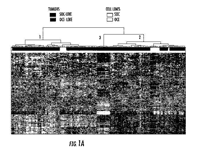
[0023] FIG. 1: Gene expression profiling of ovarian cancer cell lines and
ovarian tumor
samples identifies two major classes. A) Unsupervised hierarchical clustering
of gene expression
data of 37 cell lines and 285 human tissues. Genes with an expression level
that was at least 2-
fold different relative to the median value across tissues in at least 4 cells
were selected for
6
CA 02914026 2015-11-27
WO 2014/197543 PCT/US2014/040806
hierarchical clustering analysis (3,831 gene features). The data are presented
in matrix format in
which rows represent individual genes and columns represent each tissue. Each
square in the
matrix represents the expression level of a gene feature in an individual
tissue or cell line. The
red and green color in cells reflect relative high and low expression levels
respectively as
indicated in the scale bar (log2 transformed scale). Red, blue and black bars
above the heatmap
are human tumor samples; light blue, OCT lines; yellow bar SOC lines. Whereas
SOC cells
(yellow bars) were exclusively within tumor cluster 1 (red A bar), the OCT
cells (light blue bars)
were predominantly within tumor cluster 2 (blue bar). A small subset of tumor
samples formed a
small distinct cluster that did not include any cell lines (black bar). B) The
progression-free and
overall survival analysis data of patients with the ovarian tumors in clusters
1 and 2 in panel A.
The patients with tumors that have a gene expression profile that is similar
to OCT lines (blue
bar, cluster 2 in panel A) have a worse outcome than patients with tumors that
have gene
expression profile similar to SOC lines (red bar, cluster 1 in panel A). The
small subset of tumors
in cluster 3 that did not include any cell lines (black bar) was excluded from
the outcome
analysis.
[0024] FIG. 2: Gene expression profiling and Taxol Response of ovarian
cancer cell lines
identifies two major classes. A) Taxol response of OCT and SOC cell lines in
mRNA/RPPA
(Reverse Phase Protein Analysis) Cluster 1 vs. Cluster 2. The OCT and SOC
lines were plated in
triplicates in WIT-OC medium (5000 cells/well) in 96 well plates. The next day
20 nM Taxol
was added and metabolic activity was measured as 590/530 fluorescence via
Alamar Blue after 5
days. OCT cell lines in mRNA/RPPA Cluster 1 (blue bars), SOC cell lines in
Cluster 2 (red bars),
OCT lines in Cluster 2 (white bars). The results are representative of more
than three four
different experiments. B) Proteins that are over-expressed in OCT mRNA/RPPA
Cluster 1. The
hierarchical clustering of RPPA data from OCT and SOC lines revealed a subset
of proteins that
are over-expressed in OCT lines significantly correlated with Taxol resistance
(Cluster 1, blue
labels; Cluster 2, red labels; OCT-C4p purple label, IC-50, p<0.05, Spearman).
The data are
presented in matrix format in which rows represent cell lines and columns
represent antibody
probes for each protein. The red and green colors reflect relative high and
low expression levels
respectively. C) Hierarchical clustering of mRNA data from OCT and SOC lines.
The mRNA for
the subset of genes associated with Taxol response in the RPPA analysis was
examined. The
mRNA clustering of the cell lines was very similar to RPPA groups. The over
expressed genes
7
CA 02914026 2015-11-27
WO 2014/197543 PCT/US2014/040806
are in red, under expressed genes are in green. A detailed list of genes that
are up-regulated in
each group is provided in Table 2 (list of proteins associated with Taxol
resistance association
that are over-expressed in Taxol resistant Cluster 1 OCT cells compared to
Taxol sensitive class 2
cells in RPPA analysis). Cluster 1, blue labels; Cluster 2, red labels; OCT-
C4p purple label.
[0025] FIG. 3: Gene expression profiling of ovarian cancer cell lines
identifies two major
classes. A) Unsupervised hierarchical clustering of mRNA expression levels of
OCT (blue bars)
and SOC (red bars) ovarian cancer cell lines. The data are presented in matrix
format in which
rows represent individual genes and columns represent each cell line. Each
square in the matrix
represents the expression level of a gene feature in an individual tissue or
cell line. The red and
green color in cells reflect relative high and low expression levels
respectively as indicated in the
scale bar (log2 transformed scale). Two major clusters are observed; Cluster I
contains only
OCT cell lines (left cluster, blue only), and Cluster II contains a mixture of
SOC and OCT cell
lines (right cluster, red and blue). Interestingly, while the papillary serous
histotype almost
exclusively aligned within Cluster I (green bars), the other subtypes were
present in both clusters
(orange bars). B) The dendogram of the cell lines that make up the two
clusters in the heatmap
in panel A. The cell line names are colored as follows; first column OCT
(blue), SOC (red);
second column Papillary Serous (dark green), other histotypes (orange); third
column Papillary
Serous (dark green), Clear Cell (light blue), Endometrioid (pink), mucinous
(light green), other
histotypes (orange).
[0026] Figure 4: The proteomic profile of ovarian cancer cell lines
identifies two major
classes. A) The unsupervised clustering of protein expression (measured by
RPPA) in OCT cell
lines (blue bars) together with SOC ovarian cancer cell lines (red bars)
revealed two distinct
clusters. Rows represent cell lines and columns represent antibody probes for
each protein. The
red and green colors reflect relative high and low expression levels,
respectively. As in the
mRNA clustering, Cluster 1 contains only OCT cell lines (top half of the
heatmap, blue only),
and Cluster 2 contains a mixture of SOC and OCT cell lines (bottom half of the
heatmap, red and
blue). While the papillary serous histotype almost exclusively aligned within
Cluster 1 (green
bars), the non-papillary serous subtypes (orange bars) were divided between
Cluster 1 and
Cluster 2. B) The dendogram of the cell lines that make up the two clusters in
panel A. The cell
line names are colored as follows; Papillary Serous (green), other histotypes
(orange). See also
Figure 8.
8
CA 02914026 2015-11-27
WO 2014/197543 PCT/US2014/040806
[0027] FIG. 5: Histopathology of OCI xenografts recapitulates the original
human tumor. A-
C) H&E stained sections of primary human tumors used to create OCI-P8p
(papillary serous),
OCI-Elp (endometrioid) and OCI-C3x (clear cell). D-F) H&E stained sections of
xenografts
tumors derived by injecting SOC cells (E52, SKOV3, and TOV-112D)
subcutaneously into
immunocompromised mice. The typical features of human adenocarcinomas such as
glands,
papillae, stromal cores, and desmoplastic stroma are absent. G-0) H&E stained
sections of
xenograft tumors derived by injecting OCI cell lines (P5x, P7a, P9a, C5x, C3x,
CSp, Elp)
subcutaneously into immunocopromised mice. In papillary serous specimens note
the presence
of stromal cores and papillary architecture (G, H and I). In the endometrioid
specimen note the
presence of glands (M), which were positive for estrogen receptor (ER) and
mucin (brown),
respectively, consistent with the endometrioid phenotype (N and 0).
[0028] Figure 6: The mRNA expression profiles of OCI cell lines in Cluster
1 and Cluster 2
are associated with distinct pathways. For pathway analysis we used Ingenuity
Pathway Analysis
(IPA) to organize the 823 genes that were significantly differentiate
expressed between Cluster 1
vs. in Cluster 2 (p, 0.05) (Figure 3). A) 558 were up-regulated in Cluster 1
which were
organized in 37 core pathways in IPA (p < 0.05). B) 265 genes were up-
regulated in Cluster 2
which were organized in 37 core pathways in IPA (p < 0.05).
[0029] FIG. 7: Validation of ten probesets associated with unique genes and
over-expressed
in either OCE or FNE in two independent ovarian cancer datasets. (a)
Association of OV-like
and FT-like tumor subclassification in the Wu dataset with clinical
characteristics (P-values from
logistic regression (grade, stage as ordinal variables) and Fisher's Exact
test (histological
subtype)). (b) Association of OV-like and FT-like subgroups in the Tothill
dataset with clinical
features (P-values calculated as in (a)). (c) Kaplan-Meier plots demonstrate
significant
differences in disease-free and overall survival between OV- and FT-like
subgroups in the
Tothill data (univariate P-values from the log-rank test are displayed),In
multivariate analysis,
the OV/FT-like subgroups were independently associated with disease-free
survival (Cox
proportional hazards P=0.01) but not overall survival (P=0.34) after adjusting
for tumor grade,
stage, serous subtype, patient age and residual disease.
[0030] FIG. 8: A series of graphs and a table showing that OCI lines are
significantly more
resistant to a diverse panel of oncology drugs compared to standard cell
lines.
DETAILED DESCRIPTION
9
CA 02914026 2015-11-27
WO 2014/197543 PCT/US2014/040806
[0031] Described herein are assays, methods and kits for analyzing
sensitivity of a subject's
cancerous tumor to a drug, predicting responses of cancerous tumors to drugs,
determining the
prognosis of a subject having a cancerous tumor, and developing a personalized
therapy or
treatment strategy for the subject. The assays, methods and kits involve
analyzing gene and
protein expression signatures or profiles of a subject's cancerous tumor,
testing candidate drugs
in cancerous cells from the subject's cancerous tumor, and classifying a
subject's cancerous
tumor based on ovarian cell and fallopian tube cell cell-of-origin gene
expression signatures.
Using these methods, a suitable drug (or drugs) is identified, the subject can
be treated with that
drug, and a personalized therapy is thus developed for the subject.
Biological and Chemical Methods
[0032] Methods involving conventional molecular biology techniques are
described herein.
Such techniques are generally known in the art and are described in detail in
methodology
treatises such as Molecular Cloning: A Laboratory Manual, 3rd ed., vol. 1-3,
ed. Sambrook et al.,
Cold Spring Harbor Laboratory Press, Cold Spring Harbor, N.Y., 2001; and
Current Protocols in
Molecular Biology, ed. Ausubel et al., Greene Publishing and Wiley-
Interscience, New York,
1992 (with periodic updates). Conventional methods of culturing mammalian
cells are generally
known in the art. Methods of culturing ovarian and fallopian tube cells (e.g.,
ovarian cancer cells
and fallopian tube cancer cells), including preparation and use of WIT-OC cell
culture medium,
are described in detail in PCT application no. PCT/U52012/030446. Any WIT
culture medium
or derivative of WIT culture medium (e.g., WIT-P, WIT-I, WIT-T, WIT-OC, WIT-
OCe, WIT-L
etc.) can be used.
Methods and Assays for Analyzing Sensitivity of a Subject's Cancerous Tumor to
a Drug and
Developing a Personalized Therapy
[0033] Using the methods described herein, a prediction of a particular
drug's (e.g., oncology
drug) effect on a subject's cancerous tumor may be made, based on the
expression profile of a
particular set of proteins in the cancerous tumor, and a comparison to a
control or reference cell
line for which responsiveness to that particular drug is known. Generally, if
a subject's
cancerous tumor has a protein expression profile substantially similar to that
of a control or
reference cell line, and the control or reference cell line is responsive to
treatment with a
CA 02914026 2015-11-27
WO 2014/197543 PCT/US2014/040806
particular drug (e.g., oncology drug such as Taxol), then one can predict that
the subject's
cancerous tumor will also respond to treatment with that particular drug
(e.g., Taxol).
Conversely, if a subject's cancerous tumor has a protein expression profile
substantially similar
to that of a control or reference cell line, and the control or reference cell
line is resistant to
treatment with a particular drug (e.g., Taxol), then one can predict that the
subject's cancerous
tumor will also be resistant to treatment with that particular drug (e.g.,
Taxol).
[0034] A typical method for analyzing sensitivity of a subject's (e.g.,
mammal such as a
human) cancerous tumor to a drug (e.g., Taxol) and developing a personalized
therapy for the
subject includes the steps of: obtaining cancer cells from the subject's
cancerous tumor;
examining expression of a set of proteins or mRNAs in the cancerous cells; and
correlating
overexpression or underexpression of the set of proteins or mRNAs relative to
a control with
resistance of the subject's cancerous tumor to the oncology drug and
correlating normal
expression of the set of proteins or mRNAs relative to the control with
sensitivity of the subject's
cancerous tumor to the oncology drug. The subject may be any animal, e.g.,
mammals such as
human, bovine, canine, ovine, feline, non-human primate, porcine, etc. For
example, the subject
may be a female human having at least one (e.g., one, two, three, etc.)
ovarian cancer tumor.
The cancerous tumor may be any type of cancerous tumor. Examples of cancerous
tumors
include those from ovary, fallopian tube, lung, breast, colon, prostate,
gastrointestinal, endocrine
organ, blood, immune cell, muscle, bone, neural, endothelial, fibroblasts, or
other epithelial and
stromal tumors.
[0035] In this example of a method, the set of proteins or mRNAs includes
proteins whose
overexpression or underexpression relative to a control is associated with
resistance to the drug.
The set of proteins or mRNAs can include a subset of proteins or mRNAs whose
overexpression
is associated with resistance to the drug as well as a subset of proteins or
mRNAs whose
underexpression is associated with resistance to the drug. Typically, the drug
is a known
oncology drug. In one embodiment, the oncology drug is Taxol or vincristine,
and the set of
proteins includes at least two of: tubulin, AKT, androgen receptor, Jun
oncogene, Crystalline,
cyclin D1, epidermal fatty acid binding protein, Ets related gene, FAK,
Forkhead Box 03,
Erk/Mek, N-cadherin, mitogen-activated protein kinase 14, plasminogen
activator inhibitor type
1, paired box 2, protein kinase C-alpha, protein kinase AMP-activated Gamma 2,
phosphatase
and tensin homolog, SMAD3, Sarcoma viral oncogene homolog, signal transducer
and activator
11
CA 02914026 2015-11-27
WO 2014/197543 PCT/US2014/040806
of transcription 3, and signal transducer and activator of transcription 5
(e.g., two or more (i.e.,
two, three, four, five, six, seven, eight, nine, ten, fifteen, twenty, etc.)
of the proteins listed in
Table 1 below). In the experiments described herein, the proteins listed in
Table 1 were found to
be overexpressed in OCT lines in the mRNA/RPPA Cluster 1 and associated with
Taxol
resistance in an RPPA analysis. Use of this method is not limited to Taxol,
however. The same
approach can be applied to any other oncology drug. As shown in Figure 7, the
methods
described herein can be used for any oncology drug, e.g., Taxol, vincristine,
U0126, PJ34,
adriamycin, AS703026, 5-Fluorouracil, cisplatin, PLX4720, etc. Drugs that are
considered off-
label may also be analyzed using the methods.
[0036] Any suitable method of obtaining cancer cells from a subject's
cancerous tumor can
be used. In a typical method, cancer cells are obtained by a biopsy, needle
aspirations, ascites
fluid, or any other fluid containing tumor cells or solid tumor fragments
removed during
surgery. The cancer cells may be also obtained from a xenograft explant. In
some embodiments,
the method is used to simultaneously analyze the sensitivity of cancerous
tumors from multiple
subjects (e.g., 2, 3, 4, 5, 10, 15, 20, 25, 30, 35, 40, 50, 100, 1000, 10,000,
etc.) who have cancer.
In some embodiments, cancer cells from a plurality of subjects can be analyzed
simultaneously,
e.g., in a high-throughput format.
[0037] In the method, any suitable control sample can be used. Typically,
the control sample
is normal cells isolated from the same patient and same tissue, or cell lines
established from
other patients with a known drug response - sensitive or resistant, and
expression of the set of
proteins in the subject's cancerous cells is examined relative to expression
levels of the set of
proteins in this control sample. When referring to "overexpression" of the
proteins in the set of
proteins, what is meant is at least a two-fold increase compared to a control.
Expression of a
particular protein or set of proteins in a sample or population of cancerous
cells can be compared
to a baseline level (also known as a control level) of expression of the
particular protein or set of
proteins (e.g., a protein(s) listed in Table 1). A "baseline level" is a
control level, and in some
embodiments a normal level or a level not observed in subjects having cancer
(e.g., ovarian
cancer) or cell lines that are sensitive to a drug. Alternatively, a "baseline
level" or control level
is a level not observed in a sample from subjects having a different type of
cancer (e.g., ovarian-
like ovarian cancer) than the cancer (e.g., fallopian tube-like ovarian
cancer) of the subject
whose cancerous cells are being analyzed for sensitivity or resistance to an
oncology drug.
12
CA 02914026 2015-11-27
WO 2014/197543 PCT/US2014/040806
Therefore, it can be determined, based on the control or baseline level of
expression of the
particular protein (or set of proteins), whether a sample of cancer cells to
be evaluated for
sensitivity or resistance to a particular drug (e.g., Taxol) has a measurable
increase (i.e.,
overexpression, upregulation), decrease, or substantially no change in
expression of the
particular protein (or set of proteins), as compared to the baseline level.
[0038] Expression of a set of proteins in the cancerous cells can be
analyzed using any
suitable techniques or protocols. For example, a Reverse Phase Protein
Analysis (RPPA) assay
(Zhang et al., Bioinformatics 25, 650-654, 2009) can be used. Conventional
methods of
analyzing protein expression include enzyme-linked detection systems such as
enzyme-linked
immunosorbent assays (ELISAs), fluorescence-based detection systems, Western
blots, ELISAs,
etc. In some embodiments, protein expression can be extrapolated by analyzing
corresponding
mRNA levels. Conventional methods of analyzing mRNA levels include reverse
transcription
polymerase chain reaction (RT-PCR), quantitative PCR, Serial analysis of gene
expression
(SAGE), RNA-Seq, next-generation sequencing, northern blotting, microarrays,
etc.
[0039] In some embodiments, the steps of the method can be repeated for
different oncology
drugs until an oncology drug that the subject's cancerous tumor is sensitive
(responsive) to is
identified. If it turns out a patient's tumor is resistant to Taxol, the
method can be repeated with
a different set of proteins and another oncology drug(s) until an oncology
drug the tumor will
respond to is found. In some embodiments, after determining that a patient's
tumor is resistant to
Taxol, a second oncology drug may instead be administered to the patient
without first testing
resistance of the patient's tumor to the second oncology drug.
[0040] Once a suitable drug (or drugs) is identified, the subject can be
treated with that drug,
and a personalized therapy can be developed for the subject. More
specifically, a treatment can
be selected for the subject based at least in part on a prediction or result
suggesting that a
particular oncology drug will be effective or more effective than one or more
alternative
oncology drugs for that particular subject. For example, if the set of
proteins or mRNAs are
overexpressed or underexpressed in the subject's cancerous tumor relative to
the control sample,
it is determined that the subject's cancerous tumor is not sensitive to (i.e.,
is resistant to) the first
oncology drug (e.g. Taxol), and thus a second oncology drug (e.g.,
vincristine, U0126, PJ34,
adriamycin, A5703026, 5-Fluorouracil, cisplatin, PLX4720, etc.) different from
the first
oncology drug (e.g., Taxol) can be administered to the subject. In another
example, if the set of
13
CA 02914026 2015-11-27
WO 2014/197543 PCT/US2014/040806
proteins or mRNAs are expressed at normal levels in the subject's cancerous
tumor relative to
the control sample, it is determined that the subject's cancerous tumor is
sensitive to the first
oncology drug (e.g., Taxol) and thus, the first oncology drug (e.g., Taxol)
can be administered to
the subject. As there is great biological diversity amongst human tumors,
different tumors
having different gene signatures and molecular features, the methods described
herein are
particularly useful for personalized cancer treatment, including predicting a
subject's response to
a particular drug (e.g., oncology drug), classifying a subject's cancerous
tumor, and choosing an
appropriate treatment strategy as well as predicting the subject's
outcome/survival based on such
characterizations.
[0041] According to the methods, the drug to which the subject's cancerous
tumor is
determined to be responsive can be administered to the subject in combination
with one or more
other oncology drugs and/or treatments (e.g., chemotherapy, radiation therapy,
surgery, etc.). In
some embodiments, the method can further include determining the subject's
prognosis, e.g.,
outcome, survival, disease-free survival. In such an embodiment, the method
further includes
correlating overexpression or underexpression of the set of proteins or mRNAs
relative to the
control sample with a worse prognosis for the subject compared to a second
subject having a
cancerous tumor in which the first set of proteins are normally expressed
relative to the control
sample. Generally, what is meant by a "worse prognosis" or "worse
outcome/survival" is meant
a statistically significant shorter period without relapse, metastasis or
death due to tumor.
[0042] In these methods, after a subject is treated with a drug, at one or
more (e.g., one, two,
three, four, etc.) time points, the subject or a sample from the subject
(e.g., a biopsy, culture) can
be analyzed to determine the subject's response to the drug. In other words,
the subject or a
sample from the subject (e.g., a biopsy, culture) can be analyzed to determine
if the drug is
having a therapeutic effect on the subject, e.g., reducing tumor size and/or
tumor growth and/or
tumor markers. Any suitable methods of analyzing a sample from the subject for
the drug's
therapeutic effect can be used, including those protein and mRNA assays
described herein. Any
suitable methods for analyzing the subject to determine if the drug is having
a therapeutic effect
can be used. Such methods include, for example, physical exams, tumor
biomarkers such as
CA125, and imaging (x-rays, CT scan, PET scan, MRI etc.).
14
CA 02914026 2015-11-27
WO 2014/197543 PCT/US2014/040806
Methods and Assays for Predicting Responses of Cancerous Tumors to Drugs and
Developing
Personalized Therapy for Treatment of Cancerous Tumors
[0043] Described herein are methods (e.g., assays) for predicting a
response of a cancer
patient's cancerous tumor (e.g., ovarian cancer tumor) to a drug (e.g.,
oncology drug) and
developing a personalized therapy for the patient for treatment of the
cancerous tumor.
Generally, cancerous tumor cells obtained from a patient having a cancerous
tumor (e.g.,
obtained from a biopsy or surgery) are cultured in an appropriate medium
(e.g., WIT-OC or
WIT-FO medium) and exposed to a particular drug (or to a combination of
drugs). The effect of
the particular drug (or combination of drugs) on survival and proliferation of
the cancerous
tumor cells is examined in order to make a prediction of the particular drug's
(or the combination
of drugs') likely effect on the patient's cancerous tumor. Using the method, a
treatment can be
selected for the patient based at least in part on a prediction or result
suggesting that a particular
drug (e.g., oncology drug) will be effective or more effective than one or
more alternative drugs
for that particular patient. Such methodology can be used to determine a
patient-specific
response to one or more therapeutic strategies that have been approved for the
treatment of the
medical condition being treated in the patient (e.g., ovarian cancer), as well
as therapies that may
be utilized off-label. Use of the prediction methods described herein allows
for the identification
of optimal personalized treatment strategies for a cancer patient.
[0044] In one embodiment, the method includes predicting a response of a
cancer patient's
(e.g., a female human having an ovarian cancer tumor) cancerous tumor to a
drug (e.g., oncology
drug) and developing a personalized therapy for the patient for treatment of
the cancerous tumor.
A typical method includes the steps of: obtaining cancer cells from the
patient's cancerous
tumor; culturing the cancer cells in WIT-OC cell culture medium (or other WIT
culture medium
or a derivative of a WIT culture medium); contacting the cultured cancer cells
with the drug;
determining an IC50 value (or IC90 value - a dose of drug that kills at least
90% of tumor cells)
for the drug in the cultured cancer cells; and correlating an increased IC50
(or IC90) value
relative to an IC50 (or IC90) value for the drug in control cultured cells
with a poor response of
the patient's cancerous tumor to the drug and correlating a normal or low IC50
(or IC90) value
relative to the IC50 (or IC90) value for the drug in control cultured cells
with a positive response
of the patient's cancerous tumor to the drug. By a "poor response" is meant no
decrease in
CA 02914026 2015-11-27
WO 2014/197543 PCT/US2014/040806
tumor size or tumor markers. A "positive response" means a decrease in tumor
size or tumor
markers.
[0045] In one embodiment in which the patient's cancerous tumor is not
responsive to the
drug being tested, the IC50 value is increased relative to the IC50 value for
the drug in control
cultured cells, and the method further includes administering to the patient a
second drug (i.e., a
drug different from the drug tested to which the cancerous tumor cells
demonstrated a poor
response, e.g., Taxol, vincristine, U0126, PJ34, adriamycin, AS703026, 5-
Fluorouracil,
cisplatin, and PLX4720). In another embodiment, in which the patient's
cancerous tumor is
responsive to the drug being tested, the IC50 value is normal or decreased
relative to the IC50
value for the drug in control cultured cells, and the method further includes
administering the
tested drug (e.g., Taxol, vincristine, U0126, PJ34, adriamycin, AS703026, 5-
Fluorouracil,
cisplatin, and PLX4720) to the patient. The method can also be used for making
a prognosis for
the patient. In such an embodiment, the method can further include correlating
an increased
IC50 value relative to an IC50 value for the drug in control cultured cells
with a worse prognosis
for the patient compared to a second patient having a cancerous tumor in which
an IC50 value
for the drug in cultured cancer cells from the second patient is normal or
decreased relative to the
IC50 value for the drug in control cultured cells.
[0046] Although the experiments described herein involved measuring IC50 or
IC90 values,
other measurements can be taken to predict a response of a cancer patient's
cancerous tumor to a
drug. Any assay that measures survival and/or proliferation of cancer cells in
response to a drug
(e.g., Taxol) can be used. For example, cell number counts, mtt, mtx, alamar
blue, apatosis
assays, cell cycle profiles, etc. can be used.
[0047] As with the other methods described above, the cancer cells may be
obtained from a
xenograft explant, from ascites fluid, biopsy or primary solid ovarian tissue
from the subject. In
some embodiments, the method is used to simultaneously predict responses of
cancerous tumors
from multiple subjects (e.g., 2, 3, 4, 5, 10, 15, 20, 25, 30, 35, 40, 50, 100,
etc.) who have cancer
to a drug (e.g., oncology drug) or combination of drugs. As already mentioned,
a nonexhaustive
list of oncology drugs includes Taxol, vincristine, U0126, PJ34, adriamycin,
AS703026, 5-
Fluorouracil, cisplatin, PLX4720, etc.
[0048] As with the other methods described above, after a subject is
treated with a drug, at
one or more (e.g., one, two, three, four, etc.) time points, the subject or a
sample from the subject
16
CA 02914026 2015-11-27
WO 2014/197543 PCT/US2014/040806
(e.g., a biopsy, culture) can be analyzed to determine the subject's response
to the drug. In other
words, the subject or a sample from the subject (e.g., a biopsy, culture) can
be analyzed to
determine if the drug is having a therapeutic effect on the subject, e.g.,
reducing tumor size
and/or tumor growth and/or tumor markers. Any suitable methods of analyzing a
sample from
the subject for the drug's therapeutic effect can be used, including those
protein and mRNA
assays described herein. Any suitable methods for analyzing the subject to
determine if the drug
is having a therapeutic effect can be used. Such methods include, for example,
physical exams,
tumor biomarkers such as CA125, and imaging (x-rays, CT scan, PET scan, MRI
etc.).
Methods for Determining the Prognosis of a Subject Having an Ovarian Cancer
Tumor
[0049] One embodiment of a method for determining a prognosis of a subject
(e.g., female
human) having an ovarian cancer tumor involves generation of a gene expression
signature or
profile for the subject's ovarian cancer tumor, and classifying the ovarian
cancer tumor as
fallopian tube-like or ovary-like. In the experiments described in Example 3
below, a cell-of-
origin gene expression signature that distinguishes normal human ovarian (OV)
and fallopian
tube (FT) epithelial cells within the same subject (e.g., patient) was
identified, and it was shown
that application of the OV vs. FT cell-of-origin gene signature to gene
expression profiles of
primary ovarian cancers permits identification of distinct OV and FT-like
subgroups among
these cancers. The experiments further showed that the normal FT-like tumor
classification
correlated with a significantly worse disease-free survival, and thus,
applying this classification
to a gene expression signature or profile of a subject's cancerous tumor can
be used for
determining a prognosis for the subject (e.g., female human).
[0050] In one example of such a method, the method includes the steps of:
obtaining a
sample from the subject's tumor; subjecting the sample to gene expression
profiling resulting in
an expression profile including a first set of genes that are upregulated in
fallopian tube cells
relative to ovarian cells and a second set of genes that are upregulated in
ovarian cells relative to
fallopian tube cells; determining expression levels of the first and second
sets of genes; and
correlating an upregulation of the first set of genes and normal expression of
the second set of
genes with a worse disease-free survival prognosis (e.g., statistically
significant shorter period
without relapse, metastasis or death due to tumor) relative to a second
subject having an ovarian
cancer tumor in which the first set of genes are not upregulated and the
second set of genes are
17
CA 02914026 2015-11-27
WO 2014/197543 PCT/US2014/040806
upregulated. In the method, the first set of genes typically includes all of
DOK5, CD47, HS6ST3,
DPP6, and OSBPL3, as these genes were found to be overexpressed in cultured
fallopian tube
cells compared to cultured ovarian cells. If other genes are also found to be
overexpressed in
cultured fallopian tube cells compared to cultured ovarian cells, the first
set of genes can then
include one or more (e.g., one, two, three, four, five) of DOK5, CD47, HS6ST3,
DPP6, and
OSBPL3 in combination with one or more other genes that are overexpressed in
cultured
fallopian tube cells compared to cultured ovarian cells. The second set of
genes typically
includes STC2, SFRP1, SLC35F3, SHMT2, and TMEM164, as these genes were found
to be
overexpressed in cultured ovarian cells compared to cultured fallopian tube
cells. If other genes
are also found to be overexpressed in cultured ovarian cells compared to
cultured fallopian tube
cells, the second set of genes can then include one or more (e.g., one, two,
three, four, five) of
STC2, SFRP1, SLC35F3, SHMT2, and TMEM164, in combination with one or more
other genes
that are overexpressed in cultured ovarian cells compared to cultured
fallopian tube cells.
However, any suitable genes can be analyzed, as long as they are
differentially expressed
between fallopian tube cells and ovarian cells. Quantitative sensitive methods
such as PCR and
RNA sequencing, for example, can be used to examine other suitable genes that
are differentially
expressed between fallopian tube and ovary; gene expression profiling can be
performed using
any suitable methods, including any of those described herein.
[0051] In an embodiment in which the first set of genes in the expression
profile is
upregulated but the second set of genes is not upregulated, the method can
further include
classifying the subject's ovarian cancer tumor as fallopian tube-like. In
another embodiment in
which the second set of genes in the expression profile is upregulated but the
first set of genes is
not upregulated, the method can further include classifying the subject's
ovarian cancer tumor as
ovary-like. As shown in the experiments described in Example 3 below,
fallopian tube-like
tumors were of significantly higher stage, higher grade and were predominantly
composed of
serous adenocarcinomas, while in contrast, ovary-like tumors included non-
serous subtypes and
lower grade cancers. Thus, the correlation can be made between a subject's
ovarian cancer
tumor being a fallopian tube-like tumor, and a poor prognosis for the subject.
If the subject's
ovarian cancer tumor is ovary-like, the subject is expected to have a better
prognosis, (a longer
period without relapse, metastasis or death due to tumor).
18
CA 02914026 2015-11-27
WO 2014/197543 PCT/US2014/040806
[0052] As with the other methods described herein, the method can further
include treating
the subject with one or more oncology drugs and/or treatments (e.g.,
chemotherapy, radiation
therapy, surgery, etc.). After the subject is treated with a drug, at one or
more (e.g., one, two,
three, four, etc.) time points, the subject or a sample from the subject
(e.g., a biopsy, culture) can
be analyzed to determine the subject's response to the drug. In other words,
the subject or a
sample from the subject (e.g., a biopsy, culture) can be analyzed to determine
if the drug is
having a therapeutic effect on the subject, e.g., reducing tumor size and/or
tumor growth and/or
tumor markers. Any suitable methods of analyzing a sample from the subject for
the drug's
therapeutic effect can be used, including those protein and mRNA assays
described herein. Any
suitable methods for analyzing the subject to determine if the drug is having
a therapeutic effect
can be used. Such methods include, for example, physical exams, tumor
biomarkers such as
CA125, and imaging (x-rays, CT scan, PET scan, MRI etc.).
Kits
[0053] Kits for analyzing sensitivity of a subject's cancerous tumor to an
oncology drug
(predicting a response of a cancer patient's cancerous tumor to an oncology
drug) and
developing a personalized therapy for the subject are described herein. A
typical kit for
determining if a subject's cancerous tumor is sensitive or resistant to a
particular oncology drug
(e.g., Taxol) includes at least one control such as one more OCT lines as an
internal control(s);
instructions for use; and WIT medium, or a derivative of WIT medium. Although
an OCT line is
typically included as a control, any suitable control(s) can be used.
Additionally, the kit may
contain one or more (e.g., one, two, three, four, five, ten, twenty, etc.)
probes. For example, the
kit may include one or more probes for use in a multiplexed PCR assay, for
example, in which
several probes are used simultaneously. Probes that are specific for
particular proteins can be
used. For example, the one or more probes can be at least two probes specific
to at least two
(e.g., two, three, four, five, six, etc.) of the following proteins: tubulin,
AKT, androgen receptor,
Jun oncogene, Crystalline, cyclin D1, epidermal fatty acid binding protein,
Ets related gene,
FAK, Forkhead Box 03, Erk/Mek, N-cadherin, mitogen-activated protein kinase
14,
plasminogen activator inhibitor type 1, paired box 2, protein kinase C-alpha,
protein kinase
AMP-activated Gamma 2, phosphatase and tensin homolog, SMAD3, Sarcoma viral
oncogene
homolog, signal transducer and activator of transcription 3, and signal
transducer and activator of
19
CA 02914026 2015-11-27
WO 2014/197543 PCT/US2014/040806
transcription 5. Optionally, kits may also contain one or more of the
following: containers which
include positive controls, containers which include negative controls,
photographs or images of
representative examples of positive results and photographs or images of
representative
examples of negative results.
Data and Analysis
[0054] Use of the assays, methods and kits described herein may employ
conventional
biology methods, software and systems. Useful computer software products
typically include
computer readable medium having computer-executable instructions for
performing logic steps
of a method. Suitable computer readable medium include floppy disk, CD-
ROM/DVD/DVD-
ROM, hard-disk drive, flash memory, ROM/RAM, magnetic tapes and etc. The
computer
executable instructions may be written in a suitable computer language or
combination of several
languages. Basic computational biology methods are described in, for example
Setubal and
Meidanis et al., Introduction to Computational Biology Methods (PWS Publishing
Company,
Boston, 1997); Salzberg, Searles, Kasif, (Ed.), Computational Methods in
Molecular Biology,
(Elsevier, Amsterdam, 1998); Rashidi and Buehler, Bioinformatics Basics:
Application in
Biological Science and Medicine (CRC Press, London, 2000) and Ouelette and
Bzevanis
Bioinformatics: A Practical Guide for Analysis of Gene and Proteins (Wiley &
Sons, Inc., 2nd
ed., 2001). See U.S. Pat. No. 6,420,108.
[0055] The assays, methods and kits described herein may also make use of
various
computer program products and software for a variety of purposes, such as
reagent design,
management of data, analysis, and instrument operation. See, U.S. Pat. Nos.
5,593,839,
5,795,716, 5,733,729, 5,974,164, 6,066,454, 6,090,555, 6,185,561, 6,188,783,
6,223,127,
6,229,911 and 6,308,170. Additionally, the embodiments described herein
include methods for
providing data (e.g., experimental results, analyses) and other types of
information over networks
such as the Internet.
EXAMPLES
[0056] The present invention is further illustrated by the following
specific examples. The
examples are provided for illustration only and should not be construed as
limiting the scope of
the invention in any way.
CA 02914026 2015-11-27
WO 2014/197543 PCT/US2014/040806
Example 1 ¨ An in vitro test for Taxol sensitivity in ovarian tumor cell lines
that retain the
phenotype of primary tumors.
[0057] The inability to establish stable cell lines from the vast majority
of human tumors has
limited the use of in vitro models to study human cancer. Currently available
tumor cell lines
fail to represent the biological diversity of human tumors. We previously
developed a cell
culture medium and methods that enabled us to routinely establish cell lines
in more than 95% of
cases and from diverse subtypes of ovarian tumors. Importantly, the 25 ovarian
tumor cell lines
described herein retained the genomic landscape and histopathology of the
original tumors, and
their molecular features.
[0058] Described herein is the use of these cell lines to predict a
patient's response
(including patients' responses) to drugs. We have determined that the drug
response of the cell
lines we have established correlated with patient outcomes. Thus, tumor cell
lines derived using
this methodology represent a significantly improved new platform to test and
potentially predict
patient response to treatment. A robust and efficient model system that
predicts patient response
to various drugs would greatly improve development of new drugs for
personalized treatment of
cancer patients. The cell lines we established represent a more malignant,
drug-resistant cancer
phenotype than has been previously represented in tumor cell line panels.
Thus, tumor cell lines
derived using this methodology represent a significantly improved new platform
to study human
tumor biology and treatment.
[0059] We previously developed a new culture system for common human
cancers both by
the ostensible need for improved model systems and by the encouraging results
with a new
chemically-defined culture medium (WIT) we described previously (Ince et al.,
Cancer Cell /2,
160-170, 2007). This medium provides all the essential nutrients for
maintaining basic cellular
metabolism without undefined supplements such as serum, pituitary extract,
feeder layers,
conditioned medium or drugs (Ince et al., Cancer Cell /2, 160-170, 2007). In
WIT medium
normal human breast epithelial cells could reach beyond seventy population
doublings, a nearly
1021-fold expansion of cell numbers. These results encouraged us to
hypothesize that perhaps
human tumors could also be grown routinely in such a medium.
[0060] For the purposes of this report, all ovarian cancer cell lines
derived using standard
culture medium and methods will collectively be referred to as "standard
ovarian carcinoma" cell
lines, or SOC cell lines, including the 26 SOC lines available from the
American Tissue Type
21
CA 02914026 2015-11-27
WO 2014/197543 PCT/US2014/040806
Collection (ATCC) and the European Collection of Cell Cultures (ECACC). The
set of ovarian
cancer cell lines derived using WIT-OC medium will be referred to as "OCT"
cell lines. In two
cases the bulk of the tumor mass was located in the fallopian tubes, these
cell lines are referred to
as "FCI" cell lines.
RESULTS
[0061] mRNA gene expression profile of the OCT tumor cell lines resembles
human tumors
with distinct clinical characteristics. Examination of the OCT and SOC cell
line panel together
with 285 human ovarian tumor specimens revealed three distinct patient
clusters. Patient Cluster
1 included only OCT lines, and Cluster 2 included all the SOC lines. None of
the cell lines were
in Cluster 3 (Figure la). The distribution of the cell lines within human
tumor samples was
identical to the in vitro cell line clusters, except a single cell line (OCT-
C4p), strongly indicating
that the in vitro phenotype of these cell lines may reflect relevant in vivo
clinical differences.
Furthermore, the comparison of the clinical outcomes of these two groups of
patients revealed
that the patients with OCT-like tumors in Cluster 1 had a significantly
shorter progression free
and overall survival than tumors in Cluster 2 with an SOC-like profile in
multivariate analysis
(Figure lb).
[0062] Response of tumor cell lines in mRNA/RPPA Clusters 1 and 2 to Taxol:
The striking
correlation between poor patient outcomes and OCT lines in mRNA/RPPA Cluster 1
prompted us
to test the response of these cell lines to Taxol and Cisplatin, which are two
of the most
commonly-used drugs for ovarian cancer. We selected a panel of lines that
correspond to OCT
lines in mRNA/RPPA Cluster 1 and SOC lines in mRNA/RPPA Cluster 2; each panel
included
examples of different tumor subtypes (PS, CC, CS, E, M), and tissue sources
(solid tumors,
ascites fluid, and xenograft explants). In these experiments we observed that
the IC50 for Taxol
in OCT lines in mRNA/RPPA Cluster 1 ranged > 10-100 nM, which was > 5-10 fold
higher than
the IC50 values in SOC lines in mRNA/RPPA Cluster 2. The SOC IC50 values for
Taxol in
these experiments were consistent with previous reports. The subset of OCT
lines in Cluster 2
were also more sensitive to Taxol compared to OCT lines in Cluster 1, similar
to SOC lines
(Figure 2a). Both OCT and SOC lines were plated in WIT-OC medium for the above
experiments. Thus, we infer that the differences in drug response are not a
consequence of
different growth media. Importantly, we found that the response to another
microtubule
22
CA 02914026 2015-11-27
WO 2014/197543 PCT/US2014/040806
inhibiting drug, Vincristine, was similarly different between OCT and SOC
lines. In contrast, we
did not find a significant difference in the response to Cisplatin between OCT
and SOC lines.
[0063] In order to explore the basis for the relative Taxol resistance of
OCT cells we
compared the protein profiles Clusterl/OCI cells with Cluster2/SOC lines since
they had the
largest IC50 differences. There was a strong correlation between protein
expression levels and
Taxol response of 46 proteins and IC50 values. Among these, we concentrated on
22
proteins that were over-expressed in Cluster 1 (Figure 2b, Table 1).
Reassuringly, Tubulin,
which is the target of Taxol, was in this group of proteins. Furthermore, 11
additional proteins in
this group had been previously associated with Taxol resistance in disparate
studies including
AKT, p38, AKT, PTEN, Src, SMAD3, STAT3, STAT5. The unsupervised hierarchical
clustering of the mRNA microarray data including the list of genes from the
resistance-
associated protein signature was also able to distinguish identical cell line
groups in Clusters 1
and 2 (Figure 2c). Using functional protein network association software, we
found that the
majority of these over-expressed proteins either directly or indirectly
interact with each other.
The amino acid sequences of these proteins are well known in the art.
[0064] Table 1 - The list of proteins that are over-expressed in OCT lines
in the
mRNA/RPPA Cluster 1 and associated with Taxol resistance in RPPA analysis.
RPPA Evidence for
Antibody Gene Name Association with Potential Interactome Role
Probe Taxol Resistance
Murphy et al.,
Biochimica et
Biophysica Acta
1784 (2008) Tubulin over-expressed and mutated in
Taxol
1184-1191; resistant cells (Sangraijrang
Chemotherapy
a.Tubulin Tubulin
L'esperance (2000)46:327-334; Orr Oncogene (2003)
22,
International 7280-7295)
Journal of
Oncology 29: 5-
24,2006
Lin et al. Br. J. Rapamycin with paclitaxel displayed
synergistic
Cancer (2003) effects (Aissat et al., Cancer
Chemother
88:973-980; Liu Pharmacol (2008) 62:305-313; Liu et
al.,
AKT AKT et al., Oncogene Oncogene (2006c) 25:3565-3575).
(2006c) 25:3565¨ Constitutively active Akt contributes to
3575; Jiang et al., Vincristine Resistance (Zhang Cancer
Drug Resist Investigation, 28:156-165,2010). Akt
induces
Updat. (2008) survival in paclitaxel treated cells
(Bava et al.,
23
CA 02914026 2015-11-27
WO 2014/197543 PCT/US2014/040806
11(3): 63-76; The International Journal of
Biochemistry &
Bava 2009 Cell Biology 43 (2011) 331-341). Akt
directly
regulates the transcriptional activity of c-Jun
(Shin et al., Mol Cancer Res. 2009, 7(5):745-
54)
Androgen Androgen receptor is activated by STAT3
AR.C19.
Receptor (Ueda J Biol Chem. 2002, 277(9):7076-
85).
Paclitaxel-
resistant Human
A physical interaction of Stat3 with c-Jun has
Ovarian Cancer
been reported both in vitro and in vivo. Stat3
Cells Undergo c-
and c-Jun cooperated to yield maximal enhancer
Jun NH2-terminal
c.JUN_pS Jun Kinase-mediated function, point mutations of Stat3
within the
73 oncogene interacting domains blocked both
physical
Apoptosis (Zhou
interaction of Stat3 with c-Jun and their
Biol Chem. Vol.
277, No. 42, cooperation (Zhang et al., Mol Cell
Biol. 1999,
19(10):7138-46)
39777-39785,
2002)
Subunits of crystallin interact with tubulin
subunits to regulate the equilibrium between
Crystalline Crystalline
tubulin and microtubules (Houck Clark JI
(2010) PLoS ONE 5(7): e11795).
Cyclin D1 promotes anchorage-independent cell
survival by inhibiting FOX03-mediated anoikis
Cyclin.D1 Cyclin D1
(Gan et al., Cell Death Differ. 2009, 16(10):
1408-1417).
Epidermal Liu et al., J E-FABP expression that is blocked by
mitogen-
E.FABP.0 fatty acid Neurochem. 2008, activated protein kinase kinase (MEK)
inhibitor
20. binding 106(5): 2015¨ U0126 (Liu et al., J Neurochem. 2008,
106(5):
protein 2029 2015-2029).
Lu et al., J Expression of EGR-1 mediated by p38MAPK
Huazhong Univ pathway plays a critical role in
paclitaxel
Ets Related
Erg. 1_2_3 Sci Technolog resistance of ovarian carcinoma cells (Lu
et al.,
Gene
Med Sci. 2008, J Huazhong Univ Sci Technolog Med Sci.
2008,
28(4):451-5 28(4):451-5)
Halder et al., Clin Docetaxel induces FAK cleavage in taxane-
FAK_pY3 Cancer Res sensitive ovarian cancer cells but not
in taxane-
FAK
97 2005;11:8829- resistant cells (Halder et al., Clin
Cancer Res
8836 2005;11:8829-8836).
ERK promotes tumorigenesis by inhibiting
Forkhead
FOX03a FOX03a (Yang et al., nature cell biology vol.
Box 03
10(2), 2008).
McDaid Cancer MEK inhibitor CI-1040 potentiates
efficacy of
MAPK_p Res 65:2854- Taxol in xenograft tumor modes (McDaid,
Erk/Mek
T202 2860, 2005; 2005). RNAi screening identified Erkl
as
Bauer et al. Breast enhancing paclitaxel activity (Bauer et al.
24
CA 02914026 2015-11-27
WO 2014/197543 PCT/US2014/040806
Cancer Research Breast Cancer Research 2010, 12:R41).
2010, 12:R41; Xu Inactivation of ERK is necessary for the
2009 enhancement of paclitaxel cytotoxicity
by
U0126 (McDaid Cancer Res 65:2854-2860,
2005).
Rosano et al.,
N-Cadherin is over-expressed in Taxol resistant
N.Cadheri Cancer Res; 17(8);
N-Cadherin
n 2350-60. 2011 cells (Rosano et al., Cancer Res;
17(8); 2350¨
60. 2011 AACR).
AACR
Constitutive increase of p38-MAPK was found
in vincristine-resistant cells. Inhibition of p38-
Mitogen- MAPK by SB202190 reduced increased the
p38_pT18 activated sensitivity of cells to chemotherapy
(Guo et al.,
protein BMC Cancer 2008, 8:375; Lu et al., J
Huazhong
kinase 14 Univ Sci Technolog Med Sci. 2008,
28(4):451-
5). p38 MAP kinase phosphorylates c-Jun (Lo et
al., Mol Nutr Food Res. 2007, 51(12):1452-60).
Plasminoge MEK/ERK1/2 and SMAD3 was essential for
n activator PAT-1 induction initiated by
microtubule
PAI.1
inhibitor disruption (Samarakoon et al., Cell
Signal. 2009
type 1 June ; 21(6): 986-995).
PAX2 expression correlated with enhanced
Paired Box
PAX2 Buttiglieri 2003 resistance against apoptotic signals and with
the
2
proinvasive phenotype (Buttiglieri 2003).
Purified protein kinase C phosphorylates
Protein
microtubule-associated protein 2. (Akiyama et
PKCa Kinase C -
al., J Biol Chem (1986) Vol. 261, No. 33,
alpha
15648-15651).
Protein
Kinase
PRKAG2 AMP-
Activated
Gamma 2
Phosphatase Silencing Akt in PTEN-mutated prostate
cancer
PTEN.138
and Tensin cells enhances the antitumor effects of
Taxol
G50.
homolog (Priulla et al., The Prostate 67:782-
789 (2007)).
Increased
expression in
Paclitaxel resistant SMAD3 binds to microtubules (Dong et al.,
cells (Kashkin et Molecular Cell, Vol. 5, 27-34, 2000). SMAD3
SMAD3 SMAD3 al., Doklady and SMAD4 cooperate with c-Jun/c-Fos to
Biochemistry and mediated transcription (Zhang et al., Nature.
Biophysics, 2011, 1998, 394(6696):909-13).
Vol. 437, pp. 105-
108)
SRC Sarcoma Knockdown of Src SRC activates STAT3 (Cao 1996). STAT3
CA 02914026 2015-11-27
WO 2014/197543 PCT/US2014/040806
viral enhanced siRNA inhibited Bc1-2 expression (Choi
et al.,
oncogene paclitaxel- Exp Mol Med. 2009, 41(2):94-101). Bc1-
2
homolog mediated growth down-regulation is associated with
Paclitaxel
inhibition in reesistance (Ferlini et al., Molecular
ovarian cancer Pharmacology Vol. 64, No. 1, 51-58,
2003).
cells (Le et al., Constitutive activation of Stat3 by
the Src
Cancer Biology & causes growth of breast carcinoma cells (Garcia
Therapy 12:4, Oncogene (2001) 20, 2499 -2513).
Dasatinib
260-269, 2011; has synergistic activity with
paclitaxel in
Chen 2005) ovarian cancer cells (Teoh, et al.
Gynecologic
Oncology 121 (2011) 187-192).
STAT3 is activated by ERK1 and and induces
Signal STAT3 activation AKT. STAT3 binds the C-terminal
tubulin (Ng
Transducer through Src leads et al Biochem J. 2009, 425(1):95-
105).
STAT3 and to Taxol resistance Knockdown of Stat3 reduces AKT1
expression
Activator of (Hawthorne Mol (Park et al., J Biol Chem. Vol. 280,
No. 47,
Transcriptio Cancer Res 2009, 38932-38941, 2005) STAT3 is induced by
n 3 7(4)) Src (Zhang et al., JBC Vol. 275, No.
32,
24935-24944, 2000).
Signal
Transducer
and STAT5 was shown to activate cyclin D1
gene
STAT5 expression (Magne et al., Mol Cell
Biol. 2003,
Activator of
23(24):8934-45).
Transcriptio
n 5
[0065] Table 2 ¨ Proteins with Taxol resistance association over-expressed
in Cluster 1
Proteins with Taxol resistance association over-expressed in Cluster 1
Function and Association with Taxol Resistance
Reference
a. Tubulin Tubulin: Target for binding of Taxol.
Murphy et al.,
Biochimica et
Biophysica Acta
1784 (2008) 1184-
1191;
L'esperance
International Journal
of Oncology 29: 5-
24,2006
26
CA 02914026 2015-11-27
WO 2014/197543 PCT/US2014/040806
AKT Rapamycin with paclitaxel displayed synergistic Akt induces
effects survival in
(Aissat 2008; Liu 2006; Zhang 2010; Priulla 2007) paclitaxel treated
Akt directly regulates the transcriptional activity of c- cells (Bava
Jun (Shin 2009) 2011).
c.JUN_p573 Stat3 and c-Jun cooperate to yield maximal enhancer
function.
Cyclin.D1 Cyclin D1 promotes anchorage-independent cell (GAN 2009).
survival
by inhibiting FOX03-mediated anoikis
E.FABP.C20. E-FABP expression that is blocked by mitogen- Liu 2008
activated
protein kinase kinase (MEK) inhibitor U0126.
Erg.1_2_3 Lu 2008
FAK_pY397 Halder 2005
FOX03a ERK promotes tumorigenesis by inhibiting FOX03a (Yang 2008)
MAPK_pT202 McDaid 2005, Xu
2009
N.Cadherin Rosano 2011
P38_pT180 Constitutive increase of p38-MAPK was found in (Guo 2008, Lu
vincristine-resistant cells. Inhibition of p38- 2008, Lo 2007)
MAPK by SB202190 increased the sensitivity
of cells to chemotherapy
PAX2 Buttiglieri 2003
PTEN.138G50. Priulla 2007
SMAD3 SMAD3 binds to microtubules (Dong 2000) SMAD3 Kashkin 2011
and
SMAD4 cooperate with c-Jun/c-Fos to
mediated transcription (Zhang 1998)
SRC SRC activates STAT3 (Cao 1996) STAT3 siRNA Teoh 2011, Le
inhibited 2011, Fournier
Bc1-2 expression (Choi 2009). Constitutive 2011, Chen
activation of Stat3 by the Src causes growth of 2005,
breast carcinoma cells (Garcia 2001). Hawthorne 2009
27
CA 02914026 2015-11-27
WO 2014/197543 PCT/US2014/040806
STAT3 STAT3 is activated by ERK1 and induces AKT. Hawthorne 2009
STAT3 binds the C-terminal tubulin (Ng 2009).
Knockdown of Stat3 reduces AKT1 expression
(Park 2005)
[0066] As described in Figures 1 and 2, we developed a test to tell which
patients will
respond to Taxol, which is the first line drug for ovarian cancer (it is also
used for many other
cancers including breast). In one embodiment this test may be in the form of
analyzing the
expression of the proteins that are listed in Figure 1 in patient tumors. In
another embodiment, it
may take the form of making cell lines from patients and carrying out an in
vitro test to
determine the IC50 on the cell lines as we show in these figures.
Example 2 ¨ Characterization of novel ovarian tumor cell lines that retain the
phenotype of
primary tumors
[0067] Currently available human tumor cell line panels consist of a small
number of lines
that generally fail to retain the phenotype of the original patient tumor. We
have developed a cell
culture medium that enables us to routinely establish cell lines from diverse
subtypes of human
ovarian cancers with >95% percent efficiency. Importantly, the 25 ovarian
tumor cell lines
described here retained the genomic landscape, histopathology, and molecular
features of the
original tumors. Furthermore, the molecular profile and drug response of these
cell lines
correlated with distinct groups of primary tumors with different outcomes.
Thus, tumor cell lines
derived using this new methodology represent a significantly improved new
platform to study
human tumor biology and treatment.
[0068] Human carcinomas that grow uncontrollably in the body are
paradoxically difficult to
grow in cell culture. A robust and efficient cell line model system that
predicts a patient's
response to various drugs would greatly improve development of new drugs for
personalized
treatment of cancer patients. The cell lines we established capture the in
vivo heterogeneity of
human ovarian tumors and correspond to a more malignant, drug-resistant cancer
phenotype than
standard ovarian cancer cell lines.
[0069] We set out to develop a new culture system for common human cancers,
driven both
by the clear need for improved in vitro models and by the encouraging results
with the WIT
medium that we described previously (Ince et al., Cancer Cell /2, 160-170,
2007). These results
encouraged us to hypothesize that perhaps human tumors could also be grown
consistently in
28
CA 02914026 2015-11-27
WO 2014/197543 PCT/US2014/040806
such a medium. This report characterizes the phenotypic properties of 25 new
continuous OCT
derived using cell culture media (WIT-OC) optimized for human ovarian cancer
subtypes.
RESULTS
[0070] Tumor cells fail to thrive in standard cell culture media.
Consistent with prior
reports, we were able to establish tumor cell lines in standard culture media
only with less than
one percent success rate. In the single successful case, the ovarian tumor
line OCI-Ul a was
derived in RPMI medium; in which a brief period of rapid growth (days 0-20),
was followed by
growth arrest (days 20-40), widespread cell death (days 40-50), and the
eventual emergence of
rapidly growing rare clones that gave rise to a continuous cell line (days 60-
90). Importantly, the
copy number variants (CNV) measured in the genome of the cell line grown in
RPMI varied
significantly from those found in the starting tumor cell population,
consistent with clonal
outgrowth of select subpopulations or the acquisition of additional genetic
aberrations during
tissue culture. Consistent with the experience of others in this field, over
the course of this nearly
ten year-long study, this was the only patient tumor specimen that yielded a
continuous ovarian
tumor cell line using standard media.
[0071] Optimization of culture conditions for ovarian tumor cells in WIT
medium. We also
discovered that the original WIT medium developed for breast cells (Ince et
al., Cancer Cell /2,
160-170, 2007) did not support the growth of either normal ovarian cells or
tumors of the ovary.
Normal human breast cells, like most normal epithelium, are never in direct
contact with blood
or serum under physiologic conditions. Accordingly, the medium we developed
for normal
breast cells was completely devoid of serum in order to more closely
approximate the
physiologic environment. In contrast, normal ovarian and fallopian tube
epithelial cells are
known to be directly in contact with normal peritoneal fluid, which contains a
physiologic serum
protein concentrations that can be as high as fifty percent of the levels
present in the circulating
blood. Indeed, in many cases the ovarian tumors grow in malignant ascites
fluid that has
concentrations of proteins and growth factors that are actually higher than
those present in
serum. Thus, we added serum into WIT medium in order to mimic the physiologic
growth
conditions of malignant ovarian cells. However, supplementation of WIT medium
with serum
was not sufficient for growth of ovarian tumor cells, without additional
factors. After testing
many modifications over the years, we found that a combination of factors
including particular
concentrations of serum, insulin, hydrocortisone, EGF, cholera toxin, estrogen
were also
29
CA 02914026 2015-11-27
WO 2014/197543 PCT/US2014/040806
necessary but not sufficient to culture different ovarian tumor types with
high efficiency. In
addition to medium optimization, it was necessary to optimize 02 levels and
the cell attachment
surfaces, because while endometrioid and mucinous histotypes of ovarian tumors
were best
cultured in low 02 conditions (5-10%), the serous, clear cell and other
subtypes proliferated best
in ambient 02 (18-21%). Lastly, we found that a modified plastic surface
(Primaria, BD)
performed best for the culture of primary ovarian tumors.
[0072] It is worth noting that during the course of this work we discovered
that optimizing
individual culture medium variables one at a time resulted in small
improvements in culture
success. Nevertheless, several components that had little effect on culture
success by themselves
had a large combined effect. However, these synergistic effects were difficult
to predict and
empirically testing all possible combinations of multiple components was
prohibitive due to very
large number of permutations. In addition, optimization of conditions on one
tumor sample did
not ensure universal success, since a particular variable was sometimes
dispensable for some
tumor samples, and absolutely essential for the successful culture of others.
Lastly, effects of
changing culture conditions or medium formulation sometimes became apparent
after successive
passages. Thus, a very time consuming aspect of this process was the need to
test combinations
of variables in multiple primary ovarian cancer samples over many passages to
ensure broad
applicability across all ovarian cancer subtypes. These four factors: (1) the
non-obvious nature
of synergistic combinatorial effects of medium components, (2) the need to
test long-term effects
of each variable over many months, (3) the necessity to test each variable in
multiple tumor
samples, and (4) the very large number of permutations to test, precluded an
incremental
systematic approach to the development of WIT-OC media. Nevertheless, once the
medium and
cell culture conditions were optimized, only one sample out of twenty-six
failed to generate a
cell line.
[0073] OCI cell lines encompass the major histological subtypes of ovarian
cancer.
Adenocarcinoma of the ovary is a heterogeneous disease that is comprised of
many
histopathological subtypes with distinct features. In many cases the original
subtype of tumor
that gave rise to most of the "standard ovarian carcinoma" (SOC) cell lines is
unknown. In this
study we used the small subset of SOC cell lines in which the histologic
subtype is known. In
order to distinguish the cell lines derived using WIT-OC medium from SOC
lines, they will be
referred to as "OCI" cell lines. In two cases the bulk of the tumor was
located in the fallopian
CA 02914026 2015-11-27
WO 2014/197543 PCT/US2014/040806
tubes ¨ these cell lines are referred to as "FCI" cell lines. The capital
letter after the ovarian
carcinoma designation "OCT" refers to the histological subtype of the original
tumor. The OCT
panel includes papillary serous (P), clear cell (C), endometrioid (E),
mucinous (M) cancers, and
rare types such as carcinosarcoma (CS) and dysgerminoma (D). Together, the P,
C, E and M
subtypes account for more than ninety percent of ovarian adenocarcinomas;
accordingly this
panel of cell lines is broadly representative of ovarian cancer. The lower
case letter at the end of
each cell line name refers to tissue source; 14 of the cell lines were
established from primary
solid tumors (p), seven from ascites fluid (a), and four from primary mouse
xenografts derived
from direct implantation of human tumors into immunocompromised mice (x). All
25 OCT lines
were able to form colonies in soft agar, consistent with retention of a
transformed phenotype in
culture.
[0074] In WIT-OC medium the tumor cells were able to proliferate
immediately, suggesting
that most of the tumor cells proliferated without significant in vitro clonal
selection or a need to
acquire additional genomic or epigenetic aberrations. Furthermore, it was
possible to culture
these cells continuously for 30-100 population doublings with no decrease in
growth rate; we
have not yet identified an upper limit of population doublings.
[0075] Standard media fail to support OCT cell lines. We observed that none
of the OCT
lines we tested could be cultured in existing standard media. In contrast, all
of the SOC lines we
tested could be cultured in WIT-OC medium. Until now, none of the standard
media support the
culture of all of the existing SOC lines, making it difficult to compare a
large panel of SOC lines
with one another because they require being cultured in a variety of different
media. Our results
indicate that WIT-OC medium has the potential to serve as a universal culture
medium for SOC
lines facilitating comparisons across cell lines.
[0076] OCT cell lines mirror the genomic landscape of the original tumor.
Major genetic
alterations may accumulate during cell culture in standard media. In order to
compare tumor vs.
cell line genomes, we examined their loss-of-heterozygosity (LOH) profiles and
found that each
OCT cell line exhibited remarkable similarity to its corresponding uncultured
tumor sample. In
several cases there were especially striking similarities between the cell
line and tumor (OCI-
M1p/TM1, OCT-P2a/TP2, OCT-C2p/TC2, OCT-EP1/TEP1). Two of the OCT lines (OCT-
Ulp and
P5x) and their matched tumors had large-scale alterations that involved whole
chromosome
arms. In contrast, the remaining ten OCT lines and their matched tumors
contained genomic
31
CA 02914026 2015-11-27
WO 2014/197543 PCT/US2014/040806
regions of LOH that spanned narrow regions of the chromosomes. There was a
more than 90%
identity between the LOH pattern of the uncultured tumor and the matching cell
lines, except in
four cases, there were significant differences in the LOH profile between
cultured cells and the
primary tumor. The overall CVN trends were similar between OCT lines and
ovarian tumors; in
both data sets CNV trend was copy number gain in chromosomes 2, 19, 20 and
copy number loss
in chromosomes 4, 9, 13, 15, and 18. The remaining chromosomes had a more
complex pattern.
Interestingly, while copy number losses were predominant in short arm of
chromosomes 3 and 8,
gains were predominant in the long arm, and this pattern was replicated in OCT
lines. These
results are consistent with the LOH comparison between OCT lines and their
matched tumors and
indicate that the genomic landscape of primary tumors are preserved in OCT
lines. Because the
fragment of tumor from which the cell line is established is necessarily
different than the
fragment of tumor from which the DNA is isolated, it is possible that intra-
tumoral genetic
heterogeneity may be responsible for some of these differences. It is also
possible that in a few
cases some of the changes may be due to accumulation of genetic alterations
during culture, even
though there was no noticeable difference in the growth rate among these lines
and the other OCT
lines.
[0077] We were not able to compare the DNA of the SOC lines to the matched
original
tumor DNA because these cells were established decades ago and the original
tumor sample is
not available. Next we compared copy number variation (CNV) patterns of OCT
cells with the
ovarian tumors analyzed in the TCGA dataset. Consistent with the LOH analysis,
the overall
CNV trends of the OCT lines was similar to primary tumor samples.
[0078] A persistent problem in the cell culture field has been cross-
contamination and
misidentification of lines, existing in up to 15-20% of published reports. The
close genomic
match between OCT lines and the original tumor tissues ensure that the OCT
lines were each
derived from a unique patient. Furthermore, we sequenced the mitochondrial DNA
of the OCT
and SOC lines to provide a permanent unique identifier for authentication of
these cell lines.
Overall, these results indicate that a majority of the OCT cell lines
faithfully preserve the genetic
alterations present in the tissue.
[0079] OCT and SOC lines possess different gene expression signatures.
Unsupervised
hierarchical clustering of the mRNA expression data of 25 OCT and six SOC
lines revealed two
32
CA 02914026 2015-11-27
WO 2014/197543 PCT/US2014/040806
major clusters; 558 and 265 genes were found up-regulated in clusters 1 and 2
respectively
(Figure 3, Table 3).
[0080] Table 3 - List of 20 most up and down regulated mRNAs in Cluster 1.
The complete
dataset is available through the NIH' s Gene Expression Omnibus, GEO accession
number
GSE40785.
Cluster#1, Up regulated genes fold
HSD11B1 hydroxysteroid (11-beta) dehydrogenase 1 32.20
DIRAS 3 DIRAS family, GTP-binding RAS-like 3 27.35
HSD11B1 hydroxysteroid (11-beta) dehydrogenase 1 23.89
HSD11B1 hydroxysteroid (11-beta) dehydrogenase 1 22.85
COL1A2 collagen, type I, alpha 2 22.61
COL1A2 collagen, type I, alpha 2 19.01
COL1A1 collagen, type I, alpha 1 12.53
THBS2 thrombospondin 2 11.51
ANXA8 annexin A8 11.46
BDKRB1 bradykinin receptor B1 11.34
RARRES 1 retinoic acid receptor responder 1 9.23
COL5A1 collagen, type V, alpha 1 9.11
C13orf33 chromosome 13 open reading frame 33 8.58
ANXA8 annexin A8 (ANXA8) 8.40
SPARC secreted protein, acidic, cysteine-rich (osteonectin)
8.27
SERPINE1 serpin peptidase inhibitor 1 8.18
CPA4 carboxypeptidase A4 7.85
RARRES 1 retinoic acid receptor responder 1 7.80
WISP1 WNT1 inducible signaling pathway protein 1 7.74
L00652846 imilar to Annexin A8 (Vascular anticoagulant-beta) 7.53
Cluster #1, Down regulated genes fold
CD24 CD24 molecule (CD24) 0.17
QPRT quinolinate phosphoribosyltransferase 0.29
IGF2BP3 insulin-like growth factor 2 mRNA binding protein 3 0.32
PITX1 paired-like homeodomain transcription factor 1 0.35
SPINT2 serine peptidase inhibitor, Kunitz type, 2 0.38
IMPA2 inositol(myo)-1(or 4)-monophosphatase 2 0.40
CLDN1 claudin 1 0.41
CTSL2 cathepsin L2 0.41
HIS T1H4C histone cluster 1, H4c 0.43
ABLIM1 actin binding LIM protein 1 0.43
AURKB aurora kinase B 0.43
CMTM8 CKLF-like MARVEL TM 8 0.43
SDC1 syndecan 1 (SDC1), transcript variant 1 0.44
UBE2C ubiquitin-conjugating enzyme E2C 0.44
AIF1L allo graft inflammatory factor 1-like 0.44
33
CA 02914026 2015-11-27
WO 2014/197543 PCT/US2014/040806
HES4 hairy and enhancer of split 4 (Drosophila) 0.44
UBE2C ubiquitin-conjugating enzyme E2C 0.44
CDCA5 cell division cycle associated 5 0.44
DBNDD1 dysbindin, dystrobrevin binding protein 1 0.44
CDCA7 cell division cycle associated 7 0.45
[0081] Table
4 - List of 20 up and down regulated mRNAs in Cluster 2. The complete
dataset is available at the NIH's Gene Expression Omnibus, GEO accession
number GSE40785.
Cluster #2, Up regulated genes fold
TACSTD1 tumor-associated calcium signal transducer 1 12.42
EPCAM epithelial cell adhesion molecule 9.81
SPINT2 serine peptidase inhibitor, Kunitz type, 2 9.24
S100A4 S100 calcium binding protein A4 8.67
TACSTD2 tumor-associated calcium signal transducer 2 7.71
CDH1 cadherin 1, type 1, E-cadherin 6.49
MAL mal, T-cell differentiation protein 6.16
ZIC2 Zinc family member 2 (Drosophila) 6.03
ClOorf58 chromosome 10 open reading frame 58 5.98
MAL2 mal, T-cell differentiation protein 2 5.94
S100A4 S100 calcium binding protein A4 5.93
UGT2B7 UDP glucuronosyltransferase 2 family, polypeptide B7 5.72
APOE apolipoprotein E 5.71
SPP1 secreted phosphoprotein 1 5.70
GLDC glycine dehydrogenase 5.48
SPP1 secreted phosphoprotein 1 5.44
UCP2 uncoupling protein 2 (mitochondrial, proton carrier) 5.39
MDK midkine (neurite growth-promoting factor 2) 5.39
ALDH 1A1 aldehyde dehydrogenase 1 family, member Al 5.13
FAM84B family with sequence similarity 84, member B 5.08
Cluster #2, Down regulated genes fold
TMEM98 transmembrane protein 98 0.07
EFEMP1 EGF-containing fibulin-like extracellular matrix protein 1
0.07
DCN decorin (DCN), transcript variant C 0.07
CDH11 cadherin 11, type 2, OB-cadherin (osteoblast) 0.09
MT1E metallothionein lE (MT1E) 0.10
COL3A1 collagen, type III, alpha 1 (COL3A1) 0.10
ALDH1A3 aldehyde dehydrogenase 1 family, member A3 0.10
IGFBP4 insulin-like growth factor binding protein 4 0.11
COL5A2 collagen, type V, alpha 2 0.11
PDPN podoplanin (PDPN) 0.12
PLOD2 procollagen-lysine, 2-oxoglutarate 5-dioxygenase 2 0.12
SPOCK1 sparc/osteonectin, (testican) 1 0.12
FLNC filamin C, gamma (actin binding protein 280) 0.12
CRISPLD2 cysteine-rich secretory protein LCCL domain containing 2 0.14
34
CA 02914026 2015-11-27
WO 2014/197543 PCT/US2014/040806
DKK3 dickkopf homolog 3 (Xenopus laevis) ( 0.14
RAC2 rho family, small GTP binding protein Rac2 0.14
SGK1 serum/glucocorticoid regulated kinase 1 0.15
DPYSL3 dihydropyrimidinase-like 3 0.15
SGK1 serum/glucocorticoid regulated kinase 1 0.15
PLIN2 perilipin 2 0.15
[0082] Cluster 1 contained only OCT lines (Figure 3a-b). Most of the OCT
papillary serous
lines were in this cluster (10/12) (Figure 3a-b, Cluster 1). In contrast,
Cluster 2 was
predominantly composed of non-papillary serous tumors (10/13), and contained
the entire SOC
panel of cell line samples (12/12) (Figure 3a-b).
[0083] Consistent with the above results, others have shown that while mRNA
profiles of
human serous cancers constitute a distinct group, the profiles of a small
subset of endometrioid
and clear cell tumors overlap those of papillary serous tumors. Reminiscent of
this pattern, some
endometrioid and clear cell OCT lines were associated with papillary serous-
dominant Cluster 1,
suggesting that some tumors classified as endometrioid and clear cell
histologically may have a
papillary serous-like gene expression signature (Figure 3b, Cluster 1). The
mRNA expression
profile of OCT lines in Cluster 2 resembled the SOC lines (Figure 3a-b).
Notably, three out of
four xenograft-derived OCT-lines were in Cluster 2 (Figure 3a-b, C3x, C5x, and
P5x), suggesting
that the cell lines derived from xenograft explants (Cluster 2) have distinct
expression profiles
compared to cell lines established from primary tumors (Cluster 1).
[0084] Importantly, SOC lines that were cultured in both standard media and
WIT-OC
clustered next to one another, indicating that the differences observed
between OCT and SOC
lines were not due to differences in constituents contained in the culture
medium. (Figure 3b).
The mRNA probes that were up-regulated and down regulated in cluster 1 vs.
cluster 2 involved
37 and 41 pathways respectively (p < 0.05) (Figure 6a); including NF-kB,
CXCR4, IGF-1, Rho-
GDI, ILK, and IL-8 signaling (cluster 1); Notch, BRCA, GADD45, Granzyme and
Stathmin
signaling (cluster 2) among others (Figure 6b). In summary, Cluster 1
generally correlated with
papillary serous histology, OCT cell lines and primary tumor-derived lines;
Cluster 2 correlated
with non-papillary serous histology, SOC cell lines and xenograft explants.
[0085] Protein and mRNA expression profiles identify the same OCT cell line
clusters. We
next examined the expression levels of 226 proteins representing major
signaling pathways using
Reverse Phase Protein Analysis (RPPA). The unsupervised hierarchical
clustering of the protein
CA 02914026 2015-11-27
WO 2014/197543 PCT/US2014/040806
expression data revealed two major clusters; once again Cluster 1 contained
only OCT lines as
well as most of the papillary serous lines (10/12) (Figure 4). In contrast,
Cluster 2 was
predominantly non-papillary serous lines (10/13), and contained all of the SOC
lines (6/6)
(Figure 4). See Tables 5 and 6 for lists of differentially expressed genes
between Clusters 1 and
2. To assess reproducibility of these phenotypes, the protein extracts were
prepared in triplicate
from three different passages across two experiments; in each case we observed
that the
replicates from each cell line clustered together. Comparison of the cell
lines' mRNA and RPPA
profiles also revealed a remarkable degree of consistency in molecular
phenotypes. The mRNA
and RPPA clusters were identical with one exception (OCT-C4p). These results
indicate that the
molecular differences between OCT vs. SOC lines are stable and reproducible
across passages
and analytical platforms.
Table 5- PATHWAYS UPREGULATED IN CLUSTER 1
Ingenuity Canonical Pathways Molecules
IGFBP4,VCAM1,CTGF,TNFRSF1A,FGF2,ACTA2,B
Hepatic Fibrosis / Hepatic Stellate Cell
AMBI,VEGFC,IGFBP5,IL1R1,MYL9,TGFBR2,COL
Activation 1A2,COL1A1,CCL2,TIMP1,COL3A1
Inhibition of Matrix Metalloproteases
TIMP4,MMP23B,TIIVIP1,RECK,THBS2,TFPI2,LRP1
RAC2,PARVA,RALA,TS PANS ,AS AP1,DIRAS 3 ,AC
TA2,ITGA5,TLN1,MYLK,MYL9,RRAS2,RND3,RH
Integrin Signaling OU,CAV1,ZYX,CAPN2
Chondroitin and Dermatan
Biosynthesis CHSY3,CHPF,CSGALNACT1
VCAM1,RRAS 2,CCL2,RND3,TNFRS FlA,DIRAS 3,
HMGB1 Signaling RHOU,IL1R1,SERPINE1,PLAT
TGFBR2,RAC2,CDH2,RRAS2,RND3,TUBB6,TNFR
Germ Cell-Sertoli Cell Junction SF1A,DIRAS 3 ,TUBB 2A,ACTA2,RHOU,ZYX,RAB 8
Signaling B
PARVA,TNFRSF1A,DIRAS3,ACTA2,VEGFC,HIF1
A,MYL9,TGFB1I1,RND3,FLNC,S NAI2,RHOU,IRS 2
ILK Signaling ,PTGS2
36
CA 02914026 2015-11-27
WO 2014/197543 PCT/US2014/040806
RAC2,VCAM1,DIRAS3,VEGFC,MAP4K4,IRAK3,G
NAI2,MYL9,GNB4,RRAS2,RND3,RHOU,GNG2,PT
IL-8 Signaling G52
Ascorbate Recycling (Cytosolic) GLRX,GSTO1
DIRAS3,ACTA2,ITGA5,CDH11,GNAI2,MYL9,GN
RhoGDI Signaling B4,CDH2,RND3,PIP5K1C,RHOU,GNG2,ARHGEF3
Regulation of Actin-based Motility by MYLK,MYL9,RAC2,RND3,PIP5K1C,DIRAS3,ACT
Rho A2,RHOU
GNAI2,S1PR3,GNB4,RRAS2,RALA,LPAR1,CAV1,
Gai Signaling RGS4,GNG2,FPR1
Epithelial Adherens Junction MYL9,TGFBR2,CDH2,NOTCH2,RRAS2,TUBB6,S
Signaling NAI2,ACTA2,TUBB2A,ACVR1,ZYX
Glioma Invasiveness Signaling TIMP4,RRAS2,RND3,TIMP1,DIRAS3,RHOU
IGFBP4,IGFBP6,NEDD4,CTGF,RRAS2,IGFBP5,IRS
IGF-1 Signaling 2,CYR61
UDP-N-acetyl-D-glucosamine
Biosynthesis II UAP1,GFPT2
Glycogen Biosynthesis II (from UDP-
D-Glucose) UGP2,GBE1
DIRAS3,ACTA2,ITGA5,CDH11,GNAI2,MYLK,MY
L9,GNB4,CDH2,RND3,PIP5K1C,RHOU,GNG2,AR
Signaling by Rho Family GTPases HGEF3
IL33,TGFBR2,GHR,RRAS2,TNFRSF1A,TNFAIP3,
NF-KB Signaling MAP4K4,IL1R1,IRAK3,CARD11,TNFSF13B
Complement System SERPING1,CD59,C1S,CFI
TGFBR2,GNB4,RRAS2,MMP23B,RND3,TNFRSF1
Colorectal Cancer Metastasis A,DIRAS3,RHOU,VEGFC,GNG2,PTGS2,PTGER2,L
Signaling RP1,WNT5A
Remodeling of Epithelial Adherens
Junctions RALA,TUBB6,ACTA2,TUBB2A,ZYX,MAPRE3
GNAI2,S1PR3,RND3,DIRAS3,CASP1,RHOU,CASP
Sphingosine-l-phosphate Signaling 4,SMPD1
Virus Entry via Endocytic Pathways RAC2,RRAS2,ITSN1,FLNC,ACTA2,CAV1,ITGA5
37
CA 02914026 2015-11-27
WO 2014/197543 PCT/US2014/040806
GNAI2,MYL9,GNB4,RRAS2,RND3,DIRAS3,CXCL
CXCR4 Signaling 12,RHOU,ITPR1,GNG2
Semaphorin Signaling in Neurons RND3,DPYSL3,DPYSL4,DIRAS3,RHOU
Role of Macrophages, Fibroblasts and VCAM1,TNFRSF1A,FGF2,CXCL12,VEGFC,IRAK3
Endothelial Cells in Rheumatoid ,IL1R1,IL33,R0R2,TRAF3IP2,RRAS2,CCL2,DKK3,
Arthritis LRP1,TNFSF13B,WNT5A
Dermatan Sulfate Biosynthesis CHSY3,CHPF,CSGALNACT1,HS35T3A1,DSE
Ephrin B Signaling GNAI2,RAC2,GNB4,ITSN1,CXCL12,GNG2
Prostanoid Biosynthesis PTGIS,PTGS2
Regulation of Cellular Mechanics by
Calpain Protease RRAS2,ITGA5,TLN1,CAPN2,CAST
Actin Nucleation by ARP-WASP
Complex RRAS2,RND3,DIRAS3,RHOU,ITGA5
SLIT3,RAC2,PAPPA,PLXNA3,ITSN1,TUBB2A,CX
CL12,VEGFC,ITGA5,MYL9,GNAI2,GNB4,ADAMT
56,RRAS2,TUBB6,ABLIM3,RTN4,ADAM19,GNG2,
Axonal Guidance Signaling BMP1,WNT5A
GNAI2,MYLK,MYL9,GNB4,RRAS2,RND3,DIRAS3
Thrombin Signaling ,RHOU,ARHGEF3,ITPR1,GNG2
Role of IL-17F in Allergic
Inflammatory Airway Diseases TRAF3IP2,CCL2,RPS6KA2,CXCL6
GNAI2,RAC2,CD99,TIMP4,VCAM1,MMP23B,JAM
Leukocyte Extravasation Signaling 3,TIIVIP1,ACTA2,CXCL12,THY1
Cholecystokinin/Gastrin-mediated
Signaling IL33,RRAS2,RND3,DIRAS3,RHOU,PTGS2,ITPR1
Antiproliferative Role of Somatostatin
Receptor 2 GNB4,RRAS2,CDKN1A,GNG2,NPR2
Chondroitin Sulfate Biosynthesis (Late
Stages) CHS Y3,CHPF,CSGALNACT1,HS 3S T3A1
GNAI2,RAC2,GNB4,RRAS2,ITSN1,CXCL12,ITGA
Ephrin Receptor Signaling 5,VEGFC,MAP4K4,GNG2
FAK Signaling RRAS2,ASAP1,ACTA2,ITGA5,TLN1,CAPN2
Intrinsic Prothrombin Activation
Pathway COL1A2,COL1A1,COL3A1
38
CA 02914026 2015-11-27
WO 2014/197543 PCT/US2014/040806
GNAI2,RRAS2,TUBB6,LPAR1,ACTA2,TUBB2A,C
Gap Junction Signaling AV1,ITPR1,NPR2
Sulfate Activation for Sulfonation PAPSS2
Fatty Acid a-oxidation ALDH1A3,PTGS2
Chemokine Signaling GNAI2,CCL13,RRAS2,CCL2,CXCL12
PPAR Signaling IL33,RRAS2,TNFRSF1A,MAP4K4,IL1R1,PTGS2
Colanic Acid Building Blocks
Biosynthesis UGP2,PMM1
LPS/IL-1 Mediated Inhibition of RXR IL33,ALDH1A3,TNFRSF1A,ACSL4,IL1R1,ABCC3,
Function PAPSS2,HS3ST3A1,ABCA1 ,GST01,MA0A
IL33,COL1A1,RRAS2,TNFRSF1A,MAP4K4,IL1R1,
IL-6 Signaling TNFAIP6
Caveolar-mediated Endocytosis
Signaling ITSN1,FLNC,ACTA2,CAV1,ITGA5
IL-17 Signaling TRAF3IP2,RRAS2,CCL2,TIIVIP1,PTGS2
Chondroitin Sulfate Biosynthesis CHSY3,CHPF,CSGALNACT1,HS35T3A1
Triacylglycerol Biosynthesis PPAPDC1A,GPAM,PPAP2A
Breast Cancer Regulation by GNAI2,GNB4,RRAS2,TUBB6,PPP1R3C,CDKN1A,
Stathminl TUBB2A,ARHGEF3,ITPR1,GNG2
Tryptophan Degradation X
(Mammalian, via Tryptamine) ALDH1A3,MA0A
Putrescine Degradation III ALDH1A3,MA0A
Glutathione Redox Reactions II GLRX
IL33,COL1A2,COL1A1,VCAM1,CCL2,CXCL12,C0
Atherosclerosis Signaling L3A1
Toll-like Receptor Signaling TICAM2,TNFAIP3,MAP4K4,IRAK3
y-linolenate Biosynthesis II (Animals) CYB5R3,ACSL4
Coagulation System SERPINE1,BDKRB1,PLAT
MYL9,GNB4,RRAS2,RALA,RND3,DIRAS3,RHOU,
Phospholipase C Signaling ITGA5,ARHGEF3,ITPR1,GNG2
Arsenate Detoxification I
(Glutaredoxin) GSTO1
Melatonin Degradation II MAOA
39
CA 02914026 2015-11-27
WO 2014/197543 PCT/US2014/040806
RAC2,RALA,APH1B,DIRAS3,HIF1A,TGFBR2,GN
AI2,RRAS2,RND3,CDKN1A,RHOU,ARHGEF3,LRP
Molecular Mechanisms of Cancer 1,BMP1,WNT5A
fMLP Signaling in Neutrophils GNAI2,GNB4,RRAS2,ITPR1,GNG2,FPR1
TR/RXR Activation KLF9,COL6A3,SLC16A2,HIF1A,RCAN2
a-Adrenergic Signaling GNAI2,GNB4,RRAS2,ITPR1,GNG2
MYLK,MYL9,RAC2,RRAS2,PIP5K1C,FGF2,ACTA
Actin Cytoskeleton Signaling 2,ITGA5,TLN1,ARHGAP24
Dopamine Degradation ALDH1A3,MA0A
Bladder Cancer Signaling MMP23B,RRAS2,FGF2,CDKN1A,VEGFC
Pyruvate Fermentation to Lactate LDHA
Tetrapyrrole Biosynthesis II ALAD
Glycerol Degradation I GK
Purine Ribonucleosides Degradation
to Ribose-1-phosphate PGM2
Tyrosine Degradation I FAH
G Beta Gamma Signaling GNAI2,GNB4,RRAS2,CAV1,GNG2
CCR3 Signaling in Eosinophils GNAI2,MYLK,GNB4,RRAS2,ITPR1,GNG2
TGFBR2,GHR,RRAS2,ACVR1,MAP4K4,IL1R1,GK,
PPARa/RXRa Activation ABCA1
RhoA Signaling MYLK,MYL9,LPAR1,RND3,PIP5K1C,ACTA2
Agrin Interactions at Neuromuscular
Junction RAC2,RRAS2,ACTA2,ITGA5
IL-1 Signaling GNAI2,GNB4,IL1R1,IRAK3,GNG2
p38 MAPK Signaling IL33,TGFBR2,TNFRSF1A,IL1R1,RPS6KA2,IRAK3
GNAI2,MYL9,TGFBR2,GNB4,HAND2,RRAS2,RN
Cardiac Hypertrophy Signaling D3,DIRAS3,RHOU,GNG2
IL33,SERPING1,RRAS2,TNFRSF1A,C15 ,OSMR,IL
Acute Phase Response Signaling 1R1,SERPINE1
MSP-RON Signaling Pathway CCL2,ACTA2,MST1
Thioredoxin Pathway TXNRD1
GDP-glucose Biosynthesis PGM2
GDP-mannose Biosynthesis PMM1
GDNF Family Ligand-Receptor RRAS2,DOK4,IRS2,ITPR1
CA 02914026 2015-11-27
WO 2014/197543 PCT/US2014/040806
Interactions
PTEN Signaling TGFBR2,RAC2,GHR,RRAS2,CDKN1A,ITGA5
RRAS2,RND3,DIRAS3,CDKN1A,RHOU,ITPR1,WN
Glioblastoma Multiforme Signaling T5A
Gaq Signaling GNB4,RND3,DIRAS3,RHOU,RGS4,ITPR1,GNG2
LXR/RXR Activation IL33,CCL2,TNFRSF1A,IL1R1,PTGS2,ABCA1
Glucose and Glucose-l-phosphate
Degradation PGM2
Sphingomyelin Metabolism SMPD1
Sertoli Cell-Sertoli Cell Junction RRAS2,TUBB6,JAM3,TNFRSF1A,ACTA2,TUBB2A
Signaling ,ITGA5,RAB8B
GNAI2,TGFBR2,RCAN1,GNB4,RRAS2,ITPR1,GN
Role of NFAT in Cardiac Hypertrophy G2,RCAN2
Paxillin Signaling PARVA,RRAS2,ACTA2,ITGA5,TLN1
TGFBR2,VCAM1,CCL13,RRAS2,CCL2,SMARCA2,
Glucocorticoid Receptor Signaling SGK1,CDKN1A,PTGS2,SERPINE1,FKBP5
Serotonin Degradation ALDH1A3,CSGALNACT1,MA0A
HIFI a Signaling MMP23B,RRAS2,VEGFC,HIF1A,LDHA
Clathrin-mediated Endocytosis SH3BP4,PIP5K1C,FGF2,ACTA2,ITGA5,DAB2,VEG
Signaling FC,SH3KBP1
RRAS2,RND3,DIRAS3,RHOU,VEGFC,HIF1A,RPS6
mTOR Signaling KA2,RPS27L
VDR/RXR Activation IGFBP6,WT1,CDKN1A,IGFBP5
Ceramide Signaling S1PR3,RRAS2,TNFRSF1A,SMPD1
Role of IL-17A in Arthritis CCL2,PTGS2,CXCL6
Calcium Transport I ATP2B4
Heme Biosynthesis II ALAD
UDP-N-acetyl-D-galactosamine
Biosynthesis II UAP1
Pancreatic Adenocarcinoma Signaling TGFBR2,RALA,CDKN1A,VEGFC,PTGS2
TREM1 Signaling CCL2,CASP1,ITGA5
G Protein Signaling Mediated by
Tubby GNB4,GNG2
41
CA 02914026 2015-11-27
WO 2014/197543 PCT/US2014/040806
Role of Tissue Factor in Cancer CTGF,RRAS2,VEGFC,RPS6KA2,CYR61
Human Embryonic Stem Cell
Pluripotency 51PR3,TGFBR2,FGF2,ACVR1,WNT5A,BMP1
Role of Osteoblasts, Osteoclasts and
IL33,COL1A1,DKK3,TNFRSF1A,ITGA5,IL1R1,LR
Chondrocytes in Rheumatoid Arthritis P1,WNT5A,BMP1
Role of JAK2 in Hormone-like
Cytokine Signaling GHR,IR52
Noradrenaline and Adrenaline
Degradation ALDH1A3,MA0A
Glycogen Degradation II PGM2
TGF- f3 Signaling TGFBR2,RRAS2,ACVR1,SERPINE1
Circadian Rhythm Signaling ARNTL,BHLHE40
Role of NFAT in Regulation of the GNAI2,RCAN1,GNB4,RRAS2,ITPR1,GNG2,RCAN
Immune Response 2
Oncostatin M Signaling RRAS2,0SMR
Neuregulin Signaling RRAS2,DCN,ITGA5,ERRFI1
GNAI2,RRAS2,CASP1,CASP4,PTGER2,PTGS2,ITP
Endothelin-1 Signaling R1
Role of JAK1 and JAK3 in yc
Cytokine Signaling IL7R,RRAS2,IR52
Factors Promoting Cardiogenesis in
Vertebrates TGFBR2,ACVR1,LRP1,BMP1
IL-17A Signaling in Fibroblasts TRAF3IP2,CCL2
Eicosanoid Signaling PTGIS,PTGER2,PTGS2
PAK Signaling MYLK,MYL9,RRAS2,ITGA5
Apoptosis Signaling RRAS2,TNFRSF1A,MAP4K4,CAPN2
MYL9,RCAN1 ,ACTA2,TPM2,ITPR1,RCAN2,ATP2
Calcium Signaling B4
Glycogen Degradation III PGM2
Histamine Degradation ALDH1A3
Guanosine Nucleotides Degradation
III A0X1
VEGF Signaling RRAS2,ACTA2,VEGFC,HIF1A
Ga12/13 Signaling MYL9,CDH2,RRAS2,LPAR1,CDH11
ERK5 Signaling RRAS2,SGK1,RPS6KA2
Notch Signaling NOTCH2,APH1B
Role of IL-17A in Psoriasis CXCL6
Fatty Acid Activation ACSL4
Urate Biosynthesis/Inosine 5'-
phosphate Degradation A0X1
42
CA 02914026 2015-11-27
WO 2014/197543 PCT/US2014/040806
NRF2-mediated Oxidative Stress RRAS2,ACTA2,HSPB8,A0X1,FKBP5,TXNRD1,GS
Response TO1
p53 Signaling WT1,SNAI2,CDKN1A,SERPINE2
Fcy Receptor-mediated Phagocytosis
in Macrophages and Monocytes MY05A,RAC2,ACTA2,TLN1
SAPK/JNK Signaling RAC2,RRAS2,MAP4K4,GNG2
Netrin Signaling RAC2,ABLIM3
Role of MAPK Signaling in the
Pathogenesis of Influenza RRAS2,CCL2,PTGS2
Aldosterone Signaling in Epithelial
Cells NEDD4,PIP5K1C,SGK1,HSPB8,ITPR1,HSPB6
Phenylalanine Degradation IV
(Mammalian, via Side Chain) MAOA
Adenosine Nucleotides Degradation II A0X1
CCR5 Signaling in Macrophages GNAI2,GNB4,GNG2
Tight Junction Signaling MYLK,MYL9,TGFBR2,JAM3,TNFRSF1A,ACTA2
Mechanisms of Viral Exit from Host
Cells NEDD4,ACTA2
IL-10 Signaling IL33,MAP4K4,IL1R1
eNOS Signaling LPAR1,CAV1,VEGFC,ITPR1,BDKRB1
Role of
Hypercytokinemia/hyperchemokinemi
a in the Pathogenesis of Influenza IL33,CCL2
Dermatan Sulfate Biosynthesis (Late
Stages) HS3ST3A1,DSE
CDP-diacylglycerol Biosynthesis I GPAM
Oxidative Ethanol Degradation III ALDH1A3
Melanoma Signaling RRAS2,CDKN1A
GNB4,SGK1,CASP1,CASP4,CAPN2,ITPR1,GNG2,S
Huntington's Disease Signaling NAP25
GNAI2,S1PR3,GPER,PDE7B,LPAR1,RGS4,PTGER
cAMP-mediated signaling 2,FPR1
Nicotine Degradation III CS GALNACTLA OX1
Phosphatidylglycerol Biosynthesis II
(Non-plastidic) GPAM
Purine Nucleotides Degradation II
(Aerobic) A0X1
Mitochondrial L-carnitine Shuttle
Pathway ACSL4
Ethanol Degradation IV ALDH1A3
Nitric Oxide Signaling in the
Cardiovascular System CAV1,VEGFC,ITPR1
43
CA 02914026 2015-11-27
WO 2014/197543 PCT/US2014/040806
Relaxin Signaling GNAI2,GNB4,PDE7B,GNG2,NPR2
GNAI2,S 1PR3,GPER,RRAS 2,PDE7B,LPAR1,RGS 4,
G-Protein Coupled Receptor Signaling PTGER2,FPR1
Differential Regulation of Cytokine
Production in Macrophages and T
Helper Cells by IL-17A and IL-17F CCL2
Hepatic Cholestasis IL33,TNFRSF1A,IL1R1,IRAK3,ABCC3
TNFR1 Signaling TNFRSF1A,TNFAIP3
Wnt/I3-catenin Signaling TGFBR2,CDH2,DKK3,ACVR1,LRP1,WNT5A
Lipid Antigen Presentation by CD1 CD1D
GADD45 Signaling CDKN1A
Regulation of IL-2 Expression in
Activated and Anergic T Lymphocytes TGFBR2,RRAS2,CARD11
Renin-Angiotensin Signaling RRAS2,CCL2,PTGER2,ITPR1
Gas Signaling GNB4,GPER,PTGER2,GNG2
Corticotropin Releasing Hormone
Signaling GNAI2,PTGS2,ITPR1,NPR2
CNTF Signaling RRAS2,RPS6KA2
Dendritic Cell Maturation IL33,COL1A2,COL1A1,TNFRSF1A,COL3A1,CD1D
Androgen Signaling GNAI2,GNB4,TGFB1I1,GNG2
Nicotine Degradation II CS GALNACTLA OX1
Amyloid Processing APH1B,CAPN2
Superpathway of Melatonin
Degradation CSGALNACT1,MA0A
Type II Diabetes Mellitus Signaling TNFRSF1A,ACSL4,IRS2,SMPD1
Glycolysis I EN02
14-3-3-mediated Signaling RRAS2,TUBB6,TNFRSF1A,TUBB2A
GNAI2,MYLK,MYL9,TGFBR2,GNB4,PDE7B,FLN
Protein Kinase A Signaling C,PPP1R3C,KDELR3,PTGS2,ITPR1,GNG2
Differential Regulation of Cytokine
Production in Intestinal Epithelial
Cells by IL-17A and IL-17F CCL2
Glutathione-mediated Detoxification GSTO1
Gluconeogenesis I EN02
P2Y Purigenic Receptor Signaling
Pathway GNAI2,GNB4,RRAS2,GNG2
44
CA 02914026 2015-11-27
WO 2014/197543 PCT/US2014/040806
Thrombopoietin Signaling RRAS2,IR52
Tumoricidal Function of Hepatic
Natural Killer Cells SRGN
Estrogen-mediated S-phase Entry CDKN1A
Thyroid Hormone Metabolism II (via
Conjugation and/or Degradation) CS GALNACT 1
PI3K/AKT Signaling RRAS2,CDKN1A,ITGA5,PTGS2
Role of JAK family kinases in IL-6-
type Cytokine Signaling OSMR
Calcium-induced T Lymphocyte
Apoptosis CAPN2,ITPR1
ErbB4 Signaling RRAS2,APH1B
Death Receptor Signaling TNFRSF1A,MAP4K4
ATM Signaling CDKN 1A,TDP 1
Antiproliferative Role of TOB in T
Cell Signaling TGFBR2
D-myo-inositol (1,4,5)-Trisphosphate
Biosynthesis PIP5K1C
Chronic Myeloid Leukemia Signaling TGFBR2,RRAS2,CDKN1A
Role of BRCA1 in DNA Damage
Response SMARCA2,CDKN1A
B Cell Development IL7R
TNFR2 Signaling TNFAIP3
Retinoate Biosynthesis I ALDH1A3
Fatty Acid 13-oxidation I ACSL4
Ethanol Degradation II ALDH1A3
Table 6- PATHWAYS UPREGULATED IN CLUSTER 2
Ingenuity Canonical Pathways Molecules
Cell Cycle Control of Chromosomal MCM3,MCM6,MCM2,CDT1,CDK4,ORC6,MCM4,
Replication CDK2,MCM7
Estrogen-mediated S-phase Entry CCNE1,CDK4,E2F5,CDK1,E2F2,CDK2
Cell Cycle Regulation by BTG Family
Proteins PRMT1,CCNE1,CDK4,E2F5,E2F2,CDK2
Role of BRCA1 in DNA Damage
Response FANCD2,FANCG,E2F5,RBBP8,RFC5,E2F2,RFC3
Cyclins and Cell Cycle Regulation CCNE1,CDK4,WEE1,E2F5,CDK1,E2F2,CDK2
Role of CHK Proteins in Cell Cycle
Checkpoint Control E2F5,RFC5,CDK1,E2F2,CDK2,RFC3
GADD45 Signaling CCNE1,CDK4,CDK1,CDK2
CA 02914026 2015-11-27
WO 2014/197543 PCT/US2014/040806
Superpathway of Cholesterol
Biosynthesis ACAT2,TM7 S F2,HMGCS 1,LBR
Hereditary Breast Cancer Signaling FANCD2,FANCG,CDK4,WEE1,RFC5,CDK1,RFC3
Myo-inositol Biosynthesis ISYNA1,IMPA2
Cell Cycle: Gl/S Checkpoint
Regulation CCNE1,CDK4,E2F5,E2F2,CDK2
Pyridoxal 5'-phosphate Salvage
Pathway NEK2,CDK4,TTK,CDK1,CDK2
dTMP De Novo Biosynthesis TYMS,SHMT1
DNA damage-induced 14-3-3a
Signaling CCNE1,CDK1,CDK2
Zymosterol Biosynthesis TM7SF2,LBR
Cell Cycle: G2/M DNA Damage
Checkpoint Regulation WEE1,CKS 1B ,TOP2A,CDK1
Glioblastoma Multiforme Signaling CCNE1,PLCG2,CDK4,FZD3,E2F5,E2F2,CDK2
STMN1,CCNE1,ARHGEF16,E2F5,PPP1R14A,CDK
Breast Cancer Regulation by Stathminl 1,E2F2,CDK2
Salvage Pathways of Pyrimidine
Ribonucleotides NEK2,CDK4,TTK,CDK1,CDK2
Folate Transformations I MTHFD2,SHMT1
Regulation of Cellular Mechanics by
Calpain Protease CCNE1,CDK4,CDK1,CDK2
Glutaryl-CoA Degradation ACAT2,HSD17B8
Ketogenesis ACAT2,HMGCS 1
ATM Signaling FANCD2,CBX5,CDK1,CDK2
Mevalonate Pathway I ACAT2,HMGCS 1
Cholesterol Biosynthesis I TM7SF2,LBR
Cholesterol Biosynthesis II (via 24,25-
dihydrolanosterol) TM7SF2,LBR
Cholesterol Biosynthesis III (via
Desmosterol) TM7SF2,LBR
Notch Signaling JAG2,DLL3,HEY1
Pancreatic Adenocarcinoma Signaling CCNE1,CDK4,E2F5,E2F2,CDK2
Small Cell Lung Cancer Signaling CCNE1,CDK4,CKS1B,CDK2
Granzyme B Signaling LMNB1,PARP1
Mismatch Repair in Eukaryotes RFC5,RFC3
Superpathway of
Geranylgeranyldiphosphate
Biosynthesis I (via Mevalonate) ACAT2,HMGCS 1
Tryptophan Degradation III
(Eukaryotic) ACAT2,HSD17B8
Glycine Biosynthesis I SHMT1
Glutamate Dependent Acid Resistance GAD1
46
CA 02914026 2015-11-27
WO 2014/197543 PCT/US2014/040806
Tyrosine Biosynthesis IV PCBD 1
Fatty Acid f3-oxidation III (Unsaturated,
Odd Number) ECI1
Glioma Signaling PLCG2,CDK4,E2F5,E2F2
Antiproliferative Role of TOB in T Cell
Signaling CCNE1,CDK2
Mitotic Roles of Polo-Like Kinase PLK4,WEE1,CDK1
Creatine-phosphate Biosynthesis CKB
Methylmalonyl Pathway PCCB
Phenylalanine Degradation I (Aerobic) PCBD1
Fatty Acid f3-oxidation I ECI1,HSD17B8
Superpathway of Methionine
Degradation PRMT1,PCCB
Tetrahydrofolate Salvage from 5,10-
methenyltetrahydrofolate MTHFD2
2-oxobutanoate Degradation I PCCB
Glutamate Degradation III (via 4-
aminobutyrate) GAD1
Folate Polyglutamylation SHMT 1
NAD Biosynthesis from 2-amino-3-
carboxymuconate Semialdehyde QPRT
Superpathway of Serine and Glycine
Biosynthesis I SHMT 1
Pentose Phosphate Pathway (Non-
oxidative Branch) RPIA
Glycine Cleavage Complex GLDC
Selenocysteine Biosynthesis II
(Archaea and Eukaryotes) SARS2
Salvage Pathways of Pyrimidine
Deoxyribonucleotides TK1
CCNE1,FANCD2,CDK4,ARHGEF16,FZD3,E2F5,E
Molecular Mechanisms of Cancer 2F2,CDK2
Phosphatidylcholine Biosynthesis I CHKA
Phosphatidylethanolamine Biosynthesis
II CHKA
Histidine Degradation III MTHFD2
Ketolysis ACAT2
Aryl Hydrocarbon Receptor Signaling CCNE1,CDK4,CDK2,MCM7
Factors Promoting Cardiogenesis in
Vertebrates CCNE1,FZD3,CDK2
Apoptosis Signaling PLCG2,CDK 1 ,PARP 1
Pentose Phosphate Pathway RPIA
Glycine Betaine Degradation SHMT1
Chronic Myeloid Leukemia Signaling CDK4,E2F5,E2F2
HGF Signaling ELF3,PLCG2,CDK2
47
CA 02914026 2015-11-27
WO 2014/197543
PCT/US2014/040806
Tight Junction Signaling CDK4,CGN,CNKSR3,PARD6A
NAD biosynthesis II (from tryptophan) QPRT
DNA Double-Strand Break Repair by
Non-Homologous End Joining PARP1
Isoleucine Degradation I ACAT2
Pyrimidine Deoxyribonucleotides De
Novo Biosynthesis I TYMS
Methionine Degradation I (to
Homocysteine) PRMT1
Extrinsic Prothrombin Activation
Pathway F12
Glutathione Redox Reactions I GPX4
Granzyme A Signaling HMGB2
Cysteine Biosynthesis III (mammalia) PRMT1
D-myo-inositol (1,4,5)-trisphosphate
Degradation IMPA2
Dopamine Receptor Signaling PPP1R14A,PCBD1
Mitochondrial Dysfunction UCP2,CYC1,GPX4
HER-2 Signaling in Breast Cancer CCNE1,PARD6A
Superpathway of D-myo-inositol
(1,4,5)-trisphosphate Metabolism IMPA2
Reelin Signaling in Neurons ARHGEF16,PAFAH1B3
Prostate Cancer Signaling CCNE1,CDK2
D-myo-inositol (1,4,5)-Trisphosphate
Biosynthesis PLCG2
Systemic Lupus Erythematosus
Signaling LSM2,SNRPB,PLCG2,SNRPF
Intrinsic Prothrombin Activation
Pathway F12
FXR/RXR Activation SDC1,FGFR4
TR/RXR Activation UCP2,STRBP
Sonic Hedgehog Signaling CDK1
G Protein Signaling Mediated by
Tubby PLCG2
Serotonin Receptor Signaling PCBD1
p53 Signaling CDK4,CDK2
Inhibition of Angiogenesis by TSP1 SDC1
tRNA Splicing PDE9A
Coagulation System F12
Estrogen Biosynthesis HSD17B8
Wnt/I3-catenin Signaling 50X4,FZD3,BCL9
tRNA Charging SARS2
Inhibition of Matrix Metalloproteases SDC1
Netrin Signaling ABLIM1
Mechanisms of Viral Exit from Host
Cells LMNB1
48
CA 02914026 2015-11-27
WO 2014/197543
PCT/US2014/040806
Transcriptional Regulatory Network in
Embryonic Stem Cells FOXCl
FcyRIIB Signaling in B Lymphocytes PLCG2
Melanoma Signaling CDK4
Role of Oct4 in Mammalian Embryonic
Stem Cell Pluripotency PARP1
MSP-RON Signaling Pathway F12
ERK/MAPK Signaling ELF3,PLCG2,PPP1R14A
Primary Immunodeficiency Signaling UNG
GABA Receptor Signaling GAD1
Synaptic Long Term Potentiation PLCG2,PPP1R14A
PTEN Signaling FGFR4,CNKSR3
Ephrin A Signaling EFNA1
CD27 Signaling in Lymphocytes SIVA1
Semaphorin Signaling in Neurons SEMA4D
D-myo-inosito1-5-phosphate
Metabolism PLCG2,PPP1R14A
TREM1 Signaling PLCG2
Thrombopoietin Signaling PLCG2
Phospholipases PLCG2
ErbB2-ErbB3 Signaling ETV4
Ovarian Cancer Signaling CDK4,FZD3
Cardiac f3-adrenergic Signaling PDE9A,PPP1R14A
ErbB4 Signaling PLCG2
Human Embryonic Stem Cell
Pluripotency FGFR4,FZD3
Retinoic acid Mediated Apoptosis
Signaling PARP1
Estrogen-Dependent Breast Cancer
Signaling HSD17B8
Hypoxia Signaling in the
Cardiovascular System UBE2C
Angiopoietin Signaling GRB14
Non-Small Cell Lung Cancer Signaling CDK4
Erythropoietin Signaling PLCG2
CCR5 Signaling in Macrophages PLCG2
Melatonin Signaling PLCG2
Growth Hormone Signaling PLCG2
GDNF Family Ligand-Receptor
Interactions PLCG2
Chemokine Signaling PLCG2
Macropinocytosis Signaling PLCG2
Basal Cell Carcinoma Signaling FZD3
49
CA 02914026 2015-11-27
WO 2014/197543 PCT/US2014/040806
[0086] OCT cell lines reproduce human tumor histopathology as mouse
xenografts. To
examine the in vivo tumor phenotype of the OCT lines, we injected each into
immunocompromised mouse hosts. The microscopic features of tumors have long
been used to
describe ovarian tumor subtypes. The most common malignant ovarian tumor
subtype, papillary
serous carcinoma, displays finger-like structures (papillae) that consist of
central stromal cores
giving rise to smaller branches lined by a malignant epithelium with minimal
cytoplasm and very
large, high grade, round nuclei (Figure 5a). Endometrioid adenocarcinoma named
for its
similarity to endometrial tissue, features glands organized around central
lumina surrounded by
elongated malignant epithelial cells with abundant cytoplasm (Figure 5b).
Clear cell carcinoma
typically forms back-to-back micro-cysts, glands and/or papillae that are
lined with cells with
abundant cytoplasm that appears clear in H&E stains due to excess cytoplasmic
glycogen (Figure
5c). Instead of glycogens, mucinous cancers have high levels of mucin in their
cytoplasm. These
differences in tumor morphology reflect relevant differences in gene
expression and clinical
features. However, recapitulating architectural features of primary tumors has
been an elusive
goal in most xenograft tumor models.
[0087] The SOC lines generally produce poorly differentiated xenograft
tumors in mice
without distinctive histopathologic features of specific ovarian tumor
subtypes (Figure 5 d-f). In
contrast, the OCT lines produced tumor xenografts with a histopathology
strongly resembling the
original human tumor (Figure 5 g-o). OCT-P5x, P7a and P9a were established
from human
papillary serous carcinoma, and they recapitulated the papillary serous-like
specific architecture
in immunocompromised mice (Figure 5, g-i). The OCT-C3x and C5x lines were
established from
human clear cell, and they formed microcysts and papillae lined by clear cells
in mice (Figure 5,
j and k). The OCT-CSp line was established from a poorly differentiated
carcinosarcoma and it
formed a poorly differentiated tumor in mice (Figure 5 1). The OCT-Elp line
was established
from an endometrioid adenocarcinoma and formed estrogen receptor positive
tumors with a
glandular architecture, recapitulating the original tumor phenotype (Figure 5,
m-o).
[0088] In summary, quite remarkably, the OCT lines formed tumors that were
morphologic
phenocopies of corresponding human ovarian carcinomas at the histopathologic
level, unlike
SOC lines, which generally lack this characteristic (Figure 5 d-f).
[0089] mRNA profile of OCT lines identify human tumors with different
outcomes.
Clustering analysis of the OCT and SOC cell line panel together with 285 human
ovarian tumor
CA 02914026 2015-11-27
WO 2014/197543 PCT/US2014/040806
specimens revealed two distinct patient clusters. Patient Cluster P1 included
only OCT lines, and
Cluster P2 included all the SOC lines (Figure 1). The distribution of the cell
lines within human
tumor samples was identical to the in vitro cell line clusters, with the
exception of a single cell
line (OCT-C4p), indicating that the in vitro phenotype of these cell lines
conform to in vivo
clinical tumor phenotypes . Furthermore, the comparison of the clinical
outcomes of these two
groups of patients revealed that the patients with OCT-like tumors in Cluster
1 had a shorter
progression-free and overall survival than patients in Cluster 2 with a SOC-
like profile (Figure
1).
[0090] In vitro Taxol response of OCT lines correlate with patient outcome.
The striking
correlation between poor patient outcomes and OCT lines in mRNA/RPPA Cluster 1
prompted us
to test the response of these cell lines to Taxol and Cisplatin, which are two
of the most
commonly used drugs for the treatment of ovarian cancer. We selected a panel
of lines that
correspond to the OCT lines in mRNA/RPPA Clusters 1 and 2 and the SOC lines in
mRNA/RPPA Cluster 2; each panel included examples of different tumor subtypes
(P, C, CS, E,
M), and tissue sources (solid tumors, ascites fluid, and xenograft explants).
Both OCT and SOC
lines were plated in WIT-OC medium for the above experiments. In these
experiments we
observed that the OCT lines in mRNA/RPPA Cluster 1 were less sensitive to
Taxol than SOC
lines in mRNA/RPPA Cluster 2 (Figure 2). The subset of OCT lines in Cluster 2,
similar to SOC
lines in the same cluster, was also more sensitive to Taxol compared to OCT
lines in Cluster 1
(Figure 2). These results were confirmed with a full dose-response curve. In
contrast, we did not
find a significant difference in the response to Cisplatin between OCT and SOC
lines.
[0091] To explore the possible basis for the relative Taxol resistance of
OCT cells, we
compared the protein profiles of Cluster 1/OCI lines with Cluster2/SOC lines
(Figure 2, Table 7).
Cluster 1 drug-resistant lines over-expressed several proteins that have been
previously
associated with Taxol resistance including Tubulin (the target of Taxol),
PAX2, Cox2, PAI.1,
AKT, PTEN, SMAD3 and activated Erk (MAPKpT202). Cluster 2 drug-sensitive cell
lines
displayed higher levels of several pro-apoptotic proteins, e.g. Bim, SMAC-
DIABLO, and
cleaved caspase 7, and lower levels of inactive phosphorylated BAD (pS112), a
BH3-only pro-
apoptotic protein; these proteins could render Cluster 2 cells more sensitive
to Taxol-induced
apoptosis. High Bim levels were anti-correlated with activated Erk
(MAPKpT202), which
phosphorylates Bim and targets it for ubiqutination and degradation (Table 2).
These results
51
CA 02914026 2015-11-27
WO 2014/197543 PCT/US2014/040806
suggest that OCT cell lines may be a valuable addition to the existing SOC
cell lines for
preclinical studies of ovarian cancer drug response.
[0092] Table 7 -
Protein profiles of Clusterl/OCT lines with Cluster2/SOC lines
Mean std dev
Mean std dev
Cluster 2 Cluster 2
Label Clusterl Clusterl fold change p-value
(OCT)
(OCI+SOC (OCT) (OCI+SOC
) )
0.9366730 0.5922555 0.0605667 0.0368488 1.5815351
PAX2
98 3'95E-09
31 71 18 86
9.1117941 0.7870099 2.2285043 1.1904015 11.577737
PAI.1
26 3'41E-05
32 25 4 43
Collagen.V 1.2571142 0.9139388 0.1262879 0.0977003 1.3754904
5.05E-05
I 05 63 46 32 8
ab_Crystall 8.5607304 4.7419808 1.6107026 0.8514472 1.8053068
5.88E-05
me 37 21 3 9 46
0.1406291 0.3199965 0.0388492 0.1017437 0.4394706 0.00026029
ACC_pS79
23 96 29 44 84 5
0.2287363 0.4093426 0.0161200 0.1164870
0 5587894 0'00032934
p9ORSK
27 38 81 22 ' 2
.
0.1477740 0.1155153 0.0094221 0.0152436 1.2792592 0.00043961
n
N.Cadhen
37 17 25 29 51 1
0.9140139 0.5045561 0.0393992 0.1439107 1.8115206 0.00064167
PTEN
57 92 12 83 44 5
0.4569104 0.6855724 0.0476987 0.1382071 0.6664655 0.00069921
PKCa
25 35 57 03 73 1
c.JUN_p57 1.0061365 0.7088280 0.1158703 0.1218581 1.4194368
0.00070545
3 89 11 07 54 35
1.0031201 0.7745137 0.0820592 0.0981286 1.2951612 0.00088404
a.Tubulin
87 22 39 33 86 7
0.4368590 0.6070333 0.0390481 0.1058153 0.7196623 0.00093856
AMPKa
37 67 2 91 13 1
PTEN.138 1.2402635 0.6034266 0.3659376 0.1863261 2.0553676 0.00094555
G50 85 29 21 56 7
8
0.1421130 0.3990446 0.0253710 0.2190526 0.3561332 0.00123669
NBS 1
7 18 57 3 84 4
0.4717980 0.6456762 0.0394460 0.1107102 0.7307037 0.00125441
AIB1
82 64 88 01 73 9
0.2419882 0.4532313 0.0265454 0.1603564 0.5339178 0.00133889
Cyclin.E1
87 17 69 61 42 4
BAD_pS 11 0.6195474 0.4273065
0.0442539 0.1084841 1.4498899 0.00193575
2 86 73 38 18 03
5
5.3857521 3.6330161 0.7597752 0.8735837 1.4824465 0.00197904
S6_pS235
87 99 59 16 11 9
52
CA 02914026 2015-11-27
WO 2014/197543 PCT/US2014/040806
0.0888898 0.0778519 0.0049119 0.0055785 1.1417813 0.00199391
HSP27
87 33 26 92 74 2
0.2508942 0.3220614 0.0108531 0.0483903 0.7790261 0.00224117
XRCC1
53 01 26 67 5 8
4.3272787 9.3995286 2.1703068 3.3356615 0.4603718 0.00225938
Cyclin.B1
84 62 5 29 91 3
1.4806953 0.9758334 0.1327177 0.2566556 1.5173648 0.00295469
AKT
9 41 74 62 78 3
0.6466036 0.5075439 0.0851910 0.0580618 1.2739855 0.00308453
YBI
54 53 09 6 33 7
2.1746275 4.2683458 0.9052243 1.4040067 0.5094778 0.00331381
Cyclin.B1
32 19 9 96 22 1
MAPK_pT 2.2673924 1.1440629 0.6479654 0.4288882 1.9818772 0.00346105
202 47 82 98 4 95 3
0.5500548 0.3392957 0.0347367 0.1130087 1.6211661 0.00393103
PKCa
04 66 91 81 3 2
0.3164082 0.4202020 0.0093142
0 0823791 0'7529906 0.00422777
ZNF342
05 25 26 ' 73 2
0.0864688 0.0982187 0.0036743 0.0083038 0.8803703 0.00486527
Cyclin.E
44 13 99 72 65 7
0.1678956 0.2584678 0.0149950 0.0742274 0.6495802 0.00542292
JUNB
21 82 54 64 09 7
0' 1471975 0.0045321 0.0189624 0.8352323 0.00711651
Stathmin 0.1229442
97 15 69 86 4
SMAD3_p 0.9499992 0.6731109 0.0780139 0.1766670 1.4113559 0.00757445
S423 23 96 36 33 69 1
0.0627525 0.0967159 0.0053059 0.0323038 0.6488331 0.00793390
BIM.V
16 57 06 03 25 3
0.8192190 0.6366481 0.0724810 0.1237583 1.2867689 0.00831507
SMAD3
72 67 5 91 15 5
p9ORSK_p 0.1553820 0.2260285 0.0037493 0.0659704 0.6874442 0.00922614
T359 49 73 12 72 76 7
SRC_pY52 0.3000306 0.3300555 0.0202427 0.0185014 0.9090306 0.01018110
7 51 76 86 52 99 5
0.0808503 0.0898669 0.0052942 0.0061834 0.8996674 0.01116276
Cyclin.E2
27 04 14 88 38 6
0.1410957 0.1769493 0.0022681 0.0313254 0.7973793 0.01288503
HSP70
61 57 28 12 34 8
PKCa_pS6 0.5754576 0.3936867 0.0739470 0.1238267 1.4617145 0.01323510
57 67 62 78 66 49 2
0.1306254 0.1466124 0.0072867 0.0127978 0.8909578 0.01392992
LCK
96 36 25 04 16 1
1.8178387 0.9834418 0.7550438 0.4531398 1.8484456
COX2
22 0'01395324
12 12 42 61
ERa_pS 118 0.1725406 0.2071100 0.0103269 0.0308057 0.8330866 0.01430593
53
CA 02914026 2015-11-27
WO 2014/197543 PCT/US2014/040806
3 71 74 41 25 5
0.2655420 0.3548836 0.0132517 0.0891320 0.7482509 0.01462809
AR.N20
06 03 88 7 86 5
MAPK_pT 1.1137321 0.6596976 0.2725570 0.2293110 1.6882463 0.01464984
202 61 74 66 46 07 5
p9ORSK_p 0.1440410 0.2052742 0.0054343 0.0617106 0.7017005 0.01477815
T359 53 51 98 5 39 9
0.2618281 0.2075883 0.0205194 0.0430213 1.2612853 0.01513124
AR.C19
55 5 97 25 99 3
SMAC_DI 0.5360007 0.7533805 0.0453236 0.2064116 0.7114608 0.01579221
ABO 28 08 26 07 39 3
0.2908652 0.2679309 0.0128443 0.0169155 1.0855981
CD20
18 0'01672375
88 07 54 57
. 0' 4241053 0.2051188 0.1320363 0.1938724
2.0676078 0.01696437
Fibronectm
5 55 7 86 3 7
0.1309915 0.1799068 0.0060280 0.0514400 0.7281079 0.01920442
AR.N20
96 41 1 04 19 4
0.2297984 0.2794151 0.0218192 0.0414303 0.8224265
ATRIP
47 0'02035317
41 55 9 95
Caveolin.1 4'7556525 2.1994038 1.6290138 2.9912288 2.1622462 0.02054152
77 03 19 88 28 9
c.Myc_pT5 0.4225028 0.5287678 0.0586685 0.0961566 0.7990328 0.02106591
8 72 35 16 45 52 1
MKLP.1.D 0.2303095 0.2630336 0.0147077 0.0286968 0.8755896 0.02115718
17 84 9 42 33 81 8
0.4104420 0.3639853 0.0232365 0.0396873 1.1276333 0.02126761
Erg.1_2_3
75 98 35 73 53 9
0.4010268 0.8250964 0.0589567 0.5257375 0.4860363 0.02289875
CHK2
79 33 34 14 75 7
0.4923005 0.7510553 0.1518822 0.2887994 0.6554783 0.02356995
CASK
03 19 86 82 52 8
0.0358263 0.1242944 0.0042262 0.0959926 0.2882380 0.02537904
p53
97 69 37 78 64 6
Cofilin_pS 0.3462372 0.2284374 0.0926727
0.0878754 1'5156760 0.02548589
3 09 82 79 01 8
BOP1.N16 0'4292265 0.3850850 0.0398976 0.0281963 1.1146279 0.02556180
79 51 47 26 97 1
0.2229647 0.2971904 0.0358434 0.0681219 0.7502419 0.02590271
PLK1
33 52 83 82 11 7
TSC2_pT1 0.8935605 0.6933259 0.1085785 0.1639759 1.2888030 0.02767547
462 44 06 51 6 52 7
1.2298733 0.8444394 0.2198406 0.4195706 1.4564375 0.02793264
p21
5 92 38 85 09 1
0.5488144 0.4602434 0.0428335 0.0815004 1.1924438 0.02804173
FOX03a
34 15 09 33 58 1
54
CA 02914026 2015-11-27
WO 2014/197543 PCT/US2014/040806
Caspase.7.c 0.0890032 0.1032853 0.0036373 0.0146032 0.8617221 0.02879298
leaved 48 18 04 9 66
6
FAK_pY39 0.7354722 0.5611562 0.0944584 0.1747975 1.3106371 0.02994590
7 19 46 16 51 43
9
0.4429264 0.3762004 0.0391915 0.0632792 1.1773681 0.03806617
Cofilin
77 81 05 58 83
8
BRCA2
0.1054139 0.1004199 0.0031448 0.0045835 1.0497317 0.03811151
68 14 6 85 08
7
1.5283737 1.2185496 0.3298447 0.2673495 1.2542564 0.04174291
eIF4E
54 91 15 8 04
3
Telomerase 0'1520655 0.1683607 0.0125982 0.0149878 0.9032125 0.04602182
08 04 25 3 91
9
0.1726276 0.2997364 0.0152522 0.2014662 0.5759314 0.04874159
BC1.XL
38 35 75 69 47
2
CHK2_pT6 0.1366090 0.2032465 0.0231322 0.0833098 0.6721343
23 0'0499792
8 11 92 63 29
[0093]
Here we present a method for propagating a diverse array of ovarian carcinoma
cell
types. Use of this method, in the form of a novel medium, has yielded to date
a panel of 25 new
ovarian cancer cell lines that are extensively characterized, with
histopathological and molecular
analysis that includes whole genome profiles of DNA and RNA data and protein
arrays.
[0094]
The molecular profile of OCT cell lines we describe here demonstrate a
remarkable
consistency and robustness across the DNA, mRNA and protein profiles, and
recapitulate
clinically relevant patient populations. For example, a subset of the OCT cell
lines have a gene
expression profile that resembles tumors from patients with worse outcomes and
are more
resistant to Taxol, a first line treatment for ovarian cancer. This result
shows that the in vitro
drug responses of these OCT lines may indeed correlate closely with in vivo
patient responses to
drug treatments. Such a correlation between in vitro cell line data and in
vivo patient data is
especially encouraging since it has been recognized that such correlations are
rare with standard
tumor cell lines, which tend to be more drug-sensitive than human tumors,
leading to false-
positive hits in cell culture based drug screens. The closer correlation
between OCT lines and
human ovarian tumors is perhaps not surprising, since we observed that
cytological,
morphological and molecular features of the OCT lines and their xenograft
tumors resembled
specific subtypes of human ovarian cancer, which has not been the case for
most SOC lines.
[0095]
A robust and efficient culture system yielding cancer cell populations that
predict
patient responses to various drugs will greatly improve development of new
drugs for
CA 02914026 2015-11-27
WO 2014/197543 PCT/US2014/040806
personalized treatment of cancer patients. Our results suggest that this
methodology can be
adopted to culture other tumor types such as leukemias, breast cancers,
pancreatic cancers,
gastrointestinal sarcomas, etc. The methodology described herein can be
adapted for
personalized oncology where the drug sensitivity profile of each patient's
tumor can be assessed
real-time in cultured tumor cells, and this information can be used to guide
treatment decisions.
METHODS
[0096] Primary tumor culture and cell lines: Fresh tumor tissue fragments
were minced and
plated on Primaria (BD Biosciences) plates before and after digestion with 1
mg/ml collagenase
(Roche). The tumor cells were cultured in WIT nutrient medium described
previously (Ince et
al., Cancer Cell /2, 160-170, 2007), supplemented with insulin,
hydrocortisone, EGF, cholera
toxin, and serum. We refer to this version of the medium as WIT-OC. This
formulation was
supplemented with 17 f3-estradiol for endometrioid and mucinous tumors. The
papillary serous,
clear cell, dysgerminoma, and carcinosarcoma tumors were cultured in 5% CO2
and regular 02 at
37 `C as monolayers attached to Primaria culture plates. The endometrioid and
mucinous tumors
were cultured in 5% CO2 and low 02 at 37 `C as monolayers attached to Primaria
culture plates.
The tumor cells were passaged at a ¨ 1:3 ratio once a week and plated into a
new flask at
approximately lx104 cells/cm2. During the initial weeks of culture, (-1-5) the
plates were treated
with diluted trypsin first, in order to deplete stromal cells. The remaining
cells that are still
attached to the culture plate were treated with 0.25% trypsin for sub-
culturing. In general, tumor
cultures were free of stromal and normal cell types within 4-6 passages (see
Supplemental
Methods for further details of culture methods). The SOC cell lines were grown
as per the
instructions of the vendor. OCI lines will be available from the Ince
laboratory upon publication.
All study procedures were approved by the Institutional Review Boards at the
Brigham and
Women's Hospital (BWH) and Massachusetts General Hospital (MGH) to collect
discarded
tissues.
[0097] Protein, DNA and RNA analysis: Protein expression was analyzed by
RPPA, as
described previously (Hu et al., Bioinformatics 23, 1986-1994, 2007).
Replicate data were
averaged, log2 transformed, median centered and subjected to hierarchical
clustering using un-
centered Pearson correlation in Cluster (v. 3.0) and Java TreeView (v. 1.1.1).
mRNA expression
for the cell lines was measured using the Illumina HumanHT-12 v4 Expression
BeadChip
platform. The gene expression data for 285 ovarian tumor samples were obtained
from the Gene
56
CA 02914026 2015-11-27
WO 2014/197543 PCT/US2014/040806
Expression Omnibus (GEO) (accession number: GSE9899) and normalized by RMA
method.
The genomic DNA from tumors and cell lines were analyzed with Affymetrix 250K
Sty chips.
The copy number analysis was performed using the Molecular Inversion Probe
(MIP) 330k
microarrays from Affymetrix.
[0098] Drug sensitivity experiments: The relative sensitivities of OCT and
SOC cell lines to
chemotherapy drugs was measured by seeding an equal number of cells in six
replicates in 96-
well black-walled clear bottom Corning plates at 3000 cells/well, and allowing
attachment in
WIT-OC for 12h. Both OCT and SOC cell lines were exposed to drugs in WIT-OC
medium. The
cell lines were cultured in the presence of drug or vehicle control for 96h.
The fraction of
metabolically active cells after drug treatment was measured by incubation
with 2:10 (v/v)
CellTiter-Blue reagent (Promega Cat# G8081) in media for 2h, and the reaction
was stopped by
addition of 3% SDS. Fluorescence was measured in a SpectraMax M5 plate reader
(Molecular
Devices, CA) using SoftMax software (555EX/585EM).
[0099] Analysis of tumorigenicity: Single-cell suspensions were prepared in
a Matrigel: WIT
mixture (1:1) and 1-5 million cells per 100 1 volume were injected in one
intraperitoneal and
two subcutaneous sites per mouse. Tumor cell injections were performed on 6-8
week old female
immunodeficient nude (Nu/Nu) mice (Charles River Laboratories International,
Inc, Wilmington,
MA). Tumors were harvested 5 to 9 weeks after implantation. Tumor
histopathology was
assessed with hematoxylin and eosin stained sections of formalin-fixed
paraffin-embedded
(FFPE) tissues. All mouse studies adhered to protocols reviewed and approved
by either the
BWH or MGH Institutional Animal Care and Use Committee.
Supplemental Methods
[00100] Cell Culture Medium: Several different media have been previously used
to culture
human breast and ovarian cells including RPMI, DMEM, Ham's F12, MCDB-105,
McCoy's 5A,
and MCDB-170 (MEGM). In general only a small percent of primary ovarian or
breast cancer
samples can be established as cell lines using these standard cell culture
media. Consistent with
this, we failed to establish any permanent human ovarian cancer cell lines
using these standard
media to culture cells from more than one hundred tumors. Therefore, we
explored the use of a
chemically-defined serum-free cell culture media (WIT) that we had previously
developed to
support growth of human breast epithelial cells derived from the normal tissue
as described in
Ince et al., Cancer Cell /2, 160-170, 2007.
57
CA 02914026 2015-11-27
WO 2014/197543 PCT/US2014/040806
[00101] WIT media include a family of novel chemically-defined cell culture
media that can
support long-term growth of normal and transformed human breast cells without
undefined
components such as serum, feeder-layers, tissue extracts or pharmacological
reagents (Ince et al.,
Cancer Cell /2, 160-170, 2007). Using a version of this medium optimized for
normal cells
(WIT-P), we were able to culture human breast epithelial cells (BPEC) for more
than seventy
population doublings during six months of continuous culture, a nearly 1021-
fold expansion of
cell number (Ince et al., Cancer Cell /2, 160-170, 2007). In contrast, in
standard medium these
normal breast epithelial cells ceased growing after several passages (Ince et
al., Cancer Cell /2,
160-170, 2007). We initially tested a version of the medium (WIT-T) optimized
for transformed
human breast cells (BPLER) (Ince et al., Cancer Cell /2, 160-170, 2007) to
establish human
ovarian tumor cell lines, but were unsuccessful with more than a dozen tumor
samples using this
medium.
[00102] Next, we examined whether modifications in the concentration of
distinct
components of WI-T medium would support the growth of primary ovarian tumors
cells. First,
we reasoned that low levels of serum may be required to mimic the physiologic
environment of
normal ovary, fallopian tube and ovarian cancers in the peritoneal cavity.
Normal human breast
cells, like most epithelium, never contact blood or serum directly under
physiologic conditions
and thus the WIT medium we developed for normal breast epithelial cells was
completely devoid
of serum. In contrast, the ovaries and fallopian tubes are directly in contact
with normal
peritoneal fluid, which contains a physiologic serum protein concentration
that can be as high as
fifty percent of the circulating blood. Importantly, concentrations of
proteins and growth factors
in human ascites fluid present in ovarian cancer patients can be higher than
serum. The addition
of serum to WIT medium proved to be necessary, but not sufficient for growth
of ovarian tumor
cells; additional factors had to be optimized.
[00103] One of the difficulties associated with optimizing media is the non-
obvious
synergistic combinatorial effects of individual components. In many cases,
individual additives
did not increase ovarian cancer culture success incrementally; but cell growth
increased
exponentially when all components were added at optimal concentrations. After
many years of
optimization, we found that inclusion of insulin, hydrocortisone, EGF, and
cholera toxin in
addition to fetal bovine serum to WIT-T media showed broad efficacy in
supporting the growth
of the different ovarian tumor subtypes.
58
CA 02914026 2015-11-27
WO 2014/197543 PCT/US2014/040806
[00104] Some ovarian tumor subtypes, such as endometrioid and mucinous
cancers, express
estrogen receptor (ER), and addition of f3-Estradiol (E2) enhanced the growth
of these tumors
subtypes.
[00105] We also had to optimize 02 levels because while papillary serous and
clear cell
tumors proliferated best in ambient 02 (18 to 21%), ER+ endometrioid and
mucinous tumors
proliferated best at low 02 levels (5 to 10%), lower 02 levels (1%) were
detrimental.
Furthermore, culture at lower 02 levels (5%) was necessary to maintain ER
expression. It has
been shown that estrogens can play a role as pro-oxidants or anti-oxidants
depending on the cell
types. This might be one reason ER+ endometrioid and mucinous OCT lines in
100nM f3-
Estradiol maintain their phenotype best in low 02 levels.
[00106] A very time consuming aspect of this process was the need to validate
the ability of
each medium to support the derivation of multiple primary ovarian cancer
samples to ensure that
the final formula has applicability across the broad spectrum of specimens and
cancer subtypes.
While some ingredients had little effect on culture efficiency for some
samples, they were
absolutely essential for others. Importantly, the effects of removing these
components became
more apparent after multiple passages. Hence, effects of each ingredient had
to be tested over
many passages.
[00107] The cell attachment surfaces was also important; while uniformly
negatively charged
regular tissue culture plastic produced variable results, a modified cell
culture plastic with mixed
positive and negative charges (Primaria, BD) helped in preserving cell
morphology and
heterogeneity.
[00108] All of these factors - the non-obvious nature of combinatorial
outcomes, the need to
test conditions in multiple passages and in multiple cell lines, and the very
large number of
conditions to test - precluded an incremental approach to medium development.
Furthermore,
even leaving aside 30 to 50 differences between WIT and standard media, and
just concentrating
on the seven differences between WIT-T vs. WIT-OCe, at three concentrations
would require
examining 262,144 combinations. Therefore, it is not possible to
systematically test each of
these variations even in retrospect.
[00109] Despite these daunting odds, our empiric efforts led to identification
of a combination
of insulin, hydrocortisone, EGF, cholera toxin, serum, f3-Estradiol, 02 and
cell culture flasks that
supported long term culture of a majority of primary ovarian cancers. The
medium optimized for
59
CA 02914026 2015-11-27
WO 2014/197543 PCT/US2014/040806
human ovarian tumors, named WIT-OC (-0Ce when f3-Estradiol added), contains
final
concentrations of EGF (0.01 ug/mL, E9644, Sigma-Aldrich, St. Louis, MO),
Insulin (20 ug/mL,
10516, Sigma-Aldrich), Hydrocortisone (0.5 ug/mL, H0888, Sigma-Aldrich),
25ng/mL Cholera
Toxin (227035, Calbiochem, EMD Millipore, Billerica, MA) together with 2 - 5 %
heat
inactivated fetal bovine serum (HyClone, Thermo Fisher Scientific, Waltham,
MA). With the
current formulation of WIT-OC we were able to culture ovarian tumors with
relatively high
efficiency (25/26).
[00110] Lastly, we observed that none of the OCI lines we tested could be
cultured in existing
standard media. In contrast, all of the SOC lines we tested could be cultured
in WIT-OC
medium. To our knowledge, none of the standard media support the culture of
all of the existing
SOC lines, thus, it has been difficult to compare a large panel of SOC lines
with each other. Our
results indicate that WIT-OC medium has the potential to serve as a universal
culture medium
for SOC lines facilitating comparisons across cell lines.
[00111] Tumor Tissue Collection and Clinical Information: All study procedures
were
approved by the Internal Review Board at the Brigham and Women's Hospital to
collect
discarded tissues. In this initial study we concentrated on developing methods
for successful
culture of human ovarian tumors. For this purpose we used anonymized discarded
human tissue
and did not have access to clinical patient follow up information
retrospectively. A prospective
study with larger number of patients and clinical follow up will be needed to
examine the direct
comparison of individual patients to treatment and in vitro response of their
corresponding cell
line, which is underway.
[00112] Establishment of Cell Lines: Tumors are complex tissues composed of
many cell
types including stromal cells such as fibroblasts, endothelial cells,
leukocytes, macrophages as
well as normal epithelial cells that are intermingled with tumor cells. Among
these, fibroblasts
have historically been the easiest cells to grow in standard culture medium.
In general serum
promotes fibroblast growth and inhibits epithelial cell proliferation. When
tumor tissue is
cultured in medium with high serum content, typically there is an exponential
growth of
fibroblasts such that in a few weeks the fibroblasts completely overtake the
culture plate, and
soon all other cell types including tumor cells are eliminated. For this
reason we used low levels
of serum (2 %) to culture ovarian tumor cells during the initial passages (1-
5) to suppress
fibroblast growth. Another difference between epithelial cell and fibroblasts
is adherence to
CA 02914026 2015-11-27
WO 2014/197543 PCT/US2014/040806
tissue culture plastic; in general epithelial cells are more strongly adherent
to the culture flasks
and require higher concentrations of trypsin to release them. Thus, it is
possible to treat the plates
with diluted trypsin first (0.05%), which selectively removes stromal cells.
Afterwards, the
epithelial cells that are still attached to the culture plate can be were
treated with 0.25% trypsin
for sub-culturing. WIT-OC was designed to support epithelial tumor cell
proliferation and
suppress fibroblast growth. However, in general it takes 4-6 passages with
differential
trypsinization to establish tumor cultures free of stromal and normal cell
types. Afterwards the
FBS levels were increased to 5% to increase tumor cell proliferation.
[00113] All OCT cell lines we cultured for at least 20-25 population
doublings. In several
cases, we carried out a formal population doubling analysis, which showed that
the OCT lines can
proliferate for at least 120 days (-60 population doublings). Even though the
mRNA extracts
(Figure 3) and the protein extracts (Figure 4) were prepared at different
times (passages) by
different investigators and he drug sensitivity experiments (Figure 1) were
carried out separately
by different authors, we observed a remarkable degree for consistency between
mRNA, Protein
profiles and drug response. These results indicate that these cell lines have
a robust and stable
phenotype.
[00114] Clonal Selection: Mindful of the possibility of clonal selection, we
carefully
monitored all OCT cultures for emergence of fast growing colonies, eliminated
plates with too
few starting cells and avoided partial trypsinization of plates during sub-
culturing of OCT lines.
[00115] Measures of cell proliferation: In many previous reports, the
cumulative number of
cell passages has been used to indicate successful establishment of cell
lines. However, it is
important to note that the number of passages is not adequate by itself to
verify net increase in
tumor cell numbers. The cell passage number refers to the number of times the
cells are
successfully lifted from one plate and seeded into a new culture plate. This
indicates that at least
some of the cells can tolerate the transfer and are still alive. However,
passage number does not
necessarily correlate with increased cell numbers. For example, we were able
to passage the
tumor cell line OCT-05x in MCDB-105/M199 for nearly 20 passages. However, the
population
doubling curve of these cells stayed flat after 7 passages. Thus, there was no
net increase
between passages 7 and 20. Hence these cells, when grown using MCDB-105/199,
could not
provide a practical platform to carry out many experiments. The utility as a
platform is better
assessed by measuring population doublings.
61
CA 02914026 2015-11-27
WO 2014/197543 PCT/US2014/040806
[00116] An objective comparison of results from different studies can be made
with previous
work in terms of 'population doublings', or the log2 of the number of cells
harvested less the log2
of the number of cells seeded; hence 2 cells expand to 1,024 cells in 10
population doublings
(210=
1,024). Each 10 population doublings is approximately equal to 3 orders of
magnitude
(x103) net increase in cell numbers, and so 20 population doublings would be
close to a 1
million-fold increase and 30 population doublings would be close to a 1
billion fold increase in
net cell numbers. We have achieved 30-100 population doublings with OCT lines,
with no
decrease in cell growth rate. At this point we ended long-term cell growth
experiments, thus the
upper limit of population doublings that can be achieved is likely to be much
greater with OCT
lines. Sixty population doublings would be approximately equal to 1020-fold
expansion in cell
numbers (¨ 100 quintillion cells) more than adequate for any research use of
these cell lines.
[00117] The growth rate plateau that is seen during the culture of tumor cells
in standard
media is linked to the long lag time between the initial plating of tumor
tissue and the emergence
of a cell line. This is a significant variable in evaluating the efficiency
and practicality of a
culture system, and has significant implications for the quality of the cell
lines. For example, it
was reported that on average it took more than five months (21 weeks) before
tumor cells could
be passaged for the first time, which is similar to our experience using RPMI
medium
(Verschraegen et al., Clinical cancer research: an official journal of the
American Association for
Cancer Research 9, 845-852, 2003).
[00118] In standard cell culture medium both normal and ovarian tumor cells
are growth
arrested within the initial several passages. Since the growth arrest due to
telomere-shortening
occurs typically after 50-70 passages, this type of early growth arrest is due
to inadequate
culturing conditions.
[00119] Soft agar colony assay: In order to confirm that the OCT cell lines we
established
maintained their transformed phenotype in culture we carried out anchorage
independent growth
assays in soft agar. Since normal cells are in capable of forming soft agar
colonies, this is an
excellent method to ensure that we have indeed established tumor cell lines.
For these assays,
well bottoms of a 12-well plate were sealed with 0.6% agar prepared in WIT-OC
medium to
prevent monolayer formation. Cells from established cultures (passage 6-8)
were harvested. A
single cell suspension in 0.4% agar in WIT-OC medium was added and allowed to
set at room
62
CA 02914026 2015-11-27
WO 2014/197543 PCT/US2014/040806
temperature, and placed in 37 `C incubators with 5% CO2. The cells were fed
with 0.4% agar in
WIT-OC at 2 weeks, and colony formation was assessed 2-4 weeks after plating.
[00120] Alternatively, tumor cells were grown in suspension cultures. The
tumor spheres were
grown in WIT-OC medium with 2% B27 supplement (Gibco), 20 ng/ml EGF, 20 ng/ml
bFGF
(BD Biosciences), 4 ug/ml heparin, and 0.5% methyl cellulose. For sphere
formation
experiments, 15,000-20,000 cells/well were plated into 6-well ultra-low
attachment plates
(Corning), fed at days 1, 3,5, and spheres were counted at day 7.
[00121] LOH Analysis: The genomic DNA of tumor tissues were extracted from
paraffin
sections or, when available, from fresh tissues. The fresh tumor tissues were
homogenized
directly in RLT+ cell lysis buffer (Qiagen). The DNA was extracted from the
lysates using the
Qiagen All-Prep mini kit. Briefly, DNA is cleaved with Styl, and the fragments
are PCR
amplified. The purified products were further fragmented with DNaseI,
biotinylated, hybridized
to a chip, and fluorescently labeled with phycoerythrin-conjugated
streptavidin with signal
amplification. Inferred LOH analysis was performed using dCHIP software and
employed the
hidden Markov model with a reference heterozygosity rate of 0.2.
[00122] LOH segment analyses was performed using the Affymetrix Genotyping
Console
(version 4.1.4.840). The BRLMM algorithm was used for genotyping (score
threshold=0.5,
prior size=10,000, DM threshold=0.17). Unpaired sample analysis was performed
for CN and
LOH using 20 female samples taken from the HapMap samples that Affymetrix has
provided for
the platform (default configuration, i.e. quantile normalization, 0.1Mb
genomic smoothing).
Then the Segment Reporting Tool within the software was run to get the
filtered result
(minimum number of markers per segment=5, minimum genomic size of a
segment=100kbp).
Finally, after synchronizing the probe sets for all the samples, we further
summarized the LOH
calls for every 60kbp region along the chromosomes. When there were no LOH
calls in such a
region for all samples, the region was excluded from the final table.
[00123] Copy Number Analysis: The copy number analysis was performed using the
Molecular Inversion Probe (MIP) 330k microarrays from Affymetrix. MIP probes
are
oligonucleotides in which the two end sequences are complementary to two
adjacent genomic
sequences; these two ends anneal to the genomic DNA in an inverted fashion
with a single base
between them. In copy number analysis the genomic DNA is hybridized to the MIP
probe and
the reaction split into two separate tubes containing nucleotide mixes
(triphosphates of either
63
CA 02914026 2015-11-27
WO 2014/197543 PCT/US2014/040806
Adenine + Thymine or Cytosine + Guanine). With the addition of polymerase and
ligase, the
MIP probe circulates in the presence of the nucleotide complementary to the
allele on the
genome. Genomic DNA is limiting in the reaction such that the number of
circulated probes
proportionally reflects the absolute amount of template DNA. After
circularization, unused
probes and genomic DNA are eliminated from the reaction by exonuclease leaving
only
circularized probes. These probes are then amplified, labeled, detected, and
quantified by
hybridization to tag microarrays; tags are designed to have low cross
hybridization. The data was
analyzed using the Nexus 5.1 software from BioDiscovery.
[00124] In order to compare CNV patterns of OCT cells with the ovarian tumors
in the TCGA
dataset, we downloaded the MSKCC Agilent 1M Copy Number Variation data from
the TCGA
data portal. This set included 497 copy-number segmentation files generated
from 487 TCGA
ovarian samples using the CBS algorithm. We randomly selected 100 files and
merged
individual copy number profile into a single consensus using an interval-
merging algorithm that
sums together the mean log2 intensity values in overlapping intervals.
[00125] Similarly, we generated individual copy number profile for 25 ovarian
cell-line
samples using Affymetrix MIP array and the CBS algorithm in the DNAcopy
package in R
bioconductor. A consensus copy number profiling was generated from these 25
samples using
the same interval-merging algorithm. Method for Copy Number/LOH.
[00126] RNA expression analysis: Total RNA was extracted from each cell line
in triplicate
(different passages from the same cell line) using the RNeasy Mini kit
(Qiagen, Valencia, CA)
according to the manufacturer's instructions. RNA was checked with a size
fractionation
procedure using a capillary electrophoresis instrument (Bioanalyzer 2100,
Agilent Technologies,
Santa Clara, CA) to ensure high quality and RNA concentrations were estimated
using the
Nanodrop ND-1000 (Nanodrop Technologies Inc, Wilmington, DE). Gene expression
for the cell
lines was measured using the Illumina HumanHT-12 v4 Expression BeadChip
platform. Raw
signals of all the built-in controls were checked as quality control for the
performance of the
arrays. The sample-independent controls were used to check hybridization and
signal generation
and the housekeeping genes were used as sample-dependent controls. After
background
subtraction, the data were normalized across arrays using quantile
normalization (Bolstad et al.,
2003). The average signal intensities were used for gene expression profiling.
64
CA 02914026 2015-11-27
WO 2014/197543 PCT/US2014/040806
[00127] Gene expression data for 285 ovarian tumor samples were also obtained
from the
Gene Expression Omnibus (GEO) (accession number: GSE9899). The samples were
assayed
using the Affymetrix HG-U133 Plus 2.0 platform. The data were normalized by
RMA method
(Irizarry et al., Nucleic acids research 31, e15, 2003). The two data sets
were combined by
matching gene symbols. The data were median-centered for each sample. Genes
with an
expression level that had at least a 2-fold difference relative to the median
value across tissues in
at least 4 cell lines were selected. This resulted in 3831 genes for further
analysis.
[00128] In Figure 3 the combined samples were clustered using hierarchical
clustering to see
whether the cell lines could be grouped with patient samples with different
clinical outcomes.
This was done using a Spearman correlation coefficient based distance matrix
and Ward's
minimum variance based agglomeration algorithm. The sample tree was cut into
three main
branches (Figure 1). Cluster P2 has all the SOC cell lines (and a few OCT cell
lines), and all the
OCT cell lines were grouped in Cluster Pl. Branch 3 was not included in
survival analysis
because it is small and has no cell line samples. The Kaplan-Meier curves for
progression free
survival and overall survival were plotted in Figure 1. All the statistical
analysis, after the raw
data had been generated from the platform vendor software, was performed in R
(Team, 2011).
[00129] Comparison of the mRNA expression of cell lines with primary tumor
tissue is
challenging, because primary tumors are a heterogeneous mix of normal cells,
tumor cells,
stromal cell, inflammatory cells, apoptotic cells, blood vessels, necrotic
matrix etc. Furthermore,
cell lines in culture have a much higher cell in cycle in exponential growth
phase compared to
tumors. Hence, comparisons of tissues with cell lines generally result in cell
cycle, stroma,
matrix genes, inflammatory genes dominating the profile. Since we had limited
fresh tumor
material that was mostly used for optimizing culture conditions, we did not
have enough tissue
material to carry out microdissection that may address some of these problem.
For these reasons
we were not able to compare the primary tumor tissue with cell lines.
[00130] Methods used for microarray data based pathway enrichment analysis:
For pathway
analysis in Figure 6 we used average value of expressions in log2 transformed
microarray data,
for each gene on each sample, detected by different probes to denote the
consensus gene
expression. In MATLAB 2010b, student's t-test p-value and fold-change value
were calculated
for each gene with the partition of clusters 1 and 2. Gene names and their
affiliated p values,
fold-change values were imported into Ingenuity Pathway Analysis (IPA). By
setting cutoffs as
CA 02914026 2015-11-27
WO 2014/197543 PCT/US2014/040806
0.05 and 1 for p and fold-change values (log2-based, either up- or down-
regulation), 823 genes
were identified as significantly differentiate expressed. 558 and 265 genes
were found up-
regulated in clusters 1 and 2 respectively. Using the 'core analysis' module
in IPA, 37 and 41
pathways were found significantly enriched (with the p value < 0.05 by IPA)
correspondingly for
cluster 1 and 2 as up-regulated.
[00131] Protein expression analysis: In Figure 4 the cell lysates were
immobilized on
nitrocellulose coated slides, and each slide was incubated with an antibody
specific for a protein
of interest. The protein lysates were prepared in a lysis buffer containing
SDS and protease
inhibitors. Semi-confluent wells in 6-well plates were lysed in 125 uL lysis
buffer on ice in
triplicate (at least two different passages from the same cell line). Sample
concentrations were
adjusted after BCA measurements. Each sample was spotted onto the slide in
dilution series (5
dilutions), and the slides were probed with 156 (first experiment) and 191
(second experiment)
primary antibodies and the signal intensity was captured by a biotin
conjugated secondary
antibody and amplified by a DakoCytomation-catalyzed system. The slides were
scanned,
analyzed and quantitated using MicroVigene software (Vigene Tech inc. Carlise,
MA) to
generate spot signal intensities, which were processed by the R package
SuperCurve. Protein
concentrations were derived from the supercurve for each lysate by curve-
fitting and normalized
by median polish. The antibodies utilized in this study were primarily
targeting proteins involved
in PI3K/Akt pathway or were otherwise cancer related signaling pathways. The
signal intensity
data was collected and normalized using software specifically developed for
RPPA analyses.
Replicate data were averaged, log2-median centered, hierarchically clustered
(Cluster 3.0), and
visualized in heatmaps (Java TreeView 1.1.1). Two-sided Student's tests of log
transformed
RPPA values were performed using the t.test function in bioConductor/R.
[00132] Cell Line Unique Identifier mtDNA: A common problem in cell culture is
cross-
contamination or misidentification of cells. In repeated studies since 1970s,
it has been shown
that 15-25 % of cell lines are contaminated with a second line, or is
completely misidentified. In
the 1970s and 1980s, it was shown that over 100 cancer cell lines were
actually HeLa cells. An
effective cell culture quality and identity control is required in order to
avoid inter- and intra-
species contamination of cell lines and their further propagation and
dissemination. However,
vigilant monitoring against misidentification and cross-contamination is
possible by developing a
practical "unique identifier" for the cells by the establishing laboratory.
66
CA 02914026 2015-11-27
WO 2014/197543 PCT/US2014/040806
[00133] We generated mtDNA sequence evidence that 16 cell lines examined in
this
manuscript are from unrelated individuals. Thus, the OCT cell lines can be
verified by the
recipient laboratories and can be monitored for purity and integrity. This
will significantly reduce
the incidence of cell line contamination and misidentification. The control
region of the human
mtDNA is highly polymorphic due to a rapid rate of evolution. The mtDNA does
not undergo
recombination and is present in high copy number per cell. For this reason,
its analysis is very
useful for the identification of cell lines.
[00134] DNA was extracted using the QIAamp DNA Mini Kit using standard
methods. The
HVI and HVII segments were amplified by PCR using specific primers. The two
segments were
directly sequenced by capillary electrophoresis on both strands. Nucleotide
substitutions and
insertions/deletions were found by comparison with Cambridge reference
sequence (NCBI
Reference Sequence NC_012920.1). PCR amplification was performed in 50 pi with
a Bio-Rad
thermocycler (Applied Biosystems Inc., USA). The PCR product amplified from D-
loop mtDNA
was detected by electrophoresis on a 1% agarose gel with 1X TBE buffer at 120
V and 60mA for
60 min and under UV transillumination after ethidium bromide staining, and
photographed. After
purification with the QIAquick Gel Extraction Kit (QIAGEN, USA), all of the
PCR products
were sequenced (Operon, Petaluma, CA) in both directions using the same
primers as PCR. After
nucleotide sequencing, sequence variations were determined by comparison with
the Cambridge
reference sequence using CLUSTALW2.
[00135] Drug sensitivity experiments: The relative sensitivities of OCT and
SOC cell lines to
Taxol was measured by seeding 3000 cells/well in six replicates in 96-well
black-walled clear
bottom Corning plates and allowing attachment in WIT-OC for 12h. Both OCT and
SOC cell
lines were cultured in the presence of Taxol dosages ranging from 1 to 800nM
(or vehicle
control) in WIT-OC medium for 120h. The fraction of metabolically active cells
after drug
treatment was measured by incubation with 2:10 (v/v) CellTiter-Blue reagent
(Promega Cat#
G8081) in media for 2h, and the reaction was stopped by addition of 3% SDS.
Fluorescence was
measured in SpectraMax M5 plate reader (Molecular Devices, CA) using SoftMax
software
(555EX/585EM). In case of high variation among calculated values for four
independent assays,
four additional independent assays were performed to allow defining and
discarding outliers.
[00136] Lethal Dose Analysis: Data was analyzed using GraphPad Prism 5
Software, and
values were fit to a dose response-inhibition curve with variable slope
(sigmoidal with four
67
CA 02914026 2015-11-27
WO 2014/197543 PCT/US2014/040806
parameters). The cell viability as response r between bottom (B) and top (T)
values (B < r < T)
was assumed to depend on concentration (C) via a general Hill equation for
inhibition as in
equation (1)
n
(1) r = B + (T ¨ B)
cnc+icrsto
where /C50 is the concentration producing a response that is halfway between
Bottom and Top
(notation as used in Prism) and n is the Hill coefficient. T was constrained
to be constant and
equal to 100 and B to be equal or greater than zero. Accordingly, the
concentration that produces
a given response r (viability) can be calculated from equation (2), and after
obtaining B, T, IC50,
and n from fitting the data, this was used to estimate LD90 values
corresponding to
concentrations causing 90% lethality (r = 10% viable cells).
7.-B )1/n
(2) Cr = /C50
Example 3 - Gene expression signature of normal cell-of-origin predicts
ovarian tumor
outcomes tumors
[00137] Most epithelial ovarian cancers are thought to arise from different
cells in the ovarian
or fallopian tube epithelium. We hypothesized that these distinct cells-of-
origin may play a role
in determining ovarian tumor phenotype and also could inform the molecular
classification of
ovarian cancer. To test this hypothesis, we developed new methods to isolate
and culture paired
normal human ovarian (OV) and fallopian tube (FT) epithelial cells from
multiple donors
without cancer and identified a cell-of-origin gene expression signature that
distinguished these
cell types within the same patient. Application of the OV versus FT cell-of-
origin gene signature
to gene expression profiles of primary ovarian cancers permitted
identification of distinct OV
and FT-like subgroups among these cancers. Importantly, the normal FT-like
tumor
classification correlated with a significantly worse disease-free survival.
This work describes a
new experimental method for culture of normal human OV and FT epithelial cells
from the same
patient. These findings provide new evidence that cell-of-origin is an
important source of ovarian
tumor heterogeneity and the associated differences in tumor outcome.
[00138] Studies investigating the molecular basis of ovarian tumor
heterogeneity have
identified distinct transcriptional subtypes of ovarian cancer based on their
gene expression
68
CA 02914026 2015-11-27
WO 2014/197543 PCT/US2014/040806
signatures. Understanding the source of this molecular heterogeneity is
crucial to highlight
aberrant genes or pathways that could be targeted to improve treatment
outcomes through
subtype-stratified care. Our objective was to investigate the role of ovarian
and fallopian tube
cell-of-origin in determining the associated tumor behavior and to define
their contribution to the
molecular heterogeneity observed in ovarian cancer. Towards this goal, we
developed a new cell
culture medium and methods to culture and propagate normal ovarian epithelium
and fallopian
tube epithelium as paired cultured cells from the same individuals. We then
identified a gene
signature that distinguished normal ovarian epithelium and fallopian tube
epithelium from the
same patients and applied this information to classify primary ovarian tumors
as fallopian tube
(FT)-like and ovarian epithelial (0V)-like; this classification was predictive
of patient outcome.
These findings provide new evidence that cell-of-origin is an important source
of ovarian tumor
heterogeneity and the associated differences in tumor phenotype.
Materials and Methods
[00139] Tissue collection and culture of normal human fallopian tube and
ovarian epithelium.
Scrapings from the normal ovary and fallopian tube were collected using a
kittner (e.g., Aspen
Surgical) from two postmenopausal donors who were being treated at the Brigham
and Women's
Hospital for benign gynecologic disease following an IRB approved protocol to
collect discarded
tissues (see Supplementary Methods below). The cells used in this study are
primary cell cultures
established directly from tissue samples during the course of this study by
the investigators.
Collected cells were immediately placed in WIT-fo cell culture media and
transferred to a tissue
culture flask with a modified surface (Primaria, BD, Bedford, MA) and
incubated at 37 with 5%
CO2 in ambient air. WIT medium was previously described (Stemgent, Cambridge,
MA) and
WIT-fo is a modified version of this medium optimized for fallopian tube and
ovarian epithelial
cells (see Supplementary Methods below). After 10-15 days, during which the
medium was
changed every 2-3 days, cells were lifted using 0.05% trypsin at room
temperature (-15 seconds
exposure), then trypsin was inactivated in 10% serum-containing medium,
followed by
centrifugation of cells in polypropylene tubes (500xg, 4 minutes) to remove
excess trypsin and
serum. Subcultures were established by seeding cells at a minimum density of 1
x104/cm2 (a split
ratio of 1:2 was generally applied, i.e. one flask of cells was split and
seeded into two equivalent-
sized flasks). However, we highly recommend counting cells to seed at the
required minimum
69
CA 02914026 2015-11-27
WO 2014/197543 PCT/US2014/040806
density rather than relying on a split ratio. Medium was replaced 24 hrs after
re-plating cells and
every 48-72 hours thereafter.
[00140] To culture ovarian epithelial cells, we tested several previously
described cell culture
media (14-16), including a 1:1 mixture of MCDB 105/Medium 199 with a range of
5-10% fetal
bovine serum, 2 mM 1-glutamine with and without 10 ng/ml epidermal growth
factor, and
Dulbecco's modified Eagle's medium (DMEM)/Ham's F-12 (1:1 mixture) with 10-15%
fetal
bovine serum. In neither case were we able to propagate ovarian epithelial
cells beyond a few
population doublings. For fallopian tube epithelium culture we tested several
previously
described media (17-19), a 1:1 mixture of DMEM/Ham's F12, supplemented with
0.1% BSA,
5% serum (1:1 mix of 2.5% fetal bovine serum plus 2.5 % Nu Serum) and 17f3
estradiol, or a
slightly modified version of this medium supplemented with 2% serum
substitute. None of the
above-mentioned traditional cell culture media that we tested supported long-
term propagation of
normal epithelial cells from human ovary or fallopian tube. Cell
immortalization and
transformation of the normal cells with defined genetic elements, and the
analysis of
tumorigenicity was carried out as previously described (see Supplementary
Methods below).
[00141] Western blotting, live cell imaging and FACS. Protein expression was
determined by
immunoblotting of total cell proteins on Bis-Tris gels (Invitrogen, Carlsbad,
CA) that were
transferred onto PVDF membranes and probed with antibodies for Cytokeratin 7
(MAB3554)
(Millepore, Billerica, MA), PAX8 (10336-1-AP) (ProteinTech Group, Inc,
Chicago, IL), FOXJ1
(HPA005714), HOXA5 (ab82645) (Abcam, Cambridge, MA), and HOXC6 (ab41587)
(Abcam)
and f3-Actin (clone AC-15) (Sigma-Aldrich, St. Louis, MO) (see Supplementary
Methods
below). Cells were grown for two days on fluorodishes (World Precision
Instruments, Sarasota,
FL) and images of live cells were taken at 40x magnification using the Nikon
TE2000-U
inverted microscope. Fluorescence activated cell sorting (FACS) analysis using
a FACS Aria
multicolor high speed sorter (BD) was used to quantify ovarian and fallopian
tube cells that were
GFP positive following infection with pmig-GFP-hTERT.
[00142] Expression profiling and microarray analysis. Total RNA was extracted
using the
RNeasy Mini kit (Qiagen, Valencia, CA) and quality was verified using a
Bioanalyzer (Agilent
Technologies, Santa Clara, CA). Between 5-15 g of RNA was used to generate
biotinylated
cDNA target that was hybridized to Affymetrix HG U133 Plus 2.0 micro arrays
(Affymetrix Inc.,
CA 02914026 2015-11-27
WO 2014/197543 PCT/US2014/040806
Santa Clara, CA) at the Dana-Farber Cancer Institute Microarray Core Facility.
Microarray CEL
files are available at GEO (G5E37648).
[00143] The OV/FT signatures were compared to publically available datasets
that were
generated with similar methods in order to minimize platform related and
methodological bias.
Hence, datasets generated from analyzing total unamplified RNA isolated from
fresh frozen
ovarian cancers and profiled using the same (HG U133 Plus 2.0) or a similar
(HG U133A)
Affymetrix microarray platform were used in these comparisons. We also
prioritized datasets
with the largest number of samples (TCGA) and those which contained non-serous
tumors (Wu
et al., (Cancer Cell 2007; 11: 321-33, Tothill et al., Clin Cancer Res 2008;
14: 5198-208).
Affymetrix microarrays of four hTERT immortalized cell lines (OCE, FNE) from
two patients as
well as publically available ovarian cancer datasets by Wu et al. (Cancer Cell
2007; 11: 321-33)
(GEO Series accession number G5E6008) and Tothill et al. (Clin Cancer Res
2008; 14: 5198-
208) (G5E9891) were independently normalized using vsnrma (Huber et al.,
Bioinformatics
2002; 18 Suppl 1: S96-104). The TCGA mRNA expression data was normalized by
the TCGA
consortium.
[00144] We applied hierarchical clustering based on global expression profiles
to the
OCE/FNE cells and observed the strongest separation by patient (1 or 2) then
by cell type (ovary
or fallopian tube). To identify genes that were differentially expressed
between paired hTERT
immortalized human fallopian tube vs ovarian epithelium in the same patients,
we applied a
modified t-test (False Discovery Rate (FDR) adjusted P<0.05) using the
duplicate correlation
function in Limma to block for patient differences.
[00145] To classify human ovarian tumors as fallopian tube (FT)-like and ovary
(0V)-like
from three publically available gene expression datasets, we selected the most
highly significant
ten probesets with unique gene symbols that were over-expressed in either FNE
or OCE and
calculated the sum of the normalized expression values of these ten probesets
in two ovarian
cancer datasets by weighting FNE genes by (+1) and OCE genes by (-1) to
calculate an overall
signature expression score for each tumor (a higher score tumor is more FT-
like). We then fit a
bimodal distribution of Gaussian curves to this score using mixture modeling
to classify ovarian
tumors as OV-like or FT-like.
[00146] We compared the clinical characteristics of patient tumors classified
as FT-like or
OV-like using ordinal logistic regression (grade, stage) or Fisher's Exact
Test (histologic
71
CA 02914026 2015-11-27
WO 2014/197543 PCT/US2014/040806
subtype). Kaplan-Meier plots and univariate P-values using the log-rank test
as well as
multivariate Cox proportional hazards tests were calculated to evaluate the
association of the FT/
OV-like classification with survival. All analyses were conducted using R
version 2.10.1.
Results
[00147] Establishment of normal ovarian and fallopian tube epithelial
cultures. The normal
human ovarian epithelium and fallopian tube epithelium cells were collected
from separate
scrapings of the ovarian surface and the fimbriated end of the fallopian tube
using an endoscopic
kittner from two postmenopausal patients undergoing surgery for benign
gynecologic conditions
at the Brigham and Women's Hospital following an IRB-approved protocol to
collect discarded
tissues.
[00148] In order to culture normal human primary ovarian and fallopian tube
epithelial cells,
we modified the chemically-defined WIT medium that previously described. Next,
we compared
the long term growth of these cells in this modified medium optimized for
fallopian tube and
ovary cells (WIT-fo) with other media that have been previously used to
culture ovarian
epithelium and fallopian tube epithelium (Auersperg et al., Lab Invest 1994;
71: 510-8 and
Comer et al., Hum Reprod 1998; 13: 3114-20) by plating cells from the same
donor in replicate
plates in either WIT-fo or control media conditions. It was possible to
propagate both normal
ovarian epithelium and fallopian tube epithelium in WIT-fo medium beyond 10
population
doublings, which corresponds to >1000-fold net increase in cell numbers. In
contrast, neither
ovarian epithelium, nor fallopian tube epithelium could be propagated in
traditional media
beyond a few population doublings. We were not able to establish long-term
cultures of normal
ovarian epithelium or fallopian tube epithelium in any of the previously
described media,
including the unmodified WIT medium that was originally optimized to culture
normal human
breast cells (Ince et al., Cancer Cell 2007; 12:160-170) (see Supplementary
Methods below).
[00149] To determine the origins of the cultured ovarian and fallopian tube
epithelial cell
populations, we investigated cell subtype specific markers in sections of
normal human ovarian
and fallopian tube formalin-fixed paraffin embedded (FFPE) tissues from 6
patients. Three
antibodies (PAX8, FOXE and CK7) distinguished ovarian surface from ovarian
inclusion cyst
epithelium, and ciliated epithelium from non-ciliated fallopian tube
epithelium (see
Supplementary Methods below). Both ovarian surface and inclusion cyst
epithelia were CK7 .
72
CA 02914026 2015-11-27
WO 2014/197543 PCT/US2014/040806
The ovarian surface epithelium was PAX8- (mesothelial phenotype), except rare
cells that were
PAX8+; in contrast, the epithelium in >75% of the ovarian inclusion cysts was
entirely composed
of PAX8+ cells (Mullerian phenotype) (see Supplementary Methods below). In the
fallopian
tube, non-ciliated epithelium was CK7 /PAX8 /FOXJ1- , and the ciliated cells
were
CK7- /PAX8- /FOXJ1+ which is most consistent with the staining profiles of
ovarian inclusion
cyst epithelium and non-ciliated fallopian tube epithelium and these cultured
cells are hereafter
referred to as OC (ovarian epithelium) and FN (fallopian tube non-ciliated).
[00150] Immortalization of ovarian and fallopian tube epithelial cultures.
Next, we
introduced hTERT into the OC and FN cells to create immortalized derivatives
(OCE and FNE
cells, respectively). Immunoblotting showed that cultured ovarian and
fallopian tube epithelium
were CK7 /PAX8 /F0XJ1- as expected and immunofluorescence confirmed that all
of the OCE
and FNE cells had a uniform PAX8 /FOXJ1- phenotype. The OCE and FNE cells
could be
distinguished based on HOXA5 and HOXC6 expression. Western blotting confirmed
that OCE
cells are HOXA5 /HOXC6+ while in contrast these proteins were not detectable
in FNE cells.
[00151] The immortalized OCE and FNE cells were cultured continuously beyond
40
population doublings, which corresponds to a ¨1012-fold net increase in cell
numbers. In
contrast, replicate plates of the same cells cultured in standard media (see
Supplementary
Methods below) or when transferred to unmodified WIT medium ceased growing
after a few
passages.
[00152] Immortalization of normal human ovarian epithelial cultures has been
previously
attempted using viral oncogenes such as HPV E6/E7 and 5V40T/t (Maines-Bandiera
et al., Am J
Obstet Gynecol 1992; 167: 729-35 and Tsao et al., Exp Cell Res 1995; 218: 499-
507) however
this method also increases genetic instability and can cause the accumulation
of DNA mutations
that could significantly alter the gene expression profiles in the
immortalized cells as compared
with their finite lifespan counterparts. Furthermore, these SV40T/t and E6/E7
transformed cells
are not immortal because the genetic instability eventually results in crisis
and cell death within
weeks to several months of continuous culture. Thus, using the WIT-fo media we
have
developed the first practical and robust system that allows long-term culture
of hTERT
immortalized OCE and FNE cells.
[00153] Application of a cell-of-origin (ovary vs. fallopian tube) gene
signature to classify
patient ovarian cancers. Based on studies suggesting that some 'ovarian'
cancers may arise in
73
CA 02914026 2015-11-27
WO 2014/197543 PCT/US2014/040806
the fallopian tube and others in the ovarian epithelium, we reasoned that it
would be of value to
determine if FNE and OCE cells expressed different gene signatures and that
this cell-of-origin
signature could be tested for its potential utility to distinguish these
distinct subgroups of human
ovarian cancer. Gene expression profiles of FNE and OCE cells were examined
using HG U133
Plus 2.0 arrays. Application of a modified t-test (FDR adjusted P<0.05) using
Limma while
blocking for patient differences identified 632 and 525 probesets that were
significantly up-
regulated in FNE or OCE cells, respectively.
[00154] From this list we selected the top ten most highly significant
differentially expressed
probesets with unique gene symbols. Five of these genes are over-expressed in
cultured fallopian
tube cells (FNE genes: DOK5, CD47, HS6ST3, DPP6, OSBPL3) and the other five
genes are
over-expressed in cultured ovarian cells (OCE genes: STC2, SFRP1, SLC35F3,
SHMT2,
TMEM164). In preliminary analyses we determined that including additional
probes with less
significant differential expression was counterproductive; the inclusion of 20
or 100 highly
significant probesets only appeared to introduce noise into the
classification. Hence, all further
analyses were carried out with the ten probesets.
[00155] Nucleic acid sequences for these genes are well known in the art and
can be found in
the National Center for Biotechnology Information (NCBI) database by their
names and/or
accession numbers. Examples of accession numbers (NCBI Reference Sequence
numbers) for
these genes are as follows: Homo sapiens docking protein 5 (DOK5): NM_018431;
Homo
sapiens CD47 molecule (CD47): NM_001777, NM_198793, BC037306; Homo sapiens
heparan
sulfate 6-0-sulfotransferase 3 (HS6ST3): NM_153456, NM_205551, XM_926275,
XM_931159, XM_941593, XM_945293; Homo sapiens dipeptidyl-peptidase 6 (DPP6):
NM_130797, NM_001936.4, NM_001039350.2, NM_001290253.1, NM_001290252.1; Homo
sapiens oxysterol-binding protein-like protein OSBPL3 (OSBPL3): AF392444,
XM_005249698.1, NM_015550.3, NM_145320.2, NM_145321.2, NM_145322.2; Homo
sapiens stanniocalcin 2 (STC2): NM_003714; Homo sapiens secreted frizzled-
related protein 1
(SFRP1): NM_003012.4, BC036503.1; Homo sapiens solute carrier family 35,
member F3
(SLC35F3): NM_173508, XM_939358; Homo sapiens serine hydroxymethyltransferase
2
(mitochondrial) (SHMT2): NM_005412, NR_029416.1, NR_029415.1, NR_029417.1,
NM_001166356.1, NM_001166357.1, NM_001166359.1, NM_001166358.1, NR_048562.1;
and Homo sapiens transmembrane protein 164 (TMEM164): NM_017698, XM_001714477,
74
CA 02914026 2015-11-27
WO 2014/197543 PCT/US2014/040806
XM_002343838, NM_032227. These sequences and accession numbers are
incorporated herein
by reference in their entirety.
[00156] In order to investigate the cellular origins of human ovarian cancers,
we examined the
expression profile of these ten probesets in previously published datasets. An
overall signature
expression score was calculated for each sample by weighting genes that are up-
regulated in
FNE by (+1) and genes that are up-regulated in OCE by (-1) and a bimodal
distribution of
Gaussian curves was applied to these scores together with mixture modeling to
predict two
subpopulations; those that were FT-like with high scores or OV-like with low
scores.
[00157] We first validated the ten probeset FNE vs. OCE cell-of-origin
signature in previously
published datasets that had profiled normal human fallopian tube epithelium
and normal ovarian
surface epithelium. It is worth noting that in the previously published
studies a single cell type
was analyzed; the normal ovarian cells or the tubal cells were profiled in
separate studies. Thus,
different collection methods and analysis platforms were used, and ovarian and
tubal cells were
not patient matched. Despite these differences, the ten probeset FNE vs. OCE
cell-of-origin
signature correctly classified all of the microdissected FTE samples as
fallopian tube (FT)-like
(n=12) and all of the cultured OSE cells as ovary (0V)-like (n=6) in two
different datasets. In
addition, all four uncultured normal OSE scrapes in the Wu et al. (Cancer Cell
2007; 11: 321-33)
dataset were correctly classified as OV-like.
[00158] The ten probeset gene signature was next used to classify 99 manually
microdissected
serous, endometrioid, clear cell and mucinous ovarian carcinomas in the Wu et
al. (Cancer Cell
2007; 11: 321-33) dataset. Due to platform differences 8/10 probesets were
available for
analysis. The FNE vs. OCE signature expression scores visualized in a density
plot showed a
clear bimodal distribution which supports our binary classification of ovarian
tumors into FT-like
and OV-like subgroups. Comparisons of the clinical features of FT-like and OV-
like tumors
demonstrated that FT-like tumors were of significantly higher stage, higher
grade and were
predominantly composed of serous adenocarcinomas (P<0.001 for all comparisons)
(Fig. 7a). In
contrast, OV-like tumors included non-serous subtypes and lower grade cancers.
[00159] To further validate these results, we next evaluated the FNE vs. OCE
cell-of-origin
signature in a second ovarian tumor dataset (Tothill et al.) (Clin Cancer Res
2008; 14: 5198-208)
which included mostly serous cancers (n=246) with a small subgroup of
endometrioid tumors
(n=20). In the Tothill dataset we observed a left skewing in the signature
expression scores,
CA 02914026 2015-11-27
WO 2014/197543 PCT/US2014/040806
which is consistent with a small subgroup of OV-like tumors. The tumors in the
Tothill dataset
were not microdissected, potentially causing a low signal to noise ratio due
to stromal gene
expression, and included just serous and endometrioid tumor subtypes. Thus,
the lack of distinct
bimodality of the scores in the Tothill dataset is likely due to these
factors. In contrast, the tumor
samples in the Wu dataset represented a diverse distribution of histological
subtypes and were
purified with microdis section, thus allowing a more direct comparison of the
patient tumor
signature with a signature derived from cultured epithelial cells without the
interference of
stromal signals.
[00160] Nonetheless, in the Tothill dataset, the FT-like subgroup was
significantly enriched
for serous tumors (P<0.01) and contained more advanced stage tumors (P=0.07)
(Fig. 7b).
However, no association with tumor grade was found (P=0.87) possibly because
the Tothill data
set included few low grade lesions. We also evaluated the associations with
tumor histological
subtype, grade and stage in the Wu and Tothill datasets using the continuous
score from the cell-
of-origin signature and observed similar results to those based on the FT/OV-
like bipartition.
[00161] Analyses of patient survival in the Tothill dataset demonstrated that
FT-like tumors
had significantly worse disease-free survival (univariate log-rank P<0.001)
and overall survival
(univariate P=0.0495) (Fig. 7c). In multivariate analysis, after adjusting for
tumor grade, stage,
serous subtype, patient age and residual disease, the OV/FT-like subgroups
were associated with
disease-free survival (Cox proportional hazards P=0.01), but not overall
survival (P=0.34).
[00162] Most ovarian cancer gene expression datasets are composed of high
grade serous
tumors that are thought to arise in the fallopian tube. These datasets
underrepresent borderline
and low grade tumors, as well as various histological subtypes such as
endometrioid, clear cell,
mucinous and transitional ovarian cancers. For example, the TCGA dataset
(Nature 2011; 474:
609-15) includes only serous high grade tumors (n=491). In this dataset 6/10
probesets were
available for analysis due to platform differences. With these 6 probesets we
found that the FNE
vs. OCE signature classified 43 TCGA tumors as OV-like and 448 tumors as FT-
like. Hence,
perhaps not surprisingly, there was little variability in the signature scores
and there was no
association of these subgroups with patient survival in the TCGA dataset. In
the Tothill dataset,
20 high grade serous tumors were classified as OV-like and 217 tumors as FT-
like. These results
are consistent with the notion that most high grade serous carcinomas may
indeed arise in the
76
CA 02914026 2015-11-27
WO 2014/197543 PCT/US2014/040806
fallopian tube, but also highlight the limitations of these datasets in order
for evaluating the FNE
vs. OCE cell-of-origin signature.
[00163] To directly test the influence of the normal cell-of-origin on the
associated tumor
phenotype, we also created transformed derivatives of hTERT immortalized FNE
and OCE cells
by sequential introduction of SV40 Large T/small t (SV40T/t) antigen and H-Ras
as we
described before (Ince et al., Cancer Cell 2007; 12: 160-170 and Hahn et al.,
Nature 1999; 400:
464-8); these tumorigenic cells are hereafter referred to as FNLER and OCLER,
respectively.
Equal numbers of transformed FNLER and OCLER cells were injected into the
intraperitoneal
space and subcutaneous sites of 24 immunodeficient nude (Nu/Nu) mice (12 mice
per cell type).
Necropsy analyses of mice after 5-9 weeks after injection revealed similar
rates of xenograft
formation, total tumor burden and tumor histopathology (poorly differentiated
with focal
micropapillary-like architecture) in both cell types. Both FNLER and OCLER
derived tumors
were highly invasive into the surrounding intraperitoneal tissues (FNLER
invasion).
Examination of the lungs from mice bearing tumors (>0.5g) revealed striking
differences in the
propensity to develop lung metastases; FNLER formed metastases in the lungs of
67% of mice
(n=6) while isogenic OCLER formed metastases in only 13% of the mice (n=8)
(P=0.04, Mann-
Whitney test). The number of metastatic cells in each set of lungs was also
higher in mice
bearing FNLER tumors. The presence of the metastatic cells were confirmed in
FFPE mouse
lungs with H&E and immunohistochemical staining for p53 and SV40. No
difference was
observed in the total tumor burden or the time of tumor incubation in mice
that were examined
for lung metastases. These in vivo data, combined with our previous
observations that FT-like
patient tumors were associated with worse outcome, suggests that the normal
cell of origin may
indeed play a role in determining the associated tumor phenotype.
Supplementary Methods
[00164] Tissue collection. All study procedures were approved by the Internal
Review Board
to collect discarded tissues. The study protocol allowed limited access to
clinical information to
exclude women who had an increased genetic risk for ovarian cancer or those
currently taking
medications that could modify their ovaries or fallopian tubes. During the
optimization period,
we tested various medium formulations and cell collection methods over several
years, which
were tested on a total of 37 samples, including 18 tissue fragments that were
collected at the
77
CA 02914026 2015-11-27
WO 2014/197543 PCT/US2014/040806
pathology suite following surgery and 19 tissue scrapes that were collected in
the operating
room.
[00165] The tissue fragments collected following surgery were dissociated
mechanically or
enzymatically and plated in various formulations of WIT-fo medium. With this
approach we
were not able to establish any short term ovarian cells in culture, and only
three fallopian tube
cultures could be established. In contrast, collecting surface scrapings
during surgery was more
successful. In this approach the scrapings from the normal ovary and fallopian
tube (fimbriated
end) were collected during the surgery using an endoscopic kittner (e.g.,
Aspen Surgical) from
patients undergoing surgery for benign gynecologic conditions. Among these
patients we were
able to establish cells in culture from the fallopian tube fimbria in
approximately 75% of the
cases, and approximately 30% from the ovarian surface epithelium. However, in
many cases
paired normal ovarian surface and fallopian tube epithelial cells from the
same patient were not
available, either because only one of the tissues could be sampled, or they
were not both disease
free or one of them was removed in a previous surgery. We were also using
these samples to
optimize WIT-fo medium formulations therefore even in cases where both tissues
were
collected, one of the cell pairs was sometimes lost due to growth arrest or
cell death during the
optimization period. However, once all conditions were optimized we were able
to establish
paired ovarian surface and fallopian tube epithelial cell lines from two
patients who were 56 and
65 years old and did not have any type of gynecologic cancer. The ovaries and
fallopian tubes
were disease free.
[00166] Ovarian surface epithelium: The normal ovarian surface epithelium is
very delicate
such that even gentle handling during surgery immediately strips away most of
the normal
surface epithelium. In order to collect the surface lining of the ovary, the
cells need to be
collected before the organ is handled extensively by the surgeon or the
pathologist during routine
surgical procedures. The ovarian inclusion cyst epithelium is sometimes
located directly adjacent
to the ovarian surface with no cell layers in between, or may be separated
from the surface by
just a few stromal cells and on occasion the cysts open up to the surface
focally. Hence, a firm
scraping of the ovarian surface can detach inclusion cyst epithelium.
[00167] Fallopian tube fimbria epithelium: To establish paired ovarian surface
epithelium and
fallopian tube epithelium cultures, we collected specimens in the operating
room before their
removal from the patient. Fallopian tube epithelial cells were collected using
an endoscopic
78
CA 02914026 2015-11-27
WO 2014/197543 PCT/US2014/040806
kittner, by rolling the fimbria around the end of the kittner. Cells were
immediately placed into
the WIT-fo cell culture media and then transferred into a small tissue culture
dish (e.g., 1 or 2
wells of a 6-well plate to maximize cell density) and placed in a tissue
culture incubator as soon
as possible. It has been easier to establish fallopian tube epithelial
cultures from specimens that
have been removed from patients, likely due to the abundance of epithelial
cells in the fallopian
tube fimbria compared to the ovarian surface epithelium.
[00168] Culture of primary normal human fallopian tube and ovarian epithelium.
The cells
that were collected from fallopian tube and ovary were immediately placed in
WIT-fo cell
culture media and transferred to a tissue culture flask with a modified
surface treatment
(Primaria, BD Biosciences, Bedford, MA) and incubated at 37 with 5% CO2 in
ambient air.
Please note that we strongly recommend the use of these culture plates since
in our experience it
will be nearly impossible to grow these cells in regular tissue culture
plastic ware. Incubating the
cells in lower 02 levels did not improve the results, nor were we able to
establish long term
cultures using regular tissue culture plastic. WIT-fo is a modified version of
WIT medium that
we previously described (Bast et al., Nat Rev Cancer 2009; 9: 415-28)
(Stemgent, Cambridge,
MA). In order to adapt WIT medium for ovarian and fallopian tube epithelial
cells it was
modified with several supplements to a final concentration of 0.5 to 1% serum.
The normal
human epithelial cells are normally not in direct contact with blood or serum
under physiologic
conditions. Thus, the medium we use for most normal cells is completely serum-
free in order to
mimic physiologic conditions. However, cells on the surface of normal ovary
and the fimbriated
end of the fallopian tubes are directly in contact with normal peritoneal
fluid which contains
physiologic serum proteins. Indeed, the concentration of these serum proteins
can be as high as
fifty percent of the circulating blood. Thus, we added serum into WIT medium
in order to mimic
the physiologic growth conditions of normal ovarian cells. In addition to low
concentrations of
serum (0.5 - 1%), the WIT medium was supplemented with EGF (0.01 ug/mL, Sigma,
E9644),
Insulin (20 ug/mL, Sigma, 10516), Hydrocortisone (0.5 ug/mL, Sigma H0888) and
25ng/mL
Cholera Toxin (Calbiochem, 227035) in order to prepare WIT-fo medium. After 10-
15 days,
during which the medium was changed every 2-3 days, cells were lifted from the
tissue culture
plastic ware using 0.05% trypsin at room temperature while continuously
checking and tapping
the tissue culture flask to dislodge cells and therefore minimize exposure to
trypsin (-15-30
seconds exposure to trypsin or longer times only if necessary). Trypsin was
inactivated using
79
CA 02914026 2015-11-27
WO 2014/197543 PCT/US2014/040806
medium containing 10% serum, followed by centrifugation of cells in
polypropylene tubes
(500xg, 4 minutes) to remove excess trypsin and serum. Subcultures were
established by seeding
cells at a minimum density of 1 x10⁴/cm2 (a split ratio of 1:2 was generally
applied, i.e. one flask
of cells was split and seeded into two equivalent-sized flasks). However we
highly recommend
counting cells to seed at the required minimum density rather than relying on
a split ratio.
Medium was replaced 24 hrs after re-plating cells and every 48-72 hours
thereafter. Primary cell
cultures were generally split every 1-2 weeks or when cells reached ¨90-95%
density.
[00169] The normal ovarian surface and fallopian tube epithelial cells were
cultured in WIT-
fo medium beyond 10 population doublings, while replicate plates of the same
cells cultured
under standard media conditions stopped growing after a few passages. In many
previous reports
the success in culturing normal ovarian and fallopian tube epithelium has been
described as the
number of cell passages that was achieved. It is worth noting that cell
passage number refers to
the number of times the cells are successfully lifted from one plate and
seeded into a new culture
plate. This indicates that at least some of the cells can tolerate the
transfer and are still alive.
However, passage number does not necessarily correlate with proliferation and
an associated net
increase in the number of cells. For example, we were able to 'passage'
fallopian tube epithelium
for nearly 60 days, with 4 passages in control medium. However, the curve was
almost flat after
14 days and there was no net increase in the number of cells.
[00170] A fair comparison with our results would be in terms of 'population
doublings', or the
log2 of the number of cells harvested less the number of cells seeded; hence 2
cells expand to
1,024 cells in 10 population doublings (210= 1,024). Each 10 population
doublings is
approximately equal to 3 orders of magnitude (x103) net increase in cell
numbers, 20 population
doublings would be close to a 1 million fold increase and 30 population
doublings would be
close to a 1 billion fold increase in net cell numbers. In contrast, cell
passages may be equal to
almost no net increase in cell numbers. For example, ovarian epithelial cells
grown in WIT-fo
medium reach 14 population doublings in 7 passages (42 days), an 8,192-fold
increase in net cell
number. In standard control medium the same cells could be passaged 7 times
(42 days) as well,
however, they only had 2.4 population doublings which is equal to a 5.3-fold
net increase in cell
numbers, thus the same cells increased in number 1,546 times more in WIT-fo
than in standard
medium (8,192+5.3=1,526).
CA 02914026 2015-11-27
WO 2014/197543 PCT/US2014/040806
[00171] To culture ovarian epithelial cells, we tested several previously
described cell culture
media (Ince et al., Cancer Cell 2007; 12: 160-170; Visvader et al., Nature
2011; 469: 314-22;
Dubeau, Lancet, Oncol 2008; 9: 1191-7), including a 1:1 mixture of MCDB
105/Medium 199
with a range of 5-10% fetal bovine serum, 2 mM 1-glutamine with and without 10
ng/ml
epidermal growth factor, and Dulbecco's modified Eagle's medium (DMEM)/Ham's F-
12 (1:1
mixture) with 10-15% fetal bovine serum. In neither case were we able to
propagate ovarian
epithelial cells beyond a few population doublings. The ovarian epithelial
cell growth rates that
we observed when using the MCDB 105/Medium 199/10% fetal bovine serum control
medium
(-2 population doublings) were within the lower range (2-12 population
doublings using MCDB
105/Medium 199/15% fetal bovine serum) previously reported by Auersperg et al.
(J Cell
Physiol 1997; 173: 261-5 and Lab Invest 1994; 71: 510-8).
[00172] For fallopian tube epithelium culture we tested several previously
described media
(Piek et al., J Pathol 2001; 195: 451-6; Lee et al., J Pathol 2007; 211: 26-
35; Kindelburger et al.,
Am J Surg Pathol 2007; 31: 161-9; and The Cancer Genome Atlas Research Network
Nature
2011; 474: 609-15), a 1:1 mixture of DMEM/Ham's F12, supplemented with 0.1%
BSA, 5%
serum (1:1 mix of 2.5% fetal bovine serum plus 2.5 % Nu Serum) and 17f3
estradiol, or a slightly
modified version of this medium supplemented with 2% serum substitute. None of
the above-
mentioned traditional cell culture media that we tested supported long-term
propagation of
normal epithelial cells from human ovary or fallopian tube. Additional notes
on culturing
primary normal human fallopian tube and ovarian epithelial cells include:
= Maintain the cells in large media volumes, which are greater than typical
volumes;
e.g. T25 flask = 10m1s; T75 flask = 28m1s
6cm plate = 4m1s; 10cm plate = 15mls
= Change media every 2-3 days, or sooner if the media turns a
yellowish/brown color.
= Trypsinization: Cells trypsinize quickly (<1 min when adding trypsin at
room
temperature); inactivate trypsin as soon as cells come off the flask,
otherwise cells will
not survive. Trypsinize using freshly defrosted 0.05% trypsin, followed by
trypsin
inactivation in 10-20% serum containing media (aliquot trypsin & freeze for
this
purpose). Alternatively, `Tryple Express' (BD) for trypsinization (designed
for serum-
81
CA 02914026 2015-11-27
WO 2014/197543 PCT/US2014/040806
free cell cultures) can be used to detach the cells from the plate (following
the
manufacturer's instructions).
= Cell seeding density :_Minimum seeding density? 10,000 cells per cm2 of
growth area
(tissue culture plate), e.g. seed 400,000 cells into one T25 flask.
= Freezing cells: `Bambanker' freeze down media works well (Bambanker,
produced by
Lymphotec Inc, is distributed by Wako Laboratory Chemicals) (follow
manufacturer's
instructions for use). Cells can also be frozen in 10% DMSO in media
containing 20%
serum, but this method is not as optimal as Bambanker. Freeze cells in a "Mr
Freeze"
container (Nalgene) (as per manufacturer's instructions).
[00173] Retroviral infections. Amphotropic retroviruses (for pmig-GFP-hTERT)
were
produced by transfection of the 293T producer cell line with 1 lug of
retroviral vector and 1 i_tg
total of the packaging (pUMVC3) and envelope (pCMV-VSV-G) plasmids at an 8:1
ratio using
Fugene 6 (Roche Applied Science, Indianapolis, IN). Viral supernatants were
harvested at 24 and
48 hrs and used to infect primary ovarian surface and fallopian tube
epithelial cells with 8 [tg/m1
polybrene. Retroviruses were sequentially introduced to recipient cells in
individual steps in the
following order: pmig-GFP-hTERT, pBABE-zeo-5V40-ER and pBABE-puro-H-ras V12.
Selection of infected cells was performed serially and drug selection (500
[tg/m1 zeocin (zeo) and
1 [tg/m1 puromycin (puro)) was used to purify polyclonal infected populations
after each
infection. Primary ovarian surface epithelial cells were immortalized with
hTERT between
passages 2 to 6 and transformed between passages 26 to 30. Primary fallopian
tube epithelial
cells were transduced with hTERT between passages 1 to 4 and transformed at
passage 16. Cells
immortalized with hTERT and those that were transduced with 5V40 and/ or H-ras
were cultured
in WIT-fo media on Primaria tissue culture ware (BD Biosciences). All
protocols involving the
use of retroviruses were approved by the Committee on Microbiological Safety.
[00174] Immortalized ovarian surface and fallopian tube epithelial cell lines
(containing only
the pmig-GFP-hTERT vector) will are referred to as OCE and FNE and fully
transformed
derivatives as OCLER and FNLER following the introduction of vectors encoding
hTERT (E),
5V40 early region (L) and HRas (R).
[00175] Analysis of tumorigenicity and metastasis.
The protocol for tumorigenesis
experiments in immunocompromised mice was approved by the Harvard Standing
Committee on
Animals. All experiments were performed in compliance with relevant
institutional and national
82
CA 02914026 2015-11-27
WO 2014/197543 PCT/US2014/040806
guidelines and regulations. Single-cell suspensions were prepared in a
Matrigel: WIT-fo mixture
(1:1) and 1 million cells per 100 pi volume were injected in one
intraperitoneal (IP) and two
subcutaneous (SC) sites per mouse. Tumor cell injections were performed on 6-8
week old
female immunodeficient nude (Nu/Nu) mice (Charles River Laboratories
International, Inc,
Wilmington, MA). Tumors were harvested 5 to 9 weeks after implantation of
tumorigenic cells
from tissue culture into IP and SC sites in nude mice. Tumor histopathology
was assessed from
hematoxylin and eosin stained sections from formalin-fixed paraffin-embedded
(FFPE) tissues.
Immunohistochemistry was carried out on FFPE tissues using cell type specific
markers (CK7,
FOXJ1, PAX8) to determine immunostaining patterns in mouse OCLER and FTLER
xenografts
as well as normal human ovaries and fallopian tubes (discarded tissues
collected under an IRB-
approved protocol). Metastasis of GFP-expressing tumor cells to lungs was
analyzed initially
using an Olympus SZX16 Stereo Fluorescence microscope in fresh tissues,
followed by
microscopic examination of hematoxylin and eosin stained sections of FFPE
tissues. The
presence of tumor cells in mouse lungs was confirmed by immunostaining for
5V40 LT (v-300)
and p53 (FL-393) (Santa Cruz Biotechnology, Santa Cruz, CA). Immunostaining
was carried out
using the conventional ABC technique.
[00176] Live cell imaging and fluorescence activated cell sorting. Cells were
grown for two
days on untreated fluorodishes (World Precision Instruments, Sarasota, FL) and
images of live
cells were taken at 40x magnification with oil immersion using the Nikon
TE2000-U inverted
microscope and EZ-C1 software (Nikon) for image acquisition. Fluorescence
activated cell
sorting (FACS) analysis using a FACS Aria multicolor high speed sorter (BD
Biosciences, San
Jose, CA) was applied to quantify the proportion of ovarian and fallopian tube
cells that were
GFP positive following infection with pmig-GFP-hTERT.
[00177] Microarray data normalization and analysis. Affymetrix microarrays of
hTERT
immortalized cell lines (OCE, FNE) and publically available ovarian cancer
datasets by Wu et al.
(Cancer Cell 2007; 11: 321-33) (GEO Series accession number G5E6008) and
Tothill et al. (Clin
Cancer Res 2008; 14: 5198-208) (GEO Series accession number G5E9891) were
independently
normalized using the variance stabilization method (vsnrma) in R. We also used
the TCGA
mRNA expression data that was normalized by the TCGA consortium (Nature 2011;
474: 609-
15). Comparisons of gene expression between cell lines were performed using 12
Human
Genome U133 Plus 2 microarrays (HG U133Plus2.0, Affymetrix, Santa Clara, CA)
measuring
83
CA 02914026 2015-11-27
WO 2014/197543 PCT/US2014/040806
54,675 probes. Samples that were arrayed included two biological replicates
(paired hTERT
immortalized OCE and FNE cells from two patients) and three experimental
replicates (different
passages) from each cell line. Microarray CEL files are available at GEO
(G5E37648).
[00178] We first applied complete linkage hierarchical clustering (euclidean
distance) based
on global gene expression profiles and observed the strongest separation by
patient (1 or 2) and
the next subdivision of samples was by cell type (ovary or fallopian tube). To
identify genes that
were differentially expressed between epithelial cells of fallopian tube vs
ovarian origin, we
applied a modified t-test (P<0.05) using Linear Models for Microarray Data
(Smyth et al., Stat
Appl Genet Mol Biol 2004; 3: Article3) (Limma) and corrected for the False
Discovery Rate
(FDR). Setting the FDR adjusted P-value cutoff < 0.05, 1,157 probesets varied
significantly
between immortalized (FNE vs OCE) cells. Since we observed differences between
patients in
unsupervised hierarchical clustering analysis, we applied the duplicate
correlation function in
Limma (Auersperg et al., Lab Invest 1994; 71: 510-8) to identify
differentially expressed genes
between FNE and OCE while blocking for patient differences.
[00179] To classify human ovarian tumors as fallopian tube (FT)-like and ovary
(0V)-like
within three publically available ovarian cancer datasets (detailed above), we
sorted the FNE vs
OCE genelist based on FDR-adjusted P-values and selected ten probesets with
unique gene
symbols that were over-expressed in either FNE or OCE and calculated the sum
of the
normalized expression values of these ten probesets in two ovarian cancer
datasets by weighting
FNE probesets by (+1) and OCE probesets by (-1); specifically, the sum of the
normalized
expression values of OCE genes were subtracted from the sum of expression
values of FNE
genes to calculate a score for each tumor (e.g. a higher score tumor is more
FT-like). We then fit
a bimodal distribution of Gaussian curves using mixture modeling to this score
to identify two
groups of tumors within each dataset, those that were more OV-like or FT-like.
[00180] We first performed this clustering in the Wu et al. (Cancer Cell 2007;
11: 321-33)
dataset that contains expression profiles of 99 fresh frozen, microdissected
epithelial ovarian
cancers (including many non-serous histologic subtypes) arrayed on a similar
platform
(Affymetrix HG U133A). Eight of the 10 selected probesets were available for
analysis due to
array platform differences. We used this cell-of-origin signature to define FT-
like and OV-like
subpopulations in the Wu data (as discussed above) and visualized the
distribution of these
scores using density plots to determine the validity of this classification.
We evaluated the
84
CA 02914026 2015-11-27
WO 2014/197543 PCT/US2014/040806
clinical characteristics of patient tumors classified as FT-like/OV-like and
calculated their
associated P-values using ordinal logistic regression (grade, stage) or
Fisher's Exact Test
(histologic subtype).
[00181] The cell-of-origin signature was further validated in the Tothill
(Clin Cancer Res
2008; 14: 5198-208) dataset which includes 246 serous and 20 endometrioid
fresh frozen
malignant tumors (not microdissected) that were arrayed on the HG U133 Plus
2.0 platform
(Affymetrix) and importantly in this dataset gene expression patterns can be
linked with patient
survival data. Similar methods for Gaussian mixture modeling and tumor
classification as
described above were applied to the Tothill dataset. To assess whether the FT-
like/OV-like
classification was associated with differences in patient disease-free and
overall survival, we
constructed Kaplan-Meier plots and calculated univariate P-values using the
log-rank test. We
then applied a Cox proportional hazards test, adjusting for grade, stage,
serous histologic
subtype, patient age and residual disease, to determine multivariate
statistical significance.
[00182] Lastly, using the same methods described above we tested the FNE vs.
OCE cell-of-
origin signature in the TCGA dataset (Nature 2011; 474: 609-15), which
includes 491 serous
high grade tumors (tumors that were missing stage/grade were excluded). In the
TCGA dataset
6/10 probesets were available due to platform differences. All microarray and
survival analyses
were conducted using R version 2.10.1.
[00183] Mesothelial versus Mullerian phenotypes of ovarian epithelium. The
ovarian surface
epithelium is in general very similar to the flat or cuboidal cells of the
mesothelium that lines the
peritoneal surfaces, and has a predominantly mesothelial-like morphology. A
second
subpopulation of ovarian epithelial cells with columnar and/or ciliated
epithelium that is
consistent with a Mullerian phenotype can be occasionally identified on the
ovarian surface. The
ovarian inclusion cyst epithelium is traditionally thought to arise from an
invagination of the
ovarian surface epithelium into the underlying stroma and both mesothelial and
Mullerian
phenotypes have been observed in the ovarian inclusion cyst epithelium. The
ovarian epithelial
(OCE) cells that we cultured exhibited a Mullerian phenotype among these cell
types. Ovarian
epithelial cells with a Mullerian phenotype in normal adult ovaries may result
from exposure of
the ovarian epithelium to the microenvironment or hormonal milieu in the
ovarian cortex or may
originate in the uterus or fallopian tube, and implant themselves onto the
ovarian surface by the
retrograde flow of the endometrial cells, exfoliation or direct contact via
tubal adhesions. We
CA 02914026 2015-11-27
WO 2014/197543 PCT/US2014/040806
predominantly observed Mullerian phenotype ovarian epithelium only in the
ovarian inclusion
cysts, thus favoring that OCE cells may originate in Mullerian phenotype
ovarian inclusion cyst
epithelium. However, a recent study by Li et al. (Mod Pathol 2011) described
rare Mullerian
phenotype cells on the ovarian surface in addition to the inclusion cysts.
Hence the cultured OCE
cells also may have originated from Mullerian phenotype epithelial cells on
the ovarian surface
in addition to the cyst epithelium.
[00184] In summary, we demonstrated that the cell-of-origin may mediate
important
differences in ovarian tumor phenotype. In light of other findings that
suggest that the same
oncogenes can have vastly different phenotypic consequences depending on the
cell-of-origin, an
approach that combines the ongoing efforts to survey the genetic mutational
spectrum in various
types of tumors with contextual information about the cell-of-origin and
differentiation state of
each tumor is needed to evaluate the effects of different genetic aberrations.
In ovarian cancer,
this information may assist to devise approaches for personalized medicine
based on the cell-of-
origin classification and to address the role of site-of-origin in cancer
prevention models. Here
we describe a new culture system that will greatly improve our ability to
study the role played by
different cells-of-origin in the pathogenesis of ovarian carcinomas.
Example 4¨ Panel of Oncology Drugs
[00185] Referring to Figure 8, our results show that OCT lines are
significantly more resistant
to a diverse panel of oncology drugs compared to standard cell lines. In these
experiments,
ATCC ovarian tumor lines (SKOV3, OV90, TOV-1120 and A2780) and OCT ovarian
tumor
lines (FCI-P2p, OCI-P5x, OCI-P2a, OCI-C4p, OCI-P7a, OCI-05x, OCI-CSp, FCI-Plp,
OCI-
P9a1, OCI-P8p, OCI-P3a, OCI-P 1 a) were plated in WIT-OC medium (5000
cells/well) in 96
well plates. The next day serial dilutions of Taxol, Vincristine, U0126, PJ34,
Adriamycin,
AS703026, 5-fluorouracil, Cisplatin, PLX4720 and PJ34 were added. The number
of viable cells
was measured as 590/530 florescence via Alamar Blue after 72-144 hrs
incubation, depending on
drug. We found that the OCT lines were in general more resistant to inhibition
of cell
proliferation by Poly (ADP-ribose) polymerase (PARP) inhibitor PJ34, which may
be consistent
with the low level of response seen to this drug in the clinic so far. OCT
lines were also more
resistant to MAPK inhibitor U0126 and DNA intercalating drug Adriamycin and 5-
fluorouracil.
86
CA 02914026 2015-11-27
WO 2014/197543 PCT/US2014/040806
Thus, the methods described herein of analyzing sensitivity of a subject's
cancerous tumor to an
oncology drug and developing a personalized therapy for the subject can be
used for any drug.
Other Embodiments
[00186] Any improvement may be made in part or all of the assays, kits, and
method steps.
All references, including publications, patent applications, and patents,
cited herein are hereby
incorporated by reference. The use of any and all examples, or exemplary
language (e.g., "such
as") provided herein, is intended to illuminate the invention and does not
pose a limitation on the
scope of the invention unless otherwise claimed. Any statement herein as to
the nature or
benefits of the invention or of the preferred embodiments is not intended to
be limiting, and the
appended claims should not be deemed to be limited by such statements. More
generally, no
language in the specification should be construed as indicating any non-
claimed element as being
essential to the practice of the invention. Although the experiments described
herein pertain to
ovarian cancer, the assays, method and kits described herein can be applied to
any cancer. This
invention includes all modifications and equivalents of the subject matter
recited in the claims
appended hereto as permitted by applicable law. Moreover, any combination of
the above-
described elements in all possible variations thereof is encompassed by the
invention unless
otherwise indicated herein or otherwise clearly contraindicated by context.
87