Note: Descriptions are shown in the official language in which they were submitted.
CA 02914384 2015-12-09
WaveLight GmbH 1A-125 752
DEVICE FOR LASER TREATMENT OF A HUMAN EYE
Technical Field
The present disclosure relates in general to the generation of incisions in a
human cornea by means of focussed, pulsed laser radiation. It relates in
particular to the preparation of a LASIK flap whilst avoiding the generation
of a
so-called opaque bubble layer (OBL).
Background
Frequently, a so-called LASIK (laser in-situ keratomileusis) technique is used
to
correct defects of vision of the human eye (for example, short-sightedness or
long-sightedness or astigmatism). In this case, a small cover disk (generally
referred to as a flap) is first parted off from adjoining corneal tissue, the
flap
remaining connected to the corneal tissue in a hinge region. This enables the
flap to be easily folded away in order to expose the tissue regions of the
cornea underneath, and enables the flap to be easily folded back following an
ablation of the exposed tissue regions by means of focussed UV laser
radiation.
Removal of material in the ablation procedure causes the surface of the cornea
to have an altered shape, after the flap has been folded back, and thus causes
the cornea, and consequently the eye system overall, to have a different
refractive behaviour. Through appropriate definition of the ablation profile,
it
is possible to achieve an at least significant reduction in visual
defectiveness
and, at best, even almost complete correction.
To generate incisions by means of focussed laser radiation in transparent or
translucent material (transparent/translucent for the laser radiation), the
physical effect of so-called laser-induced optical breakdown is used. The
breakdown results in a photodisruption of the irradiated tissue in the region
of
the focus of the laser radiation. The interaction of the incident laser
radiation
with the irradiated corneal tissue causes local vaporization of the tissue in
the
focal point. This may result in the development of gases, which - unless
dissipated outwards - collect in internal cavities or are absorbed by
adjoining
corneal tissue. It has been found that, if gases that develop during
production
of the flap remain in the cornea in the case of LASIK treatment of the human
eye, this may result in problems in the subsequent ablation procedure. In this
case, the gases may result in development of a so-called opaque bubble layer
1
CA 02914384 2015-12-09
WaveLight GmbH 1A-125 752
(OBL). The development of an OBL may make it more difficult, or even
impossible, to precisely track the eye by means of an eye tracker. It is to be
noted in this case that laser systems used for the ablation of corneal tissue
(as
in a LASIK treatment) generally comprise an eye tracker, in order to capture
eye movements during the laser treatment and to guide the laser radiation
according to the captured movements. Normally, the eye trackers include at
least one camera, and appropriate image analysis software for analysing the
images recorded by the camera and for capturing changes in the eye position.
In this case, characteristic features of the eye (for example, particular
points
on the iris and/or the centre of the pupil and/or the apex of the cornea
and/or
the limbus) are analysed by the image analysis software. It has been found
that gas accumulations (e.g. an OBL) remaining in the cornea, which have
occurred during production of the flap, may impede the capturing of such
characteristic features of the eye.
Summary of Example Embodiments
An object of the present invention is to avoid, or at least reduce, the
occurrence of an OBL in the case of production of the LASIK flap by laser
means.
One aspect of the present invention is an apparatus for laser treatment of a
human eye, comprising: a source of pulsed laser radiation; a control device
configured to control a focus of the laser radiation in space and time to
generate an incision figure that defines: a corneal flap connected to
adjoining
corneal tissue in a hinge region and having a flap underside parted-off from
adjoining corneal tissue by a bed incision; a first auxiliary channel
extending
from the hinge region to an outer surface of the eye and adapted to remove
gases that develop during the generation of the bed incision; and a second
auxiliary channel extending along an edge of the bed incision , wherein the
second auxiliary channel is connected to the first auxiliary channel and
extends
beyond the hinge region, wherein the control device is configured to generate
the second auxiliary channel prior to the bed incision.
Even before production of the bed incision commences, the first auxiliary
channel and the second auxiliary channel provide a possibility for removing
gases that develop during the production of the bed incision. Thus, during
2
CA 02914384 2015-12-09
WaveLight GmbH 1A-125 752
each phase of the production of the bed incision, the gases can be removed
out of the region of the bed incision in a simplified manner, via the second
auxiliary channel, into the first auxiliary channel and towards the surface of
the
cornea.
It may be provided that the second auxiliary channel extends continuously into
a region of the bed incision edge that is opposite the hinge region.
In this case, the second auxiliary channel may extend continuously over the
entire part of the bed incision edge that is located outside of the hinge
region.
This improves the previously described removal of the gases to the effect
that,
in each region within the bed incision, the shortest possible distance to the
second auxiliary channel is provided.
For an optimum removal of gases, it may further be provided that the second
auxiliary channel forms a closed annular channel, which has a channel portion
that runs rectilinearly in the hinge region, and runs in the form of an arc
outside of the hinge region. In this case, the production of the rectilinear
channel portion may be prescribed, at least partially, by the program
instructions, before the production of the arcuate channel portion.
The second auxiliary channel may have a height that is substantially constant
over its length. In this case, the height may describe a difference between a
deeper corneal region and a less deep corneal region, starting from the
surface
of the cornea. Alternatively, the second auxiliary channel may have a height
that varies over its length.
For simplified removal of gases through the second auxiliary channel, the
height of the second auxiliary channel may correspond to a plurality of damage
zones, produced by photodisruption, that are disposed above one another. As
an alternative to this, it may be provided that the channel height of the
second
auxiliary channel corresponds only to a single damage zone produced by
photodisruption. The second auxiliary channel may have, for example, a
channel height of not less than 5 pm or 10 pm or 15 pm. Further, the second
auxiliary channel may have, for example, a channel height of not more than
35 pm or 30 pm or 25 pm.
3
CA 02914384 2015-12-09
WaveLight GmbH 1A-125 752
It may further be provided that the second auxiliary channel reaches into
deeper corneal regions and less deep corneal regions, relative to the bed
incision. As an alternative to this, the second auxiliary region may reach
only
into deeper corneal regions or less deep corneal regions, relative to the bed
incision.
The flap defined by the incision figure may have a flap edge that is parted
off
from adjoining corneal tissue by a lateral incision located outside of the
hinge
region, wherein the control device is configured to cause generation of the
lateral incision after the bed incision. As an alternative to this, the
control
device may be configured to cause generation of the lateral incision after the
second auxiliary channel and before the bed incision. The lateral incision may
adjoin the second auxiliary channel and lead as far as the eye surface. In
this
case, it may be provided that the lateral incision adjoins the second
auxiliary
channel rectilinearly.
To further improve the removal of the gases that develop during the
generation of the bed incision, the control device may be configured to
generate the bed incision through movement of the focus along a plurality of
mutually parallel, rectilinear scan lines, wherein line directions of the scan
lines
extend transversely with respect to an imaginary hinge axis of the hinge
region. In this case, the scan lines may run at least approximately
perpendicularly in relation to the hinge axis. Thus, in particular, the gases
in
the region of the hinge axis can escape into the second auxiliary channel and
into the first auxiliary channel in a simplified manner.
Further, the control device may be configured to cause, for a first group of
scan lines, a progression of the focus from a scan line to a respectively next
scan line of the first group in a first direction and, for a second group of
scan
lines, a progression of the focus from a scan line to a respectively next scan
line of the second group in a direction opposite to the first direction. In
this
case, the first direction may correspond to a movement along the hinge axis.
It may be provided that an area of the bed incision is substantially divided
by
the first and second groups into halves adjoining one another at an imaginary
4
CA 02914384 2015-12-09
WaveLight GmbH 1A-125 752
centre line perpendicular to the hinge axis, wherein for each of the first and
second groups the progression of the focus is effected in a direction away
from
the centre line.
The first auxiliary channel may extend into corneal depths beneath the bed
incision. The portion of the first auxiliary incision that is located deeper
in
cornea, relative to the bed incision, may have a function of a gas reservoir.
The gases that develop during the production of the bed incision can be stored
temporarily by the gas reservoir when the removal capacity of the portion of
the first auxiliary channel located less deeply in the cornea, relative to the
bed
incision, has been exhausted. It may further be provided that the first
auxiliary channel extends into corneal depths beneath the second auxiliary
channel, or that the point of the first auxiliary channel that is deepest in
the
cornea corresponds to the point of the second auxiliary channel that is
deepest
in the cornea.
Another aspect of the present invention is a method for laser treatment of a
human eye, comprising steps of: providing pulsed laser radiation; directing
the
laser radiation at a human cornea to be treated; controlling a focus of the
laser
radiation in space and time to generate: a corneal flap connected to adjoining
corneal tissue in a hinge region and having a flap underside parted-off from
adjoining corneal tissue by a bed incision; a first auxiliary channel
extending
from the hinge region to an outer eye surface and adapted to remove gases
that develop during the generation of the bed incision; and a second auxiliary
channel extending along an edge of the bed incision, wherein the second
auxiliary channel is connected to the first auxiliary channel and extends
beyond
the hinge region, wherein the second auxiliary channel is generated prior to
the bed incision.
Brief Description of the Drawings
Additional features, advantages or elements of the present invention may be
gathered from the following description of the accompanying drawings, in
which:
CA 02914384 2015-12-09
WaveUght GmbH 1A-125 752
Figure 1 shows a schematic block representation of an
embodiment of a device for laser treatment of a
human eye;
Figures 2A and 28 show embodiments of a corneal incision figure in the
laser treatment of a human eye; and
Figures 3A and 38 show embodiments of scan patterns of the focus
movement according to the provided time sequence
for producing a corneal incision figure.
Detailed Description of Example Embodiments
Figure 1 shows a block representation of an embodiment of a device, denoted
in general by 10, for laser treatment of a human eye 12. The device 10 in this
case comprises a control device 14, a laser arrangement 16 and a patient
adapter 17.
The laser arrangement 16 comprises a laser source 18, which generates a laser
beam 20 having pulse durations that are, for example, in the femtosecond
range. The laser beam has a suitable wavelength for producing a laser-
induced optical breakdown in the corneal tissue of the eye 12. The laser beam
20 may have a wavelength in the range of from 300 nm (nanometers) to
1900 nm, e.g. a wavelength in the range of from 300 nm to 650 nm, 650 nm
to 1050 nm, 1050 nm to 1250 nm, or 1100 nm to 1900 nm. The laser beam
20 may additionally have a focal diameter of 5 pm or less.
A beam expansion optical system 22, a scanner device 24, a mirror 26 and a
focussing objective 28 are disposed behind the laser source 18 in the
direction
of propagation of the laser beam 20 (indicated by the arrows in Fig. 1). The
beam expansion optical system 22 serves to enlarge the diameter of the laser
beam 20 generated by the laser source 18. In the embodiment shown, the
beam expansion optical system 22 is a Galilean telescope, which comprises a
concave lens (lens having a negative refractive power), and a convex lens
(lens
having a positive refractive power) that is disposed behind the concave lens
in
the direction of propagation of the laser beam 20. The lenses may be a plano-
concave lens and a plano-convex lens, whose planar sides are disposed facing
6
CA 02914384 2015-12-09
WaveLight GmbH 1A-125 752
towards each other. In another embodiment, the expansion optical system
may comprise, as an alternative to the Galilean telescope, for example a
Keplerian telescope, which has two convex lenses.
The scanner device 24 is designed to control the position of a focus of the
laser beam 20 (radiation focus) in the transversal direction and in the
longitudinal direction. In this case, the transversal direction describes the
direction that is transverse in relation to the direction of propagation of
the
laser beam 20 (denoted as the x-y plane), and the longitudinal direction
describes the direction of propagation of the laser beam 20 (denoted as the z-
direction). For the purpose of transversally deflecting the laser beam 20, the
scanning device 24 may comprise, for example, a pair of galvanometrically
actuated deflection mirrors that can be tilted about mutually perpendicular
axes. As an alternative or in addition to this, the scanner device 24 may have
an electro-optical crystal or other components suitable for transversally
deflecting the laser beam 20. The scanner device 24 may additionally
comprise a lens that is longitudinally adjustable or that has a variable
refractive
power, or a deformable mirror, in order to influence the divergence of the
laser
beam 20 and, consequently, the longitudinal alignment of the radiation focus.
In the embodiment shown, the components for controlling the transversal
alignment and longitudinal alignment of the radiation focus are represented as
an integral component. In another embodiment, the components may be
disposed separately along the direction of propagation of the laser beam 20.
Thus, for example, an adjustable mirror may be disposed in front of the beam
expansion optical system 22, in the direction of propagation, for the purpose
of
controlling the longitudinal alignment of the radiation focus.
The mirror 26 is an immovable deflection mirror, which is designed to direct
the laser beam 20 in the direction of the focussing objective 28. In addition
or
as an alternative to this, further optical mirrors and/or optical elements,
for
deflecting and diffracting the laser beam 20, may be disposed in the beam
path.
The focussing objective 28 is designed to focus the laser beam 20 on to the
region of the cornea of the eye 12 to be treated. The focussing objective 28
in
this case may be, for example, an F-Theta objective. The focussing objective
7
CA 02914384 2015-12-09
WaveLight GmbH 1A-125 752
28 is detachably coupled to the patient adapter 17. The patient adapter 17
comprises a conical carrier sleeve 30, which is connected to the focussing
objective 28 via a coupling formation (not represented), and a contact element
32, which is attached to the narrower underside of the carrier sleeve 30 that
faces towards the eye 12. The contact element 32 in this case may be
attached to the carrier sleeve 30 in a non-detachable manner (e.g. by adhesive
bonding) or in a detachable manner (e.g. by screwed connection). The contact
element 32 has an underside, denoted as a bearing contact surface 34, which
faces towards the eye 12. In the embodiment shown, the bearing contact
surface 34 is realized as a planar surface. During the laser treatment of the
eye 12, the contact element 32 is pressed against the eye 12, or the eye 12 is
sucked on to the bearing contact surface 34 by negative pressure, in such a
manner that at least the region of the cornea of the eye 12 to be treated is
flattened.
The control device 14 comprises a memory 36, in which at least one control
program 38, having program instructions, is stored. The laser source 18 and
the scanner device 24 are controlled by the control device 14 in accordance
with the program instructions. The control program 38 in this case contains
program instructions that, when executed by the control device 14, cause the
radiation focus to be moved in time and space in such a manner that an
incision figure is produced in the cornea of the eye 12 to be treated. The
incision figure may comprise a LASIK flap and additional auxiliary channels
for
avoiding an OBL.
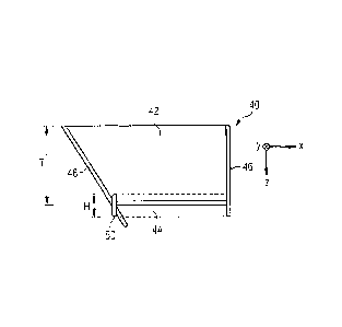
Figures 2A and 2B show embodiments of a corneal incision figure, denoted in
general by 40, in a laser treatment of the eye 12. The laser treatment may be
performed by means of the device shown in Figure 1. Figure 2A shows a top
view, and Figure 2B shows a cross-sectional view of the corneal incision
figure
40.
In Figure 2A, a flattening region is denoted by a circle line 41 indicated by
long
line segments. The flattening region 41 describes the region of the eye 12
that
bears on the bearing contact surface 34 of the contact element 32 and that is
flattened for laser treatment (cf. Figure 1). The flattening region 41 may
have
a contour other than a circle. The contour is influenced, for example, by
8
CA 02914384 2015-12-09
WaveLight GmbH 1A-125 752
differing radii of curvature in the main meridian directions of the surface of
the
cornea.
The incision figure 40 represented defines a flap, which is denoted in general
by 42. The flap 42 comprises a flap underside, which is parted off by a bed
incision 44 from adjoining corneal tissue located deeper in the longitudinal
direction, starting from the surface of the cornea, and a flap sheath that is
parted off by a lateral incision 46 from corneal tissue that adjoins in the
transversal direction.
In the embodiment shown, the bed incision 44 extends over a circle segment
of a circle area, the circle segment being delimited by an approximately
rectilinear chord of a circle and by an arcuate circle arc. In another
embodiment, the bed incision 44 may extend over an entire circle area, or the
arcuate edge portion may be other than a circle arc (e.g. elliptical). In the
embodiment shown, the lateral incision 46 extends along the entire arcuate
edge portion of the bed incision 44. In the region of the rectilinearly
extending
edge portion of the bed incision 44, the flap 42 is connected to the adjoining
corneal tissue in less deep regions, relative to the bed incision 44. The
transition region (hinge region) between the flap 42 and the adjoining corneal
tissue forms a hinge that allows the flap 42 to fold away in such a manner
that
the deeper corneal tissue is exposed for an ablating laser treatment. In the
embodiment shown, a notional hinge axis A of the hinge corresponds
approximately to the rectilinearly extending edge portion of the bed incision
44.
For the purpose of removing gases that develop during the production of the
bed incision, the incision figure 40 additionally comprises a first auxiliary
channel 48 and a second auxiliary channel 50. The course of the first
auxiliary
channel 48 is outside of the flap 42, going out from the hinge region as far
as
the eye surface. In this case, in the embodiment shown, the first auxiliary
channel 48 has a substantially constant width W1. In another embodiment, the
first auxiliary channel 48 may have, for example, a greater width in the hinge
region and a lesser width in the region of the eye surface (or vice versa).
9
CA 02914384 2015-12-09
WaveLight GmbH 1A-125 752
In order that the gases are removed rapidly and completely from the region of
the bed incision 44, the second auxiliary channel 50 is provided, via which
the
gases get into the first auxiliary channel 48 in a simplified manner. In the
embodiment shown, the second auxiliary channel 50 runs continuously along
the entire edge of the bed incision 44. It is thereby possible, in particular,
to
improve the removal of gases from regions of the bed incision 44 that are
produced closer to the edge portion of the bed incision 44 that is opposite
the
hinge region. The second auxiliary channel 50 in this case has a channel
portion extending rectilinearly in the hinge region, and a channel portion
extending in the form of an arc outside of the hinge region. In another
embodiment, it may be provided that the second auxiliary channel 50 does not
extend along the entire edge of the bed incision 44. It may be provided, for
example, that the rectilinearly extending channel portion of the second
auxiliary channel 50 extends only within the portions of the hinge region in
which the first auxiliary channel 48 does not extend (as explained more fully
in
the following with reference to Figure 3B).
It may be provided that the second auxiliary channel 50 has a width W2 that is
substantially constant along its direction of extent. The width W2 may
correspond to a single photodisruptive damage zone or to a plurality thereof
positioned next to each other. The width W2 may assume, for example, values
of approximately 5 pm or 10 pm.
Figure 2B shows a cross-sectional view of the corneal incision figure 40 in
the
flattening region 41 of the eye according to Figure 2A, along a straight line
within a region delimited by the dotted lines in Figure 2A.
In the embodiment shown, the bed incision 44 extends out from the surface of
the cornea at a substantially constant corneal depth. The depth of the bed
incision 44 in this case corresponds to the desired thickness T of the flap
42.
In this case, the thickness T may assume, for example, values in the range of
from 60 pm to 150 pm, such as, for example, 60 pm, 80 pm, 100 pm, 120 pm
or 150 pm. As an alternative to this, the flap 42 may have, for example, a
lesser thickness in the hinge region and a greater thickness in the region
opposite the hinge region (or vice versa). It may be provided that the height
CA 02914384 2015-12-09
WaveLight GmbH 1A-125 752
of the bed incision 44 corresponds to a single damage zone produced by
photodisruption. In this case, the height may be approximately 5 pm.
The second auxiliary channel 50 has a height H that is substantially constant
over its length, as shown in Figure 2B by the incisions through the second
auxiliary channel 50 that are represented on opposite sides of the bed
incision
44. In order to simplify the removal of the gases through the second auxiliary
channel 50, in the embodiment shown the channel height H corresponds to a
plurality of damage zones, produced by photodisruption, that are disposed one
above the other. Thus, the channel height H may assume, for example, values
of not less than 5 pm or 10 pm or 15 pm. Moreover, the channel height H
may correspond to not more than 35 pm or 30 pm or 25 pm. In another
embodiment, a channel height H that varies over the length of the second
auxiliary channel 50 may be provided.
In the embodiment shown, the second auxiliary channel 50 reaches into
deeper corneal regions and less deep corneal regions, relative to the bed
incision 44. In this case, it may be provided that the second auxiliary
channel
50 is produced in such a depth that the bed incision 44 adjoins the second
auxiliary channel 50 approximately in the region of the central longitudinal
extent of the latter. In another embodiment, the second auxiliary channel 50
may reach, for example, only into deeper corneal regions or only into less
deep
corneal regions, relative to the bed incision 44.
The first auxiliary channel 48 extends from the surface of the cornea into
corneal depths beneath the bed incision 44 (as also represented in Fig. 2A by
the dashed line indicated by short line segments). In this case, it can
extend,
for example, into regions that are deeper by 5 pm, 10 pm, 15 pm or 20 pm.
Gases can be stored temporarily in the portion of the first auxiliary channel
48
located beneath the bed incision 44. It is thus possible, for example, to
avoid
accumulation of gases in the region of the bed incision when the removal
capacity of the portion of the first auxiliary channel 48 located above the
bed
incision 44, has been exhausted.
The first auxiliary channel 48 is connected to the bed incision 44 and to the
second auxiliary channel 50. In the embodiment shown, it is provided that the
11
CA 02914384 2015-12-09
WaveLight GmbH 1A-125 752
first auxiliary channel 48 adjoins the edge of the bed incision 44 in the
hinge
region. In another embodiment, it may be provided that the first auxiliary
channel 48 does not adjoin the edge of the bed incision 44, and is connected
to the bed incision 44, for example, via the connection to the second
auxiliary
channel 50.
In the embodiment shown, the lateral incision 46 adjoins the second auxiliary
channel 50 rectilinearly, and leads as far as the surface of the cornea. In an
alternative embodiment, the lateral incision 46 may also lead obliquely to the
eye surface. An angle between the second auxiliary channel 50 and the lateral
incision 60 may assume values of between 140 and 1800, such as, for
example, 140 , 160 or 180 . The width of the lateral incision 46 may
correspond to the width W2 of the second auxiliary channel 50, or differ from
it. The width of the lateral incision 46 may correspond, for example, to a
single damage zone produced by photodisruption.
Figures 3A and 3b show embodiments of scan patterns of the movement of the
radiation focus according to the time sequence, provided by the program
instructions, for producing the incision figure 40 (e.g. according to Figures
2A
and 2B). The bed incision 44, the first auxiliary channel 48 and the second
auxiliary channel 50 are represented.
In the embodiments shown, the program instructions provide for the
production of the first auxiliary channel 48 before the production of the flap
42, and then the production of the second auxiliary channel 50. Thus, even
before commencement of the production of the bed incision 44, a possibility
exists for removing the gases, developed during the production of the bed
incision 44, out of the region of the bed incision 44, to the surface of the
cornea.
For the purpose of producing the first auxiliary channel 48, the radiation
focus
progresses, scan line by scan line, out from the surface of the cornea in the
direction of regions located deeper in the cornea, as indicated by the arrow
60
shown in Figure 3A. The scan lines, denoted by 62, run, approximately
rectilinearly and parallel to each other, transversely in relation to the
direction
of extent of the first auxiliary channel 48. In another embodiment, the scan
12
CA 02914384 2015-12-09
WaveLight GmbH 1A-125 752
lines 62 may run in the direction of extent of the first auxiliary channel 48.
The portion of the first auxiliary channel 48 that extends into corneal depths
beneath the bed incision 44 is not represented, for reasons of clarity.
In the embodiment shown in Figure 3A, the second auxiliary channel 50 forms
a closed annular channel (cf. also Figure 2A). For this purpose, starting from
one end of the hinge region, the channel portion that runs rectilinearly in
the
hinge region is produced first, and then the channel portion that runs in the
form of an arc outside of the hinge region is produced, as marked by the
arrows denoted by 64.
Unlike Figure 3A, in the embodiment shown in Figure 3B the second auxiliary
channel 50 does not run within the portion of the hinge region into which the
first auxiliary channel 48 extends. Starting from a first edge of the first
auxiliary channel 48 (marked by the point 66), the rectilinear channel portion
in the hinge region that adjoins in the negative y direction is produced
first,
then the channel portion running in the form of an arc outside of the hinge
region is produced, and finally a second channel portion of the second
auxiliary
channel 50 is produced, which portion extends as far as a second edge of the
first auxiliary channel 48 (marked by the point 68) that is opposite the first
edge. The direction in which the second auxiliary channel 50 is produced is
indicated by the arrows 70. The first auxiliary channel 48 is connected to the
second auxiliary channel 50, at least in the edge region (points 66, 68).
In another embodiment, a movement of the radiation focus that differs from
the embodiments shown in Figures 3A and 3B may be provided by the program
instructions. For example, the closed annular channel according to Figure 3A
may be produced starting from a point within the portion of the hinge region
into which the first auxiliary channel 48 extends. Moreover, the direction of
the focus movement indicated by the arrows may be reversed, at least
partially.
For the purpose of producing the flap 42, the bed incision 44 is first
applied.
In the embodiments shown in Figures 3A and 3B, the bed incision 44 is
produced, in accordance with the program instructions, by means of a
movement of the radiation focus along rectilinear and mutually parallel scan
13
CA 02914384 2015-12-09
WaveLight GmbH 1A-125 752
lines, whose line direction runs approximately perpendicularly in relation to
the
hinge axis A. In accordance with a provided time sequence of the program
instructions, a first scan line 72 is firstly produced with reference to the
control
of the radiation focus according to Figure 1, which scan line corresponds to a
notional bed-incision central line that is perpendicular to the hinge axis A.
The
radiation focus then progresses from one scan line to the respectively next
scan line, starting from the bed-incision central line, in the positive or
negative
y direction, and after that in the opposite y direction. The directions of
production are indicated by the arrows 74 and 76. It is to be noted that the
first scan line 72 may have the same transversal extent as the other scan
lines
of the bed incision 44, and is represented in a more pronounced manner only
for reasons of clarity. In an alternative embodiment, it may be provided that
the portions of the bed incision 44 reaching out from the bed-incision central
line 72 in the positive and the negative y directions are applied
approximately
simultaneously. For this purpose, for example, the device 10 according to
Figure 1 could additionally comprise an arrangement for splitting the laser
beam 20, and supplementary arrangements for controlling the radiation focus
in the transversal and longitudinal directions, and for focussing the laser
beam
20.
Furthermore, for example, a time sequence for the movement of the radiation
focus may be provided, according to which the radiation focus progresses,
scan line by scan line, starting from a point of minimum y extent of the bed
incision 44, in the direction of maximum y extent (or vice versa).
In another embodiment, it may be provided that the line direction of the scan
lines corresponds, at least approximately, to the direction of the hinge axis
A.
In this case, the production of the bed incision may follow a time sequence of
the movement of the radiation focus, according to which the radiation focus
progresses, for example, scan line by scan line, increasingly in the direction
away from the hinge region.
It may be provided that the lateral incision 46 (not represented) is produced
after the bed incision 44, or that the lateral incision 46 is produced after
the
second auxiliary channel 50 and before the bed incision 44. It is to be noted
14
CA 02914384 2015-12-09
WaveLight GmbH 1A-125 752
that no limitation whatsoever to a particular time sequence of incision
production and channel production is intended.