Note: Descriptions are shown in the official language in which they were submitted.
- 1 -
THERAPEUTICS FOR THE INDUCTION OF ENDOGENOUS
STEROIDOGENESIS AND METHODS ASSOCIATED WITH THEIR
IDENTIFICATION
TECHNOLOGICAL FIELD
The present disclosure concern peptide-based agents promoting endogenous
steroidogenesis and particularly the production of testosterone as well as
associated
therapeutic applications especially suited for the prevention, treatment
and/or alleviation of
symptoms associated with hypogonadism. The peptide-based agents comprise 14-3-
3E
binding motifs and are shown to limit or impede the association between the 14-
3-3E protein
and the VDAC1 protein. The present disclosure also provides corresponding
screening
assays based for identifying further therapeutic agents for promoting
endogenous
steroidogenesis especially suited for the prevention, treatment and/or
alleviation of
symptoms associated with hypogonadism.
BACKGROUND
Reduced serum testosterone (T) is common among subfertile and infertile young
men,
including most men diagnosed with idiopathic infertility. Reduced T is also
common in aging
men, with T levels declining at age 40 and been low in the majority of men
older than 60.
Reduced T is often associated with mood changes, fatigue, depression,
decreased lean body
mass, reduced bone mineral density, increased visceral fat, metabolic
syndrome, decreased
libido and reduced sexual function. T replacement therapy (TRT) is used
clinically to restore
T levels. TRT can treat symptoms associated with low T. However, TRT may
increase the
risk and aggressiveness of prostate cancer, augment the incidence of adverse
cardiovascular events, favor obesity and depression and even increase the rate
of mortality
in patients. Therefore is not recommended for patients at high risk of such
diseases.
Moreover, long-term TRT can suppress luteinizing hormone (LH) production,
making this
approach inappropriate for men who wish to have children. Fluctuating T
levels, skin
irritation, and T transfer to others through skin contact are additional
disadvantages of TRT.
The molecular mechanisms that govern androgen formation in testicular Leydig
cells remain
Date Recue/Date Received 2020-12-16
CA 02914755 2015-12-07
WO 2014/197979
PCT/CA2014/050467
- 2 -
unclear. Identification of these mechanisms will facilitate development of new
approaches for
inducing endogenous T synthesis voiding exogenous T treatment.
T production is regulated by LH and its secondary messenger, cAMP. Cholesterol
import
from cytosolic sources into mitochondria is a hormone-sensitive and rate-
limiting step of
steroidogenesis. Cholesterol is cleaved into pregnenolone by CYP11A1 in
mitochondria, and
steroidogenesis begins. Cholesterol import into mitochondria is mediated by a
hormone-
induced multiprotein complex called the transduceosome, which is composed of
cytosolic
and outer mitochondrial membrane (OMM) proteins that control the rate of
cholesterol entry
into the OMM. These proteins include the cytosolic mitochondria-targeted,
hormone-induced
steroidogenic acute regulatory protein (STAR), the OMM high-affinity
cholesterol-binding
protein translocator protein (TSPO), which contains a cytosolic cholesterol
recognition/interaction domain (CRAC) and the OMM voltage-dependent anion
channel
protein (VDAC1). Recent studies shed light on the importance of interactions
between STAR,
TSPO and VDAC1, suggesting that cholesterol import into mitochondria relies on
the function
and physical interactions between components of the transduceosome. The nature
and
dynamics of transduceosome protein-protein interactions remain unknown.
The 14-3-3 family of adaptor proteins were recently shown to have binding
motifs on
important functional sites in STAR, TSPO, and VDAC1 and 14-3-3y was identified
as a
regulator of STAR activity. However this hormone-induced 14-3-3 isoform was
shown to
function in a transient manner at the initiation of steroidogenesis, to delay
the maximum
STAR activity. Indeed, the function of 14-3-3y is terminated as it dissociates
from STAR,
allowing for maximal steroid production. In these studies, the levels of the
14-3-3 family
isoform, were found to be increased in Leydig cell mitochondria during
steroidogenesis. This
isoform mediates in a tissue/target-specific manner, cell functions such as
neural
development, adipocyte differentiation, protein trafficking, cell cycle,
apoptosis and cell
signaling. Levels of 14-3-3E, formerly known as mitochondrial import
stimulating factor 25,
are also high in human testes but its function in this tissue is unknown.
It would be desirable to be provided with a therapeutic agent capable of
upregulating
endogenous steroid production, such as testosterone production, without
altering luteinizing
hormone levels, to avoid or limit the side-effects listed above. It would also
be desirable to be
provided with screening assays for determining if a putative agent is capable
of upregulating
endogenous steroid production.
SUMMARY
One aim of the present disclosure is to provide agents capable of promoting
endogenous
steroidogenesis (and particularly the production of testosterone) without
altering the
CA 02914755 2015-12-07
WO 2014/197979
PCT/CA2014/050467
- 3 -
endogenous production of the luteinizing hormone. These agents can be used for
the
prevention, treatment and/or alleviation of symptoms associated with
hypogonadism. As it
will be shown herein, isolated peptides comprising the amino acid sequence
RVTQSNF
(SEQ ID NO: 5), a 14-3-3E binding motif, can limit or impede the interaction
between the 14-
3-3E protein and the VDAC1 protein and, consequently favor endogenous
steroidogenesis
(such as testosterone production). The present disclosure also provides
corresponding
screening assays based for identifying further therapeutic agents for
promoting endogenous
steroidogenesis especially suited for the prevention, treatment and/or
alleviation of
symptoms associated with hypogonadism.
According to a first aspect, the present disclosure provides an isolated
peptide having the
amino acid sequence RVTQSNF (SEQ ID NO: 5). In an embodiment, the isolated
peptide
has the amino acid sequence SKSRVTQSNFAVG ( SEQ ID NO: 30). In an another
embodiment, the serine residue at position 5 of SEQ ID NO: 5 or at position 8
of SEQ ID NO:
30 of the isolated peptide is phosphorylated.
According to a second aspect, the present disclosure provides a chimeric
peptide having the
isolated peptide described herein fused to a cell penetrating peptide. In an
embodiment, the
cell penetrating peptide is from a TAT protein and can have the amino acid
sequence
YGRKKRRQRRR (SEQ ID NO: 29). In another embodiment, the carbon/ terminus of
the cell
penetrating peptide is fused to the amino terminus of the isolated peptide. In
still another
embodiment, the isolated peptide is fused to the cell penetrating peptide by a
linker. In still
another embodiment, the linker can comprise at least one amino acid, such as,
for example,
a glycine residue.
According to a third aspect, the present disclosure provides a delivery system
comprising (i)
the isolated peptide described herein or the chimeric peptide described herein
and (ii) a cell
.. penetration enhancer.
According to a fourth aspect, the present disclosure provides the isolated
peptide described
herein, the chimeric peptide described herein or the delivery system described
herein for use
in therapy.
According to a fifth aspect, the present disclosure provides a method for
promoting the
endogenous production of a steroid in a cell. Broadly, the method comprises
contacting the
cell with at least one of the isolated peptide described herein, the chimeric
peptide described
herein, the delivery system described herein or a nucleic acid molecule
impeding the
expression of a 14-3-3E protein. The methods described herein if designed to
promote the
endogenous production of the steroid in the cell. In an embodiment, the
nucleic acid
molecule encodes a siRNA specific or comprises a combination of siRNAs
specific for a
CA 02914755 2015-12-07
WO 2014/197979
PCT/CA2014/050467
- 4 -
transcript encoding a 14-3-3E protein. In an embodiment, the steroid is
testosterone. In still
another embodiment, the cell is in vitro. In yet another embodiment, the cell
is in vivo and the
method further comprises administering the isolated peptide, the chimeric
peptide, the
delivery system or the nucleic acid molecule to a subject in need thereof
comprising the cell.
In some embodiments, the subject is a mammal and/or a male. In yet another
embodiment,
the cell is from a testis, such as, for example a Leydig cell. In yet a
further embodiment, the
method is for the prevention, treatment and/or alleviation of symptoms of a
condition
associated with hypogonadism. Conditions associated with hypogonadism include,
but are
not limited to infertility, aging, decreased libido, sexual dysfunction,
altered mood, fatigue,
decreased lean body mass, decreased bone mineral density, increased visceral
fat or
metabolic syndrome.
According to a sixth aspect, the present disclosure provides the use of the
isolated peptide
described herein, the chimeric peptide described herein, the delivery system
described
herein or a nucleic acid molecule impeding the expression of a 14-3-3E protein
for promoting
the endogenous production of a steroid in a cell. In an embodiment, the
nucleic acid
molecule is a siRNA or comprises a combination of siRNAs specific for a
transcript of a 14-3-
3E protein. In another embodiment, the steroid is testosterone. In a further
embodiment, the
cell is in a subject such as, for example, a mammal and/or a male. In another
embodiment,
the cell is from a testis, such as, for example, a Leydig cell. In yet a
further embodiment, the
use is for the prevention, treatment and/or alleviation of symptoms of a
condition associated
with hypogonadism. Conditions associated with hypogonadism include, but are
not limited to
infertility, aging, decreased libido, sexual dysfunction, altered mood,
fatigue, decreased lean
body mass, decreased bone mineral density, increased visceral fat or metabolic
syndrome.
According to a seventh aspect, the present disclosure provides a method for
determining the
usefulness of an agent for promoting endogenous steroid production in a cell.
Broadly, the
method comprises: (a) combining the agent, a 14-3-3E protein and a VDAC1
protein; (b)
determining if the agent promotes or impedes the formation and/or stability of
a complex
between the 14-3-3E protein and the VDAC1 protein; and (c) characterizing the
agent (i) as
being useful for promoting endogenous steroid production if the agent impedes
the formation
and/or stability of the complex or (ii) as lacking utility to promote
endogenous steroid
production if the agent promotes the formation and/or stability of the
complex. In an
embodiment, the steroid is testosterone. In another embodiment, the combining
step is
conducted in vitro in a cell, such as, for example, a MA-10 cell. In a further
embodiment, the
combining step is conducted ex vivo in a tissue, such as, for example, an
isolated testis. In
yet another embodiment, the combining step is conducted in an animal. In still
another
embodiment, the cell is from or in a testis, such as, for example, a Leydig
cell.
CA 02914755 2015-12-07
WO 2014/197979
PCT/CA2014/050467
- 5 -
BRIEF DESCRIPTION OF THE DRAWINGS
Having thus generally described the nature of the invention, reference will
now be made to
the accompanying drawings, showing by way of illustration, a preferred
embodiment thereof,
and in which:
Figure 1 illustrates that 14-3-3E is a negative regulator of steroidogenesis.
(a)
Immunohistochemistry (ICC) indicates that 14-3-3E is present in MA-10 cells
(third column)
and that this protein partially localizes with mitochondria (second column).
MA-10 nucleus is
also shown (first column). Results are shown with respect to the time of
incubation of the
cells with 8-Br-cAMP in minutes (0 = first row, 120 = second row) (131)
Immunoblot results of
MA-10 cells stimulated with 8-Br-cAMP for indicated time points (in minutes)
show 14-3-3E
expression (first row) and quantification relative to GAPDH control protein
(second row). (b2)
Immunoblot analysis corresponding to the immunoblot of Figure 1b1. Results are
shown as
the ratio of 14-3-3E to GAPDH protein in function of incubation time (in
minutes) with 8-Br-
cAMP. (c1) Immunoblot analysis indicating the levels of 14-3-3E protein
compared to GAPDH
in MA-10 cells in the absence of siRNA (mock), transfected with scrambled
(Scr) siRNA as
negative and positive control, respectively, or transfected with a mixture of
14-3-3E specific
siRNA at different concentrations (20 nM, 10 nM, or 5 nM). * P<0.05 and **
P<0.01 (c2)
Immunoblot indicating the levels of 14-3-3E protein and GAPDH in MA-10 cells
in the
absence of siRNA (mock), transfected with scrambled (Scr) siRNA as negative
and positive
control, respectively, or transfected with a mixture of 14-3-3E specific siRNA
at different
concentrations (20 nM, 10 nM, or 5 nM). (d) MA-10 cells were transfected with
10 nM 14-3-3E
siRNA and further stimulated with 8-Br-cAMP for 0, 30, 60, and 120 min, and
progesterone
levels were measured at each time point. Results are shown for the mock
treatment (white
bars), the treatment with the scrambled siRNA (grey bars) and the treatment
with the 14-3-3E
siRNA (black bars).* P<0.05
Figure 2 illustrates that TSPO, STAR, and VDAC1 are targets of 14-3-3E. (a, b,
c, d) Cell
immunoprecipitation (Duolink technology) indicates the dynamics of the
interactions between
14-3-3E with TSPO, STAR and VDAC1. Images show the cell nucleus (first column
of each
panel), mitochondria (second column of each panel), and endogenous protein-
protein
interactions (third column of each panel) between 14-3-3E-TSPO (a), 14-3-3E-
STAR (b), and
14-3-3E-VDAC1 (c) the background signal (d) of the Duolink assay in MA-10
cells, and the
merge of the three previous columns (last column). (e, f, g) Corresponding
analysis of the
cell immunoprecipitation assays shown in Figures 2a to 2c for the 14-3-3E-TSPO
interaction
(e), the 14-3-3E-STAR interaction (f) or the 14-3-3E-VDAC1 interaction (g).
Results are shown
as the signal of the interacting proteins per cell in function of time of
incubation with 8-Br-
cAMP (in minutes).* P<0.05) and *** P<O. 001.
CA 02914755 2015-12-07
WO 2014/197979
PCT/CA2014/050467
- 6 -
Figure 3 illustrates that blocking the interaction between 14-3-3E and VDAC1
negates the
regulatory role of 14-3-3E in steroidogenesis. (a, b) The Duolink assay was
performed to
measure protein-protein interactions between 14-3-3E and endogenous VDAC1 in
untreated
MA-10 cells (control) or cells treated with the TVS35, TVG35, TVS167, or
TVS167 peptides,
after 8-Br-cAMP (120 min) treatment which induces maximum 14-3-3E-VDAC1
interaction
(third column of each panel). Staining of nucleus (first column of each panel)
and
mitochondria! (second column of each panel) are also shown.
lmmunohistochemistry is whon
in (a) while the corresponding analysis is shown in (b, * =, ** =). (c)
Progesterone levels in
control MA-10 cells or cells treated with 1V535, TVS167, or TV5167 were
measured after 8-
Br-cAMP (120 min) treatment. Levels of progesterone were normalized to protein
content
and further compared to the levels in control cells, as fold increase. ***
P<0.001 (d, e, f, g,
j). The impact of blocking interactions between 14-3-3E-VDAC1 on other
transduceosome
protein-protein interactions was studied in the presence of TVS167. MA-10
cells were treated
with TVS167 and 8-Br-cAMP (120 min). The interactions between 14-3-3E-STAR (d,
e in
which *** P<0.001), TSPO-VDAC1 (f, g in which ** P<0.01) and 14-3-3E-TSPO (h,
I in which
** P<0.01) were measured, as endogenous protein-protein interactions.
Histograms show the
sum of protein-protein interactions in Z-stacks as signal/cell ratio. (j) The
physiological impact
of the increase in 14-3-3E-TSPO interactions on cholesterol binding to TSPO
was studied.
Progesterone levels in control (untreated), TVS167-treated, and combination 19-
Atriol/TVS167-treated MA-10 cells were measured after 8-Br-cAMP stimulation
(120 min). *
P<0.05.
Figure 4 shows the effect of ex vivo and in vivo administration of TVS167 on T
production.
(a) Testes dissected from adult Sprague-Dawley rats were cultured in media
supplemented
with or without TVG167 or TVS167 and/or hCG (120 min). T levels were measured.
Results
are shown as T levels (in ng/testes) for control treatment (white bars),
treatment with
TVG167 (gray bars) or treatment with TVS167 (black bars) before and after hCG
treatment.
** P<0.01, *** P<0.001, #44 P=0.01) (b, c, d) Adult Sprague-Dawley rats were
injected in one
testis with water or 150 ng TVG167 or TVS167. A pump releasing H20, 75 ng/24 h
TVG167,
or 75 ng/24 h TVS167 was connected to the injected testis. Animals were
dissected after 24
hrs. Intratesticular T levels were measured (ng/mL) in treated (black bars)
and control (white
bars) testes (b in which ** P<0.01 ). Serum T levels (in ng/mL in c in which *
P<0.05) and
serum LH levels (in mIU/mL in d in which * P<0.05 ) were also measured in H20-
treated
(white bars), TVG167-treated (gray bars) and TVS-167-treated (black bars)
animals. (e)
Duolink assay was performed on the testes sections. lmmunofluorescence images
show the
merge of nucleus channel and protein-protein interaction channel indicating
that, in the
CA 02914755 2015-12-07
WO 2014/197979
PCT/CA2014/050467
- 7 -
presence of TVS167 peptide, the interactions of 14-3-3E and VDAC1 in rat
testes are
removed.
Figure 5 shows that the effect of TVS167 in vivo is LH-independent. (a, b)
Adult Sprague-
Dawley rats were given i.p. injections of H20 or Cetrorelix (0.4 mg/day).
Animals were either
dissected on day 4 (no pump) or treated with H20 (if given H20 i.p.) or TVS167
(if given
Cetrorelix i.p.) through a bolus injection and pump installation to one testis
and dissected 24
his after pump installation. T levels(provided in ng/nnL) in the
intratesticular fluid (a in which *
P<0.05) of testis connected to a pump (+), not connected to a pump (-), or
both testes (no
pump and Total) and in serum were measured (b in which * P<0.05). (c, d) Adult
Sprague-
Dawley rats were injected i.p. with either H20 or Cetrorelix for 0-4 days and
on day 4, one
testis per animal was given a bolus injection of 8.5 pg FGIN-1-27 to induce
acute
steroidogenesis in the absence or presence of LH signaling. T levels (provided
in ng/mL) in
intratesticular (c in which * P<0.05) and plasma (d in which * P<0.05 were
measured in
animal treated with H20 (white bars) or Cetrorelix (grey bars) 2 his post
injection, showing a
significant increase.
Figure 6 illustrates a modeling representation of the human VDAC1 14-3-3E
motif containing
S167. (a) Putative models 14-3-3E and VDAC1 were mapped in indicated species
showing a
high degree of homology. (b) Macromolecular docking among two proteins. The
docking site
of 14-3-3E in the VDAC1 structure in Mus muscu/us was predicted. 1VS167 was
shown to
dock onto open and non-ligand-bound 14-3-3E at the site to which VDAC1 also
bound to this
protein, suggesting that 1V167 can block this interaction. Due to a high
percentage of
homology between the 3-D structures of human 14-3-3E and VDAC1, the same
docking sites
were predicted in these species. The ribbon representative of each protein and
surface
mapping of electrostatic potential of mouse 14-3-3E are shown. The molecular
surface are
corresponding to negative, positive and neutral charged regions, respectively.
(c, d) The
molecular docking studies show TVS167 targets the 14-3-3E binding groove as
well as the
right shoulder that interacts with VDAC1(6b). (e) Mutation of S167 to G167
removes the
ability of the TV peptide to dock outside the binding groove. (f) The
phosphorylated TVS167
docked within the binding groove.
Figure 7 provides a comparison of 14-3-3E protein profile in adrenal gland
versus testis. (a)
lmmunofluorescence images illustrates the nucleus and 14-3-3E expression in
sections of
adult mice adrenal gland (first row) and testes (second row) indicating higher
levels of protein
expression in interstitial cells of testes compared to adrenal glands. (b)
Immunoblot (b1) and
corresponding analysis (bl) show that the expression levels of 14-3-3E
compared to GAPDH
are significantly higher in protein lysate extracted from interstitial testes
compared of adrenal
glands of adult rats. (c) Adult rats were implanted with H20 (white bar) or
TVS167 (black bar)
CA 02914755 2015-12-07
WO 2014/197979
PCT/CA2014/050467
- 8 -
releasing pumps abdominally, this pump was directed to one testis and induces
T levels in
rat testes and plasma (Fig. 4d, e, f). Corticosterone levels (measured as
ng/mL) produced by
the adrenal gland cortex were measured in these rats indicating insignificant
changes
compared to control. (d) Public microarray data comparing mRNA levels of 14-3-
3E in human
tissues indicates higher mRNA in human interstitial cells compared to adrenal
gland. (e)
Duolink assay was performed to study the protein-protein interactions between
14-3-3E and
VDAC1 in NCL-H296R human adrenocortical cell line. Immunofluorescence images
illustrate
the nucleus (first column), mitochondria (second column) and protein-protein
interactions
(third column) indicating that the protein-protein interaction signal is not
as high as in MA-10
cells and rat testes sections and that despite a decrease in the interactions
of 14-3-3E and
VDAC1 in these cells in the presence of TVS167 (second row), this effect is
not significant
(el). A corresponding analysis is also presented (e2).
Figure 8 illustrates the characteristics of the 14-3-3E binding motifs. (a)
TSPO, STAR and
VDAC1 each contain 2 to 3 in silico predicted 14-3-3 binding motif as
indicated. The amino
acid sequence of these motifs is shown based on the sequence homology with
mode I
(RSXpSXP or SEQ ID NO: 1) or ll (R)0((pSXP or SEQ ID NO: 2) of the classic 14-
3-3
motifs. These motifs on all three proteins are suboptimal, varying by 1 to 2
amino acids from
the classic 14-3-3 motifs. (b) MA-10 cells were treated with 8-Br-cAMP for 0,
15, 30, 60 and
120 minutes. Cross-linking (CL) was performed with photo-activatable leucine
and
methionine and UV light. Cell lysates were immunoprecipitated with 14-3-3E
anti-sera (IF).
Immunoblot analysis confirms the previous results (Fig 2) and shows the
dynamics of 14-3-
3E interactions with other 14-3-3 isoforms (14-3-3 pan), TSPO (b2), STAR (bl)
and VDAC1
(b2) during steroidogenesis, C lane indicates these interactions in native MA-
10 cell protein
lysates.
Figure 9 provides an ininiunofluorescence image of control MA-10 cells (a) and
MA-10 cells
treated with the fluorescent FAM-TVS167 chimeric peptide (b), indicating the
high efficiency
of these peptides to penetrate cell membranes.
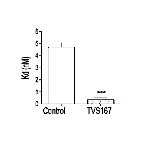
Figure 10 illustrates that 14-3-3E regulates the affinity of TSPO for its drug
ligand PK11195.
(a) Levels (measured as pmol/mg protein) of TSPO bound to PK11195 in the
absence (.) or
presence (0) of the TV5167 peptide at different doses of 3H PK 11195. (b) Kd
(measured as
nM), affinity-1 of TSPO for PK 11195, was measured in the absence (control,
white bar) or
presence (TV5167, black bar) of TVS167. *** P<0.001(c) Bmõ (measured as
prnol/mg
protein), the available binding site of TSPO for PK 11195, was measured in the
absence
(control, white bar) or presence (TV167, black bar) of TVS167.
CA 02914755 2015-12-07
WO 2014/197979
PCT/CA2014/050467
- 9 -
Figure 11 shows that TVS167 induces T levels in a dose response manner. One
testis of
each adult Sprague-Dawley rat was injected with a bolus of water, TVG167, or
different
doses of TVS167. The other testis was used as a control. T levels were
measured after 2 hrs
in the intratesticular fluid of each treated testis (black bars) and, control
testis (white bars) (a).
* P<0.05 . Serum LH levels (b) were also monitored. * or # P<0.05.
Figure 12 shows that the administration of the TVS167 peptides does not induce
toxic
histological modifications to the testis. Rat testes sections were obtained
from wild type adult
Sprague-Dawley rats and rats injected with a bolus does of H20 or 150 ng
TVS167 following
implantation of H20 or TVS167 (75ng/24h) releasing pump. The pumps were
directed to one
testis. Animals were dissected 24 h post pump implantation and sectioned.
Hennatoxylin
staining of these sections indicates no histological difference between testes
of these
animals (a, control, b, H20 pump and c, 1VS167 pump).
Figure 13 provides the alignment of amino acid sequences of 14-3-3E (a) and
VDAC1 (b)
showing high degree of conservation for both proteins and that the 14-3-3
binding motif is
conserved in the three mammalian species as indicated.
DETAILED DESCRIPTION
Throughout this application, various terms are used and some of them are more
precisely
defined herein.
14-3-3E protein. As used in the context of the present disclosure, the 14-3-3E
protein is
encoded by the YWHAE gene and is an adapter protein involved in the regulation
of a large
spectrum of both general and specialized signaling pathways. The 14-3-3E
protein binds to a
large number of partners, usually by recognition of a phosphoserine or
phosphothreonine
motif. In the context of the present disclosure, the 14-3-3E protein has been
shown to interact
with the VDAC1 protein and such interaction modulates (e.g. decreases)
endogenous steroid
production, such as endogenous testosterone protein. As shown herein,
particularly in Figure
13a, the 14-3-3E protein is largely conserved amongst mammals. The 14-3-3E
protein has
been documented in humans (Accession Number P62258), in mouse (Accession
Number
P62259) as well as in rats (Accession P62260). In some embodiments, the 14-3-
3E protein
comprises or consists of the consensus sequence shown in Figure 13a or any one
of the
sequences presented set forth in SEQ ID NO: 17, 18 or 19.
Antagonist. This term, as used herein, refers to an agent that impedes or
decreases the
formation and/or stability of an hetero-complex between the 14-3-3E protein
and the VDAC1
protein. An antagonist can also be a compound which decreases the stability of
a 14-3-
3E/VDAC1 complex, which downregulates the expression of a 14-3-3E-encoding
gene, which
limits the expression of a 14-3-3E-encoding transcript (e.g., mRNA), which
dowregulates the
CA 02914755 2015-12-07
WO 2014/197979
PCT/CA2014/050467
- 10 -
expression of a VDAC1-encoding gene, which limits the expression of a VDAC1-
encoding
transcript (e.g., mRNA), which favors the degradation of the 14-3-3E
polypeptide and/or
which favors the degradation of the VDAC1 polypeptide. In the context of this
disclosure,
such antagonists are considered useful for the prevention, treatment and/or
alleviation of
symptoms of conditions associated with hypogonadism.
Conditions associated with hypogonadism. Hypogonadism is understood as
diminished
functional activity of the gonads (e.g., the testes and ovaries) resulting in
diminished sex
hormone (e.g., testosterone, estradiol, biosynthesis progesterone, DHEA, anti-
Mullerian
hormone, activin and inhibin) . Low androgen (e.g., testosterone) levels can
be referred to as
hypoandrogenism and low estrogen (e.g., estradiol) can be referred to as
hypoestrogenisrn.
Conditions associated with hypogonadism include, but are not limited to
infertility (due to
defective or insufficient spermatogenesis or ovulation), aging, decreased
libido, sexual
dysfunction, altered mood, fatigue, decreased lean body mass, decreased bone
mineral
density, increased visceral fat and metabolic syndrome.
Pharmaceutically effective amount or therapeutically effective amount. These
expressions
refer to an amount (dose) effective in mediating a therapeutic benefit to a
subject (for
example prevention, treatment and/or alleviation of symptoms a condition
associated with
hypogonadism). The pharmaceutically effective amount can be used in
relationship to the
antagonist as described herein. It is also to be understood herein that a
"pharmaceutically
effective amount" may be interpreted as an amount giving a desired therapeutic
effect, either
taken in one dose or in any dosage or route, taken alone or in combination
with other
therapeutic agents.
Prevention, treatment and alleviation of symptoms. These expressions refer to
the ability of a
method or an agent to limit the development, progression and/or symptornology
of a
condition associated with hypogonadism. Broadly, the prevention, treatment
and/or
alleviation of symptoms encompass the lack of reduction of symptoms associated
with
hypogonadism, such as, for example, infertility (due to defective or
insufficient
spermatogenesis or ovulation), aging, decreased libido, sexual dysfunction,
altered mood,
fatigue, decreased lean body mass, decreased bone mineral density, increased
visceral fat
and metabolic syndrome.
Reaction vessel. The reaction vessel is an in vivo or in vitro discrete unit
for characterizing a
potential therapeutic agent. When a potential therapeutic agent is being
screened, the
contact between the agent, the 14-3-3E protein and the VDAC1 protein must be
made under
conditions suitable and for a period of time sufficient to allow, when
possible, interactions
between the agent the 14-3-3E protein and the VDAC1 protein. Suitable in vitro
environments
CA 02914755 2015-12-07
WO 2014/197979
PCT/CA2014/050467
- 11 -
can include, for example, a cell-free environment is combined in a reaction
media comprising
the appropriate reagents to enable the various measurements. Other suitable in
vitro
environments include cell-based assays (comprising, for example, using testis
cells such as
Leydig cells) or tissue-based assays (for example using isolated testis).
RNA interference. RNAi is a post-transcriptional gene silencing process that
is induced by a
miRNA or a dsRNA (a small interfering RNA; siRNA) and has been used to
modulate gene
expression. Generally, RNAi is being performed by contacting cells with a
double stranded
siRNA ou a small hairpin RNA (shRNA). However, manipulation of RNA outside of
cells is
tedious due to the sensitivity of RNA to degradation. It is thus also
encompassed herein a
deoxyribonucleic acid (DNA) compositions encoding small interfering RNA
(siRNA)
molecules, or intermediate siRNA molecules (such as shRNA), comprising one
strand of an
siRNA. Accordingly, the present disclosure provides an isolated DNA molecule,
which
includes an expressible template nucleotide sequence encoding an intermediate
siRNA,
which, when a component of an siRNA, mediates RNA interference (RNAi) of a
target RNA
(e.g., a mRNA or transcript encoding the 14-3-3E protein). The suppression of
gene
expression caused by RNAi may be transient or it may be more stable, even
permanent.
VDAC1 protein. As used in the context of the present disclosure, the VDAC1
protein, also
known as voltage-dependent anion-selective channel protein 1, is encoded by
the VDAC1
gene and forms a channel through the mitochondrial outer membrane and the
plasma
membrane. As shown herein, the VDAC1 protein can interact with the TSPO
protein to form
a mitochondrial channel for the transport of cholesterol. As also shown
herein, the VDAC1
protein comprises 14-3-3E binding motifs (e.g., KTKSEN (SEQ ID NO: 13) and
RVTQSNF
(SEQ ID NO: 14) as shown in Fig. 8a) and is capable of binding to the 14-3-3E
protein. Such
interaction between the VDAC1 protein and the 14-3-3E protein has been shown
to modulate
(e.g., decrease) endogenous steroid production, particularly endogenous
testosterone
protein. As also shown herein, and particularly in Figure 13b, the VDAC1
protein is largely
conserved amongst mammals. It has been documented in humans (Accession Number
NP_003365), in mouse (Accession Number NP_035824) as well as in rats
(Accession
NP_112643). In some embodiments, the VDAC1 protein comprises or consists of
the
consensus sequence shown in Figure 13b or any one of the sequences presented
set forth
in SEQ ID NO: 20, 21 or 22. It is worth noting that the serine residue located
at position 167
of the VDAC1 protein sequence (as shown in any one of SEQ ID NO: 20 to 22 and
which
corresponds to the serine residue in RVTQSNF (SEQ ID NO: 5)) is involved in
the
association between the 14-3-3E protein and the VDAC1 protein.
CA 02914755 2015-12-07
WO 2014/197979
PCT/CA2014/050467
- 12 -
Agents for promoting endogenous steroidogenesis production
The present disclosure provides novel peptides comprising a 14-3-3E binding
motif,
peptidomimetics of such peptides as well as chimeric peptides comprising such
peptides. As
it will be shown herein, these peptides are capable of limiting the
interaction between the 14-
.. 3-3E protein and the VDAC1 protein and ultimately favor the endogenous
steroidogenesis (in
vitro or in vivo), particularly the production of testosterone.
In an embodiment, the peptides described herein are capable of binding to the
14-3-3E
protein, and in an embodiment, are capable of binding to the 14-3-3E protein
in a region (or in
the vicinity of the region) where the STAR and/or the VDAC1 protein binds to
the 14-3-3E
.. protein. In a further embodiment, the peptides are capable of limiting or
inhibiting the binding
of the STAR and/or the VDAC1 protein to the 14-3-3E protein. In some
embodiments, the
peptides are derived from a mouse VDAC1 protein (SEQ ID NO: 20) and possesses
a serine
residue at a location corresponding to 167 in the mature mouse VDAC1 protein
which
corresponds to a serine residue at a location corresponding to 180 of the
immature mouse
VDAC1 protein (e.g., whose transcript has not been spliced). This serine
residue is
conserved in the human VDAC1 protein (e.g., residue located at position 167 of
SEQ ID NO:
21) as well as in the rat VDAC protein (e.g., residue located at position 167
SEQ ID NO: 22).
In an embodiment, the peptide is derived from the mouse VDAC1 species and
corresponds
to the amino acid residues located between positions 163 and 169 of SEQ ID NO:
20. In
another embodiment, the peptide is derived from a human VDAC1 species and
corresponds
to the amino acid residues located between positions 163 and 169 of SEQ ID NO:
21. In still
another embodiments, the peptide is derived from a rat VDAC1 species and
corresponds to
the amino acid residues located between positions 163 and 169 of SEQ ID NO:
22. In yet
another embodiment, the peptide can be chemically synthesized to have or
consist of the
following amino acid sequence RVTQSNF (SEQ ID NO: 5).
Particularly advantageous peptides are those having or consisting of the amino
acid
sequence RVTQSNF (SEQ ID NO: 5) as well as peptidomimetic versions thereof. As
shown
herein, the peptide having or consisting of the amino acid sequence RVTQSNF
(SEQ ID NO:
5) is capable of limiting or impeding the physical association between the 14-
3-3E protein and
the VDAC1 protein which in return allows endogenous steroid production (such
as
testosterone production) without modulating the production of the luteinizing
hormone. As
also shown herein, the serine residue of the RVTQSNF (SEQ ID NO: 5) is
important for such
biological activity since the replacement of such serine residue (by a glycine
residue for
example) abrogates the peptide's ability to upregulate endogenous steroid
production. In an
.. embodiment, the peptide can have or consist of the amino acid sequence of
SKSRVTQSNFAVG ( SEQ ID NO: 30). As also shown herein, the serine residue at
position 8
CA 02914755 2015-12-07
WO 2014/197979
PCT/CA2014/050467
- 13 -
of SEQ ID NO: 30 is important for such biological activity since the
replacement of such
serine residue (by a glycine residue for example) abrogates the peptide's
ability to upregulate
endogenous steroid production. In some embodiments, the peptide can be
phosphorylated at
least at one amino acid residue. For example, the peptide can be
phosphorylated at the
serine residue located at position 5 of the amino acid sequence RVTQSNF (SEQ
ID NO: 5)
or at position 8 of the amino acid sequence SKSRVTQSNFAVG (SEQ ID NO: 30). The
presence of a phosphorylated serine residue on the peptide can, in some
embodiments,
increases the affinity of the peptide for the 14-3-3E protein and ultimately
further enhance or
prolong endogenous steroid production. In an embodiment, the isolated peptide
is at least 7,
8, 9, 10, 11, 12 or 13 amino acid long. In yet another embodiment, the
isolated peptide is no
more than 13, 12, 11, 10, 9, 8 or 7 amino acid long.
The present disclosure also provides a chimeric peptide (as well as
corresponding
peptidomimetic versions thereof) having or consisting of the peptide described
herein (which
may or may not be phosphorylated) fused to a cell penetrating peptide. As used
herein, the
term "cell penetrating peptide" refers to a peptide capable of enhancing
penetration across
certain cellular structures, such as the cytoplasmic membrane, the
mitochondrial membrane
or the nuclear membrane. Some penetrating peptides can be specific or derived
from a
protein transduction domain. Other penetrating peptide can be specific or
derived from a
growth factor or a hormone. An exemplary targeting/penetrating peptide can be
a blood-
brain-barrier (BBB)-permeant, amyloid-targeting/penetrating peptide such as
KKLVFFWGC
or a cell penetrating fragment thereof (as presented in US Patent Serial
Number 7,803,351).
Another exemplary penetrating peptides include, but are not limited to, the
TAT protein or a
cell penetrating fragment thereof (such as, for example YGRKKRRQRRR (SEQ ID
NO: 29).
A further exemplary penetrating peptide is antenapedia or a cell-penetrating
fragment
thereof. In an embodiment, the cell penetrating peptide of the chimeric
peptide is at least 5,
6, 7, 8, 9, 10 or 11 amino acid long. In still another embodiment, the cell
penetrating peptide
of the chimeric peptide is no more than 11, 10, 9, 8, 7, 6 or 5 amino acid
long.
In the chimeric peptides described herein, the isolated peptide and the cell
penetrating
peptide can be directly fused (e.g., linked) to one another. However, in
another embodiment,
the isolated peptide can be indirectly fused (e.g., linked) to the cell
penetrating peptide via a
linker (such as an amino acid linker, a glycine linker for example). The
linker can be, for
example, at least 1, 2, 3, 4 or 5 amino acid long.
In an embodiment of the chimeric peptide described herein, the carboxy
terminus of the cell
penetrating peptide is fused to the amino terminus of the isolated peptide.
The amino acid
sequence of an exemplary chimeric peptide is provided at SEQ ID NO: 7. In such
chimeric
peptide, the carboxy terminus of a TAT protein was fused to a linker (e.g., a
single glycine
CA 02914755 2015-12-07
WO 2014/197979
PCT/CA2014/050467
- 14 -
residue). The carboxy terminus of the linker was fused to a peptide comprising
the 14-3-3E
binding motif SKSRVTQSNFAVG (SEQ ID NO: 30). Such chimeric peptide was shown
to
alter the formation of a complex between the 14-3-3E protein and the VDAC1 as
well as to
increase steroid production ex vivo and in vivo. The chimeric peptide can also
be
phosphorylated at a single amino acid residue or at a plurality of amino acid
residues. In the
embodiment of the chimeric peptide presented in SEQ ID NO: 7, the serine
residue at
position 20 can be phosphorylated. The chimeric peptide can be made using
recombinant
expression in a transgenic host or can be synthetically synthesized.
The present disclosure also provides a delivery system comprising the isolated
peptide
described herein or the chimeric peptide (as well as peptidomimetic version of
such peptide
or chimeric peptide) described herein and a cell penetration enhancer. As used
herein, a "cell
penetration enhancer", when complexed with the isolated peptide or the
chimeric peptide,
facilitates the passage of the peptide across a cellular structure or a
cellular membrane (such
as the cytoplasmic membrane, the mitochondrial membrane or the nuclear
membrane for
example). Exemplary embodiments of the delivery system include, but are not
limited to, viral
delivery systems, nanoparticles and liposomes.
The present disclosure also provides nucleic acid molecules (e.g., encoding
siRNAs) capable
of impeding the expression of the 14-3-3E protein which, in some conditions,
can be useful
for promoting endogenous production of a steroid (such as testosterone for
example). In the
context of the present disclosure, siRNA are double stranded RNA molecules
from about 10
to about 30 nucleotides (for example between 12 to 28 nucleotides long, more
preferably 13
to 20 nucleotides long, even more preferably 16 to 19 nucleotides long)
recognized for their
ability to specifically interfere with the expression of the 14-3-3E protein.
In one embodiment,
siRNAs of the present disclosure are 12, 13, 14, 15, 16, 17, 18, 19, 20, 21,
22, 23, 24, 25,
26, 27 or 28 nucleotides in length. As used herein, siRNA molecules need not
to be limited to
those molecules containing only RNA, but further encompass chemically modified
nucleotides and non-nucleotides. An siRNA molecule can be assembled from two
nucleic
acid fragments wherein one fragment comprises the sense region and the second
fragment
comprises the antisense region of siRNA molecule (such as, for example, the
siRNA
sequences presented in Table 2). The sense region and antisense region can
also be
covalently connected via a linker molecule. The linker molecule can be a
polynucleotide
linker or a non-polynucleotide linker. Exemplary siRNA pairs include, but are
not limited to
the siRNAs having or consisting of the nucleic acid sequence of SEQ ID NO: 23
and 24, the
siRNAs having or consisting of the nucleic acid sequence of SEQ ID NO: 25 and
26 or the
siRNAs having or consisting of the nucleic acid sequence of SEQ ID NO: 27 or
29.
CA 02914755 2015-12-07
WO 2014/197979
PCT/CA2014/050467
- 15 -
Therapeutic applications
As shown herein, disrupting the formation of a complex between the 14-3-3E
protein and the
VDAC1 protein, for example by using a 14-3-3E antagonist, can be useful in
promoting
endogenous steroid production. As also shown herein, the TSPO-VDAC complex
facilitates
the import of cholesterol in mitochondria, which is a rate-limiting step in
steroid biosynthesis.
Without wishing to be bound to theory, it is assumed that by inhibiting the
formation of a
complex between the 14-3-3E protein and the VDAC1 protein, the latter has
increased
availability to form a complex with TSPO to ultimately increase the import of
cholesterol in
mitochondria. As such, the expression of all steroid formed from cholesterol
could be
affected (e.g., increased) by modulating the interaction between the 14-3-3E
protein and the
VDAC1 protein. Because the isolated peptide and chimeric peptides (optionally
presented in
a delivery system) as well as the nucleic acid molecules described herein have
the ability to
promote the endogenous production of a steroid, they can be advantageously
used to
modulate levels of steroid production in a cell, in an organism and in some
embodiments, in
therapy. For example, in a therapeutic application, the isolated peptide, the
chimeric peptide
(optionally presented in a delivery system) or the nucleic acid molecule (or
combination
thereof) is contacted with a cell (either in vitro or in vivo) under
conditions suitable for
promoting the endogenous production of the steroid (such as, for example,
testosterone).
In therapeutic applications, the isolated peptide, the chimeric peptide, the
delivery system or
the nucleic acid molecule can optionally be formulated in a pharmaceutical
composition with
a pharmaceutically acceptable excipient. Further, the isolated peptide, the
chimeric peptide,
the delivery system or the nucleic acid molecule can optionally be formulated
for being
administered as a topical composition designed to be applied on the skin, such
as a cream or
a gel. In addition, the isolated peptide, the chimeric peptide, the delivery
system or the
nucleic acid molecule can optionally be formulated for being administered as
an injection
either for subcutaneous, intravenous, intramuscular or intratesticular
administration.
The therapeutic applications described herein can be applied to increase or
stimulate the
production of any metabolite of the substrate cholesterol in the pathway of
steroid
biosynthesis. In an embodiment of the therapeutic applications described
herein, the steroid
can be pregnenolone, progesterone, testosterone or other steroids formed from
the substrate
cholesterol during steroid biosynthesis. Evidently, testosterone formation
involves
pregnenolone, progesterone, 17-hydroxyprogestreone and the end product
testosterone
which could further be metabolized to estradiol. In the female, the end
products could be
progesterone and estrogen. Furthermore, since the mechanism of action
described herein
relates to steroidogenesis in general, a further application can be made to
induce
CA 02914755 2015-12-07
WO 2014/197979
PCT/CA2014/050467
- 16 -
neurosteroid formation (pregnenolone and progesterone metabolites for
example). As such,
in an embodiment, the steroid can be a neurosteroid.
In an embodiment, the therapeutic applications comprise promoting endogenous
steroid
production in a subject in need thereof (for example, a subject having a low
steroid level or a
declining steroid level). Broadly, the method comprises contacting an agent
capable of
impeding the formation and/or stability of an intracellular complex between a
14-3-3E protein
and a VDAC1 protein (e.g., a 14-3-3E protein antagonist) so as to promote
endogenous
steroid production. In an embodiment, the therapeutic agent is capable of
limiting or inhibiting
the expression of the 14-3-3E protein. Such agent include, but is not limited
to, a siRNA or a
combination of siRNAs capable of specifically inhibiting the expression of
transcripts
encoding the 14-3-3E protein (such as, for example, the triple combinations of
siRNA
described below). Alternatively, the therapeutic agent is capable of limiting
or inhibiting the
interaction between the 14-3-3E protein and the VDAC1 protein. For example,
such
therapeutic agent includes, but is not limited to, the isolated peptide
described herein, the
chimeric described herein or the delivery system described herein.
In addition, in the therapeutic applications described herein, the treated
cell can be in vitro or
in vivo. In the latter embodiment, the method can comprise administering the
isolated
peptide, the chimeric peptide, the delivery system or the nucleic acid
molecule to a subject in
need thereof. The isolated peptide, the chimeric peptide, the delivery system
or the nucleic
acid molecule is administered at a therapeutic effective amount to achieve the
desired
results.
In the therapeutic applications described herein, the subject can be a mammal
and, in a
further embodiment, a male. In an embodiment, the male is at least 30 years
old or, in a
further embodiment, at least 50 years old. Testosterone production in the
males declines
after the age of 30 years old and there is annual decline of 1-20 in total
testosterone levels.
Thus, testosterone replacement therapy (in this case induction of endogenous T
production)
could be applicable at any time when testosterone decline begins and/or the
symptoms
associated with testosterone decline (low libido and erection, low lean mass,
reduced
energy, central adiposity, lack of coping with stressors, etc. are indicative
of testosterone
decline). These symptoms are more prominent with aging and are more commonly
seen in
men over 50 where the cardiovascular disease, metabolic syndrome and
depression are
added in the list of the phenotypes associated with testosterone decline.
Moreover, even at
ages younger than 30 years old, the use of the therapeutic agent described
herein could
assist in cases of male infertility due to hypogonadism.
CA 02914755 2015-12-07
WO 2014/197979
PCT/CA2014/050467
- 17 -
In the therapeutic applications described herein, the treated cell can be from
or located in a
testis and, in a further embodiment, the treated cell can be a Leydig cell. In
another
embodiment, the cell can be from or located in an ovary, an adrenal gland
and/or a brain.
The therapeutic applications described herein can be used for the prevention,
treatment
and/or alleviation of symptoms of a condition associated with a decline in a
steroid level. One
exemplary condition associated to a decline in a steroid level is
hypogonadism. Such
conditions include, but are not limited to infertility, subfertility, aging,
decreased libido, sexual
dysfunction, altered mood, fatigue, decreased lean body mass, decreased bone
mineral
density, increased visceral fat, metabolic syndrome.
The therapeutic applications described herein can be used for the prevention,
treatment
and/or alleviation of symptoms associated with a condition associated to a
decline in steroid
levels, for example, a decline in neurosteroid levels. Such conditions
include, but are not
limited to anxiety disorders and depression.
In an embodiment, the therapeutic applications described herein can be used
for the
prevention, treatment and/or alleviation of symptoms associated with a
condition associated
to a decline in steroid levels. Such conditions include, but are not limited
to, depression,
organ failure, cardiac muscle stiffness, low energy, hematocrit, and coping
with stressors.
In some embodiments, the isolated peptides and chimeric peptides provided
herewith can
penetrate cell membranes easily with high transfection efficiency and within a
short period of
time. In alternate embodiments, the isolated peptides and chimeric peptides
are active in
vitro and in vivo in inducing steroid formation by testicular Leydig cells. In
another
embodiment, the isolated peptides and chimeric peptides can induce endogenous
T levels in
a manner comparable to that induced by the gonadotropin luteinizing hormone
(LH).
Administration is by any of the routes normally used for introducing the
therapeutic agents
into ultimate contact with circulation (blood or cerebrospinal fluid for
example) or tissue cells.
The therapeutic agents described herein can be administered in any suitable
manner,
preferably with the pharmaceutically acceptable carriers or excipients. The
terms
"pharmaceutically acceptable carrier", "excipients" and "adjuvant" and
"physiologically
acceptable vehicle" and the like are to be understood as referring to an
acceptable carrier or
adjuvant that may be administered to a patient, together with a compound of
this disclosure,
and which does not destroy the pharmacological activity thereof. Further, as
used herein
"pharmaceutically acceptable carrier" or "pharmaceutical carrier" are known in
the art and
include, but are not limited to, 0.01-0.1 M and preferably 0.05 M phosphate
buffer or 0.8%
saline. Additionally, such pharmaceutically acceptable carriers may be aqueous
or non-
aqueous solutions, suspensions, and emulsions. Examples of non-aqueous
solvents are
CA 02914755 2015-12-07
WO 2014/197979
PCT/CA2014/050467
- 18 -
propylene glycol, polyethylene glycol, vegetable oils such as olive oil, and
injectable organic
esters such as ethyl oleate. Aqueous carriers include water, alcoholic/aqueous
solutions,
emulsions or suspensions, including saline and buffered media. Parenteral
vehicles include
sodium chloride solution, Ringer's dextrose, dextrose and sodium chloride,
lactated Ringer's
.. or fixed oils. Intravenous vehicles include fluid and nutrient
replenishers, electrolyte
replenishers such as those based on Ringer's dextrose, and the like.
Preservatives and other
additives may also be present, such as, for example, antimicrobials,
antioxidants, collating
agents, inert gases and the like.
As used herein, "pharmaceutical composition" means therapeutically effective
amounts
(dose) of the agent together with pharmaceutically acceptable diluents,
preservatives,
solubilizers, emulsifiers, adjuvants and/or carriers. A "therapeutically
effective amount" as
used herein refers to that amount which provides a therapeutic effect for a
given condition
and administration regimen. Such compositions are liquids or lyophilized or
otherwise dried
formulations and include diluents of various buffer content (e.g., Tris-HCI,
acetate,
phosphate), pH and ionic strength, additives such as albumin or gelatin to
prevent absorption
to surfaces, and detergents (e.g., Tween 20Tm, Tween 80Tm, Pluronic FS8TM,
bile acid salts).
The pharmaceutical composition can comprise pharmaceutically acceptable
solubilizing
agents (e.g., glycerol, polyethylene glycerol), anti-oxidants (e.g., ascorbic
acid, sodium
metabisulfite), preservatives (e.g., thimerosal, benzyl alcohol, parabens),
bulking substances
or tonicity modifiers (e.g., lactose, mannitol), covalent attachment of
polymers such as
polyethylene glycol to the protein, complexation with metal ions, or
incorporation of the
material into or onto particulate preparations of polymeric compounds such as
polylactic acid,
polyglycolic acid, hydrogels, etc., or onto liposomes, microemulsions,
micelles, unilamellar or
multilamellar vesicles, erythrocyte ghosts, or spheroplasts. Such compositions
will influence
.. the physical state, solubility, stability, rate of in vivo release, and
rate of in vivo clearance.
Controlled or sustained release compositions include formulation in lipophilic
depots (e.g.,
fatty acids, waxes, oils). Also comprehended by the disclosure are particulate
compositions
coated with polymers (e.g., poloxamers or poloxamines).
Suitable methods of administering such nucleic acid molecules are available
and well known
to those of skill in the art, and, although more than one route can be used to
administer a
particular composition, a particular route can often provide a more immediate
and more
effective reaction than another route.
The therapeutic agents of the present disclosure may be administered, either
orally or
parenterally, systemically or locally. For example, intravenous injection such
as drip infusion,
intramuscular injection, intervertebral injection, intraperitoneal injection,
intratesticular,
subcutaneous injection, suppositories, intestinal lavage, oral enteric coated
tablets, and the
CA 02914755 2015-12-07
WO 2014/197979
PCT/CA2014/050467
- 19 -
like can be selected, and the method of administration may be chosen, as
appropriate,
depending on the age and the conditions of the patient. The effective dosage
is chosen from
the range of 0.01 mg to 100 mg per kg of body weight per administration.
Alternatively, the
dosage in the range of 1 to 1000 mg, preferably 5 to 50 mg per patient may be
chosen.
Screening applications
A mechanism of action of the 14-3-3E protein is provided herewith and suggests
that
disrupting the interaction between 14-3-3E and VDAC1 negates the negative
effects on 14-3-
3E on endogenous steroid production. As such, the present disclosure provides
a screening
assay for identify potential therapeutic agents for promoting endogenous
steroid production.
The screening method allows the determination of the usefulness of a putative
agent for the
promotion of endogenous steroid production. Broadly, the method comprises
combining the
agent with a 14-3-3E protein and a VDAC1 protein, determining if the agent
promotes or
impedes the formation and/or stability of a complex between the 14-3-3E
protein and the
VDAC1 protein and characterizing the agent based on this determination. If the
agent
impedes the formation and/or stability of the complex between the 14-3-3E
protein and the
VDAC1 protein, then the agent is characterized as being useful for promoting
endogenous
steroid production. On the other hand, if the agent promotes the formation
and/or stability of
the complex between the 14-3-3E protein and the VDAC1 protein, then the agent
is
characterized as lacking utility to promote endogenous steroid production.
In the screening methods described herein, the steroid can be pregnenolone,
progesterone,
testosterone or other steroids formed from the substrate cholesterol during
steroid
biosynthesis, such as a neurosteroid for example.
In the screening methods described herein, the combining step can occur in
cell (either in
vitro or ex vivo). The cell can be derived from a testis (such as the MA-10
cell) and/or be a
Leydig cell. Alternatively, the cell can be derived from an ovary, an adrenal
or a brain. The
cell can be in a tissue-like state, for example from an ex vivo isolated
testis. Alternatively, the
combining step can be conducted in an non-human animal (a rodent for example).
The screening methods described herein are useful to identify agents for the
prevention,
treatment and/or alleviation of symptoms of a condition associated with
hypogonadism (for
example infertility, subfertility, aging, decreased libido, sexual
dysfunction, altered mood,
fatigue, decreased lean body mass, decreased bone mineral density, increased
visceral fat
and/or metabolic syndrome).
In order to determine if an agent would be useful for preventing hypogonadism,
an agent to
be screened is contacted with uncomplexed (e.g., free) 14-3-3E proteins and
VDAC1
proteins. In order to determine if an agent would be useful for treating
and/or alleviating the
CA 02914755 2015-12-07
WO 2014/197979
PCT/CA2014/050467
- 20 -
symptoms of hypogonadism, the 14-3-3E protein and the VDAC1 protein are first
contacted
and allowed to interact to form a complex and then an agent to be screened is
added. This
contact may occur by placing the agent, the 14-3-3E protein and the VDAC1
protein in a
reaction vessel. In the assays, the reaction vessel can be any type of
container that can
accommodate the measurement of a parameter of the complex between the 14-3-3E
protein
and the VDAC1 protein (for example the level of formation or of dissociation
of the complex
and/or the stability of the complex).
For screening applications, a suitable in vitro environment for the screening
assay described
herewith can be a cell-free environment or a cultured cell. In an embodiment,
the cultured
cell should be able to maintain viability in culture. In such embodiment, the
cultured cell(s)
should express the 14-3-3E protein and the VDAC1 protein. The cell is
preferably derived
from a steroid-producing tissue (primary cell culture or cell line) and even
more preferably is
a testis cell, such as a Leydig cell. If a primary cell culture is used, the
cell may be isolated or
in a tissue-like structure. A further suitable environment is a non-human
model, such as an
.. animal model. If the characterization of the agent occurs in a non-human
model, then the
model is administered with the agent. Various dosage and modes of
administration may be
used to fully characterize the agent's ability to prevent, treat and/or
alleviate the symptoms of
hypogonadism.
Once the contact has occurred, a measurement or value of a parameter of the of
the
complex between the 14-3-3E protein and the VDAC1 protein is determined. This
parameter
can be, without limitation, the presence or the absence the complex, the rate
of association
of the complex and/or the rate of dissociation of the complex. This assessment
may be made
directly in the reaction vessel (by using a probe for example) or on a sample
of such reaction
vessel. Even though a single parameter is required to enable the
characterization of the
agent, it is also provided that more than one parameter of the complex may be
measured.
The measuring step can rely on the addition of a quantifier specific to the
parameter to be
assessed to the reaction vessel or a sample thereof. The quantifier can
specifically bind to
the complex, the free 14-3-3E protein and/or the free VDAC1 protein that is
being assessed.
In those instances, the amount of the quantifier that specifically bound (or
that did not bind)
to the complex, the 14-3-3E protein and/or the VDAC1 protein is used to
provide a
measurement of the parameter of the complex.
The amount of the complex, the free 14-3-3E protein and/or the free VDAC1
protein can be
measured for example, through an antibody-based technique (such as a Western
blot, an
ELISA or flow cytometry), a micro-array, spectrometry, MRM mass spectrometry,
etc. In one
embodiment, this assay is performed utilizing antibodies specific to the
complex, the free 14-
CA 02914755 2015-12-07
WO 2014/197979
PCT/CA2014/050467
- 21 -
3-3E protein and/or the free VDAC1 protein. Methods for detecting such
antibody-target
complexes, in addition to those described above for the GST-immobilized
complexes, include
immunodetection of complexes, immunoprecipitation as well as enzyme-linked
assays.
In some embodiments, it is also possible to evaluate the ability of screened
agent to limit or
inhibit the physical association of the 14-3-3E protein to the VDAC1 protein.
To identify such
agents, a reaction mixture containing the 14-3-3E protein to the VDAC1 protein
is prepared,
under conditions and for a time sufficient, to allow the two polypeptides to
form complex. In
order to test if an agent which impedes the interaction between the 14-3-3E
protein and the
VDAC1 protein, the reaction mixture can be provided in the presence and
absence of the test
agent. The test agent can be initially included in the reaction mixture, or
can be added at a
time subsequent to the formation of the 14-3-3ENDAC1 protein complex. The
formation of
any complexes between the target product and the cellular or extracellular
binding partner is
then detected. This type of assay can be accomplished, for example, by
coupling one of the
components, with a label such that binding of the labeled component to the
other can be
determined by detecting the labeled compound in a complex. A component can be
labeled
with 1251, 35s, 14C, or 3H, either directly or indirectly, and the
radioisotope detected by direct
counting of radio-emission or by scintillation counting. Alternatively, a
component can be
enzymatically labeled with, for example, horseradish peroxidase, alkaline
phosphatase, or
luciferase, and the enzymatic label detected by determination of conversion of
an appropriate
substrate to product. The interaction between two molecules can also be
detected, e.g.,
using a fluorescence assay in which at least one molecule is fluorescently
labeled. One
example of such an assay includes fluorescence energy transfer (FET or FRET
for
fluorescence resonance energy transfer). A FET binding event can be
conveniently
measured through standard fluorometric detection means well known in the art
(e. g., using a
fluorimeter). Another example of a fluorescence assay is fluorescence
polarization (FP). In
another embodiment, the measuring step can rely on the use of real-time
Bionnolecular
Interaction Analysis (BIA).
In one embodiment of the screening applications, the 14-3-3E protein or the
VDAC1 protein
can be associated onto a solid phase. Examples of such solid phase include
microtiter
plates, test tubes, array slides, beads and micro-centrifuge tubes. Following
incubation, the
solid phases are washed to remove any unbound components, the matrix
immobilized in the
case of beads, complex determined either directly or indirectly.
Alternatively, the screening assays can be conducted in a liquid phase. In
such an assay, the
reaction products are separated from unreacted components, by any of a number
of
standard techniques, including but not limited to: differential
centrifugation; chromatography
(gel filtration chromatography, ion-exchange chromatography) and/or
electrophoresis. Such
CA 02914755 2015-12-07
WO 2014/197979
PCT/CA2014/050467
- 22 -
resins and chromatographic techniques are known to one skilled in the art.
Further,
fluorescence energy transfer may also be conveniently utilized, as described
herein, to
detect binding without further purification of the complex from solution.
In addition to cell-based and in vitro assay screening systems, non-human
organisms, e.g.
transgenic non-human organisms or a model organism, can also be used. A
transgenic
organism is one in which a heterologous DNA sequence is chromosomally
integrated into the
germ cells of the animal. A transgenic organism will also have the transgene
integrated into
the chromosomes of its somatic cells. Organisms of any species, including, but
not limited to:
yeast, worms, flies, fish, reptiles, birds, mammals (e.g. mice, rats, rabbits,
guinea pigs, pigs,
micro-pigs, and goats), and non-human primates (e.g. baboons, monkeys,
chimpanzees)
may be used in the methods described herein.
In another assay format, the specific activity or level of the 14-3-3E protein
and the VDAC1
protein complex, normalized to a standard unit, may be assayed in a cell-free
system, a cell
line, a cell population or animal model that has been exposed to the agent to
be tested and
compared to an unexposed control cell-free system, cell line, cell population
or animal model.
Once the measurement has been made, it is extracted from the reaction vessel
and the
value of the parameter of the complex can optionally be compared to a control
value. In an
embodiment, the control value is associated with a lack of prevention,
treatment and/or
alleviation of symptoms of hypogonadism. In such assay format, agents useful
in the
prevention, treatment and/or alleviation of symptoms of hypogonadism are able,
when
compared to the control, decrease the formation or stability the complex.
Still in such assay
format, the agents are not considered to be useful if the agent maintain or
increase the
formation of the complex.
In another embodiment, the control value is associated with the prevention,
treatment and/or
alleviation of symptoms of hypogonadism. In such assay format, agents useful
in the
prevention, treatment and/or alleviation of symptoms of hypogonadism are, when
compared
to the control, able to maintain or decrease the formation of the complex.
Still in such assay
format, the agents are considered not to be useful if the agent increases or
favors the
formation the complex.
In the screening methods, the control value may be the parameter of the
complex in the
absence of the agent. In this particular embodiment, the parameter of the
complex can be
measured prior to the combination of the agent with the complex or in two
replicates of the
same reaction vessel where one of the screening system does not comprise the
agent. The
control value can also be the parameter of the complex in the presence of a
control agent
that is known not to prevent/treat/alleviate the symptoms of hypogonadism.
Such control
CA 02914755 2015-12-07
WO 2014/197979
PCT/CA2014/050467
- 23 -
agent may be, for example, a pharmaceutically inert excipient. The control
value can also be
the parameter of complex obtained from a reaction vessel comprising cells or
tissues from a
healthy subject (e.g., age- and sex-matched) that is not afflicted by
hypogonadism.
The comparison can be made by a subject or in a comparison module. Such
comparison
module may comprise a processor and a memory card to perform an application.
The
processor may access the memory to retrieve data. The processor may be any
device that
can perform operations on data. Examples are a central processing unit (CPU),
a front-end
processor, a microprocessor, a graphics processing unit (PPUNPU), a physics
processing
unit (PPU), a digital signal processor and a network processor. The
application is coupled to
the processor and configured to determine the effect of the agent on the
parameter of the
complex with respect to the control value. An output of this comparison may be
transmitted to
a display device. The memory, accessible by the processor, receives and stores
data, such
as measured parameters of the complex or any other information generated or
used. The
memory may be a main memory (such as a high speed Random Access Memory or RAM)
or
an auxiliary storage unit (such as a hard disk, a floppy disk or a magnetic
tape drive). The
memory may be any other type of memory (such as a Read-Only Memory or ROM) or
optical
storage media (such as a videodisc or a compact disc).
Once the determination and optionally the comparison has been made, then it is
possible to
characterize the agent. This characterization is possible because, as shown
herein, impeding
the formation of the complex is associated with increased steroid production
in vivo or in
vitro.
The characterization can be made by a subject or with a processor and a memory
card to
perform an application. The processor may access the memory to retrieve data.
The
processor may be any device that can perform operations on data. Examples are
a central
processing unit (CPU), a front-end processor, a microprocessor, a graphics
processing unit
(PPU/VPU), a physics processing unit (PPU), a digital signal processor and a
network
processor. The application is coupled to the processor and configured to
characterize the
agent being screened. An output of this characterization may be transmitted to
a display
device. The memory, accessible by the processor, receives and stores data,
such as
measured parameters of the complex or any other information generated or used.
The
memory may be a main memory (such as a high speed Random Access Memory or RAM)
or
an auxiliary storage unit (such as a hard disk, a floppy disk or a magnetic
tape drive). The
memory may be any other type of memory (such as a Read-Only Memory or ROM) or
optical
storage media (such as a videodisc or a compact disc).
CA 02914755 2015-12-07
WO 2014/197979
PCT/CA2014/050467
- 24 -
The present disclosure also provides screening systems for performing the
characterizations
and methods described herein. These systems comprise a reaction vessel for
placing the 14-
3-3E protein and the VDAC1 protein and the and the agent, a processor in a
computer
system, a memory accessible by the processor and an application coupled to the
processor.
The application or group of applications is(are) configured for receiving a
test value of a
parameter of the complex in the presence of the agent; comparing the test
value to a control
value and/or characterizing the agent in function of this comparison.
The present disclosure also provides a software product embodied on a computer
readable
medium. This software product comprises instructions for characterizing the
agent according
to the methods described herein. The software product comprises a receiving
module for
receiving a test value the complex in the presence of an agent; a comparison
module
receiving input from the measuring module for determining if the test value is
lower than,
equal to or higher than a control value; a characterization module receiving
input from the
comparison module for performing the characterization based on the comparison.
In an embodiment, an application found in the computer system of the system is
used in the
comparison module. A measuring module extracts/receives information from the
reaction
vessel with respect to the test value of the complex. The receiving module is
coupled to a
comparison module which receives the value(s) of the level of the complex and
determines if
this value is lower than, equal to or higher than a control value. The
comparison module can
.. be coupled to a characterization module.
In another embodiment, an application found in the computer system of the
system is used in
the characterization module. The comparison module is coupled to the
characterization
module which receives the comparison and performs the characterization based
on this
comparison.
In a further embodiment, the receiving module, comparison module and
characterization
module are organized into a single discrete system. In another embodiment,
each module is
organized into different discrete system. In still a further embodiment, at
least two modules
are organized into a single discrete system.
The present invention will be more readily understood by referring to the
following examples
which are given to illustrate the invention rather than to limit its scope.
EXAMPLE
Cell culture, treatments, and steroid measurement. MA-10 mouse Leydig tumor
cells were
maintained in DMEM/Ham's nutrient mixture F12 (Gibco, Burlington, ON)
supplemented with
5% fetal bovine serum, 2.5% hoarse serum, and 1% penicillin and streptomycin
at 37 C and
CA 02914755 2015-12-07
WO 2014/197979
PCT/CA2014/050467
- 25 -
3.7% CO2. Cells were incubated in serum-free cell culture media supplemented
with 1 mM 8-
Bromo-cAMP (Enzo Biosciences, Farmingdale, NY) or 1.36 pM (50 ng/ml) hCG for
15, 30,
60, and 120 min or as indicated. Human NCL-H295R cells were maintained in
DMEM/F12
media supplemented with 2.5% Ultroser G (Pall Biospera, Mississauga, ON), 1%
penicillin/streptomycin (Invitrogen, Carlsbad, CA), and 1% ITS+ Premix (BD
Biosciences,
Franklin Lakes, NJ) in a humidified chamber at 37 C and 5% CO2.
Experiments involving the TV peptides included 1 x 10³ MA-10 or NCL-H295R
cells, which
were initially cultured for 24 hrs. Serum-free media were supplemented with
250 nM TV
fusion peptides, and cells were incubated for 90 min after optimization. For
the experiments
with 19-Atriol, 1 x 10³ MA-10 cells were cultured for 24 hrs. Serum-free media
were
supplemented with 10 pM 19-Atriol and cells were treated for 120 min (Gwynne
etal. 1982).
In the siRNA transfection studies, 4 x 10⁵ MA-10 cells were plated in gelatin-
coated 100-mm
cell culture dishes and incubated for 24 hrs. Cells were transfected with 5,
10, or 20 nM of a
mixture of three pre-designed siRNA (IDT, San Jose, CA) sequences (Table 1)
using
Lipofectamine RNAiMAXTm (Invitrogen, Carlsbad, CA) and Opti-MEMTm transfection
medium.
DMEM/F12 culture media without antibiotics was added to reach 5 mL for the 72-
h
incubation period. HPRT siRNA and scrambled siRNA were transfected at 10 nM
each and
served as positive and negative control, respectively (IDT, San Jose, CA). The
optimum
concentration of 14-3-3E siRNA, which was used for further studies, was 10 nM
siRNA (Table
1), which achieved 55-75% KID (Table 1).
Table 1. Sequences of the 14-3-3E specific siRNA
siRNA sequence SEQ ID NO:
5'-GCAAGAUCAUCAUUAGAA -3', 23,
3'-UUUCCAUUUCUAAUGAUG -5 24
5'-GGGAGGAGAGGACAAAUU -3', 25,
3'-CAUCUUUAAUUUGUCCUC -5 26
5'-AGAUGAGAAUCAGUGAGA -3' 27,
3'-UAUUUCGUCUCACUGAUU -5' 28
Progesterone or testosterone levels were measured with specific RIAs in
triplicate using
commercial anti-sera from rabbit and sheep, respectively (MP Bionnedicals,
Santa Ana, CA).
Protein levels in MA-10 cell lysates were determined with the Bradford dye
assay (Bio-Rad).
LH and corticosterone levels were measured with rat-specific ELISA kits
(CUSABIO, Wuhan,
China).
CA 02914755 2015-12-07
WO 2014/197979
PCT/CA2014/050467
- 26 -
Animals studies. Animals were handled according to protocols approved by the
McGill
University Animal Care and Use Committees. 60- to 64-day-old male Sprague-
Dawley rats
were purchased from Charles River, Senneville, QC. Rats were provided standard
diets and
tap water ad libitum and were maintained in controlled conditions (24 C, 12-h
light, 12-h dark
schedule). Animals were euthanized, and the testes were either immediately
used for organ
culture or intratesticular fluid collection, or testes and adrenal glands were
snap-frozen for
sectioning. Bolus injections with water or indicated doses of TV peptides were
performed
after administering isofluorane anesthesia. Rats were injected with either
water or the
indicated dose of TV fusion peptide. When FGIN-1-27 (Sigma-Aldrich, St.Louis,
MO) was
.. used, 8.5 pg in 50 p1(390 pM) of the compound in 1% DMSO solution
containing Tween-
20Tm was injected into one of the testes of 68-72 day old Sprague-Dawley rats.
Animals were
incubated in standard conditions for 2 hrs. Surgeries were performed on 60- to
64-day-old
rats bolus injection of water or TV peptides. Alzet mini-osmotic pumps
releasing 75 ng of
filled with water or a TV peptide or water over a course of 24 hrs was
surgically implanted in
the abdomen. The pumps were connected to a polyethylene catheter tubing
(Alzet) and
directed to the injected testis. The pump released 75 ng TV peptide to the
testis over a
course of 24 hrs at the rate of 1 p1/hour. GnRH antagonist, CetrorelixTM
(Sigma Aldrich) was
injected into 59-day-old rats intraperitoneally (i.p.) at 0.4 mg/animal/day
for 0-4 days (Horvath
et al. 2004). Animal dissection was performed after CO2 anesthesia. Cardiac
puncture was
used for blood collection, and further centrifugation yielded serum
separation. Testes were
dissected and either decapsulated for intratesticular fluid collection or snap-
frozen for
sectioning.
Ex vivo organ culture. 60- to 64-day-old Sprague-Dawley rats were dissected.
Testes were
collected, weighed and decapsulated. A gentle mechanical disruption was
performed,
.. keeping the tubular structures intact. Testes were cultured in DMEM/F12
media with or
without 250 nM TV peptides and incubated for 90 min with or without 1.36 pM
hCG at 3.7%
CO2 and 34 C.
lmmunocytochemistry and con focal microscopy. MA-10 cells (2 x 10⁴) were
plated in 96-well
glass-bottom dishes (Fluorodish) in triplicate until 60% confluent. Time-
course treatments
with 1 mM cAMP were undertaken. At the end of the incubations cells were fixed
in 4 C
methanol for 3-5 minutes, permeabilized with 10% Triton X100TM for 3 minutes,
and blocked
with 10% goat serum for 1 hour. 14-3-3E antibody (1:50) and VDAC1 antibody
(1:140,
Abeam, Cambridge, UK) were added overnight at 4 C. The wells were washed the
next day
with lx PBS and incubated in secondary anti-mouse IgG F(a1:02 Fragment (Alexa
Fluor
4881m Conjugate, Green) and anti-rabbit IgG F(a13')2 Fragment (Alexa Fluor
555TM Conjugate,
CA 02914755 2015-12-07
WO 2014/197979
PCT/CA2014/050467
- 27 -
Red) (Cell Signaling Technology, Danver, MA) for one h. Cells were washed,
DAPI was
added for nuclear staining, and cells were maintained in ultra-pure water.
Confocal
microscopy was performed with an Olympus Fluoview FV1000 Laser Confocal
Microscope at
100x magnification.
Immunohistochemistry. Testes and adrenal glands from adult mice were purchased
from
Cytochem Inc, Montreal, QC. Fluorescent staining was performed on testes and
adrenal
sections after fixing the tissues as previously explained (Aghazadeh et al.
2012), using 4%
formaldehyde. Briefly, sections were permeabilized with 1% Triton X100TM,
blocked with
10% goat serum in 1% BSA for 1 hour, and incubated with 14-3-3E antibody
(1:50) overnight
at 4 C in a humidity chamber. The following day, cells were washed in lx PBS
and incubated
with and secondary anti-mouse IgG F(ab')2 Fragment (Alexa Fluor 488TM
Conjugate, Green;
Invitrogen, Carlsbad, CA) for 1 hour. Hoechst (Enzo Biosciences, Farmingdale,
NY) was
used for nuclear staining. Sections were maintained in one drop of mounting
media
(Invitrogen, Carlsbad, CA), and Images were captured with an Olympus inverted
microscope
with 20x and 40x lenses. Hematoxylin staining was performed on testes sections
from 60- to
64-day-old Sprague-Dawley rats 24 hrs post pump implantation surgery. Tissues
were snap-
frozen and sectioned to be 4-6-pm thick (Cytochem Inc, Montreal, QC).
Immunoblot analysis. Immunoblot analysis was performed on protein lysates of
MA-10 cells
and testicular interstitial cells and adrenal glands from 60-day-old Sprague-
Dawley rats.
Briefly, for MA-10 cell lysate extraction, 6 x 10⁵ cells were cultured in six-
well gelatin-coated
plates in triplicate for 24 hrs and treated with cAMP for a time course. Cells
were washed
with lx PBS and harvested. For testicular interstitial cell lysates, testes
were decapsulated,
mechanically disrupted in RPM! 1640 media (Sigma-Aldrich, St.Louis, MO), and
incubated in
media containing 0.05% collagenase/dispase (Roche Diagnostics, Basel,
Switzerland),
0.005% soybean trypsin inhibitor, and 0.001% deoxyribonuclease I (Sigma-
Aldrich, St.Louis,
MO) for 20 min at 34 C. The supernatant was collected, filtered, and
centrifuged at 900 rpm
for 10 min at 25 C. The Leydig-cell-enriched pellet was snap-frozen until
further use. Adrenal
glands were snap-frozen for later use. MA-10 cell pellets, interstitial cells,
and adrenal glands
were mechanically homogenized in RIPATM lysis buffer (Cell Signaling
Technology, Danver,
MA), and protein levels were measured by the Bradford protein assay (Bio-Rad).
MA-10
protein lysate (10 pg) and interstitial or adrenal protein lysates (15 pg)
were solubilized and
immunoblot was performed as previously described (Aghazadeh etal. 2012).
In silico prediction of the presence of the 14-3-3 binding motif. The presence
of the following
types of 14-3-3 binding motifs were assessed manually in Mus muscu/us VDAC1,
TSPO, and
STAR: mode I, RSXpSXP (SEQ ID NO: 1); mode II: RXXXpSXP (SEQ ID NO: 2), in
which R
CA 02914755 2015-12-07
WO 2014/197979
PCT/CA2014/050467
- 28 -
is Arginine, S is serine, X is any amino acid, P is proline, and T is
threonine; mode III:
pS/pTX1-2-CO2H, in which X is not proline (Yaffe etal. 1997).
Cross-linking studies. MA-10 cells (1 x 10⁶) were plated in gelatin-coated 100-
mm Petri cell
culture dishes overnight. Media were replaced with Dulbecco's Modified Eagle's
Limiting
Media supplemented with 10% FBS and 105 mg/L photo-leucine and 30 mg/L photo-
nnethionine (Thermo Scientific, Waltham, MA). Cells were incubated for 22 hrs
followed by
cAMP time-course treatment. Cross-linking was performed immediately after each
time point
with a 3UV lamp (UVP) for 16 min at 365 nm at a distance of 1 cm from the
surface of the
Petri dishes.
Co-immunoprecipitation. Co-immunoprecipitation was performed with DynabeadsTM
Co-
Immunoprecipitation Kit (Invitrogen, Carlsbad, CA). The 14-3-3E specific
antibody
(Gegenbauer etal. 2012) was coupled with the DynabeadsTM, yielding 10 mg/mL of
antibody-
coupled beads according to the manufacturer's instructions. Cross-linked MA-10
protein
lysates were harvested in extraction buffer A (1x immunoprecipitation buffer,
1 M NaCI, and
protease inhibitor without EDTA), and 0.15 mg or 0.5 mg protein was
precipitated with 14-3-
3E antibody-coupled beads rotating at 4 C for 1 hr to study 14-3-3E
dimerization or target
binding, respectively. The precipitated samples were loaded onto 4-20% Tris-
glycine gels.
MA-10 cell lysate (10 pg) was used as a control. Immunoblotting was performed
as
previously described with the following antibodies: anti-14-3-3 pan (1:1000,
Santa Cruz
Biotechnology, Santa Cruz, CA), anti-TSPO (1:1000 dilution (Trott etal.
2010)), anti-STAR
(1:5000 dilution (Ritchie etal. 2010)), and anti-VDAC1 (2 pg/mL, Abcam,
Cambridge, UK).
In cell IP and con focal microscopy. DuolinkTM technology was used
(Fredriksson et al. 2002)
for in cell IF. In this assay, primary antibodies raised in different species
were incubated with
cells or tissue sections of interest. Species-specific secondary antibodies
were conjugated to
short oligonucleotide tails, which form a circular oligonucleotide strand upon
addition of a
ligase. A polymerase was added to amplify this nucleotide strand followed by
addition of a
fluorescent tag, which hybridizes with this strand. Fluorescent signals were
captured by
confocal microscopy and measured with 0-link software. MA-10 cells were
cultured at 1 x
103 per well, in a 96-well glass-bottom dish (Fluorodish, Sarasota, FL) in
triplicate and
incubated overnight. The next day, cells were treated with cAMP and
immediately fixed after
each time point in 3.7% formaldehyde for 15 min. Sections of adult rat testes
were obtained
and fixed in 3.7% formaldehyde for 20 min. Fixed MA-10 cells and sections of
rat testes were
washed and permeabilized with 1% Triton XlOOTM for one minute. DuolinkTm ll
Red Starter
Kit (0Link Biosciences, Uppsala, Sweden) was used according to the
manufacturer's
instructions. MA-10 cells were incubated with a combination of mouse anti-14-3-
3E (1:150)
CA 02914755 2015-12-07
WO 2014/197979
PCT/CA2014/050467
- 29 -
and either rabbit anti-STAR (1:150), rabbit anti-TSPO (1:150), or rabbit anti-
VDAC1 (1:140)
or with a different combination of rabbit anti-TSPO and mouse anti-VDAC1
(1:140) overnight
at 4 C. A combination of mouse anti-14-3-3E and rabbit anti-VDAC1 was used for
rat tissue
sections. Mitochondria and nuclei were stained with Mito-ID and Hoechst,
respectively (Enzo
Biosciences, Farmingdale, NY) for 30 min at 37 C. MA-10 cells were maintained
in Ultra-
pure water, and tissue sections were maintained in one drop of mounting media.
Protein-
protein interactions were detected with Olympus FluoviewTM FV1000 Laser
Confocal
Microscope at 100x magnification. Z-stacks were captured from the bottom to
the top of MA-
cell nuclei. The sum of signals from all Z-stacks were measured with 0-link
software and
10 divided by the number of cells in the corresponding image to obtain the
signal/cell ratio for a
minimum of 60 cells. One image of each of the rat sections was captured from
the middle of
the nucleus, which is the area of maximum focus with the 100x lens.
Radioligand binding assays. Binding of [31-1]-PK 11195 to 5 mg MA-10 cell
homogenate was
performed as described previously (Kramer et al. 1997). Specific [31-I]-PK
11195 binding was
analyzed with the iterative nonlinear curve-fitting program in GraphPad Prism
5TM
TV peptide design and labeling. TV Peptides were designed with an 11-mer of
the HIV-1
virus trans-activator of transcription protein (TAT) (Nagahara et al. 1998)
followed by a
glycine residue and amino acids 28-40 (containing 14-3-3 motif KTKSEN (SEQ ID
NO: 4)) or
amino acids 160-172 (containing 14-3-3 motif RVTQSNF (SEQ ID NO: 5)) of
mVDAC1.
Serine residues in 14-3-3 motifs are important for 14-3-3 binding. Thus,
peptides were
named according to the serine residue in the peptide. TAT-VDAC1 fusion peptide
S35 was
named TVS35 (SEQ ID NO: 6), and TAT-VDAC1 fusion peptide S167 was named TVS167
(SEQ ID NO: 7). S35 and S167 were mutated to glycine residues as controls, and
1VG35
(SEQ ID NO: 8) and TVG167 (SEQ ID NO: 9) were synthesized. TV5167 was labeled
with 6-
fluorescein (FAM) 488. Peptide synthesis and labeling were outperformed at the
Sheldon
Biotechnology Center, McGill University, Montreal, QC. Peptide sequences are
shown in
Table 2.
CA 02914755 2015-12-07
WO 2014/197979
PCT/CA2014/050467
- 30 -
Table 2. Sequence of TV fusion peptides. The table provides the name of the
TAT-VDAC1
chimeric peptides. The amino acid sequence of the TV peptide and the location,
on the
mouse VDAC1 protein are shown.
Peptide Name Sequence 14-3-3 motif (aa #)
YGRKKRRQRRR-G-KLDLKTKSENGLE KTKSEN (32-37)
TVS35
(SEQ ID NO: 6) (SEQ ID NO: 4)
TVS35G YGRKKRRQRRR-G-KLDLKTKGENGLE N. A.
(SEQ ID NO: 8)
TVS167 YGRKKRRQRRR-G-SKSRVTQSNFAVG RVTQSNF (163-169)
(SEQ ID NO: 7) (SEQ ID NO: 5)
TVS167G YGRKKRRQRRR-G-SKSRVTQGNFAVG N. A.
(SEQ ID NO: 9)
Protein sequence alignment, homology modeling, and molecular docking. The
amino acid
sequences for the 14-3-3E and VDAC1 proteins in Mus musculus, Homo sapiens and
Rattus
norvegicus were aligned using ClustalW. The selected VDAC1 14-3-3 binding
motif
containing S167 was conserved between these three species. Coordinates of
human and
mouse VDAC1 and human 14-3-3E were from the Brookhaven Protein Database (PDB:
2JK4
and 3EMN for VDAC1; 2BR9 for 14-3-3E). The coordinates of the putative three-
dimensional
structure of mouse 14-3-3E in the absence of ligand were predicted via an
automated
comparative protein modeling server (Swiss-Model, Basel,
Switzerland)
(http://www.expasy.ch) at the University of Geneva. The optimized mode used
the
coordinates of the human 14-3-3y protein (PDB: 4E2E) as a template. The
putative 3-D
structures of the rat VDAC1 and 14-3-3E proteins were predicted in a similar
fashion using
mouse VDAC1 (PDB: 3EMN) and human 14-3-3E (PDB: 2BR9) as templates,
respectively
50. The 3-D coordinates of the TVS167 peptide (phosphorylated and non-
phosphorylated)
were prepared with the PyMOL Molecular Graphics System V. 1.3 (SchrOdinger,
Portland,
OR). The 3D coordinates of the Ph-TVS167 were extracted from the crystal
structure of
human 14-3-3E in complex with phospho-peptide ligand (PDB: 2BR9), and then
virtually
mutated. Docking of the peptide with the 14-3-3E protein was performed with
AutoDock-vina
51. Protein-protein docking between mouse VDAC1 and 14-3-3E was performed with
HEX V
6.3. All docking results and protein structures are presented either in the
PyMOL Molecular
Graphics System and/or in Swiss-PDB viewer 4.1.
CA 02914755 2015-12-07
WO 2014/197979
PCT/CA2014/050467
- 31 -
Statistical analysis. Experimental results were examined for significance by
two-tailed t-tests
(Figs. lb, c, 2f, g, 3b, c, e, g, i,j, 4b, c, f, 5a, b, 7b, c) or one-way
ANOVA (Figs. id, 2e, 3a, e,
f) with GraphPad Prism 5 software. Cells from three independent passages (n=3)
were
included for each experiment that involved MA-10 and NCL-H295R cells, and each
passage
was examined in triplicate in each experiment. In the ex vivo testes cultures,
three and four
animals were included for the control and TVS167 or TVG167 treatment
respectively (n=3 or
4). For bolus dose-response testicular injections, three animals were used for
each dose
(n=3). Five animals (n=5) were included for each pump installation of
different groups
involving water, TVS167, or TVS167-releasing pumps. Three animals (n=3) were
included
per group for i.p. injections of water or CetrorelixTm. Six animals (n=6) were
included per
group for water or Cetrorelix injections followed by water or TVS167 pump
installation.
14-3-3E negatively regulates steroidogenesis. The hormone-responsive MA-10
mouse Leydig
tumor cells were used to study the role of 14-3-3E in steroidogenesis.
Immunocytochemistry
(ICC) indicated that 14-3-3E was present and partially colocalized with
mitochondria in MA-10
mouse tumor Leydig cells (Fig. la). lmmunoblot analysis indicated that the
cAMP analog 8-
Bromo-cAMP (8-Br-cAMP), which triggers maximal steroidogenesis, did not alter
14-3-3E
levels in MA-10 cells after 120 min of treatment which is a time point at
which the increase in
the rate of steroidogenesis is highest (Fig. lb), However, Blue-Native
polyacrylamide gel
electrophoresis followed by immunoblot of isolated mitochondrial complexes
from hormone-
treated MA-10 cells showed a 5-fold induction in 14-3-3E levels compared to
control; a finding
confirmed by mass spectrometry of the isolated protein complexes (Aghazadeh et
al. 2012).
To understand the physiological role of 14-3-3E in steroidogenesis, 14-3-3E
was knocked
down with specific small interfering RNA (siRNA) (Table 1). Scrambled and
hypoxanthine-
guanine phosphoribosyl transferase siRNAs served as negative and positive
controls,
respectively. MA-10 cells were transfected with 5, 10, or 20 nM siRNA to
optimize
knockdown (KID). Ultimately, 10 nM siRNA achieved 55-75% KID of 14-3-3E and
was
selected for transfection (Fig. 1c). After transfecting 14-3-3E siRNA, cells
were treated with 8-
Br-cAMP. Media were collected, and progesterone production was measured by
radioimmunoassay (RIA). Treatment with 8-Br-cAMP for 120 min increased
progesterone
formation by 3-fold in cells with 14-3-3E KID compared to controls, suggesting
that 14-3-3E
negatively regulated steroidogenesis (Fig. 1d). These results suggest that,
unlike 14-3-3y,
14-3-3E levels are not induced by hormone treatment and this protein may act
as a negative
regulator blocking maximal steroid formation.
Identification of targets of 14-3-3E negative regulation. In silico analysis
of mouse TSPO,
VDAC1, and STAR confirmed 14-3-3 binding motifs (mode I, RSXpSXP (SEQ ID NO:
1);
CA 02914755 2015-12-07
WO 2014/197979
PCT/CA2014/050467
- 32 -
mode II: RXXXpSXP (SEQ ID NO: 2)) on TSPO adjacent to the CRAC domain on VDAC1
at
its dimerization and lateral sites and on STAR at its cleavage and activation
sites (Fig. 8a).
All motifs varied by one to two amino acids from the classic 14-3-3 motif,
leading to high
affinity but transient binding to 14-3-3 proteins. To identify isoform-
specific targets of 14-3-3E,
in vitro co-immunoprecipitation (co-IP) was performed using magnetic beads
coupled with
14-3-3E isoform-specific anti-sera 29 (Fig. 8b). MA-10 cells were treated with
8-Br-cAMP.
Transient interactions between 14-3-3E and target proteins were strengthened
by cross-
linking with photo-activatable amino acids and ultraviolet (UV) light. Protein
lysates
precipitated with 14-3-3E anti-sera were separated by SDS-PAGE, and
interactions of 14-3-
3E with TSPO, STAR, and VDAC1 were assessed. Dimerization of 14-3-3E was
reduced by
8-Br-cAMP and reached a minimum after 120 min. Interactions between 14-3-3E
and TSPO
were triggered by 8-Br-cAMP but were not time-dependent. In contrast,
interactions between
14-3-3E and STAR or VDAC1 peaked after 15-30 min and 120 min, respectively
(Fig. 8b).
Co-IP did not appear to be sufficiently sensitive to capture 14-3-3E targets,
therefore, in cell
IF (Fredriksson et al. 2002) was performed. Interactions between 14-3-3E and
TSPO were
triggered within 15 min of 8-Br-cAMP treatment and were maintained (Fig. 2a,
e). Interactions
between 14-3-3E and STAR increased at earlier time points but decreased after
120 min of 8-
Br-cAMP treatment (Fig. 2b, f). This pattern was opposite to that observed for
14-3-3E and
VDAC1, as these proteins had reduced interactions at earlier treatment times
and
significantly increased interactions at 120 min (Fig. 2c, g). Interestingly,
the negative
regulatory function of 14-3-3E in steroidogenesis was also observed at 120
min. Thus,
VDAC1 appears to be a 14-3-3E target and mediator of 14-3-3E effects. The
background
signal is shown in Fig. 2d.
Identification and manipulation of 14-3-3E-VDAC1 interactions. To identify the
14-3-3E-
specific site of interaction with VDAC1, part of HIV transcription factor 1
(TAT) was fused with
each of the in sitico-predicted 14-3-3-binding motifs on VDAC1 to create TAT-
VDAC1 Ser35
(TVS35) and TAT-VDAC1 Ser167 (TVS167) (Table 2). The TAT sequence easily
penetrates
the cell membrane (Nagahara et al. 1998) and shuttles the conjugated peptide.
Serine
residues are important for 14-3-3 binding (Aitken 2006) and were mutated to
create TAT-
VDAC1 Gly35 (TVG35) and TAT-VDAC1 Gly167 (TVG167) control peptides (Table 2).
TVS167 was fluorescently labeled. Confocal microscopy indicated that 250 nM
TVS167
penetrated into all of the MA-10 cells within 90 min (Fig. 9). MA-10 cells
were then incubated
in media with TV peptides. Maximal I4-3-3E-VDAC1 interactions were induced by
120 min of
8-Br-cAMP treatment. Cells were fixed, and in cell IP studies were performed
to measure
interactions between 14-3-3E and endogenous VDAC1 in the presence of each
peptide.
TVS35 and TVS167 competed with VDAC1 for binding 14-3-3E, resulting in reduced
CA 02914755 2015-12-07
WO 2014/197979
PCT/CA2014/050467
- 33 -
interactions between endogenous 14-3-3E and VDAC1 (Fig. 3a, b). TVG35 also
interacted
with 14-3-3E. However, TVG167-14-3-3E interactions were much lower, allowing
endogenous
VDAC1 and 14-3-3E to interact (Fig. 3a, b).
Disruption of 14-3-3E-VDAC1 interaction at S167, but not S35, induced steroid
formation
similar to that observed by 14-3-3E K/D (Fig. 3c). The interactions between 14-
3-3E-STAR
and TSPO-VDAC1 were measured in the presence of TVS167 and 8-Br-cAMP.
Disruption of
VDAC1-14-3-3E interaction inhibited binding of STAR to 14-3-3E (Fig. 3d, e),
suggesting that
VDAC1 and STAR interact with 14-3-3E at the same site, explaining the opposite
patterns of
interactions between these proteins and 14-3-3E (Fig. 2b, c, f, g).
Interestingly, TSPO-VDAC1
interactions were increased in the presence of TVS167. These data suggest that
the 14-3-3E
scaffold intercalates between TSPO and VDAC1, blocking interactions that
mediate
cholesterol import across the OMM. Therefore, 14-3-3E buffers cholesterol
import to
mitochondria (Fig. 3f, g). TVS167 increased interactions between 14-3-3E and
TSPO by 1.6-
fold (Fig. 3h, i). Cholesterol binding to TSPO was blocked with 19-Atriol, a
drug that targets
the CRAC domain and decreases steroidogenesis. The stimulatory effect of
TVS167 was
blocked by 19-Atriol, suggesting that interactions between 14-3-3E and TSPO
affected
cholesterol binding to TSPO (Fig. 3j). The binding of PK 11195, the most
prominent TSPO
drug ligand was altered showing increased affinity and reduced binding
capacity (Fig. 10).
Thus, 14-3-3E appears to regulate the microenvironment of TSPO. Therefore the
interaction
between 14-3-3E and TSPO is critical for steroidogenesis.
TV peptides penetrate rat testes and induce T production ex vivo and in vivo
in an LH-
independent manner. Testes from adult Sprague-Dawley rats were collected and
cultured ex
vivo to assess the potential of the TV peptides to penetrate the testes and
induce Leydig cell
androgen formation. Testes were maintained in media with 250 nM TV peptides
for 90 min
followed by treatment with or without hCG for 120 min. TV peptides penetrated
the rat testes,
and TVS167 treatment induced T production (Fig. 4a). Interestingly, testes
treated with
TVS167 produced significantly more T than hCG-treated controls. Rats were then
injected in
one testis with water, 150 ng TVG167, or 15, 150, 300, or 1500 ng TVS167. The
contralateral testis was used as a control. Blood and testes were collected 2
hrs after
injection, and T levels were measured in the intratesticular fluid and serum
by RIA. T levels
increased in a dose-dependent manner in testes treated with TV peptides,
whereas the
contralateral testes of the same animals used as control, had reduced T
levels. This trend
began at 300 ng dose and reached significance with 1500 ng TVS167, suggesting
that LH
negative feedback is triggered at these doses (Fig. 11a). Therefore LH levels
were
measured, showing that in the presence of 300-1500 ng TVS167 increased T
production led
CA 02914755 2015-12-07
WO 2014/197979
PCT/CA2014/050467
- 34 -
to the suppression of circulating LH levels (Fig. 11b). Thus, 150 ng TVS167,
the highest dose
that does not affect LH levels was further used. To induce and maintain T
production over
longer periods of time, a 150-ng bolus of T was injected into one testis of
each rat. Alzet
mini-osmotic pumps containing water (TV peptide diluent), TVG167, or TVS167
were
surgically implanted in the abdomen and directed to the injected testis with a
small catheter.
The contralateral testis served as a control. Pumps released 75 ng TVS/G167
(250 nM) over
24 hrs. T levels in interstitial fluid and serum and also circulating LH
levels were measured.
Animals treated with TVS167 had significantly higher levels of T in
interstitial fluid and serum,
whereas LH levels were significantly reduced compared to controls (Fig. 4b, c,
d). In vivo
VDAC1-14-3-3E interactions were studied through in cell IP in the testes of
control and rats
implanted with water- or TVS167-releasing pumps. TVS167 treatment blocked 14-3-
3E-
VDAC1 interactions (Fig. 4e). No morphological differences were seen between
rat testis
from controls or from animals implanted with water or TVS167-releasing pumps
(Fig. 12).
The in vivo induction of T by TVS167 is LH-independent. It was then examined
if TVS167
triggers T production in animals with very low LH levels. The gonadotropin-
releasing
hormone (GnRH) antagonist CetrorelixTM was intraperitoneally (i.p.) injected
into 60-64 day-
old rats at 0.4 mg/day for four days as previously described (Horvath et a/.
2004). T levels
were completely suppressed in the testes and serum four days after injection
(Fig. 5a, b; no
pump). Next, CetrorelixTM was injected i.p. for 0-4 days while on day 3
animals were injected
in one testis with 150 ng TVS167 and implanted with TVS167-releasing pump.
Animals were
sacrificed after 24 hrs. T levels increased by 12-, 8-, or 20-fold,
respectively, in testes
connected to a TVS167 pump, testes not connected to a pump (due to diffusion)
and in both
testes together (Fig. 5a +, -, Total). TVS167 treatment increased circulating
T by 5-fold (Fig.
5b).
To further assess the link between VDAC1 and TSPO function in vivo, the acute
effect of
TVS167 was compared to that of a well characterized high affinity TSPO drug
ligand, N,N-
dihexy1-2-(4-fluorophenyl)indole-3-acetamide (FGIN-1-27) (Perheentupa et al.
2009), in the
absence of LH signaling. Sprague-Dawley rats of 68-72 days old were given an
i.p. injected
of either H20 or 0.4 mg CetrorelixTM for 0-4 days. On day 4, a bolus
intratesticular injection of
8.5 pg FGIN-1-27 or its solvent (control) was given to one testis per animal.
To assess the
acute effect of this TSPO drug ligand on steroidogenesis intratesticular and
circulating T
levels were measured after 2 hrs. The results obtained showed a significant
increase in
intratesticular T levels in both control and Cetrorelix-treated rats (Fig. 5c)
whereas plasma T
levels were increased only in the Cetrorelix-treated animals (Fig. 5d). It
should be noted that
CA 02914755 2015-12-07
WO 2014/197979
PCT/CA2014/050467
- 35 -
a 2 hrs bolus intratesticular injection of TVS167 peptide to control Sprague-
Dawley rats also
resulted in an increase in circulating T levels (Fig. 11a).
Prediction of TVS167 binding to 14-3-3E in different species. Sequence
alignment showed
high (>99%) homology between Mus musculus, Homo sapiens and Rattus norvegicus
14-3-
3E and VDAC1 proteins. The VDAC1 14-3-3E binding motif that contains S167 was
conserved across species (Fig. 13a, b). The three-dimensional (3-D) structures
of human,
mouse, and rat VDAC1 and 14-3-3E were also highly conserved (Fig. 6a). Protein-
protein
docking predicted the binding site of 14-3-3E on VDAC1. Surface mapping of
electrostatic
potential in 14-3-3E indicates the VDAC1 binding site is involved in protein-
protein interaction
(Fig. 6b). These data also indicate that S167 is in the mitochondrial membrane
suggesting
that the interactions of 14-3-3c with this residue might decrease the
conductance of VDAC1
which in turn could affect cholesterol transport. Molecular docking of 14-3-3
c with TVS167
shows the peptide binding within the binding groove with -5.9 kcal/mol
affinity (Fig. 6c) and to
the right "shoulder" of the 14-3-3E, the same site as VDAC1 binding site, with
-5.8 kcal/mol
affinity (Fig. 6d). Similar molecular docking studies indicated that if S167
is mutated to G, the
TVG167 peptide falls out of the binding groove, indicating that the mutated
peptide is not the
favorable ligand of the protein (Fig. 6e). As the most favorable binding site
of 14-3-3 proteins
to their targets occurs mostly at phosphorylated serine residues, docking
studies were
performed using the phosphorylated TVS167 peptide (Ph-S167) indicating that
the top 9
conformations are all within the binding groove, with 5.5 kcal/mol affinity
(Fig. 6f). Therefore,
the most favorable peptide ligand for 14-3-3 c is Ph-TVS167.
Specificity of TVS167 effects on testicular steroidogenesis. TAT peptides pass
through all
cell types (Nagahara et al. 1998), and 14-3-3E is ubiquitously expressed in
mammalian
tissues. However, expression levels and function of 14-3-3E are tissue-
specific. To gain
insights into the effects of TVS167 in other steroid-synthesizing tissues,
such as the adrenal
gland, 14-3-3E expression was studied in three mammalian species.
lmmunohistochemistry
was performed on adult mouse testes and adrenals. Testicular interstitial
cells were enriched
with 14-3-3E, whereas the levels and concentrations of 14-3-3E were lower in
the
steroidogenic adrenal cortex (Fig. 7a). In adult rats, levels of 14-3-3E were
significantly higher
in testes than adrenals (Fig. 7b). The effect of TVS167 on corticosterone
production was
examined in sera from rats implanted with TVS167-releasing pumps for 24 hrs.
Significantly
higher intratesticular and circulating T levels were found compared to control
rats implanted
with water-releasing pumps (Fig. 4d, e). Circulating corticosterone levels
were not
significantly changed by TVS167 (Fig. 7c). Published microarray data indicated
that 14-3-3E
mRNA levels were higher in human testes than human adrenals (Fig. 7d). Similar
to MA-10
CA 02914755 2015-12-07
WO 2014/197979
PCT/CA2014/050467
- 36 -
cells, human adrenocortical NCL-H295R cells were treated with or without
TVS167 followed
by in cell IP to examine 14-3-3E-VDAC1 interactions. Interactions between 14-3-
3E and
VDAC1 were decreased by TVS167. However, this decrease was not significant,
possibly
because 14-3-3E levels are lower in NCL-H295R cells (Fig. 7e).
.. T contributes to quality-of-life and well-being. Male development,
virilisation, sexual
differentiation, and fertility rely on T production throughout life. A
progressive decline in T
begins at age 40 (andropause) and continues in aging males. This decline is
due in part to
an age-related reduction in steroidogenesis by testicular Leydig cells, i.e.,
primary
hypogonadism. A low level of T has been linked with several chronic and life-
threatening
diseases and is also a major cause of male infertility. TRT shows clinical
benefit in patients
with andropause in order to ameliorate muscle mass and strength, bone density,
libido and
quality of erection. However, TRT is not recommended for patients with non-
treated or at
high risk of prostate cancer, breast cancer, sleep apnea, infertility,
cardiovascular problems
and hematocrit over 50%. Therefore alternative therapies with fewer side
effects are needed.
The transduceosome mediates the rate-limiting step of steroidogenesis, which
is cholesterol
import from the cytosol to the mitochondria. Defects in aging Leydig cells
involve this rate-
limiting step. It was speculated that a scaffold protein at the OMM allows
spatial and
temporal regulation of protein-protein interactions, leading to cholesterol
import and steroid
formation. It was previously reported that 14-3-3E is present in the
mitochondria of
steroidogenic cells. Induction of steroidogenesis triggered movement of 14-3-
3E from the
cytosol to the OMM. KID studies indicated that 14-3-3E is a negative regulator
of
steroidogenesis and a potential target for inducing T biosynthesis. 14-3-3E is
primarily in the
cytosol associated with STAR at the beginning of steroidogenesis. VDAC1 then
competes
with STAR to bind to 14-3-3E. This promotes relocalization of 14-3-3E to
mitochondria and
intercalation between TSPO and VDAC1, resulting in reduced cholesterol import
into
mitochondria. We believe that competition between STAR and VDAC1 for 14-3-3E
binding
balances the interactions between TSPO and 14-3-3E. Disruption of this balance
affects the
TSPO microenvironment, which disrupts the binding of cholesterol to this
protein. Negative
regulation by 14-3-3E occurs in response to interactions with VDAC1. S167 on
VDAC1 was
the critical amino acid for these interactions. Analyses of in cell protein-
protein interactions
indicated that 1V5167 competed with endogenous VDAC1 to reduce interactions
with 14-3-
3E, leading to increased steroidogenesis. Consequently, serum levels of T were
elevated.
However, this increase was not as pronounced as that observed in testes. Thus,
this
approach did not jeopardize the ability of testes to retain T and allowed a
controlled amount
.. of T to be released into circulation.
CA 02914755 2015-12-07
WO 2014/197979
PCT/CA2014/050467
- 37 -
GnRH antagonists induce chemical castration by removing LH signaling and
blocking T
production. CetrorelixTM blocked LH release and T production in adult rats.
Testicular infusion
of TVS167 to the testes of CetrorelixTm-treated rats increased T by 20-fold
despite a lack of
LH. Thus, protein-protein interactions are critical for the production of T by
Leydig cells in the
absence of LH. These results suggest that TVS167 is a potential therapy for
treating primary
hypogonadism and for maintaining physiological T levels during situations,
such as aging,
without requiring exogenous administration of T.
The data presented point to TSPO as the downstream target of the 14-3-3E-VDAC
interactions mediating cholesterol delivery into mitochondria for
steroidogenesis. Indeed,
1VS167 affected the 14-3-3E-TSPO interactions, increased the TSPO drug ligand
affinity
while reducing its binding capacity, and the stimulatory effect of TVS167 on
steroid formation
was blocked by 19-Atriol, a drug blocking cholesterol binding to TSPO.
Moreover, FGIN-1-
27, a high affinity TSPO drug ligand induced acute T formation in vivo both in
control and
CetrorelixTm-treated rats, although to a lesser extent that TVS167, in
agreement with recent
findings in aged Brown-Norway rat Leydig cells, a model of male hypogonadism.
Although the signal transduction mechanisms mediating the effect of LH on
Leydig cell
steroidogenesis are well known, the results of this example suggests that
alternative
mechanisms via intracellular peptide mediators may be involved in the
production of T. The
presence of such natural intracellular peptides able to regulate protein-
protein interactions
and cell signal transduction was recently demonstrated.
Homo sapiens, Mus musculus and Rattus norvegicus 14-3-3E and VDAC1 proteins
showed
high degrees of homology. In addition, the VDAC1 14-3-3E binding motif that
contains S167
had a 100% across-species homology. Thus, it is highly probable that the
bioactive TV
presented herein will also induce T formation in humans. In fact, surface
mapping of 14-3-3E
indicated that TVS167 binds to the open structure of 14-3-3E and blocks
docking of 14-3-3E
to VDAC1 in all species.
The adrenal is second only to the gonads as a major site of steroid synthesis.
Although these
tissues share common signaling mechanisms for regulating steroid formation,
there are
some differences. For example, different lipoproteins supply cholesterol for
steroidogenesis,
and the adrenal has a rapid stress response to hormones. Tissue-specific roles
of 14-3-3E
are well-established 14-3-3E levels were much lower in the adrenals than in
the testes. In
addition, interactions between 14-3-3E and VDAC1 were lower in human
adrenocortical cells
compared to testes. Moreover, TVS167 significantly increased circulating T
levels in rats but
CA 02914755 2015-12-07
WO 2014/197979
PCT/CA2014/050467
- 38 -
failed to induce corticosteroid levels. These results demonstrate the
specificity of TVS167 for
maintaining testicular function and T formation without affecting the adrenal
steroidogenesis.
Taken together the results presented herein not only unveiled a novel
mechanism regulating
androgen biosynthesis but also identified a novel therapeutic target which
upon activation
allows for the recovery of the ability of the testis to form androgens. Thus,
the identified lead
peptide TVS167 offers a new potential means for treating primary hypogonadism
and for
maintaining physiological T levels when needed, without requiring exogenous
administration
of T. Considering that in addition to androgens the testis makes a number of
other
physiologically important steroids, it is obvious that activating the
endogenous mechanism of
steroid production in testis may offer additional benefits to the
administration of a
testosterone analog.
While the invention has been described in connection with specific embodiments
thereof, it
will be understood that the scope of the claims should not be limited by the
preferred
embodiments set forth in the examples, but should be given the broadest
interpretation
consistent with the description as a whole.
REFERENCES
US Patent Serial Number 7,803,351
Aghazadeh, Y. et al. Hormone-induced 14-3-3gamma adaptor protein regulates
steroidogenic acute regulatory protein activity and steroid biosynthesis in MA-
10 Leydig cells.
J. Biol. Chem. (2012).
Aitken, A. 14-3-3 proteins: a historic overview. Semin. Cancer Biol. 16, 162-
172 (2006).
Fredriksson, S. et al. Protein detection using proximity-dependent DNA
ligation assays. Nat.
Biotechnol. 20, 473-477 (2002).
Gegenbauer, K., Elia, G., Blanco-Fernandez, A., & Smolenski, A. Regulator of G-
protein
signaling 18 integrates activating and inhibitory signaling in platelets.
Blood 119, 3799-3807
(2012).
Gwynne, J.T. & Strauss, J.F., Ill The role of lipoproteins in steroidogenesis
and cholesterol
metabolism in steroidogenic glands. Endocr. Rev. 3, 299-329 (1982).
Horvath, J.E., Toiler, G.L., Schally, A.V., Bajo, A.M., & Groot, K. Effect of
long-term treatment
with low doses of the LHRH antagonist Cetrorelix on pituitary receptors for
LHRH and
gonadal axis in male and female rats. Proc. Natl. Acad. Sci. U. S. A 101, 4996-
5001 (2004).
CA 02914755 2015-12-07
WO 2014/197979
PCT/CA2014/050467
- 39 -
Kramer, B., Rarey, M., & Lengauer, T. CASP2 experiences with docking flexible
ligands
using FlexX. Proteins Suppl 1, 221-225 (1997).
Nagahara, H. et al. Transduction of full-length TAT fusion proteins into
mammalian cells:
TAT-p27Kip1 induces cell migration. Nat. Med. 4, 1449-1452 (1998).
Perheentupa, A. & Huhtaniemi, I. Aging of the human ovary and testis. Mol.
Cell Endocrinol.
299, 2-13 (2009).
Ritchie, D.W. & Venkatraman, V. Ultra-fast FFT protein docking on graphics
processors.
Bioinformatics. 26, 2398-2405 (2010).
Trott, 0. & Olson, A.J. AutoDock Vina: improving the speed and accuracy of
docking with a
new scoring function, efficient optimization, and multithreading. J. Comput.
Chem. 31, 455-
461 (2010).
Yaffe, M.B. et al. The structural basis for 14-3-3:phosphopeptide binding
specificity. Cell 91,
961-971 (1997).