Note: Descriptions are shown in the official language in which they were submitted.
CA 02915897 2015-12-16
WO 2015/000062
PCT/CA2014/000543
TITLE OF THE INVENTION
EGFR Antibody Conjugates
FIELD OF THE INVENTION
The present invention relates to an immunoconjugate that targets EGFR-
expressing
cancer cell populations and comprises an anti-EGFR antibody, such as
panitumumab or
cetuximab, conjugated to a microtubule damaging agent such as a maytansinoid.
BACKGROUND TO THE INVENTION
The conjugation of cell-binding proteins, such as antibodies, to potent cell-
killing agents
to enhance their anti-cancer activity, and provide the so-called "magic
bullets" has had
mixed clinical results.
Relative to naked antibodies, immunoconjugates often show enhanced cell-
killing
potency, and this increases their activity against cancer cells expressing the
antibody-
targeted antigen. The same increase in potency is also seen, however, in
normal cells that
express that same antigen. Of particular concern is the increased cytotoxicity
against the
rapidly proliferating tissues, such as skin. For example, a CD44v6-targeting
immunoconjugate consisting of a maytansinoid and CD44v6 antibody was very
active
against cancer cells but was discontinued because of severe skin toxicities,
such as toxic
epidermal necrolysis, which occurred as a result of enhanced activity of the
immunoconjugate against skin cells also expressing CD44v6 (Tijink et al., Clin
Cancer
Res, 2006, 12:6064).
Another cell surface protein, epidermal growth factor receptor, or EGFR, is an
attractive
target for the development of anti-cancer immunoconjugates because of the
antigen's
expression by many tumors and its rapid internalization. However, because EGFR
is also
expressed by skin tissues, EGFR-targeting agents, such as the antibodies
cetuximab and
panitumumab, also show levels of skin toxicities that either demand dose
reduction or in
some cases are so severe as to warrant discontinuation of treatment.
1
CA 02915897 2015-12-16
WO 2015/000062
PCT/CA2014/000543
With immunoconjugates, the toxicity of these antibodies is amplified through
conjugation
to a potent cell toxin. This exacerbates the problem for antibodies that are
already
inherently toxic to normal cells. For instance, conjugation with a toxic
maytansinoid
caused severe toxicity against skin cells when delivered via a CD44v6
antibody. As a
result, development of anti-EGFR immunoconjugates based on approved anti-EGFR
antibodies has not been pursued because of concerns over enhanced skin
toxicity of such
immunoconjugates. Alternative strategies are being pursued for the development
of anti-
EGFR immunoconjugates.
Anti-EGFR immunoconjugates are now being designed specifically to address
these
safety concerns. These conjugates are based on antibodies that target a
mutated but
naturally occurring version of EGFR, known as EGFRvIII, or on conformational
forms of
the EGFR, both of which predominate on tumour cells and not on skin cells
(US 7628986, and US 7589180, respectively). For example, anti-EGFR antibody
MAb806 is an antibody that targets an EGFR epitope found only on cancer cells,
and
potentially offers an advantage over the current EGFR antibodies, which all
display
significant binding to normal organs such as skin in humans. With this
specificity, it is
recognized that "the most important advantage of MAb 806 compared to current
EGFR
antibodies, is that MAb 806 can be directly conjugated to cytotoxic agents",
an approach
not feasible with other EGFR antibodies since the "cytotoxic conjugation would
almost
certainly induce severe toxicity" (US 7589180). An immunoconjugate comprising
EGFR
MAb 806 linked to an anti-microtubule payload is currently in phase I clinical
testing in
patients with advanced solid tumours.
Efforts continue through screening for naked anti-EGFR antibodies, and
immunoconjugates thereof, to identify those with partial antagonistic activity
against
EGFR and reduced activity against keratinocytes (see US 2012/0156217),
immunoconjugates based on "masked" anti-EGFR antibodies that are
preferentially
activated in the tumour microenvironment (WO 2009/025846), and
immunoconjugates
based on antibodies with medium affinity that preferentially accumulate in the
tumor and
2
CA 02915897 2015-12-16
WO 2015/000062
PCT/CA2014/000543
not normal tissues (WO 2012/100346). All of these strategies are aimed at
reducing
toxicities toward skin and other organs expressing EGFR, because currently
approved
anti-EGFR antibodies were deemed unsuitable for development as
immunoconjugates.
An object of the present invention is to provide an immunoconjugate useful to
treat
EGFR+ disease cells including EGFR+ cancer cells and tumours comprising them.
Another object of the present invention is to provide a method for
potentiating the
cytotoxicity of an EGFR antibody toward disease cells selectively. By this
method,
potentiation of toxicity toward normal cells is essentially avoided.
SUMMARY OF THE INVENTION
The present invention draws from the unexpected initial finding that
conjugation of the
anti-EGFR antibody cetuximab to a toxic maytansinoid through a non-cleavable
linker
yields an immunoconjugate that displays enhanced cytotoxicity against cancer
cells
without a corresponding increase in cytotoxicity against skin cells. The
inventor
demonstrates that the activity of some anti-EGFR antibodies against both
cancer and
keratinocytes is strongly potentiated by linking to maytansinoids and other
cell-killing
agents, whereas the activity of maytansinoid-conjugated cetuximab is
potentiated only
against cancer cells and not against keratinocytes. In these studies,
conjugates of
cetuximab with cell-killing agents other than anti-microtubule toxins, such as
saporin,
were found, as expected, to have an attendant and significantly enhanced
toxicity toward
keratinocytes.
It is further demonstrated that another antibody having full antagonist
activity at EGFR,
i.e. panitumumab, also demonstrates selective potentiation at EGFR+ cells when
conjugated to an anti-microtubule payload such as a maytansinoid, in showing
toxicity to
EGFR+ cancer cells while sparing EGFR+ normal cells such as keratinocytes.
On the basis of these and other findings herein disclosed, the present
invention enables
the selection of components essential to yield an immunoconjugate that
comprises an
EGFR antibody and a toxic payload that potentiates antibody activity toward
cancer cells
3
CA 02915897 2015-12-16
WO 2015/000062
PCT/CA2014/000543
but not toward normal cells such as keratinocytes. Immunoconjugates having
this
property require the selection of an EGFR antibody that is a full antagonist,
a toxin that is
an anti-microtubule agent, and a linker that most desirably is not cleavable.
By applying
these criteria, there is provided an EGFR antibody-based immunoconjugate that
has
significant therapeutic activity against EGFR+ cancer cells without a
corresponding
increase in toxicity against EGFR+ normal cells including skin cells such as
keratinocytes. Lack of additional potentiation of the toxicity against skin
cells is critical
because naked EGFR-targeting antibodies are already characterized by high
prevalence
dermatologic toxicities and it is beneficial not to potentiate these side
effects.
Accordingly, in a general aspect, there is provided an immunoconjugate
comprising an
antibody having full antagonist activity at EGFR, and a toxin conjugated
therewith
through a non-cleavable linker, the immunoconjugate having a cytotoxic effect
relative to
a naked form of the antibody that is essentially not potentiated with respect
to EGFR+
keratinocytes. The cytotoxic effect of the immunoconjugate with respect to
EGFR+
cancer cells desirably is potentiated. The toxic payload desirably is an anti-
microtubule
toxin.
Also in a general aspect, there is provided a method useful to potentiate the
anti-cancer
activity of an EGFR antibody without potentiating the effect thereof on normal
EGFR+
cells, the method comprising:
(i) selecting, for conjugation, an EGFR antibody that is a full EGFR
antagonist and competes with cetuximab for binding to EGFR;
(ii) selecting, for delivery by the EGFR antibody, an anti-microtubule
toxin;
(iii) selecting, for coupling the selected EGFR antibody and the anti-
microtubule toxin, a linker; and
producing an immunoconjugate that incorporates the linker between the antibody
and the
toxin, thereby providing an immunoconjugate having a cytotoxicity that is
potentiated
against EGFR+ disease cells and essentially not potentiated against normal
EGFR+
keratinocyte cells.
4
CA 02915897 2015-12-16
WO 2015/000062
PCT/CA2014/000543
In one particular aspect, there is provided an immunoconjugate comprising
cetuximab
and a toxin conjugated therewith, the immunoconjugate having a cytotoxic
effect relative
to naked cetuximab that is (1) enhanced with respect to EGFR+ cancer cells,
and (2)
substantially not enhanced with respect to EGFR+ keratinocytes, wherein the
immunoconjugate comprises cetuximab and an anti-microtubule toxin such as
maytansinoid DM-1 conjugated by a non-cleavable linker. In alternative
embodiments,
the cetuximab is an equivalent of cetuximab, such as an EGFR-binding fragment
of
cetuximab, or an EGFR-binding variant of cetuximab that incorporates one or
two or
more benign substitutions in the antibody constant region or framework region
without
affecting the antibody binding to the receptor or antibody conjugate-mediated
cell killing.
For example, chimeric cetuximab can be further humanized using standard
methods to
create a more human-like version of the antibody. Alternatively, a fully human
anti-
EGFR antibody, such as necitumumab also known as IMC-11F8, which is considered
to
be functionally equivalent to cetuximab can be developed by screening of a
human Fab
library for an antibody that can bind and strongly inhibit EGFR and competes
with
cetuximab for receptor binding (Li S., Kussie P., Fergusson KM, Structural
basis for EGF
receptor inhibition by the therapeutic antibody IMC-11F8. Structure. 2008
Feb ;16(2) :216-27).
In another particular aspect, there is provided an immunoconjugate comprising
panitumumab and a toxin conjugated therewith, the immunoconjugate having a
cytotoxic
effect relative to naked panitumumab that is (1) enhanced with respect to
EGFR+ cancer
cells, and (2) substantially unaltered with respect to EGFR+ keratinocytes,
wherein the
immunoconjugate comprises panitumumab and an anti-microtubule toxin such as
maytansinoid DM-1 conjugated by a non-cleavable linker. In alternative
embodiments,
the panitumumab is an EGFR-binding fragment of panitumumab, or is a variant of
panitumumab that incorporates one, two, or more benign substitutions yet
maintains
EGFR binding and inhibitory characteristics of the panitumumab parent.
In preferred embodiments, conjugation of the antibody and toxin is achieved
using a non-
cleavable linker. With non-cleavable linkers, release of the cytotoxic payload
occurs by
CA 02915897 2015-12-16
WO 2015/000062
PCT/CA2014/000543
intracellular destruction of the drug conjugate by lysosomes. A non-cleavable
linker is
substantially resistant to acid-induced cleavage, light-induced cleavage,
peptidase-
induced cleavage, esterase-induced cleavage, or disulfide bond cleavage,
whereas
cleavable linkers, which can be used optionally but less desirably, are
linkers that can be
cleaved by one or more of these recited cleaving agents. Examples of such non-
cleavable
linkers include those that are or can be derived from a haloacetyl-based
moiety selected
from the group consisting of N-succinimidy1-4-(iodoacety1)-aminobenzoate
(STAB), N-
succinimidyl iodoacetate (SIA), N-succinimidyl bromoacetate (SBA), and N-
succinimidyl 3-(bromoacetamido)propionate (SBAP). Alternatively, the non-
cleavable
linker is or is derived from a maleimido-based moiety selected from the group
consisting
of N-succinimidyl 4-(maleimidomethyl)cyclohexanecarboxylate (SMCC), N-
succinimidy1-4-(N-maleimidomethyl)-cyclohexane-1-carboxy-(6-amidocaproate) (LC-
SMCC), K-maleimidoundecanoic acid N-succinimidyl ester (KMUA), y-
maleimidobutyric acid N-succinimidyl ester (GMBS), c-maleimidcaproic acid N-
hydroxysuccinimide ester (EMCS), m-maleimidobenzoyl-N-hydroxysuccinimide ester
(MB S), N-(a-maleimidoacetoxy)-succinimide ester (AMAS), succinimidy1-6-(f3-
maleimidopropionamido)hexanoate (SMPH), N-succinimidyl 4-(p-maleimidopheny1)-
butyrate (SMPB), and N-(p-maleimidophenyl)isocyanate (PMPI); another non-
cleavable
linker is maleimidocaproyl. (See US 2005/0169933; , Yoshitake et al, 101 Eur.
J.
Biochem. 395-399 (1979); Hashida et al, J. Applied Biochem. 56-63 (1984); and
Liu et
al, 18 690-697 (1979), and Doronina et al,. Bioconjugate Chem., 2006 Jan-
Feb;17(1):114-24 for additional details.)
In a preferred embodiment, conjugation is achieved using a non-cleavable cross-
linking
reagent as linker such as succinimidyl 4-(N-maleimidomethyl)-cyclohexane-1-
carboxylate (SMCC). Other useful forms of non-cleavable linkers include N-
Succinimidyl iodoacetate (SIA), sulfo-SMCC, m-
maleimidobenzoyl-N-
hydroxysuccinimide ester (MBS), sulfo-MBS and succinimidyl-iodoacetate, as
described
in the literature, which introduce 1-10 reactive groups. (see, Yoshitake et
al, 101 Eur. J.
Biochem. 395-399 (1979); Hashida et al, J. Applied Biochem. 56-63 (1984); and
Liu et
al, 18 690-697 (1979)). Particularly useful for linking auristatins as anti-
microtubule
6
CA 02915897 2015-12-16
WO 2015/000062
PCT/CA2014/000543
toxin are the non-cleavable maleimidocaproyl linkers described by Doronina et
al, in
Bioconjugate Chem., 2006 Jan-Feb;17(1):114-24).
In another aspect, there is provided a pharmaceutical composition comprising
the present
EGFR antibody-based immunoconjugate in an amount cytotoxic to EGFR+ cancer
cells,
and a pharmaceutically acceptable carrier.
In a further aspect, there is provided the use of the present pharmaceutical
composition
for the treatment of EGFR+ cancer cells. In a related aspect, there is
provided a method
for treating a subject presenting with an EGFR+ cancer cell, comprising
administering
the present immunoconjugate to the subject in an amount cytotoxic to the EGFR+
cancer
cell.
These and other aspects of the present invention are now described in greater
detail with
reference to the accompanying drawings in which:
BRIEF DESCRIPTION OF THE DRAWINGS
Figure 1 is a graph showing cytotoxic activity against keratinocytes of naked
anti-EGFR
antibodies and maytansinoid conjugates thereof. About 2-3,000 cells/well were
seeded in
a flat bottom 96-well tissue culture plate, and incubated with various
concentrations of
test article in culture media for 5 days at 37 C. Viability of the remaining
cells was
determined by WST-8 based colorimetric assay.
Figure 2 is a graph showing cytotoxic activity against cancer cell line of
naked anti-
EGFR antibodies and maytansinoid conjugates thereof. About 2-3,000 cells/well
were
seeded in a flat bottom 96-well tissue culture plate, and incubated with
various
concentrations of test article in culture media for 5 days at 37 C. Viability
of the
remaining cells was determined by WST-8 based colorimetric assay.
Figures 1 and 2 herein are reproductions of figures presented in US
2012/0156217.
7
CA 02915897 2015-12-16
WO 2015/000062
PCT/CA2014/000543
Figure 3 is a graph showing cytotoxic activity against H226 cancer cell line
of naked anti-
EGFR antibody cetuximab and maytansinoid conjugates thereof. About 2-3,000
cells/well were seeded in a flat bottom 96-well tissue culture plate, and
incubated with
various concentrations of test article in culture media for 72 h at 37 C.
Viability of the
remaining cells was determined via alamar blue cell viability assay.
Figure 4 is a graph showing cytotoxic activity against A431 cancer cell line
of naked anti-
EGFR antibody cetuximab and maytansinoid conjugates thereof. About 2-3,000
cells/well were seeded in a flat bottom 96-well tissue culture plate, and
incubated with
various concentrations of test article in culture media for 72 h at 37 C.
Viability of the
remaining cells was determined via alamar blue cell viability assay.
Figure 5 is a graph showing the cytotoxic activity of naked anti-EGFR antibody
cetuximab and maytansinoid conjugates thereof against normal keratinocyte cell
line
HaCaT. About 2-3,000 cells/well were seeded in a flat bottom 96-well tissue
culture
plate, and incubated with various concentrations of test article in culture
media for 5 days
at 37 C. Viability of the remaining cells was determined via alamar blue cell
viability
assay.
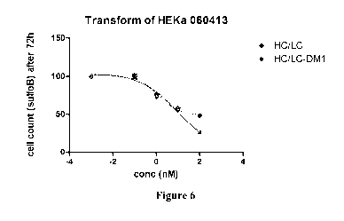
Figure 6 is a graph showing the cytotoxic activity of naked anti-EGFR antibody
cetuximab and maytansinoid conjugates thereof against primary keratinocytes. 2-
3,000
cells/well were seeded in a flat bottom 96-well tissue culture plate, and
incubated with
various concentrations of test article in culture media for 72 h or 5 days at
37 C. Viability
of the remaining cells was determined via alamar blue cell viability assay.
Figure 7 is a graph showing the cytotoxic activity of naked anti-EGFR antibody
panitumumab and maytansinoid conjugates thereof against primary keratinocytes.
2-
3,000 cells/well were seeded in a flat bottom 96-well tissue culture plate,
and incubated
with various concentrations of test article in culture media for 72 h or 5
days at 37 C.
Viability of the remaining cells was determined via alamar blue cell viability
assay.
8
CA 02915897 2015-12-16
WO 2015/000062
PCT/CA2014/000543
Figure 8 is a graph showing the cytotoxic activity of naked anti-EGFR antibody
panitumumab and maytansinoid conjugates thereof against primary keratinocytes.
2-
3,000 cells/well were seeded in a flat bottom 96-well tissue culture plate,
and incubated
with various concentrations of test article in culture media for 72 h or 5
days at 37 C.
Viability of the remaining cells was determined via alamar blue cell viability
assay.
Figures 9-13 show results with immunoconjugates that incorporate elements not
selected
according to the criteria prescribed herein, as discussed in Example 4.
Figure 9 shows that panitumumab and cetuximab are very strong EGFR activation
blockers and thus will not be potentiated on normal keratinocytes by
conjugation,
whereas partially antagonistic antibodies such as J2898A is potentiated (See
Figure 10,
and 11).
Figure 10 shows that partially antagonistic anti-EGFR antibody by IMGN
(J2989A) is
potentiated by conjugation (IMGN289) on normal keratinocytes (see also Setiady
et al,
Proceedings of the 104th Annual Meeting of the American Association for Cancer
Research; 2013 Apr 6-10; Washington, DC. Philadelphia (PA): AACR; Cancer Res
2013;73(8 Suppl):Abstract nr 5463).
Figure 11 shows the toxicity of cetuximab mutant antibody, 6-LC, (having
reduced
affinity to make it a partially antagonistic against EGFR) is potentiated by
conjugation to
payload via non-cleavable linker (6LC-DM1).
Figure 12 shows panitumumab, and panitumumab-DM1 effects on keratinocytes, and
reveals that conjugation of panitumumab via non-cleavable linker to DM1
(Avid300-
DM1) does not increase its activity against normal cells; 2C9-DM1 is cetuximab-
DM1
conjugate used as a control.
Figure 13 shows that conjugation of cetuximab to MMAE anti-microtubule payload
by a
cleavable linker (valine-citruline) potentiates its toxicity against both
normal cells and
9
CA 02915897 2015-12-16
WO 2015/000062
PCT/CA2014/000543
MDA-MB-468 cancer cells (Cetux 2C9-MMAE), whereas conjugation via non-
cleavable
linker (SMCC) only (Cetux2C9-DM1) potentiates anti-cancer activity, thus
demonstrating a favorable therapeutic window.
DETAILED DESCRIPTION OF THE PREFERRED EMBODIMENTS
The present immunoconjugates are based on antibodies that bind to the human
epidermal
growth factor receptor (hEGFR), a protein that is presented on the surface of
many
different cell types including particularly skin cells such as keratinocytes.
As used
herein, the term "hEGFR" refers to any protein that comprises the expressed
and
processed product of the human her-1 gene, wherein the protein is designated
as
UniProtKB/Swiss-Prot P00533. The term EGFR is used generically herein, and
refers to
the wild type protein and all naturally occurring variants thereof. The term
"wtEGFR" is
used more specifically with reference only to the wild type form of human
EGFR. The
term "EGFRvIII" refers to the EGFR variant protein that comprises the
expressed and
processed product of a variant of the her-1 gene lacking exons 2-7, and thus
includes only
the polypeptide sequence encoded by exons 1 and 8 of her-1. The term "domain
III" is
not related to EGFRvIII, and instead refers to a location within EGFR, and
represents an
extracellular site that is key for EGF ligand binding, and binding of highly
antagonistic
antibodies cetuximab and panitumumab (Voigt et al, 2012 November; 14(11): 1023-
1031).
The present immunoconjugates comprise an EGFR antibody that is a full
antagonist at the
EGFR. An EGFR antibody that is a "full antagonist" is an antibody that blocks
completely or nearly so the transmission of a signal that is stimulated, in
the normal
course, by the EGF ligand through wtEGFR to the wtEGFR-coupled tyrosine
kinase.
EGFR antibodies that are full antagonists are particularly EGFR antibodies
that bind
directly to EGFR domain III. EGFR antibodies having these properties and an
EGFR
binding affinity of 5 nanomolar (nM) or less are particularly preferred for
inclusion in the
present immunoconjugates. By this measure, and as shown in Figure 9, at least
the
following known antibodies are not suitable for use in the present
immunoconjugates:
CA 02915897 2015-12-16
WO 2015/000062
PCT/CA2014/000543
J2898A, intellimab 6-LC (a cetuximab variant taught in WO 2012/100346),
nimotuzumab, and matuzumab.
It is found that when a full antagonist EGFR antibody is selected to deliver a
toxic
payload, the killing effect is potentiated only on EGFR+ cancer cells and not
on EGFR+
normal cells such as keratinocytes. This is not the case when the EGFR
antibody is a
partial antagonist, i.e., an antibody that allows transmission of some EGF-
mediated
signal. When conjugated to a toxin, these partial antagonist antibodies show a
potentiation of the killing effect at both normal cells that are EGFR+ and
cancer cells that
are EGFR+.
The present immunoconjugates are based more particularly, and in one
embodiment, on
the hEGFR antibody known as cetuximab, now commercially available from Eli
Lilly
and Company under the trade name Erbitux . Cetuximab is a recombinant,
human/mouse
chimeric IgG1 antibody that binds specifically to the extracellular domain of
wtEGFR.
The amino acid sequences of the CDRs for both the heavy chain of cetuximab
(SEQ ID
Nos.1-3) and the light chain of cetuximab (SEQ ID Nos. 4-6) are listed herein.
Also
listed are the amino acid sequences of the heavy chain variable region (SEQ ID
No.7) and
of the light chain variable region (SEQ ID No.8) of cetuximab, together with
the amino
acid sequences of the complete heavy chain (SEQ ID No. 9) and complete light
chain
(SEQ ID No.10) of cetuximab.
Cetuximab variants useful herein are highly antagonistic EGFR-binding agents
that
compete with cetuximab for binding to human EGFR. Useful cetuximab variants
have
been mentioned hereinabove, and include fragments of cetuximab comprising the
EGFR
binding sites of cetuximab, such as all of the light chain and heavy chain
CDRs herein
recited. Other cetuximab variants useful here in are cetuximab variants that
incorporate
one, two or more substitutions outside the antigen binding domains, such as in
the
framework region or in the constant region (Fc). Such substitutions are benign
in the
sense that they do not reduce cytotoxicity relative to cetuximab per se.
11
CA 02915897 2015-12-16
WO 2015/000062
PCT/CA2014/000543
In another embodiment, the present immunoconjugates are based on the EGFR
antibody
known as panitumumab, now commercially available and sold under the trade name
Vectibix . Panitumumab is a recombinant, fully human IgG2 antibody that binds
specifically to the extracellular domain of wtEGFR. The amino acid sequences
of the
heavy and light chains of panitumumab are listed in US 6,235,883 and US
7,807,798. A
useful alternative to panitumumab is an EGFR binding variant thereof that
competes with
panitumumab for EGFR binding, such as a fragment of panitumumab that
incorporates its
antigen binding sites but has an otherwise lost or altered constant region.
The present immunoconjugates can also be based on still other EGFR antibodies
provided they show full antagonist activity as defined above, such as EGFR
antibodies
that bind selectively to domain III of EGFR, and any other EGFR antibodies
that compete
with EGF and block fully or nearly so the transmission of EGF-stimulated
downstream
signalling.
In the present immunoconjugates, the full antagonist EGFR antibody, such as
cetuximab
or panitumumab, is conjugated to an anti-microtubule toxin such as
maytansinoid toxin.
By "anti-microtubule toxin" is meant an agent having cell toxicity mediated by
interference with the microtubule structures important for cell mitosis, such
as by
inhibiting the formation of tubulin or by inhibiting the organization thereof.
Included within this toxin family are the maytansinoids and auristatins, and
many other
agents developed more recently and having the same mechanism of action. The
auristatins in particular block cell replication by inhibiting polymerization
of tubulin and
are thus anti-mitotic. The structure of an auristatin useful herein and known
as MMAE,
or vedotin, is shown below:
HT
0
? 0
N
H H
12
CA 02915897 2015-12-16
WO 2015/000062
PCT/CA2014/000543
There are various forms of maytansinoids that are useful. These are all based
on the
complex structure of a natural molecule, maytansine.
Particularly useful are the maytansinoids including DM-1 and DM-4. In a
specific
embodiment, the toxin coupled to the EGFR MAb is DM-1 having the structure
shown
infra.
Also useful as anti-microtubule toxins are dolostatins, auristatins,
tubulysins and
cryptophycins. Specific examples of useful species within each genus include
dolostatin
10, monomethyl dolostatin 10, auristatin E, monomethyl auristatin E (MMAE),
auristatin
F, monomethyl auristatin F, HTI-286, tubulysin M, as well as the tubulin
binders such as
tubulysin IM-1, tubulysin IM-2, tubulysin IM-3, colchicine DA, and
maytansinoids AP-3,
DM-1 and DM-4.
Conjugates of a full antagonist EGFR antibody such as cetuximab or
panitumumab, and
an anti-microtubule toxin such as a maytansinoid or auristatin can be formed
using any
technique presently known or later developed that couples a linker that is
"non-
cleavable". These linkers remain intact, and retain the antibody and toxin in
covalent
association, throughout conditions normally encountered following
administration to a
subject, including extracellular environments. More specifically, non-
cleavable linkers
result in ADC constructs for which release of the cytotoxic payload is
achieved by
destruction of the antibody by intracellular lysosomes.
Methods of linker integration are described for instance in US 5,208,020; US
8,088,387;
and US 6,441,163. A preferred method is to modify the EGFR antibody, e.g.,
cetuximab,
with succinimidyl 4(N-maleimidomethyl)-cyclohexane-1-carboxylate (SMCC) to
introduce maleimido groups followed by reaction of the modified antibody with
a thiol-
containing maytansinoid to give a thioether-linked conjugate. The resulting
chemical
structures are shown below. Conjugates with 1 to 10 drug molecules per
antibody
molecule result.
13
CA 02915897 2015-12-16
WO 2015/000062
PCT/CA2014/000543
o
iton
: 0
0
WO fah \
0
s.../L.0
116 11
28
AF-SMCC-DMI
.41} Antibody
0
0
(.2
0
CI 0
Nic0 'N
0
=.* ZS
Ab-S1.4=DMI
Ab - Antibody
Other useful forms of non-cleavable linkers include N-Succinimidyl iodoacetate
(SIA),
sulfo-SMCC, m-maleimidobenzoyl-N-hydroxysuccinimide ester(MBS), sulfo-MBS and
succinimidyl-iodoacetate, as described in the literature, to introduce 1-10
reactive groups.
(see, Yoshitake et al, 101 Eur. J. Biochem. 395-399 (1979); Hashida et al, J.
Applied
Biochem. 56-63 (1984); and Liu et al, 18 690-697 (1979)). Particularly useful
for linking
auristatins as anti-microtubule toxin are the non-cleavable maleimidocaproyl
linkers
described by Doronina et al, in Bioconjugate Chem., 2006 Jan-Feb;17(1):114-
24).
In a specific embodiment, the immunoconjugate comprises cetuximab linked to DM-
1 by
an SMCC linker.
14
CA 02915897 2015-12-16
WO 2015/000062
PCT/CA2014/000543
In another specific embodiment, the immunoconjugate comprises panitumumab
linked to
DM-1 by an SMCC linker.
Thus, in a general aspect, the invention provides a method useful to
potentiate the anti-
cancer activity of an EGFR antibody without potentiating the effect thereof on
normal
EGFR+ cells, the method comprising:
(i) selecting, for conjugation, an EGFR antibody that is a full EGFR
antagonist;
(ii) selecting, for delivery by the EGFR antibody, an anti-microtubule
toxin;
(iii) selecting, for coupling the selected EGFR antibody to the anti-
microtubule
toxin, a linker that is non-cleavable; and
incorporating the linker between the antibody and the toxin to provide an
immunoconjugate having a cytotoxic activity that is potentiated against EGFR+
disease
cells but not against normal EGFR+ cells.
As will be appreciated from the preceding disclosure, the full antagonist EGFR
antibody
is preferably cetuximab or panitumumab. The anti-microtubule toxin is
preferably an
auristatin or a maytansinoid, and is most preferably DM-1. The non-cleavable
linker is
preferably SMCC.
In another specific embodiment, there is the proviso that the full antagonist
EGFR
antibody is not cetuximab. In a further specific embodiment, there is the
proviso that the
full antagonist EGFR antibody is not panitumumab.
Therapeutic formulations of the conjugate are prepared for therapeutic use
directly or for
storage by mixing the conjugate having the desired degree of purity with
optional
pharmaceutically acceptable carriers, excipients or stabilizers (Remington's
Pharmaceutical Sciences, 16th edition, Osol, A. Ed. [1980]), in the form of
lyophilized
formulations or aqueous solutions. Acceptable carriers, excipients, or
stabilizers are
nontoxic to recipients at the dosages and concentrations employed, and include
buffers
such as phosphate, citrate, and other organic acids; antioxidants including
ascorbic acid
CA 02915897 2015-12-16
WO 2015/000062
PCT/CA2014/000543
and methionine; preservatives (such as octadecyldimethylbenzyl ammonium
chloride;
hexamethonium chloride; benzalkonium chloride, benzethonium chloride; phenol,
butyl,
or benzyl alcohol; alkyl parabens such as methyl or propyl paraben; catechol;
resorcinol;
cyclohexanol; 3-pentanol; and m-cresol); low molecular weight (less than about
10
residues) polypeptides; proteins such as serum, albumin, gelatin, or
immunoglobulins;
hydrophilic polymers such as polyvinylpyrrolidone; amino acids such as
glycine,
glutamine, asparagines, histidine, arginine or lysine; monosaccharides,
disaccharides, and
other carbohydrates including glucose, mannose, or dextrins; chelating agents
such as
EDTA; sugars such as sucrose, marmitol, trehalose or sorbitol; salt-forming
counter-ions
such as sodium; metal complexes (e.g., Zn-protein complexes); and/or non-ionic
surfactants such as TWEEN, PLURONICS or polyethylene glycol (PEG).
The active ingredients to be used for in vivo administration must be sterile.
This is readily
accomplished by filtration through sterile membranes.
Sustained-release preparations may be prepared. Suitable examples of sustained-
release
include semipermeable matrices of solid hydrophobic polymers containing the
conjugate,
which matrices are in the form of shapes articles, e.g., films or
microcapsules. Examples
of sustained-release matrices include polyesters, hydrogels (for example, poly
(2-
hydroxyethyl-methacrylate), polylactides (U.S. Pat. No. 3,773,919), copolymers
of L-
glutamic acid and ethyl-L-glutamate, non-degradable ethylene-vinyl acetate,
degradable
lactic acid-glycolic acid copolymers such as injectable microspheres composed
of lactic
acid-glycolic acid copolymer and leuprolide acetate, and poly-D-(-)-3-
hydroxybutyric
acid. While polymers such as ethylene-vinyl acetate and lactic acid-glycolic
acid enable
release of molecules for over 100 days, certain hydrogels release proteins for
shorter time
periods.
The conjugates are useful to treat EGFR+ disease cells. Such treatment results
in a
reduction in the number, size or distribution of such disease cells in
subjects presenting
with them. In embodiments, the conjugates are used to treat EGFR+ disease
cells that are
EGFR+ cancer cells and tumours comprising them. Such treatment results
preferably in a
16
CA 02915897 2015-12-16
WO 2015/000062
PCT/CA2014/000543
reduction in the number, size, volume or distribution of such cancer cells and
tumours
comprising them, or at least in a reduction in the rate at which such disease
cells increase
in number, size, volume or distribution of such cells and tumours in subjects
presenting
with them.
Subjects presenting with EGFR+ cancer cells can be identified with the aid of
assays that
detect the receptor, as protein or as nucleic acid precursor (DNA or RNA) in
physiological samples such as biopsied tissue. A suitable test for EGFR
protein is the
commercially available and FDA approved Dako EGFR pharmDx test kit.
For the treatment of subjects presenting with EGFR+ cancer cells, the
appropriate dosage
of the conjugate will depend on the type of disease to be treated, as defined
above, the
severity and course of the disease, whether the agent is administered for
preventative or
therapeutic purposes, previous therapy, the patients clinical history and
response to the
agent, and the discretion of the attending physician. The agent is suitably
administered to
the patient at one time or over a series of treatments.
For example, depending on the type and severity of the disease, about 1 g/kg
to 15
mg/kg (e.g., 0.1-20 mg/kg) of conjugate is a candidate dosage for
administration to the
patient, whether, for example, by one or more separate administrations, or by
continuous
infusion. A typical daily dosage might range from about 1 jig/kg to 500 mg/kg
or more,
depending on the factors mentioned above. For repeated administrations over
several
days or longer, depending on the condition, the treatment is sustained until a
desired
suppression of disease symptoms occurs. However, other dosage regimens may be
useful.
The progress of this therapy is easily monitored by conventional techniques
and assays.
It will thus be appreciated that an effective amount of the immunoconjugate is
an amount
effective alone or as part of a treatment regimen that retards or inhibits the
rate of growth
or proliferation of EGFR+ disease cells.
An EGFR+ disease cell is a disease cell that presents EGFR on its surface as
detectable
for instance by EGFR antibody binding, or by detection of intracellular mRNA
encoding
17
CA 02915897 2015-12-16
WO 2015/000062
PCT/CA2014/000543
her-I. Particular EGFR+ disease cells include those having on their surface an
abnormally high density and/or activity of EGFR molecules, or the presence of
the
EGFRvIII variant of EGFR.
It may be useful to administer to administer the present conjugates by
intravenous
infusion first as loading dose, followed by maintenance dose, such as at an
initial dose of
4mg/kg over 90 minutes, then 2 mg/kg over 30 minutes, once weekly for as many
as 52
weeks, with follow up as required. In the specific case of the panitumumab
conjugate,
dosing might be based on that utilized for panitumumab per se, which comprises
6mg/kg
given once every two weeks as a one hour infusion.
The conjugates are useful in the treatment of a variety of cancers, to inhibit
the growth or
proliferation of EGFR+ cancer cells and tumours comprising them, including
hematopoietic cell cancers and solid tumours. Conditions or disorders to be
treated
include benign or malignant tumors (e.g., renal, liver, kidney, bladder,
breast, gastric,
ovarian, colorectal, prostate, pancreatic, lung, vulva, and thyroid); hepatic
carcinomas;
sarcomas; glioblastomas; and various head and neck tumors; leukemias and
lymphoid
malignancies. In particular embodiments, the antibody or bivalent fragment are
used in
the treatment of such cancer cells that express EGFRvIII, as determined by the
screening
assays herein described. In particular embodiments, the cancer cells are EGFR+-
presenting cancer cells that include head and neck cancers and especially
squamous cell
carcinoma of the head and neck, colorectal cancers, gastrointestinal cancers,
brain
tumours including glioblastomas, and tumours of the lung including non-small-
cell lung
carcinoma, and of the breast, pancreas, esophagus, kidney, ovary, cervix and
prostate. In
specific embodiments, the EGFR+ cancer is one for which cetuximab has received
FDA
marketing approval, such as squamous cell carcinoma of the head and neck and
colorectal
cancers.
It will be appreciated that subjects who could benefit from the present method
include
mammals including humans as well as livestock, and pets.
18
CA 02915897 2015-12-16
WO 2015/000062
PCT/CA2014/000543
Other therapeutic regimens may be combined with the administration of the
conjugates of
the instant invention. For example, the patient to be treated may also receive
radiation
therapy, such as external beam radiation. Alternatively, or in addition, a
chemotherapeutic agent may be administered to the patient. Preparation and
dosing
schedules for such chemotherapeutic agents may be used according to
manufacturers'
instructions or as determined empirically by the skilled practitioner.
Preparation and
dosing schedules for such chemotherapy are also described in Chemotherapy
Service Ed.,
M. C. Perry, Williams & Wilkins, Baltimore, Md. (1992). The chemotherapeutic
agent
may precede, or follow administration or the conjugate or may be given
simultaneously
therewith. The conjugate may be combined with any anti-cancer toxins, or any
other
suitable drug particularly including irinotecan (CPT-11), cisplatin,
cyclophosphamide,
melphalan, dacarbazine, doxorubicin, daunorubicin, and topotecan, as well as
tyrosine
kinase inhibitors, including particularly EGFR kinase inhibitors such as
AG1478 ((4-(3-
chloroanilino-6,7-dimethoxyquinazoline), gefitinib (Iressa0), erlotinib
(TarcevaS),
lapatinib (Tykerb0), canertinib (PD183805, Pfizer), PKI-166 (Novartis),
PD158780 and
pelitinib.
It may also be desirable to administer antibodies or conjugates against other
tumor
associated antigens or their ligands, such as antibodies which bind to the
ErbB2
(including trastuzumab marketed as Herceptine, and pertuzumab marketed as
Omnitarge), ErbB3, ErbB4, or vascular endothelial factor (VEGF), and/or
antibodies
that bind to EGF or TGFa.
In another embodiment of the invention, an article of manufacture containing
conjugate
in an amount useful for the treatment of the disorders described herein is
provided. The
article of manufacture comprises a container and a label. Suitable containers
include, for
example, bottles, vials, syringes, and test tubes. The containers may be
formed from a
variety of materials such as glass or plastic. The container holds a
composition which is
effective for treating the condition and may have a sterile access port (for
example the
container may be an intravenous solution bag or vial having a stopper
pierceable by a
hypodermic injection needle). The label on or associated with the container
indicates that
19
CA 02915897 2015-12-16
WO 2015/000062
PCT/CA2014/000543
the composition is used for treating a cancer condition. The article of
manufacture may
further compromise a second container compromising a pharmaceutically-
acceptable
buffer, such as phosphate-buffered saline, Ringer's solution and dextrose
solution. It may
further include other matters desirable from a commercial and use standpoint,
including
other buffers, diluents, filters, needles, syringes, and package inserts with
instructions for
use.
An anti-cancer immunoconjugate according to the invention may be administered
with a
pharmaceutically-acceptable diluent, carrier, or excipient, in unit dosage
form. Unit
doses of the conjugate are suitably 50mgs, 100mgs, 150mgs, 200mgs, 250mgs,
300mgs
and 400mgs. The drug can be formulated in single use vials at a concentration
such as
20mg/mL, for instance 100mg in 5mL vehicle such as 0.9% saline, 200mg in 10mL
or
400mg in 20mL.
Any appropriate route of administration can be employed, for example,
parenteral,
intravenous, subcutaneous, intramuscular, intracranial, intraorbital,
ophthalmic,
intraventricular, intracapsular, intraspinal, intracisternal, intraperitoneal,
intranasal,
aerosol, pulmonary, or oral administration.
Example 1 ¨ Preparation of Cetuximab-SMCC-DM1 conjugate (A-H)
A. PREPARATION AND MEASUREMENT OF CETUXIMAB ANTIBODY
Cetuximab is obtained from the open market, or is produced as described in WO
2012/100346, for conjugation to DM1 using the non-cleavable heterobifunctional
cross-
linking reagent SMCC.
Cetuximab antibody was then buffer exchanged into 50 mM potassium phosphate,
50
mM sodium chloride, 2 mM EDTA; pH 6.5 buffer (Buffer A). All buffers in this
experiment were tested to be free of endotoxin using a chromogenic Limulus
amoebocyte
lysate (LAL) method (Cambrex). The concentration of antibody was measured
using an
extinction coefficient of 1.45 mL/mg/cm at 280 nm and a molecular weight of
145,781g.
CA 02915897 2015-12-16
WO 2015/000062
PCT/CA2014/000543
B. PREPARATION AND MEASUREMENT OF SMCC STOCK SOLUTION
A 20 mM solution of SMCC (6.69 mg/mL) (Concortis Biosystems Corp.) was
prepared
in DMSO. The solution was diluted 1/40 in Assay Buffer and the absorbance of
the
samples was measured at 302 nm. The concentration of the stock solution was
calculated
using a molar extinction coefficient of 602/M/cm.
C. PREPARATION AND MEASUREMENT OF DM1 STOCK SOLUTION
A 10 mM solution of DM1 (free thiol form; Concortis Biosystems Corp.) was
prepared in
DMA (7.37 mg/mL). The absorbance of dilutions of the stock solution in ethanol
was
measured at 280 nm. The concentration of stock DM1 was calculated by using a
molar
extinction coefficient of 5700/M/cm at 280 nm. The concentration of free ¨SH
in the
stock DM1 preparation was measured using Elman's reagent (DTNB). Dilutions of
the
stock solution were prepared in Assay buffer made to 3% (v/v) DMA, and then
100 mM
DTNB in DMSO (1/100th volume) was added. The increase in absorbance at 412 nm
was
measured against a reagent blank and the concentration was calculated using an
extinction coefficient of 14150/M/cm. The concentration of ¨SH resulting from
the
Elman's assay was used to represent the DM1 stock concentration in
calculations for
conjugation conditions.
D. MODIFICATION OF CETUXIMAB WITH SMCC CROSSLINKER
The antibody was modified using a 7.5-fold molar excess of SMCC at 20 mg/mL
antibody. The reaction was carried out in Buffer A (95% v/v) with DMSO (5%
v/v) for 2
hours at room temperature with stirring.
E. G25 CHROMATOGRAPHY TO REMOVE EXCESS SMCC
The cetuximab-SMCC reaction mixture was gel-filtered through a 1.5x4.9 cm pre-
packed
column of Sephadex G25 resin equilibrated in Buffer A. The load and elution
volumes
were according to manufacturer's instructions (Amersham Biosciences). The
concentration of the modified antibody solution was assayed
spectrophotometrically
using the extinction co-efficient described above.
21
CA 02915897 2015-12-16
WO 2015/000062
PCT/CA2014/000543
F. CONJUGATION OF CETUXIMAB-SMCC WITH DM1
The modified antibody was reacted with a 1.7-fold excess of DM1 over linker
(assuming
linkers per antibody). The reaction was carried out at 10 mg/mL antibody
concentration
in Buffer A (94% v/v) with DMA (6% v/v). After addition of DM1, the reaction
was
incubated at room temperature for 16.5 hours with stirring.
G. CONJUGATION PURIFICATION BY G25 CHROMATOGRAPHY
The conjugation reaction mixture was gel-filtered through a 1.5x4.9 cm pre-
packed
column of Sephadex G25 resin equilibrated in 1 x phosphate buffered saline
(PBS), pH
6.5 (Buffer B). The load and elution volumes were according to manufacturer's
instructions (Amersham Biosciences). The number of DM1 molecules linked per
mole of
cetuximab was determined by measuring absorbance at both 252 nm and 280 nm of
the
eluted material. The DM1/antibody ratio was found to be 2 and 4. The resulting
conjugate
was analyzed for binding and cytotoxicity.
H. TESTING OF CETUXIMAB-SMCC-DM1
The cell lines used in these studies have the following characteristics:
A431: human epithelial carcinoma of vulva cell line; available from ATCC;
plated at
4000 cells/well in DMEM-10%FBS, 100[11/well in 96 well plate.
H226: lung squamous cell carcinoma cell line; available at ATCC; plated at
4000
cells/well in RPMI-10%FBS, 100[d/well in 96-well culture plate.
MDA-MB-468: mammary gland/breast; derived from metastatic site: pleural
effusion;
available from ATCC; plated at 4000 cells/well in ATCC-formulated Leibovitz's
L-15
Medium (Catalog No. 30-2008) with added fetal bovine serum to a final
concentration of
10%; 100[d/well in 96 well plate.
22
CA 02915897 2015-12-16
WO 2015/000062
PCT/CA2014/000543
HaCaT: in vitro spontaneously transformed keratinocytes from histologically
normal
skin; available from Chinese Center for Type Culture Collection of Wuhan
University;
plated at 2000 cells/well in DMEM-10%FBS, 1000/well in 96-well culture plate.
HEKa: Normal Human Primary Epidermal Keratinocytes, adult; available from
PromoCell GmbH; plated at 4000cells/well in EpiLife media 100 1/well, EpiLife
medium #MEP1500CA, Invitrogen +HKGS human keratinocyte growth supplement
(#S-001-5)
Binding studies showed that the conjugation of antibody to DM1 did not affect
the KD;
both naked cetuximab antibody and cetuximab-SMCC-DM1 conjugate had the same
binding affinity to EGFR by surface plasmon resonance (-0.3 nM, Table 1).
Evaluation
of the in vitro cytotoxicity of the sample showed that the cetuximab-SMCC-DM1
conjugate is significantly more cytotoxic compared to cetuximab against cancer
cell lines
(Figs 3&4), but that surprisingly cetuximab-SMCC-DM1 and naked cetuximab have
the
same cytotoxic activity against keratinocytes (Figs 5&6).
Table 1: effects of conjugation to DM1 on cetuximab KD
Sample # of experiments EGFR ecd KD (nM)
Commercial cetuximab 3 0.27 +1- 0.02
Cetuxima b-SMCC-DM 1 3 0.28 +/- 0.02
The effect of DM1 conjugation on different EGFR MAbs (huML66 and huEGFR-7R) is
demonstrated in ImmunoGen's published US patent application 2012/0156217, and
Figures 1 and 2 herein are reproductions of figures appearing in that
application. More
particularly, as shown in Figure 1, conjugation of anti-EGFR antibodies huML66-
SMCC-
DM1 and huEGFR-7R to maytansinoids to create huML66-SMCC-DM1 and huEGFR-
7R-SMCC-DM1 immunoconjugates increases cytotoxic activity of against normal
keratinocytes. Similarly, and as expected, conjugation of anti-EGFR antibodies
huML66
and huEGFR-7R to maytansinoids to create huML66-SMCC-DM1 and huEGFR-7R-
SMCC-DM1 immunoconjugates significantly potentiates cytotoxic activity of the
antibodies against H226 cancer cell line (Figure 2).
23
CA 02915897 2015-12-16
WO 2015/000062
PCT/CA2014/000543
As shown in Figures 3 and 4, DM1 conjugation of anti-EGFR antibody cetuximab
(HC-
LC) to create cetuximab-SMCC-DM1 immunoconjugate (HC-LC/DM1) significantly
potentiates cytotoxic activity of the antibody against cancer cells, as
expected and shown
in Figure 3. As shown in Figure 4, and also as expected, conjugation of anti-
EGFR
antibody cetuximab (HC-LC) to maytansinoids to create cetuximab-SMCC-DM1
immunoconjugate (HC-LC/DM1) significantly potentiates cytotoxic activity of
the
antibody against cancer cells.
However, and quite surprisingly, as shown in Figures 5 and 6, conjugation of
anti-EGFR
antibody cetuximab (HC-LC) to maytansinoids to create cetuximab-SMCC-DM1
immunoconjugate (HC-LC/DM1) did not increase cytotoxicity against the
keratinocytes
as is reflected in substantially the same IC50 values for these agents (IC50
of 0.8269 for
conjugate vs. IC50 of 0.4324 for naked antibody; Fig 5, HaCaT - normal human
keratinocytes cell line & IC50 of 2.210 for conjugate vs. IC50 of 0.9348 for
naked
antibody, Fig 6; HEKa- primary human keratinocyte cells). As used herein, IC50
refers to
the concentration of drug required to kill 50% of cells.
The measure of whether cytotoxicity is increased or not is determined above by
comparing IC50 values produced using the alamar blue cell viability assay
(each
experiment performed in triplicates in a 96-well plate). A difference within
one log order
is deemed not significant and is considered to reveal no change in
cytotoxicity, whereas a
difference greater than one log order reveals a significant change in
cytotoxicity.
Example 2 - Evaluation in Primates
Cynomolgus monkey species has been previously demonstrated to be a valuable
and
highly predictive model for evaluating anti-EGFR antibodies toxicities,
including
dermatologic side-effects. The high level of correlation between Cynomolgus
monkey
and human toxicity data for EGFR-targeting antibodies is in part due high
homology
between the monkey and human EGFR receptors that results in very similar KD
values
for the antibodies:
24
CA 02915897 2015-12-16
WO 2015/000062
PCT/CA2014/000543
Table 1: Binding affinity of cetuximab and panitumumab to human and
cynomologus
EGFR is consistent across the species. Apparent antibody affinity defined as
the antibody
concentration (picomolar, pM) required to achieve half-maximal binding in
ELISA with
recombinant extracellular domains of human EGFR (huEGFR), and Cynomolgus
monkey
EGFR (cyEGFR).Adapted from Koefoed et al, MABs. 2011 Nov-Dec; 3(6):584-595.
Apparent affnity, EL1SA assay
Domain 1118 Binding
ft4
Antigens a> At
s.o
human sEGFR (DM) 12 32
eynoMOlgus NECFR (pM) 14 32
The cetuximab-SMCC-DM1 immunoconjugate was tested in primates particularly to
identify any significant potentiation of antibody side-effects by the
conjugated toxin on
normal cells. In particular, close attention was paid to dermatologic side-
effects that are
qualitatively different and/or significantly exacerbated relative to what is
expected with
the naked fully antagonistic anti-EGFR antibody.
The study initially made use of two Cynomolgus macaques, all males that were
sedated
with ketamine and anesthetized with isoflurane to facilitate IV administration
of the
antibody-drug conjugate administered at 10mg/kg. Animals were then observed
for 5
days prior to sacrifice for general signs of acute toxicity, and examined
further.
The detailed histopathology analysis revealed no severe dermatologic
toxicities or other
severe and qualitatively new dermatologic side-effects other than those that
are expected
as a result of naked cetuximab treatment. Also, no other severe on-target
toxicities were
observed in these animals demonstrating safety of ADCs of present invention
and
absence of significant exacerbation of the naked antibody toxicities.
CA 02915897 2015-12-16
WO 2015/000062
PCT/CA2014/000543
Subsequent to successful completion of the acute primate study, repeat-dose
safety study
has been initiated in additional animals. The ADC was administered once every
three
weeks at 10mg/kg dose over 4 cycles. This dose and schedule was selected based
on
dosing schedules commonly used with other ADCs in primates and humans (Poon KA
et
al, Preclinical safety profile of trastuzumab emtansine (T-DM1): mechanism of
action of
its cytotoxic component retained with improved tolerability. Toxicol App!
Pharmacol.
2013 Dec 1;273(2):298-313; FDA Package Insert for KadcylaTM, accessed February
24,
2014: http://www.accessdata.fda. gov/drugsatfda_doc
s/labe1/2013/1254271b1.pdf)
With the exception of reversible liver enzyme elevations which is an expected
class side-
effect of ADCs, no other severe and/or qualitatively new dermatologic or other
on-target
side-effects other than those that are already expected as a result of naked
cetuximab
treatment (cetuximab cynomolgus monkey data available at:
http ://www.accessdata.fda.gov/drugsatfda_docs/b1a/2004/125084_ERBITUX_PHARMR
P3.PDF) have been observed in the ongoing study to date. Severe skin toxicity
and toxic
epidermal necrolysis previously reported with anti-CD44v6 ADC (which also uses
SMCC-DM1 payload technology and targets an antigen expressed by normal
keratinocytes) has not been observed in this study (Tijink et al., Clin Cancer
Res, 2006,
12:6064). These safety data further validates in vitro results that on-target
toxicities of
fully antagonist naked anti-EGFR antibodies is not significantly potentiated
as result of
conjugation to the cytotoxic payloads.
Example 3¨ Preparation of Panitumumab-SMCC-DM1 conjugate (A-H)
A DM-1 conjugated panitumumab was prepared essentially as described above for
the
cetuximab counterpart. More particularly,
A. PREPARATION AND MEASUREMENT OF PANITUMUMAB ANTIBODY
Panitumumab is obtained from the open market, or is produced as described in
US
6,235,883 or US 7,807,798 for conjugation to DM1 using the non-cleavable
heterobifunctional cross-linking reagent SMCC.
26
CA 02915897 2015-12-16
WO 2015/000062
PCT/CA2014/000543
Panitumumab antibody was then buffer exchanged into 50 mM potassium phosphate,
50
mM sodium chloride, 2 mM EDTA; pH 6.5 buffer (Buffer A). All buffers in this
experiment were tested to be free of endotoxin using a chromogenic Limulus
amoebocyte
lysate (LAL) method (Cambrex). The concentration of antibody was measured
using an
extinction coefficient of 1.45 mL/mg/cm at 280 nm and a molecular weight of
145,781g.
B. PREPARATION AND MEASUREMENT OF SMCC STOCK SOLUTION
A 20 mM solution of SMCC (6.69 mg/mL) (Concortis Biosystems Corp.) was
prepared
in DMSO. The solution was diluted 1/40 in Assay Buffer and the absorbance of
the
samples was measured at 302 nm. The concentration of the stock solution was
calculated
using a molar extinction coefficient of 602/M/cm.
C. PREPARATION AND MEASUREMENT OF DM1 STOCK SOLUTION
A 10 mM solution of DM1 (free thiol form; Concortis Biosystems Corp.) was
prepared in
DMA (7.37 mg/mL). The absorbance of dilutions of the stock solution in ethanol
was
measured at 280 nm. The concentration of stock DM1 was calculated by using a
molar
extinction coefficient of 5700/M/cm at 280 nm. The concentration of free ¨SH
in the
stock DM1 preparation was measured using Elman's reagent (DTNB). Dilutions of
the
stock solution were prepared in Assay buffer made to 3% (v/v) DMA, and then
100 mM
DTNB in DMSO (1/100th volume) was added. The increase in absorbance at 412 nm
was
measured against a reagent blank and the concentration was calculated using an
extinction coefficient of 14150/M/cm. The concentration of ¨SH resulting from
the
Elman's assay was used to represent the DM1 stock concentration in
calculations for
conjugation conditions.
D. MODIFICATION OF PANITUMUMAB WITH SMCC CROSSLINKER
The antibody was modified using a 7.5-fold molar excess of SMCC at 20 mg/mL
antibody. The reaction was carried out in Buffer A (95% v/v) with DMSO (5%
v/v) for 2
hours at room temperature with stirring.
27
CA 02915897 2015-12-16
WO 2015/000062
PCT/CA2014/000543
E. G25 CHROMATOGRAPHY TO REMOVE EXCESS SMCC
The panitumumab-SMCC reaction mixture was gel-filtered through a 1.5x4.9 cm
pre-
packed column of Sephadex G25 resin equilibrated in Buffer A. The load and
elution
volumes were according to manufacturer's instructions (Amersham Biosciences).
The
concentration of the modified antibody solution was assayed
spectrophotometrically
using the extinction co-efficient described above.
F. CONJUGATION OF PANITUMUMAB-SMCC WITH DM1
The modified antibody was reacted with a 1.7-fold excess of DM1 over linker
(assuming
linkers per antibody). The reaction was carried out at 10 mg/mL antibody
concentration
in Buffer A (94% v/v) with DMA (6% v/v). After addition of DM1, the reaction
was
incubated at room temperature for 16.5 hours with stirring.
G. CONJUGATION PURIFICATION BY G25 CHROMATOGRAPHY
The conjugation reaction mixture was gel-filtered through a 1.5 x4.9 cm pre-
packed
column of Sephadex G25 resin equilibrated in 1 x phosphate buffered saline
(PBS), pH
6.5 (Buffer B). The load and elution volumes were according to manufacturer's
instructions (Amersham Biosciences). The number of DM1 molecules linked per
mole of
panitumumab was determined by measuring absorbance at both 252 nm and 280 nm
of
the eluted material. The DM1/antibody ratio was found to be 2 and 4. The
resulting
conjugate was analyzed for binding and cytotoxicity.
H. TESTING OF PANITUMUMAB-SMCC-DM1
The cell lines used in these studies have the following characteristics:
MDA-MB-468: mammary gland/breast; derived from metastatic site: pleural
effusion;
available from ATCC; plated at 4000 cells/well in ATCC-formulated Leibovitz's
L-15
Medium (Catalog No. 30-2008) with added fetal bovine serum to a final
concentration of
10%; 1001A/well in 96 well plate.
28
CA 02915897 2015-12-16
WO 2015/000062
PCT/CA2014/000543
HaCaT: in vitro spontaneously transformed keratinocytes from histologically
normal
skin; available from Chinese Center for Type Culture Collection of Wuhan
University;
plated at 2000 cells/well in DMEM-10%FBS, 100111/well in 96-well culture
plate.
In vitro studies with keratinocytes demonstrate that conjugation of
panitumumab to an
anti-microtubule toxin via non-cleavable linker does not potentiate the
toxicity of the
antibody. The resulting ADC has a similar safety profile on keratinocytes as
the naked
panitumumab, as well as another ADC based on a fully antagonistic antibody,
cetuximab.
On MDA-MB-468 cancer cells, significant potentiation of anticancer activity of
panitumumab is observed as a result of conjugating the antibody to anti-
microtubule
toxin via non-cleavable linker. Similar to toxicity observations, panitumumab-
based ADC
was similarly active as cetuximab-based ADC.
Example 4 ¨ Results with partial antagonist EGFR MAb conjugates
The effect of selecting immunoconjugate components different from those
recommended
herein is shown in accompanying drawings. Figure 9 shows that a partial
antagonist
EGFR antibody, designated J2989A has an activity that is potentiated on normal
keratinocytes when conjugated to DM-1. Figure 10 shows, similarly, that
another partial
antagonist antibody, designated 6-LC (a substitution variant of cetuximab
having lower
relative affinity for EGFR) has an activity that is also potentiated at normal
keratinocytes
when conjugated to DM-1. Figure 11 shows that the effect of panitumumab of
keratinocytes is not potentiated on keratinocytes when conjugated to DM1,
while the anti-
cancer effect of the conjugate is potentiated dramatically (Figure 12). And,
Figure 13
shows that conjugation of cetuximab to MMAE by a cleavable linker (valine-
citrulline)
potentiates its toxicity against normal cells and MDA-MB-468 cancer cells,
whereas
conjugation via non-cleavable linker (SMCC) potentiates anti-cancer activity.
Thus, in these studies, only fully antagonistic anti-EGFR antibodies were not
potentiated
on normal cells by payload conjugation via non-cleavable linkers, whereas
toxicity
against normal cells of partial antagonist EGFR antibodies was potentiated by
the
conjugation. Moreover, we subsequently tested the effect on this activity
profile of the
29
CA 02915897 2015-12-16
WO 2015/000062
PCT/CA2014/000543
cleavable vs. non-cleavable linkers, while maintaining payload mechanism of
action the
same (i.e. anti-microtubule agent). The antibodies were conjugated via
cleavable linker to
anti-microtubule payload, MMAE as described by Doronina et al, Nature
Biotechnology
Nov 7, 2003, Vol 21: pp 778-784. Cleavable linker data demonstrate that when
the fully
antagonistic antibodies are conjugated to their payloads via cleavable
linkers, their
toxicity against normal cells is potentiated. Thus, a safe anti-EGFR ADC
should
incorporate a strongly antagonistic anti-EGFR antibody linked to an anti-
microtubule
payload by a non-cleavable linker.
The scope of the claims should not be limited by the preferred embodiments set
forth in
the examples, but should be given the broadest interpretation consistent with
the
description as a whole.
CA 02915897 2015-12-16
WO 2015/000062
PCT/CA2014/000543
Referenced Sequences
SEQ ID No. Description
1 Cetuximab heavy chain CDR1
NYGVH
2 Cetuximab heavy chain CDR2
VIWSGGNTDYNTPFTS
3 Cetuximab heavy chain CDR3
ALTYYDYEFAY
4 Cetuximab light chain CDR1
RASQSIGTNIH
Cetuximab light chain CDR2
ASESIS
6 Cetuximab light chain CDR3
QQNNNWPTT
7 Cetuximab heavy chain variable region OW
QVQLKQSGPGLVQPSQSLSITCTVSGFSLTNYGVHWVRQSPGKGLEWLGVIWSGGNTDYNTPFTS
RLSINKDNSKSQVFFKMNSLQSNDTAIYYCARALTYYDYEFAYWGQGTLVTVSA
8 Cetuximab light chain variable region (VO
DILLTQSPVILSVSPGERVSFSCRASQSIGTNIHWYQQRTNGSPRLLIKYASESISGIPSRFSGS
GSGTDFTLSINSVESEDIADYYCQQNNNWPTTFGAGTKLELK
9 Cetuximab complete heavy chain
QVQLKQSGPGLVQPSQSLSITCTVSGFSLTNYGVHWVRQSPGKGLEWLGVIWSGGNTDYNTPFTS
RLSINKDNSKSQVFFKMNSLQSNDTAIYYCARALTYYDYEFAYWGQGTLVTVSAASTKGPSVFPL
APSSKSTSGGTAALGCLVKDYFPEPVTVSWNSGALTSGVHTFPAVLQSSGLYSLSSVVTVPSSSL
GTQTYICNVNHKPSNTKVDKRVEPKSCDKTHTCPPCPAPELLGGPSVFLFPPKPKDTLMISRTPE
VTCVVVDVSHEDPEVKFNWYVDGVEVHNAKTKPREEQYNSTYRVVSVLTVLHQDWLNGKEYKCKV
SNKALPAPIEKTISKAKGQPREPQVYTLPPSRDELTKNQVSLTCLVKGFYPSDIAVEWESNGQPE
NNYKTTPPVLDSDGSFFLYSKLTVDKSRWQQGNVFSCSVMHEALHNHYTQKSLSLSPGK
Cetuximab complete light chain
DILLTQSPVILSVSPGERVSFSCRASQSIGTNIHWYQQRTNGSPRLLIKYASESISGIPSRFSGS
GSGTDFTLSINSVESEDIADYYCQQNNNWPTTFGAGTKLELKRTVAAPSVFIFPPSDEQLKSGTA
SVVCLLNNFYPREAKVQWKVDNALQSGNSQESVTEQDSKDSTYSLSSTLTLSKADYEKHKVYACE
VTHQGLSSPVTKSFNRGE
31