Note: Descriptions are shown in the official language in which they were submitted.
81793769
- 1 -
CATHETER ANCHORING DEVICE AND METHOD
RELATED APPLICATIONS
This Application claims priority under 35 U.S.C. 119(e) to U.S. Provisional
Application Serial No. 61/841,048, entitled "CATHETER ANCHORING DEVICE AND
METHOD" filed on June 28, 2013.
BACKGROUND
Technical Field
The present disclosure relates to medical devices and more particularly to the
process
of anchoring medical catheters to the skin of human patients to prevent
movement of a catheter
after insertion into the body. The catheter anchors described may also be used
in a similar
fashion for veterinary use.
Discussion of the Related Art
One of the most common medical procedures performed each year is the insertion
of
catheters into the body for the purpose of delivering fluids to or extracting
fluids from a
specific part of the body and/or extracting air from a specific part of the
body. Examples of
catheters include but are not limited to central venous catheters (CVC) which
deliver fluids
intravenously to a vein typically in the chest, neck, or groin; peripherally
inserted central
catheters (PICC lines) which deliver fluids intravenously typically in the
arms; chest tubes
which extract fluids and/or air from the chest cavity; gastrostomy tubes (G-
tubes) which
deliver fluids to the stomach; jejunostomy tubes (J-tubes) which deliver
fluids to the jejunum;
and Hickman catheters which are used in chemotherapy and hemodialysis. An
additional type
of catheter uses a wire to deliver current to a specific part of the body. A
trans-venous
pacemaker wire that is placed temporarily into the heart is an example of a
wire-based catheter
that delivers current to the heart instead of fluids. Each of these and other
catheters and lines
that enter into the body should be stably anchored to the patient's skin so
that their precise
placement into the vein, heart, chest cavity, stomach, jejunum, etc. is not
disturbed by
movement of the patient so that the catheter or line can achieve its intended
delivery or
extraction purpose. Furthermore, it is important that the catheter or line
remain in its inserted
location to prevent damage to the patient including tearing of the skin,
dislodging of the
catheter, rupturing of the vein, accidental removal, or other consequential
damage from the
unintended movement of the catheter. For this purpose, typically a catheter
anchor is attached
to the patient's skin near the catheter or line insertion site, and the
catheter or line is
Date Recue/Date Received 2020-11-16
CA 02917078 2015-12-24
WO 2014/210565 PCT/US2014/044742
-2-
mechanically tethered to this anchor to prevent the catheter or line from
being moved or
disturbed.
A common methodology for anchoring the catheter or line to the patient's skin
is the
use of a catheter anchor that can be sutured to the patient's skin. These
catheter anchors come
in various sizes and configurations to accommodate catheters of different
diameters and some
are affixed directly to the catheters themselves, but they typically work in
the same fashion.
They are typically applied by a physician due to the skill needed to suture
them to the patient.
In typical use a physician will insert a catheter into a patient to a
particular depth or position.
Next the physician will place a form-fitting non-slip pliable sleeve typically
made of silicone
around the catheter near the insertion point into the body. The physician then
places, for
example, a hard plastic cap that is keyed to the silicone sleeve over the
sleeve. This cap
typically has two holes that are parallel to the patient's skin when in
position. Once the cap is
in position, the physician will suture the cap using a straight or curved
exposed needle and
non-dissolvable suture thread to the patient's skin using the two holes in the
cap. The
physician should be careful not to penetrate the skin too deeply so as not
cause excessive
bleeding, nerve damage, or other physical damage to the patient but should
also penetrate
deeply enough to securely anchor the catheter. The suturing process involves
inserting the
needle through the skin on each side of the cap, drawing the suture threads
through the skin,
and then tying the loops of thread to each side of the catheter via their
respective holes. Due to
the dangers associated with the open needle, it is critical that the patient
remain still during the
insertion procedure. The process of attaching the catheter to the patient's
skin in ideal
circumstances takes approximately 3 minutes after the catheter has been
properly positioned.
If a patient is not still during the procedure, then the suturing process can
take considerably
longer. After the physician completes the suturing process, medical adhesive
tape is then
applied over the sutured catheter anchor to further secure the catheter anchor
and to cover the
puncture wounds made by the needle to reduce the risk of contamination and
subsequent
infection. Once the catheter anchor has been sutured to the patient, it is
difficult to move or
adjust the catheter. The exact position of the catheter is almost always
verified by x-ray. If the
physician needs to reposition the catheter at all (which happens frequently),
then the physician
has to cut and remove the sutures, remove the catheter anchor cap, reposition
the catheter, and
repeat the entire suturing process as described above. This results in
additional wounds in the
patient's skin and tissue, consequently multiplying the risk of infection or
the aforementioned
other risks to the patient. A catheter anchor as described can usually remain
in place on a
patient's skin for a limited time (typically up to a week). During that time,
the wound area
81793769
-3-
created by the needle punctures and the catheter insertion site must be
cleaned as frequently as
.
necessary with a disinfecting cleaning solution such as Betadmnvie to reduce
the risk of infection
to the patient. Millions of catheters of all different sizes and types are
inserted into patients in
the US every year, most requiring at least one catheter anchor per insertion.
There are several safety risks to both the physician and the patient involved
with the
insertion of a typical sutured catheter anchor. One risk is a needlestick
injury to the physician.
If the physician who is wearing protective surgical gloves comes in contact
with the patient's
blood via puncturing of the physician's skin by the infected needle, the
physician may contract
any number of diseases born by the patient. In some cases this may lead to a
chronic or life-
threatening disease including HIV infection or Hepatitis which are passed from
one human to
another by blood to blood contact. The risks associated with a needlestick
injury can be very
serious. In many hospitals, it is a requirement that a physician undergo
expensive testing for
possible infection whenever there is a contaminated or suspected contaminated
needlestick
injury. The risk of a needlestick injury is significant when using an
unprotected needle even
when the patient is completely still. If a patient is agitated or unstable,
the risk of a needlestick
injury is greatly increased due to the unpredictable motion of the patient. A
patient can also be
subject to a needlestick injury if the physician unwittingly punctures his or
her skin during the
suturing process and then contaminates the patient with the infected needle.
Needlestick injuries have become a very serious health risk for medical
professionals.
According to the CDC, more than 800,000 needlestick injuries take place in the
US alone each
year, and this number does not reflect the numerous needlestick injuries that
go unreported. Of
these 800,000 needlestick injuries, more than 380,000 happen to hospital-based
medical
personnel, resulting in more than 1,000 cases of serious infections to
physicians or other
medical practitioners every year.
Therefore, any measure that can reduce needlestick injuries is both
potentially
lifesaving and highly cost effective. Due to the serious risks associated with
needlestick
injuries including blood-borne infections of fatal and incurable diseases,
Congress enacted the
US Needlestick Safety and Prevention Act which mandates the use of safer
alternative
methodologies to conventional needles whenever it is possible to do so.
Insurance companies
also follow the same safety guidelines.
Another significant risk to the patient during and after the insertion of a
sutured catheter
anchor is infection. While the physician takes great care not to contaminate
the needle or the
wound sites made by the needle, infections can and do enter the patient's
bloodstream via the
wound sites. This can lead to significant complications for the patient
depending on the type
Date Recue/Date Received 2020-11-16
CA 02917078 2015-12-24
WO 2014/210565 PCT/US2014/044742
-4-
of infection and the patient's health, and in some cases may become life-
threatening or lead to
death. Reducing infection is important during the insertion of any catheter
and catheter anchor.
Any time a needle enters, exits, and then re-enters the skin, the risk of
contamination increases,
and consequently the risk of infection to the patient increases. Additionally,
each time a needle
enters the skin, a new wound site is generated; and with each additional wound
site, the risk of
infection increases. Therefore, the drawing of the exposed needle or suture
thread through the
patient's skin and out again increases the risk of infection to the patient by
both increasing the
number of wound sites and by potentially drawing contaminants into the
patient's skin which
can come in contact with the patient's bloodstream. This can even occur when
the suturing
process is conducted in a clean hospital environment.
The risk of damage done by the insertion of the needle is yet another safety
issue. Even
a skilled physician can damage the patient's skin, underlying tissue, nerves,
blood vessels, or
worse if the patient moves unexpectedly during the time that the needle has
penetrated the
patient's body. The needle insertion typically is only done between a depth of
3 and 5 mm
below the surface of the skin (depending on the insertion location on the
body) to reduce
bleeding and nerve pain or damage which takes great care and skill by the
physician. Given
the typical time that is needed to suture a catheter anchor and the numerous
types of conditions
under which a catheter anchor might be applied, it is not uncommon for the
needle to cause an
injury to the patient which could be minor or significant. Older patients who
have very thin
skin (i.e. shallow epidermis and dermis layers) and minimal fat tissue in the
subcutaneous layer
of the skin (hypoden-nis) are particularly at risk for this type of injury
especially for catheter
insertions in the neck.
The depth of penetration into the skin is an important factor for patient
comfort and for
mitigating consequential damage such as excessive bleeding and tearing of the
skin. The top
two layers of the skin (the epidermis and dermis layers) are typically 2 ¨ 4
mm in depth
depending on the location on the body. Since the majority of the nerve endings
lie at the
junction of the epidermis and dermis layers, it is desirable to penetrate
through the dermis layer
and into the subcutaneous layer of the skin to avoid excessive discomfort and
to reduce the risk
of tearing the skin while the catheter anchor is in place. Penetrating the
skin deep into the
subcutaneous layer runs the risk of reaching the underlying muscle layer or
bone depending on
the insertion location, and excessive bleeding may occur due to the presence
of larger blood
vessels, veins, and arteries. The depth of the skin will also vary based on
the age of the patient
(due to decreased amounts of fat cells), the general health of the patient,
and the body mass
index of the patient. The skin in the neck, for example, is most frequently
thinner than the skin
CA 02917078 2015-12-24
WO 2014/210565 PCT/US2014/044742
-5-
in the chest. Therefore, it would be desirable to have a reliable penetration
depth of the sharps
to reduce the aforementioned negative consequences of a needle insertion,
while maximizing
the holding strength of the catheter anchor.
There have been numerous attempts to create alternative catheter anchors to
the
common sutured catheter anchor. One type uses an adhesive backed base which
adheres to the
patient's skin. While no needles or sharps are employed in this methodology,
the drawbacks to
this type of catheter anchor are significant. First, the adhesive can cause
significant irritation
to the skin of some patients. Second, removal of the adhered catheter anchor
can cause
significant damage to the patient's skin including tearing, and the removal
process can be slow
and painstaking, sometimes requiring the use of harsh chemicals. Third,
adhesive-type
catheter anchors are difficult to apply to wet, sweaty. or compromised skin.
And the nature of
the adhesive makes it not strong enough to hold most sizes of catheters on the
patient's skin,
making it suitable only for normally taped applications such as PICC lines.
Some
manufacturers of these adhesive-type catheter anchors acknowledge their
weaknesses in their
own instruction literature and recommend them only for use as a substitute for
taped
applications, making them unsuitable for catheter applications that normally
require sutured
catheter anchors.
Another type of catheter anchor employs a single-sided sharp or set of sharps
that
penetrate the skin and then re-emerge through the skin to lock into a plastic
base which
contains the anchoring mechanism for the catheter. U.S. Pat. No. 6,572,587 to
Lerman et al.
teaches one such method. U.S. Pat. No. 7,914,498 to Daniels, Jr. et al teaches
another very
similar method. In these examples and others, the exposed tip of the sharp
which exits the skin
to engage with the housing during insertion into the patient's skin becomes a
potential risk for
infection to the patient. While the tip of the sharp is exposed to the air
(i.e. during the entire
time the catheter anchor is attached to the patient) it may be exposed to
contaminants. In order
to remove the catheter anchor from the patient, the exposed portion of the
sharp is drawn back
underneath the skin and through the underlying tissue, potentially exposing
the contaminated
tip to the patient's bloodstream and increasing the risk of infection over the
common suturing
methodology.
One significant reason that sharp or needle-based catheter anchors have not
been
successful in supplanting the sutured catheter anchor is that none have solved
the issue of
eliminating or significantly reducing needlestick injuries. These types of
catheter anchors can
cause needlestick injuries either before, during, or after insertion, and this
lack of full
protection against needlestick injuries may be responsible for the lack of
adoption of these
CA 02917078 2015-12-24
WO 2014/210565 PCT/US2014/044742
-6-
methodologies. Lerman et al. describes the shortcomings of numerous prior art
catheter
anchors that fail to protect the operators and patients from needlestick
injuries. Lerman et al.
also claims to have reduced the risk of needlestick injury with its invention,
but Lerman lacks
any failsafe mechanism to prevent a needlestick injury during insertion or
removal.
Furthermore, once the contaminated device has been removed from a patient,
there is nothing
to prevent the operator or any other person who may come in contact with the
device from
deploying its sharps and potentially incurring a needlestick injury. In
addition, there is no
mechanism that prevents the reuse of the device which could cause grave injury
after
contamination. In fact, Lerman et al. even teaches that its device may be re-
inserted into a
patient's skin after removal as a methodology for anchoring if a catheter has
to be repositioned.
Daniels, Jr. et al. does not even mention the risks of needlestick injuries
nor teaches any
methodology to reduce the risk of needlestick injuries.
SUMMARY
In nearly all medical procedures improvements in speed are beneficial to
improving the
efficiency of the delivery of care for medical practitioners ¨ especially
physicians. Moreover,
improvements in speed of a medical procedure can be life-saving in trauma and
triage
situations in the hospital, in the field, or in battlefield situations.
Embodiments of the present
disclosure substantially improve the speed and reliability of the insertion of
a catheter
anchoring device.
Embodiments can prevent the sharps from being deployed accidentally during the
insertion process. Embodiments can also lock the points of the sharps within
the device
securely before the device is removed from the surface of the patient's skin
thereby reducing
any chance of a needlestick injury to the operator and/or any subsequent
personnel who come
in contact with it.
An important factor for patient comfort after insertion is the relative
positions of the
pointed ends of the sharps. If the pointed ends of the sharps remain free
after insertion under
the skin, then the patient may experience discomfort during movement akin to
having a splinter
imbedded in the skin known as the "splinter effect." To eliminate the splinter
effect, an
embodiment has the point of each pair of opposing sharps nest into each other.
This nesting
prevents the sharp point of each needle from irritating the patient's skin
while inserted. This
essentially closed arc has the further benefit of creating a stronger anchor
than even a surgical
staple where the points nearly meet but do not overlap.
CA 02917078 2015-12-24
WO 2014/210565 PCT/US2014/044742
-7-
With a patient completely motionless, it typically takes a skilled physician
approximately three minutes to completely attach a sutured catheter anchor to
the patient once
a catheter has been inserted into the body. If the patient is agitated or less
than ideal conditions
are present, this process can take considerably longer. The catheter anchor
should also be
precisely placed before suturing, as it is difficult to reposition the
catheter after the catheter
anchor is sutured into place. When using a sutured catheter anchor the
greatest risk from the
procedure, particularly one in which the patient is not motionless, is an
inadvertent needlestick
injury. Any attempt to speed up the process of inserting the catheter anchor
using an
unprotected needle (e.g. in a trauma situation where time is of the essence)
greatly increases
the chances of a needlestick injury.
The embodiments overcome several shortcomings of the sutured catheter. First,
the
insertion process of the embodiments nominally takes only about 10 ¨ 15
seconds in total to
fasten the catheter anchor to the patient's skin and to secure the catheter to
the catheter anchor.
Second, once the catheter anchor is secured, the catheter can be repositioned
as often as needed
by releasing the catheter clamping mechanism, repositioning the catheter to
its new desired
position, and then relocking the catheter clamping mechanism which also takes
just a matter of
seconds to accomplish. Third, the sharps are not exposed to the physician or
other medical
personnel at any time. The sharps can only be deployed when the catheter
anchor is lying on
the surface of the patient's skin and the catheter has already been inserted
into the patient (due
to the catheter anchor's interlock mechanism), preventing the sharps from
penetrating anything
but the patient's skin. During the time that the catheter anchor is attached
to the patient, the
sharps are nested safely in pairs below the patient's skin under the catheter
anchor. During the
removal process, the sharps are retracted automatically via a spring-loaded
mechanism, and the
tips of the sharps become permanently and completely encased within the
catheter anchor
housing before the catheter anchor can be lifted from the patient's skin.
Therefore, there is no
time when the ends of the sharps are exposed to the physician or other medical
personnel,
eliminating any chance of a needlestick injury.
This substantial increase in speed of insertion yields numerous benefits. In
trauma and
triage situations, the time savings can be critical to the patient's survival.
In nearly all
situations, the physician will save valuable time to perform other procedures
and duties,
increasing his/her efficiency. If a patient is not completely motionless, the
substantial increase
in speed enables the physician to insert the catheter anchor in a matter of
seconds ¨
dramatically reducing the risk of injury to the patient or physician over
conventional suturing.
In any instance where a catheter needs to be repositioned or adjusted after
insertion (which
CA 02917078 2015-12-24
WO 2014/210565 PCT/US2014/044742
-8-
happens frequently), the time savings is quite substantial as the sutured
catheter anchor would
have to be removed and a new one sutured in its place in the new position,
wasting a
significant amount of time. Embodiments of the present disclosure, which do
not have to be
removed to allow for the re-positioning of the catheter, obviate these risks.
Embodiments also provide for easy and reliable insertion into a patient. As
previously
described, the depth of penetration into the skin is very important to reduce
the risk of injury
and/or discomfort to the patient. Suturing with a conventional straight or
curved needle within
the confines of that very shallow and specific depth takes great skill,
patience, and dexterity;
and this procedure is typically only performed by physicians. Embodiments
greatly simplify
the insertion process and make the insertion more reliable. The depth of
penetration of the
curved sharps is preset to fall within the narrow range of the dermis and
subcutaneous layer of
the skin depending on the location of insertion on the body (i.e.
approximately 4 - 5 mm at full
depth) so that the practitioner does not have to worry about penetrating the
skin too shallowly
or too deeply, consequently eliminating the risks associated with penetration
outside of the
desired safe range. The operator need not have the skill or dexterity that a
physician who
sutures would have, for the difficulty and precision of an unprotected needle
insertion is
eliminated. This makes embodiments suitable for anchoring all types of
catheters including
PICC lines. In addition, embodiments can be applied quickly when time is of
the essence such
as in trauma situations. The reliability of operation of the embodiments
extends to patients
with lacerated or damaged skin, patients who are agitated, patients who have
wet or sweaty
skin, and patients with an abundance of hair on their skin. Thus, regardless
of the location of
insertion, the condition of the patient's skin, and the motion of the patient,
the operator can
quickly, easily, and reliably insert the catheter anchor securely and safely
into the patient and
secure the catheter in place. Since the position of the catheter is fully
adjustable after the
catheter anchor has been inserted, the precision of placement associated with
a conventional
sutured catheter is obviated.
Another advantage of the embodiments is that they provide a completely self-
contained
device which does not require any additional insertion or extraction
instruments or tools. The
self-contained catheter anchor reduces the chances of contamination and
consequent infection
since the sharps are only exposed as they are being deployed into the
patient's skin. Unlike a
conventional sutured catheter anchor or a stapled catheter anchor (such as
taught by U.S. Pat.
No. 5,730,758 to Allgeyer) there are no exposed sharps or needles and no tools
which may be
dropped or contaminated during the insertion or extraction process.
Furthermore, any tools or
additional materials (such as the needle or suture) needed to insert the
catheter anchor could be
CA 02917078 2015-12-24
WO 2014/210565 PCT/US2014/044742
-9-
lost or misplaced during the procedure, further slowing the process of
insertion or extraction.
Proprietary tools or instruments are also likely to increase the cost of the
procedure.
The embodiments add other benefits to the process of anchoring catheters to
patients. The
embodiments provide a device for attaching an apparatus to a body which:
(a) Can be securely attached to the body without the risk of a needlestick
injury to the
physician or patient;
(b) Can be safely detached from the patient's body without the risk of a
needlestick injury
to the physician or patient;
(c) Is completely self-contained and does not require additional implements
for insertion
or removal;
(d) Reduces the risk of infection;
(e) Can be securely attached very quickly and safely on all types of skin
surfaces
regardless of the physical motion of the patient;
(f) Can be removed quickly and safely on all types of skin surfaces without
harming the
patient's skin;
(g) Can quickly and reliably secure a catheter in place;
(h) Has the ability to allow the catheter to be released for readjustment or
movement and
then re-secured without detaching the device from the patient's skin as many
times as
needed;
(i) Has a predictable and reliable insertion depth into the patient's skin
which minimizes
discomfort to the patient during the insertion process and during the entire
duration of
its attachment to the patient;
(j) Is small and compact so it will be less obtrusive to the patient, will not
interfere with
care of the patient, and will not inhibit the movement or mobility of the
patient;
(k) Can be operated easily and reliably by an operator with straightforward
training;
(1) Contains a mechanical failsafe interlock mechanism which prevents the
sharps from
deploying until the device is properly placed on the patient's skin and the
catheter is in
place;
(m) Contains a mechanical failsafe interlock mechanism that automatically
secures the
sharps safely within the housing permanently upon actuating the removal
mechanism
and that prevents any reuse or accidental injury from the device;
(n) Can be deployed by the operator with a single hand (either right or left);
(o) Can be utilized for a wide range of catheters of various diameters and
types; and
(p) Can be produced reliably and inexpensively so as to make it disposable.
CA 02917078 2015-12-24
WO 2014/210565 PCT/US2014/044742
Embodiments work in conjunction with many prior art catheters of any size or
type. A
catheter is first inserted into its desired area of the body of a patient by
the appropriate means.
Once the catheter is in place, the operator places the apparatus over the
catheter on the surface
of the skin of the patient in the desired insertion location. Using a
squeezing motion of the
operator's thumb and forefinger, the operator depresses the buttons on either
end of the device
which in turn precisely deploy two sets of diametrically opposed sharps into
the patient's skin
at a controlled depth, securely affixing the device to the patient's body. The
catheter is then
locked into place on the device by deploying the catheter locking mechanism.
At any time, the
operator may reposition the catheter by releasing the catheter locking
mechanism,
repositioning the catheter, and re-engaging the catheter locking mechanism.
The apparatus
does not have to be removed from the patient's skin to permit repositioning or
readjustment of
the catheter.
The apparatus can be easily and quickly removed from a patient's skin when the
operator desires to do so. After the operator releases two safety mechanisms
(which prevent
accidental deployment), the diametrically opposed sharps instantly,
automatically, and safely
retract into the housing of the device, and the pointed ends of the sharps are
permanently and
safely contained within the housing so that no one can come in contact with
them. The
removed device can be safely disposed of with or without the catheter
attached.
Embodiments contain multiple failsafe mechanisms to virtually eliminate the
risk of
needlestick injuries before the device is attached to the patient's skin,
during the insertion
process, during the entire time it is attached to the patient's skin, during
removal from the
patient's skin, and after the device has been removed from the patient's skin.
Consequently,
neither the operator nor anyone else is at risk for a needlestick injury at
any time through the
use of the device.
Embodiments vastly improve on the speed and safety with which a catheter
anchor can
be attached to a patient's skin over suturing methodology. Embodiments also
significantly
improve the speed and safety with which a catheter may be re-secured to the
patient's skin
after the catheter has been repositioned over the suturing methodology.
Furthermore, the
embodiments are easier and more reliable to operate than a conventional
sutured catheter
anchor, and embodiments reduce the risk of infection over a conventional
sutured catheter.
Other objects and advantages of the embodiments will become apparent from the
following description of the embodiments in conjunction with the accompanying
drawings.
Reference numbers identifying the same parts are used throughout the drawings.
CA 02917078 2015-12-24
WO 2014/210565 PCT/US2014/044742
-11-
According to one embodiment described herein, an anchor device is provided.
The
anchor device comprises a housing having a bottom surface and at least one
pair of sharps
within the housing. Each sharp in the at least one pair of sharps has an end
configured to
pierce a skin surface. The anchor device further comprises a locking mechanism
configured to
maintain the end of each sharp within the housing when the locking mechanism
is engaged and
to enable each sharp to protrude from the bottom surface and to pierce a skin
surface when the
locking mechanism is disengaged.
In some embodiments, the locking mechanism is disengaged by contacting the
bottom
surface to the skin surface. In some embodiments, the locking mechanism is
disengaged by a
catheter.
In some embodiments, the anchor device further comprises at least one button
configured to be pressed by an operator to move the at least one pair of
sharps when the
locking mechanism is disengaged. In some embodiments, the at least one button
moves
parallel to the bottom surface of the housing.
In some embodiments, the ends of each pair of sharps contact each other
underneath the
skin surface. In some embodiments, the ends of each pair of sharps are
configured to be
nested. In some embodiments, the ends of each pair of sharps touch at a depth
from the skin
surface. In some embodiments, the depth corresponds to a dermis layer. In some
embodiments, the depth corresponds to a subcutaneous layer. In some
embodiments, the depth
corresponds to the range of approximately 4 millimeters to approximately 5
millimeters.
In some embodiments, the anchor device further comprises a cavity configured
to
position a catheter and a catheter locking mechanism configured to secure the
catheter within
the cavity when the catheter locking mechanism is engaged and to allow the
catheter to be
repositioned when the catheter locking mechanism is disengaged. In some
embodiments, the
catheter locking mechanism is engaged after the end of each sharp pierces the
skin surface. In
some embodiments, the catheter locking mechanism includes securing the
catheter between an
inner surface of the cavity and a catheter clamp. In some embodiments, the
cavity is scaled to
fit a dimension of the catheter.
In some embodiments, the anchor device further comprises at least one release
mechanism configured to retract the end of each sharp into the housing. In
some
embodiments, the end of each sharp is permanently contained within the housing
when the at
least one release mechanism is engaged. In some embodiments, the at least one
release
mechanism includes a release bar having a first position and a second
position, when the
release bar is positioned from the first position to the second position the
end of each sharp
CA 02917078 2015-12-24
WO 2014/210565 PCT/US2014/044742
-12-
retracts into the housing. In some embodiments, the release bar permanently
resides in the
second position after being positioned from the first position to the second
position. In some
embodiments, the at least one pair of sharps retract simultaneously into the
housing.
In some embodiments, the at least one pair of sharps have a radial
configuration. In
some embodiments, the at least one pair of sharps have a linear configuration.
In some
embodiments, the at least one pair of sharps have a helical configuration.
In some embodiments, the bottom surface includes a membrane and the end of
each
sharp is configured to pierce the membrane. In some embodiments, the membrane
is silicone.
In some embodiments, the sharps are stainless steel. In some embodiments, the
sharps
are coated with a layer of nickel.
According to another embodiment described herein, the anchor device comprises
a
housing having a bottom surface, a cavity in the bottom surface. a locking pin
configured to
displace away from the bottom surface further into the housing in response to
an object being
disposed in the cavity when the bottom surface contacts a skin surface, and at
least one pair of
sharps within the housing. Each sharp in the at least one pair of sharps has
an end configured
to pierce the skin surface.
A further embodiment described herein is directed to an anchoring method. The
method comprises placing an anchoring device having a bottom surface on a skin
surface
where the bottom surface contacts the skin surface, releasing a locking
mechanism of the
anchoring device, extending at least one pair of sharps from the bottom
surface of the
anchoring device, and piercing the skin surface with the at least one pair of
sharps when the
locking mechanism is released, each sharp in the at least one pair of sharps
having an end
configured to pierce the skin surface.
In some embodiments, the method further comprises moving the at least one pair
of
sharps to have each pair of sharps meet at a depth below the skin surface. In
some
embodiments, the locking mechanism is released by displacing a locking pin
with a catheter.
In some embodiments, the method further comprises pressing at least one button
on the
anchoring device when the locking mechanism is released to engage the end of
each sharp to
pierce the skin surface.
In some embodiments, the method further comprises pressing at least one button
on the
anchoring device to release the locking mechanism by gathering skin in a
cavity in the bottom
surface of the anchoring device and displacing a locking pin by the gathered
skin.
In some embodiments, the method further comprises positioning a catheter in a
cavity
in the anchoring device and engaging a catheter locking mechanism to secure
the catheter to
81793769
- 13 -
the catheter anchoring device. In some embodiments, the method further
comprises releasing
the catheter locking mechanism to unlock the catheter's position. In some
embodiments, the
method further comprises repositioning the catheter to a new position and re-
engaging the
catheter locking mechanism to secure the catheter at the new position.
In some embodiments, the method further comprises retracting the end of each
sharp
into the anchoring device. In some embodiments, the retracting permanently
maintains the end
of each sharp in the anchoring device.
According to another embodiment of the present invention, there is provided an
anchor device for a catheter, the device comprising: a housing having a bottom
surface; at
least one pair of sharps, each sharp in the at least one pair of sharps having
an end configured
to pierce a skin surface; and a locking mechanism configured to maintain the
end of each
sharp within the housing when the locking mechanism is engaged and to enable
each sharp to
protrude from the bottom surface and to pierce a skin surface when the locking
mechanism is
disengaged; wherein at least one release bar is configured to move from a
first position to a
second position, wherein the at least one pair of sharps is configured to
protrude from the
bottom surface when the at least one release bar is in the first position and
remain enclosed
within the housing when the at least one release bar is in the second
position; and at least one
spring is configured to apply a force to the at least one pair of sharps when
the at least one
pair of sharps protrude from the bottom surface and retract the at least one
pair of sharps
within the housing in response to moving the at least one release bar from the
first position to
the second position.
According to still another embodiment of the present invention, there is
provided an
anchoring method, the method comprising: positioning an anchoring device
having a housing
and a bottom surface; releasing a locking mechanism of the anchoring device;
extending at
least one pair of sharps from the bottom surface of the anchoring device,
wherein at least one
release bar is moved from a first position to a second position, the at least
one pair of sharps
being configured to protrude from the bottom surface when the at least one
release bar is in
the first position and remain enclosed within the housing when the at least
one release bar is in
the second position; and a force is applied by at least one spring to the at
least one pair of
sharps when the at least one pair of sharps protrude from the bottom surface;
and the at
Date Recue/Date Received 2020-11-16
81793769
- 13a -
least one pair of sharps is retracted within the housing by the at least one
spring in response to
moving the at least one release bar from the first position to the second
position.
BRIEF DESCRIPTION OF THE DRAWINGS
FIG. 1 is a perspective view of a catheter anchoring device in the undeployed
position
according to one embodiment;
FIG. 2 is a cross-sectional side view of the catheter anchoring device of FIG.
1 in the
undeployed position;
FIG. 3 is a longitudinal cross-sectional bottom view of the catheter anchoring
device of FIG. 1
in the undeployed position;
FIG. 4 is another perspective view of the catheter anchoring device of FIG. 1
in the
undeployed position;
FIG. 5 is an exploded view of the catheter anchoring device of FIG. 1;
FIG. 6 is a side view of the catheter anchoring device of FIG. 1 on the
surface of a patient's
skin with a catheter in the catheter channel prior to deployment;
FIG. 7 is a side view of the catheter anchoring device of FIG. 1 in the
deployed position with
the catheter unlocked and a cross-sectional view of the layers of the
patient's skin;
FIG. 8 is an end view of the catheter anchoring device of FIG. 1 in the
deployed position;
FIG. 9 is a longitudinal cross-sectional side view of the catheter anchoring
device of FIG. 1 in
the deployed position showing the latching mechanism;
FIG. 10 is a side view of the catheter anchoring device of FIG. 1 in the
deployed position with
the catheter locking mechanism deployed;
Date Recue/Date Received 2020-11-16
CA 02917078 2015-12-24
WO 2014/210565 PCT/US2014/044742
-14-
FIG. ills a longitudinal cross-sectional side view of the catheter anchoring
device of FIG. 1 in
the released position;
FIG. 12 is a perspective view of the reverse side of the catheter anchoring
device of FIG. 1 in
the released position;
FIG. 12A is a perspective view of the catheter anchoring device of FIG. 1 with
the catheter
mechanism deployed;
FIG. 13-20 illustrate another embodiment of a catheter anchoring device.
DETAILED DESCRIPTION
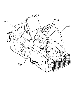
FIG. 1 is a perspective view of a catheter anchoring device in the undeployed
position
according to one embodiment. The pointed ends of each pair of parallel radial
sharps 4 are
fully enclosed within housing 100 of the catheter anchoring device as shown in
the cross-
sectional side view of the catheter anchoring device in FIG. 2. Each pair of
two parallel radial
sharps 4 are joined by a common crossbar 4a which is anchored to a pivoting
wing 9 via a tang
9a integrated into pivoting wing 9. In one embodiment of the catheter
anchoring device each
pair of radial sharps 4 and their respective crossbar 4a are formed from a
continuous piece of
wire stock. Alternatively, each individual radial sharp 4 can be joined by a
separate crossbar
member. The pointed ends of each radial sharp 4 are sharpened in opposite
directions to each
other shown in FIG. 2 and are sharpened to easily penetrate a patient's skin
with light pressure.
Two buttons 6 are diametrically opposed to each other on opposite ends of the
catheter
anchoring device and linked through a pair of parallel racks 12 and a pinion
gear 7a shown as
item 7 in FIG. 3, and both racks 12 are linked together through the common
rotation of pinion
gear 7a. Pinion gear 7a is held in place by and rotates about a keyed vertical
axle 101
integrated into housing 100 which enables the pinion gear 7a to be inserted
into the assembled
catheter anchoring device. The key in vertical axle 101 in turn prevents
vertical movement of
pinion gear 7a after rotation caused by the assembly of racks 12 as shown in
FIG. 4. Each
button 6 is an integral member of one end of rack 12 and perpendicular to the
rack. Reference
is made to FIG. 5 which is an exploded diagram of the catheter anchoring
device. Catheter
locking pin 8 contains an integral leaf spring which applies downward force
toward the bottom
of housing 100 in its unsprung position and prohibits the turning of pivoting
wings 9 when the
catheter channel 2 is empty (i.e. no catheter is present) by mechanically
interfering with the
rotation of pivoting wings 9. It should be noted that caps 102a and 102b, in
one embodiment,
CA 02917078 2015-12-24
WO 2014/210565 PCT/US2014/044742
-15-
are slid into place onto housing 100 and locked in place during the assembly
process to become
integral members of housing 100.
When a catheter 1 (i.e. one which conveys fluids, gases, or electrical current
or any
combination thereof) of correct diameter is placed in catheter channel 2 and
the bottom surface
of the catheter anchoring device is pressed against the surface of the skin of
a patient by the
operator (as shown in FIG. 6), the catheter locking pin 8 will be displaced
upward orthogonal
to the bottom surface of housing 100, compressing its integral leaf spring and
disengaging the
locking channel in both pivoting wings 9 thus allowing the rotation of
pivoting wings 9. The
rotation of pivoting wings 9 allows the racks 12 to move inward into housing
100 when
buttons 6 are pressed inward parallel to the surface of the patient's skin.
Pinion gear 7a keeps
the two racks 12 and two pivoting wings 9 in synchronization with one another.
The sharpened tips of both pairs of parallel radial sharps 4 remain locked
within
housing 100 of the device until the buttons 6 are pressed toward each other as
shown in FIG. 2.
Both pivoting wings 9 rotate about a common axle 10 in opposite directions. A
torsion spring
16 also rotates about axle 10 and is captured between pivoting wings 9. The
ends of torsion
spring 16 are mechanically coupled to each pivoting wing 9 so that equal and
opposite force is
applied to each pivoting wing 9 to cause rotation toward its fully open
position which is
limited by a preset mechanical interference between both pivoting wings 9. In
the disengaged
position, torsion spring 16 is slightly tensioned. The torsion spring 16 is
further tensioned by
the rotation of the pivoting wings 9 away from their fully disengaged
position. Each pivoting
wing 9 contains an integral pinion gear 9b at the opposite end from its
respective tang 9a.
Each integral pinion gear 9b rotates perpendicular to the rotation of pinion
gear 7a and engages
the integral teeth of rack 12 on the top surface of the common rack 12. Each
rack 12 also has
teeth on the perpendicular surface facing the center of the device which
engages pinion gear
7a. No motion of the pivoting wings 9 can take place until catheter locking
pin 8 has been
displaced by a catheter 1 placed within the catheter channel 2 and the
catheter anchoring
device has been pressed firmly against the surface of the patient's skin by
the operator. When
the catheter locking pin 8 has been pushed upwards against its integral leaf
spring to its
unlocked position, the buttons 6 can be pressed inward toward each other. As
buttons 6 are
pressed toward each other, racks 12 move parallel to the surface of the
patient's skin while
simultaneously engaging pivoting wings 9 via their respective integral pinion
gears 9b. The
parallel rack and pinion gear 7 synchronizes the motion of the pivoting wings
9 so that each
pivoting wing 9 moves with equal force and distance relative to the surface of
the patient's
skin. As buttons 6 move closer together, pivoting wings 9 rotate around common
axle 10, and
CA 02917078 2015-12-24
WO 2014/210565 PCT/US2014/044742
-16-
pivoting wings 9 are rotated down toward the top surface of the housing of the
device. This in
turn drives the sharpened ends of each pair of radial sharps 4 through their
respective grooves
13 in the housing 100. As the sharpened end of each radial sharp 4 protrudes
below the bottom
surface of housing 100 though its respective outlet hole 14 as shown in FIG.
2, the sharpened
point of each radial sharp 4 pierces membrane 15 that covers the entire bottom
surface of
housing 100 of the catheter anchoring device. Membrane 15 is made of a soft
and pliable
material that will not irritate the skin such as silicone. Membrane 15 may be
nominally 0.5 ¨ 1
mm thick. When each radial sharp 4 penetrates the membrane 15, the membrane 15
will seal
around the outer diameter of the radial sharp 4. This in turn will keep
biological fluids and
contaminants (blood, exudates, effluence, etc.) from entering the housing and
potentially
causing an infection to the patient. As the radial sharps 4 are driven through
the bottom
surface of membrane 15, the pointed ends of sharps 4 pierce the surface of the
patient's skin
and penetrate through the epidermis and dermis layers into the subcutaneous
layer of the
patient's skin as shown in FIG. 7.
When buttons 6 have been engaged fully and are flush with housing 100 of the
device,
the two pairs of diametrically opposed radial sharps 4 will be fully deployed
underneath the
patient's skin at a maximum depth of approximately 4 ¨ 5 mm in the
subcutaneous layer of the
patient's skin. The oppositely sharpened tips of each pair of diametrically
opposed radial
sharps 4, having mating oppositely-formed angular tips, will nest with each
other at the nadir
below the surface of the patient's skin, consequently forming a virtually
solid arc as seen in
FIG. 7. The two pairs of parallel diametrically opposed sharps 4 spaced by
nearly the full
width of housing 100 as shown in FIG. 8 will form a solid and secure anchor to
the patient's
skin. When buttons 6 have been fully engaged, the integral pawl at the end of
each locking
arm 9c will latch on the bottom surface of a release bar 18 located in its
first detent position in
housing 100 as shown in FIG. 9, locking each pivoting wing 9 into the fully
deployed position
until released by the operator. The top surfaces of pivoting wings 9 will be
flush with the top
surface of housing 100 in the fully deployed position.
With the catheter anchoring device fully attached to the patient and locked in
position,
the catheter 1 can be secured to the catheter anchoring device by means of
catheter lock 19. In
the unlocked position, catheter lock 19 is held in place by a pair of
diametrically opposed
integral pawls in integrated handles 19b located on either side of catheter
lock 19. Each pawl
engages a respective detent 103 on each respective side of housing 100.
Catheter lock 19
rotates along an axis in housing 100 parallel to the catheter 1 and directly
above catheter
channel 2. The concave surface of catheter clamp 19a of catheter lock 19 is
coated with a thin
CA 02917078 2015-12-24
WO 2014/210565 PCT/US2014/044742
-17-
layer of non-slip pliable material such as silicone. The inner concave face of
catheter channel
2 that is opposite the concave surface of catheter clamp 19a may also be
coated with a thin
layer of non-slip pliable material such as silicone to provide additional
frictional resistance to
catheter 1 when catheter clamp 19a is in its locked position. When handles 19b
on either side
of catheter lock 19 are pushed down toward the patient by the operator (e.g.
using the
operator's thumb and forefinger), each pawl in each respective handle 19b are
released from its
respective detent 103. As the operator pushes down on handles 19b, the
catheter clamp 19a
will rotate toward the catheter 1 in catheter channel 2, compressing the side
of the catheter 1
very slightly but not inhibiting the flow in catheter 1 and gripping the
catheter 1 between the
catheter clamp 19a and the opposite concave inner sidewall of catheter channel
2. Catheter
lock 19 will lock into the sides of housing 100 via a detent 104 on either
side of housing 100
engaged by each respective pawl in handle 19b when the top surface of handles
19b are flush
with the top surface of housing 100. In addition to locking the catheter lock
19 in place, the
detents 104 and mating channels contained within housing 100 for handles 19b
prohibit over-
travel of the catheter lock 19 which in turn prohibits the catheter clamp 19a
from inhibiting the
flow in catheter 1. FIG. 10 shows the side view of the catheter anchoring
device with catheter
lock 19 locked in place by handles 19b having engaged detents 104 and catheter
1 securely
gripped between the concave inner sidewall of catheter channel 2 and the
deployed catheter
clamp 19a. The resistance provided by the tensioned concave and silicone-
coated catheter
clamp l 9a and the opposite inner concave sidewall of the catheter channel 2
(which may also
be coated with silicone) on either side of the catheter 1 within catheter
channel 2 along the
entire length of catheter channel 2 are sufficient to keep the catheter 1
securely clamped in
place (i.e. no movement of the catheter can take place once catheter lock 19
has been rotated
into its detent locked position).
Once the catheter 1 has been locked in place, it will remain secured until the
catheter 1
and the catheter anchoring device are removed by the operator. The catheter 1
can be axially
repositioned, if necessary, without removing the catheter anchoring device
from the patient's
skin. In the event of such repositioning the operator can unlock catheter lock
19 by pulling the
two handles 19b of catheter lock 19 away from the side of housing 100 slightly
and orthogonal
to the side of housing 100 and in the opposite direction from each other. This
action releases
the pawls in handles 19b from their respective locked detents 104, and the
operator can then
rotate the two handles 19b in a direction away from the surface of the
patient's skin to release
the tension on the catheter clamp 19a and its grip on the catheter 1. This
rotational motion will
reposition the integral pawls in handles 19b into detents 103 which will keep
catheter clamp
CA 02917078 2015-12-24
WO 2014/210565 PCT/US2014/044742
-18-
19a away from the catheter 1. With the catheter 1 now free to move within the
catheter
channel 2, the operator can reposition the catheter 1 to its new position, and
catheter lock 19
can be re-locked via the methodology described above to re-secure the catheter
1 in its new
position. This process may be repeated as many times as needed by the
operator.
During the time that the catheter anchoring device is attached to the patient,
the surface
of the skin surrounding the insertion points of the sharps 4 may be cleaned
with an appropriate
disinfecting solution such as Betadine. A saturated swab or pad of
disinfectant may be wiped
and/or squeezed at the surface of the patient's skin adjacent to the sides of
the housing 100.
The disinfectant will wick under the bottom surface of the catheter anchoring
device near the
sharp insertion sites, keeping them free of potential infections. This process
can be repeated as
needed while the catheter anchoring device is attached to the patient's skin.
The catheter anchoring device may be removed easily at any time after it is
attached to
the patient's skin by the operator. The process for removal has been
specifically designed to
be easy but deliberate to operate in order to obviate an accidental removal of
the catheter
anchoring device that could have deleterious repercussions for the patient. A
safety
mechanism is employed which requires both of release bars 18 to be actuated in
order to
disengage the locks that hold the catheter anchoring device in place.
Therefore, actuating only
one release bar will not disengage the locking mechanism that keeps the sharps
4 in their
deployed positions which in turn keeps the catheter anchoring device securely
attached to the
patient's skin.
To remove the catheter anchoring device from the patient's skin, the operator
grasps
either one of two release bars 18 protruding from the opposite sides of
housing 100 (the order
of actuation is inconsequential) between his/her thumb and forefinger via its
integral ridged
grip and pulls release bar 18 to its second detent position. Pulling the
release bar 18 to its
second detent position accomplishes two mechanical functions simultaneously.
First, the pawl
at the end of locking arm 9c in the respective pivoting wing 9 will be
unlatched from the
underside of release bar 18 via a slot in the release bar 18 that is slid into
position in the second
detent position. Second, release bar 18 is permanently locked into a detent in
housing 100 via
a pawl on the underside of release bar 18 which is attached to release bar 18
by a flexible
member as shown in FIG. 5. Consequently, release bar 18 cannot be re-engaged
once the
operator has pulled release bar 18 into the second detent position,
prohibiting the reuse of the
intentionally disposable catheter anchoring device. Once the first release bar
18 has been
locked into its second detent position, the operator repeats the procedure
with the second
release bar 18 on the opposite side of housing 100. Pulling the second release
bar 18 into its
CA 02917078 2015-12-24
WO 2014/210565 PCT/US2014/044742
-19-
second detent position accomplishes the same two mechanical functions as the
first release bar
18 as described above. Upon unlatching of the pawl at the end of locking arm
9c in the
respective second pivoting wing 9, the tensioned torsion spring 16 instantly
recoils to its
relaxed and undeployed position, pulling both pivoting wings 9 to their
original undeployed
positions via their integral pinion gears 9b acting on their respective racks
12. As pivoting
wings 9 spring back toward their undeployed positions, the two pairs of
parallel sharps 4 are
retracted from the patient's skin nearly instantly and the sharpened ends of
sharps 4 are
encapsulated completely within housing 100 within their respective grooves 13
as shown in the
cross-sectional view in FIG. 11. Torsion spring 16 has sufficient force to
remove all four
radial sharps 4 simultaneously from the patient's skin virtually
instantaneously. Torsion spring
16 also simultaneously forces racks 12 to their respective undeployed
positions where a pawl
on a flexible member at the internal end of each respective rack 12 latches
onto each respective
release bar 18 in its second detent position, permanently locking racks 12,
pivoting wings 9,
and pinion gear 7a of mechanism 7 in the undeployed position thus preventing
any re-
engagement of the catheter anchoring device. Consequently, the sharpened ends
of radial
sharps 4 which are now fully contained and locked within housing 100 cannot be
exposed to
the operator or anyone else ever again, completely obviating any chance for an
inadvertent
needlestick injury.
In the unlikely event that the mechanism to automatically release the spring-
loaded
sharps 4 from the patient's skin fails to operate as intended, a backup fail
safe mechanism can
be manually manipulated by the operator to release radial sharps 4 from the
patient's skin. A
small opening 20 (as shown in FIG. 12) in each button 6 enables the operator
to insert a small
tool such as a Kelly clamp or the head of a small flat-bladed screwdriver into
opening 20 to
force the pivoting wing 9 to disengage. This will force the second pivoting
wing 9 to
disengage, allowing torsion spring 16 to return both pivoting wings 9 to their
undeployed
positions. In this scenario, the operator should apply a small amount of force
to overcome the
malfunctioning release bar 18. Even using this manual backup procedure, the
operator is fully
protected from an inadvertent needle stick injury since the torsion spring 16
will still perform as
intended. Additionally, a hole in each release bar 18 is provided as a backup
mechanism as a
means for pulling the release bar 18 into the second detent position in the
event that the
operator cannot pull release bar 18 with his or her fingers (e.g. if the
release bar 18 becomes
slippery due to liquid, blood, or effluence). The operator may also use a
Kelly clamp or other
small tool to grasp the release bar 18 via the hole in its surface to pull
release bar 18 into its
second detent position.
81793769
-20-
The methodology described above for removal enables the operator to dispose of
the
catheter anchoring device with the catheter 1 still attached. Alternatively,
the operator may
unlock catheter lock 19 before engaging the release mechanism via release bars
18 as
previously described. Using this methodology, the catheter anchoring device
and catheter 1
can be removed from the patient and disposed of separately. In either
scenario, the protection
against an inadvertent needlestick injury to the operator or anyone else is
exactly the same.
In the embodiment described above, housing 100 and all of the internal
components as
seen in FIG. 5 with the exception of the parts noted above that are made of a
pliable material
and torsion spring 16 may be constructed from any sterilizable, rigid material
that is medically
safe to be in contact with a patient's skin including but not limited to
plastics and/or metals.
Torsion spring 16 is intended (but not limited) to be constructed of a
suitable metal such as
stainless steel or spring steel with the proper spring properties. Radial
sharps 4 and integrated
crossbar 4a may be formed from a single piece of wire stock suitable for
insertion into the skin
of a patient such as hardened surgical stainless steel. Radial sharps 4 can
also be coated with
a layer of nickel or other suitable material which has properties that reduce
the risk of
infection. It is the intention of this embodiment that radial sharps 4 are
rigid and very difficult
to bend or deform. Membrane 15 and the concave inner surfaces of catheter
clamp 19a and the
sidewall of catheter channel 2 may be made of any material suitable for
medical use that is
pliable and has frictional characteristics similar to silicone and that will
also not irritate the
skin. In one embodiment of the catheter anchoring device radial sharps 4 and
their integrated
crossbars 4a are made of hardened and tempered surgical stainless steel coated
with a layer of
nickel to reduce the risk of infection, and membrane 15 and the concave inner
surface of
catheter clamp 19a and the concave inner surface of the sidewall of catheter
channel 2 are
made of silicone. In this one embodiment all other components of the catheter
anchoring
device except axle 10 and torsion spring 16 as described above are made from a
rigid injection
molded plastic. In this one embodiment axle 10 is made from surgical stainless
steel. One of
ordinary skill in the art will recognize that a variety of materials and
combinations thereof
could be used to achieve the properties of the various components of the
catheter anchoring
device described.
The embodiment described above contains two pairs of diametrically opposed
radial
sharps 4 that move coaxially. In another embodiment, there may be one pair of
diametrically
opposed radial sharps or three or more pairs of radial sharps. The two pair of
diametrically
opposed radial sharps 4 described above are parallel along the same radius of
curvature to each
other. In another embodiment, the two pair of diametrically opposed radial
sharps 4 may be
Date Recue/Date Received 2020-11-16
CA 02917078 2015-12-24
WO 2014/210565 PCT/US2014/044742
-21-
rotated away from each other by a small angle so that when viewed from either
end of the
catheter anchoring device they form a slightly obtuse angle relative to the
center of housing
100 and do not rotate coaxially. This embodiment can add further stability to
the catheter
anchoring mechanism especially in applications for large diameter catheters.
In another
embodiment, either or both of the concave inner surfaces of the catheter clamp
19a and the
inner sidewall of catheter channel 2 can be coated with a layer of silicone or
other non-slip
material. In another alternate embodiment catheter lock 19 can utilize a
sliding mechanism to
move it from its unlocked position into its locked position rather than the
rotating mechanism
as described. In another embodiment catheter lock 19 can utilize a spring-
loaded push on/push
off mechanism. In another embodiment, channels may be incorporated into
housing 100 that
facilitate the delivery of disinfecting fluids to the sharp insertion sites
and/or that facilitate the
drainage of fluids and wound exudates from the sharp insertion sites.
In another embodiment the buttons 6 when fully deployed may extend outward
slightly
from housing 100 as shown in FIG. 12A rather than rest flush with housing 100.
The increased
button depth prevents the user's fingers from getting too close to the
pivoting wings 9 and
minimizing the risk that a user's surgical glove will be inadvertently caught
in the pivoting
wings 9 as they close and latch during deployment of the catheter anchoring
device.
In another embodiment the removal of the catheter anchoring device may be
accomplished by pushing both release bars 18 inward toward housing 100 rather
than pulling
the release bars 18 away from housing 100. This is accomplished by orienting
the exposed
grips of each release bar 18 through the opposite side of housing 100 from the
side of housing
100 as described in the embodiment above. In this embodiment the user pushes
the release
bars 18 from their first detent positions toward the housing to their second
detent positions.
The resulting mechanical actuation as described in the embodiment above is
then exactly the
same upon the second release bar 18 reaching its second detent position
provided that the first
release bar 18 is also in its second detent position. Similar to the
embodiment as described
above the order of actuation of each release bar 18 is inconsequential. In
another embodiment
the release bars 18 may be oriented so that one release bar must be pushed
toward housing 100
to move it from its first detent position to its second detent position while
the second release
bar 18 must be pulled away from housing 100 to move it from its first detent
position to its
second detent position. In this embodiment as well the order of actuation of
each release bar to
remove the catheter anchoring device is inconsequential.
In another embodiment each pair of radial sharps 4 are independently connected
to each
pivoting wing 9 and no crossbar 4a connects the pair of radial sharps 4 to
that pivoting wing 9.
CA 02917078 2015-12-24
WO 2014/210565 PCT/US2014/044742
-22-
In this embodiment the mechanical operation of the radial sharps 4 attached to
the pivoting
wing 9 is the same as described in the first embodiment.
Furthermore, embodiments can to be used to anchor many types and sizes of
catheters,
drains, electrical catheters such as transvenous pacemaker wires, or nearly
any type of other
medical conduit that delivers fluids, medicines, or gases to the human body or
extracts fluids or
gases from the human body -- any of such catheters or conduits which may be
anchored to a
patient's skin while in service. The embodiment described above illustrates a
typical example
of a catheter anchoring device for a specific catheter size (i.e. the diameter
of the catheter).
The embodiment described above can be modified to accommodate any specific
size of
catheter, drain, or medical conduit. For example, for nearly any small
diameter catheter, drain,
or conduit, the size of the catheter channel 2, inner concave sidewall of the
catheter channel 2,
and catheter lock mechanism 19 would be scaled appropriately to accommodate
the specific
catheter diameter. In larger diameter catheter applications such as chest
tubes, for example, the
entire size of the catheter anchoring device could be scaled and/or the
catheter locking
mechanism 19 as described above made larger to appropriately accommodate the
size of the
catheter 1 and provide sufficient anchoring strength to securely hold the
catheter 1 in place.
In practice, an operator (typically a physician in the United States) will
insert a catheter
1 into a patient using a known methodology. Once the catheter 1 has been
inserted, the
operator will remove the catheter anchoring device from its factory-sealed
package. The
catheter anchoring device is fully sterile when it is removed from its sealed
packaging. Due to
the mechanical interlock fail safe mechanism described above, the four pointed
ends of the
radial sharps 4 are safely and securely encased within housing 100 of the
catheter anchoring
device and cannot be deployed accidentally in any way before the device is
properly positioned
on the patient's skin. The operator cleans the surface of the skin with a
disinfecting solution
such as Betadine where the catheter anchoring device is to be placed. The
operator may also
apply a topical anesthetic on the patient's skin. The operator then places the
catheter anchoring
device on the patient's disinfected skin near the insertion site for the
catheter 1. The catheter
anchoring device is positioned over the catheter 1 which lies parallel to the
surface of the skin
so that the catheter 1 lies lengthwise within catheter channel 2. With the
outlets for the radial
sharps 4 safely pressed against the patient's skin, the mechanical interlock
failsafe mechanism
is released by the presence of the appropriately-sized catheter 1 within
catheter channel 2
which displaces catheter locking pin 8 as the operator presses the catheter
anchoring device
toward the patient's skin. With one hand the operator grasps the two buttons 6
between his/her
CA 02917078 2015-12-24
WO 2014/210565 PCT/US2014/044742
-23-
thumb and forefinger; and while applying light pressure toward the surface of
the patient's
skin, the operator squeezes buttons 6 inward deploying radial sharps 4.
The sharpened tips of radial sharps 4 penetrate the membrane 15 on the bottom
surface
of the catheter anchoring device through outlet holes 14 and enter the surface
of the patient's
skin. As each radial sharp 4 penetrates membrane 15, the pliable silicone
material self-seals
around the outer diameter of each radial sharp 4, prohibiting blood, exudates,
and other
contaminants from being drawn into the separate grooves 13 which guide each
individual
radial sharp 4. This self-sealing process reduces the risk of infection to the
patient while the
catheter anchoring device is attached to the patient's skin. As the actuation
mechanism is
engaged by the operator, the radial sharps 4 penetrate the epidermis, dermis,
and subcutaneous
layers of the skin. When buttons 6 have been fully engaged by pressing them
toward each
other and the buttons 6 are flush with housing 100, a positive detent caused
by the latching of
the pawl on the end of each pivoting wing locking arm 9c onto its respective
release bar 18 will
be felt by the operator (and an audible click will be heard by the operator as
well) to let the
operator know that the catheter anchoring device has been locked securely in
place. As the
catheter anchoring device is locked into its fully deployed position, the
oppositely-sharpened
ends of each pair of diametrically opposed radial sharps 4 nest into each
other forming a nearly
solid arc at a preset depth of approximately 4 ¨ 5 mm beneath the surface of
the patient's skin
in the subcutaneous layer. This nesting of each pair of the sharpened ends of
each pair of
radial sharps 4 eliminates the "splinter effect" for the patient. Penetration
of the radial sharps 4
to the subcutaneous layer provides maximum holding strength for the catheter
anchoring
device while reducing potential risks to the patient as previously enumerated
and reduces
discomfort to the patient while the catheter anchoring device is attached to
the patient's skin.
When the catheter anchoring device has been secured to the patient's skin, the
operator then
locks the catheter 1 in place to the catheter anchoring device by pushing down
lightly on the
handles 19b of catheter lock 19 (e.g. using the operator's thumb and
forefinger). When the
pawls in each handle 19b engage their respective detents 104 in housing 100,
the operator will
feel and hear the positive engagement of catheter lock 19 to let him/her know
that the catheter
1 is fully locked. The operator may also visually confirm that the catheter 1
is locked in place
by catheter lock 19 by noticing that the top surfaces of handles 19b are flush
with the top
surface of housing 100. As previously described, the engagement of catheter
lock 19 very
slightly compresses the catheter 1 (without inhibiting its flow) between the
concave inner
surface of catheter clamp 19a and the inner concave sidewall of catheter
channel 2 opposite it.
CA 02917078 2015-12-24
WO 2014/210565 PCT/US2014/044742
-24-
Since one or both concave surfaces are coated in silicone which has non-slip
properties, the
catheter 1 is held securely once gripped within catheter channel 2 by catheter
lock 19.
After confirming the insertion depth of the catheter 1 by x-ray or other means
or for any
other reason, the operator may reposition the catheter 1 after releasing
catheter lock 19 using
the procedure described above. Once the catheter -1 has been placed in its new
position by the
operator, the catheter l can be re-secured using the catheter locking
procedure described
above. This process can be repeated as many times as necessary by the operator
without
having to detach the catheter anchoring device from the patient's skin. The
unlocking
mechanism has been designed to be very deliberate in actuation so that the
catheter lock 19
cannot be accidentally disengaged by the patient or anyone else, for an
accidental dislodgement
of the catheter 1 could have serious consequences for the patient. As is
standard practice with
sutured catheters, the catheter anchoring device can also be covered in
medical tape that is
attached to the patient's skin after the catheter anchoring device has been
secured to the
patient's skin if desired. While the catheter anchoring device is attached to
the patient's skin,
each pair of sharpened ends of the radial sharps 4 are securely nested into
each other 4 - 5 mm
below the surface of the patient's skin directly underneath the body of the
catheter anchoring
device. Consequently, radial sharps 4 which are now contaminated with the
patient's blood are
prevented from causing an accidental needlestick injury to the operator, any
other medical
personnel, or anyone else while the catheter anchoring device is attached to
the patient's skin.
Unlike sutured catheters and other conventional catheter anchoring devices,
the ends of the
sharps 4 of the embodiments only penetrate the skin once and remain embedded
in the skin
(i.e. they are not exposed to the air and/or contaminants) until the catheter
anchoring device is
removed from the patient's skin when sharps 4 are withdrawn from the skin.
This further
reduces the previously enumerated risks of infection which can be very serious
or even fatal
for the patient. While the catheter anchoring device is attached to the
patient's skin, the four
small wound sites as well as the catheter anchoring device itself can be
cleaned as often as
needed using the procedure described above to reduce the risk of infection.
When the operator wishes to remove the catheter 1 from the patient, the
operator may
either detach the catheter 1 from the catheter anchoring device via unlocking
catheter lock 19
or the operator may leave the catheter anchoring device attached to the
catheter 1 for disposal
with the catheter 1. Under the first scenario, the operator first disengages
catheter lock 19 via
the procedure described above. In either scenario, the operator will grasp
either of the two
release bars 18 with his/her thumb and forefinger using its integral ridged
grip and pull it
slightly away from housing 100 to its second detent position. The operator
will feel release bar
CA 02917078 2015-12-24
WO 2014/210565 PCT/US2014/044742
-25-
18 lock into its second detent position, and release bar 18 will be locked in
place prohibiting
over-travel or redeployment of release bar 18. The operator then grasps the
sides of the
catheter anchoring device with one hand; and with the other hand, the operator
grasps the
second release bar 18 with his/her thumb and forefinger using its integral
ridged grip. When
the operator pulls the second release bar 18 away from housing 100 to its
second detent
position, the fail safe mechanism will unlock. Immediately upon the unlocking
of the fail safe
mechanism. the tensioned torsion spring 16 will instantly retract all four
radial sharps 4 from
the patient's skin and safely secure their sharpened ends completely within
housing 100. Since
the catheter anchoring device prevents removal from the patient's skin before
all of the
sharpened ends of radial sharps 4 have been safely locked within housing 100,
the potential for
needlestick injuries to the operator or any other personnel are reduced. Due
to the mechanical
failsafe interlock that is engaged upon release of radial sharps 4 from the
patient's skin, the
pointed ends of radial sharps 4 are locked within housing 100 of the catheter
anchoring device
and are prevented from being accidently redeployed. Therefore, no needlestick
injury can
occur to the operator, other medical personnel, or anyone else after the
catheter anchoring
device has been removed from the patient's skin. The catheter anchoring device
can then be
disposed of safely and properly with or without the catheter attached as
described above.
As previously described, the process of attaching the catheter anchoring
device can be
accomplished in just a few seconds easily by the operator. If less than ideal
conditions exist or
if a patient is unable to or is unwilling to remain motionless during the
insertion procedure,
then the extremely quick installation procedure will permit the operator to
attach the catheter
anchor nearly instantly without risk of a needlestick injury to the operator
or injury to the
patient. The process of unlocking and relocking the catheter 1 in catheter
lock 19 in order to
reposition the catheter can also be accomplished in a matter of a few seconds.
Lastly, the
removal process also takes only a few seconds by the operator to achieve.
In another embodiment, a pair of helical sharps may secure the catheter
anchoring
device to the patient's skin in place of the two pairs of radial sharps and
their associated
pivoting wings previously described. Both helical sharps are wound in the same
hand, either
both wound clockwise or both wound counter-clockwise. Each helical sharp is
mounted to and
allowed to slide in a vertical groove in a drum with an integral pinion gear
that is coaxial with
its respective helical sharp, and its axis of rotation is orthogonal to the
patient's skin. The
helical sharps are guided by helical grooves in the housing. The pitch of each
helical sharp is
different thus allowing the drums and their sharps to be mounted next to each
other. When the
pinion gear on the drum is turned by its respective rack, the drum turns the
helical sharp in the
CA 02917078 2015-12-24
WO 2014/210565 PCT/US2014/044742
-26-
direction of its winding. This rotates the sharp about its axis; and guided by
the grooves in the
housing, the sharp is driven downward toward the patient's skin. The pointed
end of each
helical sharp is sharpened to easily penetrate the patient's skin. At full
deployment, the
pointed end of each sharp penetrates to the subcutaneous layer of the
patient's skin. The
windings of the pair of helical sharps are designed so that at full deployment
their respective
tips come to rest touching each other.
In this embodiment of the catheter anchoring device, a mechanical failsafe
mechanism
similar to the first embodiment is employed. Due to the fully deployed
geometry of the helical
sharps, the catheter channel is positioned next to the pair of helical sharps
(i.e. off-center) in
the housing. In the undeployed position, the sharpened tips of the helical
sharps are fully
encased within the housing, and the failsafe mechanism prevents the deployment
of the sharps
until a proper deployment condition has been achieved. A catheter locking pin
prevents the
movement of the rack and pinion gears until a catheter of proper size is
located within the
catheter channel and the housing is pressed against a patient's skin. When the
catheter locking
pin releases the rack, the operator can push the buttons toward the centerline
of the device
parallel to the surface of the patient's skin which compresses a spring
element. The movement
of each rack rotates each drum which turns the sharpened end of each helical
sharp downward
through its respective helical groove in the housing and out through its
respective outlet in the
housing. The bottom of the housing is completely covered with a layer of
silicone similar to
the first embodiment. As the buttons are pressed inward, the sharpened ends of
the helical
sharps pierce the membrane and then penetrate the surface of the patient's
skin. When the
buttons are fully compressed (i.e. they are flush with the housing), the
sharps reach their
maximum depth of penetration into the patient's skin and the tips of each
sharp come to rest
touching point to point in the subcutaneous layer of the patient's skin. A
pawl at the end of
each rack latches onto its respective release bar in its first detent position
to lock the catheter
anchoring device securely on the patient's skin in a manner similar to the
first embodiment.
Similar to the first embodiment previously described, the catheter can be
released,
repositioned, and re-secured as needed via a catheter locking mechanism as
previously
described. When the operator wishes to remove the catheter anchoring device,
the operator
pulls both release bars in a similar manner to the first embodiment to their
second detent
positions. Upon release of the second release bar by moving it into its second
detent position,
the pinion gears driving the helical sharps will be freed to rotate. The
compressed spring
element will instantly relax to its unsprung position which will rotate the
drums, retracting the
helical sharps from the skin and completely encasing them within the housing.
The pawls at
CA 02917078 2015-12-24
WO 2014/210565 PCT/US2014/044742
-27-
the ends of the racks will latch onto the release bars in their locked second
detent positions,
prohibiting the catheter anchoring device and its helical sharps from being
redeployed. The
catheter anchoring device can then be safely disposed of by the operator. This
embodiment
protects the operator, the patient, and any other personnel from an
inadvertent needlestick
injury in the same manner as the first embodiment.
In another embodiment of the catheter anchoring device, the catheter locking
mechanism is an integral part of the failsafe interlock mechanism. In this
embodiment, the
rotating catheter locking mechanism described in the first embodiment is
replaced by a linear
push on/push off slide mechanism incorporated into one of the two buttons. A
first button
operates a pinion gear through two integral parallel racks so that pushing
this button inward
actuates both pivoting wings and their respective radial sharps
simultaneously. Similar to the
first embodiment a mechanical failsafe mechanism prevents the deployment of
the sharps until
the catheter anchoring device has been placed over a catheter of proper size
on the patient's
skin and pressure has been placed on the device toward the patient's skin.
When this condition
has been achieved, the catheter locking pin disengages the respective channels
in the pivoting
wings, releasing the rack and pinion gear mechanism and enabling the operator
to deploy the
sharps by grasping the device between the operator's thumb and forefinger and
pressing the
first button inward toward the centerline of the device. The second button on
the device
remains locked in place until the first button has been fully depressed, the
radial sharps have
been fully deployed, and the pawls at the ends of the locking arms of the
pivoting wings have
latched onto their respective release bars.
Once the catheter anchoring device is attached to the patient's skin and in
its locked
fully-deployed position, the interlock for the second button is disengaged
allowing the second
button to operate. Pushing the spring-loaded second button to its second
detent position slides
the concave end of the catheter clamp against the catheter and grips the
catheter between the
catheter clamp and the concave sidewall of the catheter chamber in a manner
similar to the first
embodiment. Pushing the button inward slightly releases the button from its
second detent
position, and the compressed spring returns the button to its first undeployed
position. This in
turn releases the catheter from the catheter clamp so that it can be
repositioned and then
subsequently re-secured by the operator. The operator may repeat this
procedure as many
times as needed.
The operator may remove the catheter anchoring device from the patient's skin
in a
manner similar to that of the first embodiment described above by pulling both
release bars
into their second detent positions. Upon release of the second release bar,
the radial sharps
CA 02917078 2015-12-24
WO 2014/210565 PCT/US2014/044742
-28-
automatically and nearly instantly fully retract from the patient's skin, and
the pointed ends of
the radial sharps are locked permanently and safely within the housing of the
catheter
anchoring device so that no needlestick injury can occur.
In another embodiment of the catheter anchoring device two pairs of linearly
opposed
sharps are employed to secure the catheter anchoring device to the patient's
skin. In this
embodiment, a housing section 200 is connected to a housing section 210 by an
integral feature
in housing section 210 which mates with a channel 201 (not shown) in housing
section 200 in a
first detent position shown in FIG. 13. A coiled spring 30 is connected on one
end to housing
section 200 and on the other end to housing section 210, and in its relaxed
position, coil spring
30 keeps housing section 200 and housing section 210 a preset distance apart.
Housing section
200 and housing section 210 have flat bottom surfaces that are co-planar, and
each bottom
surface (202 and 211 respectively) is covered with a layer of non-slip pliable
material such as
silicone.
To fasten the catheter anchoring device in this embodiment to the patient's
skin, the
operator places the flat bottom side of the device on the patient's skin with
the longitudinal
axis of the device perpendicular to the catheter 100 that has already been
inserted into the
patient. Using the operator's thumb and forefinger, the operator grips both
buttons 32 and 33
on opposite ends of the device. Using light downward pressure to press the
silicone covered
bottom of the catheter anchoring device onto the patient's skin, the operator
squeezes the two
housing sections 200 and 210 toward each other. The non-slip surface of the
silicone will grip
the surface of the skin by friction. As the two sections of the housing are
pushed toward each
other, the skin is gathered between the two housing sections and will be
forced upward and fill
skin cavity 34 formed between the housing sections. Skin cavity 34 is designed
so that the
maximum height of the skin gathered is approximately 5 mm. When housing
sections 200 and
210 have reached a preset minimum distance between them as shown in FIG. 14
and the
patient's gathered skin has filled skin cavity 34, a skin locking pin 35
(similar to the catheter
locking pin in the first embodiment) will be pushed upward against its
integral spring by the
gathered skin thereby releasing the failsafe interlock mechanism. The release
of the failsafe
interlock mechanism will then enable a pawl in the integral member of housing
210 to latch
onto a detent in housing section 200 which will lock the two housing sections
together
permanently.
If the operator has failed to gather a sufficient amount of the patient's skin
in skin
cavity 34, then skin locking pin 35 will prevent the failsafe mechanism from
releasing and the
two sections of the housing from locking together. In this event the operator
may relax the grip
CA 02917078 2015-12-24
WO 2014/210565 PCT/US2014/044742
-29-
on the buttons 32 and 33, and the compressed coiled spring 30 will return the
catheter
anchoring device to its original undeployed position so that the operator can
reattempt to
properly deploy the device.
When the skin interlock has been satisfied by the presence of the gathered
skin in skin
cavity 34 and skin locking pin 35 has been released, buttons 32 and 33 are
mechanically freed
to move farther inward. Each button acts on a pair of parallel linear sharps
36 that are
completely encased within their respective housing section until deployed. The
parallel linear
sharps 36 can be joined on their non-sharp ends by a crossbar 36a (not shown)
that is
perpendicular to both sharps. In one embodiment of the catheter anchoring
device, each pair of
linear sharps 36 and its respective integral crossbar 36a are formed from one
continuous piece
of surgical stainless steel wire stock. The pointed ends of the sharps 36 are
sharpened to easily
penetrate the skin, and each pair is sharpened with mating congruent angles
for nesting with
the opposing pair of sharps 36. Each button acts on each pair of linear sharps
36 through an
integral feature that is perpendicular to the surface of the button. Each
button also compresses
a coiled spring 37 (not shown) as it is pressed toward the center of the
catheter anchoring
device. When pushed by its respective button, each pair of linear sharps 36
slides toward skin
cavity 34 through a channel in its respective housing section. The sidewalls
of skin cavity 34
are coated with a thin membrane of silicone so that when the pointed ends of
each pair of
parallel linear sharps 36 penetrate the sidewall of skin cavity 34 through
their respective outlet
holes, the membrane will seal around the outer diameter of each linear sharp
36, keeping
blood, effluence, and wound exudate from entering the housing.
As the operator squeezes buttons 32 and 33 toward each other, the two pairs of
linear
sharps 36 pierce the membrane of the sidewalls of skin cavity 34 and penetrate
the gathered
skin of the patient in skin cavity 34. When buttons 32 and 33 are fully
depressed and flush
with their respective sides of the housing, each pair of sharps 36 will
protrude from the
sidewall of the housing by half the width of skin cavity 34 parallel to the
bottom surface of the
housing as shown in FIG. 15. The sharpened points of each opposed pair of
sharps 36 will nest
with each other due to their mating congruent angles in the subcutaneous layer
of the patient's
skin gathered in skin cavity 34, forming a nearly continuous linear anchor
that will secure the
catheter anchoring device to the patient's skin as shown in FIG. 16 and reduce
the splinter
effect while the catheter anchoring device is attached to the patient's skin.
When the buttons
are fully deployed, a pawl at the end of each integral feature of buttons 32
and 33 latches onto
release bar 38 (not shown) to lock the buttons and the two pairs of parallel
linear sharps 36 in
place.
CA 02917078 2015-12-24
WO 2014/210565 PCT/US2014/044742
-30-
The operator then places catheter 100 in the curved catheter holder 39 as
shown in FIG.
17. Catheter holder 39 has tabs emanating from its sides that hold the
catheter 100 within its
groove. The concave groove surface of catheter holder 39 is coated with a
layer of non-slip
material such as silicone.
The operator then slides cover 40 toward the center of the catheter anchoring
device so
that it mates with cover 41 as shown in FIG. 18. Cover 40 has features on the
underside of its
leading edge which will gently press and hold the catheter 100 into the groove
of catheter
holder 39 when cover 40 is closed but not impede the flow in the catheter 100.
This coupled
with the non-slip surface of catheter holder 39 keeps the catheter 100 secured
so that it cannot
move while cover 40 is closed. A pair of integral pawls in cover 40 lock into
a mating pair of
detents in housing section 210 to keep cover 40 locked in place.
The operator may reposition the catheter 100 at any time, if necessary,
without
removing the catheter anchoring device from the patient's skin. The operator
can unlock cover
40 when it is in its closed position by using a sliding motion to move cover
40 to its open
position, thereby releasing its integrated pawls from their mating detents in
housing section
210. The catheter 100 can then be repositioned by the operator since the
catheter 100 is free to
move within catheter holder 39 while cover 40 is open. The catheter 100 can be
re-secured by
closing cover 40 and locking it into its closed detent position. This
procedure can be repeated
as often as necessary.
When the operator wishes to remove the catheter anchoring device from the
patient's
skin, the operator slides cover 41 away from cover 40, overcoming its two
integral pawls and
their mating pair of detents in housing section 200 that hold cover 41 in its
first detent position.
This movement as shown in FIG. 19 exposes release button 38a at one end of
release bar 38.
The operator then pushes release button 38a which moves release bar 38 into
its second detent
position which accomplishes two mechanical actions simultaneously. First, the
pawls at the
ends of the integral members of buttons 32 and 33 will be unlatched from
release bar 38, and
the compressed coil springs 37 acting on each button assembly will return
buttons 32 and 33 to
their uncompressed state nearly instantly. This will automatically and nearly
instantaneously
withdraw the opposing sets of parallel linear sharps 36 from the patient's
skin and completely
encase the two pairs of parallel linear sharps 36 within their respective
sides of the housing.
Second, release bar 38 will be permanently locked into its second detent
position in housing
section 200 via a pawl. In order to prohibit the reuse of the catheter
anchoring device, pawls
on the integral members of buttons 32 and 33 will engage release bar 38, and
in turn, this will
permanently lock the two pairs of parallel linear sharps 36 safely and
completely within their
CA 02917078 2015-12-24
WO 2014/210565 PCT/US2014/044742
-31-
respective sides of the housing. The operator can then safely remove the
locked and secured
catheter anchoring device from the patient's skin without the risk of a
needlestick injury to
anyone and safely dispose of the catheter anchoring device. FIG. 20 shows the
catheter
anchoring device in its fully extracted position with the catheter 100 still
secured between
catheter holder 39 and cover 40. Alternatively, the catheter 100 can be
removed prior to
extraction of the catheter anchoring device by sliding cover 40 to its open
position and
removing the catheter 100 from catheter holder 39.
In the embodiment described above two pairs of parallel linear sharps are
specified.
Alternatively, this embodiment may contain one set of opposing linear sharps
or three or more
sets of opposing parallel linear sharps.
In all of the embodiments described, the sharps cannot be deployed before the
catheter
anchoring device has been placed safely onto the patient's skin and an
additional failsafe
condition has been satisfied. The catheter anchoring device has to be properly
located on the
patient's skin and in the case of all but the last embodiment, a catheter of
appropriate size has
to be within the catheter channel before the catheter locking pin will release
the failsafe
mechanism. In the last embodiment a similar failsafe mechanism requires that a
sufficient
amount of the patient's gathered skin must be in the skin cavity before the
skin locking pin will
release the failsafe mechanism. It is only under this condition that the
sharps can be deployed,
and consequently, the pointed ends of the sharps can be exposed only to the
patient's skin.
Therefore, neither the operator nor anyone else can be exposed to the pointed
ends of the
sharps, obviating any risk of an inadvertent needlestick injury. While the
catheter anchoring
device is attached to the patient's skin, there is no risk of an inadvertent
needlestick injury to
the operator or anyone else because the points of the sharps are safely below
the housing
within the subcutaneous layer of the patient's skin. And since the pointed
ends of the sharps
are fully retracted from the patient's skin and fully encased and permanently
locked within the
housing before the catheter anchoring device can be removed from the patient's
skin, there is
no risk to the operator or anyone else of an inadvertent needlestick injury
after the catheter
anchoring device has been removed from the patient's skin.
It should be noted that all of the embodiments described use similar
techniques as the
first embodiment to reduce the risk of infection to the patient. Each of the
embodiments may
use materials similar to the first embodiment as described. Someone of
ordinary skill in the art
will recognize that there are many variations and combinations of the
embodiments described
that can yield the desired attributes of the specific catheter anchoring
device embodiments
CA 02917078 2015-12-24
WO 2014/210565 PCT/US2014/044742
-32-
described herein, and the specific embodiments described herein are intended
to be illustrative
but are not meant to limit the scope of the invention.
It should be noted that conventional catheter anchoring devices for preventing
needlestick injuries can easily have their sharps accidentally engaged prior
to insertion and/or
after their removal processes have been performed because they do not contain
failsafe
interlock mechanisms to such actions. Consequently, operators who use some
conventional
catheter anchoring devices could be vulnerable to inadvertent needlestick
injuries and their
resultant risks. Furthermore, there is nothing to prevent these conventional
catheter anchoring
devices from being redeployed after removal from a patient's skin which could
not only result
in an accidental needlestick injury but could also result in contaminating the
patient with a
serious infection. Unlike conventional catheter anchoring devices, the chance
of a needlestick
injury during the insertion or removal process or anytime the catheter
anchoring device is not
attached to the patient (either prior to insertion or after removal) is
reduced since the
mechanical interlock mechanism locks the pointed ends of the sharps safely and
completely
within the housing and prohibits the pointed ends of the sharps from being
exposed in any way
to anyone. The failsafe mechanical interlock only enables deployment of the
sharps if the
catheter anchoring device is on the surface of a patient's skin (where the
ends of the sharps can
only be exposed to the patient) and a catheter of proper diameter is present
in the catheter
channel as would be the case in a proper application of the catheter anchoring
device.
Furthermore, the only time that the pointed ends of the sharps can be exposed
is when they are
beneath the surface of the patient's skin, and consequently, they do not pose
a risk of a
needlestick injury to the operator or anyone else. The pointed ends of the
sharps are retracted
(which occurs instantly by spring action) in order to remove the catheter
anchoring device from
the patient's skin, thus reducing the risk of a needlestick injury to the
operator or anyone else.
It should be noted that while the foregoing has been described for use with
human
patients, embodiments of the catheter anchoring device may be used for
veterinary patients in a
similar fashion.
The preceding description of the invention has been described with reference
to various
specific embodiments for the purposes of illustration and description, but it
is not intended to
be exhaustive or to limit the invention to the precise form disclosed.
Numerous modifications
and variations are possible within the scope and spirit of the inventive
concepts described.
Having thus described at least one illustrative embodiment of the invention,
various
alterations, modifications, and improvements will readily occur to those
skilled in the art. Such
alterations, modifications, and improvements are intended to be within the
spirit and scope of the
CA 02917078 2015-12-24
WO 2014/210565 PCT/US2014/044742
-33-
invention. Accordingly, the foregoing description is by way of example only
and is not intended
as limiting. The invention is limited only as defined in the following claims
and the equivalents
thereto.
What is claimed is: