Note: Descriptions are shown in the official language in which they were submitted.
CA 02917958 2016-01-11
WO 2015/013451 PCT/US2014/047892
COMPOSITIONS AND METHODS FOR UPREGULATING HIPPOCAMPAL
PLASTICITY AND HIPPOCAMPUS-DEPENDENT LEARNING AND MEMORY
CROSS REFERENCE TO RELATED APPLICATIONS
This application claims the benefit of co-pending United States Provisional
Patent
Application No. 61/857,306, filed July 23, 2013, and entitled COMPOSITIONS AND
METHODS FOR UPREGULATING HIPPOCAMPAL PLASTICITY AND
HIPPOCAMPUS-DEPENDENT LEARNING AND MEMORY, United States
Provisional Patent Application Serial No. 61/888,420, filed October 8, 2013
and entitled
COMPOSITIONS AND METHODS FOR UPREGULATING HIPPOCAMPAL
PLASTICITY AND HIPPOCAMPUS-DEPENDENT LEARNING AND MEMORY, and
United States Provisional Patent Application Serial No. 61/930,388, filed
January 22, 2014
and entitled COMPOSITIONS AND METHODS FOR OPTIMIZING NEURONAL
SYNAPTIC TRANSMISSION, all of which are incorporated herein by reference in
their
entireties.
FIELD OF THE INVENTION
Particular aspects relate generally to hippocampus-dependent learning and
memory, and in more particular aspects to compositions and methods for
upregulating
hippocampal plasticity and hippocampus-dependent learning and memory in a
subject by
administering a therapeutic composition comprising a gas-enriched (e.g.,
oxygen enriched)
electrokinetically generated fluid comprising charge-stabilized oxygen-
containing
nanostructures, as disclosed herein. Additional aspects relate to methods for
enhancing
the synaptic maturation of neurons by enriching the density and size of
dendritic spines
(e.g., comprising enhancing at least one of the length of primary axons, the
number of
collaterals, or the number of tertiary branches). Additional aspects relate
generally to
neurons and neuronal synaptic transmission, and more particularly to
compositions and
methods for optimizing or enhancing neuronal synaptic transmission. Further
aspects
relate to methods for enhancing intracellular oxygen delivery or utilization
(particularly in
neurons), and methods for enhancing ATP synthesis (e.g., at presynaptic and/or
postsynaptic terminals). Additional aspects relate to combination therapies.
1
CA 02917958 2016-01-11
WO 2015/013451 PCT/US2014/047892
BACKGROUND OF THE INVENTION
Increased calcium influx through ionotropic glutamate receptors and the
upregulation of plasticity-associated molecules in hippocampal neurons are two
important
events in the process of hippocampus-dependent spatial learning and memory.
Additionally, increased density of dendritic spines and enhanced synaptic
transmission through ionotropic glutamate receptors are important events of
synaptic
plasticity and eventually in the process of hippocampus-dependent spatial
learning and
memory.
Hippocampal neuron function is also implicated in neurodegenerative disease.
Alzheimer's disease (AD), for example, is the most common neurodegenerative
disorder
in the aged population characterized by impairments in memory and cognition.
An
extensive loss of hippocampal neurons (1) is the hallmark of this disease. The
death of
hippocampal neurons is often associated with and the strong downregulation of
many
functional genes (2) involved in ion conductance (3, 4), synapse formation
(5), dendritic
arborization (6), long term potentiation (7, 8), and long term depression (8,
9). Impaired
calcium influx through ionotropic glutamate receptors including NMDA and AMPA
receptors is directly linked to the loss of hippocampal learning and memory
(10). Analysis
of postmortem AD brains showed that expression of NMDA subunits including NR1,
NR2A, and NR2B was altered in susceptible brain regions including hippocampus
(11).
Downregulation of immediate early genes (IEGs) (12) including arc, zit:268,
homer-1, c-
fos and inhibition of synaptic genes (13-15) including psd-95, synpo, adam-10
was also
reported to be downregulated in AD brain. In addition, oxidative (16) and
nitrosylative
(17, 18) damages in different hippocampal proteins also have been implicated
in the loss
of function and eventually death of hippocampal neurons. Many pharmacological
compounds have been tested in the treatment of these progressive
neurodegenerative
diseases including cholinesterase inhibitors and memantine, but most of them
generate
several side effects, perhaps because of lower metabolic activities of elderly
population, or
perhaps because of toxicity because they are metabolized.
Aside from treating neurodegenerative diseases, however, there is a pronounced
need in the art for compositions and methods to enhance neuroplasticity and
learning in
the general population (in addition to enhancing neuroplasticity and learning
in the context
of neurodegenerative diseases).
2
CA 02917958 2016-01-11
WO 2015/013451 PCT/US2014/047892
SUMMARY OF THE INVENTION
According to particular aspects, the disclosed electrokinetically-altered
fluids (e.g.,
RNS60) control or modulate (e.g., increase or enhance) the synaptic plasticity
of
hippocampal neurons by inducing calcium influx via NMDA- and AMPA-sensitive
ionotropic glutamate receptors. RNS60, but neither NS nor PNS, stimulates the
expression of NR2A, NR2B subunits NMDA and G1uR1 subunit of AMPA receptors
along with other plasticity-associated molecules including Arc, P5D95, and
CREB.
Particular aspects, therefore, provide a method for enhancing hippocampal
plasticity and hippocampus-dependent learning and/or memory, comprising
administering
to a subject in need thereof a therapeutically effective amount of an
electrokinetically
altered aqueous fluid comprising an ionic aqueous solution of charge-
stabilized oxygen-
containing nanostructures (e.g., nanobubbles) predominantly having an average
diameter
of less than about 100 nanometers and stably configured in the ionic aqueous
fluid in an
amount sufficient for enhancing hippocampal plasticity and hippocampus-
dependent
learning and/or memory in the subject.
Particular aspects, therefore, provide a method for enhancing hippocampal-
mediated learning and memory, comprising administering to a subject in need
thereof a
therapeutically effective amount of an ionic aqueous solution of charge-
stabilized oxygen-
containing nanostructures having an average diameter of less than 100
nanometers for
enhancing hippocampal-mediated learning and memory in the subject.
In particular aspects of the methods, the ionic aqueous solution comprises
dissolved oxygen in an amount of at least 8 ppm, at least 15, ppm, at least 25
ppm, at least
ppm, at least 40 ppm, at least 50 ppm, or at least 60 ppm oxygen at
atmospheric
pressure. In particular aspects of the methods, the percentage of dissolved
oxygen
25 molecules present in the solution as the charge-stabilized oxygen-
containing
nanostructures is a percentage selected from the group consisting of greater
than: 0.01%,
0.1%, 1%, 5%; 10%; 15%; 20%; 25%; 30%; 35%; 40%; 45%; 50%; 55%; 60%; 65%;
70%; 75%; 80%; 85%; 90%; and 95%. In particular aspects of the methods, the
amount of
dissolved oxygen present in charge-stabilized oxygen-containing nanostructures
is at least
30 8 ppm, at least 15, ppm, at least 20 ppm, at least 25 ppm, at least 30
ppm, at least 40 ppm,
at least 50 ppm, or at least 60 ppm oxygen at atmospheric pressure. In
particular aspects
of the methods, the majority of the dissolved oxygen is present in the charge-
stabilized
oxygen-containing nanostructures. In particular aspects of the methods, the
charge-
3
CA 02917958 2016-01-11
WO 2015/013451 PCT/US2014/047892
stabilized oxygen-containing nanostructures have an average diameter of less
than a size
selected from the group consisting of: 90 nm; 80 nm; 70 nm; 60 nm; 50 nm; 40
nm; 30
nm; 20 nm; 10 nm; and less than 5 nm. In particular aspects of the methods,
the ionic
aqueous solution comprises a saline solution. In particular aspects of the
methods, the
solution is superoxygenated.
In particular aspects of the methods, the charge-stabilized oxygen-containing
nanostructures comprise charge-stabilized oxygen-containing nanobubbles having
an
average diameter of less than 100 nanometers.
In particular aspects of the methods comprise modulating at least one of
cellular
membrane potential and cellular membrane conductivity in hippocampal cells of
the
subject.
In particular aspects of the methods, enhancing learning and/or memory,
comprises
enhancing learning and/or memory in at least one group selected from the group
consisting
of normal subjects, subject recovering from neurological trauma, and subjects
with
learning disorders. In particular aspects of the methods, the learning
disorder comprises
one selected from the group consisting of, dyslexia, dyscalculia, dysgraphia,
dyspraxia
(sensory integration disorder), dysphasia/aphasia, auditory processing
disorder, non-verbal
learning disorder, visual processing disorder, and attention deficit disorder
(ADD). In
particular aspects of the methods, neurological trauma comprises at least one
of accidents
or injury to the brain, stroke, oxygen deprivation, drowning, and asphyxia.
In particular aspects of the methods, administration promotes modulating
(e.g.,
upregulating, in hippocampal neurons, of expression, amount or activity levels
of at least
one neuronal plasticity protein selected from the group consisting of NR2A
and/or NR2B
subunits NMDA receptors, G1uR1 (glurl) subunit of AMPA receptors, Arc (arc),
PSD95,
CREB (creb): IEGs including arc, zit:268, and c-fos; NMDA receptor subunits
including
nr1, nr2a, nr2b, and nr2c; AMPA receptor subunit glurl; neurotrophic factors
and their
receptors including bdnf, nt3, nt5, and ntrk2; adenylate cyclases (adcyl and
adcy8);
camk2a, aktl; ADAM-10, Synpo and homer-1.
In particular aspects of the methods, administration promotes modulating
(e.g.,
downregulating expression, amount or activity levels of at least one protein
selected from
the group consisting of Gria2, Ppplca, Ppp2ca, and Ppp3ca, proteins encoded by
genes
known to support long-term depression.
4
CA 02917958 2016-01-11
WO 2015/013451 PCT/US2014/047892
Particular aspects of the methods comprise combination therapy, wherein at
least
one additional therapeutic agent is administered to the patient. In particular
aspects of the
methods, the at least one additional therapeutic agent is selected from the
group consisting
of: glatiramer acetate, interferon-13, mitoxantrone, natalizumab, inhibitors
of MMPs
including inhibitor of MMP-9 and MMP-2, short-acting 132-agonists, long-acting
132-
agonists, anticholinergics, corticosteroids, systemic corticosteroids, mast
cell stabilizers,
leukotriene modifiers, methylxanthines, 132-agonists, albuterol, levalbuterol,
pirbuterol,
artformoterol, formoterol, salmeterol, anticholinergics including ipratropium
and
tiotropium; corticosteroids including beclomethasone, budesonide, flunisolide,
fluticasone,
mometasone, triamcinolone, methyprednisolone, prednisolone, prednisone;
leukotriene
modifiers including montelukast, zafirlukast, and zileuton; mast cell
stabilizers including
cromolyn and nedocromil; methylxanthines including theophylline; combination
drugs
including ipratropium and albuterol, fluticasone and salmeterol, budesonide
and
formoterol; antihistamines including hydroxyzine, diphenhydramine, loratadine,
cetirizine,
and hydrocortisone; immune system modulating drugs including tacrolimus and
pimecrolimus; cyclosporine; azathioprine; mycophenolatemofetil; and
combinations
thereof In particular aspects of the methods, the at least one additional
therapeutic agent
is an anti-inflammatory agent.
In particular aspects of the methods, modulation of at least one of cellular
membrane potential and cellular membrane conductivity comprises modulating at
least
one of cellular membrane structure or function comprising modulation of at
least one of an
amount, conformation, activity, ligand binding activity and/or a catalytic
activity of a
membrane associated protein. In particular aspects of the methods, the
membrane
associated protein comprises at least one selected from the group consisting
of receptors,
ion channel proteins, intracellular attachment proteins, cellular adhesion
proteins, and
integrins. In particular aspects of the methods, the receptor comprises a
transmembrane
receptor. In particular aspects of the methods, modulating cellular membrane
conductivity
comprises modulating whole-cell conductance. In particular aspects of the
methods,
modulating whole-cell conductance comprises modulating at least one voltage-
dependent
contribution of the whole-cell conductance.
In particular aspects of the methods, modulation of at least one of cellular
membrane potential and cellular membrane conductivity comprises modulating a
calcium
dependent cellular messaging pathway or system. Particular aspects of the
methods
5
CA 02917958 2016-01-11
WO 2015/013451 PCT/US2014/047892
comprise modulating calcium influx through ionotropic glutamate receptors
(e.g.,
comprises at least one NMDA and/or AMPA receptor).
In particular aspects of the methods, modulation of at least one of cellular
membrane potential and cellular membrane conductivity comprises modulating
intracellular signal transduction comprising modulation of phospholipase C
activity.
In particular aspects of the methods, modulation of at least one of cellular
membrane potential and cellular membrane conductivity comprises modulating
intracellular signal transduction comprising modulation of adenylate cyclase
(AC) activity.
Particular aspects of the methods comprise administration to a cell network or
layer, and further comprising modulation of an intercellular junction therein.
In particular aspects of the methods, the solution comprises at least one of a
form
of solvated electrons, and electrokinetically modified or charged oxygen
species. In
particular aspects of the methods, the form of solvated electrons or
electrokinetically
modified or charged oxygen species are present in an amount of at least 0.01
ppm, at least
0.1 ppm, at least 0.5 ppm, at least 1 ppm, at least 3 ppm, at least 5 ppm, at
least 7 ppm, at
least 10 ppm, at least 15 ppm, or at least 20 ppm. In particular aspects of
the methods, the
electrokinetically altered oxygenated aqueous fluid comprises solvated
electrons
stabilized, at least in part, by molecular oxygen.
In particular aspects of the methods, the ability of the solution to modulate
of at
least one of cellular membrane potential and cellular membrane conductivity
persists for at
least two, at least three, at least four, at least five, at least 6, at least
12 months, or longer
periods, in a closed gas-tight container.
In particular aspects of the methods, treating/administering comprises
administration by at least one of topical, inhalation, intranasal, oral,
intravenous (IV) and
intraperitoneal (IP).
In particular aspects of the methods, the charge-stabilized oxygen-containing
nanostructures are formed in a solution comprising at least one salt or ion
from Tables 1
and 2 disclosed herein.
In particular aspects of the methods, the subject is a mammal, preferably a
human.
Additional aspects provide a method for enhancing the synaptic maturation of
neurons by enriching the density and size of dendritic spines, comprising
administering to
a neuron or subject in need thereof a therapeutically effective amount of an
ionic aqueous
solution of charge-stabilized oxygen-containing nanostructures having an
average
6
CA 02917958 2016-01-11
WO 2015/013451 PCT/US2014/047892
diameter of less than 100 nanometers sufficient for enhancing the synaptic
maturation of
neurons by enriching the density and size of dendritic spines. Particular
embodiments
comprise enhancing at least one of the length of primary axons, the number of
collaterals,
or the number of tertiary branches. In certain aspects, the ionic aqueous
solution
-- comprises dissolved oxygen in an amount of at least 8 ppm, at least 15,
ppm, at least 25
ppm, at least 30 ppm, at least 40 ppm, at least 50 ppm, or at least 60 ppm
oxygen at
atmospheric pressure. In certain aspects, the percentage of dissolved oxygen
molecules
present in the solution as the charge-stabilized oxygen-containing
nanostructures is a
percentage selected from the group consisting of greater than: 0.01%, 0.1%,
1%, 5%;
-- 10%; 15%; 20%; 25%; 30%; 35%; 40%; 45%; 50%; 55%; 60%; 65%; 70%; 75%; 80%;
85%; 90%; and 95%. In certain aspects, the amount of dissolved oxygen present
in charge-
stabilized oxygen-containing nanostructures is at least 8 ppm, at least 15,
ppm, at least 20
ppm, at least 25 ppm, at least 30 ppm, at least 40 ppm, at least 50 ppm, or at
least 60 ppm
oxygen at atmospheric pressure. In certain aspects, the majority of the
dissolved oxygen is
-- present in the charge-stabilized oxygen-containing nanostructures. In
certain aspects, the
charge-stabilized oxygen-containing nanostructures have an average diameter of
less than
a size selected from the group consisting of: 90 nm; 80 nm; 70 nm; 60 nm; 50
nm; 40 nm;
30 nm; 20 nm; 10 nm; and less than 5 nm. In certain aspects, the ionic aqueous
solution
comprises a saline solution. In certain aspects, the solution is
superoxygenated. In certain
-- aspects, the neurons are hippocampal neurons. Certain aspects comprise
administration to
neurons ex vivo, in vivo or in vitro.
In certain aspects, the charge-stabilized oxygen-containing nanostructures
comprise charge-stabilized oxygen-containing nanobubbles having an average
diameter of
less than 100 nanometers.
Further aspects comprise methods for maintaining, growing or enhancing the
synaptic maturation of neurons in culture.
Yet further aspects relate to optimizing or enhancing neuronal synaptic
transmission, and/or for enhancing intracellular oxygen delivery or
utilization (particularly
in neurons), and methods for enhancing ATP synthesis (e.g., at presynaptic
and/or
-- postsynaptic terminals).
Determining the biological variables that control both electrical and chemical
synaptic transmission between nerve cells, or between nerve terminals and
muscular or
glandular systems, has been a very significant area of physiological
exploration over the
7
CA 02917958 2016-01-11
WO 2015/013451 PCT/US2014/047892
decades. Chemical synaptic transmission has had the added attraction of
addressing both
the transmission gain of the event, as well as the excitatory or inhibitory
nature of the
junction and its activity-dependent potentiation or depression.
Provided are methods for optimizing or enhancing neurotransmission (neuronal
synaptic transmission), comprising administrating an electrokinetically-
altered ionic
aqueous solution comprising charge-stabilized oxygen-containing nanostructures
(e.g.,
oxygen-containing nanobubbles) having an average diameter of less than 100 nm
in an
amount and for a time period sufficient for modulating at least one
presynaptic and/or
postsynaptic response.
Additional aspects provide a method for optimizing or enhancing
neurotransmission, comprising contacting neurons with, or administrating to a
subject
having neurons, an eleetrokinetieally-altered ionic aqueous solution
comprising charge-
stabilized oxygen-containing nanostructures having an average diameter of less
than 100
nm in an amount and for a time period sufficient for enhancing intracellular
oxygen
delivery or utilization, wherein a method for optimizing neuronal synaptic
transmission is
afforded.
Further aspects provide a method for enhancing intracellular oxygen delivery
or
utilization, comprising contacting cells with, or administrating to a subject
having cells, an
electrokinetically-altered ionic aqueous solution comprising charge-stabilized
oxygen-
containing nanostructures having an average diameter of less than 100 nm in an
amount
and for a time period sufficient for enhancing intracellular oxygen delivery
or utilization in
the cells.
For the above methods for optimizing or enhancing neurotransmission,
representative presynaptic and/or postsynaptic response include, but are not
limited to, for
example, at least one of: increased of spontaneous transmitter release;
modification of
noise kinetics; increase in a postsynaptic response (e.g., absent an increase
in presynaptic
ICa++ amplitude); decrease in synaptic vesicle density and/or number at active
zones;
increase in the number of clathrin-coated vesicles, and/or large endosome like
vesicles
near junctional sites; increase in ATP synthesis (e.g., at the presynaptic and
postsynaptic
terminals); or enhanced recovery of postsynaptic spike generation.
In particular aspects, the electrokinetically-altered ionic aqueous solutions
optimize
synaptic transmission without producing over abnormal over-release effects.
8
CA 02917958 2016-01-11
WO 2015/013451 PCT/US2014/047892
In particular aspects, the effect of artificial seawater (ASW) based on RNS60,
a
physically modified isotonic saline that has been electrokinetically altered
to include
charge-stabilized oxygen containing nanobubbles, has been shown to enhance
and/or
optimize neurotransmission.
BRIEF DESCRIPTION OF THE DRAWINGS
Figures lA through 1H show the effects of RNS60, PNS60, and NS on NMDA and
AMPA-dependent calcium influx in cultured mouse hippocampal neurons.
Figures 2A through 2K show the effects of RNS60 in the expression of
plasticity-
associated proteins in mouse hippocampal neurons.
Figures 3A through 3Dviii show the effects of RNS60 on the expression of
plasticity-associated genes in cultured mouse hippocampal neurons.
Figures 4A through 4D show the role of PI3K pathway in RNS60-mediated
upregulation of plasticity-associated genes in mouse hippocampal neurons.
Figures 5A through 5D show that activation of PI3K regulates both NMDA- and
AMPA-sensitive calcium influx in RNS60-treated mouse hippocampal neurons.
Figures 6A through 6J show the effect of RNS60 on the expression of plasticity-
associated molecules in vivo in the hippocampus of 5XFAD transgenic animals.
Figures 7A through 7K show the effect of RNS60, NS, PNS60, and RNS10.3 on
the number, size, and maturation of dendritic spines in hippocampal neurons.
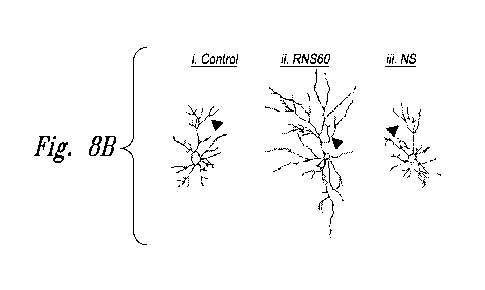
Figures 8A through 8F show that RNS60 stimulates the length, and collaterals
of
primary axon in cultured hippocampal neurons.
Figures 9A, 9B(i)-9B(iii) and 9C-9E show activation of PI3K regulates
morphological plasticity in RNS60-treated mouse hippocampal neurons.
Figure 10 shows, according to particular exemplary aspects, an example of
increased evoked transmitter release in a hypoxic synapse following electrical
stimulation
of the pre synaptic terminal.
Figures 11A-11E show, according to particular exemplary aspects, high-
frequency
stimulation in Control and RNS60 ASW.
Figures 12A-12C show, according to particular exemplary aspects, synaptic
noise
recorded in Control ASW and RNS60 ASW.
9
CA 02917958 2016-01-11
WO 2015/013451 PCT/US2014/047892
Figures 13A-13E show, according to particular exemplary aspects, a voltage
clamp
study indicating that RNS60 increases transmitter release without modifying
calcium
current or its relationship with transmitter release.
Figures 14A-14F show, according to particular exemplary aspects, direct
determination of increased ATP synthesis at the presynaptic and postsynaptic
terminals
using Luciferin/Luciferase light emission.
Figure 15 shows, according to particular exemplary aspects, reduction of
spontaneous synaptic release following oligomycin administration. Plot of
noise
amplitude as a function of frequency (note double log coordinates). Red is
Control ASW,
green is 7 min after addition of oligomycin and blue is 22 min after
oligomycin
administration and 12 min after changing superfusion to RNS60 ASW. Black is
extracellular recording.
Figures 16A-16C show, according to particular exemplary aspects,
electronmicrographs of a synaptic junction following RNS60 ASW superfusion.
Figures 17A and 17B show, according to particular exemplary aspects,
statistical
determination of synaptic vesicle numbers in synapses superfused with RNS60
ASW. Fig.
8A shows a plot of the number of CCV as a function of size. Fig. 8B shows the
number of
large vesicles as a function of size.
Figures 18A-18C show, according to particular exemplary aspects, the
ultrastructure of squid giant synapse active zones following oligomycin
injection.
Figures 19A-19C show, according to particular exemplary aspects, the effect of
RNS60 and olygomycin on synaptic vesicle numbers.
DETAILED DESCRIPTION OF THE INVENTION
Upregulating/Enhancing Hippocampal Plasticity and Hippocampus-Dependent
Learning
and Memory
Certain embodiments disclosed herein relate to providing compositions and
methods for upregulating hippocampal plasticity and hippocampus-dependent
learning and
memory, comprising administering, to a subject (e.g., a mammal or human) in
need
thereof, a therapeutic composition comprising an electrokinetically-altered,
gas-enriched
(e.g., oxygen enriched) aqueous fluid.
Particular aspects provide a method for enhancing hippocampal plasticity and
hippocampus-dependent learning and memory, comprising administering to a
subject in
CA 02917958 2016-01-11
WO 2015/013451 PCT/US2014/047892
need thereof a therapeutically effective amount of an electrokinetically
altered aqueous
fluid comprising an ionic aqueous solution of charge-stabilized oxygen-
containing
nanostructures having an average diameter of less than about 100 nanometers
and stably
configured in the ionic aqueous fluid in an amount sufficient for enhancing
hippocampal
plasticity and hippocampus-dependent learning and memory to provide a method
for
enhancing hippocampal plasticity and hippocampus-dependent learning and memory
in
the subject.
Increased calcium influx through ionotropic glutamate receptors and the
upregulation of plasticity-associated molecules in hippocampal neurons are two
important
events in the process of hippocampus-dependent spatial learning and memory.
Here we
have undertaken an innovative approach to upregulate hippocampal plasticity.
Applicants'
RNS60 fluid, for example, is an isotonic saline solution generated by
subjecting normal
saline to a patented type of Taylor-Couette-Poiseuille (TCP) flow under
elevated oxygen
pressure (see, e.g., Applicants' issued U.S. Patent Nos. 7,832,920, 7,919,534,
8,410,182,
8,445,546, 8,449,172, and 8,470,893, all incorporated herein by reference in
their
respective entireties).
RNS60, but neither NS (normal saline) nor PNS60 (saline containing excess
oxygen without TCP modification) stimulates the NMDA- and AMPA-sensitive
calcium
influx in cultured hippocampal neurons. Using mRNA-based targeted gene array,
real-
time PCR, and immunoblot and immunofluorescence analysis, we further
demonstrate that
RNS60 stimulates the upregulation of many plasticity-associated proteins in
cultured
hippocampal neurons. Finally, RNS60 treatment increased plasticity-associated
proteins
and calcium influx in the hippocampus of 5XFAD transgenic mouse model of
Alzheimer's
disease (AD). These results describe a novel property of RNS60 in stimulating
hippocampal plasticity, which may be helpful in treating AD and other
dementias.
According to particular aspects, the disclosed electrokinetically-altered
fluids (e.g.,
RNS60) control or modulate (e.g., increase or enhance) the synaptic plasticity
of
hippocampal neurons by inducing calcium influx via NMDA- and AMPA-sensitive
ionotropic glutamate receptors. RNS60, but neither NS nor PNS, stimulates the
expression of NR2A, NR2B subunits NMDA and G1uR1 subunit of AMPA receptors
along with other plasticity-associated molecules including Arc, P5D95, and
CREB.
11
CA 02917958 2016-01-11
WO 2015/013451 PCT/US2014/047892
It is believed that plasticity decreases in various conditions including, but
not
limited to, old age and in patients with AD. Therefore, exploring ways to
boost plasticity
generally, including in conditions of learning disorders and in AD or aging is
an important
area of research. Although there are other drugs and approaches for improving
brain
function, here we introduce a simple saline-based agent to augment plasticity.
Upon
subjecting normal saline to Taylor-Couette-Poiseuille (TCP) turbulence in the
presence of
elevated oxygen pressure, Revalesio Corporation (Tacoma, WA) has generated
RNS60,
which does not contain any active pharmaceutical ingredient (19, 20). Due to
TCP
turbulence, RNS60 contains charge-stabilized nanostructures consisting of,
e.g., an oxygen
nanobubble core surrounded by an electrical double-layer at the liquid/gas
interface (19,
20).
Here we delineate the first evidence that ionic fluid or saline generated due
to TCP
turbulence is capable of improving plasticity in cultured hippocampal neurons
and in vivo
(e.g., in the hippocampus of 5XFAD transgenic mice).
Our conclusion is based on the following:
First, as shown in Example 7, we observed that RNS60 induced the number, size,
and maturation of dendritic spines in cultured hippocampal neurons, indicating
a
beneficial role of RNS60 in regulating the synaptic efficacy of neurons;
Second, as shown in Example 7, RNS60 increased the axonal length and
collaterals
in neurons further corroborating the role of RNS60 in stimulating the
morphological
plasticity of neurons.
Third, as shown in working Example 3, RNS60 did not alter the calcium
dependent
excitability of hippocampal neurons, but rather stimulated inbound calcium
currents in
these neurons through ionotropic glutamate receptor. This indicates that RNS60
modulates plasticity-related activities.
Fourth, as shown in working Example 4, RNS60 induced the expression of a broad
spectrum of plasticity-associated molecules in hippocampal neurons.
Fifth, as shown in working Example 4, RNS60 augmented the levels of several
genes, proteins of which stimulate signaling pathways (adenylate cyclase, CAM
kinase II
and Akt) for the activation of CREB, the master regulator of plasticity.
Sixth, as shown in working Example 4, proteins encoded by several genes such
as
Gria2 , Ppp 1 ca, Ppp2ca, and Ppp3ca are known to support long-term depression
(35). It is
12
CA 02917958 2016-01-11
WO 2015/013451 PCT/US2014/047892
interesting to see that RNS60 down-regulated the expression of Gria2, Ppplca,
Ppp2ca,
and Ppp3ca in hippocampal neurons.
Seventh, as shown in working Example 6, RNS60 treatment increased the
expression of plasticity-associated molecules and augmented calcium influx in
vivo in the
hippocampus of 5XFAD transgenic mice. These results indicates that RNS60
provides as
a therapeutic agent in boosting plasticity in patients in need thereof,
including subject with
learning and/or memory disorders, and including subjects with neuronal injury,
and those
with AD and other dementias.
A growing body of evidence suggests that the excessive activation of glutamate-
operated NMDA receptors in postsynaptic neurons is the primary factor of
progressive
neuronal loss in AD (28). Different noncompetitive and uncompetitive NMDA
receptor
blockers are being used for the treatment of AD (36). However prolonged use of
these
drugs eventually destroys the normal excitability of these receptors, which is
essential for
the viability of these neurons. Moreover, these specific inhibitors of NMDA
receptors
generate a wide range of side effects including chest pain, nausea, increased
heart rate,
breathing trouble, lowered urination, and different digestive disorders
because of their
poor metabolic clearance among older populations (37, 38). In contrast, RNS60
for
example, produces almost no side effects as chemically it is identical to
isotonic saline
with additional oxygen.
As presently disclosed in working Examples 2 through 6, RNS60 treatment
generated high amplitude NMDA-dependent calcium oscillations both in cell
culture and
in vivo experiments. Since high amplitude calcium wave corresponds to the
excitability of
ionotropic receptors, if follows that RNS60 does not alter the normal
excitability of
NMDA receptors. Moreover, RNS60 induced the expression many growth supportive
molecules including CREB, BDNF and NTRs, which are required for the survival
of
neurons; synaptic proteins including P5D95, ADAM-10, and Synpo, which are
required
for the maintenance of synaptic structure; receptor proteins including NR2A,
G1uR1, and
NR2B, which are needed for calcium excitability of the postsynaptic neurons;
and IEGs
such as c-FOS, Arc, Homer 1, and Zif-268 essential for neuroplasticity,
leading to memory
consolidation (39-41).
Signaling mechanisms leading to plasticity are becoming clear. It has been
found
that master regulator cAMP response element-binding (CREB) plays an important
role in
13
CA 02917958 2016-01-11
WO 2015/013451 PCT/US2014/047892
plasticity and promoters of different plasticity-associated genes harbor
multiple cAMP
response elements (CRE) (42-45). Applicants have demonstrated that RNS60
induces the
activation of CREB in microglial cells via type IA phosphatidylinositol 3-
kinase (PI3K) in
microglial cells. PI3K is a key signaling molecule implicated in the
regulation of a broad
array of biological responses including cell survival (34). For class IA PI3K,
the p85
regulatory subunit acts as an interface by interacting with the IRS-1 through
its SH2
domain and thus recruits the p110 catalytic subunit (p110a/13) to the cell
membrane, which
in turn activates downstream signaling molecules like Akt/protein kinase B and
p70
ribosomal S6 kinase (34). On the other hand, for class IB PI3K, p1107 is
activated by the
engagement of G-protein coupled receptors. The p1 1 Oy then catalyzes the
reaction to
release phosphatidylinositol (3,4,5)-triphosphate as the second messenger,
using
phosphatidylinositol (4,5)-bisphosphate as the substrate, and activates
downstream
signaling molecules (33).
Herein we demonstrate, in working Example 5, that RNS60 induces the activation
of both the subunits of type IA PI-3K (p110a and p11 0f3) without modulating
type IB PI-
3K p 1 107 in primary hippocampal neurons, indicating the specific activation
of type IA
p110a/13 PI3K in neurons. Furthermore, abrogation of RNS60-mediated
upregulation of
NR2A and G1uR1 and stimulation of calcium influx in hippocampal neurons by
inhibitors
of PI3K indicates that RNS60 increases NMDA- and AMPA-sensitive calcium
current via
PI3K.
According to particular aspects, applicants herein demonstrate, for the first
time,
that RNS60 treatment upregulates plasticity-associated molecules and calcium
influx in
cultured hippocampal neurons and in vivo (e.g., in the hippocampus of 5XFAD
mice).
These results demonstrate and confirm a new hippocampal neuron plasticity
boosting
property of applicants' fluids (e.g., RNS60) and provide a new use for
applicants'
modified saline for stimulating synaptic plasticity in all types of subjects
as disclosed
herein.
Optimizing Neuronal Synaptic Transmission
According to particular exemplary aspects, RNS60, a physically modified saline
containing charge-stabilized oxygen-containing nanostructures (e.g., charge-
stabilized
oxygen-containing nanobubbles), has significant function-optimizing properties
for
optimizing neuronal synaptic transmission.
14
CA 02917958 2016-01-11
WO 2015/013451 PCT/US2014/047892
According to particular aspects, RNS60 represents a class of bioactive agents
relating to the physical structure of water and an increased oxygen caring
ability (in the
form of charge-stabilized oxygen-containing nanostructures, e.g., charge-
stabilized
oxygen-containing nanobubbles having an average diameter less than 100 nm),
with no
added chemical molecules and yet has proven cytoprotective and anti-
inflammatory
effects in different models of neurodegeneration through direct effects on
glial cells as
well as modulation of T cell subsets (Khasnavis S.2012; Mondal, S, 2012).
Without being
bound by mechanism, and together with the results described herein, this
suggests that
RNS60 exerts pleiotropic effects that are not based on interaction with a
specific receptor,
but rather that RNS60 is a facilitator of physiological function that require
a different
appellative. Functionally, as shown herein, RNS60 is able to optimize synaptic
transmission without affecting normal function, and without any deleterious
side effects
(as has been demonstrated in previous studies in other systems including human
use where
no deleterious effects have been seen).
Preferred embodiments. Particular aspects provide a method for optimizing
neurotransmission, comprising contacting neurons with, or administrating to a
subject
having neurons, an electrokinetic ally- altered ionic aqueous solution
comprising charge-.
stabilized oxygen-containing nanostructures having an average diameter of less
than 100
nm in an amount and for a time period sufficient for modulating at least one
presynaptic
and/or postsynaptic response, wherein a method for optimizing neuronal
synaptic
transmission is afforded. In certain aspects, modulating at least one
presynaptic and/or
postsynaptic response comprises an increase of spontaneous transmitter
release. In certain
aspects, modulating at least one presynaptic and/or postsynaptic response
comprises a
modification of noise kinetics. In certain aspects, modulating at least one
presynaptic
and/or postsynaptic response comprises an increase in a postsynaptic response
(e.g.,
without an increase in presynaptic ICa amplitude). In certain aspects,
modulating at
least one presynaptic and/or postsynaptic response comprises a decrease in
synaptic
vesicle density and/or number at active zones, and may further comprise an
increase in the
number of clathrin-coated vesicles, and large endosome like vesicles in the
vicinity of the
junctional sites. In
certain aspects, modulating at least one presynaptic and/or
postsynaptic response comprises a marked increase in ATP synthesis leading to
synaptic
transmission optimization. In certain aspects, modulating at least one
presynaptic and/or
postsynaptic response comprises an enhanced or more vigorous recovery of
postsynaptic
CA 02917958 2016-01-11
WO 2015/013451 PCT/US2014/047892
spike generation.
In certain aspects, modulating at least one presynaptic and/or
postsynaptic response comprises increased ATP synthesis at the presynaptic and
postsynaptic terminals. In particular embodiments the charge-stabilized
oxygen
containing nanostructures having an average diameter of less than 100 nm
comprise
charge-stabilized oxygen-containing nanobubbles having an average diameter of
less than
100 nm.
Additional aspect provide a method for optimizing neurotransmission,
comprising
contacting neurons with, or administrating to a subject having neurons, an
electrokinetically-altered ionic aqueous solution comprising charge-stabilized
oxygen-
containing nanostructures having an average diameter of less than 100 nm in an
amount
and for a time period sufficient for enhancing intracellular oxygen delivery
or utilization,
wherein a method for optimizing neuronal synaptic transmission is afforded. In
certain
aspects, optimizing neuronal synaptic transmission comprises an increase of
spontaneous
transmitter release.
In certain aspects, optimizing neuronal synaptic transmission
comprises a modification of noise kinetics. In certain aspects, optimizing
neuronal
synaptic transmission comprises an increase in a postsynaptic response (e.g.,
without an
increase in presynaptic ICa++ amplitude). In certain aspects, optimizing
neuronal synaptic
transmission comprises a decrease in synaptic vesicle density and/or number at
active
zones, and may further comprise an increase in the number of clathrin-coated
vesicles, and
large endosome like vesicles in the vicinity of the junctional sites. In
certain aspects,
optimizing neuronal synaptic transmission comprises a marked increase in ATP
synthesis.
In certain aspects, optimizing neuronal synaptic transmission comprises an
enhanced or
more vigorous recovery of postsynaptic spike generation. In certain aspects,
optimizing
neuronal synaptic transmission comprises increased ATP synthesis at the
presynaptic and
postsynaptic terminals. In particular embodiments the charge-stabilized
oxygen
containing nanostructures having an average diameter of less than 100 'n.m
comprise
charge-stabilized oxygen-containing nanobubbles having an average diameter of
less than
100 nm.
Further aspect provide a method for enhancing intracellular oxygen delivery or
utilization, comprising contacting cells with, or administrating to a subject
having cells, an
electrokinetically-altered ionic aqueous solution comprising charge-stabilized
oxygen-
containing nanostructures having an average diameter of less than 100 nm in an
amount
and for a time period sufficient for enhancing intracellular oxygen delivery
or utilization in
16
CA 02917958 2016-01-11
WO 2015/013451 PCT/US2014/047892
the cells. In particular aspects, the cells are nerve cells (e.g., mammalian,
human or other;
any organism or animal comprising neurons and neuronal transmission). In
particular
aspects, enhancing intracellular oxygen delivery or utilization provides for
optimizing
neuronal synaptic transmission. In particular aspects, optimizing neuronal
synaptic
transmission comprises an increase of spontaneous transmitter release. In
particular
aspects, optimizing neuronal synaptic transmission comprises a modification of
noise
kinetics. In particular aspects, optimizing neuronal synaptic transmission
comprises an
increase in a postsynaptic response (e.g., without an increase in presynaptic
ICa++
amplitude). In particular aspects, optimizing neuronal synaptic transmission
comprises a
decrease in synaptic vesicle density and/or number at active zones. Particular
aspects may
further comprise an increase in the number of clathrin-coated vesicles, and
large endosome
like vesicles in the vicinity of the junctional sites. In particular aspects,
optimizing
neuronal synaptic transmission comprises a marked increase in ATP synthesis.
In
particular aspects, optimizing neuronal synaptic transmission comprises an
enhanced or
more vigorous recovery of postsynaptic spike generation. In particular
aspects, optimizing
neuronal synaptic transmission comprises increased ATP synthesis at the
presynaptic and
postsynaptic terminals. In particular embodiments the charge-stabilized
oxygen-
containing nanost.ructures having an average diameter of less than 100 nm
comprise
charge-stabilized oxygen-containing nanobubbles having an average diameter of
less than
100 nm.
Consistent with the above, the disclosed results concerning single spike
synaptic
transmission (Fig. 10; working Example 9), as well as the response to
repetitive
presynaptic terminal activation (Fig. 11; working Example 10) indicate that
the ability of
RNS60 to maintain and optimize synaptic transmission within normal parameters
is not
accompanied by abnormal responses indicating the absence of overdose or side
effects.
This conclusion is also supported by the increase in spontaneous release that
reaches a
maximum level following a single superfusion of RNS60 that is maintained for a
period of
minutes and decays slowly after superfusion with Control ASW (Fig. 12; working
Example 11). Similar results were found with the increase in spontaneous
transmitter
30 release (Fig. 12; working Example 11).
With respect to the mechanism of action of RNS60 in the claimed methods, the
possibility that it could be modifying channel kinetics, and in particular
calcium currents,
was rendered unlikely by the voltage clamp results which indicate that
synaptic
17
CA 02917958 2016-01-11
WO 2015/013451 PCT/US2014/047892
optimization is not correlated with any change in the time course or amplitude
of the
inward calcium current responsible for the transmitter release (Fig. 13;
working Example
12).
Without being bound by mechanism, RNS60 likely changes available energy level,
via ATP increase and that such event is accompanied by an increase in synaptic
transmission effectiveness (Fig. 14; working Example 13). An additional
unexpected
finding was that of the noise frequency change in the presence of RNS60 (Fig.
12C;
working Example 11). The fact that at the level of spontaneous release there
is a clear
change in the noise profile, seen as a reduction of high frequency noise and
an increase of
low frequency noise (Fig. 12C; working Example 11), appears to correlate with
the change
in the synaptic vesicle size distribution (Fig. 17; working Example 15).
Without being
bound by mechanism, the transmitter delivery kinetics may be different between
normal
vesicular profiles and that of the larger endosome related vesicles. The
latter may have a
slower release kinetics that may explain the change in noise frequency towards
lower
frequency with an accompanying noise level amplitude increase.
From a morphological perspective, it is known that increased expression of the
brain vesicular monoamine transporter VMAT2 regulates vesicle phenotype and
quantal
size (Pothos FN et.al, 2000). As shown in RNS60 superfused terminals (Fig. 16
A;
working Example 15), large vesicles with different shapes and sizes are
observed in the
analyzed terminals. These structures are never observed in the current control
synapses
(Fig. 15B; working Example 14) or in terminals studied in former experiments
(Heuser,
J.E. & Reese, T.S., 1973).
Neurotransmitter release requires a well-known set of steps concerning
synaptic
vesicle exo- and endocytosis (Heuser, J.E. and Reese T.S., 1973). It has been
shown in
previous work that dinamin/synaptophysin complex disruption results in a
decrease of
transmitter release, resulting from a depletion of synaptic vesicle recycling
(Daly C., et al.
2000). It has also been observed that, under these conditions, the number of
CCVs
actually increased suggesting the existence of another vesicle endocytosis
mechanism with
a faster time course than the classical clathrin pathway (Daly et al. 2000).
This finding
was further corroborated by the injection of Rabfilin 3A and/or one of its
fragments which
affect the distribution of membranes of the endocytotic pathway in the squid
presynaptic
terminal in a multifunctional fashion (Burns ME et al., 1998). This is
consistent with
18
CA 02917958 2016-01-11
WO 2015/013451 PCT/US2014/047892
previous observations following different domains manipulation of the synaptic
vesicle
protein synaptotagmin (Mikoshiba K. et al.1995; Fukuda M. et al, 1995).
Although the synaptic hyperactivity demonstrated herein after RNS60
administration is accompanied by a significant decrease in synaptic vesicles
numbers at
the active zone, the presynaptic terminal area at the vicinity of the active
zone showed a
large number of CCVs, the amount of membrane retrieved by CCVs may not be
sufficient
to maintain of the large amount of transmitter release observed during the
augmented
synaptic release described here. However, the large number of endosomal
vesicles (up to
300 nm in diameter) that were imaged in the immediate vicinity of the active
zone could
be part of the enhanced synaptic transmitter released observed under these
conditions.
This was supported by the presence of such "larger vesicles" at the active
zone
intermingled with usual synaptic vesicle profiles (Fig. 17; working Example
15). Since
such vesicles appear throughout the active zone vicinity, it suggests that the
endocytotic
mechanism responsible for their presence may be independent of the clathrin or
caveolin
pathway (reviewed by Mayor S. and Pagano R.E. 2007).
The fact that both spontaneous release levels as well as the amplitude of the
evoked synaptic potentials are increased significantly indicates that while
the probability
of release of regular sized vesicles may be slightly decreased, the release of
the larger
vesicular component may actually be increased. Such a change in the
distribution of
vesicular size, favoring the larger endosomal vesicular profiles over the
smaller clathrin
related vesicles, confirms a similar morphological analysis of vesicular size
distribution
following high level synaptic activation (Hayashi M et al 2008, as reviewed by
Saheki,Y
and De Camili P. 2012).
This change in vesicular size distribution may provide a possible explanation
for
the fact that the nature of the spontaneous synaptic noise was modified after
RNS 60
administration, as shown in Figure 12 and as discussed in the description of
synaptic noise
and its relation to time course of synaptic miniature potentials and vesicular
size (working
Example 11).
Mitochondria are energy-supplier organelles, strikingly abundant in chemical
synapses (Palay, S11956, Talbot J.D. et al., 2003). In squid the presynaptic
terminal
mitochondria lies in close juxtaposition to presynaptic calcium channels
(Pivovarova NB.
et al., 1999). Energy supply to neurons in the form of oxygen and glucose and
its final
product¨mitochondrial generated ATP, is largely used for reversing the ion
influxes
19
CA 02917958 2016-01-11
WO 2015/013451 PCT/US2014/047892
underlying synaptic and action potentials (Attwell D. and Laughlin SB. 2001).
Here
Applicants tested whether inhibition of mitochondria ATP with oligomycin,
modified the
effect of RNS60 on synaptic transmission.
Mitochondria may be blocked with drugs that do not alter mitochondrial
membrane
potential ('lim), such as oligomycin or with depolarizing 'Pm inhibitors.
Ru360, an inhibitor
of the mitochondrial uniporter was not used because in some terminals Ru360
appears to
inhibit Ca2 influx across the plasma membrane (David G.1999). The use of CCCP
or
Antimycin Al was also avoided as these are also 'Pm depolarizing agents, and
because both
of them can potentially affect transmitter release from presynaptic terminals,
since these
agents block mitochondrial calcium uptake.
Concomitant application of RNS60 and the complex V mitochondrial blocker
(olygomicin) failed to induce increments in spontaneous release as determined
by synaptic
noise power spectrum analysis (Fig. 15; working Example 14). These experiments
suggest that RNS60 mechanism of action is dependent on mitochondrial ATP
production,
potentially by providing, or facilitating provision of oxygen in a more
efficient manner.
Concerning the mechanism of action of RNS60, it may be significant that a
block
of mitochondrial ATP synthesis results in an inactivation of the RNS60 effect
on synaptic
transmission. These findings further indicate that the reduction of ATP
synthesis is
accompanied by a lack of response of synaptic release mechanism by RNS60.
These
findings indicate that RNS60 likely does not operate directly on the vesicular
release
mechanism, but rather indirectly via an increased synthesis of ATP by the
mitochondrial
system. This has been shown to have a significant effect on both the
availability of
vesicular organelles and on their movement on to the active zone at the
presynaptic
compartment in the synaptic junction region (Ivanikov MV. et al. 2010).
According to particular aspects, therefore, which respect to optimizing
neurotransmission, RNS60 is an ATP synthesis optimizer via facilitation of
oxygen
transport into the mitochondrial system, with minimal increase in
intracellular free radical
level.
Electrokinetically-generated fluids:
"Electrokinetically generated fluid," as used herein, refers to Applicants'
inventive
electrokinetically-generated fluids generated, for purposes of the working
Examples
herein, by the exemplary Mixing Device described in detail in Applicants'
issued patents
CA 02917958 2016-01-11
WO 2015/013451 PCT/US2014/047892
(see, e.g., Applicants' issued U.S. Patent Nos. 7,832,920, 7,919,534,
8,410,182, 8,445,546,
8,449,172, and 8,470,893, all incorporated herein by reference in their
respective
entireties). The electrokinetic fluids, as demonstrated by the data disclosed
and presented
herein, represent novel and fundamentally distinct fluids relative to prior
art non-
electrokinetic fluids, including relative to prior art oxygenated non-
electrokinetic fluids
(e.g., pressure pot oxygenated fluids and the like). As disclosed in various
aspects herein,
the electrokinetically-generated fluids have unique and novel physical and
biological
properties including, but not limited to the following:
In particular aspects, the electrokinetically altered aqueous fluid comprise
an ionic
aqueous solution of charge-stabilized oxygen-containing nanostructures
substantially
having an average diameter of less than about 100 nanometers and stably
configured in the
ionic aqueous fluid in an amount sufficient to provide, upon contact of a
living cell by the
fluid, modulation of at least one of cellular membrane potential and cellular
membrane
conductivity.
In preferred aspects, RNS60 is a physically modified normal saline (0.9%)
solution
generated by using a rotor/stator device, which incorporates controlled
turbulence and
Taylor-Couette-Poiseuille (TCP) flow under high oxygen pressure (see
Applicants U.S.
Patent Nos. 7,832,920, 7,919,534, 8,410,182, 8,445,546, 8,449,172, and
8,470,893, all
incorporated herein by reference in their entireties for their teachings
encompassing
Applicants' device, methods for making the fluids, and the fluids per se).
In particular aspects, electrokinetically-generated fluids refers to fluids
generated
in the presence of hydrodynamically-induced, localized (e.g., non-uniform with
respect to
the overall fluid volume) electrokinetic effects (e.g., voltage/current
pulses), such as
device feature-localized effects as described herein.
In particular aspects said
hydrodynamically -induced, localized electrokinetic effects are in combination
with
surface-related double layer and/or streaming current effects as disclosed and
discussed
herein.
In particular aspects the administered inventive electrokinetically-altered
fluids
comprise charge-stabilized oxygen-containing nanostructures in an amount
sufficient to
provide modulation of at least one of cellular membrane potential and cellular
membrane
conductivity.
In certain embodiments, the electrokinetically-altered fluids are
superoxygenated (e.g., RNS-20, RNS-40 and RNS-60, comprising 20 ppm, 40 ppm
and 60
21
CA 02917958 2016-01-11
WO 2015/013451 PCT/US2014/047892
ppm dissolved oxygen, respectively, in standard saline). In particular
embodiments, the
electrokinetically-altered fluids are not-superoxygenated (e.g., RNS-10 or
Solas,
comprising 10 ppm (e.g., approx. ambient levels of dissolved oxygen in
standard saline)).
In certain aspects, the salinity, sterility, pH, etc., of the inventive
electrokinetically-altered
fluids is established at the time of electrokinetic production of the fluid,
and the sterile
fluids are administered by an appropriate route. Alternatively, at least one
of the salinity,
sterility, pH, etc., of the fluids is appropriately adjusted (e.g., using
sterile saline or
appropriate diluents) to be physiologically compatible with the route of
administration
prior to administration of the fluid. Preferably, and diluents and/or saline
solutions and/or
buffer compositions used to adjust at least one of the salinity, sterility,
pH, etc., of the
fluids are also electrokinetic fluids, or are otherwise compatible.
In particular aspects, the inventive electrokinetically-altered fluids
comprise saline
(e.g., one or more dissolved salt(s); e.g., alkali metal based salts (Li+,
Na+, K+, Rb+, Cs+,
etc.), alkaline earth based salts (e.g., Mg++, Ca++), etc., or transition
metal-based positive
ions (e.g., Cr, Fe, Co, Ni, Cu, Zn, etc.,), in each case along with any
suitable anion
components, including, but not limited to F-, Cl-, Br-, I-, PO4-, SO4-, and
nitrogen-based
anions. Particular aspects comprise mixed salt based electrokinetic fluids
(e.g., Na+, K+,
Ca++, Mg++, transition metal ion(s), etc.) in various combinations and
concentrations,
and optionally with mixtures of couterions. In particular aspects, the
inventive
electrokinetically-altered fluids comprise standard saline (e.g., approx. 0.9%
NaC1, or
about 0.15 M NaC1). In particular aspects, the inventive electrokinetically-
altered fluids
comprise saline at a concentration of at least 0.0002 M, at least 0.0003 M, at
least 0.001
M, at least 0.005 M, at least 0.01 M, at least 0.015 M, at least 0.1 M, at
least 0.15 M, or at
least 0.2 M. In particular aspects, the conductivity of the inventive
electrokinetically-
altered fluids is at least 10 [tS/cm, at least 40 [tS/cm, at least 80 [tS/cm,
at least 100 [LS/cm,
at least 150 [tS/cm, at least 200 [tS/cm, at least 300 [tS/cm, or at least 500
[tS/cm, at least 1
mS/cm, at least 5, mS/cm, 10 mS/cm, at least 40 mS/cm, at least 80 mS/cm, at
least 100
mS/cm, at least 150 mS/cm, at least 200 mS/cm, at least 300 mS/cm, or at least
500
mS/cm. In particular aspects, any salt may be used in preparing the inventive
electrokinetically-altered fluids, provided that they allow for formation of
biologically
active salt-stabilized nano structure s
(e .g. , salt-stabilized oxygen-containing
nanostructures) as disclosed herein.
22
CA 02917958 2016-01-11
WO 2015/013451 PCT/US2014/047892
According to particular aspects, the biological effects of the inventive fluid
compositions comprising charge-stabilized gas-containing nanostructures can be
modulated (e.g., increased, decreased, tuned, etc.) by altering the ionic
components of the
fluids, and/or by altering the gas component of the fluid.
According to particular aspects, the biological effects of the inventive fluid
compositions comprising charge-stabilized gas-containing nanostructures can be
modulated (e.g., increased, decreased, tuned, etc.) by altering the gas
component of the
fluid. In preferred aspects, oxygen is used in preparing the inventive
electrokinetic fluids.
In additional aspects mixtures of oxygen along with at least one other gas
selected from
Nitrogen, Oxygen, Argon, Carbon dioxide, Neon, Helium, krypton, hydrogen and
Xenon.
As described above, the ions may also be varied, including along with varying
the gas
constitutent(s).
Given the teachings and assay systems disclosed herein (e.g., cell-based
cytokine
assays, patch-clamp assays, etc.) one of skill in the art will readily be able
to select
appropriate salts and concentrations thereof to achieve the biological
activities disclosed
herein.
TABLE 1. Exemplary cations and anions.
Common Cations:
Name Formula Other name(s)
Aluminum Al '3
Ammonium NH4 '
Barium B a+2
Calcium Ca+2
Chromium(II) Cr+2
Chromous
Chromium(III) Cr+3
Chromic
Copp er(I) Cu + Cuprous
Copp er(II) Cu 2 Cupric
Iron(II) Fe 2 Ferrous
Iron(III) Fe 3 Ferric
Hydrogen H+
Hydronium H30+
Lead(II) Pb+2
Lithium Li+
Magnesium m g +2
23
CA 02917958 2016-01-11
WO 2015/013451
PCT/US2014/047892
Manganese(II) Mn 2 Manganous
Manganese(III) Mn '3 Manganic
Mercury(I) Hg2 2 Mercurous
Mercury(II) Hg 2 Mercuric
Nitronium NO2
Potassium K+
+
Silver Ag
Sodium Na+
Strontium Sr+2
Tin(II) Sn+2
Stannous
Tin(IV) Sn+4
Stannic
Zinc Zn+2
Common Anions:
Simple ions:
Hydride if Oxide 02
FluorideF Sulfide 52
ChlorideCl- Nitride N3
Bromide Br
Iodide E
Oxoanions:
Arsenate As043 Phosphate P043-
Arsenite As033-
Hydrogen phosphate HP042-
Dihydrogen phosphate H2PO4
Sulfate 5042 Nitrate NO3
Hydrogen sulfate H504- Nitrite NO2-
,
Thiosulfate 02._.i=-\ 32-
Sulfite 5032
PerchlorateC104- Iodate 103
Chlorate C103- Bromate Br03-
Chlorite C102Hypochlorite 0C1-
Hypobromite OBr-
Carbonate C032 ChromateCr042-
Hydrogen carbonate- 2-
HCO3- Dichromate Cr2v7
or Bicarbonate
24
CA 02917958 2016-01-11
WO 2015/013451 PCT/US2014/047892
Anions from Organic Acids:
Acetate CH3C00- formate HC00-
Others:
Cyanide CN- Amide NH2-
Cyanate OCN- Peroxide 022-
Thiocyanate SCN- Oxalate C2042
HydroxideOFF Permanganate Mn04-
TABLE 2. Exemplary cations and anions.
Monoatomic Cations
Formula Charge Name
H ' 1+ hydrogen ion
.................... -r- ................................................ -4
1_,1 1+ lithium ion
Na ' 1+ sodium ion
K ' 1+ potassium ion
Cs ' 1+ cesium ion
Ag ' 1+ silver ion
mg2+
2+ magnesium ion
Ca2 ' 2+ calcium ion
t=
Sr2+ 2+ strontium ion
Ba2+
2+ barium ion
Zn2+
2+ zinc ion
.................... -r- ................................................. -4
Cd2+ 2+ cadmium ion
Al3+ 3+ aluminum ion
Polyatomic Cations
Formula Charge Name
NH4+ 1+ ammonium ion
H30 ' 1+ hydronium ion
Multivalent Cations
Formula Charge 1Name
.......................................................................... -4
Cr2+ i 2 chromium(II) or chromous ion
CA 02917958 2016-01-11
WO 2015/013451 PCT/US2014/047892
Cr3+
i 3 j chromium(III)or chromic ion
Mn2+
2 manganese(H) or manganous ion
,
Mn4+
4 Imanganese(IV) ion
Fe2+
2 iron(II) or ferrous ion
. .
Fe3+
3 , iron(III) or ferric ion
Co2+
2 cobalt(II) or cobaltous ion
Co3+
3 cobalt(II) or cobaltic ion
....................................................................... -4
Ni2+ 2 .nickel(II) or nickelous ion
Ni3+ i 3 , nickel(III) or nickelic ion
Cu + i 1 copper(I) or cuprous ion
....................................................................... -4
Cu2+
i 2 copper(II) or cupric ion
Sn2+
i 2 , tin(II) or atannous ion
Sn4+
4 .tin(IV) or atannic ion
Pb2 2 lead(II) or plumbous ion
Pb4+ 4 lead(IV) or plumbic ion
Monoatomic Anions
Formula TCharge Name
if 1- i hydride ion
, ......................................................................
F 11-fluoride ion
........................................ , .......................... -4
Cl- T1- Ichloride ion
Br- 1- Ibromide ion
I- 1- iodide ion
........................................ , ........................... -4
02- 2- loxide ion
........................................ : .......
S2- 2- 1 su lfide ion
N3- 3- Initride ion
Polyatomic Anions
Formula Charge Name
01-1- 1- hydroxide ion
....................................................................... -4
CN- 1- Icyanide ion
.................. .................................... ..................
.................................. ...... .
......................................
26
CA 02917958 2016-01-11
WO 2015/013451 PCT/US2014/047892
SCN- 1- thiocyanate ion
C2H302- 1- 1 acetate ion
C10- 1- hypochlorite ion
C102- 1- chlorite ion
C103- 1 - 1 chlorate ion
C104- 1- perchlorate ion
NO2 1- nitrite ion
NO3- 1- nitrate ion
................................................................
Mn042- 2- 1permanganate ion
C032- 12- 1 carbonate ion
C2042- 12- 1 oxalate ion
Cr042- 12- 1 chromate ion
rµ 2-
Cr2k.17 1 2- Idichromate ion
S032- 12- sulfite ion
S042- 12- 1 sulfate ion
P033- phosphite ion
P043- 13- phosphate ion
The present disclosure sets forth novel gas-enriched fluids, including, but
not
limited to gas-enriched ionic aqueous solutions, aqueous saline solutions
(e.g., standard
aqueous saline solutions, and other saline solutions as discussed herein and
as would be
recognized in the art, including any physiological compatible saline
solutions), cell culture
media (e.g., minimal medium, and other culture media) useful in the treatment
of diabetes
or diabetes related disorders. A medium, or media, is termed "minimal" if it
only contains
the nutrients essential for growth. For prokaryotic host cells, a minimal
media typically
includes a source of carbon, nitrogen, phosphorus, magnesium, and trace
amounts of iron
and calcium. (Gunsalus and Stanter, The Bacteria, V. 1, Ch. 1 Acad. Press
Inc., N.Y.
(1960)). Most minimal media use glucose as a carbon source, ammonia as a
nitrogen
source, and orthophosphate (e.g., PO4) as the phosphorus source. The media
components
can be varied or supplemented according to the specific prokaryotic or
eukaryotic
organism(s) grown, in order to encourage optimal growth without inhibiting
target protein
production. (Thompson et al., Biotech. and Bioeng. 27: 818-824 (1985)).
In particular aspects, the electrokinetically altered aqueous fluids are
suitable to
modulate 13C-NMR line-widths of reporter solutes (e.g., Trehelose) dissolved
therein.
27
CA 02917958 2016-01-11
WO 2015/013451 PCT/US2014/047892
NMR line-width effects are in indirect method of measuring, for example,
solute
'tumbling' in a test fluid as described herein in particular working Examples.
In particular aspects, the electrokinetically altered aqueous fluids are
characterized
by at least one of: distinctive square wave voltametry peak differences at any
one of -
0.14V, -0.47V, -1.02V and -1.36V; polarographic peaks at -0.9 volts; and an
absence of
polarographic peaks at -0.19 and -0.3 volts, which are unique to the
electrokinetically
generated fluids as disclosed herein in particular working Examples.
In particular aspects, the electrokinetically altered aqueous fluids are
suitable to
alter cellular membrane conductivity (e.g., a voltage-dependent contribution
of the whole-
cell conductance as measure in patch clamp studies disclosed herein).
In particular aspects, the electrokinetically altered aqueous fluids are
oxygenated,
wherein the oxygen in the fluid is present in an amount of at least 15, ppm,
at least 25
ppm, at least 30 ppm, at least 40 ppm, at least 50 ppm, or at least 60 ppm
dissolved oxygen
at atmospheric pressure. In particular aspects, the electrokinetically altered
aqueous fluids
have less than 15 ppm, less that 10 ppm of dissolved oxygen at atmospheric
pressure, or
approximately ambient oxygen levels.
In particular aspects, the electrokinetically altered aqueous fluids are
oxygenated,
wherein the oxygen in the fluid is present in an amount between approximately
8 ppm and
approximately 15 ppm, and in this case is sometimes referred to herein as
"Solas."
In particular aspects, the electrokinetically altered aqueous fluid comprises
at least
one of solvated electrons (e.g., stabilized by molecular oxygen), and
electrokinetically
modified and/or charged oxygen species, and wherein in certain embodiments the
solvated
electrons and/or electrokinetically modified or charged oxygen species are
present in an
amount of at least 0.01 ppm, at least 0.1 ppm, at least 0.5 ppm, at least 1
ppm, at least 3
ppm, at least 5 ppm, at least 7 ppm, at least 10 ppm, at least 15 ppm, or at
least 20 ppm.
In particular aspects, the electrokinetically altered aqueous fluids are
characterized
by differential (e.g., increased or decreased) permittivity relative to
control, non-
electrokinetically altered fluids. In preferred aspects, the
electrokinetically altered
aqueous fluids are characterized by differential, increased permittivity
relative to control,
non-electrokinetically altered fluids. Permittivity (8) (farads per meter) is
a measure of the
ability of a material to be polarized by an electric field and thereby reduce
the total electric
field inside the material. Thus, permittivity relates to a material's ability
to transmit (or
28
CA 02917958 2016-01-11
WO 2015/013451 PCT/US2014/047892
"permit") an electric field. Capacitance (C) (farad; coulomb per volt), a
closely related
property, is a measure of the ability of a material to hold charge if a
voltage is applied
across it (e.g., best modeled by a dielectric layer sandwiched between two
parallel
conductive plates). If a voltage V is applied across a capacitor of
capacitance C, then the
charge Q that it can hold is directly proportional to the applied voltage V,
with the
capacitance C as the proportionality constant. Thus, Q = CV, or C = QN. The
capacitance of a capacitor depends on the permittivity 8 of the dielectric
layer, as well as
the area A of the capacitor and the separation distance d between the two
conductive
plates. Permittivity and capacitance are mathematically related as follows: C
= 8 (Aid).
When the dielectric used is vacuum, then the capacitance Co = 80 (Aid), where
80 is the
permittivity of vacuum (8.85 x 10⁻¹² F/m). The dielectric constant (k), or
relative
permittivity of a material is the ratio of its permittivity 8 to the
permittivity of vacuum 80,
so k = 8/co (the dielectric constant of vacuum is 1). A low-k dielectric is a
dielectric that
has a low permittivity, or low ability to polarize and hold charge. A high-k
dielectric, on
the other hand, has a high permittivity. Because high-k dielectrics are good
at holding
charge, they are the preferred dielectric for capacitors. High-k dielectrics
are also used in
memory cells that store digital data in the form of charge.
In particular aspects, the electrokinetically altered aqueous fluids are
suitable to
alter cellular membrane structure or function (e.g., altering of a
conformation, ligand
binding activity, or a catalytic activity of a membrane associated protein)
sufficient to
provide for modulation of intracellular signal transduction, wherein in
particular aspects,
the membrane associated protein comprises at least one selected from the group
consisting
of receptors, transmembrane receptors (e.g., G-Protein Coupled Receptor
(GPCR), TSLP
receptor, beta 2 adrenergic receptor, bradykinin receptor, etc.), ion channel
proteins,
intracellular attachment proteins, cellular adhesion proteins, and integrins.
In certain
aspects, the effected G-Protein Coupled Receptor (GPCR) interacts with a G
protein a
subunit (e.g., Gas, Gai 5 Gag, and Ga12).
In particular aspects, the electrokinetically altered aqueous fluids are
suitable to
modulate intracellular signal transduction, comprising modulation of a calcium
dependent
cellular messaging pathway or system (e.g., modulation of phospholipase C
activity, or
modulation of adenylate cyclase (AC) activity).
29
CA 02917958 2016-01-11
WO 2015/013451 PCT/US2014/047892
In particular aspects, the electrokinetically altered aqueous fluids are
characterized
by various biological activities (e.g., regulation of cytokines, receptors,
enzymes and other
proteins and intracellular signaling pathways) described in the working
Examples and
elsewhere herein.
In particular aspects, the electrokinetically altered aqueous fluids display
synergy
with glatiramer acetate interferon-13, mitoxantrone, and/or natalizumab. In
particular
aspects, the electrokinetically altered aqueous fluids reduce DEP-induced TSLP
receptor
expression in bronchial epithelial cells (BEC).
In particular aspects, the electrokinetically altered aqueous fluids inhibit
the DEP-
induced cell surface-bound MMP9 levels in bronchial epithelial cells (BEC).
In particular aspects, the biological effects of the electrokinetically
altered aqueous
fluids are inhibited by diphtheria toxin, indicating that beta blockade, GPCR
blockade and
Ca channel blockade affects the activity of the electrokinetically altered
aqueous fluids
(e.g., on regulatory T cell function).
In particular aspects, the physical and biological effects (e.g., the ability
to alter
cellular membrane structure or function sufficient to provide for modulation
of
intracellular signal transduction) of the electrokinetically altered aqueous
fluids persists
for at least two, at least three, at least four, at least five, at least 6
months, or longer
periods, in a closed container (e.g., closed gas-tight container at
atmospheric pressure; and
preferable at 4 degrees C).
According to particular aspects, the charge-stabilized oxygen containing
nanostructures (nanobubbles) having an average diameter of less than 100 nm of
the
electrokinetically altered aqueous fluids persist for at least two, at least
three, at least four,
at least five, at least 6 months, or longer periods, in a closed container
(e.g., closed gas-
tight container at atmospheric pressure; and preferable at 4 degrees C), which
accounts for,
and correlates with the stability of the biological activity of the fluid.
Therefore, further aspects provide said electrokinetically-generated solutions
and
methods of producing an electrokinetically altered oxygenated aqueous fluid or
solution,
comprising: providing a flow of a fluid material between two spaced surfaces
in relative
motion and defining a mixing volume therebetween, wherein the dwell time of a
single
pass of the flowing fluid material within and through the mixing volume is
greater than
0.06 seconds or greater than 0.1 seconds; and introducing oxygen (02) into the
flowing
CA 02917958 2016-01-11
WO 2015/013451 PCT/US2014/047892
fluid material within the mixing volume under conditions suitable to dissolve
at least 20
ppm, at least 25 ppm, at least 30, at least 40, at least 50, or at least 60
ppm oxygen into the
material, and electrokinetically alter the fluid or solution. In certain
aspects, the oxygen is
infused into the material in less than 100 milliseconds, less than 200
milliseconds, less
than 300 milliseconds, or less than 400 milliseconds. In particular
embodiments, the ratio
of surface area to the volume is at least 12, at least 20, at least 30, at
least 40, or at least 50.
Yet further aspects, provide a method of producing an electrokinetically
altered
oxygenated aqueous fluid or solution, comprising: providing a flow of a fluid
material
between two spaced surfaces defining a mixing volume therebetween; and
introducing
oxygen into the flowing material within the mixing volume under conditions
suitable to
infuse at least 20 ppm, at least 25 ppm, at least 30, at least 40, at least
50, or at least 60
ppm oxygen into the material in less than 100 milliseconds, less than 200
milliseconds,
less than 300 milliseconds, or less than 400 milliseconds. In certain aspects,
the dwell
time of the flowing material within the mixing volume is greater than 0.06
seconds or
greater than 0.1 seconds. In particular embodiments, the ratio of surface area
to the
volume is at least 12, at least 20, at least 30, at least 40, or at least 50.
Additional embodiments provide a method of producing an electrokinetically
altered oxygenated aqueous fluid or solution, comprising use of a mixing
device for
creating an output mixture by mixing a first material and a second material,
the device
comprising: a first chamber configured to receive the first material from a
source of the
first material; a stator; a rotor having an axis of rotation, the rotor being
disposed inside
the stator and configured to rotate about the axis of rotation therein, at
least one of the
rotor and stator having a plurality of through-holes; a mixing chamber defined
between the
rotor and the stator, the mixing chamber being in fluid communication with the
first
chamber and configured to receive the first material therefrom, and the second
material
being provided to the mixing chamber via the plurality of through-holes formed
in the one
of the rotor and stator; a second chamber in fluid communication with the
mixing chamber
and configured to receive the output material therefrom; and a first internal
pump housed
inside the first chamber, the first internal pump being configured to pump the
first material
from the first chamber into the mixing chamber. In certain aspects, the first
internal pump
is configured to impart a circumferential velocity into the first material
before it enters the
mixing chamber.
31
CA 02917958 2016-01-11
WO 2015/013451 PCT/US2014/047892
Further embodiments provide a method of producing an electrokinetically
altered
oxygenated aqueous fluid or solution, comprising use of a mixing device for
creating an
output mixture by mixing a first material and a second material, the device
comprising: a
stator; a rotor having an axis of rotation, the rotor being disposed inside
the stator and
configured to rotate about the axis of rotation therein; a mixing chamber
defined between
the rotor and the stator, the mixing chamber having an open first end through
which the
first material enters the mixing chamber and an open second end through which
the output
material exits the mixing chamber, the second material entering the mixing
chamber
through at least one of the rotor and the stator; a first chamber in
communication with at
least a majority portion of the open first end of the mixing chamber; and a
second chamber
in communication with the open second end of the mixing chamber.
Additional aspects provide an electrokinetically altered oxygenated aqueous
fluid
or solution made according to any of the above methods. In particular aspects
the
administered inventive electrokinetically-altered fluids comprise charge-
stabilized
oxygen-containing nanostructures in an amount sufficient to provide modulation
of at least
one of cellular membrane potential and cellular membrane conductivity. In
certain
embodiments, the electrokinetically-altered fluids are superoxygenated (e.g.,
RNS-20,
RNS-40 and RNS-60, comprising 20 ppm, 40 ppm and 60 ppm dissolved oxygen,
respectively, in standard saline). In particular embodiments, the
electrokinetically-altered
fluids are not-superoxygenated (e.g., RNS-10 or Solas, comprising 10 ppm
(e.g., approx.
ambient levels of dissolved oxygen in standard saline). In certain aspects,
the salinity,
sterility, pH, etc., of the inventive electrokinetically-altered fluids is
established at the time
of electrokinetic production of the fluid, and the sterile fluids are
administered by an
appropriate route. Alternatively, at least one of the salinity, sterility, pH,
etc., of the fluids
is appropriately adjusted (e.g., using sterile saline or appropriate diluents)
to be
physiologically compatible with the route of administration prior to
administration of the
fluid. Preferably, and diluents and/or saline solutions and/or buffer
compositions used to
adjust at least one of the salinity, sterility, pH, etc., of the fluids are
also electrokinetic
fluids, or are otherwise compatible therewith.
The present disclosure sets forth novel gas-enriched fluids, including, but
not
limited to gas-enriched ionic aqueous solutions, aqueous saline solutions
(e.g., standard
aqueous saline solutions, and other saline solutions as discussed herein and
as would be
32
CA 02917958 2016-01-11
WO 2015/013451 PCT/US2014/047892
recognized in the art, including any physiological compatible saline
solutions), cell culture
media (e.g., minimal medium, and other culture media).
According to particular aspects of the methods and fluids above, the charge-
stabilized oxygen-containing nanostructures comprise charge-stabilized oxygen-
containing
nanobubbles predominantly having an average diameter less than 100 nm.
According to
particular aspects, the charge-stabilized oxygen-containing nanobubbles are
stable to
persist in solution for at least months in a closed container at atmospheric
pressure.
Methods of Treatment
The term "treating" or "administering" refers to, and includes, reversing,
alleviating, inhibiting the progress of, or preventing a disease, disorder or
condition, or one
or more symptoms thereof and "treatment" and "therapeutically" refer to the
act of
treating, as defined herein.
A "therapeutically effective amount" is any amount of any of the compounds
utilized in the course of practicing the invention provided herein that is
sufficient to
reverse, alleviate, inhibit the progress of, or prevent a disease, disorder or
condition, or one
or more symptoms thereof
Certain embodiments herein relate to therapeutic compositions and methods of
treatment for a subject by enhancing hippocampal plasticity and hippocampal-
mediated
learning and memory, as disclosed herein.
Combination therapy:
Additional aspects provide the herein disclosed inventive methods, further
comprising combination therapy, wherein at least one additional therapeutic
agent is
administered to the patient. In certain aspects, the at least one additional
therapeutic agent
is and anti-inflammatory agent, as disclosed herein.
Exemplary relevant Molecular Interactions:
Conventionally, quantum properties are thought to belong to elementary
particles
of less than 10-1 meters, while the macroscopic world of our everyday life is
referred to as
classical, in that it behaves according to Newton's laws of motion.
33
CA 02917958 2016-01-11
WO 2015/013451 PCT/US2014/047892
Recently, molecules have been described as forming clusters that increase in
size
with dilution. These clusters measure several micrometers in diameter, and
have been
reported to increase in size non-linearly with dilution. Quantum coherent
domains
measuring 100 nanometers in diameter have been postulated to arise in pure
water, and
collective vibrations of water molecules in the coherent domain may eventually
become
phase locked to electromagnetic field fluctuations, providing for stable
oscillations in
water, providing a form of 'memory' in the form of excitation of long lasting
coherent
oscillations specific to dissolved substances in the water that change the
collective
structure of the water, which may in turn determine the specific coherent
oscillations that
develop. Where these oscillations become stabilized by magnetic field phase
coupling, the
water, upon dilution may still carry 'seed' coherent oscillations. As a
cluster of molecules
increases in size, its electromagnetic signature is correspondingly amplified,
reinforcing
the coherent oscillations carried by the water.
Despite variations in the cluster size of dissolved molecules and detailed
microscopic structure of the water, a specificity of coherent oscillations may
nonetheless
exist. One model for considering changes in properties of water is based on
considerations
involved in crystallization.
A protonated water cluster typically takes the form of H '(H20)õ. Some
protonated
water clusters occur naturally, such as in the ionosphere. Without being bound
by any
particular theory, and according to particular aspects, other types of water
clusters or
structures (nanoclusters, nanocages, nanobubbles) are possible, including
nanostructures
comprising oxygen (and possibly stabilized electrons imparted to the inventive
output
materials). Oxygen atoms may be caught in the resulting structures. The
chemistry of the
semi-bound nanocage or nanobubble allows the oxygen and/or stabilized
electrons to
remain dissolved for extended periods of time. Other atoms or molecules, such
as
medicinal compounds, can be combined for sustained delivery purposes. The
specific
chemistry of the solution material and dissolved compounds depend on the
interactions of
those materials.
As described previously in Applicants' WO 2009/055729, "Double Layer Effect,"
"Dwell Time," "Rate of Infusion," and "Bubble size Measurements," the
electrokinetic
mixing device creates, in a matter of milliseconds, a unique non-linear fluid
dynamic
interaction of the first material and the second material with complex,
dynamic turbulence
34
CA 02917958 2016-01-11
WO 2015/013451 PCT/US2014/047892
providing complex mixing in contact with an effectively enormous surface area
(including
those of the device and of the exceptionally small gas bubbles; nanobubbles of
less than
100 nm) that provides for the novel therapeutic effects described herein.
Additionally,
feature-localized electrokinetic effects (voltage/current) were demonstrated
using a
specially designed mixing device comprising insulated rotor and stator
features (also see,
e.g., Applicants' issued U.S. Patent Nos. 7,832,920, 7,919,534, 8,410,182,
8,445,546,
8,449,172, and 8,470,893, all incorporated herein by reference in their
respective
entireties).
As well-recognized in the art, charge redistributions and/or solvated
electrons are
known to be highly unstable in aqueous solution. According to particular
aspects,
Applicants' electrokinetic effects (e.g., charge redistributions, including,
in particular
aspects, solvated electrons) are surprisingly stabilized within the output
material (e.g.,
saline solutions, ionic solutions). In fact, as described herein, the
stability of the properties
and biological activity of the inventive electrokinetic fluids (e.g., RNS-60
or Solas
(processed through device but with no added Oxygen) can be maintained for
months in a
gas-tight container, indicating involvement of dissolved gas (e.g., oxygen) in
helping to
generate and/or maintain, and/or mediate the properties and activities of the
inventive
solutions. Significantly, the charge redistributions and/or solvated electrons
are stably
configured in the inventive electrokinetic ionic aqueous fluids in an amount
sufficient to
provide, upon contact with a living cell (e.g., mammalian cell) by the fluid,
modulation of
at least one of cellular membrane potential and cellular membrane conductivity
(see, e.g.,
cellular patch clamp working Example 23 from WO 2009/055729 and as disclosed
herein).
As described herein under "Molecular Interactions," to account for the
stability and
biological compatibility of the inventive electrokinetic fluids (e.g.,
electrokinetic saline
solutions), Applicants have proposed that interactions between the water
molecules and
the molecules of the substances (e.g., oxygen) dissolved in the water change
the collective
structure of the water and provide for nanoscale structures (e.g.,
nanobubbles), including
nanostructure (e.g., nanobubbles) comprising oxygen and/or stabilized
electrons imparted
to the inventive output materials. Without being bound by mechanism, the
configuration
of the nanostructures (e.g., nanobubbles) in particular aspects is such that
they: comprise
(at least for formation and/or stability and/or biological activity) dissolved
gas (e.g.,
CA 02917958 2016-01-11
WO 2015/013451 PCT/US2014/047892
oxygen); enable the electrokinetic fluids (e.g., RNS-60 or Solas saline
fluids) to modulate
(e.g., impart or receive) charges and/or charge effects upon contact with a
cell membrane
or related constituent thereof; and in particular aspects provide for
stabilization (e.g.,
carrying, harboring, trapping) solvated electrons in a biologically-relevant
form.
According to particular aspects, and as supported by the present disclosure,
in ionic
or saline (e.g., standard saline, NaC1) solutions, the inventive
nanostructures comprise
charge stabilized nanostructures (e.g., nanobubbles) (e.g., average diameter
less that 100
nm) that may comprise at least one dissolved gas molecule (e.g., oxygen)
within a charge-
stabilized hydration shell. According to additional aspects, the charge-
stabilized hydration
shell may comprise a cage or void harboring the at least one dissolved gas
molecule (e.g.,
oxygen). According to further aspects, by virtue of the provision of suitable
charge¨
stabilized hydration shells, the charge-stabilized nanostructure and/or charge-
stabilized
oxygen-containing nanostructures may additionally comprise a solvated electron
(e.g.,
stabilized solvated electron).
According to particular aspects of the present invention, Applicants' novel
electrokinetic fluids comprise a novel, biologically active form of charge-
stabilized
oxygen-containing nanostructures (e.g., nanobubbles), and may further comprise
novel
arrays, clusters or associations of such structures (e.g., of such
nanobubbles).
According to a charge-stabilized microbubble model, the short-range molecular
order of the water structure is destroyed by the presence of a gas molecule
(e.g., a
dissolved gas molecule initially complexed with a nonadsorptive ion provides a
short-
range order defect), providing for condensation of ionic droplets, wherein the
defect is
surrounded by first and second coordination spheres of water molecules, which
are
alternately filled by adsorptive ions (e.g., acquisition of a 'screening shell
of Na ' ions to
form an electrical double layer) and nonadsorptive ions (e.g., Cl- ions
occupying the
second coordination sphere) occupying six and 12 vacancies, respectively, in
the
coordination spheres. In under-saturated ionic solutions (e.g., undersaturated
saline
solutions), this hydrated 'nucleus' remains stable until the first and second
spheres are
filled by six adsorptive and five nonadsorptive ions, respectively, and then
undergoes
Coulomb explosion creating an internal void containing the gas molecule,
wherein the
adsorptive ions (e.g., Na ' ions) are adsorbed to the surface of the resulting
void, while the
nonadsorptive ions (or some portion thereof) diffuse into the solution (Bunkin
et al.,
36
CA 02917958 2016-01-11
WO 2015/013451 PCT/US2014/047892
supra). In this model, the void in the nanostructure is prevented from
collapsing by
Coulombic repulsion between the ions (e.g., Na ' ions) adsorbed to its
surface. The
stability of the void-containing nanostructures is postulated to be due to the
selective
adsorption of dissolved ions with like charges onto the void/bubble surface
and diffusive
equilibrium between the dissolved gas and the gas inside the bubble, where the
negative
(outward electrostatic pressure exerted by the resulting electrical double
layer provides
stable compensation for surface tension, and the gas pressure inside the
bubble is balanced
by the ambient pressure. According to the model, formation of such
microbubbles
requires an ionic component, and in certain aspects collision-mediated
associations
between particles may provide for formation of larger order clusters (arrays)
(/d).
The charge-stabilized microbubble model suggests that the particles can be gas
microbubbles, but contemplates only spontaneous formation of such structures
in ionic
solution in equilibrium with ambient air, is uncharacterized and silent as to
whether
oxygen is capable of forming such structures, and is likewise silent as to
whether solvated
electrons might be associated and/or stabilized by such structures.
According to particular aspects, the inventive electrokinetic fluids
comprising
charge-stabilized nanostructures and/or charge-stabilized oxygen-containing
nanostructures are novel and fundamentally distinct from the postulated non-
electrokinetic, atmospheric charge-stabilized microbubble structures according
to the
microbubble model. Significantly, this conclusion is unavoidable, deriving, at
least in
part, from the fact that control saline solutions do not have the biological
properties
disclosed herein, whereas Applicants' charge-stabilized nanostructures provide
a novel,
biologically active form of charge-stabilized oxygen-containing
nanostructures.
According to particular aspects of the present invention, Applicants' novel
electrokinetic device and methods provide for novel electrokinetically-altered
fluids
comprising significant quantities of charge-stabilized nanostructures in
excess of any
amount that may or may not spontaneously occur in ionic fluids in equilibrium
with air, or
in any non-electrokinetically generated fluids. In particular aspects, the
charge-stabilized
nanostructures comprise charge-stabilized oxygen-containing nanostructures.
In
additional aspects, the charge-stabilized nanostructures are all, or
substantially all charge-
stabilized oxygen-containing nanostructures, or the charge-stabilized oxygen-
containing
37
CA 02917958 2016-01-11
WO 2015/013451 PCT/US2014/047892
nanostructures the major charge-stabilized gas-containing nanostructure
species in the
electrokinetic fluid.
According to yet further aspects, the charge-stabilized nanostructures and/or
the
charge-stabilized oxygen-containing nanostructures may comprise or harbor a
solvated
electron, and thereby provide a novel stabilized solvated electron carrier. In
particular
aspects, the charge-stabilized nanostructures and/or the charge-stabilized
oxygen-
containing nanostructures provide a novel type of electride (or inverted
electride), which
in contrast to conventional solute electrides having a single organically
coordinated cation,
rather have a plurality of cations stably arrayed about a void or a void
containing an
oxygen atom, wherein the arrayed sodium ions are coordinated by water
hydration shells,
rather than by organic molecules. According to particular aspects, a solvated
electron may
be accommodated by the hydration shell of water molecules, or preferably
accommodated
within the nanostructure void distributed over all the cations. In certain
aspects, the
inventive nanostructures provide a novel 'super electride' structure in
solution by not only
providing for distribution/stabilization of the solvated electron over
multiple arrayed
sodium cations, but also providing for association or partial association of
the solvated
electron with the caged oxygen molecule(s) in the void¨the solvated electron
distributing
over an array of sodium atoms and at least one oxygen atom. According to
particular
aspects, therefore, 'solvated electrons' as presently disclosed in association
with the
inventive electrokinetic fluids, may not be solvated in the traditional model
comprising
direct hydration by water molecules. Alternatively, in limited analogy with
dried electride
salts, solvated electrons in the inventive electrokinetic fluids may be
distributed over
multiple charge-stabilized nanostructures to provide a 'lattice glue' to
stabilize higher
order arrays in aqueous solution.
In particular aspects, the inventive charge-stabilized nanostructures and/or
the
charge-stabilized oxygen-containing nanostructures are capable of interacting
with cellular
membranes or constituents thereof, or proteins, etc., to mediate biological
activities. In
particular aspects, the inventive charge-stabilized nanostructures and/or the
charge-
stabilized oxygen-containing nanostructures harboring a solvated electron are
capable of
interacting with cellular membranes or constituents thereof, or proteins,
etc., to mediate
biological activities.
38
CA 02917958 2016-01-11
WO 2015/013451 PCT/US2014/047892
In particular aspects, the inventive charge-stabilized nanostructures and/or
the
charge-stabilized oxygen-containing nanostructures interact with cellular
membranes or
constituents thereof, or proteins, etc., as a charge and/or charge effect
donor (delivery)
and/or as a charge and/or charge effect recipient to mediate biological
activities. In
particular aspects, the inventive charge-stabilized nanostructures and/or the
charge-
stabilized oxygen-containing nanostructures harboring a solvated electron
interact with
cellular membranes as a charge and/or charge effect donor and/or as a charge
and/or
charge effect recipient to mediate biological activities.
In particular aspects, the inventive charge-stabilized nanostructures and/or
the
charge-stabilized oxygen-containing nanostructures are consistent with, and
account for
the observed stability and biological properties of the inventive
electrokinetic fluids.
In particular aspects, the charge-stabilized oxygen-containing nanostructures
substantially comprise, take the form of, or can give rise to, charge-
stabilized oxygen-
containing nanobubbles. In particular aspects, charge-stabilized oxygen-
containing
clusters provide for formation of relatively larger arrays of charge-
stabilized oxygen-
containing nanostructures, and/or charge-stabilized oxygen-containing
nanobubbles or
arrays thereof
In particular aspects, the charge-stabilized oxygen-containing
nanostructures can provide for formation of hydrophobic nanobubbles upon
contact with a
hydrophobic surface.
In particular aspects, the charge-stabilized oxygen-containing nanostructures
substantially comprise at least one oxygen molecule. In certain aspects, the
charge-
stabilized oxygen-containing nanostructures substantially comprise at least 1,
at least 2, at
least 3, at least 4, at least 5, at least 10 at least 15, at least 20, at
least 50, at least 100, or
greater oxygen molecules. In particular aspects, charge-stabilized oxygen-
containing
nanostructures comprise or give rise to nanobubbles (e.g., hydrophobid
nanobubbles) of
about 20 nm x 1.5 nm, comprise about 12 oxygen molecules (e.g., based on the
size of an
oxygen molecule (approx 0.3 nm by 0.4 nm), assumption of an ideal gas and
application
of n=PV/RT, where P=1 atm, R=0.082 0571.atm/mol.K; T=295K; V=pr2h=4.7x10⁻²² L,
where r=10x10-9 m, h=1.5x10⁻⁹ m, and n=1.95x10⁻²² moles).
In certain aspects, the percentage of oxygen molecules present in the fluid
that are
in such nanostructures, or arrays thereof, having a charge-stabilized
configuration in the
ionic aqueous fluid is a percentage amount selected from the group consisting
of greater
39
CA 02917958 2016-01-11
WO 2015/013451 PCT/US2014/047892
than: 0.1%, 1%; 2%; 5%; 10%; 15%; 20%; 25%; 30%; 35%; 40%; 45%; 50%; 55%; 60%;
65%; 70%; 75%; 80%; 85%; 90%; and greater than 95%. Preferably, this
percentage is
greater than about 5%, greater than about 10%, greater than about 15%f, or
greater than
about 20%. In additional aspects, the substantial size of the charge-
stabilized oxygen-
containing nanostructures, or arrays thereof, having a charge-stabilized
configuration in
the ionic aqueous fluid is a size selected from the group consisting of less
than: 100 nm;
90 nm; 80 nm; 70 nm; 60 nm; 50 nm; 40 nm; 30 nm; 20 nm; 10 nm; 5 nm; 4 nm; 3
nm; 2
nm; and 1 nm. Preferably, this size is less than about 50 nm, less than about
40 nm, less
than about 30 nm, less than about 20 nm, or less than about 10 nm.
In certain aspects, the inventive electrokinetic fluids comprise solvated
electrons.
In further aspects, the inventive electrokinetic fluids comprises charge-
stabilized
nanostructures and/or charge-stabilized oxygen-containing nanostructures,
and/or arrays
thereof, which comprise at least one of: solvated electron(s); and unique
charge
distributions (polar, symmetric, asymmetric charge distribution). In certain
aspects, the
charge-stabilized nanostructures and/or charge-stabilized oxygen-containing
nanostructures, and/or arrays thereof, have paramagnetic properties.
By contrast, relative to the inventive electrokinetic fluids, control pressure
pot
oxygenated fluids (non-electrokinetic fluids) and the like do not comprise
such
electrokinetically generated charge-stabilized biologically-active
nanostructures and/or
biologically-active charge-stabilized oxygen-containing nanostructures and/or
arrays
thereof, capable of modulation of at least one of cellular membrane potential
and cellular
membrane conductivity.
Routes and Forms of Administration
In particular exemplary embodiments, the gas-enriched fluid of the present
invention may function as a therapeutic composition alone or in combination
with another
therapeutic agent such that the therapeutic composition enhances hippocampal
plasticity
and hippocampal-mediated learning and memory. The therapeutic compositions of
the
present invention include compositions that are able to be administered to a
subject in
need thereof In certain embodiments, the therapeutic composition formulation
may also
comprise at least one additional agent selected from the group consisting of:
carriers,
CA 02917958 2016-01-11
WO 2015/013451 PCT/US2014/047892
adjuvants, emulsifying agents, suspending agents, sweeteners, flavorings,
perfumes, and
binding agents.
As used herein, "pharmaceutically acceptable carrier" and "carrier" generally
refer
to a non-toxic, inert solid, semi-solid or liquid filler, diluent,
encapsulating material or
formulation auxiliary of any type. Some non-limiting examples of materials
which can
serve as pharmaceutically acceptable carriers are sugars such as lactose,
glucose and
sucrose; starches such as corn starch and potato starch; cellulose and its
derivatives such
as sodium carboxymethyl cellulose, ethyl cellulose and cellulose acetate;
powdered
tragacanth; malt; gelatin; talc; excipients such as cocoa butter and
suppository waxes; oils
such as peanut oil, cottonseed oil; safflower oil; sesame oil; olive oil; corn
oil and soybean
oil; glycols; such as propylene glycol; esters such as ethyl oleate and ethyl
laurate; agar;
buffering agents such as magnesium hydroxide and aluminum hydroxide; alginic
acid;
pyrogen-free water; isotonic saline; Ringer's solution; ethyl alcohol, and
phosphate buffer
solutions, as well as other non-toxic compatible lubricants such as sodium
lauryl sulfate
and magnesium stearate, as well as coloring agents, releasing agents, coating
agents,
sweetening, flavoring and perfuming agents, preservatives and antioxidants can
also be
present in the composition, according to the judgment of the formulator. In
particular
aspects, such carriers and excipients may be gas-enriched fluids or solutions
of the present
invention.
The pharmaceutically acceptable carriers described herein, for example,
vehicles,
adjuvants, excipients, or diluents, are well known to those who are skilled in
the art.
Typically, the pharmaceutically acceptable carrier is chemically inert to the
therapeutic
agents and has no detrimental side effects or toxicity under the conditions of
use. The
pharmaceutically acceptable carriers can include polymers and polymer
matrices,
nanoparticles, microbubbles, and the like.
In addition to the therapeutic gas-enriched fluid of the present invention,
the
therapeutic composition may further comprise inert diluents such as additional
non-gas-
enriched water or other solvents, solubilizing agents and emulsifiers such as
ethyl alcohol,
isopropyl alcohol, ethyl carbonate, ethyl acetate, benzyl alcohol, benzyl
benzoate,
propylene glycol, 1,3-butylene glycol, dimethylformamide, oils (in particular,
cottonseed,
groundnut, corn, germ, olive, castor, and sesame oils), glycerol,
tetrahydrofurfuryl alcohol,
polyethylene glycols and fatty acid esters of sorbitan, and mixtures thereof
As is
41
CA 02917958 2016-01-11
WO 2015/013451 PCT/US2014/047892
appreciated by those of ordinary skill, a novel and improved formulation of a
particular
therapeutic composition, a novel gas-enriched therapeutic fluid, and a novel
method of
delivering the novel gas-enriched therapeutic fluid may be obtained by
replacing one or
more inert diluents with a gas-enriched fluid of identical, similar, or
different composition.
For example, conventional water may be replaced or supplemented by a gas-
enriched fluid
produced by mixing oxygen into water or deionized water to provide gas-
enriched fluid.
In certain embodiments, the inventive gas-enriched fluid may be combined with
one or more therapeutic agents and/or used alone. In particular embodiments,
incorporating the gas-enriched fluid may include replacing one or more
solutions known
in the art, such as deionized water, saline solution, and the like with one or
more gas-
enriched fluid, thereby providing an improved therapeutic composition for
delivery to the
subject.
Certain embodiments provide for therapeutic compositions comprising a gas-
enriched fluid of the present invention, a pharmaceutical composition or other
therapeutic
agent or a pharmaceutically acceptable salt or solvate thereof, and at least
one
pharmaceutical carrier or diluent. These pharmaceutical compositions may be
used in the
prophylaxis and treatment of the foregoing diseases or conditions and in
therapies as
mentioned above. Preferably, the carrier must be pharmaceutically acceptable
and must
be compatible with, i.e. not have a deleterious effect upon, the other
ingredients in the
composition. The carrier may be a solid or liquid and is preferably formulated
as a unit
dose formulation, for example, a tablet that may contain from 0.05 to 95% by
weight of
the active ingredient.
Possible administration routes include oral, sublingual, buccal, parenteral
(for
example subcutaneous, intramuscular, intra-arterial, intraperitoneally,
intracisternally,
intravesically, intrathecally, or intravenous), rectal, topical including
transdermal,
intravaginal, intraoccular, intraotical, intranasal, inhalation, and injection
or insertion of
implantable devices or materials.
Administration Routes
Most suitable means of administration for a particular subject will depend on
the
nature and severity of the disease or condition being treated or the nature of
the therapy
being used, as well as the nature of the therapeutic composition or additional
therapeutic
42
CA 02917958 2016-01-11
WO 2015/013451 PCT/US2014/047892
agent. In certain embodiments, oral or topical administration is preferred.
Formulations suitable for oral administration may be provided as discrete
units,
such as tablets, capsules, cachets, syrups, elixirs, chewing gum, "lollipop"
formulations,
microemulsions, solutions, suspensions, lozenges, or gel-coated ampules, each
containing
a predetermined amount of the active compound; as powders or granules; as
solutions or
suspensions in aqueous or non-aqueous liquids; or as oil-in-water or water-in-
oil
emulsions.
Additional formulations suitable for oral administration may be provided to
include fine particle dusts or mists which may be generated by means of
various types of
metered dose pressurized aerosols, atomizers, nebulisers, or insufflators. In
particular,
powders or other compounds of therapeutic agents may be dissolved or suspended
in a
gas-enriched fluid of the present invention.
Formulations suitable for transmucosal methods, such as by sublingual or
buccal
administration include lozenges patches, tablets, and the like comprising the
active
compound and, typically a flavored base, such as sugar and acacia or
tragacanth and
pastilles comprising the active compound in an inert base, such as gelatin and
glycerine or
sucrose acacia.
Formulations suitable for parenteral administration typically comprise sterile
aqueous solutions containing a predetermined concentration of the active gas-
enriched
fluid and possibly another therapeutic agent; the solution is preferably
isotonic with the
blood of the intended recipient. Additional formulations suitable for
parenteral
administration include formulations containing physiologically suitable co-
solvents and/or
complexing agents such as surfactants and cyclodextrins. Oil-in-water
emulsions may
also be suitable for formulations for parenteral administration of the gas-
enriched fluid.
Although such solutions are preferably administered intravenously, they may
also be
administered by subcutaneous or intramuscular injection.
Formulations suitable for urethral, rectal or vaginal administration include
gels,
creams, lotions, aqueous or oily suspensions, dispersible powders or granules,
emulsions,
dissolvable solid materials, douches, and the like. The formulations are
preferably
provided as unit-dose suppositories comprising the active ingredient in one or
more solid
carriers forming the suppository base, for example, cocoa butter.
Alternatively, colonic
washes with the gas-enriched fluids of the present invention may be formulated
for colonic
43
CA 02917958 2016-01-11
WO 2015/013451 PCT/US2014/047892
or rectal administration.
Formulations suitable for topical, intraoccular, intraotic, or intranasal
application
include ointments, creams, pastes, lotions, pastes, gels (such as hydrogels),
sprays,
dispersible powders and granules, emulsions, sprays or aerosols using flowing
propellants
(such as liposomal sprays, nasal drops, nasal sprays, and the like) and oils.
Suitable
carriers for such formulations include petroleum jelly, lanolin,
polyethyleneglycols,
alcohols, and combinations thereof Nasal or intranasal delivery may include
metered
doses of any of these formulations or others. Likewise, intraotic or
intraocular may
include drops, ointments, irritation fluids and the like.
Formulations of the invention may be prepared by any suitable method,
typically
by uniformly and intimately admixing the gas-enriched fluid optionally with an
active
compound with liquids or finely divided solid carriers or both, in the
required proportions
and then, if necessary, shaping the resulting mixture into the desired shape.
For example a tablet may be prepared by compressing an intimate mixture
comprising a powder or granules of the active ingredient and one or more
optional
ingredients, such as a binder, lubricant, inert diluent, or surface active
dispersing agent, or
by molding an intimate mixture of powdered active ingredient and a gas-
enriched fluid of
the present invention.
Suitable formulations for administration by inhalation include fine particle
dusts or
mists which may be generated by means of various types of metered dose
pressurized
aerosols, atomizers, nebulisers, or insufflators. In particular, powders or
other compounds
of therapeutic agents may be dissolved or suspended in a gas-enriched fluid of
the present
invention.
For pulmonary administration via the mouth, the particle size of the powder or
droplets is typically in the range 0.5-10 uM, preferably 1-5 uM, to ensure
delivery into the
bronchial tree. For nasal administration, a particle size in the range 10-500
uM is
preferred to ensure retention in the nasal cavity.
Metered dose inhalers are pressurized aerosol dispensers, typically containing
a
suspension or solution formulation of a therapeutic agent in a liquefied
propellant. In
certain embodiments, as disclosed herein, the gas-enriched fluids of the
present invention
may be used in addition to or instead of the standard liquefied propellant.
During use,
these devices discharge the formulation through a valve adapted to deliver a
metered
44
CA 02917958 2016-01-11
WO 2015/013451 PCT/US2014/047892
volume, typically from 10 to 150 [iL, to produce a fine particle spray
containing the
therapeutic agent and the gas-enriched fluid. Suitable propellants include
certain
chlorofluorocarbon compounds, for example,
dichlorodifluoromethane,
trichlorofluoromethane, dichlorotetrafluoroethane and mixtures thereof
The formulation may additionally contain one or more co-solvents, for example,
ethanol surfactants, such as oleic acid or sorbitan trioleate, anti-oxidants
and suitable
flavoring agents. Nebulisers are commercially available devices that transform
solutions
or suspensions of the active ingredient into a therapeutic aerosol mist either
by means of
acceleration of a compressed gas (typically air or oxygen) through a narrow
venturi
orifice, or by means of ultrasonic agitation. Suitable formulations for use in
nebulisers
consist of another therapeutic agent in a gas-enriched fluid and comprising up
to 40% w/w
of the formulation, preferably less than 20% w/w. In addition, other carriers
may be
utilized, such as distilled water, sterile water, or a dilute aqueous alcohol
solution,
preferably made isotonic with body fluids by the addition of salts, such as
sodium
chloride. Optional additives include preservatives, especially if the
formulation is not
prepared sterile, and may include methyl hydroxy-benzoate, anti-oxidants,
flavoring
agents, volatile oils, buffering agents and surfactants.
Suitable formulations for administration by insufflation include finely
comminuted
powders that may be delivered by means of an insufflator or taken into the
nasal cavity in
the manner of a snuff In the insufflator, the powder is contained in capsules
or cartridges,
typically made of gelatin or plastic, which are either pierced or opened in
situ and the
powder delivered by air drawn through the device upon inhalation or by means
of a
manually-operated pump. The powder employed in the insufflator consists either
solely of
the active ingredient or of a powder blend comprising the active ingredient, a
suitable
powder diluent, such as lactose, and an optional surfactant. The active
ingredient typically
comprises from 0.1 to 100 w/w of the formulation.
In addition to the ingredients specifically mentioned above, the formulations
of the
present invention may include other agents known to those skilled in the art,
having regard
for the type of formulation in issue. For example, formulations suitable for
oral
administration may include flavoring agents and formulations suitable for
intranasal
administration may include perfumes.
The therapeutic compositions of the invention can be administered by any
CA 02917958 2016-01-11
WO 2015/013451 PCT/US2014/047892
conventional method available for use in conjunction with pharmaceutical
drugs, either as
individual therapeutic agents or in a combination of therapeutic agents.
The dosage administered will, of course, vary depending upon known factors,
such
as the pharmacodynamic characteristics of the particular agent and its mode
and route of
administration; the age, health and weight of the recipient; the nature and
extent of the
symptoms; the kind of concurrent treatment; the frequency of treatment; and
the effect
desired. A daily dosage of active ingredient can be expected to be about 0.001
to 1000
milligrams (mg) per kilogram (kg) of body weight, with the preferred dose
being 0.1 to
about 30 mg/kg. According to certain aspects daily dosage of active ingredient
may be
.001 liters to 10 liters, with the preferred dose being from about .01 liters
to 1 liter.
Dosage forms (compositions suitable for administration) contain from about 1
mg
to about 500 mg of active ingredient per unit. In these pharmaceutical
compositions, the
active ingredient will ordinarily be present in an amount of about 0.5-95%
weight based
on the total weight of the composition.
Ointments, pastes, foams, occlusions, creams and gels also can contain
excipients,
such as starch, tragacanth, cellulose derivatives, silicones, bentonites,
silica acid, and talc,
or mixtures thereof Powders and sprays also can contain excipients such as
lactose, talc,
silica acid, aluminum hydroxide, and calcium silicates, or mixtures of these
substances.
Solutions of nanocrystalline antimicrobial metals can be converted into
aerosols or sprays
by any of the known means routinely used for making aerosol pharmaceuticals.
In
general, such methods comprise pressurizing or providing a means for
pressurizing a
container of the solution, usually with an inert carrier gas, and passing the
pressurized gas
through a small orifice. Sprays can additionally contain customary
propellants, such as
nitrogen, carbon dioxide, and other inert gases. In addition, microspheres or
nanoparticles
may be employed with the gas-enriched therapeutic compositions or fluids of
the present
invention in any of the routes required to administer the therapeutic
compounds to a
subject.
The injection-use formulations can be presented in unit-dose or multi-dose
sealed
containers, such as ampules and vials, and can be stored in a freeze-dried
(lyophilized)
condition requiring only the addition of the sterile liquid excipient, or gas-
enriched fluid,
immediately prior to use. Extemporaneous injection solutions and suspensions
can be
prepared from sterile powders, granules, and tablets. The requirements for
effective
46
CA 02917958 2016-01-11
WO 2015/013451 PCT/US2014/047892
pharmaceutical carriers for injectable compositions are well known to those of
ordinary
skill in the art. See, for example, Pharmaceutics and Pharmacy Practice, J. B.
Lippincott
Co., Philadelphia, Pa., Banker and Chalmers, Eds., 238-250 (1982) and ASHP
Handbook
on Injectable Drugs, Toissel, 4th ed., 622-630 (1986).
Formulations suitable for topical administration include lozenges comprising a
gas-
enriched fluid of the invention and optionally, an additional therapeutic and
a flavor,
usually sucrose and acacia or tragacanth; pastilles comprising a gas-enriched
fluid and
optional additional therapeutic agent in an inert base, such as gelatin and
glycerin, or
sucrose and acacia; and mouth washes or oral rinses comprising a gas-enriched
fluid and
optional additional therapeutic agent in a suitable liquid carrier; as well as
creams,
emulsions, gels and the like.
Additionally, formulations suitable for rectal administration may be presented
as
suppositories by mixing with a variety of bases such as emulsifying bases or
water-soluble
bases. Formulations suitable for vaginal administration may be presented as
pessaries,
tampons, creams, gels, pastes, foams, or spray formulas containing, in
addition to the
active ingredient, such carriers as are known in the art to be appropriate.
Suitable pharmaceutical carriers are described in Remington's Pharmaceutical
Sciences, Mack Publishing Company, a standard reference text in this field.
The dose administered to a subject, especially an animal, particularly a
human, in
the context of the present invention should be sufficient to effect a
therapeutic response in
the animal over a reasonable time frame. One skilled in the art will recognize
that dosage
will depend upon a variety of factors including the condition of the animal,
the body
weight of the animal, as well as the condition being treated. A suitable dose
is that which
will result in a concentration of the therapeutic composition in a subject
that is known to
affect the desired response.
The size of the dose also will be determined by the route, timing and
frequency of
administration as well as the existence, nature, and extent of any adverse
side effects that
might accompany the administration of the therapeutic composition and the
desired
physiological effect.
It will be appreciated that the compounds of the combination may be
administered:
(1) simultaneously by combination of the compounds in a co-formulation or (2)
by
alternation, i.e., delivering the compounds serially, sequentially, in
parallel or
47
CA 02917958 2016-01-11
WO 2015/013451 PCT/US2014/047892
simultaneously in separate pharmaceutical formulations. In alternation
therapy, the delay
in administering the second, and optionally a third active ingredient, should
not be such as
to lose the benefit of a synergistic therapeutic effect of the combination of
the active
ingredients. According to certain embodiments by either method of
administration (1) or
(2), ideally the combination should be administered to achieve the most
efficacious results.
In certain embodiments by either method of administration (1) or (2), ideally
the
combination should be administered to achieve peak plasma concentrations of
each of the
active ingredients. A one pill once-per-day regimen by administration of a
combination
co-formulation may be feasible for some patients suffering from inflammatory
neurodegenerative diseases. According to certain embodiments effective peak
plasma
concentrations of the active ingredients of the combination will be in the
range of
approximately 0.001 to 100 uM. Optimal peak plasma concentrations may be
achieved by
a formulation and dosing regimen prescribed for a particular patient. It will
also be
understood that the inventive fluids and glatiramer acetate, interferon-beta,
mitoxantrone,
and/or natalizumab or the physiologically functional derivatives of any
thereof, whether
presented simultaneously or sequentially, may be administered individually, in
multiples,
or in any combination thereof In general, during alternation therapy (2), an
effective
dosage of each compound is administered serially, where in co-formulation
therapy (1),
effective dosages of two or more compounds are administered together.
The combinations of the invention may conveniently be presented as a
pharmaceutical formulation in a unitary dosage form. A convenient unitary
dosage
formulation contains the active ingredients in any amount from 1 mg to 1 g
each, for
example but not limited to, 10 mg to 300 mg. The synergistic effects of the
inventive fluid
in combination with glatiramer acetate, interferon-beta, mitoxantrone, and/or
natalizumab
may be realized over a wide ratio, for example 1:50 to 50:1 (inventive fluid:
glatiramer
acetate, interferon-beta, mitoxantrone, and/or natalizumab). In one embodiment
the ratio
may range from about 1:10 to 10:1. In another embodiment, the weight/weight
ratio of
inventive fluid to glatiramer acetate, interferon-beta, mitoxantrone, and/or
natalizumab in
a co-formulated combination dosage form, such as a pill, tablet, caplet or
capsule will be
about 1, i.e., an approximately equal amount of inventive fluid and glatiramer
acetate,
interferon-beta, mitoxantrone, and/or natalizumab. In other exemplary co-
formulations,
there may be more or less inventive fluid and glatiramer acetate, interferon-
beta,
48
CA 02917958 2016-01-11
WO 2015/013451 PCT/US2014/047892
mitoxantrone, and/or natalizumab. In one embodiment, each compound will be
employed
in the combination in an amount at which it exhibits anti-inflammatory
activity when used
alone. Other ratios and amounts of the compounds of said combinations are
contemplated
within the scope of the invention.
A unitary dosage form may further comprise inventive fluid and glatiramer
acetate,
interferon-beta, mitoxantrone, and/or natalizumab, or physiologically
functional
derivatives of either thereof, and a pharmaceutically acceptable carrier.
It will be appreciated by those skilled in the art that the amount of active
ingredients in the combinations of the invention required for use in treatment
will vary
according to a variety of factors, including the nature of the condition being
treated and
the age and condition of the patient, and will ultimately be at the discretion
of the
attending physician or health care practitioner. The factors to be considered
include the
route of administration and nature of the formulation, the animal's body
weight, age and
general condition and the nature and severity of the disease to be treated.
It is also possible to combine any two of the active ingredients in a unitary
dosage
form for simultaneous or sequential administration with a third active
ingredient. The
three-part combination may be administered simultaneously or sequentially.
When
administered sequentially, the combination may be administered in two or three
administrations. According to certain embodiments the three-part combination
of
inventive fluid and glatiramer acetate, interferon-beta, mitoxantrone, and/or
natalizumab
may be administered in any order.
The following examples are meant to be illustrative only and not limiting in
any
way.
EXAMPLES
EXAMPLE 1
(The electrokinetically-altered fluid solutions were determined to comprise
nanobubbles
having an average diameter less than 100 nanometers)
Experiments were performed with a gas-enriched fluid by using the diffuser of
the
present invention in order to determine a gas microbubble size limit. The
microbubble
size limit was established by passing the gas enriched fluid through 0.22 and
0.1 micron
49
CA 02917958 2016-01-11
WO 2015/013451 PCT/US2014/047892
filters. In performing these tests, a volume of fluid passed through the
diffuser of the
present invention and generated a gas-enriched fluid. Sixty milliliters of
this fluid was
drained into a 60 ml syringe. The dissolved oxygen level of the fluid within
the syringe
was then measured by Winkler titration. The fluid within the syringe was
injected through
a 0.22 micron Millipore Millex GP50 filter and into a 50 ml beaker. The
dissolved oxygen
rate of the material in the 50 ml beaker was then measured. The experiment was
performed three times to achieve the results illustrated in Table 3 below.
Table 3
DO AFTER 0.22 MICRON
DO IN SYRINGE FILTER
42.1 ppm 39.7 ppm
43.4 ppm 42.0 ppm
43.5 ppm 39.5 ppm
As can be seen, the dissolved oxygen levels that were measured within the
syringe
and the dissolved oxygen levels within the 50 ml beaker were not significantly
changed by
passing the diffused material through a 0.22 micron filter, which implies that
the
microbubbles of dissolved gas within the fluid are not larger than 0.22
microns.
A second test was performed in which a batch of saline solution was enriched
with
the diffuser of the present invention and a sample of the output solution was
collected in
an unfiltered state. The dissolved oxygen level of the unfiltered sample was
44.7 ppm. A
0.1 micron filter was used to filter the oxygen-enriched solution from the
diffuser of the
present invention and two additional samples were taken. For the first sample,
the
dissolved oxygen level was 43.4 ppm. For the second sample, the dissolved
oxygen level
was 41.4 ppm. Finally, the filter was removed and a final sample was taken
from the
unfiltered solution. In this case, the final sample had a dissolved oxygen
level of 45.4
ppm. These results were consistent with those in which the Millipore 0.22
micron filter
was used. Thus, the majority of the gas bubbles or microbubbles within the
saline solution
CA 02917958 2016-01-11
WO 2015/013451 PCT/US2014/047892
are less than 0.1 microns in size (i.e., less than 100 nanometers in diameter;
that is, the
majority of the gas bubbles are nanobubbles having an average diameter of less
than 100
nanometers).
These results were found to be applicable to ionic aqueous (e.g., water) or
saline
solutions, and have been confirmed with additional methods (e.g., AFM,
nanopipette
based experiments).
EXAMPLE 2
(Materials and methods)
Reagents: Neurobasal medium and B27 supplement were purchased from
Invitrogen (Carlsbad, CA). Other cell culture materials (Hank's balanced salt
solution,
0.05% trypsin and antibiotic-antimycotic) were purchased from Mediatech
(Washington,
DC). 5XFAD transgenic mice were purchased from Jackson Laboratory, genotyped
and
maintained in our animal care facility. Super array kit for analyzing mouse
plasticity genes
was purchased from SAbiosciences. Primary antibodies, their sources and
concentrations
used are listed in Table 4. Alexa-fluor antibodies used in immunostaining were
obtained
from Jackson ImmunoResearch and IR-dye-labeled reagents used for
immunoblotting
were from Li-Cor Biosciences.
Table 4. Antibodies, sources, applications, and dilutions used.
Antibody Manufacturer Catalog# Host Application Dilution/Amount
NR2A Cell Signaling 4205 Rabbit WB, ICC/IF WB 1:500
IF 1:100
GLUR1 Cell Signaling 8850 Rabbit WB, ICC/IF WB 1:500
IF 1:100
I3-actin Abcam Ab6276 Mouse WB 1:6000
CREB Cell Signaling 9197S Rabbit WB 1:500
P5D95 Abcam Ab2723 Mouse WB, ICC/IF WB 1:1000
IF 1:100
PI3 Kinase Cell Signaling 4249S Rabbit WB 1:1000
p110a
PI3 Kinase Santa Cruz sc-7175 Rabbit WB 1:200
p11013 Biotechnology
PI3 Kinase Santa Cruz Sc-166365 Mouse WB 1:200
p110y Biotechnology
WB, Western blot; ICC, immunocytochemistry; IHC, immunohistochemistry; IF,
immunofluorescence; ChIP, chromatin immunoprecipitation.
51
CA 02917958 2016-01-11
WO 2015/013451 PCT/US2014/047892
Animals: B6SJI.:-Tg(APPSwFIL0n,PSENI *M1461,*L286V)6799Vasa transgenic
(5XFAD) mice were purchased from Jackson Laboratories (Bar Harbor, ME). Male
5XFAD and non-transgenic mice were used for experimentation. Animals were
maintained, and experiments were conducted in accordance with National
Institutes of
Health guidelines and were approved by the Rush University Medical Center
Institutional
Animal Care and Use Committee. Antibodies against NR2A (#4205), G1uR1 (#8850),
and CREB (#9197) were purchased from cell signaling and Arg3.1 antibody was
purchased from Abcam (ab23382). Super array kit for analyzing mouse plasticity
genes
was purchased from SAbiosciences (PAMM-126Z).
Preparation of RNS60: RNS60 was generated at Revalesio (Tacoma, WA) using
Taylor-Couette-Poiseuille (TCP) flow as previously described (19, 20).
Briefly, sodium
chloride (0.9%) for irrigation, USP pH 5.6 (4.5-7.0, Hospira), was processed
at 4 C and a
flow rate of 32 mL/s under 1 atm of oxygen back-pressure (7.8 mL/s gas flow
rate), while
maintaining a rotor speed of 3,450 rpm. Chemically, RNS60 contains water,
sodium
chloride, 50-60 parts/million oxygen, but no added active pharmaceutical
ingredients.
The following controls for RNS60 were also used in this study: a) NS, normal
saline from the same manufacturing batch. This saline contacted the same
device surfaces
as RNS60 and was bottled in the same way and b) PNS60, saline with same oxygen
content (55 5 ppm) that was prepared inside of the same device but was not
processed
with TCP flow. Careful analysis demonstrated that all three fluids were
chemically
identical (19). Liquid chromatography quadrupole time-of-flight mass
spectrometric
analysis also showed no difference between RNS60 and other control solutions
(19). On
the other hand, by using atomic force microscopy, we studied nanobubble
nucleation in
RNS60 and other saline solutions and observed that RNS60 displays a unique
surface
nanobubble nucleation profile relative to that of control saline solutions
(19). This same
relative pattern of nucleation nanobubble number and size was observed when
positive
potentials were applied to AFM surfaces with the same control solutions,
suggesting the
involvement of charge in stabilization of nanobubbles in RNS60 (Fig. 1A).
Isolation and maintenance of Mouse Hippocampal neurons: Hippocampal
neurons were isolated from fetuses (E 18) of pregnant female Ppara null and
strain-
matched wild-type littermate mice as described by us (21, 22). Briefly,
dissection and
isolation procedures were performed in an ice-cold, sucrose buffer solution
(sucrose 0.32
M, Tris 0.025 M; pH 7.4). The skin and the skull were carefully removed from
the brain
52
CA 02917958 2016-01-11
WO 2015/013451 PCT/US2014/047892
by scissors followed by peeling off the meninges by a pair of fine tweezers. A
fine
incision was made in the middle line around the circle of Willis and medial
temporal lobe
was opened up. Hippocampus was isolated as a thin slice of tissue located near
the
cortical edge of medial temporal lobe. Hippocampal tissues isolated from all
fetal pups (n
>10) were combined together and homogenized with 1 ml of Trypsin for 5 min at
37 C
followed by neutralization of trypsin (21, 22). The single cell suspension of
hippocampal
tissue was plated in the poly-D-lysine pre-coated 75 mm flask. Five minutes
after plating,
the supernatants were carefully removed and replaced with complete neurobasal
media.
The next day, 10 [iM AraC was added to remove glial contamination in the
neuronal
culture. The pure cultures of hippocampal neurons were allowed to
differentiate fully for
9-10 days before treatment (Fig. 1B).
Measurement of spine density and size: For counting spine density, E18
hippocampal neurons were stained with Alexa-647 conjugated phalloidin
(Cat#A22287)
together with MAP2. Only densely stained neurons were selected for the
counting. Each
cell was magnified at 400x magnification using Olympus BX-51 fluorescence
microscope
and the total length of the dendrite was measured. The number of spines on all
the
dendrites counted under oil immersion. As some of the spines were hidden under
the
dendrite, only those spines that protruded laterally from the shafts of the
dendrites into the
surrounding area of clear neuropil were selected for the counting. The spine
density of a
pyramidal neuron was calculated by dividing the total number of spines on a
neuron by the
total length of its dendrites, and was expressed as the number of spines/10
ILLM dendrite.
The size of the dendritic spines was measured by calculating the ratio of mean
fluorescent
intensity (MFI) of the spine head and MFI of the dendritic shaft.
Measurement of axonal length and the number of collaterals: The length of the
primary axon and the number of axonal collaterals were measured by tracing of
MAP-2
stained neurons in INKSCAPETM software tracing tools. All images were scaled
under
same color intensities. For calculating the number of collaterals, images were
magnified
at 100X magnification and then the number of collaterals was measured for each
100 ILLM
long axon.
Calcium Influx Assay in Primary Mouse Hippocampal Neurons: Cultured
hippocampal neurons were loaded with Fluo4-fluorescence conjugated calcium
buffer
(Invitrogen Molecular Probes, Cat# F10471, F10472, F10473) and incubated at 37
C for
60 mins following manufacture's protocol. After that, fluorescence excitation
and
53
CA 02917958 2016-01-11
WO 2015/013451 PCT/US2014/047892
emission spectra were recorded in a Perkin¨Elmer Victror X2 Luminescence
Spectrometer
in the presence of 50 ILLM of NMDA and 50 ILLM of AMPA solutions. The
recording was
performed with 300 repeats at 0.1ms intervals.
Calcium Influx Assay in Mouse Hippocampal Slices: Male C57BL/6 animals
(n=5) were anesthetized, rapidly perfused with ice cold sterile PBS,
decapitated, and
finally the whole brain was taken out of the cranium carefully. Dorsoventral
slices of the
hippocampus were made in TPI PELCO 101 Vibratome series 1000 semi-automatic
tissue
sectioning system at a thickness of 100 micron. The slice chamber of vibratome
machine
was filled with cutting solution (sucrose 24.56 g, dextrose 0.9008 g, ascobate
0.0881 g,
sodium pyruvate 0.1650 g, and myo-inositol 0.2703 g in 500 mL distilled water)
and
continuously bubbled with 5% CO2 and 95% 02 gas mixture. The whole chamber was
kept ice cold during slicing period. Slices were then carefully transferred in
Fluo-4 dye
containing reaction buffer. The reaction buffer was made prior to the making
of brain
slices using 10 mL of artificial CSF (119 mM NaCl, 26.2 mM NaHCO3, 2.5 mM KCl,
mM NaH2PO4, 1.3 mM MgCl2, 10 mM glucose, bubbled with 5% CO2 and 95% 02
followed by the addition of 2.5 mM CaCl2) added to one bottle of Fluo-4 dye
(Cat#
F10471), and 250 mM probenecid. Before transferring slices, a flat bottom 96
well plate
(BD Falcon; Cat # 323519) was loaded with 50 iut of reaction buffer per well,
covered
with aluminum foil, and kept in a dark place. Each individual slice was placed
in each
well loaded with reaction buffer. After transferring slices, the whole plate
was re-wrapped
with aluminum foil and kept at 37 C incubator for 20 mins followed by calcium
assay in
Victor X2 instrument as discussed above.
Immunofluorescence analysis: Immunofluorescence analysis was performed as
described earlier (23, 24). Briefly, cells cultured in 8-well chamber slides
(Lab-Tek II)
were fixed with 4% paraformaldehyde for 20 min followed by treatment with cold
ethanol
(-20 C) for 5 min and 2 rinses in PBS. Samples were blocked with 3% BSA in
PBST for
min and incubated in PBST containing 1% BSA and rabbit anti-NR2A (1:100), anti-
G1uR1 (1:100), anti-P5D95 (1:100) and anti-CREB (1:100). After three washes in
PBST
(15 min each), slides were further incubated with cy2- and cy5-conjugated
secondary
30 antibodies (Jackson ImmunoResearch Laboratories, Inc.). For negative
controls, a set of
culture slides was incubated under similar conditions without the primary
antibodies. The
samples were mounted and observed under an Olympus 181 fluorescent microscope.
For
54
CA 02917958 2016-01-11
WO 2015/013451 PCT/US2014/047892
tissue staining, brains kept in 4% paraformaldehyde were sectioned in cryostat
machine
with 30 gm thickness followed by the immunostaining as described before (25).
Cellular Membrane Extraction: Neuronal membranes were isolated to determine
the recruitment of various membrane associated proteins to the membrane. Cells
were
washed with PBS and scraped in phenol-red-free HBSS to 5 mL ultracentrifuge
tubes.
The solution was then diluted with 100 mM sodium bicarbonate buffer (pH 11.5)
and spun
in an ultracentrifuge at 40,000 rpm for 1 hr at 4 C. The resultant supernatant
was aspirated
and the pellet was immersed in double-distilled H20 and SDS and stored at -80
C
overnight. The following day, the pellet was resuspended by repeated grinding
and
boiling.
Immunoblot Analysis: Immunoblot analysis was carried out as described earlier
(26). Briefly, neuronal cell homogenates were electrophoresed, proteins were
transferred
onto a nitrocellulose membrane, and protein band was visualized with Odyssey
infrared
scanner after immunolabeling with primary antibodies followed by infra-red
fluorophore-
tagged secondary antibody (Invitrogen, Carlsbad, CA).
Semi-quantitative RT-PCR: Total RNA was isolated from mouse primary
hippocampal neurons using Ultra spec-II RNA reagent (Biotecx Laboratories,
Inc.)
following manufacturer's protocol. To remove any contaminating genomic DNA,
total
RNA was digested with DNase. Semi quantitative RT-PCR was carried out as
described
earlier (27) using a RT-PCR kit from Clontech. Briefly, 1 ittg of total RNA
was reverse-
transcribed using oligo(dT)12-18 as primer and MMLV reverse transcriptase
(Clontech) in a
20-,111 reaction mixture. The resulting cDNA was appropriately-diluted, and
diluted cDNA
was amplified using following primers:
nr-2a (mouse): Sense: 5'-GAGGCTGTGGCTCAGATGCTGGATT-3' (SEQ ID
NO:1);
Anti-sense: 5'-GGCCCGGCTTGAGGT TTCAGAAAT G-3' (SEQ ID NO:2);
glurl (mouse): Sense: 5' -AATGGTGGTACGACAAGGGC-3 (SEQ ID NO:3 );
and
Anti-sense: 5 '-GGATTGCATGGACTTGGGGA-3' (SE() ID NC) :4).
Amplified products were electrophoresed on a 1.8% agarose gels and visualized
by
ethidium bromide staining.
Real-time PCR analysis: It was performed using the ABI-Prism7700 sequence
detection system (Applied Biosystems) as described earlier (25, 26) using
primers and
CA 02917958 2016-01-11
WO 2015/013451 PCT/US2014/047892
FAM-labeled probes from Applied Biosystems. The mRNA expressions of respective
genes were normalized to the level of GAPDH mRNA. Data were processed by the
ABI
Sequence Detection System 1.6 software and analyzed by ANOVA.
PCR super array analyses of plasticity-associated genes: The Mouse Synaptic
Plasticity RT2 ProfilerTM PCR Array (SA Biosciences; Cat #PAMM-126Z) profiles
the
expression of 84 key genes central to synaptic alterations during learning and
memory.
Briefly, mouse hippocampal neurons were treated with 10% (v/v) RNS60 and NS
for 24 h,
followed by isolation of total RNS using Qiagen RNA isolation kit and
synthesis of cDNA
as described previously (25, 26). Next, cDNA samples were diluted by 100 times
and then
2 1 of diluted cCNA was added in each well of 96 well array plate, followed
by the
amplification of cDNA using SYBR green technology in ABI-Prism7700TM sequence
detection system. The resulting Ct value was normalized with housekeeping gene
GAPDH and then plotted in heat map explore software.
EXAMPLE 3
(RNS60, but neither NS nor PNS60, stimulated inward calcium currents in
cultured
hippocampal neurons in the presence of NMDA or AMPA)
Inbound calcium currents through NMDA and AMPA receptors have been shown
in the art to be associated with the plasticity in hippocampal neurons. In
this example, the
effect of Applicants' electrokinetically-altered fluid (e.g., RNS60) on
calcium influx in
cultured mouse hippocampal neurons was determined.
Since the activation of ionotropic glutamate receptors is a very rapid and
transient
process, calcium influx during short time periods of RNS60 incubation was
first measured.
Interestingly, no strong induction was observed in either NMDA- (Fig.1C) or in
AMPA-
(Fig.1D) dependent calcium influx after 5, 15, and 30 minutes of incubation
with RNS60,
even though in all cases, RNS60 showed high amplitude oscillations indicating
that the
excitability of ionotropic glutamate receptors was not altered.
Next, the effect of RNS60 on NMDA and AMPA-dependent calcium influx was
examined in cultured hippocampal neurons after 24 hrs of incubation.
Interestingly,
RNS60, but neither NS nor PNS60, significantly stimulated calcium influx in
the presence
of NMDA (Fig.1E) or AMPA (Fig.1F). Moreover, prolonged incubation of
hippocampal
neurons with RNS60 resulted in high frequency calcium influx in the presence
of NMDA
56
CA 02917958 2016-01-11
WO 2015/013451 PCT/US2014/047892
(Fig.1G) or AMPA (Fig.1H) indicating, in particular aspects, that RNS60, but
not NS, is a
very potent agent in inducing postsynaptic membrane depolarization, which
eventually
leads to the formation of LTP (20) in hippocampal neurons.
Specifically, Figures lA through 1H show the effect of RNS60, PNS60, and NS on
NMDA and AMPA-dependent calcium influx in cultured mouse hippocampal neurons.
Mouse hippocampal neurons were treated with 10% (v/v) RNS60 for 5, 15, and 30
mins under serum free condition followed by treatment with 50 ILIM NMDA and
AMPA as
described under materials methods section. (A) Normalized fluorescence value
of
NMDA-dependent and (B) AMPA-dependent calcium influx monitored for 300 repeats
over 90 sec period of time in cultured hippocampal neurons. Next, NMDA-
dependent (C)
and AMPA- dependent (D) calcium influx in primary neurons after 24 h of RNS60,
NS,
and PNS60 treatment was analyzed. The result is mean of three independent
experiments.
Oscillograms of (E) NMDA-driven and (F) AMPA-driven calcium currents in RNS60
and
NS-treated primary neuronal cultures. Results are mean SD of three
independent
experiments.
EXAMPLE 4
(RNS60 was shown to have an effect on the expression of plasticity-associated
molecules
in hippocampal neurons)
Since RNS60 failed to induce the activation of ionotropic calcium channels in
neurons after a short-term incubation, it can be assumed that it is not
involved in the
transient phosphorylation of NMDA and AMPA receptors subunits. On the other
hand,
after 24 h of incubation, RNS60 induced NMDA- and AMPA-dependent calcium
influx.
Therefore, the effect of RNS60 on the expression of plasticity-associated
genes in cultured
hippocampal neurons was investigated. Time-dependent mRNA analysis shows that
RNS60 was capable of increasing NR2A and G1uR1 within 2 h of incubation (Fig.
2A-B).
However, the level of upregulation of both NR2A and G1uR1 increased with time
until the
duration (24 h) of the study (Fig. 2A-B). These mRNA expression studies were
further
corroborated with protein expression analysis of NR2A, G1uR1, P5D95, and CREB
in
hippocampal neurons.
Specifically, Figures 2A through 2K show the effects of RNS60 in the
expression
of plasticity-associated proteins in mouse hippocampal neurons. (A) RT-PCR and
(B) real-
time PCR analyses of NR2A and G1uR1 genes were performed in mouse primary
57
CA 02917958 2016-01-11
WO 2015/013451 PCT/US2014/047892
hippocampal neurons at 0, 2, 6, 12, and 24 h of RNS60 (10% : v/v) treatment.
(C)
Immunofluorescene analysis of PSD95 in mouse hippocampal neurons after 24 hrs
of
RNS60 and NS treatment as described under materials and methods section. Right
panels
are magnified views of left panel pictures as shown in dotted boxes. (D) Dual
immunofluorescence analysis of G1uR1 (red) and beta tubulin (green) in mouse
primary
neurons treated with RNS60 and NS for 24 hrs. (E) Number of GluRl-
immunoreactive
spines were counted in 50 micron long neuritis of control, NS-, and RNS60-
treated
hippocampal neurons and then plotted in percent scale compared to control.
Results are
mean SD of three independent results. #p<0.01 vs. control. (F) Double
labeling of NR2A
(red) and beta tubulin (green) in mouse hippocampal neurons treated with RNS60
and NS
for 24 hrs. (G) Number of NR2A-immunoreactive spines was plotted as percent of
control
in control, NS-, and RNS60-treated neurons. Results are mean SD of three
independent
results and 44p<0.001 vs. control. Mouse primary neurons were treated with
RNS60 and
NS for 24 hrs followed by immunoblot analyses of NR2A and G1uR1 (H); CREB and
PSD-95 (I). (J and K) Representative histograms are relative densitometric
plots of
respective immunoblot analyses. dp<0.01 vs. control NR2A, bp<0.001 vs. control
G1uR1,
cp<0.01 vs. control CREB, and dp<0.01 vs. control PSD95. Results are mean SD
of three
independent experiments.
First, immunofluorescence analysis of PSD95 (Fig. 3C), G1uR1 (Fig. 3D-E), and
NR2A (Fig. 3F-G) was performed. RNS60 strongly upregulated the protein
expression of
PSD95, NR2A, and G1uR1 in the projections of hippocampal neurons (Fig. 3C-G).
Immunoblot analyses of NR2A and G1uR1 (Fig. 3H-I) along with CREB and PSD95
(Fig.
3J-K) further confirmed that RNS60 significantly stimulated the expression of
plasticity-
related proteins in hippocampal neurons. These results were specific as NS had
no effect
on the expression of these plasticity-related proteins.
Plasticity is controlled by multiple proteins. Therefore, the question of
whether
RNS60 regulated only NR2A and G1uR1 or other plasticity-associated hippocampal
molecules are also controlled by RNS60 was examined. An mRNA-based super array
analysis of plasticity-related genes in both RNS60- and NS-treated cultured
hippocampal
neurons was performed, and the results summarized in a heat-map presentation
(Fig. 3A-
B). Strikingly, 62 of 84 analyzed genes were upregulated, 9 genes were down-
regulated,
and 13 genes remained unaltered in RNS60-treated hippocampal neurons as
compared to
NS-treatment (Fig. 3C). Among the upregulated genes observed were: IEGs
including
58
CA 02917958 2016-01-11
WO 2015/013451 PCT/US2014/047892
arc, zit:268, and c-fos; synapse-associated genes including synpo, adam-10,
and psd-95;
and most interestingly genes encoding NMDA receptor subunits including nr1,
nr2a,
nr2b, and nr2c; genes of AMPA receptor subunit glurl; and genes for
neurotrophic factors
and their receptors including bdnf, nt3, nt5, and ntrk2. Furthermore, CREB is
an
important molecule for plasticity as it controls the transcription of various
plasticity-
related molecules (29, 30). It is interesting to see that RNS60 upregulates
CREB as well
as different signaling molecules that are involved in the activation of CREB.
For example,
the adenylate cyclase pathway is known to activate CREB via the cAMP ¨ protein
kinase
A (PKA) pathway (31). RNS60 treatment increases the expression of genes
encoding for
different adenylate cyclases (adcyl and adcy8) in mouse hippocampal neurons as
compared to NS treatment (Fig. 3A-B). CREB is also activated by Ca2Vcalmodulin-
dependent protein kinase II (CAM kinase II) and Akt (31, 32). Accordingly,
RNS60 also
upregulated the expression of camk2a and aka (Fig. 3A-B). In contrast to the
upregulation of plasticity-associated molecules, RNS60 treatment down-
regulated the
expression of Gria2, Ppplca, Ppp2ca, and Ppp3ca, proteins encoded by which
genes are
known to support long-term depression (Fig. 3A-C).
In order to validate some of the array-based mRNA results, quantitative real-
time
PCR analysis of eight randomly chosen genes from the list was performed,
confirming that
RNS60 indeed upregulated the mRNA expression of nr2a (Fig. 3Di), nr2b (Fig.
3Dii),
glurl (Fig. 3Diii), arc (Fig. 3Div), homer-1 (Fig. 3Dv), creb (Fig. 3Dvi),
bdnf (Fig.
3Dvii), and zit:268 (Fig. 3Dviii) by several folds in hippocampal neurons as
compared to
untreated neurons. These results were RNS60-specific, as NS-treatment did not
upregulate the expression of these genes (Fig. 3D).
Specifically, Figures 3A through 3Dviii show the effects of RNS60 on the
expression of plasticity-associated genes in cultured mouse hippocampal
neurons. Mouse
primary neurons were treated with 10% RNS60 and NS for 24 h followed by the
analyses
of plasticity-associated gene expression from total mRNA by mRNA-based super
array
technology. (A) Heatmap expression profile of 84 plasticity-associated genes
as derived
from mRNA-based array. Red represents the minimum and blue represents the
maximum
level of expression. (B) The histogram summary of expression of all
representative genes
shown in the heatmap. (C) Venn diagram summarizes the list of genes
upregulated,
downregulated, and unaltered in RNS-treated water. (D) Realtime mRNA analyses
of
randomly selected eight different genes including NR2A (i), NR2B (ii),
G1uR1(iii),
59
CA 02917958 2016-01-11
WO 2015/013451 PCT/US2014/047892
Arc(iv), Homerl(v), CREB (vi), BDNF(vii), and Zif-268 (viii) in RNS and NS-
treated
mouse hippocampal neurons under similar condition. Results are mean SD of
three
independent experiments. ap<0.001 vs. control.
According to particular aspects of the present invention, therefore, taken
together,
these results indicate and confirm that RNS60 stimulates the expression of
plasticity-
associated proteins in hippocampal neuronal cultures.
EXAMPLE 5
(R]\/S60 upregulated plasticity-associated molecules and stimulated calcium
influx in
primary mouse hippocampal neurons via phosphatidylinositol 3-kinase (PI3K))
In this Example, mechanisms by which RNS60 increases plasticity in cultured
hippocampal neurons was examined.
Applicants have observed that RNS60 activates PI3K in microglial cells (19).
Because PI3K is linked to a diverse group of cellular functions, in this
Example, the
question of whether PI3K was involved in RNS60-mediated stimulation of
plasticity in
hippocampal neurons was examined. At first, the effect of RNS60 on PI3K
activation in
hippocampal neurons was tested. Class IA PI3K, which is regulated by receptor
tyrosine
kinases, consists of a heterodimer of a regulatory 85-kDa subunit and a
catalytic 110-kDa
subunit (p85:p110a/13/6). Class IB PI3K, on the other hand, consists of a
dimer of a 101-
kDa regulatory subunit and a pllOy catalytic subunit (p101/p110y). While in
resting
condition, subunits of PI3K are located mainly in cytoplasm, upon activation,
these are
translocated to the plasma membrane (33, 34). Therefore, the activation of
class IA and
IB PI3K by the recruitment of p110a, p11013 and p 1 lOy to the plasma membrane
was
examined.
Results. Western blotting of membrane fractions for p110 subunits suggests
that
RNS60 specifically induces the recruitment of p110a and p11013, but not p110y,
to the
plasma membrane (Fig. 4A). Densitometric analysis of the p110a and p 11 op at
different
time points of RNS60 stimulation indicates significant activation of PI3K at
10 and 15 min
(Fig. 4B). On the other hand, no activation of p110a and p11013 PI3K at 5 min
of RNS60
stimulation (Fig. 4A-B) was observed. Again these results were specific as NS
remained
unable to activate p110a and p11013 PI3K at either 10 or 15 min of RNS60
stimulation.
Together, these results suggest that RNS60 activates type IA PI3K p110a and
p11013, but
CA 02917958 2016-01-11
WO 2015/013451 PCT/US2014/047892
not type IB PI3K p110y, in hippocampal neurons.
Next, to understand whether modulation of PI3K signaling pathway is involved
in
the RNS60-induced neuronal plasticity, primary mouse hippocampal neurons were
pretreated with 2 M PI3K inhibitor (LY294002) for 15 min, followed by
stimulation with
10% RNS60 or NS. After 3 h of stimulation, mRNA expression of NR2A and G1uR1
was
monitored by RT-PCR and real-time PCR. In this instance as well, RNS60
treatment
increased the expression of NR2A and G1uR1 (Fig. 4C-D). However, LY294002
abrogated RNS60-mediated increase in NR2A and G1uR1 expression in hippocampal
neurons (Fig. 4C-D).
Specifically, Figures 4A through 4D show the role of PI3K pathway in RNS60-
mediated upregulation of plasticity-associated genes in mouse hippocampal
neurons. (A)
Mouse hippocampal neurons were stimulated with RNS60 and NS for 5, 10, 15, and
30
minutes under serum-free condition followed by the immunoblot analyses of p110
a, 13,
and y in membrane fractions. (B) Relative densitometric analyses of p110 a and
0
immunoblot in same treatment condition. Results are mean SD of three
independent
experiments. ap<0.001 vs control-p110 ; bp<0.001 vs control-p110 . Cells
pretreated
with 2 iuM LY294002 for 15 min were stimulated with 10% RNS60. After 3 h of
stimulation, the mRNA expression of NR2A and G1uR1 was analyzed by semi-
quantitative RT-PCR (C) and real-time PCR (D). Results are mean SD of three
independent experiments. ap<0.001 vs control; bp<0.001 vs R]\/S60.
However, LY29402 inhibits the activation of both class lA and 1B PI3K.
Therefore, our next aim was to identify the specific class of PI3K that was
involved in the
RNS60-mediated upregulation of NR2A and G1uR1 in hippocampal neurons. We used
three different PI3K inhibitors: GDC-0941 (an inhibitor of p110a); TGX-221 (an
inhibitor
of p11013); and AS-605240 (an inhibitor of p110 y). Interestingly, the
pretreatment of a
and 0 suppressed the RNS60-stimulated expression of NR2A and G1uR1 in cultured
hippocampal neurons indicating that class 1A, not class 1B PI3K, is involved
in the
upregulation of plasticity-associated genes in RNS60-stimulated neurons.
Since the reduced expression of NR2A and G1uR1 is linked to the decreased
spine
density and axonal maturation of neurons, the role of PI3K pathway in RNS60-
mediated
increase in spine density and axonal morphologies in cultured hippocampal
neurons was
studied. Applicants observed that 15 mins. pretreatment with 2 iuM LY29402
prior to
RNS60 treatment significantly decreased the spine density in RNS60-treated,
but not in
61
CA 02917958 2016-01-11
WO 2015/013451 PCT/US2014/047892
NS-treated, hippocampal neurons (Fig. 9A). The effect was further quantified
by counting
spine density (Fig. 9C). Next, the effect of LY29402 on the axonal length and
number of
collaterals in RNS60-treated neurons was examined. Interestingly, LY29402
significantly
attenuated the length of primary axon and number of collaterals in RNS60-
treated neurons
(Fig. 9Bi-iii), which was further quantified as shown in figure 9D-E.
Specifically, Figures 9A, 9B(i)-9B(iii) and 9C-9E show activation of PI3K
regulates morphological plasticity in RNS60-treated mouse hippocampal neurons.
(A)
LY294002 pre-treated mouse hippocampal neurons were stimulated with RNS60 and
NS
for 48 hrs followed by double-immunostaining with MAP2 (green) and Phalloidin
(red) to
demonstrate the spine density. (B) Neurons were traced by Inkscape software
after 48 hrs.
of treatment with RNS and NS. (C) Spine density, axonal length, and dendritic
branches
were measured from 10 different neurons of each treatment group. *p<0.05 vs.
control
and **p<0.01 w.r.to spine density RNS60-treated neurons.
The critical event leading to the induction of long-term potentiation appears
to be
the influx of calcium ions into the postsynaptic spine. Therefore, the effect
of LY294002
on RNS60-induced calcium influx was next examined. As shown above, RNS60
treatment stimulated calcium influx in the presence of either NMDA (Fig. 5A-B)
or
AMPA (Fig. 5C-D). However, LY294002 abated the stimulatory effect of RNS60 on
NMDA- (Fig. 5A-B) and AMPA-induced (Fig. 5C-D) calcium influx.
Specifically, Figures 5A through 5D show that activation of PI3K regulates
both
NMDA- and AMPA-sensitive calcium influx in RNS60-treated mouse hippocampal
neurons. Mouse hippocampal neurons pre-treated with 2 ILLM LY294002 for 15
mins were
incubated with 10% (v/v) RNS60 for 24 h under serum free condition followed by
the
measurement of calcium influx in the presence of 50 ILLM NMDA (A) and AMPA
(B).
Representative images are (C) NMDA- and (D) AMPA-mediated oscillograms of
calcium
influx in control, RNS60-, (RNS6O+LY)-, and LY-treated primary hippocampal
neurons.
Results are mean of three independent experiments.
According to particular aspects of the present invention, therefore, taken
together,
these results indicate and confirm that RNS60 stimulates plasticity in
hippocampal
neurons through the activation of the PI3K pathway.
62
CA 02917958 2016-01-11
WO 2015/013451 PCT/US2014/047892
EXAMPLE 6
(RNS60 treatment increased the expression of plasticity-associated proteins in
vivo in the
hippocampus of 5XFAD transgenic mice)
In this Example, the effect of RNS60 treatment on the expression of these
hippocampal proteins in 5XFAD mice, an accelerated model of AD, was
investigated.
Strong down regulation of NMDA and AMPA receptor proteins and loss of
calcium excitability in hippocampal neurons are often observed in AD brain.
According
to particular aspects, Applicants conceived that reversal of these cellular
events may have
implications for hippocampal plasticity and hippocampal-dependent learning and
memory.
Therefore, the effect of RNS60 treatment on the expression of these
hippocampal proteins
in 5XFAD mice, an accelerated model of AD was investigated.
Immunoblot analyses of different hippocampal proteins in 5XFAD transgenic (TR)
and age-matched non-transgenic (NTR) mice, as well as in transgenic animals
treated with
RNS60 (TR+RNS60) or NS (TR+NS) was first performed. Immunoblot analysis
revealed
a strong down-regulation of ionotropic glutamate receptor subunits including
NR2A and
G1uR1 (Fig. 6A-B), and other plasticity-associated proteins including PSD-95
and CREB
(Fig. 6C-D), in the hippocampus of TR mice as compared to NTR mice. This
deficit was
almost completely restored by the treatment with RNS60, while NS remain
ineffective.
Consistently, immunofluorescence analysis showed that RNS60 treatment
significantly
upregulated the expression of P5D95 (Fig. 6E) and NR2A (Fig. 6Fi-iv) in the
hippocampus of TR animals. Of note, the number of signal hotspots in
representative 3D
intensity plot of RNS60-treated TR mice was similar to that of NTR mice (Fig.
6Gi-iv).
The question of whether, if similar to cultured neurons, calcium influx in
hippocampal slices of adult mice could be recorded. Consistent with decreased
expression
of plasticity-associated molecules in hippocampus of TR mice as compared to
NTR mice,
AMPA- (Fig. 6H) and NMDA-dependent (Fig. 61) calcium influx was less in
hippocampal
slices of TR mice as compared to NTR mice. However, AMPA- and NMDA-dependent
calcium influx increased in hippocampal slices of TR mice after RNS60
treatment (Fig.
6H-I). Interestingly, the level of calcium influx in hippocampal slices of
(TR+RNS) group
was very much similar to those observed in hippocampal slices of the NTR
group. As
evident from Figure 6J, RNS60 treatment evoked oscillatory amplitude in the
hippocampus of TR mice to a level that is similar to untreated NTR mice.
63
CA 02917958 2016-01-11
WO 2015/013451 PCT/US2014/047892
Specifically, Figures 6A through 5J show the effect of RNS60 on the expression
of
plasticity-associated molecules in vivo in the hippocampus of 5XFAD transgenic
animals.
Five month old transgenic mice (n=5 per group) were injected i.p. with RNS60
and NS
(300 4/mouse/2d) for 60 days. After that, animals were sacrificed and their
hippocampi
were analyzed for the expression of different plasticity-associated proteins.
Immunoblot
analyses of NR2A and G1uR1 (A); PSD-95 and CREB (C) in the hippocampal
extracts of
NTR (non-transgenic), TR (transgenic), TR+RNS, and TR+NS animals. Relative
densitometric analyses of G1uR1 and NR2A (B) & P5D95 and CREB (D). Results are
mean + SEM of five mice per group. ap<0.001 vs. control-GluRl; bp<0.005 vs.
control-
NR2A; ep<0.001 vs. TR-GluRl; . dp<0.005 vs. TR-NR2A; ep<0.005 vs. control-
P5D95;
fp<0.001 vs. control-CREB; gp<0.001 vs. TR-P5D95; hp<0.005 vs. TR-CREB. (E)
Hippocampi of NTR and TR animals fed with RNS60 and NS were stained with P5D95
(red) and beta-tubulin (green). Representative images showed the distribution
of P5D95
in the presynaptic branches of CA1 nucleus. Right side panels are the
magnified
presentations of left side images boxed under dotted white line. (F) Double
labeling of
NR2A (red) and beta tubulin (green) in CA-1 hippocampus of NTR-(i) and TR-(ii)
animals fed with RNS60-(iii) and NS-(iv). Bottom panels are magnified views of
top
panel images highlighted in dotted squares. (Gi-iv) The distribution of NR2A
in the CA-1
nucleus was shown in a 3D contour diagram as signal hotspot in Image Dig
software.
Red, yellow, green, and blue colors indicate the region with less, moderate,
high, and very
high distribution of NR2A receptors respectively. (H) AMPA- and (I) NMDA-
dependent
calcium currents were measured in the hippocampal slices of NTR, TR,
(TR+RNS60), and
(TR+NS) animals as described under materials and methods. (J) Representative
oscillograms of calcium currents in NTR and (TR+RNS60)-fed hippocampal slices.
EXAMPLE 7
(RNS60, but neither NS, PNS60 nor RNS 10.3, induced morphological plasticity
in
cultured hippocampal neurons)
Since the formation and maturation of dendritic spines contribute directly to
the
long-term enhancement of synaptic efficacy of hippocampal neurons underlying
the
formation of learning and memory, the effect of RNS60 on the number, size, and
maturation of dendritic spines was studied. First, the effect of 2%, 5% and
10% v/v
64
CA 02917958 2016-01-11
WO 2015/013451 PCT/US2014/047892
RNS60 on the spine density was analyzed. Interestingly, RNS60 dose-dependently
increased the density of dendritic spines in cultured hippocampal neurons
(Fig. 7C-D). A
detailed morphological analyses further revealed that RNS60, but not other
salines such as
NS, PNS, and RNS10.3 (Solas), stimulated the number (Fig. 7E-F), size (Fig. 7G-
H), and
maturation (Fig. 7J-K) of dendritic spines in hippocampal neurons, indicating
that RNS60
enhances the synaptic maturation of hippocampal neurons by enriching the
density and
size of dendritic spines.
Specifically, Figures 7A through 7K show the effect of RNS60, NS, PNS60, and
RNS10.3 on the number, size, and maturation of dendritic spines in hippocampal
neurons.
A) Schematic representation of RNS60. Three weeks old hippocampal neuronal
cultures
(B) were treated with 2, 5, and 10% RNS60 for two days followed by the
immunostaining
with neuronal marker MAP2 (green) and Alexa-647 conjugated phalloidin (red)
for spines
(C). Boxplot analyses for quantifying the spine density in neurons by
different doses of
RNS60 (D). Control-, RNS60-, NS-, PNS60-, and RNS10.3-treated neurons were
double-
stained with MAP2 and Phalloidin after 48 h of incubation (E). Left side
images are the
larger view of dendrites and three right side images per group show the spine
density of
dendrites collected from three separate images from each group. The spine
density (F)
was measured from Phalloidin-stained neurons and plotted as a function of 10nm
long
dendrites (G). The cartoon shows the strategy applied to measure the spine
size. (H)
Accordingly, spine size was calculated from 20 images of dendrites. (I) Spines
with head
to neck ratio of 0.6 were considered as matured spines and their number was
counted and
plotted. Number of mushroom (J) and stubby (K) spines were counted from 10
different
images and plotted for control-, RNS60-, NS-, RNS10.3-, and PNS60-treated
hippocampal
neurons.
Different morphological changes in the axon of a pre-synaptic neuron including
the
length of primary axons, number of collaterals, and number of tertiary
branches are also
associated with the long-term synaptic facilitation (Hatada, et al., J.
Neurosci 20, RC82).
Therefore, the effect of RNS60 on the enlargement of primary axon, the
formation
of new collaterals, and the number of neurons with tertiary branches was
analyzed.
Interestingly, the tracing analyses (n=10 per group) clearly indicated that
RNS60, but not
NS, significantly stimulated the elongation of primary axons (Fig. 8A and 8C),
the number
of collaterals (Fig. 8B & 8D), and the number of neurons with tertiary
branches (Fig. 8E-
F), demonstrating that RNS60 stimulates the growth of axons, which in turn is
related to
CA 02917958 2016-01-11
WO 2015/013451 PCT/US2014/047892
the increased synaptic activity.
Specifically, Figures 8A through 8F show that RNS60 stimulates the length, and
collaterals of primary axon in cultured hippocampal neurons. (A) Hippocampal
neuronal
cultures were treated with 10% RNS60 and NS for two days followed by the
immunostaining with neuronal marker MAP2. After that neurons were traced in
scalable
vector graphics (SVG) software NKSCAPETM for only primary axon (A) and for
detailed
branching (B). (C) The length of primary axon, Number of (D) collaterals per
100 m
axon, (E) branching points, and (F) tertiary branches ( plotted in a percent
scale to RNS60)
were calculated from twenty images of each treatment group. ap<0.01 vs.
control.
According to particular aspects of the present invention, therefore, taken
together,
these results indicate and confirm that RNS60 stimulates plasticity in
hippocampal
neurons in vivo, enhances the synaptic maturation of hippocampal neurons by
enriching
the density and size of dendritic spines, and enhances the length of primary
axons, number
of collaterals, and number of tertiary branches.
EXAMPLE 8
(Squid Giant Synapse Preparation, Solutions and Methods)
All experiments were carried out at the Marine Biological Laboratory in Woods
Hole, MA (MBL). As in previous research with this junction (Katz and Miledi
1967, 71,
Llinas et al., 1976, 1981, Augustine and Charlton, 1986) one squid (Loligo
paelli) stellate
ganglion was rapidly removed from the mantle following decapitation and the
stellate
ganglion was dissected from the inner surface of the mantle under running
seawater.
Following isolation, the ganglion was placed in a recording chamber and
submerged in
artificial seawater (ASW). The ganglion was set in the chamber such that both
the
presynaptic and postsynaptic terminals could be directly visualized for
microelectrode
penetration. A total of 70 synapses were studied with the number of dissected
preparation
being close to one hundred fifty; some synapses dissected were not usable as
clear
anatomical and optimal transparency is required for experimental
implementation stability.
RNS60. RNS60 is a physically modified normal saline (0.9%) solution generated
by using a rotor/stator device, which incorporates controlled turbulence and
Taylor-
Couette-Poiseuille (TCP) flow under high oxygen pressure (see Applicants U.S.
Patent
Nos. 7,832,920, 7,919,534, 8,410,182, 8,445,546, 8,449,172, and 8,470,893, all
66
CA 02917958 2016-01-11
WO 2015/013451 PCT/US2014/047892
incorporated herein by reference in their entireties for their teachings
encompassing
Applicants' device, methods for making the fluids, and the fluids per se).
Briefly, for
producing the RNS60 used in the working examples disclosed herein, sodium
chloride
(0.9%), USP pH 5.6 (4.5-7.0, Hospira), is processed using Applicants' patented
device at
4 C with a flow rate of 32 mL/s under 1 atm of oxygen backpressure (7.8 mL/s
gas flow
rate) while maintaining a rotor speed of 3,450 rpm. These conditions generate
a strong
shear layer at the interface between the vapor and liquid phases near the
rotor cavities,
which correlates with the generation of small bubbles from cavitation,
shearing and other
forces. The resulting fluid is immediately placed into glass bottles (KG- 33
borosilicate
glass, Kimble-Chase) and sealed using gray chlorobutyl rubber stoppers (USP
class 6,
West Pharmaceuticals) to maintain pressure and minimize leachables. When
tested after
24 h, the oxygen content was 55 5 ppm (ambient temperature and pressure).
Chemically,
RNS60 contains water, sodium chloride, 50-60 parts/million oxygen, but no
active
pharmaceutical ingredients. The structure and activity of the fluids is stable
for at least
months or at least years at 4 C in the closed containers at atmospheric
pressure.
Superfusion Solutions. Two standard and one physically modified artificial
seawater (ASW) solutions were used in these experiments. Salts were added to 1
liter of
distilled water or a 40m1 bottle of physically modified water such that the
final salt
composition and pH were identical in every case (423mM NaC1, KC1 8.27mM, CaC12
10mM, MgC12 50mM, buffered to 7.2 with HEPES, salinity 3.121%). ASW made with
distilled water or physically modified saline was prepared each day and keep
at 4 until
the start of the experiment. At the start of an experiment, the control ASW
and one 40m1
bottle of RNS60 ASW was removed from the refrigerator, brought to room
temperature,
and the oxygen content measured. Several synapses (5-15) were dissected and
studied
each day. All experiments were carried out at room temperature (15-18 C) as is
our
standard practice.
The physically modified saline was RNS60 ASW, made using RNS60 that
contains oxygenated nanobubbles prepared with TCP flow. The standard ASWs
were: 1)
Control ASW, made using distilled H20 with air diffusion oxygenation (without
bubbling); and 2) N530612 ASW made using unprocessed normal saline from the
same
source solution as used to make RNS60. RNS60 and N530612 were a gift from
Revalesio.
Removal of the synapse from the squid was carried out under running seawater.
All
procedures before beginning the recording sessions, the fine dissection and
synapse
67
CA 02917958 2016-01-11
WO 2015/013451 PCT/US2014/047892
impalement, were carried out using standard ASW because of the large volume of
ASW
required. In our initial experiments synaptic transmission in NS30612 was
found to be
indistinguishable from that recorded in our standard control ASW (not shown);
ASW was
used as the initial step in all experiments.
Oxygen content measurement. Oxygen measurement of each superperfusate was
determined using a Unisense MicroOptode near infrared (NM, 760-790 nal)
sensing probe
(400pm) corrected for temperature and salinity. The mean and s.e.m. of the
oxygen
content of each of the ASWs measured over 10 min were: 1) Control ASW 268 0.26
!Amon (8.57 ppm) 2) RNS60 ASW 878 0.8iimol/1 (28.1 ppm); 3) Normal Saline (NS)
266 0.18 Inno1/1 (8.5 'ppm). The oxygen content of RNS60 ASW is quite stable.
Over the
period of a typical experiment, about 30 min, oxygen content of the RNS60 ASW
decreased by about 8.7%.
General Electrophysiology.
Following stable presynaptic and postsynaptic
microelectrode impalement and the demonstration of synaptic transmission
following
presynaptic electrical stimulation the experimental procedure was initiated.
The
postsynaptic electrodes were beveled to reduce their resistance (<1 MQ) and
thus
improved the signal/noise ratio. To evaluate changes in the RC properties of
the
postsynaptic membrane, the decay constant of the falling phase of the EPSPs
was
estimated using a built in curve fit function for a decaying exponential (exp
Xoffset, Igor
Pro, Wavemetrics, Inc).
Evoked Synaptic Transmission. Single glass microelectrodes were inserted into
the
largest (most distal) presynaptic terminal and the corresponding postsynaptic
axon.
Evoked presynaptic and postsynaptic action potentials were recorded following
a standard
protocol (Llinas R. et at 1981). The synapse was activated either by
extracellular
electrical stimulation of the presynaptic axon via an insulated silver wire
electrode pair or
by direct depolarizing the presynaptic terminal through an intracellular
electrode. Nerve
stimulation was delivered as single stimulus or a train (250ms at 200Hz
delivered at 1Hz).
Spontaneous Release as Determined by Fourier analysis of Postsynaptic Noise
Level. Spontaneous transmitter release was recorded postsynaptically as noise
fluctuation
of the postsynaptic membrane potential at the synaptic junction (Lin et al.,
1990).
Synaptic noise measurements provided a second method to assess synaptic
viability, and a
probe to understand possible effects of RNS60 on spontaneous synaptic
vesicular release
kinetics. By combining electrophysiological and -ultrastructural analysis, we
further
68
CA 02917958 2016-01-11
WO 2015/013451 PCT/US2014/047892
assessed vesicular recycling properties on the synapse. This combination
together with the
use of mitochondria' inhibitors, such as oligomycin, allowed us to study the
mechanism of
RNS60 action on ATP synthesis (Lardy et al., 1958).
Synaptic noise was recorded using a Neurodata Instrument amplifier (ER-91)
with
a Butterworth filter (0.1-1 kHz). Noise analysis was based on postsynaptic
spontaneous
unitary waveform determination via two exponential functions (Verveen and
DeFelice,
1974), F(t) = a[/[e-t/id e -Cid where a is an amplitude scaling factor and -Ed
and Tr are
the decay and rise time constants respectively.
The power spectrum derived from the unitary potentials is S(f) = 2na2(rd-
tr)2/[1+47r212T2 d)(1+47r2f2t2r)] where n is the rate of unitary release f and
a, -Ed and Tr are
the same as above. The change in spontaneous release was quantified by
averaging noise
amplitude in noise frequencies between 20 and 200 Hz.
Noise Model. In order to address the noise fluctuation changes observed
following
RNS60 based ASW we implemented a numerical solution for the noise profile (Lin
et al.,
1990). As in previous studies (Lin et al 1990), the time constant for the
miniature
potential rise time was determined as having a 0.2 ms and the fall time as 1.5
ms. The
noise results following RNS60 were found to have a rise time of 0.2 and a fall
time of 2.5
msec. The parameters for the RNS60 noise profile were selected by goodness of
fit.
Voltage Clamp. The voltage clamp experiments followed a standard protocol
(Llinas et al. 1981). Briefly, two glass mi.cropipette electrodes were
inserted into the
largest (most distal) presynaptic terminal digit at the synaptic junction site
and a third.
micropipette impaled the postsynapti.c axon at the junction site (Llinas R. et
al 1981). One
of the presynaptic electrodes was used for microinjection supporting the
voltage clamp
current feedback, while the second monitored membrane potential. Presynaptic
voltage
was measured using an FET input operational amplifier (Analog Devices model
515,
Analog Devices, Inc., Norwood, Mass). Current was injected by means of a high-
speed,
high-voltage amplifier (Burr-Brown Corp, 3584JM). Total current was measured
by
means of a virtual ground circuit (Teledyne Philbrick 1439, Teledyne
Philbrick, Dedham,
Mass.). The indifferent electrode consisted of a large silver-silver chloride
plate located
across the bottom of the chamber. To eliminate polarization artifacts, current
was
measured using an Ag-AgC1 agar virtual ground electrode placed in the bath
adjacent to
the synapse. In most cases the time to plateau of the voltage microelectrode
signal ranged
from 50 to 150 las.
69
CA 02917958 2016-01-11
WO 2015/013451 PCT/US2014/047892
ATP Synthesis. ATP synthesis was determined using luciferin/luciferase light
emitting measurements (McElroy W.D.1947). Luciferase was pressure-injected
into either
the presynaptic or the postsynaptic terminal. Luciferin was added to the
superfusate.
Light emission was monitored and imaged using a single photon counting video
camera
(Argos -100 Hamamatsu Photonix). Light magnitude was determined using fifteen-
second
time integration periods, Oligomycin (0.25 mg/m1) was injected presynaptically
using 50-
100 ms pressure pulses and visualized directly using the photon counting
camera. The
volume injected was in the range of 0.5 tol pi, i.e., about 5 to 10% of the
presynaptic
terminal volume (Llinas R. et al. 1991) for a final concentration of 25.0
gg/ml, to block
ATP synthesis.
Block of ATP synthesis with oligomycin. Oligomycin (0.25mg/m1) was injected
presynaptically using 50-100 ms pressure pulses and visualized directly using
the photon
counting camera. The volume injected was in the range of 0,5 tol pl, i.e.
about 5 to 10%
of the presynaptic terminal volume (Llinas et al., 1991) for a final
concentration of 25.0
gglml, to block ATP synthesis.
Ultrastructural Studies. At the end of the electrophysiological recordings the
stellate ganglion was immediately removed from the recording chamber and fixed
by
immersion in glutaraldehyde. Only synapses showing perfect preservation were
accepted
for analysis. -Ultrastructural analysis was thus carried out on 240 active
zones (AZ) from 8
synaptic terminals, as summarized in Table I. The tissue was postfixed in
osmium
tetroxide, stained in block with uranium acetate, dehydrated and embedded in
resin
(Embed 812, EM Sciences). Ultrathin sections were collected on Pioloform (Ted
Pella,
Redding, CA) and carbon-coated single sloth grids, and contrasted with uranyl
acetate and
lead citrate. Morphometry and quantitative analysis of the synaptic vesicles
were
performed with the Image J software (NIH, EUA). Electron micrographs were
taken at an
initial magnification of 20 or 30K. They were enlarged on a computer screen to
a
magnification of 50K for counting synaptic vesicles and to 75K for counting
clathrin¨
coated vesicles (CCV). Synaptic vesicle density and the number of CCV at the
synaptic
active zones were determined as the number of vesicles per grn2.
Statistics; Morphology. The synaptic vesicle density was analyzed by one-way
ANOVA test (parametric test) followed by the Tukey test, and the CCV density
was
analyzed by the Mann¨Whitney U test (non-parametric test). Both analyzes were
realized
in the Statistical Analysis System Software 10.0 (Statistical Analysis System
institute Inc.,
CA 02917958 2016-01-11
WO 2015/013451 PCT/US2014/047892
EVA). The data is presented as average standard error).
Electrophysiology. Analysis of the electrophysiological data was carried out
in the
SPSS environment (SPSS Statistics, IBM). Several measurements of each
parameter were
made for each experiment. Statistical analysis was carried out on the grand
mean of the
mean for each synapse. The t-test or independent samples ANOVA followed by the
Tukey post-hoc test were used to determine significance. Three statistical
thresholds are
marked, P<0.05, P<0.01, P<0.001.
Database. The data for this study were obtained from a total of 75 squid
synapses
yielding eighty-five experiments as summarized in Table 1. Synapses were
included for
1 0 analysis only if they had stable 'presynaptic and 'postsynaptic resting
potentials and if the
presynaptic and postsynaptic action potentials did not show signs of
deterioration under
control conditions.
Table I. Summary of experiments comprising database for this study.
Control Control *Oligomycin
Type of Experiment Control PNS50 Control Total
RNS60 RNS60 RNS60
Low oxygen content 10 10
Evoked release: Single stimulus ¨ 5 5
Evoked release: Recuperation
4 9 5 7 25
from repetitive stimulation
Spontaneous Release (noise and
5 6 5 9 25
analysis)
Presynaptic voltage clamp ref 6 6
Intracellular ATP generation
10
(luciferin/luciferase)
Total 9 46 16 81
15 = Oligomycin was injected into the synapse.
71
CA 02917958 2016-01-11
WO 2015/013451 PCT/US2014/047892
EXAMPLE 9
(Electrophysiological Studies were performed, and showed that RNS60 ASW
rescued
synaptic transmission from low oxygen block)
Initial experiments tested the ability of presynaptic activation to generate a
post
synaptic response (Hagiwara S. and Tasaki, I, 1958 Takeuchi A. and Takeuchi N.
1962,
and Kusano K. 1968) in the presence of physically modified ASW (RNS60 ASW)
versus
control ASW. In all of the synapses studied, superfusion with RNS60 ASW
enhanced
synaptic transmission. RNS60 ASW did not modify the resting membrane potential
of the
presynaptic membrane (Table 3). This was the case after intracellular
injection of
luciferase into the presynaptic terminal. RNS60 ASW did hyperpolarize the
postsynaptic
resting potential. This was most likely due to increased activity of the Na-K.
ATPase due
to increased APT availability in the presence of RNS60. Membrane
hyperpolarization was
not seen when luciferase was injected into the postsynaptic terminal (Table
3).
RNS60 ASW rescued synaptic transmission from low oxygen block. As originally
demonstrated by Bryant SH. (1958) and Colton CA.et al (1992), when synapses
are not
properly oxygenated synaptic transmission fails within 30 min. This is due to
transmitter
depletion following hypoxia (Colton CA. et at., 1992). An initial set of
experiments was,
therefore, designed to determine if RNS60 could restore normal transmission in
hypoxi.c
synapses.
Initial experiments, providing a simple direct test of RNS60 ability to
restore synaptic
transmission relative to a Control low oxygen ASW, consisted of allowing
postsynaptic
amplitude to decline such that only small, subthreshold postsynaptic synaptic
potentials
could be elicited (Fig. 10, lower arrow). When the hypoxic synapse was
superfused with
RNS60 (RNS60 ASW) the postsynaptic potential rapidly increases in amplitude to
the
point that a postsynaptic spike could be easily evoked by each presynaptic
stimulus. The
action potential in Figure 10 was recorded three minutes after changing to
RNS60 (RNS60
.ASW). Such recordings could be made with long-term superfusion of RNS60
(RNS60
ASW), up to several hours. This demonstrates that RNS60 (RNS60 ASW) can
rapidly and
effectively restore transmission after hypoxi.c failure and does not itself
have a deleterious
effect on the transmission event as seen with oxygenated ASW (Colton et al.,
1992).
72
CA 02917958 2016-01-11
WO 2015/013451
PCT/US2014/047892
Figure 10 shows, according to particular exemplary aspects, an example of
increased evoked transmitter release in a hypoxic synapse following electrical
stimulation
of the presynaptic terminal. Note the small subthreshold synaptic potential
after 30
minutes of hypoxia and the action potential elicited 3 minutes after
superfusion with
RNS60 ASW. Insert is an amplitude magnification (x3) showing detail of the
EPSP onset
indicating change in amplitude without a change in release latency. Time,
amplitude and
postsynaptic fiber resting potential are as indicated.
EXAMPLE 10
(RNS60 ASW rescued transmission from high frequency stimulation synaptic
fatigue)
Following the demonstration that no long-term changes occutTed with
superperfusion with RNS60 ASW, a study of transmitter depletion following
repetitive
stimulation was carried out. High frequency stimulation of the squid giant
synapse leads
to a reduction of synaptic vesicles and failure of postsynaptic spike
generation that can be
restored after a period of rest (Kusano K. and Landau E.M., 1975, Weight FF.
and Erulkar
S.D., 1976; Gillespie I.J., 1979).
A set of experiments was designed to determine if RINS60 altered the time
course
of recovery from such synaptic fatigue. Trains of 50 tetanic stimuli (at
200Hz) were
applied every second until synaptic failure (no postsynaptic spike) occurred.
The synapse
was then allowed to rest and the stimulus train was again applied. The number
of spikes
elicited during each train were used as an indication of synaptic failure or
recovery, the
latter providing a quantitative measure of intracellular transmitter
replenishment. This
protocol was followed in Control ASW and in RNS60 .ASW as shown in the example
illustrated in Fig. 11.
Figures 11A-11E show, according to particular exemplary aspects, high-
frequency
stimulation in Control and RNS60 ASW. Fig. 11A shows presynaptic (red) and
postsynaptic (black) spikes generated by a repetitive presynaptic electrical
stimulation at
200 Hz (note the last stimulus fails to generate a post synaptic spike). Fig.
11B shows
failure of all postsynaptic spike generation after 100 consecutive trains
repeated at 1 Hz in
Control ASW. Fig. 11C shows same as in B, but recorded in RNS60 ASW. Fig. 11D
shows partial recovery of postsynaptic spike generation after a 30 second rest
period in
Control ASW. Fig. 11E shows partial recovery after rest period in RNS60 ASW.
Note in
73
CA 02917958 2016-01-11
WO 2015/013451 PCT/US2014/047892
D and E that in the presence of RNS60 ASW there was a more vigorous recovery
of
postsynaptic spike generation after a similar 30 sec rest period than in
Control ASW.
Similar results were obtained in four other synapses utilizing the same
stimulus paradigm.
In Control ASW, the squid giant synapse can follow transmission at a
stimulation
rate of 200Hz. As shown in Fig. 11A, a 200Hz stimulation train elicited a
presynaptic
action potential (black) and a postsynaptic action potential (red) for the
first 49 of 50
stimuli. However, when such trains were delivered at 1Hz, transmission failed
in Control
.ASW (Fig. 1.1.13) and in RNS60 ASW (Fig. 11C). A difference was seen in the
time
course of recovery in the Control and R_NS60 ASW. In example in Fig. 11 in the
Control
ASW, after a 30 sec rest period the first 12 stimuli of the train elicited a
postsynaptic spike
after which only subthreshold ERSI's were elicited (Fig. 211). However,
following
RNS60 ASW, the first 22 stimuli elicited a postsynaptic spike (Fig. 11E).
As this simple test allowed a first approximation methodology to test recovery
from hypoxia, two types of experiments were implemented: 1) Recovery from
repetitive
stimulation in non-artificially oxygenated (control) ASW, or 2) recovery in
the presence of
RNS60 ASW. The mean recovery in control ASW was 14 2.5% (n=4) and that in
RNS60
.ASW was 68 6.2% (n===9). Statistical analysis revealed that the type of .ASW
had a
significant effect on recovery (T(1,12)=6.26,p<0.0001).
These findings indicate that there was also an increase in transmitter
availability in
addition to an increase in the amount of transmitter (as indicated by the
increased HSI'
amplitude), during RNS60 ASW superfusion. This suggests that vesicular
recycling may
be modified, allowing rapid vesicular turnover and increased transmitter
availability.
EXAMPLE 11
(RNS60 ASK' increased spontaneous transmitter release)
A related set of measurements of transmitter availability and release kinetics
may
be obtained by determining the magnitude of spontaneous transmitter release
(Miledi R.,
1966, Kusano K. and Landau E.M., 1975, Mann D.W. and Joyner R.W., 1978, Lin
J.W. et
al, 1990) in the squid synapse. This measurement has often been utilized as a
measure of
vesicular availability at a given junction (Lin J.W. et al., 1990).
To determine whether RNS60 can modify such spontaneous release, synaptic noise
was measure in Control ASW and after superfusion with RNS60 ASW (Fig. 12).
Figure
74
CA 02917958 2016-01-11
WO 2015/013451 PCT/US2014/047892
12A shows that synaptic noise recorded 5 minutes (Fig. 12A, red trace) and 10
minutes
(Fig. 12A, blue trace) after superfusion with RNS60 ASW was greater than that
recorded
in Control ASW (Fig. 12 A, green trace). Fast Fourier Transform (FFT) analysis
of the
synaptic noise showed that the increased spontaneous release occurred at
frequencies over
200Hz (Fig 12B). This consistent with the function predicted by a model (Fig.
12B,
insert).
Figures 12A-12C show, according to particular exemplary aspects, synaptic
noise
recorded in Control ASW and RNS60 ASW. Fig. 12A shows recordings showing
synaptic noise across the postsynaptic membrane superfused with Control ASW
(green)
and the increase in noise amplitude 5 min (red) and 10 min (blue) after
superfusion with
RNS60 ASW as well as the background extracellular noise recorded directly from
the bath
(black). Fig. 12B shows a plot of change in noise amplitude as a function of
time for after
superfusion with RNS60 ASW. Fig. 12C shows a plot of noise amplitude as a
function of
frequency (note log scale) in Control ASW (red) and 10 min after superfusion
with RNS60
ASW (black). The insert shows model results indicating that the change in
noise plotting
could be interpreted as a change in the time course and amplitude of synaptic
miniature
noise. (e.g., for details see Lin et al., 1990.)
These results indicate a significant increase of spontaneous transmitter
release,
ranging from 20% to 80% that optimized about ten minutes after changing from
Control to
RNS60 ASW. This is shown for four synapses in Fig. 12C where synaptic noise is
plotted
as a function of time after changing to RNS60 ASW. This increase level of
spontaneous
transmitter release was maintained for the duration of the experiments, up to
25 minutes,
in accordance with the findings shown in Figures 12 and 11.
EXAMPLE 12
(Presynaptic calcium current modulation was shown not to mediate increased
transmitter
release)
The results discussed above indicate that superfusion with RNS60 ASW results
in
an increase in both evoked and spontaneous transmitter release that is
possibly related to
transmitter availability. Importantly, it also suggests that this increase
does not elevate
transmitter release beyond an optimal functional level.
CA 02917958 2016-01-11
WO 2015/013451 PCT/US2014/047892
While such findings may be the result of any of the many components of the
release process, one possible candidate is changes in presynaptic ionic
channel kinetics
following RNS60 ASW superfusion. Of these, the most likely would be modulation
of
presynaptic voltage-gated calcium current (ICa''). An increase in this
parameter could
explain many of the results described so far. Indeed, an increase in ICa ''
would influence
the degree of transmitter release by increasing the probability of vesicular
fusion at the
presynaptic terminals well as an increase spontaneous transmitter release.
Given the
possibility of implementing a presynaptic voltage clamp paradigm, (Llinas R.
et al., 1976,
1981, Augustine GJ. et al., 1986) this synapse is optimal as a research tool
to address
changes in presynaptic calcium currents.
A set of voltage clamp experiments was implemented to determine if the RNS60
modulation of transmitter release seen above is mediated by an increase in the
presynaptic
calcium current. A second issue to consider was whether the relation between
ICa'' and
transmitter release (Llinas et al., 1981) was maintained or otherwise modified
by the
presence on RNS60.
Presynaptic calcium currents were elicited by graded depolarizing step pulses
after
pharmacological block of the voltage-gated sodium and potassium conductances
(Llinas et
al., 1976; 1981a; Augustine and Charlton, 1986). Figure 13A illustrates the
presynaptic
calcium current (Pre ICa), postsynaptic EPSP, and presynaptic voltage pulse
(PreV) at
three levels of presynaptic depolarization in control (top traces, green) and
RNS60 (bottom
traces, red) ASW. The calcium current and EPSP traces are superimposed in Fig.
13B. It
is immediately apparent that the postsynaptic response amplitude was larger in
RNS60
(red) than in Control (green) ASW and that presynaptic inward calcium current
was not
significantly modified byRNS60. Note that the difference between the control
and RNS60
EPSPs for the largest presynaptic depolarization is less than that for the
middle
depolarization. This is because the presynaptic membrane is close to the
equilibrium
potential for calcium, reducing ICa++ and the EPSP amplitude (Llinas et al.,
1981a). The
EPSP amplitude is plotted in Fig. 13C for five synapses as a function of
presynaptic
voltage clamp depolarization. Each synapse has a different marker and the
EPSPs
recorded in Control ASW (green) RNS60 ASW (red) may be compared for each
synapse.
Note that the increase in transmitter release varied among synapses, but in
every case was
larger in the RNS60 ASW and reached a maximum value. Once this value was
attained,
we did not observe any further increase with protracted superfusion,
suggesting that
76
CA 02917958 2016-01-11
WO 2015/013451 PCT/US2014/047892
conditions for enhanced transmitter release had been reached. When the mean
amplitude
of the postsynaptic response in control and RNS60 ASW were compared,
significant
differences were seen at three levels of depolarization. As may be seen in
Fig. 13D,
depolarizing pulses were not exactly the same amplitude across synapses. To
calculate the
mean EPSP amplitude, the responses were assigned to one of four groups
according to the
presynaptic depolarization (two depolarization values, 16.5 mV and 25 mV, were
not
included a group). There was a significant difference in EPSP recorded in
control and
RSN60 ASW in three presynaptic depolarization groups: 38 mV, (T(1,8)=4.27,
p<.01); 43
mV, (T(1,8)=5.1, p<.001), 48 mV, (T(1,8)=3.54, p<.01). RNS60 did not change
the decay
constant of the EPSPs. This suggests that there was not a significant change
in the passive
properties (resistance or capacitance) of the postsynaptic membrane (T,
control 2.99 0.7
msec; RNS60 2.36 0.3 msec, n=9).
Thus, the results from five voltage clamp experiments clearly indicate that
the
increase in transmitter release was not accompanied by a modification of
calcium current
kinetics or magnitude. At this point the possibility was considered that the
effect of
RNS60 could be related to some aspect of vesicular availability and related
intracellular
vesicular dynamics.
Of significance here is also the fact that when compared with similar voltage
clamp
results in past experiments (Llinas et al., 1981) (Fig. 13D and E, black)
performed with
oxygenated sea water, those results superimposed on our present control. This
indicates
that the increase in transmitter release following RNS60 based ASW increases
transmitter
release beyond that expected from normally oxygenated sea water.
Figures 13A-13E show, according to particular exemplary aspects, a voltage
clamp
study indicating that RNS60 increases transmitter release without modifying
calcium
current or its relationship with transmitter release. Fig. 13A shows a set of
traces recorded
in Control ASW showing the amplitude and time course of the presynaptic
calcium current
(black), the amplitude and time course of the postsynaptic response (green)
elicited by the
rapid voltage clamp step shown in the third trace (Pre Dep, black). Fig. 13B
shows a set
of traces recorded in RNS60 ASW with the same amplitude depolarizing pulses as
in the
control set; EPSPs are red. Fig. 13C shows superposition of calcium currents
(upper
traces) and EPSPs (lower trace) from panel A for control (green) and panel B
for RNS60
(red) ASW, demonstrating that there was no change in the time course or
amplitude of the
presynaptic calcium current, but a clear increase in the EPSP amplitude in
RNS60
77
CA 02917958 2016-01-11
WO 2015/013451 PCT/US2014/047892
compared to Control ASW. Fig. 13D shows a plot of EPSP amplitude as a function
of
presynaptic depolarization step for the five synapses (the set of recordings
from each
synapse use the same marker). Fig. 13E shows a plot of mean EPSP mean and
s.e.m. for
synapses in panel D (*P<0.05, ** P<0.005, t-test).
EXAMPLE 13
(An RNS60-mediated increase of ATP synthesis at the presynaptic and
postsynaptic
terminals was determined using Luciferin/Luciferase light emission)
A set of experiments was designed to determine the time course and magnitude
of
any change in ATP levels when the superfusate was changed from Control to
RNS60
ASW. ATP levels were measured using the luciferin/luciferase protocol in which
there is
a direct correlation between light emission and ATP levels (Spielmann, H et
al., 1981).
Light measurements were made in both the presynaptic and postsynaptic elements
of the
synapse.
Figures 14A-14F show, according to particular exemplary aspects, direct
determination of increased ATP synthesis at the presynaptic and postsynaptic
terminals
using Luciferin/Luciferase light emission.
Fig. 14A shows the levels of
luciferin/luciferase light emission at control (Cont.) and at 3 and 6 minutes
following
RNS60 superfusion. Note in Figs. 14B, 14C, and 14D that the amplitude and
resting
potential recorded at the postsynaptic axon increased indicating an
optimization of
postsynaptic axon viability that is in phase with the increased level of ATP
measured at
the presynaptic terminal following RNS60 ASW. A similar increase in ATP level
could
also be observed at the postsynaptic axon under similar conditions as
illustrated in Figs.
14E and 14F. In Fig. 14E, pre (green) and postsynaptic (red) elements are
drawn. The
luciferase injected site at the postsynaptic terminal is marked in white. In
Fig. 14F the
light emission is shown after two and five minutes following RNS60
superfusion.
More specifically, there was a clear increase in ATP levels from control
levels
(Fig. 14A, Cont) as indicated by the increased light emission recorded three
and six
minutes after the superfusate was changed from Control to RNS60 ASW (Fig.
14A).
During this same period there was a small decrease in the resting potential of
the
presynaptic terminal, but no change in the action potential amplitude (Fig.
14B-D). There
was a small increase in the resting potential in the postsynaptic axon between
3 and 6
78
CA 02917958 2016-01-11
WO 2015/013451 PCT/US2014/047892
minutes after staring RNS60 superfusion. Unlike the presynaptic element, there
was
increase in the amplitude of the postsynaptic action potential (Fig. 14B-D).
The results
indicate that the increase in synaptic transmission following RNS60
superperfusion is
accompanied by an increase in ATP levels in both the presynaptic and
postsynaptic
terminals.
EXAMPLE 14
(01 igomycin, an ATP synthesis blocker, blocked RNS60-mediated increase of ATP
synthesis at the presynaptic and postsynaptic terminals)
One clear possibility to be addressed is whether the properties of RNS60
facilitated
access of oxygen to intracellular compartments more efficiently than dissolved
oxygen. If
this were the case, one immediate possibility was that RNS60 ASW could support
ATP
synthesis more efficiently than diffusion-oxygenated ASW and thus increase
vesicular
availability either by increasing clathrin activity (Augustine GJ. et al 2006)
or by non-
clathrin dependent vesicular endocytosis (Daly C. et al 1992). Given this
possibility, a set
of experiments was design to test whether blocking ATP synthesis by
interfering with
mitochondrial function induced by hypoxia (Jonas EA, 2004; Jonas EA, et al.,
2005))
would prevent modified synaptic transmitter release by RNS60 as seen in
Figures 1-5.
A reduction of ATP would be expected to reduce transmitter release since many
aspects of synaptic vesicle mobilization and recycling are mitochondrial ATP
dependent
(reviewed in Vos et al., 2010). Although several of the effects of
mitochondrial blockade
on synaptic transmission are extracellular calcium concentration related
(Talbot 2003).
Mitochondria can be blocked with drugs that do not alter mitochondrial
membrane
potential ('lini), or with depolarizing 'Pm inhibitors. Mitochondrial
depolarizing agents
affect both ATP production and mitochondrial calcium uptake. It is proposed
that most of
the effects observed in synaptic transmission by depolarizing 'Pm inhibitors
are related to
changes in calcium dynamics at the presynaptic terminal (Billups and Forsythe
et al.,
2010, Talbot et al., 2003). Oligomycin was selected for use in the present
studies, because
it inhibits ATP synthase but does not depolarize mitochondria, and is reported
to have no
effect on either cytosolic or mitochondrial calcium dynamics in several
preparations but
acts by blocking complex V (David 1999, Talbot et al., 2003).
79
CA 02917958 2016-01-11
WO 2015/013451 PCT/US2014/047892
The most sensitive measure of vesicular turnover and the overall release
apparatus
is spontaneous transmitter release as it involves the least number of steps in
its activation.
With this in mind, a set of experiments was implemented to determine the
effect of
blocking ATP syntheses on spontaneous transmitter release.
Figure 15 shows, according to particular exemplary aspects, reduction of
spontaneous synaptic release following oligomycin administration; plots of
noise
amplitude as a function of frequency (note double log coordinates). Red is
Control ASW,
green is 7 min after addition of oligomycin and blue is 22 min after
oligomycin
administration and 12 min after changing superfusion to RNS60 ASW. Black is
extracellular recording.
Specifically, presynaptic intracellular oligomycin injection (0.25mg/m1)
during
Control ASW superfusion markedly reduced spontaneous release from control
levels
(compare Fig. 15, red and green). This occurred rapidly in all experiments. A
reduction
of more than an order of magnitude occurred within the first seven minutes
after
oligomycin injection into the presynaptic terminal. Changing the superfusion
to RNS60
ASW 22 min after injection of oligomycin failed to increase spontaneous
transmitter
release (Fig. 15, blue). The blue curve in figure 15 was recorded 12 minutes
after the start
of RNS60 ASW superfusion. Similar findings in were seen in 5 experiments.
Thus,
RNS60 ASW failed to rescue synaptic transmission from the reduction due to ATP
depletion.
Figures 19A-19C show, according to particular exemplary aspects, the effect of
RNS60 and olygomycin on synaptic vesicle numbers. Fig. 19A shows the number of
lucid
small synaptic vesicles after superfusion with control (green), RNS60 (red)
and RNS60
and presynaptic injection of oligomycin (blue). Fig. 19B shows the number of
large,
irregular vesicles under the same three conditions as in panel A. Fig. 19C the
number of
clatherin-coated vesicles under the same three conditions as in panel A. *
<0.05, Mann-
Witney.
There was a statistically significant decrease in SSV number in RNS60 ASW
superfused terminals compared (Fig. 19A, red) with control terminals (Fig.
19A, green) F
(1.114) = 5.97, p<0.05). By contrast, the number of CCVs was higher in RNS60
(Fig.
19C red) than control (Fig. 19C, green) synapses but this difference did not
reach
significance. In addition, the increased number of large vesicles suggests an
increased
vesicular turnover, as would be expected from an increased ATP level at the
presynaptic
CA 02917958 2016-01-11
WO 2015/013451 PCT/US2014/047892
terminal. These results are in accordance with research on the relation
between
mitochondria and vesicular formation and availability (Ivanikov et al., 2010).
EXAMPLE 15
(Three primary differences with normal morphology were noticed at the synaptic
active
zone following RNS60 administration: 1) an increase in the number of
clathrin¨coated
vesicles (CCV), 2) increase in the number of large diameter vesicles, and 3) a
reduction of
the numbers of regular-sized synaptic vesicles at the active zone, suggesting
increased
release dynamics)
Ultrastructural analysis of RA/S60 treated synapses. Electron microscopic
analysis
of presynaptic and postsynaptic morphology revealed very well preserved
ultrastructural
changes following RNS60 ASW administration. In general terms, the
ultrastructure
demonstrated well preserved cytosolic properties as well as mitochondria'
profiles (Figs.
16 and 18). The number of synaptic vesicles and CCV were analyzed in 1 [tm2 of
each
active zone. Quantification was carried out in 20-25 active zones in 2 control
synapses and
3 RNS60 ASW synapses.
Concerning synaptic morphology three main differences with normal morphology
were noticed at the synaptic active zone following RNS60 administration: 1) an
increase
in the number of clathrin---coated vesicles (CCV), 2) increase in the number
of large
diameter vesicles (LEV), and 3) a reduction of the numbers of lucid, regular-
sized synaptic
vesicles (SSV) at the active zone, suggesting increased release dynamics.
There was a statistically significant decrease in SSV number in RNS60 ASW
superfused terminals compared with control terminals (Fig. 16A, red and
green). By
contrast, the number of CCVs was higher in RNS60 than control (Fig. 16B, red
and green)
synapses but this difference did not reach significance. In addition, a large
increase in the
number of large vesicles (Fig. 16C, red and green) suggests an increased
vesicular
turnover, as would be expected from an increased ATP level at the presynaptic
terminal.
These results are in accordance with our research on the relation between
mitochondria
and vesicular formation and availability (Ivannikov et al., 2010).
Specifically, Figures 16A-16C show, according to particular exemplary aspects,
electronmicrographs of a synaptic junction following RNS60 ASW superfusion.
Fig. 16A
shows vesicles of irregular shapes and sizes are present in the terminals.
Blue dots denote
81
CA 02917958 2016-01-11
WO 2015/013451 PCT/US2014/047892
large synaptic like vesicles, and red dots denote mark clathrin-coated
vesicles. Fig. 16B
shows a lower-magnification presynaptic and postsynaptic image, showing
postsynaptic
digit making several contacts forming active zones with the presynaptic
terminal (yellow
dots). Fig. 16C shows a large increase in the number of large vesicles (Fig.
16C, red and
green).
Figures 8A and 8B show, according to particular exemplary aspects, statistical
determination of synaptic vesicle numbers in synapses superfused with RNS60
ASW. Fig.
17A shows a plot of the number of CCV as a function of size. Fig. 17B shows
the number
of large vesicles as a function of size.
Of interest is the fact that a direct comparison of the release site
ultrastructure in a
study from over 70 different active zones in the presence and absence of RSN60
has
revealed that in the presence of RNS60 ASW the number of normal synaptic
vesicles is
significantly decreased. In addition, the number of large vesicles suggests an
increased
vesicular turnover, as would be expected from an increased ATP level at the
presynaptic
terminal. These results are in accordance with research on the relation
between
mitochondria and vesicular formation and availability (Ivanikov et al., 2010).
Block of ATP synthesis with oliogmycin prevents effects of R]\/S60. In
synapses
treated with oligomycin the results from ultrastructural analysis indicate a
marked
reduction in all synaptic vesicle types. Indeed, images from such synapses
(Fig. 18)
indicate that while the ultrastructure is not grossly altered the numbers of
vesicles of all
types in the vicinity of the active zones are very much reduced.
Figures 18A-18C show, according to particular exemplary aspects, the
ultrastructure of squid giant synapse active zones following oligomycin
injection. In Figs.
18A-18C, black arrows indicate active zones showing few, if any, synaptic
vesicles. Note
also the lack of CCV and of large vesicular profiles that are generally found
in the
presence of synapses superfused with RNS60 ASW. Note also the presence of few
vesicles scattered away from the active zone (red arrow).
The actual numbers of vesicles were quantified from four synapses and a total
of
different 180 active zones examined.
In summary of enhanced synaptic transmission aspects. Determining the
biological variables that control both electrical and chemical synaptic
transmission
between nerve cells, or between nerve terminals and muscular or glandular
systems, has
been a very significant area of physiological exploration over the decades.
Chemical
82
CA 02917958 2016-01-11
WO 2015/013451 PCT/US2014/047892
synaptic transmission has had the added attraction of addressing both the
transmission
gain of the event, as well as the excitatory or inhibitory nature of the
junction and its
activity-dependent potentiation or depression.
According to particular aspects, exposure of neurons to an electrokinetical ly-
altered ionic aqueous solution comprising charge-stabilized oxygen-containing
nanostructures (e.g., oxygen nanobubbles) (e.g., RNS60; a physically modified
isotonic
saline prepared in accordance with Applicants' U.S. Patent Nos. 7,832,920,
7,919,534,
8,410,182, 8,445,546, 8,449,172, and 8,470,893) generates an optimization of
synaptic
transmission in neurons, for example, as exemplified by synaptic transmission
at the squid
giant synapse (superfused with artificial seawater (ASW) based on isotonic
saline
comprising oxygen nanobubbles (RN S60 ASW). This was determined by examining
the
postsynaptic response to single and repetitive presynaptic spike activation,
spontaneous
transmitter release, and presynaptic voltage clamp studies. This optimization
of synaptic
transmission reached stable maxima within 5 to 10 minutes following
superfusion with the
RNS60-based .ASW.
Analysis of synaptic noise at the post-synaptic axon during RNS60 ASW
superfusion revealed an increase of spontaneous transmitter release with a
modification of
noise kinetics. This increase was maintained for the duration of the recording
time,
usually one hour. Synaptic release was assessed by electrical activation of
presynaptic
action potentials, either as single events or following 200 Hz repetitive
presynaptic
stimulation. Voltage clamp of the presynaptic terminal demonstrated an
increase in
postsynaptic response, without an increase in presynaptic ICa++ amplitude
during RNS60
ASW superfusion. Electronmicroscopic based morphometry indicated a decrease in
synaptic vesicle density and number at active zones with an increase in the
number of
clathrin-coated vesicles, and large endosome like vesicles in the vicinity of
the junctional
sites. Finally, block of mitochondrial ATP synthesis by presynaptic injection
of
oligomycin markedly reduced spontaneous release and prevented the synaptic
noise
increase seen in RNS60 ASW. At the ultrastructural level there was a large
reduction of
vesicles at the active zone at the presynaptic junction as well as a reduction
in the number
of clathrin-coated vesicles with an increase in large vesicles. The
possibility that RNS60
ASW acts by increasing mitochondrial ATP synthesis was tested by direct
determination
of ATP levels in both presynaptic and postsynaptic structures. This was
implemented
using luciferin/luciferase photon emission, which demonstrated a marked
increase in ATP
83
CA 02917958 2016-01-11
WO 2015/013451 PCT/US2014/047892
synthesis following RNS60 administration. Without being bound by mechanism,
RNS60
likely positively modulates synaptic transmission by up-regulating ATP
synthesis leading
to synaptic transmission optimization.
References; incorporated herein by reference in their respective entireties:
1. Whitehouse, P. J., Price, D. L., Struble, R. G., Clark, A. W., Coyle, J.
T., and
Delon, M. R. (1982) Science 215, 1237-1239
2. Colangelo, V., Schurr, J., Ball, M. J., Pelaez, R. P., Bazan, N. G., and
Lukiw, W. J.
(2002) J Neurosci Res 70, 462-473
3. Jacob, C. P., Koutsilieri, E., Bartl, J., Neuen-Jacob, E., Arzberger,
T., Zander, N.,
Ravid, R., Roggendorf, W., Riederer, P., and Grunblatt, E. (2007) J Alzheimers
Dis
11,97-116
4. Myers, S. J., Dingledine, R., and Borges, K. (1999) Annu Rev Pharmacol
Toxicol
39, 221-241
5. Selkoe, D. J. (2002) Science 298, 789-791
6. Shim, K. S., and Lubec, G. (2002) Neurosci Lett 324, 209-212
7. Clayton, D. A., Mesches, M. H., Alvarez, E., Bickford, P. C., and
Browning, M. D.
(2002) J Neurosci 22, 3628-3637
8. Malenka, R. C., and Bear, M. F. (2004) Neuron 44, 5-21
9. Collingridge, G. L., Peineau, S., Howland, J. G., and Wang, Y. T. Nat
Rev
Neurosci 11, 459-473
10. Olney, J. W., Wozniak, D. F., and Farber, N. B. (1998) Restor Neurol
Neurosci 13,
75-83
11. Mishizen-Eberz, A. J., Rissman, R. A., Carter, T. L., Ikonomovic, M.
D., Wolfe, B.
B., and Armstrong, D. M. (2004) Neurobiol Dis 15, 80-92
12. Desjardins, S., Mayo, W., Vallee, M., Hancock, D., Le Moal, M., Simon,
H., and
Abrous, D. N. (1997) Neurobiol Aging 18, 37-44
13. Proctor, D. T., Coulson, E. J., and Dodd, P. R. J Alzheimers Dis 21,
795-811
14. Reddy, P. H., Mani, G., Park, B. S., Jacques, J., Murdoch, G.,
Whetsell, W., Jr.,
Kaye, J., and Manczak, M. (2005) J Alzheimers Dis 7, 103-117; discussion 173-
180
84
CA 02917958 2016-01-11
WO 2015/013451 PCT/US2014/047892
15. Bell, K. F., Zheng, L., Fahrenholz, F., and Cuello, A. C. (2008)
Neurobiol Aging
29, 554-565
16. Good, P. F., Werner, P., Hsu, A., Olanow, C. W., and Perl, D. P. (1996)
Am J
Pathol 149, 21-28
17. Lipton, S. A., Choi, Y. B., Pan, Z. H., Lei, S. Z., Chen, H. S.,
Sucher, N. J.,
Loscalzo, J., Singel, D. J., and Stamler, J. S. (1993) Nature 364, 626-632
18. Selvakumar, B., Jenkins, M. A., Hussain, N. K., Huganir, R. L.,
Traynelis, S. F.,
and Snyder, S. H. Proc Natl Acad Sci USA 110, 1077-1082
19. Khasnavis, S., Jana, A., Roy, A., Mazumder, M., Bhushan, B., Wood, T.,
Ghosh,
S., Watson, R., and Pahan, K. (2012) J Biol Chem 287, 29529-29542
20. Mondal, S., Martinson, J. A., Ghosh, S., Watson, R., and Pahan, K.
(2012) PLoS
One 7, e51869
21. Jana, M., Jana, A., Pal, U., and Pahan, K. (2007) Neurochemical
research 32,
2015-2022
22. Saha, R. N., Ghosh, A., Palencia, C. A., Fung, Y. K., Dudek, S. M., and
Pahan, K.
(2009) J Immunol 183, 2068-2078
23. Roy, A., Jana, A., Yatish, K., Freidt, M. B., Fung, Y. K., Martinson,
J. A., and
Pahan, K. (2008) Free Radic Riot Med 45, 686-699
24. Mondal, S., Roy, A., Jana, A., Ghosh, S., Kordower, J. H., and Pahan,
K. J
Neuroimmune Pharmacol 7, 544-556
25. Ghosh, A., Roy, A., Liu, X., Kordower, J. H., Mufson, E. J., Hartley,
D. M.,
Ghosh, S., Mosley, R. L., Gendelman, H. E., and Pahan, K. (2007) Proc Natl
Acad
Sci USA 104, 18754-18759
26. Roy, A., Ghosh, A., Jana, A., Liu, X., Brahmachari, S., Gendelman, H.
E., and
Pahan, K. PLoS One 7, e38113
27. Roy, A., Fung, Y. K., Liu, X., and Pahan, K. (2006) J Riot Chem 281,
14971-
14980
28. Li, L., Stefan, M. I., and Le Novere, N. PLoS One 7, e43810
29. Impey, S., and Goodman, R. H. (2001) Sci STKE 2001, pel
30. Bito, H., and Takemoto-Kimura, S. (2003) Cell Calcium 34, 425-430
31. Xia, Z., and Storm, D. R. Learn Mem 19, 369-374
32. Soderling, T. R. (1999) Trends Biochem Sci 24, 232-236
CA 02917958 2016-01-11
WO 2015/013451 PCT/US2014/047892
33. Franke, T. F., Kaplan, D. R., Cantley, L. C., and Toker, A. (1997)
Science 275,
665-668
34. Koyasu, S. (2003) Nat Immunol 4, 313-319
35. Isaac, J. (2001) Neuron 32, 963-966
36. Danysz, W., and Parsons, C. G. (2003) Int J Geriatr Psychiatry 18, S23-
32
37. Geerts, H., and Grossberg, G. T. (2006) J Clin Pharmacol 46, 8S-16S
38. Rossom, R., Adityanjee, and Dysken, M. (2004) Am J Geriatr Pharmacother
2,
303-312
39. Grinevich, V., Seeburg, P. H., Schwarz, M. K., and Jezova, D. Endocr
Regul 46,
153-159
40. MacDonald, J. F., Jackson, M. F., and Beazely, M. A. (2006) Crit Rev
Neurobiol
18, 71-84
41. Medina, A. E., Liao, D. S., Mower, A. F., and Ramoa, A. S. (2001)
Neuron 32,
553-555
42. Yin, J. C., Del Vecchio, M., Zhou, H., and Tully, T. (1995) Cell 81,
107-115
43. Lee, H. K., Takamiya, K., Han, J. S., Man, H., Kim, C. H., Rumbaugh,
G., Yu, S.,
Ding, L., He, C., Petralia, R. S., Wenthold, R. J., Gallagher, M., and
Huganir, R. L.
(2003) Cell 112, 631-643
44. Guzowski, J. F., and McGaugh, J. L. (1997) Proceedings of the National
Academy
of Sciences of the United States of America 94, 2693-2698
45. Impey, S., Smith, D. M., Obrietan, K., Donahue, R., Wade, C., and
Storm, D. R.
(1998) Nature neuroscience 1, 595-601
Attwell, D., Langlin, S.B., An energy budget for signaling in the grey matter
of
the brain., J Cereb Blood Flow Metab., 2001, 21(10), 1133-1145.
Augustine, G.J. and Charlton, M.P., Calcium dependence of presynaptic calcium
current and post-synaptic response at the squid giant synapse. The Journal of
physiology,
1986. 381: p. 619-640.
Augustine, G.J.,Morgan, J.R., Villalba-Galea, C.A., Jin, S., Prasad, K.,
Lafer,
E.M., Clathrin and synaptic vesicle endocytosis: studies at the squid giant
synapse.
Biochemical Society transactions, 2006. 34(Pt 1): p. 68-72.
86
CA 02917958 2016-01-11
WO 2015/013451 PCT/US2014/047892
Billups, B., Forsythe, I.D., Presynaptic mitochondorial calcium sequestration
influences transmission at mammalian central synapses. J. Neurosci. 2002,
22(14), 840-
847.
Burns, M.E., Sasaki, T., Takai, Y., Augustine, G.J., Rabphilin-3A: A
multifunctional regulator of synaptic vesicle traffic, J. Gen. Physiol., 1998,
111(2), 243-
255.
Colton, C., Yao, J. Grossman, Y., The effect of xanthine/xanthine oxidase
generated reactive oxygen species on synaptic transmission. Free radical
research
communications, 1991. 14(5-6): p. 385-393.
Colton, C.A., J.S. Colton, and Gilbert, D.L., Oxygen dependency of synaptic
transmission at the squid Loligo pealei giant synapse. Comparative
biochemistry and
physiology. Comparative physiology, 1992. 102(2): p. 279-283.
Daly, C., Sugimori, M., Moreira, J.E., Ziff, E.B., Llinas, R., Synaptophysin
regulates clathrin-independent endocytosis of synaptic vesicles. PNAS, 2000.
97(11): p.
6120-6125.
David G., Mitochondrial clearance of cytosolic Ca2 in stimulated lizard motor
nerve terminals proceeds without progressive elevation of mitochondrial matrix
[Ca2] J
Neurosci. 1999;19:7495-7506.
Froesch, D. and Martin, R., Heterogeneity of synaptic vesicles in the squid
giant
fibre system. Brain research, 1972. 43(2): p. 573-579.
Fukuda M., Moreira, J.E., Lewis, F.M., Sugimori, M., Niinobe, M. Mikoshiba,
K.,
Llinas, R., Role of the C2B domain in vesicular release and recycling as
determined by
specific antibody injection in the squid giant synapse pretierminal., PNAS,
1995, 92(23),
10708-10712.
Gillespie, J.I., The effect of repetitive stimulation on the passive
electrical
properties of the presynaptic terminal of the squid giant synapse. Proceedings
of the Royal
Society of London. Series B, Containing papers of a Biological character.
Royal Society,
1979. 206(1164): p.293-306.
Hagiwara, S. and Tasaki, I., A study on the mechanism of impulse transmission
across the giant synapse of the squid. The Journal of physiology, 1958.
143(1): p. 114-37.
Hayashi M., Raimondi A. O'Toole E., Paradise S., Collesi C. Cremona 0.
Ferguson S. M. and De Camili P. Cell and stimulus-dependent heterogeneity of
synaptic
vesicle endocytic recycling mechanisms revealed by studies of dynamin 1-null
neurons.
87
CA 02917958 2016-01-11
WO 2015/013451 PCT/US2014/047892
PNAS 105: 2175-2180, 2008.
Heuser, J.E., Roose, T.S., Evidence for recycling of synaptic vesicle membrane
during transmitter release at the frog neuromusclar junction., J. Cell Biol.,
1973, 57(2),
317-331.
Ivannikov, M.V., Sugimori, M. and R.R. Llinas. Calcium Clearance and its
energy
requirement in cerebellar neurons, Cell Calcium 2010, 47: p. 507-513.
Jonas, E.A., Regulation of synaptic transmission by mitochondrial ion
channels.
Journal of bioenergetics and biomembranes, 2004. 36(4): p. 357-361.
Jonas, E.A.,Hickman, J.A., Hardwick, J.M., Kaczmarek, L.K., Exposure to
hypoxia rapidly induces mitochondrial channel activity within a living
synapse. The
Journal of biological chemistry, 2005. 280(6): p. 4491-4497.
Katz, B. andMiledi, R., A study of synaptic transmission in the absence of
nerve
impulses. The Journal of physiology, 1967. 192(2): p. 407-436.
Katz, B. and Miledi, R., The effect of prolonged depolarization on synaptic
transfer
in the stellate ganglion of the squid. The Journal of physiology, 1971.
216(2): p.503-512.
Khasnavis, S., Jana, A, Roy, A., Mazumder, M., Bhushan, B., Wood, T., Ghosh,
S., Watson, R., Pahan, K., Suppression of nuclear factor- B Activation and
inflamation in
microglia by phydically modified saline., J. Biol. Chem., 2012, 287(35), 29529-
29542.
Kusano, K., Further study of the relationship between pre- and postsynaptic
potentials in the squid giant synapse. The Journal of general physiology,
1968. 52(2): p.
326-45.
Kusano, K. and Landau, E.M., Depression and recovery of transmission at the
squid giant synapse. The Journal of physiology, 1975. 245(1): p. 13-32.
Lardy, H.A., Johnson D., McMurray, W.C., Antibiotics as tools for metabolic
studies. I. A survey of toxic antibiotics in respiratory, phosphorylative and
glycolytic
systems. Arch Biochem Biophys, 1958. 78:587-597.
Lin, J.W., Sugimori, M., Llinas, R.R., McGuinness T.L., Greengard, P., Effect
of
synapsin I and calcium/calmodulin-dependent protein kinase II on spontaneous
neurotransmitter release in the squid giant synapse. PNAS. 1990, 87(21), 8257-
8261.
Llinas, R., Joyner, R.W., and Nicholson, C., Equilibrium potential for the
postsynaptic response in the squid giant synapse. The Journal of general
physiology, 1974.
64: 519-535.
Llinas, R., Steinberg, I.Z. and Walton, K., Presynaptic calcium currents and
their
88
CA 02917958 2016-01-11
WO 2015/013451 PCT/US2014/047892
relation to synaptic transmission: voltage clamp study in squid giant synapse
and
theoretical model for the calcium gate. PNAS, 1976. 73(8): p. 2918- 2922.
Llinas, R., Steinberg, I.Z., and Walton, K., Relationship between presynaptic
calcium current and postsynaptic potential in squid giant synapse. Biophysical
journal,
1981. 33(3): p. 323-351.
Llinas, R., Sugimori M., Lin, J.W., Leopold, P. and Brady, S., ATP-dependent
directional movement of rat synaptic vesicles injected into the presynaptic
terminal of
squid giant synapse. PNAS, 1989. 86(14): p. 5656-60.
Llinds, R., Sugimori, M., Lin, J.W., Leopold, P. and Brady, S. ATP-Dependent
directional movement of rat synaptic vesicles injected into the presynaptic
terminal of
squid giant synapse. PNAS, 1989, Vol. 86, p 5656-5660.
Mann, D.W. and Joyner, R.W., Miniature synaptic potentials at the squid giant
synapse. Journal of neurobiology, 1978. 9(4): p. 329-335.
Mayor, S., Pagano, R.E., Pathways of clathrin-independent endocytosis., Nat
Rev
Mol Cell Biol., 2007, 8(8), 603-612.
McElroy, W.D., The Energy Source for Bioluminescence in Isolated System.
PNAS 1947. vol. 33, p342-345.
Mikoshiba, K., Fukuda, M., Moriera, J.E., Lewis, F.M., Sugimori, M., Niinobe,
M., Llinas, R., Role of C2A domain of synaptotagmin in transmitter release as
determined
by specific antibody injection into the squid giant synapse preterminal.,
PNAS, 1995,
92(23), 10703-10707.
Miledi, R., Miniature synaptic potentials in squid nerve cells. Nature, 1966.
212(5067): p. 1240-1242.
Mondal, S., Martinson, J.A., Ghosh, S., Watson, R., Pahan, K., Protection of
Tregs, suppression of Thl and Th17 cells, and amelioration of experimental
allergic
encephalomyelitis by physically ¨ Modified saline., PLoS One, 2012, 7(12)
e51869.
Morgan, J.R., Prasad, K., Hao, W., Augustine, G.J., Lafer, E.M., A conserved
clathrin assembly motif essential for synaptic vesicle endocytosis. The
Journal of
neuroscience : the official journal of the Society for Neuroscience, 2000.
20(23): p. 8667-
8676.
Palay, S.L., Synapses in the central nervous system., J Biophys Biochem
Cytol.,
1956, 2 (4 Suppl), 193-202.
Pivovarova, N.B., Hogpaisan, J., Andrews, S.B., Friel, D.D., Depolization-
induced
89
CA 02917958 2016-01-11
WO 2015/013451 PCT/US2014/047892
mitochondrial Ca accumulation in sympathetic neurons: Spatial and temporal
characteristics., J Neurosci., 1999, 19(15), 6372-6384.
Pothos, E.N., Larsen, K.E., Krantz, D.E., Liu, Y., Haycock, J.W., Setlik, W.,
Gershon, M.D., Edwards, R.H., and Sulzer D., Synaptic Vesicle Transporter
Expression
Regulates Vesicle Phenotype and Quantal Size. The Journal of Neuroscience,
2000,
20(19):7297-7306.
Sahehi Y and De Camili P Synapic Vesicle Endocytosis, Cold Spring Harbour
Perspectives in Biology 1-30 2012.
Talbot, J.D., David, G., Barrett, E.F., Inhibition of mitochondrial Ca2 uptake
affects phasic release from motor terminals differently depending on external
Ca2' J
Neurophysiol. 2003. 90:491-502.
Takeuchi, A. and Takeuchi, N. Electrical changes in pre and plostsynaptic
axons
of the giant synapse of Loligo, J. Gen. Physiol. 1962, 45: 1181-1193.
Verveen, A.A. and De Felice, L.J. Membrane Noise. Prog Biophys Mol Biol 1974,
28: 189-265.
Verstreken, P., Ly, C.V., Venken, K.J., Koh, T.W. Zhou, Y., Bellen, H.J.,
Synaptic
mitochondria are critical for mobilization of reserve pool vesicles at
Drosophila
neuromusclar junctions., Neuron, 2005, 47(3), 365-378.
Vos, M., Lauwers, E., Verstreken, P., Synaptic mitochondria in synaptic
transmission and organization of versicle pools in health and disease. Front.
Synaptic
Neurosi., 2010, 2: 139.
Weight, F.F. and Erulkar, S.D., Modulation of synaptic transmitter release by
repetitive postsynaptic action potentials. Science, 1976. 193(4257): p. 1023-
1025.
Incorporation by Reference. All of the above U.S. patents, U.S. patent
application
publications, U.S. patent applications, foreign patents, foreign patent
applications and non-
patent publications referred to in this specification and/or listed in the
Application Data
Sheet, are incorporated herein by reference, in their entirety.
It should be understood that the drawings and detailed description herein are
to be
regarded in an illustrative rather than a restrictive manner, and are not
intended to limit the
invention to the particular forms and examples disclosed. On the contrary, the
invention
includes any further modifications, changes, rearrangements, substitutions,
alternatives,
CA 02917958 2016-01-11
WO 2015/013451 PCT/US2014/047892
design choices, and embodiments apparent to those of ordinary skill in the
art, without
departing from the spirit and scope of this invention, as defined by the
following claims.
Thus, it is intended that the following claims be interpreted to embrace all
such further
modifications, changes, rearrangements, substitutions, alternatives, design
choices, and
embodiments.
The foregoing described embodiments depict different components contained
within, or connected with, different other components. It is to be understood
that such
depicted architectures are merely exemplary, and that in fact many other
architectures can
be implemented which achieve the same functionality. In a conceptual sense,
any
arrangement of components to achieve the same functionality is effectively
"associated"
such that the desired functionality is achieved. Hence, any two components
herein
combined to achieve a particular functionality can be seen as "associated
with" each other
such that the desired functionality is achieved, irrespective of architectures
or intermedial
components. Likewise, any two components so associated can also be viewed as
being
"operably connected", or "operably coupled", to each other to achieve the
desired
functionality.
While particular embodiments of the present invention have been shown and
described, it will be obvious to those skilled in the art that, based upon the
teachings
herein, changes and modifications may be made without departing from this
invention and
its broader aspects and, therefore, the appended claims are to encompass
within their scope
all such changes and modifications as are within the true spirit and scope of
this invention.
Furthermore, it is to be understood that the invention is solely defined by
the appended
claims. It will be understood by those within the art that, in general, terms
used herein,
and especially in the appended claims (e.g., bodies of the appended claims)
are generally
intended as "open" terms (e.g., the term "including" should be interpreted as
"including
but not limited to," the term "having" should be interpreted as "having at
least," the term
"includes" should be interpreted as "includes but is not limited to," etc.).
It will be further
understood by those within the art that if a specific number of an introduced
claim
recitation is intended, such an intent will be explicitly recited in the
claim, and in the
absence of such recitation no such intent is present. For example, as an aid
to
understanding, the following appended claims may contain usage of the
introductory
phrases "at least one" and "one or more" to introduce claim recitations.
However, the use
91
CA 02917958 2016-01-11
WO 2015/013451 PCT/US2014/047892
of such phrases should not be construed to imply that the introduction of a
claim recitation
by the indefinite articles "a" or "an" limits any particular claim containing
such introduced
claim recitation to inventions containing only one such recitation, even when
the same
claim includes the introductory phrases "one or more" or "at least one" and
indefinite
articles such as "a" or "an" (e.g., "a" and/or "an" should typically be
interpreted to mean
"at least one" or "one or more"); the same holds true for the use of definite
articles used to
introduce claim recitations. In addition, even if a specific number of an
introduced claim
recitation is explicitly recited, those skilled in the art will recognize that
such recitation
should typically be interpreted to mean at least the recited number (e.g., the
bare recitation
of "two recitations," without other modifiers, typically means at least two
recitations, or
two or more recitations). Accordingly, the invention is not limited except as
by the
appended claims.
92