Note: Descriptions are shown in the official language in which they were submitted.
CA 02918125 2016-01-19
SURGICAL STAPLING INSTRUMENT
HAVING ULTRASONIC ENERGY DELIVERY
BACKGROUND
1. Technical Field
[0001] The present disclosure relates generally to surgical stapling
instruments. More
specifically, the present disclosure relates to stapling instruments including
ultrasonic energy
delivery.
2. Background of Related Art
[0002] Surgical stapling instruments configured to join tissue portions
during a surgical
procedure are well known. These stapling instruments include linear end
effectors which are
oriented parallel or transverse to a longitudinal axis of the instrument.
These stapling
instruments also include circular end effectors.
[0003] Stapling instruments can include a knife that cuts tissue between
staple lines.
Alternatively, some stapling instruments include ultrasonic blades that cut
tissue between the
staple lines.
[0004] Surgical buttress material may be used in combination with stapling
instruments
to reinforce the staple lines to promote proper staple formation, reduce
bleeding, and promote
anastomosis of tissue.
1
CA 02918125 2016-01-19
SUMMARY
[0005] In an aspect of the present disclosure, an end effector includes
first and second
jaws, first and second buttresses, and an ultrasonic blade. The first jaw
includes a fastener
cartridge that has a first tissue contacting surface and a plurality of
fasteners arranged in rows
parallel to a longitudinal axis of the first jaw. The second jaw includes a
section tissue
contacting surface. The first and second jaws are moveable relative to one
another and are
configured to grasp tissue therebetween. The first buttress is attached to the
first tissue
contacting surface. The second buttress is attached to the second tissue
contacting surface.
The ultrasonic blade is activatable to weld the first buttress to the second
buttress and to
subsequently cut the welded first and second buttresses and tissue grasped
between the first
and second jaws.
[0006] In aspects, the ultrasonic blade has a first portion that is
disposed on the first tissue
contacting surface between two rows of the plurality of fasteners. The
ultrasonic blade may
have a second portion that is disposed on the second tissue contacting surface
opposing the
first portion of the ultrasonic blade. The second portion may be parallel to a
longitudinal axis
of the second jaw.
[0007] In some aspects, the plurality of fasteners are ejectable from the
fastener cartridge
and are configured to secure the first and second buttresses about tissue
grasped between the
first and second jaws. The plurality of fasteners may be staples and the
second jaw may
include an anvil for deforming the staples as the staples are ejected from the
fastener
cartridge.
[0008] In another aspect of the present disclosure, an end effector
includes a first jaw, a
second jaw, and an ultrasonic blade. The first jaw includes a fastener
cartridge that has a first
tissue contacting surface and a plurality of fasteners that are arranged in
rows parallel to a
2
CA 02918125 2016-01-19
,
longitudinal axis of the first jaw. The fastener cartridge defines a blade
channel along its
longitudinal axis. The second jaw includes a second tissue contacting surface
and a
protrusion that opposes the blade channel. The first and second jaws are
moveable relative to
one another and are configured to grasp tissue therebetween. The ultrasonic
blade has first
and second portions that are disposed within the blade channel. The first and
second portions
are each adjacent one of the opposing walls defining the blade channel. The
first and second
portions define a gap therebetween along the longitudinal axis of the first
jaw. The
protrusion is disposed within the gap when the first and second jaws are in an
approximated
configuration.
[0009] In aspects, the end effector includes a first buttress attached to
the first tissue
contacting surface and a second buttress attached to the second tissue
contacting surface. The
plurality of fasteners may be staples and the second jaw may include an anvil
for deforming
the staples as the staples are ejected from the fasteners cartridge.
[0010] In yet another aspect of the present disclosure, a method of
dissecting tissue
includes clamping tissue between opposing jaws of an end effector, ejecting
fasteners from
one of the opposing jaws, and activating an ultrasonic blade. Each of the
opposing jaws may
include a buttress attached to a tissue contacting surface. Ejecting the
fasteners from one of
the opposing jaws includes ejecting the fasteners through each of the
buttresses to fasten the
clamped tissue together. The fasteners are disposed in rows parallel to a
longitudinal axis of
the end effector. The ultrasonic blade is disposed in a blade channel disposed
along the
longitudinal axis of the end effector. Activating the ultrasonic blade cuts
the tissue and welds
the buttresses together. Clamping tissue between the opposing jaws of the end
effector may
include a protrusion on one jaw urging a portion of the tissue into the blade
channel of the
opposing jaw.
3
CA 02918125 2016-01-19
[0011] Further, to the extent consistent, any of the aspects described
herein may be used
in conjunction with any or all of the other aspects described herein.
BRIEF DESCRIPTION OF THE DRAWINGS
[0012] Various aspects of the present disclosure are described hereinbelow
with reference
to the drawings, which are incorporated in and constitute a part of this
specification, wherein:
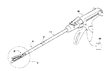
[0013] FIG. 1A is a perspective view of a manually actuated handle assembly
and a
loading unit in accordance with the present disclosure;
[0014] FIG. 1B is a perspective view of an electromechanical instrument, an
adaptor, and
the loading unit of FIG. 1A;
[0015] FIG. 2 is an enlarged view of the indicated area of detail of FIG.
1A;
[0016] FIG. 3 is a cross-sectional view taken along the section line 3-3
with jaws
approximated of FIG. 2;
[0017] FIG. 4 is a cross-sectional view taken along the section line 4-4 of
FIG. 3;
[0018] FIG. 5 is a side cross-sectional view of tissue joined with the end
effector of FIG.
2; and
[0019] FIG. 6 is a cross-sectional view similar to FIG. 4 of another end
effector provided
in accordance with the present disclosure.
4
CA 02918125 2016-01-19
..
DETAILED DESCRIPTION OF EMBODIMENTS
[0020] Embodiments of the present disclosure are now described in detail
with reference
to the drawings in which like reference numerals designate identical or
corresponding
elements in each of the several views. As used herein, the term "clinician"
refers to a doctor,
a nurse, or any other care provider and may include support personnel.
Throughout this
description, the term "proximal" refers to the portion of the device or
component thereof that
is closest to the clinician and the term "distal" refers to the portion of the
device or
component thereof that is farthest from the clinician.
[0021] FIGS. 1A and 1B illustrate a loading unit 10 having an end effector
20 in
accordance with an embodiment of the present disclosure. The loading unit 10
is configured
for connection to a manually actuated handle assembly or stapling instrument
100 such as
described in U.S. Patent No. 8,789,737 ("the '737 Patent"), which is
incorporated herein by
reference. Alternatively, the loading unit 10 can be configured for selective
connection to a
powered hand held electromechanical instrument 200 via the adaptor 210. In
such an
embodiment, the adaptor 210 of the electromechanical instrument 200 may have a
configuration similar to that of the elongated body portion 110 of the
stapling instrument 100
as shown in FIG. 1A. The loading unit 10 is releasably coupled to a distal end
112 of the
elongated body portion 110 of the manually actuated handle assembly 100 or to
a distal end
212 of the adaptor 210 of the electromechanical instrument 200. The end
effector 20 is
operatively associated with an ultrasonic generator 300. As shown in FIG. 1A,
the ultrasonic
generator 300 may be external to the stapling instrument (e.g., stapling
instrument 100 or
electromechanical instrument 200). Alternatively, as shown in FIG. 1B, the
ultrasonic
generator 300 may be incorporated into the stapling instrument (e.g., stapling
instrument 100
or electromechanical instrument 200).
CA 02918125 2916-01-19
[0022] For a detailed description of the structure and function of an
exemplary adaptor
and loading unit, please refer to commonly owned U.S. Patent Publication No.
2012/0089131. For a detailed description of the structure and function of an
exemplary
electromechanical instrument, please refer to commonly owned U.S. Patent
Publication Nos.
2012/0253329 and 2012/0323226. For a detailed description of the structure and
function of
an exemplary ultrasonic generator, please refer to commonly owned U.S. Patent
No.
8,419,758. Each of these disclosures is incorporated herein by reference in
its entirety.
[0023] Referring to FIGS. 2 and 3, the loading unit 10 includes a first or
lower jaw 22
and a second or upper jaw 24. The upper and lower jaws 22, 24 are moveable
relative to one
another between a spaced-apart configuration (FIG. 2) and an approximated
configuration
(FIG. 3). The lower jaw 22 includes a fastener cartridge 30 having a plurality
of staples 32
arranged in rows 33 on either side of a knife or lower blade channel 26 (FIG.
4). The fastener
cartridge 30 may be releasably coupled to the lower jaw 22. The upper jaw 24
includes an
anvil 40 that is configured to deform the staples 32 into formed staples as
the staples 32 are
ejected through openings 31 of the fastener cartridge 30 when the jaws 22, 24
are in the
approximated configuration as detailed below.
[0024] Alternatively, the fastener cartridge 30 of the lower jaw 22 may
include a plurality
of fasteners (not explicitly shown) and the upper jaw 24 may include a
retainer cartridge (not
shown) that includes a plurality of retainers (not shown). As the fasteners
are ejected from
the fastener cartridge 30 of the first jaw 22, each of the fasteners forms a
two-part fastener
with one of the retainers of the retainer cartridge.
[0025] With additional reference to FIG. 4, the fastener cartridge 30 and
the anvil 40 each
include a tissue contacting surface 23, 25, respectively. The end effector 20
may include a
buttress 50 releasably disposed on each of the tissue contacting surfaces 23,
25. The buttress
6
CA 02918125 2016-01-19
50 may be fabricated from a suitable biocompatible and bioabsorbable material.
The buttress
50 may be fabricated from a non-absorbent material which does not retain
fluid, or the
buttress can be made from an absorbent material. For a detailed description of
suitable
materials for surgical buttresses, please refer to commonly owned U.S. Patent
Nos.
5,542,594; 5,908,427; 5,964,774; 6,045,560; 7,823,592; and 7,938,307, and
commonly
assigned U.S. Patent Publication No. 2010/0092710, the entire contents of each
of which is
incorporated herein by reference. As detailed below, the buttress 50 detaches
from the tissue
contacting surfaces 23, 25 of the fastener cartridge 30 and the anvil 40 when
the staples 32
are ejected from the fastener cartridge 30. As discussed in greater detail
below, the buttresses
50 may promote anastomosis, reduce bleeding, provide support for the tissue to
facilitate a
higher burst pressure, and distribute pressure from the fasteners to a larger
area of tissue.
[0026] The anvil 40 defines an upper blade channel 28 that opposes the
lower blade
channel 26 of the fastener cartridge 30. An ultrasonic blade 54 is disposed in
each of the
blade channels 26, 28. The ultrasonic blades 54 are operatively associated
with an ultrasonic
generator 300 (FIG. 1A). The ultrasonic generator 300 provides ultrasonic
energy to the
ultrasonic blades 54 to ultrasonically translate the ultrasonic blades 54
within the blade
channels 26, 28. As detailed below, the ultrasonic blades 54 are configured to
cut tissue
between the jaws 22, 24 along a cut line CL (FIG. 4) in the approximated
configuration and
to weld the buttress 50 attached to the tissue contacting surface 23 of the
lower jaw 22 to
buttress 50 attached to the tissue contacting surface 25 of the upper jaw 24.
[0027] Referring to FIGS. 4 and 5, the end effector 20 is used to fasten
and divide tissue
T in accordance with the present disclosure. The jaws 22, 24 of the end
effector 20 are
approximated over tissue T to be fastened and divided. With layers of the
tissue T positioned
between the jaws 22, 24, the staples 32 are ejected from the fastener
cartridge 30 of the lower
7
CA 02918125 2016-01719
jaw 22 towards the anvil 40 of the upper jaw 24. The staples 32 pass through
the buttresses
50 and are formed in staple pockets 41 defined by the anvil 40 such that the
staples 32 are
disposed on either side of the tissue to urge the buttresses 50 towards one
another. The staple
pockets 41 deform legs of the staples 32 towards one another such that the
staples 32 fasten
the layers of tissue T to one another. The buttresses 50 compress the layers
of tissue T
therebetween to promote anastomosis of the tissue T. The ultrasonic blades 54
are then
supplied with ultrasonic energy to cut the layers of tissue T between the
ultrasonic blades 54
along the cut line CL. As the ultrasonic blades 54 cut the tissue T between
the ultrasonic
blades 54, the ultrasonic blades 54 weld the buttresses 50 together adjacent
the central cut
line CL. The weld W (FIG. 5) of the buttresses 50 helps seal the cut portion
of the tissue T.
[0028] By welding the buttresses 50 together adjacent the cut line CL of
the tissue T,
bleeding of the tissue T may be reduced when compared to anastomosis from
stapling alone.
Further, by stapling through the buttresses 50 adjacent the cut line CL, the
ultrasonic blades
54 may be used to cut and seal tissue T having a greater thickness when
compared to an
ultrasonic dissector alone.
[0029] Referring now to FIG. 6, another end effector 120 is provided in
accordance with
the present disclosure. The end effector 120 is similar to the end effector 20
detailed above
with like structures represented with similar labels, as such only the
differences will be
discussed in detail below. A lower jaw 122 includes a fastener cartridge 130
and has a
stepped tissue contacting surface 123. It is within the scope of this
disclosure, that the tissue
contacting surface 125 of the anvil 140 may also have a stepped configuration
similar to the
stepped configuration of the tissue contacting surface 123 of the fastener
cartridge 130. The
stepped configuration of the tissue contacting surfaces 123, 125 compress
tissue T between
the jaws 122, 124 in a step like manner which may allow tissue having a
greater thickness to
8
CA 02918125 2.016-01-19
be stapled and cut. The fastener cartridge 130defines a blade channel 126
between rows of
fasteners 132. Two ultrasonic blades 154 are disposed within the blade channel
126 adjacent
walls defining the blade channel 126.
[0030] The tissue contacting surface 125 of the upper jaw 124 includes a
protrusion 156
opposing the blade channel 126 of the fastener cartridge 130. The protrusion
156 extends
into the blade channel 126 when the jaws 122, 124 are in the approximated
configuration as
shown in FIG. 6. The protrusion 156 urges the tissue T into the blade channel
126 and may
compress the tissue T into the blade channel 126. As shown, the protrusion 156
has a
triangular cross-sectional shape; however, the protrusion 156 may have a
variety of shapes
that fit within the blade channel 126 of the lower jaw 122 (e.g., semi-
circular, rectangular,
pentagonal, etc.). When the tissue T is within the blade channel 126, the
fasteners 132 are
ejected from the fastener cartridge 130 and the ultrasonic blades 154 are
activated to cut and
seal the tissue T. The protrusion 156 being received within the blade channel
126 may also
align the fastener cartridge 130 and the anvil 140.
[0031] It is contemplated that the tissue contacting surfaces 123, 125 of
the fastener
cartridge 130 and the anvil 140 may include buttresses (not explicitly shown)
as detailed
above to provide additional support to the tissue T as the tissue is stapled
and cut. The
buttresses may also be welded together adjacent the cutlines of the ultrasonic
blades 154 as
detailed above.
[0032] It is also contemplated that the protrusion 156 may include an
ultrasonic blade
such that after the tissue T is stapled and sealed, the ultrasonic blade of
the protrusion 156 is
activated to cut the tissue along a tip 156a of the protrusion 156.
9
CA 02918125 2016-01-19
[0033]
While several embodiments of the disclosure have been shown in the drawings,
it
is not intended that the disclosure be limited thereto, as it is intended that
the disclosure be as
broad in scope as the art will allow and that the specification be read
likewise. For example,
in any of the embodiments disclosed herein, the surgical instrument can
include one or more
electrosurgical components, such as monopolar or bipolar components for
cutting,
cauterizing, and/or sealing tissue or buttress material. Any combination of
the above
embodiments is also envisioned and is within the scope of the appended claims.
Therefore,
the above description should not be construed as limiting, but merely as
exemplifications of
particular embodiments. Those skilled in the art will envision other
modifications within the
scope of the claims appended hereto.