Note: Descriptions are shown in the official language in which they were submitted.
1
Intraocular lens structure
Field
This disclosure relates to an intraocular lens structure (TOL), and a method
for inserting
.. such an IOL.
Background
In modern cataract procedures, also called extracapsular cataract extraction,
a hole is cut
in the anterior capsular bag. This may be done using laser devices.
Subsequently, the natural lens
is removed. In the remaining parts of the capsular bag, in many suggested
procedures an IOL is
placed. The IOL more or less maintains its position in the empty bag.
Usually, an IOL is provided with haptics. These haptics extend radially from a
lens of an
IOL. After implanting an IOL, these haptics usually engage the inside
circumference of the
remaining capsular bag part in order to more or less keep the optics, for
instance a lens, of the IOL
.. centered and positioned in the capsular bag.
For improving fixation of the position of an IOL, many designs were proposed.
US6027531 describes in its abstract "An intraocular lens for use in
extracapsular cataract
extraction has a haptic pa[r]t that surrounds the optical pa[r]t of the lens
and further contains a
groove of such shape to accommodate the anterior and posterior capsules of the
lens bag after
anterior capsulorhexis, extracapsular cataract extraction and posterior
capsulorhexis. The lens is
preferably inserted in a calibrated, circular and continuous combined anterior
and posterior
capsulorhexis, slightly smaller than the inner circumference of the groove as
to induce a stretching
of the rims of the capsular openings. This new approach is believed to prevent
the appearance of
secondary pacification of the capsules, allows a very stable fixation of the
intraocular lens and
ensures a tight separation between the anterior and posterior segment of the
eye. This new principle
of insertion is called the bag-in-the-lens technique, in contrast with the
classical lens in-the-bag
technique.". Placement of this IOL requires skills and the capsular bag may
get damaged. If after
insertion the capsular bag ruptures, the IOL will not maintain its position.
In US6881225, an intraocular lens structure for reducing complications is
described.
According to the abstract, the intraocular lens structure comprises an optic,
a support and a
closing fixture. The closing fixture is a groove or a valley formed on the
side portion of the
Date Recue/Received Date 2020-07-14
2
optic of the intraocular lens. The valley is formed by the optic and a
protrusion projecting
posteriorly from the optic. The groove or the valley in the optic is made
engaged with the posterior
capsular opening generally over the entire circumference of the groove or the
valley to close the
opening of the posterior capsule. Like most of the current IOL structures, the
structure also uses
its haptics for keeping the structure in the capsular bag. The groove holds
the posterior part of the
capsular
US5171320 describes in its abstract an intraocular lens system adapted to be
implanted
within a generally circular opening in an anterior wall of the capsular bag
which normally
contains the crystalline lens of an eye. The intraocular lens system includes
a lens body having
an annular groove which is formed in a peripheral portion thereof in a plane
substantially
perpendicular to an optical axis of the lens body. The lens body includes an
optically effective
portion located radially inside the annular groove, and an anterior lens
portion and a posterior
lens portion located on respective anterior and posterior sides of the annular
groove. The
intraocular lens system is secured in position within the circular opening
such that an annular flap
portion of the capsular bag which surrounds the circular opening is
accommodated within the
annular groove in the lens body.
EP2422746 discloses according to its abstract an intraocular implant for
placement in the
eye, e.g. as part of a cataract operation or crystalline lens extraction
refractive operation, has at a
peripheral portion of the implant a groove which engages with the lip of a
single capsulotomy only
formed in the lens capsule of the eye. The implant will normally be a lens but
may instead be a
bung or plug for occluding an opening made in the capsule. The groove may be a
continuous
groove around the periphery of the implant, or there may be a series of
individual spaced-apart
grooves formed as projections protruding from the periphery. Instead of a
single groove, a pair of
axially spaced-apart grooves may be provided, which engage with respective
capsulotomies
formed in an anterior and a posterior part of the capsule. The posterior
groove is preferably of a
smaller mean diameter than the anterior groove. The description shows an
embodiment with "a
series of projections projecting from the circumference of the lens portion",
referring to very
specific embodiments in the drawings.
Date Recue/Received Date 2020-07-14
3
Summary
A disadvantage of prior art is that placement of the IOL may be very
difficult, with a high
chance of damaging the capsular bag during the medical procedure, or may get
damaged after the
IOL is placed, or that there remains room for improvement.
The present disclosure provides an alternative IOL, which preferably further
at least partly
obviates one or more of above-described drawbacks. In particular, the IOL
described herein may
allow proper and straightforward placement. Alternatively or additionally, it
induces less damage
to the capsular bag and allows secure positioning.
In one embodiment there is provided an intra ocular lens structure (IOL) for
placement in
the capsular bag and securing the IOL in an opening in an anterior part of a
capsular bag, with an
anterior capsular bag flap surrounding said opening, said IOL having an
anterior side which in
use when the IOL is implanted in capsular bag of an eye is directed towards a
cornea of the eye,
and a posterior side which in use when the IOL is implanted in an eye is
directed towards a retina
of the eye. The IOL may comprise an optical structure comprising a perimeter.
The IOL may
furthermore comprise at least two posterior supports, coupled to and extending
away from said
perimeter of said optical structure. The posterior supports are provided for
in use providing
support surfaces for engaging a posterior surface of an anterior capsular bag
flap. The posterior
supports in use reside inside the capsular bag when the IOL is implanted in
the capsular bag.
The IOL further comprises at least two anterior supports, coupled to and
extending from said
perimeter of said optical structure, for in use residing outside the capsular
bag and extending away
from said optical structure. The anterior supports are adapted for in use
providing support surfaces
for engaging an anterior surface of an anterior capsular bag flap. A posterior
plane is defined by
the support surfaces of the posterior supports and an anterior plane defined
by the support surfaces
of the anterior supports. These planes are adapted for in use being spaced
apart at a distance
adapted for holding an anterior capsular bag flap between them for securing
the IOL in said
opening. The posterior supports and anterior supports are in perimetrical
sense or azimuthal
direction shifted with respect to one another.
It was found that due to the geometry and limited depth (approx. 0.2 mm) of
prior art
circumferential groove in known IOU s, the anterior capsular bag flap could
easily escape from
that groove resulting in IOL dislocation. Furthermore, the rotation stability
of the lens structure
may not optimal due to the prior art groove's geometry
Date Recue/Received Date 2020-07-14
4
The IOL can be inserted into the capsular bag. The anterior and posterior
supports allow
fixing the IOL with its optical structure aligned with an opening, in
particular an aperture or
orifice, in a capsular bag.
The terms "anterior" and "posterior" relate to an arrangement of features
relative to the
propagation of the light into the eye. Thus, light enters through the cornea
and passes the iris
through the pupil. Cornea and iris are here considered anterior parts of the
eye. Subsequently, the
light propagates to the retina that is located in the posterior part of the
eye.
The axis of an eye can be the optical axis, or can be the visual axis, the
line of sight, or the
pupillary axis.
An eye has a capsular bag that usually holds the natural lens. In conditions
where that
natural lens needs to be removed, an empty capsular bag remains. Usually, for
removal of the
natural lens, first an opening is made in the anterior part of the capsular 20
bag. Part of the capsular
bag membrane is removed. It leaves a through hole surrounded by a peripheral
edge defining the
perimeter. Such an opening can for instance be circular or elliptic. The
anterior membrane of the
capsular bag is thus provided with an aperture, providing an orifice that
gives access to the capsular
bag.
The part of the capsular bag that is closest to the cornea is here also
referred to as the
anterior capsular bag part. The remaining anterior capsular bag part that
surrounds the mentioned
opening is referred to as the anterior capsular bag flap. It can also be seen
as a ring of capsular bag
membrane.
The capsular bag also has a posterior part. That is the part of the capsular
bag that is closest
to the retina. The average capsular bag thickness is between 4 and 9 microns
for the posterior
capsular bag part and between 10 to 20 microns for the anterior capsular bag
part.
In a procedure for removal of the natural lens, the opening in the anterior
capsular bag can
be made using a laser cutting device. This procedure for making the opening in
the capsular bag
is also referred to as capsulotomy. This laser-assisted procedure allows a
very accurate positioning
and shape of the opening in the capsular bag. Furthermore, after removal of
the natural lens, it is
possible to subsequently make an opening in the posterior part of the capsular
bag, the posterior
opening. This may prevent post-operative posterior capsule pacification.
These two openings can
be accurately aligned. The shape of the posterior opening can be smaller than
the anterior
capsulotomy. The shape of the openings can be matched with a shape of a
perimeter of the IOL
Date Recue/Received Date 2020-07-14
5
or, more exactly stated, a perimeter about the optical structure of the IOL.
Thus, the IOL can fit in
the opening perfectly. Finally, the openings can be matched perfectly with an
optical axis of the
eye. Furthermore, if an optical axis of the IOL is aligned in a predetermined
position within the
circumference of the IOL, the optical structure of the IOL can be positioned
in an optimal manner
in the eye. Thus, the optics of the optical structure can be aligned in a
predefined manner in the
eye. For instance, optical axes may be aligned, but also other predefined
configuration may be
possible, for instance taking into account the quality of parts of the retina.
The support surfaces can be bounded areas on the anterior respectively the
posterior
supports that engage the capsular bag surface. In an embodiment, at least one
anterior support
comprises a posterior side that substantially completely engages the anterior
surface of the
anterior capsular bag part. In an embodiment, at least one posterior support
comprises an
anterior side that substantially completely engages the posterior surface of
the anterior capsular
bag part.
In an embodiment, the IOL comprises an indentation in said perimeter,
providing an
axially extending groove in the peripheral surface of said perimeter. The
indentation extends
past the edge of the capsular bag once the IOL is implanted, and extends in
the axial direction
beyond the posterior surface of the anterior supports, and beyond the anterior
surface of the
posterior supports. The indentation is provided between a posterior support
and an anterior
support.
The indentation provides an axial fluid channel. The indentation is
substantially axial.
The indentation allows fluid communication through the eye.
In another embodiment, there is provided an intra ocular lens structure (IOL)
for
placement in a capsular bag and securing the IOL in an opening in an anterior
part of the
capsular bag, with an anterior capsular bag flap at least partly surrounding
said opening, said
IOL having an anterior side which in use when the IOL is implanted in an eye
is directed
towards a cornea of the eye, and a posterior side which in use when the IOL is
implanted in an
eye is directed towards a retina of the eye. The IOL comprises:
- an optical structure;
- at least two posterior supports for when the IOL is implanted in the
capsular bag
residing in the capsular bag and extending away from said optical structure,
said
Date Recue/Received Date 2020-07-14
6
posterior supports adapted for in use providing support surfaces for engaging
a posterior
surface of an anterior capsular bag flap, and
- at least two anterior supports for when the IOL is implanted in the
capsular bag residing
outside the capsular bag and extending away from said optical structure, said
anterior
supports adapted for in use providing support surfaces for engaging an
anterior surface of
an anterior capsular bag flap,
wherein a posterior plane defined by the support surfaces of the posterior
supports and an
anterior plane defined by the support surfaces of the anterior supports are
adapted for in use being
spaced apart at a distance adapted for holding an anterior capsular bag flap
between them for
securing the IOL in said opening.
In another embodiment there is provided an intra ocular lens structure (IOL)
for
placement in a capsular bag and securing the IOL in an opening in an anterior
part of the
capsular bag, with an anterior capsular bag flap surrounding said opening,
said IOL having an
anterior side which in use when the IOL is implanted in an eye is directed
towards a cornea of the
eye, and a posterior side which in use when the IOL is implanted in an eye is
directed towards a
retina of the eye, said IOL comprising:
- an optical structure;
- at least two posterior supports for when the IOL is implanted in the
capsular bag residing
in the capsular bag and extending away from said optical structure, said
posterior supports adapted
for in use providing support surfaces for engaging a posterior surface of an
anterior capsular bag
flap, and
- at least two anterior supports for when the IOL is implanted in the
capsular bag residing
outside the capsular bag and extending away from said optical structure, said
anterior supports
adapted for in use providing support surfaces for engaging an anterior surface
of an anterior
capsular bag flap,
wherein said IOL comprises an indentation in said perimeter, providing an
axially
extending groove in the peripheral surface of said perimeter. The axially
extending groove
provides a fluid channel allowing eye fluid to pass.
In an embodiment, the IOL is formed as one part. In an embodiment, the IOL is
made from
a polymer material. In an embodiment, the IOL is foldable. The polymer
material allows the IOL
to be rolled into a roll with a diameter smaller than 2.5 mm. In order to
allow clamping of the
Date Recue/Received Date 2020-07-14
7
anterior capsular bag part, at least the anterior supports are resilient,
allowing the IOL to be
inserted in the capsular bag and subsequently bringing the anterior supports
through the
opening in the anterior capsular bag part and in engagement with the anterior
surface thereof.
In fact, this allows holding the IOL in place.
In an embodiment, the at least two posterior supports extending away from said
optical
structure are in a functionally opposite direction with respect to one
another. In an embodiment,
the at least two anterior supports extending away from said optical structure
in a functionally
opposite direction with respect to one another.
In an embodiment, the anterior plane and said posterior plane are, in
particular in
use when clamping the capsular bag, spaced apart 5-100 micron. In particular,
said posterior and
anterior planes are spaced apart 5-70 micron, more specifically 5-50 micron.
In case the support surfaces run about parallel, this distance allows a
clamping of the
anterior capsular bag flap.
The posterior supports, or at least their support surfaces, may be angled
towards the anterior
side of the 10L. In that way, after implantation in the capsular bag, the
posterior supports can urge
against the posterior surface of the capsular bag flap. The posterior supports
can be at an angle of
up to 10 .
Alternatively or in combination, the anterior supports, or at least their
support surfaces,
may be angled towards the posterior side of the IOL. In that way, after
implantation in the capsular
bag, the anterior supports can urge against the anterior surface of the
capsular bag flap. The anterior
supports can be at an angle of up to 10 .
The posterior supports and the anterior supports, in summary, thus provide
support
surfaces that are positioned, in particular that are spaced apart at a
distance,
adapted for holding an anterior capsular bag flap between them. Before the JUL
is inserted, in
particular positioned in the capsular bag, one or more of the anterior support
surfaces in axial
sense may thus even be located posterior to one or more of the posterior
support surfaces. Once
the IOL is implanted and positioned, the support surfaces will hold the
anterior capsular bag flap
between them.
In an embodiment, the posterior supports and the anterior supports are in
perimetrical sense or azimuthal direction shifted with respect to one another.
This allows
Date Recue/Received Date 2020-07-14
8
an easier manufacturing, in particular using for instance tooling or molding
technology.
Furthermore, it provides easier placement and fixation in the capsular bag
opening.
In an embodiment, the posterior supports and said anterior supports extend in
perimetrical
direction or in azimuthal direction about the optical structure. Thus, a good
support of the capsular
.. bag flap can be provided, and even a fixation of the IOL.
In an embodiment, the posterior supports and the anterior supports do not
overlap. In fact,
when viewed from the anterior side, if the anterior and posterior supports do
not overlap, tooling
can be simplified. Furthermore, it may even be possible to allow a smaller
distance between the
anterior and posterior planes. In fact, the support surface of the anterior
support may be shifted to
-100 micron past the support surface of the posterior support. In an
embodiment, the shift may be
-70 microns. In particular when the posterior support and the anterior support
are resilient, the
posterior support and the anterior support may clamp the capsular bag flap
between them, thus
fixing the IOL in the opening. Thus, when the supports do not overlap, the
distance between the
anterior and posterior plane can be between -100 and 100 micron. In an
embodiment, the distance
can be -70 to 100 microns. In particular, the distance can be between -70
micron and 70 micron.
The negative values indicate that when not in use, the anterior support may be
placed further in
posterior direction, past the posterior support. In use however, when holding
the capsular bag, the
anterior support will be at the anterior side of the anterior part of the
capsular bag, and the posterior
support will be at the posterior side of the anterior part of the capsular
bag.
In an embodiment, the IOL comprises a perimetrical surface surrounding said
optical
structure and said posterior support and said anterior support extending from
said perimetrical
surface. In particular, said perimetrical surface defines a radial surface for
when implanted
engaging a perimetrical edge of the anterior capsular bag flap which defines
the perimeter of the
opening.
This can provide alignment of the IOL. For instance, if the opening is non-
circular,
for instance elliptic, and the perimeter of the IOL matches the shape of the
opening, the
azimuthal orientation of the IOL can be fixed. Thus, specific optical
structures can be aligned.
In an embodiment, at least one selected from said posterior supports and said
anterior supports is a haptic. In particular, the haptic has an outer diameter
of 8-12 mm.
It was found that the IOL thus fits in the capsular bag. It may function as a
fail-safe if
aligning with the opening fails.
Date Recue/Received Date 2020-07-14
9
In an embodiment, the IOL is formed in one piece, its thickness and
flexibility adapted for
insertion of the IOL into the eye in a folded manner via a micro insertion.
In an embodiment, the IOL further comprises an at least partially peripheral
groove
posterior to the posterior supports. In particular, said posterior groove
opens in radial direction for
receiving, when said IOL is implanted in an eye, at least an edge of a
posterior capsular bag flap
surrounding a posterior opening in a posterior part of the capsular bag. In an
embodiment, the
posterior groove is between 0.1 and 0.3 mm deep. In particular said posterior
groove is between
0.05-0.2 mm wide. More in particular, the posterior groove is tapered.
In another embodiment, there is provided a method for fixing the intra ocular
structure
(IOL) described above into an eye, where the IOL has a perimeter about an
optical structure, the
method comprising:
- forming an opening within the anterior part of a capsular bag of an eye,
the opening having
a profile matching the perimeter of the IOL, said opening surrounded by an
anterior capsular bag
flap remaining after forming said opening;
- inserting the IOL in the eye with the posterior supports extending in said
capsular bag,
and
- taking the anterior supports out the capsular bag with the anterior
support surfaces resting
on the anterior surface of the remaining anterior part of the capsular bag
surrounding said opening
and while leaving the posterior supports inside the capsular bag, the
remaining part of the anterior
part of the capsular bag surrounding the opening positioned between the
posterior and anterior
supports, thereby securing the IOL in the opening of anterior part of the
capsular bag. The forming
the opening may also be done at a separate action. The method thus relates to
placement of the
IOL only.
In an embodiment of the method, the opening is aligned with an axis of the eye
and/or with
the optical structure of the IOL. In case the optical structure is a lens,
often an optical axis of this
lens is aligned.
In an embodiment of the method, the opening is aligned with an axis and/or an
azimuthal
axis of the eye and an optical and/or azimuthal axis of the optical structure
of the IOL.
In an embodiment of the method, the opening is circular with a center aligned
with an axis
of the eye, and/or the optical structure comprises an optical axis that is
aligned with the perimeter
of the IOL.
Date Recue/Received Date 2020-07-14
10
In an embodiment of the method, the perimeter is circular.
In an embodiment of the method, the capsular bag further comprises a posterior
part, said
method further comprises:
- forming a posterior opening in the posterior part of the capsular bag,
said posterior
opening surrounded by a posterior capsular bag flap remaining after forming
said posterior
opening;
- urging the IOL when secured in the opening in the anterior part of the
capsular bag in
posterior direction in a direction of a retina of the eye, until an inner
perimeter of the posterior
capsular bag flap that defines the posterior opening surrounds a posterior
groove in the IOL and
which at least partially surrounds the optical structure posterior of the
posterior supports, thereby
securing. Thus the posterior capsular bag flap is secured to the IOL,
posterior to the posterior
supports.
In an embodiment, the IOL comprises an indentation in said perimeter,
providing an
axially extending groove in the peripheral surface of said perimeter.
In an embodiment, this indentation is provided between a posterior support and
an
anterior support. When positioned in the opening of the capsular bag, as
explained the
peripheral edge of the capsular bag will rest around the perimeter of the IOL.
The indentation will
then provide a passage for fluid.
The term "substantially" herein, such as in "substantially opposite" or in
"substantially
consists", will be understood by the person skilled in the art. The term
"substantially" may also
include embodiments with "entirely", "completely", "all", etc. Hence, in
embodiments the adverb
"substantially" may also be removed. Where applicable, the term
"substantially" may also relate
to 90% or higher, such as 95% or higher, especially 99% or higher, even more
especially 99.5%
or higher, including 100%. The term "comprise" includes also embodiments
wherein the term
"comprises" means "consists of'.
The term "functionally" herein, such as in "functionally opposite", will be
understood by
the person skilled in the art. It includes for instance exactly opposite, but
deviations from exact
positioning are also included, as long as in operation, the feature
functionally behaves or has the
effect of being for instance substantially opposite. The term "functionally"
may therefore also
include embodiments with "entirely", "completely", "all", etc. Hence, in
embodiments the
adverb "functionally" may also be removed. Where applicable, the term
"functionally" may
Date Recue/Received Date 2020-07-14
11
also relate to 90% or higher, such as 95% or higher, especially 99% or higher,
even more
especially 99.5% or higher, including 100%.
Furthermore, the terms first, second, third and the like in the description
and in the claims,
are used for distinguishing between similar elements and not necessarily for
describing a sequential
or chronological order. It is to be understood that the terms so used are
interchangeable under
appropriate circumstances and that the embodiments described herein are
capable of operation in
other sequences than described or illustrated herein.
The devices or apparatus herein are amongst others described during operation.
As will be
clear to the person skilled in the art, the embodiments are not limited to
methods of operation or
devices in operation.
It should be noted that the above-mentioned embodiments illustrate rather than
and that
those skilled in the art will be able to design many alternative embodiments
without departing
from the scope of the teachings herein.
Many of the features of the current IOL can be combined to further improve
easy
implantation, or fixation.
The embodiments described herein may provide an apparatus, device and/or
method
comprising one or more of the characterizing features described in the
description and/or shown
in the attached drawings.
The various aspects discussed in this patent can be combined in order to
provide additional
advantages. Furthermore, some of the features can form the basis for one or
more divisional
applications.
Brief description of the drawings
Embodiments will now be described, by way of example only, with reference to
the
accompanying schematic drawings in which corresponding reference symbols
indicate
corresponding parts, and in which:
Figure 1 schematically depicts an embodiment of an IOL in
anterior view;
Figure 2 shows the embodiment of figure 1 in side view;
Figure 3 shows a detail of figure 2 as indicated;
Figure 4 shows the embodiment of figure 1 in perspective view
showing the anterior side;
Date Recue/Received Date 2020-07-14
12
Figure 5 schematically depicts a posterior side of the IOL
of figure 1,
with an alternative posterior feature;
Figure 6A shows a cross section of the IOL of figure 1 with
the posterior
feature of figure 1;
Figure 6B shows a cross section of the IOL of figure 5 with the
alternative
posterior feature;
Figure 7A shows a detail of figure 6A as indicated;
Figure 7B shows a detail of figure 6B as indicated;
Figure 8 shows yet another alternative embodiment of an
IOL in anterior
view;
Figure 9 shows an eyeball with an IOL;
Figure 10 shows a detail of figure 9 as indicated with the
IOL of figure 1;
Figure 11 shows a detail of figure 9 as indicated, but with
an IOL with an
alternative posterior feature and a posterior capsular bag part that
is intact;
Figure 12 an eye from above showing axes in the eye;
Figures 13 and 14 an alternative embodiment of the IOL of figure 8,
in front view
and in perspective partly from the rear;
Figures 15-18 a perspective view, view of a detail, a front and
rear view,
respectively, of an alternative embodiment of the IOL;
Figures 19A and 19B schematically indicate a cross section through an eye
before and
after removal of the natural lens, and figure 19C a front view of
figure 19B;
Date Recue/Received Date 2020-07-14
CA 02918617 2016-01-18
WO 2015/016705 PCT/NL2014/050519
13
Figures 20-21 a further embodiment of an JUL in perspective view and in front
view looking on the anterior side of the IOL;
Figures 22-23 yet another embodiment of an JUL in perspective view and in
front view looking on the anterior side of the IOL, and
Figures 24-25 yet another embodiment of an JUL in perspective view and in
front view looking on the anterior side of the IOL.
The drawings are not necessarily on scale.
Description of preferred embodiments
In this description, first relevant parts of the eye will be described in
figures 19A,
19B and 19C. In figures 1-11, some particular embodiments of an intraocular
lens
structure (IOL) and its position in an eye (figures 9-11) will be described,
and a
procedure for placing such an IOL in an eye.
The eye
In figures 19A and 19B, schematically a cross section through an eyeball 20 is
depicted. In figure 19A, the eyeball 20 has a cornea 21, iris 25, pupil 26,
and capsular
bag 22 with a natural lens 31. The capsular bag 22 has an anterior part 23 and
a
posterior part 24. In figure 19B, the eyeball 20 is shown after the natural
lens 31 has
been removed, leaving the empty capsular bag 22 with an opening 32, usually
having a
circular or an elliptic shape. The opening 32 is in the anterior part 23 of
the capsular
bag 22. In many cases, the centre of the opening 32 will be on an axis of the
eye. The
axis are defined in figure 12.
Figure 19C shows part of the eyeball in front view, showing the iris 25, the
anterior part 23 of the capsular bag with opening 32 and the edge of the
opening 52.
This edge 52 is also referred to as the 'perimetrical edge' 52.
In some patients, the posterior part 24 of the capsular bag 22 may not be
clear
anymore. In these cases or to generally avoid post surgery posterior capsular
opacification, additionally an opening in the posterior part 24 or the
capsular bag 22
may be made, referred to as the posterior opening, or the posterior part 24 of
the
capsular bag may be removed.
CA 02918617 2016-01-18
WO 2015/016705 PCT/NL2014/050519
14
In the previous paragraph, the adjectives 'anterior' and 'posterior' are used.
As
explained before, the terms "anterior" and "posterior" relate to an
arrangement of
features relative to the propagation of the light into the eye. Thus, light
enters cornea
and iris, which are anterior parts of the eye, and propagates to the retina
that is located
in the posterior part of the eye. Thus, for instance the capsular bag 22 has
an anterior
part 23 and a posterior part 24. The anterior part, in turn, has a surface
directed
towards the cornea 21 and the iris 25. This surface will be referred to as the
anterior
surface of the anterior part 23 of the capsular bag 22. The opposite surface,
at the
inside of the capsular bag 22, will thus be referred to as the posterior
surface of the
.. anterior part 23 of the capsular bag 22.
The intraocular lens structure (IOL)
Next, some embodiments of the intraocular lens structure (IOL) will be
.. described. Figure 1 schematically depicts an embodiment of an intra ocular
lens
structure (IOL) 1 in anterior view. The anterior side is the side of the IOL 1
that is
directed towards the cornea 21 when said IOL 1 is placed in an eye. The side
of the
IOL 1 that is directed towards the retina after the IOL is implanted in an eye
is here
referred to as the posterior side of the IOL 1. When a natural lens 31 has to
be removed
.. from an eye, usually an opening 32 is made in the anterior part 23 of the
capsular bag
22. Subsequently, the natural lens 31 is removed. In specific cases, such as
paediatric
patients, there may also be a posterior opening made in the posterior part 24
of the
capsular bag 22, the part of the capsular bag 22 that is positioned between
the natural
lens 31 and the retina. The opening 32 and the posterior opening are usually
aligned.
The openings are often circular, but other shapes may be possible, certainly
when
using laser-assisted capsulotomy. The openings are usually aligned with an
optical axis
of the eye, but other positions maybe used. Around the openings, a ring of
capsular bag
tissue or membrane remains. This ring is also referred to as a capsular bag
flap. The
ring or flap has an edge 52 bounding the perimeter of the opening 32, or in
fact
.. defining the opening 32. The opening 32 has a radial direction, running
from the centre
of the opening 32 outwards to its perimeter 52.
The IOL 1 comprises an optical structure 2. The optical structure 2 in many
cases
is a lens, in fact an anterior lens and a posterior lens. In embodiments like
the one
15
shown in figure 1, the optical structure 2 has an anterior lens structure
surface 3 and a
posterior lens structure surface 4, see figure 2. The optical structure can
further be provided
with any type of optical structure known in IOLs. In this description, the
nature of the optical
structure should further not be considered limited. The optical structure can
comprise a lens
or a closure cap. In an embodiment, both the anterior and posterior sides are
provided with a curved
surface to provide one or more lenses. Examples of lens optics are a mono
focal lens, an astigmatic
lens, a multifocal lens, an accommodative lens or a sector bifocal lens such
as for instance
disclosed in PCT/NL2012/050115. The optics may be refractive, diffractive, or
a combination
of both. Furthermore or in combination, the optical structure may comprise an
optical filter,
and/or a functional layer known to a skilled person. The optical structure may
comprise active
and/or passive elements. An example of an active element is for instance
liquid crystal optics.
An IOL 1 usually is substantially a flat structure. Its thickness is about 0.1-
1 mm.
The diameter of IOL 1 usually is about 7-12 mm. The optical structure usually
has a diameter of
between 4-7 mm. In most embodiments, the optical structure has a diameter of 5-
7 mm. The optical
structure often is biconvex.
In such a mainly flat structure, an axial sense Ax can be distinguished which
can have a
posterior direction and an anterior direction. Furthermore, a radial sense Ra
can be distinguished.
Finally, an azimuthal sense Az can be distinguished, which can have a
clockwise and
counterclockwise direction. In case the optical structure is a simple, mono
focal lens, the axial
sense is the optical axis, and the radial sense is the radial direction of the
lens. In figures 1 and 2
these are indicated. In case of other optical structures, the axial, radial
and azimuthal sense will
be clear to a skilled person.
In an embodiment, the IOL 1 is made from a polymer material. In particular,
the JUL 1
is from a polymer material that is foldable. In particular, the supports are
resilient. The JUL 1
in an embodiment is made in one piece. In particular, the IOL 1 is pliable to
allow it to be
rolled up in a small roll with a diameter smaller than 2.5 mm. In particular,
it allows rolling the
IOL up to a diameter smaller than 1.8 mm. On the other hand, the IOL is
dimensionally stable, in
particular flexible to be able to unfold from its rolled-up state and to
return to its original shape
once it is inserted in the capsular bag.
Date Recue/Received Date 2020-07-14
CA 02918617 2016-01-18
WO 2015/016705 PCT/NL2014/050519
16
The embodiment of figure 1 is further also shown in detail in figures 2-4, in
which figure 2 shows the embodiment of figure 1 in side view, figure 3 shows a
detail
of figure 2 as indicated in figure 2, and figure 4 shows the embodiment of
figure 1 in
perspective view, from the anterior side.
The IOL comprises a perimeter 7 about the optical structure 2. The perimeter 7
has a perimetrical surface. The perimeter 7 can match the shape of the opening
in the
capsular bag. If for instance the opening is circular, the perimeter can be
circular. The
size of the perimeter is such that it may be a little oversized to stretch the
size of the
capsular opening a little or matches the size of the opening. In the
embodiment of
figure 1, the optical structure 2 comprises a curved surface providing a lens.
The lens
in this embodiment is circular and has an optical axis. The perimetrical
surface here
extends parallel to the optical axis. The perimeter provides here a
cylindrical surface.
In case of a circular perimeter 7, the perimetrical surface is circle
cylindrical, in the
embodiment of figure 1 even right circle cylindrical. A non-circular shape of
the
opening and the perimeter 7 can have advantages for preventing rotation of the
IOL 1
about the optical axis. For instance, the opening can be elliptical, and the
perimeter 7
can be elliptical, matching the elliptical shape of the opening.
Alternatively, an
alignment feature, for instance a cam, can be provided at the perimeter 7, and
a
matching feature can be provided to the opening. The rotational fixation can
for
instance be advantageous in case of astigmatic optics. In an embodiment, for
instance
shown in fig 1 and figure 8, the diameter of perimeter 7 is larger than the
perimeter 10
of the optical structure 2. Perimeter 7 can for instance be 0.5-2 mm larger
than
perimeter 10 of the optical structure 2.
The IOL 1 comprises posterior supports 5, 5' here at opposite sides of the
optical
structure 2. The posterior supports 5, 5' extend away from the optical
structure. In
particular, the posterior supports 5, 5' extend away in sideward direction
with respect
to the optical structure 2. The posterior supports 5, 5' have support surfaces
13, 13',
also referred to as the support surfaces of the posterior supports 5, 5'.
These support
surfaces 13, 13' are here in a plane, referred to as the posterior plane. In
the specific
embodiment of figure 1, where the perimeter discussed above is cylindrical,
the
posterior plane is perpendicular to the cylindrical surface of the perimeter
7.
The posterior supports 5, 5' here form loops that have two ends attached to
the
perimeter 7.
CA 02918617 2016-01-18
WO 2015/016705 PCT/NL2014/050519
17
The optical structure 2 usually has a diameter of between 4-7 mm. The
perimeter
7 usually has a diameter of between 4-7 mm. In the embodiments shown in the
drawings, the anterior supports 6, 6' and the posterior supports 5, 5' are
attached to the
perimeter 7.
When the IOL 1 is implanted, the support surfaces 13, 13' of the posterior
supports 5, 5'engage the posterior surface of the anterior part 23 of the
capsular bag
22. In an embodiment, the posterior supports 5, 5' and thus at least part of
the support
surfaces can be angulated between 0-10 degrees in anterior direction. In an
embodiment, when implanted, the surface of perimeter 7 engages or almost
engages
the edge 52 of the opening in the anterior capsular bag, and the support
surface 13, 13'
of the posterior supports 5, 5' in fact nestles against the posterior surface
of the
anterior capsular bag. To that end, the support surface 13, 13' can be adapted
to hold
the surface of the capsular bag. For instance, cams or rims may be provided.
At least one of the surfaces of the posterior supports can be roughened, for
instance sand blasted, in order to prevent reflections of light.
The IOL 1 further comprises anterior supports 6, 6'. The anterior supports 6,
6'
also extend sideward with respect to the optical structure 2. The anterior
supports
provide the support surfaces 14, 14' of the anterior supports 6, 6'. When the
IOL 1 is
implanted, these anterior supports 6, 6' are outside of the capsular bag 22.
The support
surfaces 14, 14' are designed and adapted for, when the IOL 1 is implanted,
engaging
the anterior surface of the anterior part of the capsular bag. Again, these
support
surfaces 14, 14' are in a plane, referred to as the anterior plane. In an
embodiment,
when implanted, the surface of perimeter 7 engages or almost engages the edge
52 of
the opening in the anterior capsular bag, and the support surface 14, 14' of
the anterior
supports 5, 5' in fact can be made to nestle against the anterior surface of
the anterior
capsular bag. Both surfaces are thus in almost complete physical contact. To
that end,
the support surface 14, 14' can be adapted to hold the surface of the capsular
bag. For
the anterior supports to actually reach outside the capsular bag and be able
to nestle
against the anterior surface of the anterior capsular bag, usually requires
some
manipulation of the person implanting the IOL 1.
The anterior plane is functionally parallel to the posterior plane. Side view
figure
2 shows this. In particular, these planes are parallel when holding the
capsular bag 22
between them. The distance between the posterior support surfaces 14, 14' of
the
CA 02918617 2016-01-18
WO 2015/016705 PCT/NL2014/050519
18
anterior support 6, 6' and the anterior support surfaces 13, 13' of the
posterior support
5, 5' is such that they can hold the anterior part 23 of the capsular bag 22
between
them. The anterior supports 6, 6' and the posterior supports 5, 5', are
positioned such
that their support surfaces comprise a spacing 11 between them. In fact, the
distance
between the posterior plane and/or the anterior plane is adapted for holding
the anterior
capsular bag flap 23 between them for securing the JUL 1 in the opening when
the IOL
1 is implanted. In fact, the distance between the posterior plane and the
anterior plane
can be adapted to the thickness of the anterior part of the capsular bag. It
was found
that the posterior supports 5, 5' and the anterior supports 6, 6' were able to
hold the
anterior capsular bag flap between them if the distance is between 5 and 100
microns.
In particular, the posterior plane and the anterior plane are spaced apart 15-
50 microns.
The distance provides the spacing 11. In case the distance is less than 20
microns, in
particular less than 10 microns, the flap will be securely clamped and
possible rotation
of the lens prevented.
In the embodiment of figure 1, the posterior supports 5, 5' and the anterior
supports 6, 6' are staggered. In fact, when viewed from the anterior
direction, the
posterior supports 5, 5' and the anterior supports 6, 6' do not overlap. This
may also be
referred to as that the posterior supports 5, 5' and the anterior supports 6,
6' are
staggered in a perimetrical sense or azimuth sense (Az, figure 1). In this
sense,
staggered is used as in a 'staggered junction'.
In particular, when the posterior supports 5, 5' and the anterior supports 6,
6' are
staggered, the posterior plane and the anterior plane are parallel or
substantially
parallel when the anterior part of the capsular bag is held between them.
In the embodiment of figure 1, the posterior supports 5, 5' of JUL 1 are
closed
loops. In the embodiment of figure 1, the posterior supports 5, 5' of JUL 1
have a
diameter (in other words provide the JUL with a diameter) of about 8-12 mm, in
particular 7-12 mm. The thickness of the posterior support can be between 0.15-
0.4
mm. In particular, the thickness can be between 0.2-0.4 mm. More, the
thickness can
be in particular 0.20-0.35 mm. Specifically the thickness of the posterior
supports may
be between 0.25 and 0.35 mm.
Alternatively, the ends of the loops may also be removed, turning posterior
supports 5, 5' in fact each into two posterior supports, resulting in four
posterior
supports 5, 5'. The radially extended posterior supports or loop supports may
in fact
CA 02918617 2016-01-18
WO 2015/016705 PCT/NL2014/050519
19
act as safeguard if placement of IOL 1 in the opening 32 can not be
accomplished for
some reason.
The thickness of the anterior supports 6, 6' can be between 0.04 and 0.25 mm.
In
particular the thickness can be between 0.04 and 0.20 mm. More in particular,
the
thickness can be between 0.05 and 0.20 mm. Specifically, the thickness can be
between 0.05 and 0.10 mm.
In the embodiment of figure 1, the IOL 1 at or near the perimeter 7 has at
least
one in perimeter or azimuthal direction extending space 19 between a posterior
support
5, 5' and an anterior support 6, 6'. This space facilitates manufacturing, and
also
facilitates getting the anterior supports 6, 6' through the opening 32 and out
of the
capsular bag as it provides room for insertion of an instrument when inserting
and
positioning the IOL 1. In the embodiment of figure 1, at each transition from
anterior
supports 6, 6' to posterior supports 5, 5' there is a azimuthal space 19.
It was found that in order to support the posterior side of the anterior part
of the
capsular bag, the posterior supports 5, 5' extend at least about 0.5 mm away
from the
perimeter, in radial direction. In particular, the posterior supports 5, 5'
extend at least
1.0 mm in radial direction.
It was found that in order to support the anterior side of the anterior part
of the
capsular bag, at least one of the anterior supports 6, 6' extend at least
about 0.3 mm
away from the perimeter, in radial direction. In particular, the anterior
supports 6, 6'
may extend at least 0.4 mm. More in particular, the anterior supports may
extend at
least 0.5 mm in radial direction.
In the embodiment of the IOL 1 of figure 1, the IOL 1 has additional anterior
supports 8, 8'. These anterior supports are here referred to as anterior lips
8, 8'. These
in use also extend outside the capsular bag 22. They complement the other
anterior
supports 6, 6' and provide additional clamping of the anterior capsular bag
part 23.
The anterior lips 8, 8' have posterior surfaces 17, 17' that rest against the
outside of the
capsular bag 22, against the anterior surface of the anterior capsular bag
part 23. The
anterior lips 8, 8' here extend in perimeter (or azimuthal) direction about
0.1-2 mm.
The anterior lips 8, 8' extend in radial direction, i.e. away from the optical
structure 2
and the perimeter 7, about 0.1-1.3 mm. In particular it is about 0.4-1.0 mm.
Specifically, it is about 0.4-0.6 mm. In this embodiment, the anterior lips 8,
8' extend
about 0.3 mm.
CA 02918617 2016-01-18
WO 2015/016705 PCT/NL2014/050519
In figure 8, an embodiment of an IOL 1 is shown in which the anterior supports
6, 6' have an alternative shape. In this embodiment, the anterior supports 6,
6' are
provided with a support opening 18, 18'. Through these support openings 18,
18', an
instrument can be inserted for pulling the anterior supports 6, 6' back
through the
5 opening 32
in the capsular bag after the JUL was inserted in the capsular bag. The
anterior supports 6, 6' thus reach outside the capsular bag. The diameter of
the support
opening 18, 18' can be 0.2-1.5 mm. In particular, the diameter can be 0.2-1.0
mm.
In figures 6A and 6B, two different embodiments of posterior features that
influence the posterior part of the capsular bag can be seen.
10 In figures
5, 6B and 7B, showing respectively a perspective view from the
posterior side, a cross section and a detail of the cross section of figure 6B
as indicated,
the posterior side of the JUL 1 at and near the perimeter is provided with a
sharp rim
16 to prevent growth of tissue from the posterior capsular bag part. Such
growth of
tissue can cause posterior capsul opacification.
15 In figures
2, 3, 6A and 7A, an alternative embodiment of posterior features is
shown. Figure 2 shows a side view, figure 3 shows a detail as indicated,
figure 6A
shows a cross sectional view of the JUL of figure 1, and figure 7A shows a
detail as
indicated in figure 6A.
The JUL of this embodiment has a circumferential posterior groove 12,
20 extending
posterior to the posterior supports 5, 5' and the anterior supports 6, 6'. In
fact, the posterior groove 12 is here provided posterior to the posterior
surface 15, 15'
of the posterior supports 5, 5'. The posterior groove 12 is provided to
receive and hold
the edge around the posterior opening, i.e., the opening in the posterior
capsular bag.
As explained, such a posterior opening can be made by a second capsulotomy
performed on the posterior part 24 of the capsular bag 22. The edge around the
posterior opening is slipped into posterior groove 12 after the JUL 1 is
positioned in
the opening in the anterior capsular bag part. To that end, the JUL can be
gently urged
backward until the edge or rim of the posterior opening slips into the
posterior groove
12. The posterior groove 12 here has a depth of 0.1-0.3 mm. The posterior
groove 12 is
shaped to receive the edge around a posterior opening. The posterior groove 12
can be
a rectangular groove. Here it is wedge-shaped. It has walls at an angle of
between 10
and 60 degrees, in particular about 30-60 degrees, specifically 40-50 degrees.
This
CA 02918617 2016-01-18
WO 2015/016705 PCT/NL2014/050519
21
posterior groove 12 will seal the posterior opening, preventing capsule
opacification
and/or leakage of the vitreous.
The IOL positioned in the eye
Figure 9 shows in cross sectional view an eyeball with an IOL 1 in inserted
position inside capsular bag 22. The eyeball 20 has a cornea 21, an iris 25
with a pupil
26, and the capsular bag 22.
In figure 12, showing a cross section through the eye from above (N = Nasal
side, T=Temporal side), several axes of the eye 20 are defined:
1. The visual axis 51, which goes through the fixed object point and the nodal
point N of the eye. If the function of the nodal points is taken into account,
the ray,
which represents the visual axis 51, passes to the retina through the fovea
48.
2. The optical axis 47, which is perpendicular to the cornea surface and
passes
the iris 25 pupil 26 at the midpoint. Since the fovea 48 is not located
central to the
eyeball 20, the optical axis 47 differs from the visual axis 51. The optical
axis 51 is the
geometrical symmetry axis of the eye-ball system and is different from the
optical
central ray, which reaches the central point of the fovea and passes obliquely
through
the eye system.
3. The line of sight 50 is the axis, which goes through the object point and
the
centre of the entrance pupil 26. It is the ray, which passes through the
centroid of the
light bundle and is the axis of the ray cone, which enters the eye 20.
Typically, the
angle between the line of sight and the optical axis 47 lies in the range
between 3 and
8 . The centre of the entrance pupil 26 is shifted towards the nasal side due
to the
asymmetrical imaging through the cornea system and the off-axis position of
the
fovea.
4. The pupillary axis 49, which passes through the centre of the entrance
pupil 26
and is perpendicular to the front surface of the cornea.
The field of view for monocular sight covers the whole retina without the
small
portion of the blind spot. Usually humans tend to rotate the eye to the most
favourable
position where the image is generated in the fovea 48. If the eye 20 is moved
in this
way into a position of optimal orientation so that the image is in the central
part of the
fovea, the optical system of the eye is not used as a centered system.
Nevertheless, the
22
tilt is small and spherical aberration and astigmatism are the dominating
aberrations of the eye.
In figure 10, a detail of figure 9 is shown with the IOL 1 of figure 1
inserted. The IOL
1 in this example is provided with the posterior groove 12 described earlier.
Here, the posterior capsular bag 24 has the posterior opening explained
earlier. The rim of
the posterior opening is positioned in the posterior groove 12. The anterior
capsular bag flap (a
ring of capsular bag membrane material) which remains after an opening is made
in the anterior
capsular bag part 23 is held between the anterior support 6 and the posterior
support 5. The support
surface of the anterior support 6 and the support surface of the posterior
support 5 both rest against
the anterior capsular bag flap, and in fact, although perhaps not indicated
that way, may even clamp
that flap between them.
In figure 11, a detail similar to that of figure 9 is shown, but with an IOL 1
with an
alternative posterior feature. In this case, the posterior capsular bag part
24 does not have an
opening: the posterior capsular bag part 24 is intact and rests against the
posterior surface 4 of IOL
1.
In both figures 10 and 11, the posterior supports 5, 5' have a large diameter.
The IOL 1,
however, is positioned in opening 32 by means of the anterior and posterior
supports, possibly
combined with mutual fitting of perimeter 7 and the length of the perimeter of
opening 32. Thus,
the radial dimension of the posterior supports 5, 5' may be reduced.
Insertion of the IOL in an eye
Insertion of the IOL 1 described so far will be explained below. An example of
a procedure
of making the incision and implanting the IOL is as such for instance
described in US5376115. In
particular, it describes:
A surgical method gaining in popularity is the phacoemulsification technique,
that
utilizes ultrasonic vibrations to fragment the lens nucleus, thus allowing
removal of the lens
material through an incision that is approximately 3 mm long. The benefits of
a small incision are
faster visual rehabilitation, faster healing and less astigmatism than with
conventional large
incisions. A hollow titanium needle with a diameter of about 1 mm is activated
to vibrate by a
magnetostrictive ultrasonic mechanism. The mechanical vibrations transform the
lens into an
emulsion, hence the name phacoemulsification.
Date Recue/Received Date 2020-07-14
23
As the phacoemulsification technique has been refined the construction of the
incision has developed to allow sealing of the wound without the need for
sutures-- "self-sealing
incisions".
According to the reference, the technique is described for instance in J
Cataract Refract
Surg 16(5) (1990) pp. 567-577 by Menapace, R. et al and in Ophthalmology
(U.S.) 100(2) (1993)
pp. 159-163 by Ormerod, L.D. et al.
US 5376115 further describes an example of insertion of an IOL.
This may be combined with the following procedure. Before inserting the IOL 1
into
the capsular bag, first an opening is made in the anterior part of the
capsular bag. Using for
instance a laser device like the Femto laser, an opening or aperture can be
made in the anterior
membrane or anterior capsule of the capsular bag that has a precise shape and
precise position.
This procedure is also referred to as 'Capsularhexis', although recent
literature refers to a
laser-based procedure as 'Capsulotomy', and uses that term in contrast to
'Capsularhexis',
which term is then used to refer to mechanically tearing or cutting an opening
in the capsular
bag. Other laser-based procedures are currently also developing. In these
procedures, a laser
beam is directed through the cornea and into the eye, where its energy is
absorbed in an internal
structure in order to cut that structure. In one of these procedures, the
anterior capsular bag
membrane is colored with a light-absorbing agent. The absorption properties of
that light-
absorbing agent are selected in order to absorb the laser beam energy.
In many cases, for instance in case of a cataract, in a next step the cloudy
natural
lens is removed through the opening in the capsular bag. In this step, the
natural lens can be treated
with a laser first, before it is removed, for instance with a phaco
emulsification device. Removal
of the natural lens as such is known to skilled person.
In an optional next step, a posterior opening can be made in the posterior
part of the
capsular bag, in the posterior membrane or posterior capsule of the capsular
bag.
An example of such a classic Capsularhexis procedure and the use of a laser
device
in such a procedure is described in US8409182,. For instance in column 3, an
example of
steps in a
Date Recue/Received Date 2020-07-14
CA 02918617 2016-01-18
WO 2015/016705 PCT/NL2014/050519
24
Capsularhexis procedure or, more specific, a capsulotomy procedure, is
described. The
laser-assisted procedure allows accurate positioning as well as shaping of the
opening.
Furthermore, such a procedure can leave a relatively strong edge 52 around the
created
opening in the capsular bag. In particular, regarding a laser-based procedure
the
following was found.
METHODS: Capsulotomies performed by an optical coherence tomography¨
guided femtosecond laser were evaluated in porcine and human cadaver eyes.
Subsequently, the procedure was performed in 39 patients as part of a
prospective
randomized study of femtosecond laser-assisted cataract surgery. The accuracy
of the
capsulotomy size, shape, and centration were quantified and capsulotomy
strength was
assessed in the porcine eyes.
RESULTS: Laser-created capsulotomies were significantly more precise in size
and shape than manually created capsulorhexes. In the patient eyes, the
deviation from
the intended diameter of the resected capsule disk was 29 um 26 (SD) for the
laser
technique and 337 258 um for the manual technique. The mean deviation from
circularity was 6% and 20%, respectively. The centre of the laser
capsulotomies was
within 77 47 um of the intended position. All capsulotomies were complete,
with no
radial nicks or tears. The strength of laser capsulotomies (porcine subgroup)
decreased
with increasing pulse energy: 152 21 mN for 3 mJ, 121 16 mN for 6 mJ, and
113 +
23 mN for 10 mJ. The strength of the manual capsulorhexes was 65 + 21 mN.
CONCLUSION: The femtosecond laser produced capsulotomies that were more
precise, accurate, reproducible, and stronger than those created with the
conventional
manual technique.
Source: J. Cataract Refract. Surg. 2011; 37:1189-1198 Q 2011 ASCRS and
ESCRS.
Test further showed the following results.
METHODS: Ten fresh pig eyes were randomly assigned to femtosecond laser¨
assisted capsulotomy or manual capsulotomy. The capsule was immersed in
hyaluronic acid, and retractors were fixed in the capsule opening with a pull-
force
measuring device. The force necessary to break the capsulotomy was measured in
millinewtons (mN); the maximum stretching ratio was also assessed.
RESULTS: The observed mean rupture force (i.e., maximum amount of force
measured immediately before tissue rupture) was 113 mN +12 (SD) in the laser-
CA 02918617 2016-01-18
WO 2015/016705 PCT/NL2014/050519
assisted procedure and 73+ 22 mN in the manual procedure (P.05). The
stretching
ratios were 1.60+ 0.10 (femtosecond) and 1.35+ 0.04 (manual) (13.05).
CONCLUSION: In this laboratory pig-eye study, femtosecond laser¨assisted
capsulotomy resulted in a significantly stronger anterior capsule opening than
the
5 standard manually performed capsulotomy.
Source: J. Cataract Refract. Surg. 2013; 39:105-109 Q 2013 ASCRS and
ESCRS.
A very accurate positioning of an opening 32 in a capsular bag 22, and a very
accurately shape of the opening 32, allow an accurate positioning and
orientation of
10 the IOL 1 described, and are in particular advantageous when using the
current IOL or
IOL/S-IOL combination.
The IOL 1 can be used in the following way. Often, the IOL 1 is inserted in
the
capsular bag via a micro incision in the eye. Via an insertion device, the IOL
outside
the eye is rolled up and urged forward through a nozzle that fits through the
incision in
15 the eye. The rolled-up IOL 1 enters the capsular bag via the opening.
The rolled-up
IOL 1 unfolds inside the capsular bag.
Next, using a small tool, the anterior supports 6, 6' are manipulated to fold
back
through the opening 32 in the anterior capsular bag part 23 to extend outside
the
capsular bag 22. Using the same or an identical tool, the lips 8, 8' may be
manipulated
20 to also extend through the opening 32 and to reach out of the capsular
bag 22. The
posterior surfaces 17 and 17' of the lips 8, 8' will then rest on the anterior
surface of
the anterior part 23 of the capsular bag 22. If the posterior capsule is
opened as well
then in a second manoeuvre by gently pushing the IOL a little bit downward the
posterior flap will be secured in the posterior groove 12.
In figures 13 and 14, an alternative embodiment of the IOL 1 of figure 8 is
shown. In figure 13, the embodiment of figure 14 is shown partly from the rear
in
perspective. Again, similar reference numbers show similar elements.
Capsular bag distension syndrome (CBDS) is an uncommon, but well recognized
cause of reduced vision following cataract surgery. It usually presents in the
immediate
postoperative period, with shallowing of the anterior chamber, unexpected
myopic
refraction and accumulation of liquefied substance between the implanted lens
and
posterior capsule.
CA 02918617 2016-01-18
WO 2015/016705 PCT/NL2014/050519
26
The most likely mechanism of CBDS is the production of collagens from
residual lens epithelial cells or necrotic and/or apoptotic autolyzed lens
epithelial cells
or the retained viscoelastic from the surgical procedure accumulates behind
the
intraocular lens (TOL) as the IOL optic occludes the anterior capsular opening
made by
the capsulotomy. The creation of a small opening in the lens to avoid total
sealing of
the bag may avoid this post-operative complication. The opening could be
shaped in
the form of notch at the optic edge or a small hole made in the optic. It is
also possible
to create small capsulotomies when the capsule opening is made in the anterior
or
posterior capsule flaps to avoid complete sealing of the capsular opening when
using
.. the IOL described earlier.
In the embodiment of figure 13 an 14, another approach is chosen. In this
embodiment, an indentation 53 is created in the peripheral surface 7. This
indentation
53 provides an axial (Ax) groove in the perimeter 7 about the IOL. Here, the
groove as
straight in axial (Ax) direction, but amendments may be made to control flow
of fluid.
This indentation 53 creates a passage between the peripheral surface 7 and the
edge 52
of the opening 32 in the anterior part of the capsular bag 23 after insertion
of the IOL
1. Thus, a passage for fluid is provided once the IOL is inserted in the
opening 32 in
the capsular bag. In fact, even if the posterior groove 12 is provided in the
IOL, this
groove may provide a passage for fluid part once the posterior part of the
capsular bag
is inserted in the posterior groove 12. In fact, the radial extension of the
indentation
may control such a passage.
In order to provide an easy passage, the indentation 53 is provided in radial
sense
next to a posterior support 5, 5' or an anterior support 6, 6'. In the
embodiment shown
in the drawings, the indentation 53 is provided between a posterior support 5,
5' and an
anterior support 6, 6'. In this embodiment, two indentations 53 are provided,
here
opposite one another. Here, the diameter of the indentations 53 are selected
to allow
eye fluid to pass the passage. In this embodiment, the width of the
indentations 53 is
here 0.2-0.6 mm. In particular, the width is 0.25-0.5 mm. The depth of the
indentations
53 is here 0.05-0.4 mm. In particular, the depth is 0.1-0.3 mm.
In figures 15-18 a perspective view, view of a detail, a front and rear view,
respectively, of an alternative embodiment of the IOL. Again, identical
reference
numbers refer to features that are at least functionally equivalent. More in
particular, in
this embodiment the indentation 53 was modified. In this embodiment, the
position (in
CA 02918617 2016-01-18
WO 2015/016705 PCT/NL2014/050519
27
circumferential or tangential sense T) of the indentations 53 is adapted.
Furthermore
here three indentations 53 are provided. It was found that the indentations 53
resulted
in an interruption of the posterior rim 16. As already explained, the
posterior side of
the JUL 1 at and near the perimeter is provided with a sharp rim 16 to prevent
growth
of tissue from the posterior capsular bag part. Such growth of tissue can
cause
posterior capsular pacification. The indentations 53 of the earlier
embodiment of
figures 13 and 14 interrupt that rim 16, thus presenting a risk of growth of
tissue which
may start posterior capsular opasification. This tissue may for instance block
the
indentation, preventing exchange of fluids.
Here, the indentation opens at the anterior side of the JUL. The depth (in
axial
direction A, for clarity reasons also the radial direction R is indicated in
figure 15) is
selected that the indentation extends past the edge 52 of the capsular bag
once the JUL
1 is implanted. In practice, the indentation in axial direction A extends
beyond the
posterior surface 14, 14' of the anterior supports 6, 6'. In an embodiment,
the
indentation extends beyond the anterior surface 13, 13' of the posterior
supports 5, 5'.
Thus, the indentations provide a fluid channel past the capsular bag 23. The
indentations 53 here end before the posterior rim 16, leaving its edge in
tact. Thus, the
indentations 53 have a bottom or end 54. The indentations 53 extend radially R
inward
with respect to the peripheral surface 7. The supports 5, 5', 6, 6' extend
radially
outward from the peripheral surface 7. Before implantation, in an embodiment,
the
posterior surface of the anterior supports 6, 6' in an embodiment in radial
direction R
extends past the peripheral surface 7. The anterior surface of the posterior
supports 5,
5' in an embodiment in radial direction R extends past the peripheral surface
7 in
opposite direction Thus, the supports can clamp the capsular bag between them.
Figures 20-25 show various other embodiments of an JUL allowing easier
production, and easier implantation and fixation in an eye.
In these embodiments these are multiple posterior supports and multiple
anterior
supports. They are not separately indicated with an '-mark. The same parts or
features
again have the same references and will not be discussed further. Figure 20
shows a
perspective view and figure 21 shows a view from the anterior, showing the
anterior
side of the JUL.
There, the JUL has three haptics remaining in the (remainder of) the capsular
bag. The haptics provide in fact six posterior supports 5 which are two by two
coupled
28
at their radial ends. They extend further in radial (Ra) direction then the
anterior supports 6.
When viewed like in figure 21, it is clear that the supports 5, 6 do not
overlap. The through
holed 18 in the anterior supports 6 again allows the anterior supports 6 to be
brought out of the
capsular bag easily. This can provide better centering in the capsular bag.
In the embodiment of figures 22 and 23, the bottom 54 of the axial
indentations 53 are
further remote to the posterior direction then the anterior surfaces 13 of the
posterior supports.
This provides a more sure fluid channel. The axial indentations 53 in the
perimeter 7 (also referred
to as axial groove 53) may also taper in posterior direction. This may make
tooling or molding
such a lens easier.
Again, the two-by-two connected posterior supports 5 may also provide the
functionality
of haptics. Another definition may be that there are three posterior supports
that have through
openings. The posterior supports 5 and anterior supports 6 again do not
overlap. They are
azimuthally shifted.
The embodiment of figures 24 and 25 differs little from the embodiment of
figures 22 and
23. In this embodiment, the posterior supports 5 are angulated in anterior
direction. Thus, part of
their posterior surface 15 is visible in the side view of figure 24. Thus in
some cases, fixation in
the capsular bag may be improved. In the embodiment with angulation in
anterior direction, the
lens is pressed a little in posterior direction, and may rest against the
posterior capsular bag part.
Pressing more secure to the capsule may prevent posterior capsule
opacification. When a through
hole is also provided in the posterior capsular bag part, as explained
earlier, fixation in that hole
may improve.
It will also be clear that the above description and drawings are included to
illustrate some
embodiments and not to limit the scope of the disclosure. Starting from this
disclosure, many
more embodiments will be evident to a skilled person. These embodiments are
within the scope
and the essence of teachings herein and may include combinations of prior art
techniques and
the disclosure of this patent.
Date Recue/Received Date 2020-07-14
CA 02918617 2016-01-18
WO 2015/016705
PCT/NL2014/050519
29
List of reference numbers
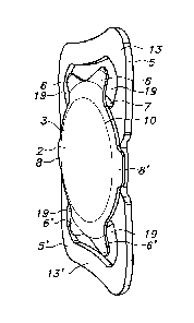
1 Intra ocular lens structure (IOL)
2 Optical structure
3 Anterior surface of the IOL
4 Posterior surface of the IOL
5, 5' Posterior supports
6, 6' Anterior supports
7 perimeter of the IOL
8, 8' Additional anterior lips
9 Outer perimeter of the optical structure
10 Perimeter of the optical structure
11 Space between the posterior plane and anterior plane
12 Posterior groove for the posterior capsular bag flap
13, 13' Anterior support surfaces of the posterior support
14, 14' posterior support surfaces of the anterior support
15 15' Posterior surfaces of the posterior support
16 Posterior rim
17, 17' Posterior surfaces of the additional anterior lips
18, 18' holes in the anterior support
19 azimuthal (Az) space between posterior and anterior supports
20 eyeball
21 Cornea
22 Capsular bag
23 Anterior part of the capsular bag
24 Posterior part of the capsular bag
25 Iris
26 pupil
31 natural lens
32 opening (in the anterior part of the capsular bag)
47 optical axis
48 fovea
49 pupillary axis
CA 02918617 2016-01-18
WO 2015/016705
PCT/NL2014/050519
50 line of sight
51 visual axis
52 perimetrical edge of the anterior capsular bag flap
53 Indentation
5 54 end of indentation