Note: Descriptions are shown in the official language in which they were submitted.
CA 02919961 2016-01-29
WO 2015/022510 PCT/GB2014/052454
ELECTRON MICROSCOPY SAMPLE SUPPORT
COMPRISING POROUS METAL FOIL
FIELD OF THE INVENTION
The present invention relates to an electron microscopy sample support; a
method of
manufacturing such an electron microscopy sample support; a method of imaging
using such an electron microscopy sample support and an apparatus operable to
perform such a method of imaging.
BACKGROUND
Electron microscopy techniques can be used to image a specimen. According to
such
techniques, a beam of electrons is used to "illuminate" a specimen. The
presence of the
specimen in the electron beam results in changes to that beam. The changes to
the
beam induced by the sample can be examined to create a magnified image of the
specimen.
In order to be illuminated by an electron beam, a specimen must be adequately
supported in that beam. Often the electrons forming the electron beam have a
high
energy and it will be appreciated that bombarding an object, for example, a
specimen
for examination, together with the support holding the specimen in position
within the
electron beam, may result in physical, chemical and/or electrical changes to
the
support and/or specimen. Such changes may impact results, including resolution
of
image, obtained through use of electron microscopy techniques.
It is desired to provide a specimen support, for use in electron microscopy,
which may
address some of the features of known specimen supports.
SUMMARY
Accordingly, a first aspect provides an electron microscopy sample support
comprising:
a support member; and a metal foil comprising a porous region; the support
member is
configured to give structural stability to the metal foil, and the porous
region of the
metal foil is configured to receive an electron microscopy sample.
The first aspect recognises that the information content in electron
micrographs of
specimens including, for example, nanoscale particles, can be limited by:
electron
beam-induced motion of individual particles; charge accumulation on the
specimen
induced by the electron beam and/ or chemical transformation of a specimen
support,
1
CA 02919961 2016-01-29
WO 2015/022510 PCT/GB2014/052454
for example, a carbon substratc. Such phenomena are illustrated schematically
in
Figures 1 to 3.
The phenomena described above may be of particular relevance in relation to
electron
cryomicroscopy, also known as cryo-electron microscopy, in which transmission
electron microscopy is used to study specimens at cryogenic temperatures.
Electron
cryomicroscopy techniques can be particularly useful in the study of frozen,
hydrated
biological specimens. The loss in information content as a result of phenomena
such as
those alluded to previously may limit the resolution of images collected by
electron
cryomicroscopy in relation to such frozen hydrated biological samples,
particularly in
relation to 3D reconstruction of images of such samples. Known techniques
impose a
limit on the accuracy of angular assignments of individual particles as a
result of the
limited information available such that smaller particles, such as those less
than ¨500
kDa, cannot be aligned reliably.
The first aspect recognises that whilst the use of metal grid supports in
electron
microscopy is known and the use of a porous carbon film is known, the use of a
fine
porous metal foil to support specimens for analysis may have advantages,
despite
appearing, in the first instance, to be a structure which might be detrimental
to
resulting images. In particular, it may be thought that housing samples in the
region of
pores provided in a metal foil would result in poorer images, since the metal
foil is not
transparent to an electron beam and may cause undue interference to an
incident
electron beam. As a result, typically porous carbon substrates have been used,
those
substrates being supported by, for example, metal grids, the metal grids being
operable
to add mechanical stability to the specimen support and to "conduct" away
electrons
from a sample as required.
A sample support in accordance with the first aspect includes a metal foil,
having
properties selected to allow it to provide, for example, sufficient secondary
electrons to
a sample located in the region of a pore in the metal foil if correctly
aligned within an
incident electron beam. Such secondary electrons may then neutralise positive
charge
accumulated in the sample during exposure to an incident electron beam. The
electron
yield of an appropriately chosen metal may act to minimise charging effects
experienced by a sample or specimen in the region of a pore of the foil.
Furthermore,
the metal may be selected such that it is substantially inert and non-reactive
in the
presence of an electron beam. The non reactive nature of an appropriately
chosen
metal may minimise mechanical deformation of the foil.
2
CA 02919961 2016-01-29
WO 2015/022510 PCT/GB2014/052454
Although the word metal has been used to describe the porous foil of the first
aspect, it
will be appreciated that a material having substantially metallic properties
as outlined
further below may also be used, for example, an appropriately chosen
semiconductor
material. Typically a metal may be considered to be any material having a free
charge
carrier concentration greater than 10e21 per cm cubed.
In one embodiment, the metal foil is arranged to be in ohmic contact with the
support
member. That is to say, the contact between the metal foil and the support
member
acts as a non-rectifying junction, thus allowing for ease of movement of any
free
electrons between the foil to the support and ameliorating charging effects
which may
result as a result of exposure of the support to an electron beam.
In one embodiment, the metal foil comprises a metal having a large proportion
of
mobile electrons not tightly bound to any particular atom in the material.
Materials of
this sort are more conductive than typical amorphous carbon. That is to say,
in some
embodiments, the metal may comprise a high conductivity metal. Accordingly,
provision of a high conductivity metal for the foil allows for relatively free
movement of
electrons within the foil, which can ameliorate charging effects which may
result as a
result of exposure of the support to an electron beam.
In one embodiment, the metal foil comprises a metal having a high total yield
of
electrons emitted by the foil for each electron incident on the foil.
Accordingly,
exposure to a high energy electron beam may allow generation of electrons
which can
fall upon a specimen or sample region, thus ameliorating charging effects
which may
result as a result of exposure of the sample to an electron beam.
In one embodiment, the metal foil comprises a metal having a high mechanical
stability. The metal foil may comprise a metal having a mechanical strength at
a
selected thickness which is sufficient to reduce motion of the sample during
exposure to
an incident electron beam to less than the inverse of the spatial resolution
desired for a
resulting image. Accordingly, the foil may be configured to be self-supporting
when
extending across a support member. The metal foil may, if chosen to have an
appropriate Young's modulus, be such that it can be relatively strong across a
span
between sides of a support member, despite being relatively thin by nature.
Furthermore, by means of choice of a foil material which has an appropriate
mechanical stability, any effects of m ech an ical distortion caused by
chemical change or
3
CA 02919961 2016-01-29
WO 2015/022510 PCT/GB2014/052454
charge imbalance to the porous metal foil may be addressed. That is to say,
the stiffer
the material, the less likely a charge imbalance can be to cause physical
warping of the
foil. Ameliorating physical warping can help to ameliorate blurring caused by
movement in micrograph images of a sample.
In one embodiment, the metal foil comprises a non-reactive metal. Accordingly,
exposure to a high energy electron beam may result in few chemical changes in
the
metal foil and thus any effects of mechanical distortion caused by chemical
changes to
the porous metal foil may be ameliorated.
In one embodiment, the metal foil comprises a metal compatible with a
biological
electron microscopy sample. Accordingly, choice of material may be such that a
reaction with a specimen is minimised. In particular, in some embodiments, the
metal
foil may be chosen to display little reaction with a biological substance.
In one embodiment, the metal foil comprises at least one of a: gold, platinum,
palladium, hafnium or rhodium metal foil. Accordingly, such materials, and
similar
materials, may be chosen since they have an appropriate: grain size, are non-
oxide
forming, have a desired Young's modulus, secondary electron yield, or other
similar
desirable and /or tunable characteristics.
In sonic embodiments, individual pores in the metal foil are dimensioned such
that
they are comparable to an area to be interrogated by an incident electron beam
of an
electron microscope. Accordingly, an electron microcope beam may be operable
to
view the entire of a single pore. In some embodiments, each pore, or hole is
dimensioned to allow the simultaneous imaging of a plurality of electron
microscopy
samples of interest in a single hole. In some embodiments, each pore is
dimensioned to
be smaller than the size of an incident electron microscope electron beam.
Accordingly,
an incident beam may be arranged to cover the sample housed in the pore and
extend
onto, or overlap, a region of metal foil surrounding the hole or pore. The
overlap of
beam onto metal foil can help to ensure uniform charge neutralisation of a
sample by
secondary electrons generated as a result of the incident electron beam onto
the foil.
In one embodiment, the porous region of the metal foil comprises a layer of
metal
including a plurality of holes. The porous region may extend across
substantially all of
the metal foil held in place by the support member. In some embodiments, only
a
region of the foil may include holes. Those holes may be regularly or
irregularly
arranged in the porous region. In some embodiments, those holes may be
substantially
4
CA 02919961 2016-01-29
WO 2015/022510 PCT/GB2014/052454
uniform in size. In some embodiments, the size of the holes may vary across
the foil.
That is to say, a plurality of porous regions may be provided on the metal
foil, each
having a different pore size. Alternatively, pores of different sizes may be
provided
across the porous region of the metal film. Accordingly, a number of
conditions can be
tested on a single grid.
In one embodiment, the holes are dimensioned to receive at least one electron
microscopy sample. Accordingly, at least one sample may be seen by an
interrogating
electron beam when the sample held in position by the support is irradiated.
That is to
say, in one embodiment, the metal foil has a thickness selected to be at least
the
smallest dimension of the electron microscopy sample.
In one embodiment, the support member comprises a substantially annular
element.
The cross-sectional shape of that annulus may, for example, be substantially
circular,
oval, rectangular or triangular. Accordingly, the metal foil may extend across
said
annular element.
In one embodiment, the support member comprises a plurality of spaced support
elements. In one embodiment, the plurality of spaced support elements are
arranged to
form a mesh. Accordingly, the support member may comprise an annular element
which supports a grid-like structure. That grid-like structure may then
support,
between adjacent mesh elements, the metal foil. Such a grid may provide
additional
structural stability to the metal foil. In one embodiment, the porous region
of the metal
foil is arranged to extend across a region of the mesh.
In one embodiment, the support member and the support elements comprise a
metal.
In one embodiment, the metal comprises at least one of: gold, platinum,
palladium, or
hafnium. Accordingly, the support may be metal or have metallic properties
which can
be selected so as to minimise charging, chemical and/or other similar motion-
inducing
processes which may occur on exposure of the support structure to a high
energy
electron beam.
In one embodiment, the support member, support elements and metal foil are all
formed from the same metal. Accordingly, the main components forming the
sample
support are formed from a material having substantially the same thermal
expansion
coefficient (TEC). As a result, stress, strain, stretching or tearing induced
in the metal
foil may be mitigated, where those changes are induced by a change in
temperature,
5
CA 02919961 2016-01-29
WO 2015/022510 PCT/GB2014/052454
such as that experienced when a sample support is reduced to, for example,
liquid
nitrogen temperatures. If the support is fabricated such that the foil is
under tension,
matching the thermal coefficient of the sample support and/or support
element(s) to
the thermal coefficient of the metal foil can help to mitigate the likelihood
of damage to
the foil and relative movement between the foil and the sample support and/or
support
element(s). Furthermore, it will be appreciated that such thermal matching
between
support components may be desirable to maintain a prescribed amount of tension
in
the metal foil membrane during cooling to keep the flexural rigidity of the
membrane
across a range of temperatures.
In one embodiment, the support further comprises a graphene layer. In some
embodiments, that graphene layer may comprise a thin film. Accordingly, a
graphene
layer is provided which may be substantially transparent to an incident
electron beam.
.. In one embodiment, the graphene layer is configured to extend across pores
in the
porous region of the metal foil. As a result of the transparent nature of
graphene,
provision of such a layer in said sample support may allow for additional
structural
stability, whilst not degrading the quality of resultant images.
In one embodiment, the graphene layer is configured to be in ohmic contact
with the
metal foil. Accordingly, the impact of the provision of a graphene layer in
the sample
support may be minimised, and the benefits of a porous metal foil maintained.
In one embodiment, the graphene layer is configured to support the electron
microscopy sample. Accordingly, the graphene layer, which may extend across
pores in
the metal foil, may be used as a surface upon which to support a sample, or
form a thin
layer containing a sample, such that the graphene surrounds, or encloses a
sample. The
continuous layer of graphene may allow for creation of a more uniform sample
containing structure.
In one embodiment, holes in the porous region of the metal foil are configured
to
receive a radiation sensitive material to be examined using electron
microscopy. The
samples to be examined may sit over, under or in the pores of the porous metal
foil.
The radiation sensitive material may comprise a protein. That protein may be
substantially destroyed by the electron microscopy process. In one embodiment,
the
radiation sensitive material comprises biological material. In one embodiment,
the
biological material is supported in said porous region of said metal foil in
vitreous ice.
6
CA 02919961 2016-01-29
WO 2015/022510 PCT/GB2014/052454
Accordingly, the structure of thc ice may not interrupt imaging of the sample
or
specimen of interest.
In some embodiments, the sample support comprises an electron cryomicroscopy
sample support. Some of the issues described herein may be of particular
relevance in
the electron cryomicroscopy field, and thus the sample support of aspects and
embodiments may find particular applicability in such a field.
A second aspect provides a method of manufacturing an electron microscopy
sample
support, the method comprising: providing a support member; and a metal foil
comprising a porous region; configuring the support member to give structural
stability
to the metal foil, and configuring the porous region of the metal foil to
receive an
electron microscopy sample.
In one embodiment, forming the metal foil may comprise metal deposition on a
template.
In one embodiment, forming the metal foil may comprise removal of the template
after
metal deposition.
In one embodiment, the method comprises arranging the metal foil to be in
ohmic
contact with the support member.
In one embodiment, the metal foil comprises a metal having a high
conductivity.
In one embodiment, the metal foil comprises a metal having a high secondary
electron
generation yield.
In one embodiment, the metal foil comprises a metal having a high mechanical
stability.
In one embodiment, the metal foil comprises a non-reactive metal.
In one embodiment, the metal foil comprises a metal compatible with a
biological
electron microscopy sample.
7
CA 02919961 2016-01-29
WO 2015/022510 PCT/GB2014/052454
In one embodiment, the metal foil comprises at least one of a: gold, platinum,
palladium, or hafnium metal foil.
In one embodiment, the porous region of the metal foil comprises a layer of
metal
including a plurality of holes.
In one embodiment, the method comprises providing holes dimensioned to receive
at
least one electron microscopy sample.
In one embodiment, the method comprises selecting a metal foil having a
thickness of
at least the smallest dimension of the electron microscopy sample.
In one embodiment, the support member comprises a substantially annular disc.
In one embodiment, the support member comprises a plurality of spaced support
elements.
In one embodiment, the method comprises arranging the plurality of spaced
support to
form a mesh.
In one embodiment, the method comprises arranging the porous region of the
metal
foil such that it extends across a region of the mesh.
In one embodiment, the support member and the support elements comprise a
metal.
In one embodiment, the metal comprises at least one of: gold, platinum,
palladium, or
hafnium.
In one embodiment, two or more of: the support, the support element(s) and the
metal
foil comprise: one or more materials having substantially matched thermal
expansion
coefficients. In one embodiment, two or more of: the support, the support
element(s)
and the metal foil comprise: the same material and have substantially matched
thermal
expansion coefficients.
In one embodiment, the method further comprises providing a graphene layer.
8
In one embodiment, the method comprises configuring the graphene layer to
extend
across pores in the porous region of the in.etal foil.
In one embodiment, the method comprises configuring the graphene layer to be
in
ohmic contact with the metal foil,
In one embodiment, the method comprises configuring the graphene layer to
support
the electron microscopy sample.
In one embodiment, the method comprises configuring holes in the porous region
of
the metal fail to receive a radiation sensitive material to be examined using
electron
microscopy.
In one embodiment, the radiation sensitive material comprises biological
material.
In one embodiment, the method comprises supporting biological material in said
porous region of said metal foil in vitreous ice.
A third aspect provides a method of imaging an electron microscopy sample
comprising: configuring the electron microscopy sample on a support according
to the
first aspect; arranging the support in an electron beam of an microscope; and
collecting
image data for analysis.
A fourth aspect provides imaging apparatus operable to provide an electron
microscopy
image of a sample, the imaging apparatus comprising: an electron microscopy
sample
mounted on a support in accordance with the first aspect; an electron beam of
a
microscope arranged to be incident on the support; and a collection device
operable to
collect image data for analysis.
Where an apparatus feature is described as being operable to provide a
function, it will
be appreciated that this includes an apparatus feature which provides that
function or
which is adapted or configured to provide that function.
9
Date Recue/Date Received 2021-03-12
CA 02919961 2016-01-29
WO 2015/022510 PCT/GB2014/052454
BRIEF DESCRIPTION OF THE DRAWINGS
Embodiments of the present invention will now be described further, with
reference to
the accompanying drawings, in which:
Figure 1 illustrates schematically electron beam-induced motion of particles
in vitreous
ice:
Figure 2 illustrates schematically electron beam-induced sample charge
accumulation;
Figure 3 illustrates schematically electron beam-induced chemical
transformation of a
support substrate;
Figure 4a illustrates schematically a sectional, plan, and side elevation of
an electron
microscopy support in accordance with one embodiment;
Figure 4b illustrates schematically a portion of the electron microscopy
support shown
in Figure 4a in more detail;
Figures 5a to Sc show optical and electron images of a device according to one
embodiment;
Figure 6 shows results of one experiment to measure gold particle motion in
vitreous
ice in example electron microscopy supports;
Figures 7a and 7b show a comparative example of a specimen imaged using a
support
in accordance with one embodiment;
Figures 8a and 8bshow experimental results illustrating reduced motion of gold
grid
supports in accordance with an arrangement compared to conventional amorphous
carbon grid supports; and
Figures 9a to 9d show experimental results illustrating average 80S ribosome
displacement from an initial position as a plot against time/ electron fluence
for data
collected in ice using a variety of microscopy support structures.
DESCRIPTION OF THE EMBODIMENTS
It has been recognised that the information content in electron micrographs of
specimens including, for example, nanoscale particles, can be limited by:
electron
beam-induced motion of individual particles; charge accumulation on the
specimen
induced by the electron beam and/or chemical transformation of a specimen
support,
for example, a carbon substrate. Such phenomena are illustrated schematically
in
Figures 1 to 3.
Figure 1 illustrates schematically electron beam-induced motion of particles
in vitreous
ice. Figure 1 illustrates particles, in this instance, proteins, embedded in
vitreous ice.
The sample is irradiated with an electron beam. The electrons forming the beam
have
CA 02919961 2016-01-29
WO 2015/022510 PCT/GB2014/052454
energy which is imparted to the protein samples when colliding with, or
passing
through, those samples. It will be appreciated that, during imaging, particles
being
studied may move both rotationally and tran slation ally upon irradiation with
the
electron beam, causing blurring in resulting captured images.
Figure 2 illustrates schematically electron beam-induced sample charge
accumulation.
Figure 2 shows schematically samples, again proteins, held in position in
vitreous ice
formed in holes in an amorphous carbon substrate supported between bars of a
metal
grid. Irradiation of the samples and support (formed, in this instance, from a
metal
grid and amorphous carbon substrate) with high energy electrons forming an
electron
beam may cause release or movement of electrons forming part of the sample
and/ or
support. The resulting movement or displacement of electrons can result in
sample
charging which may introduce electrical forces that act on the sample and
substrate,
causing particle movement and image blurring by deflection of the electron
beam.
Figure 3 illustrates schematically electron beam-induced chemical
transformation of a
support substrate. In the arrangement shown in Figure 3 particles, in this
instance,
proteins, are embedded in vitreous ice. That vitreous ice is formed in a hole
formed in
a carbon substrate. The sample is irradiated with an electron beam.
Irradiation with a
high energy electron beam may result in the electrons forming the beam acting
to break
chemical bonds in the carbon substrate, which in turn can alter the density
and shape
of the substrate. The change in density and shape of the substrate may induce
mechanical stress and motion and cause "doming" of the ice layer. That doming
of the
ice layer, supporting the particles may cause blurring in the resulting
electron
microscopy image.
Current sample supports and substrates for cryo-EM typically comprise a metal
mesh
disc, referred to as a "grid" formed from a suitable material. That suitable
material may
comprise a metal. The grid is typically covered with a thin layer of holey
amorphous
carbon. The grid may have a regular array of holes. The thin layer of holey
amorphous
carbon may comprise a regular array of holes. Irregular "lacey" carbon
substrates may
also be used. It will be appreciated that in the case of cryo-EM, vitreous ice
is often
used to encapsulate samples in holes formed in the amorphous carbon. Since ice
is an
insulator and amorphous carbon is a poor and highly variable conductor. both
accumulate significant mobile surface charge that can deflect an electron beam
and
exert strong electrostatic forces on the sample, as shown in Figure 2 and
Figure 3.
Furthermore, if amorphous carbon is irradiated by a high energy electron beam,
it may
11
CA 02919961 2016-01-29
WO 2015/022510 PCT/GB2014/052454
undergo chemical changes which may change the density and therefore the shape
of the
amorphous carbon support material, thus causing movement of individual
particles in
a sample.
.. Overview
Before discussing the embodiments in any more detail, first an overview will
be
provided. Aspects and embodiments described herein may provide an ultra-stable
sample support which may ameliorate, reduce or eliminate each of the problems
with
supports used for electron microscopy described above.
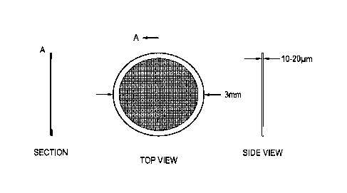
Figure 4a illustrates schematically a sectional, plan, and side elevation of
an electron
microscopy support in accordance with one embodiment. Figure 4a illustrates an
electron microscopy sample support comprising: a support element, in this
instance, a
substantially annular member together with a metal grid and a metal foil which
extends
between adjacent trusses of the metal grid. The metal foil comprises a porous
region.
The support element is configured to give structural stability to the porous
metal foil.
The porous region of the metal foil is configured to receive an electron
microscopy
sample. In some embodiments the electron microscopy support comprises a
perforated
gold foil mounted on a gold mesh grid.
Figure 4b illustrates schematically a portion of the electron microscopy
support shown
in Figure 4a in more detail. In particular, Figure 4b shows a close-up view of
a
suspended holey foil membrane on a grid. The insets of Figure 4b show the
support
structure in detail.
Figures 5a to Sc show optical and electron images of a device according to one
embodiment. The scale of each image is indicated. Figure 5a shows a
low magnification optical image of a region of a device according to one
embodiment.
The embodiment shown comprises a 3 mm gold metal mesh "grid" which is covered
in
a suspended thin foil of holey gold.
Figure 5b is a higher magnification image of the device shown in Figure 5a and
shows
an individual grid square. A regular array of holes can be seen in the thin
gold foil
between grid supports.
12
CA 02919961 2016-01-29
WO 2015/022510 PCT/GB2014/052454
Figure 5c is a transmission electron micrograph of an individual hole in the
foil of the
embodiment of Figure 5a. The hole shown contains a sample comprising 70S
ribosomes embedded in a thin layer of ice.
It will be appreciated that various parameters of a support in accordance with
aspects
and embodiments may be altered in order to construct a support suited to a
range of
electron microscopy applications of interest. In particular, parameters
including those
listed herein may be tuned to provide a support suited to a specimen of
interest:
Choice of material
In the embodiments shown in Figures 4 and 5, gold is selected to be a suitable
metal
material for both the porous metal foil and the support structure. The support
structure of the embodiment of Figure 4 and Figure 5 takes the form of a grid,
itself
mounted on an annular support. Gold is highly conductive (resistivity 2.3 [112
cm
compared to ¨140 0 jun cm for amorphous carbon) and placing the perforated
gold foil
on top of a gold grid generates a continuous electrical ground plane with no
discontinuity in the thermal expansion coefficient.
Furthermore, the number of secondary electrons generated from a gold substrate
is far
greater than from a carbon substrate, or indeed from many other candidate
metals.
The number of secondary electrons generated can be an important consideration
in the
design of a support, since secondary electrons, generated when an electron
beam of an
electron microscope hit a substrate, act to neutralize any positive surface
charge on a
specimen.
Gold has a similar mechanical stability to carbon. The Young's modulus of gold
is
comparable to that of amorphous carbon: 79 GPa for gold and ¨100 GPa for
carbon).
In contrast, gold is not subject to chemical transformation and is therefore
more stable
in an electron beam.
It will be appreciated that this combination of material properties make gold
a
particularly suited metal for the perforated foil forming the specimen
support. Other
metals having similar properties are also suitable substrates, for example,
platinum,
palladium, rhodium or hafnium.
13
CA 02919961 2016-01-29
WO 2015/022510 PCT/GB2014/052454
Thickness of gold foil
The thickness (tin Figure 4b) of the perforated metal foil layer is a tunable
parameter
of a support in accordance with aspects and embodiments described herein. The
minimum thickness of the metal foil is set by the size of the evaporated metal
grains.
Take, for example, a gold foil. The gold foil of the specimen support must be
thicker
than the gold grain to provide sufficient mechanical stability and uniform
electrical
conductivity. The thickness of the perforated metal, for example, gold, foil
also affects
the thickness of, for example, the ice which fills holes in the foil and holds
specimens in
position in those holes. The maximum metal foil thickness may be selected in
dependence upon the desired ice thickness, which in turn is set by specimen
particle
dimensions. Taking such factors into consideration gives, in one example, a
perforated
gold foil should be ¨500 A or the diameter of the specimen particle of
interest,
whichever is greater.
Aspect ratio of holes
There may be practical limitations when designing a support in relation to the
aspect
ratio of holes (t id in Figure 4) in the metal foil. Those limitations may be
dictated by,
for example, practical constraints in fabrication and the size of the field of
view of an
electron microscope of interest. The diameter (d) of the hole may, in some
embodiments, be selected to match the field of view of an electron microscope
of
interest such that the microscope electron beam can be arranged to uniformly
illuminate a hole in the metal foil, that hole containing ice in which
specimens may be
encapsulated, together with a "ring" of metal, for instance, gold,
encompassing and
encircling the edge of the perforation in the metal foil. In one embodiment,
the optimal
diameter is given by the minimal size that meets the above criteria at a
selected imaging
magnification. Larger holes may typically suffer from increased charging and
beam
induced motion since they comprise larger regions of insulating ice. For
example, for
typical imaging conditions in a modern electron microscope, at 39,000 X
magnification
and with a beam diameter of 1.2 pm, the optimal foil perforation hole size can
be
calculated to be d approximately equal to 1 pm.
Graphene devices
According to some embodiments, a sample support may further comprise a
graphene
layer. In such embodiments, a layer of graphene may be incorporated into the
support
substrate. Such a graphene layer may be arranged such that is located on top
of the
perforated metal foil. In some embodiments, the graphene layer may be arranged
to sit
between the support grid and the perforated metal foil.
14
CA 02919961 2016-01-29
WO 2015/022510 PCT/GB2014/052454
Provision of a graphene layer in the support may be such that the graphene
layer can
act to further decrease surface charge build up. That further reduction may
come as a
result of the conductive properties of graphene. According to the embodiments
in
.. which graphene is incorporated, the graphene is arranged to be in ohmic
contact with
the metal foil. The graphene may also be in ohmic contact with the support.
Provision
of a graphene layer in a support may be such that it acts to increase the
mechanical
strength of the substrate.
In a support arrangement in which a layer of graphene is provided above the
metal foil
provision of that graphene layer may be such that a substantially uniform
coating
including one or more samples or specimens for analysis may be arranged to
extend
across said graphene layer. Such an arrangement may allow control of the
thickness of
the coating layer including one or more samples. In such an arrangement, the
thickness of a coating, for example, an ice layer including protein samples,
may be
controlled independently of the thickness of the metal foil.
In a support arrangement in which a layer of graphene is provided beneath the
metal
foil, the graphene layer may be arranged such that a substance containing one
or more
samples for analysis may be supported in holes or pores in the metal film by
the
graphene.
Provision of a graphene layer in a support according to some embodiments may
be
such that the mechanical stability of the support and/or sample for analysis
is
improved.
Furthermore, selection of appropriate graphene may be such that inclusion of
such a
layer can be useful when examining biological samples, since it may allow for
the
controlled deposition of proteins on the support.
Method of production
It will be appreciated that various methods of production may be employed to
construct
a sample support in accordance with aspects and embodiments described herein.
By
way of example only, to produce one embodiment of a device in accordance with
one
embodiment, a gold grid having a suspended layer of perforated amorphous
carbon on
top can be used as a template. It will be appreciated that other types of
template can be
used, including: lacey carbon, nanoporous polycarbonate and other patterned
plastics.
CA 02919961 2016-01-29
WO 2015/022510 PCT/GB2014/052454
Gold is then evaporated onto the suspended template. The template is
subsequently
removed by exposing the device to a low energy oxygen/argon plasma. The plasma
can
be tuned such that it reacts very strongly with all carbon-containing
materials but has
no effect on the gold. Such an arrangement allows for selective removal of
substantially
all of the template layer, leaving a perforated pure gold foil directly
attached to a pure
gold mesh grid.
Figure 6 shows results of one experiment to measure gold particle motion in
vitreous
ice in example electron microscopy supports. By imaging gold particles it can
be shown
that there is reduced specimen motion when imaged on an ultra-stable sample
support
in accordance with one embodiment. The left panel of Figure 6 illustrates a
typical
motion trajectory for a specimen in ice on a typical substrate (red curve)
against a
typical trajectory for a specimen in ice on our ultra-stable grids (black
curve). The right
panel of Figure 6 shows five individual specimen trajectories determined when
using a
support in accordance with one embodiment. It can be seen that the motion is
less
than the accuracy with which the specimens can be located, that is to say,
less than one
Angstrom per time point. Note the scale on the right panel of Figure 6 is
displacement
in pixels.
Experimental Use of Support
It has been found that samples and specimens prepared on an ultra-stable
sample
supports according to one embodiment display reduced charging when placed in
an
electron beam. The "bee-swarm effect" is a fluctuation in the granularity of
images at
low magnification and is a result of surface charging. The "bee-swarm effect"
with a
typical support has been compared to the effect experienced when using an
ultra-stable
substrate according to one embodiment and it was found that the effect is
greatly
reduced, indicating that sample/specimen charging is likely to be reduced when
using a
support in accordance with aspects and embodiments described herein.
Embodiments can be designed such that during imaging an electron beam of an
electron microscope illuminates metal around the entire circumference of the
porous
hole which contains the specimen, in some cases, encapsulated in vitreous ice.
Such an
arrangement may allow for uniform generation of secondary electrons by the
metal foil.
Those secondary electrons can neutralize positive charges generated within and
on the
surface of the ice in the pore of the foil. When the electron beam is arranged
to be off-
centre with respect to the hole, the electron beam does not touch metal, for
example,
16
CA 02919961 2016-01-29
WO 2015/022510 PCT/GB2014/052454
gold, around the entire circumference of the pore and a blurring in the
resultant image
can be observed. That blurring may be indicative that secondary electrons from
the
metal, for example, gold foil, are important in neutralizing any charging
phenomenon
experienced by the specimen.
In comparison to standard EM substrates, samples and specimens prepared on
ultra-
stable supports according to some embodiments appear to have decreased
particle
motion and suffer decreased charging effects. The decreased particle motion
increases
the contrast in each resulting image due to reduced blurring. Decreased
particle
motion may be a result of, for example: high mechanical stability, reduced
force on the
ice due to reduced charge build-up, and elimination of chemical changes in the
support
which would induce stresses in a perforated foil membrane. Decreased charging
effects
may further improve the phase contrast of images by reducing charge induced
lensing
of the electron beam.
Figures 7a and 7b show a comparative example of a specimen imaged using a
support
in accordance with one embodiment. In particular, Figures 7a and 7b show 3D
electron
density maps made using the same number of images of the same protein sample
on (a)
standard holey carbon grids and (b) a support in accordance with one
embodiment.
The alpha helices of the protein sample are clearly resolved in Figure 7b as
cylindrical
regions of electron density. The diameter of apoferritin, the octahedral
protein of
molecular mass 450 kDa imaged in Figures 7a and 7b is 120 A. Alignment of a
plurality
of apoferritin images to construct a 3D image requires information at
resolutions
greater than 1/10 A. Using cryo-EM on conventional grids, it has not been
possible to
determine the 3D structure of apoferritin (Figure 7a). Using images obtained
by using a
support in accordance with one embodiment, a 3D reconstruction of apoferritin
has
been generated (Figure 7b). Such an image demonstrates that a sample support
in
accordance with aspects and embodiments described herein may be such that the
information content in electron micrographs where spatial information whose
frequency is greater than ¨1/10 A5 significantly improved.
Specimen supports in accordance with aspects and embodiments may reduce
particle
motion and/or sample charging in electron microscopy, and thus improve
information
content available from electron micrographs. Appropriately designed and
constructed
supports may lead to an increased resolution per particle and increased
accuracy of
angular assignments in 3D reconstructions of, for example, biological
specimens. This
17
CA 02919961 2016-01-29
WO 2015/022510 PCT/GB2014/052454
may enable the determination of structures of smaller and more difficult
proteins than
was previously possible using EM techniques.
Figures 8a and 8b show experimental results illustrating reduced vertical
motion of
gold grid supports in accordance with an arrangement compared to conventional
amorphous carbon grid supports. In general Figures 8a and 8b illustrate
graphically
the reduced motion of gold grids under high-energy electron irradiation when
compared to conventional amorphous carbon grids. Each point shown in the plots
of
Figure 8a and 8b represents the r.m.s. vertical displacement of a particular
hole in, for
example, the carbon substrate (upper points and lines) or gold substrate
(lower points
and lines)relative to its initial position before electron irradiation. Each
solid line is the
average displacement for multiple holes in multiple squares of one grid. It
can be seen
from Figure 8a that conventional amorphous carbon grids exhibit large degree
of
motion perpendicular to the plane of the grid under typical cryo-EM
illumination
conditions (300 keV, 16 e/ A2/s and 80K). The lower points and curves shown in
Figure 8a represent the same measurement in relation to gold grids.
Figure 8b comprises an analogous measurement and analysis performed in
relation to
amorphous carbon grid supports (upper points and lines) and a gold support
(lower
points and lines) when a typical thin layer of vitreous ice is present. It can
be seen that
the vertical motion is reduced about two-fold relative to grids without ice
and that, in
general, the motion becomes more complicated in nature. In both cases there is
on
average a 50 fold reduction in the vertical motion of the grid during the
first 16 e/ 'A2 of
irradiation for gold grids compared to use of conventional amorphous carbon
grids.
Figures 9a to 9d show experimental results illustrating average 80S ribosome
displacement from an initial position as a plot against time! electron fluence
for data
collected in ice using a variety of microscopy support structures. In
particular, Figure
9a relates to a sample supported by a continuous layer of amorphous carbon;
Figure 9b
relates to a sample without any support layer on amorphous carbon perforated
support
membranes; Figure 9c relates to a sample supported on a graphene substrate;
and
Figure 9d relates to a sample on unsupported ice on gold substrates. All plots
(Figure
9a to 9d) have the same scale. Each point (dotted lines) represents the root
mean
squared (RMS) displacement of thousands of particles from a single grid, whose
positions were measured using a five-frame running average under constant
electron
beam irradiation (300 keV; 16 e-/A2/s). The solid lines of Figures 9a to 9d
are the
linear fits to the two phases of motion. Error bars represent the standard
error of the
18
CA 02919961 2016-01-29
WO 2015/022510 PCT/GB2014/052454
mean of replicate experiments (3 separate grids in relation to Figures 9 a, b,
d and 4 for
Figure 9c).
Although illustrative embodiments of the invention have been disclosed in
detail
herein, with reference to the accompanying drawings, it is understood that the
invention is not limited to the precise embodiment and that various changes
and
modifications can be effected therein by one skilled in the art without
departing from
the scope of the invention as defined by the appended claims and their
equivalents.
The work leading to this invention has received funding from the European
Research Council under the European Union's Seventh Framework
Programme (FP7/2007-2013) / ERC grant agreement n 261151.
19