Note: Descriptions are shown in the official language in which they were submitted.
CA 02920384 2016-02-03
WO 2015/020816 PCT/US2014/048305
SYNTHETIC CHORD FOR CARDIAC VALVE REPAIR APPLICATIONS
CROSS-REFERENCE TO RELATED APPLICATIONS
Pursuant to 35 U.S.C. 119 (e), this application claims priority to the
filing
dates of: United States Provisional Patent Application Serial No. 61/948,480
filed
March 5, 2014; United States Provisional Patent Application Serial No.
61/889,331
filed October 10, 2013; and United States Provisional Patent Application
Serial No.
61/862,922 filed August 6, 2013; the disclosures of which applications are
herein
incorporated by reference.
INTRODUCTION
The mitral valve is composed of two leaflets attached to the mitral valve
annulus, which are supported at the free edge by chordae tendinae (chords)
attached to the inside wall of the left ventricle and to the papillary
muscles. However,
sometimes one or both of the valve leaflets become loose, due to loosening or
failure of one or more of these chords. The valve then prolapses, and the seal
that it
normally provides between the left atrium and left ventricle becomes
compromised,
causing the blood to flow back into the left atrium during systole.
A variety of methods have been described for placement of artificial chordae
tendineae to correct mitral valve leaflet prolapse and treat diseased mitral
valve
chordae tendineae. However, there are many technical challenges in this
surgical
procedure, especially when performed with minimally invasive techniques. The
most
common method of repairing the valves is to create synthetic chordae tendineae
from polytetrafluoroethylene (PFTE), which tendineae are fastened into place
between the papillary muscle of the heart wall and the mitral valve leaflets.
Cardiac
surgeons usually are required to perform the time-consuming process of
measuring
and cutting the necessary length of synthetic chordae tendineae material
during the
surgical procedure after they have measured the dimensions of the patient's
heart
valves. In addition, anchoring the synthetic chordae tendineae in the
papillary
muscle and securing the fasteners through the leaflets is often technically
difficult in
1
CA 02920384 2016-02-03
WO 2015/020816 PCT/US2014/048305
minimally invasive procedures, because of limitations in using 2-dimensional
video
for viewing the surgical field, limited exposure of the surgical field, and
limited
degrees of freedom using standard thoracoscopic instrumentation.
Therefore, there is considerable interest in the development of new
techniques for use in both open and minimally invasive procedures that address
the
problems of accurately and efficiently securing the valve leaflets during
cardiac
surgery.
SUMMARY
Synthetic chord devices and methods for using the same for connecting
tissues are provided. Aspects of the synthetic chord devices include a first
flexible
connector having first and second ends. Located at the first end is an
attachment
element that includes a piercing member coupled to a securing member, wherein
the
securing member transitions from a linear to a planar configuration upon
separation
of the tissue piercing member from the attachment element. A reinforcing
element is
located at the second end. The devices and methods of the invention find use
in a
variety of applications, such as cardiac valve, e.g., mitral valve, repair.
BRIEF DESCRIPTION OF THE FIGURES
FIGS. 1A and 1B provide a view of a device in accordance with an
embodiment of the invention, where the device is shown before and after
deployment, respectively.
FIG. 2 provides views of a device in accordance with an embodiment of the
invention, where the device is shown before and after deployment.
FIG. 3 provides views of a device in accordance with an embodiment of the
invention, where the device is shown before and after deployment.
FIG. 4 provides views of a device in accordance with an embodiment of the
invention, where the device is shown before and after deployment.
FIGS. 5A to 5D provide various views of a device in accordance with an
embodiment of the invention.
2
CA 02920384 2016-02-03
WO 2015/020816 PCT/US2014/048305
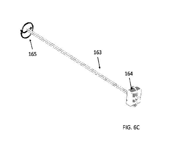
FIGS. 6A to 6D provide views of a single-arm device in an un-deployed state
in accordance with an embodiment of the invention, which FIGS. 6C and 6D
provide
views of the same device in a deployed state.
FIGS. 7A to 7D provide views of a double-arm device in an un-deployed state
in accordance with an embodiment of the invention, which FIGS. 7C and 7D
provide
views of the same device in a deployed state.
FIG. 8A provides a schematic view of the normal left side of the heart.
FIG. 8B provides a schematic view of the left side of the heart demonstrating
a ruptured chorda tendinea of the mitral valve.
FIG. 8C provides a schematic view of the left side of the heart after repair
of
the ruptured chorda tendinea of the mitral valve with embodiments of the
synthetic
chord device of the subject invention.
FIG. 8D provides a schematic view of the heart after repair of both the
ruptured chordae tendineae of the mitral valve and tricuspid valves with
embodiments of the synthetic chord device of the subject invention.
DEFINITIONS
As used herein, the term "tissue" refers to one or more aggregates of cells in
a subject (e.g., a living organism, such as a mammal, such as a human) that
have a
similar function and structure or to a plurality of different types of such
aggregates.
Tissue may include, for example, organ tissue, muscle tissue (e.g., cardiac
muscle;
smooth muscle; and/or skeletal muscle), connective tissue, nervous tissue
and/or
epithelial tissue.
The term "subject" is used interchangeably in this disclosure with the term
"patient". In certain embodiments, a subject is a "mammal" or "mammalian",
where
these terms are used broadly to describe organisms which are within the class
mammalia, including the orders carnivore (e.g., dogs and cats), rodentia
(e.g., mice,
guinea pigs, and rats), and primates (e.g., humans, chimpanzees, and monkeys).
In
some embodiments, subjects are humans. The term "humans" may include human
subjects of both genders and at any stage of development (e.g., fetal,
neonates,
infant, juvenile, adolescent, adult), where in certain embodiments the human
subject
3
CA 02920384 2016-02-03
WO 2015/020816 PCT/US2014/048305
is a juvenile, adolescent or adult. While the devices and methods described
herein
may be applied to perform a procedure on a human subject, it is to be
understood
that the subject devices and methods may also be carried out to perform a
procedure on other subjects (that is, in "non-human subjects").
The present disclosure provides embodiments of devices (e.g., a synthetic
chord device or a portion thereof, such as a flexible connector, an attachment
element, a tissue piercing member, a securing member and/or a reinforcing
element)
that are implantable. As used herein, the terms "implantable", "implanted" and
"implanting" refer or relate to the characteristic of the ability of a device
to be placed
(e.g., surgically introduced) into a physiological site (e.g., a site within
the body of a
subject) and maintained for a period of time without substantial, if any,
impairment of
function. As such, once implanted in or on a body, the devices do not
deteriorate in
terms of function, e.g., as determined by ability to perform effectively as
described
herein, for a period of 2 days or more, such as 1 week or more, 4 weeks or
more, 6
months or more, or 1 year or more, e.g., 5 years or more, up to and including
the
remaining lifetime or expected remaining lifetime of the subject or more.
Implantable
devices may also be devices that are configured (e.g., dimensioned and/or
shaped)
to fit into a physiological site (e.g., a site within the body of a subject).
For example,
in certain embodiments, an implantable device may have a longest dimension,
e.g.,
length, width or height, ranging from 0.05 mm to 150 mm, such as from 0.1 mm
to
10 mm, including from 0.5 mm to 5 mm. Implanting may also include securing an
implanted object (e.g., a prosthetic device) to one or more tissues within the
body of
the subject. Additionally, implanting may, in some instances, include all of
the
surgical procedures (e.g., cutting, suturing, sterilizing, etc.) necessary to
introduce
one or more objects into the body of a subject.
In some instances, the devices or portions thereof may be viewed as having a
proximal and distal end. The term "proximal" refers to a direction oriented
toward the
operator during use or a position (e.g., a spatial position) closer to the
operator (e.g.,
further from a subject or tissue thereof) during use (e.g., at a time when a
tissue
piercing device enters tissue). Similarly, the term "distal" refers to a
direction
oriented away from the operator during use or a position (e.g., a spatial
position)
4
CA 02920384 2016-02-03
WO 2015/020816 PCT/US2014/048305
further from the operator (e.g., closer to a subject or tissue thereof) during
use (e.g.,
at a time when a tissue piercing device enters tissue). Accordingly, the
phrase
"proximal end" refers to that end of the device that is closest to the
operator during
use, while the phrase "distal end" refers to that end of the device that is
most distant
to the operator during use.
In certain variations of the disclosed methods and associated devices, the
method, such as a method by which a synthetic cord device is used, is an open
surgical procedure. As used herein, the phrase "open surgical procedure"
refers to a
surgical procedure wherein at least one long incision (e.g., having a length
of 10 cm)
is made in the body of a subject to introduce at least one surgical instrument
and/or
visualize the surgery through the incision. In an open surgical procedure,
closure
devices, e.g., staples, sutures, etc., may be used to close at least one
incision.
In certain variations of the disclosed methods, the method is a minimally
invasive surgical procedure. As used herein, the phrase "minimally invasive
surgical
procedure" refers to a surgical procedure that is less invasive than an open
surgical
procedure. A minimally invasive surgical procedure may involve the use of
arthroscopic and/or laparoscopic devices and/or remote-control manipulation of
surgical instruments. Minimally invasive surgical procedures include
endovascular
procedures, which may be totally endovascular procedures, percutaneous
endovascular procedures, etc. Endovascular procedures are procedures in which
at
least a portion of the procedure is carried out using vascular access, e.g.,
arterial
access.
Furthermore, the definitions and descriptions provided in one or more (e.g.,
one, two, three, or four, etc.) sections of this disclosure (e.g., the
"Descriptions",
"Devices", "Methods" and/or "Kits" sections below) are equally applicable to
the
devices, methods and aspects described in the other sections.
DETAILED DESCRIPTION
Synthetic chord devices and methods for using the same for connecting
tissues are provided. Aspects of the synthetic chord devices include a first
flexible
connector having first and second ends. Located at the first end is an
attachment
5
CA 02920384 2016-02-03
WO 2015/020816 PCT/US2014/048305
element that includes a piercing member coupled to a securing member, wherein
the
securing member transitions from a linear to a planar configuration upon
separation
of the tissue piercing member from the attachment element. A reinforcing
element is
located at the second end. The devices and methods of the invention find use
in a
variety of applications, such as cardiac valve, e.g., mitral valve repair.
Before the present invention is described in greater detail, it is to be
understood that this invention is not limited to particular embodiments
described, as
such may, of course, vary. It is also to be understood that the terminology
used
herein is for the purpose of describing particular embodiments only, and is
not
intended to be limiting, since the scope of the present invention will be
limited only
by the appended claims.
Where a range of values is provided, it is understood that each intervening
value, to the tenth of the unit of the lower limit unless the context clearly
dictates
otherwise, between the upper and lower limit of that range and any other
stated or
intervening value in that stated range, is encompassed within the invention.
The
upper and lower limits of these smaller ranges may independently be included
in the
smaller ranges and are also encompassed within the invention, subject to any
specifically excluded limit in the stated range. Where the stated range
includes one
or both of the limits, ranges excluding either or both of those included
limits are also
included in the invention.
Certain ranges are presented herein with numerical values being preceded by
the term "about." The term "about" is used herein to provide literal support
for the
exact number that it precedes, as well as a number that is near to or
approximately
the number that the term precedes. In determining whether a number is near to
or
approximately a specifically recited number, the near or approximating
unrecited
number may be a number which, in the context in which it is presented,
provides the
substantial equivalent of the specifically recited number.
Unless defined otherwise, all technical and scientific terms used herein have
the same meaning as commonly understood by one of ordinary skill in the art to
which this invention belongs. Although any methods and materials similar or
6
CA 02920384 2016-02-03
WO 2015/020816 PCT/US2014/048305
equivalent to those described herein can also be used in the practice or
testing of
the present invention, representative illustrative methods and materials are
now
described.
All publications and patents cited in this specification are herein
incorporated
by reference as if each individual publication or patent were specifically and
individually indicated to be incorporated by reference and are incorporated
herein by
reference to disclose and describe the methods and/or materials in connection
with
which the publications are cited. The citation of any publication is for its
disclosure
prior to the filing date and should not be construed as an admission that the
present
invention is not entitled to antedate such publication by virtue of prior
invention.
Further, the dates of publication provided may be different from the actual
publication dates which may need to be independently confirmed.
It is noted that, as used herein and in the appended claims, the singular
forms
"a", "an", and "the" include plural referents unless the context clearly
dictates
otherwise. It is further noted that the claims may be drafted to exclude any
optional
element. As such, this statement is intended to serve as antecedent basis for
use of
such exclusive terminology as "solely," "only" and the like in connection with
the
recitation of claim elements, or use of a "negative" limitation.
Additionally, certain embodiments of the disclosed devices and/or associated
methods can be represented by drawings which may be included in this
application.
Embodiments of the devices and their specific spatial characteristics and/or
abilities
include those shown or substantially shown in the drawings or which are
reasonably
inferable from the drawings. Such characteristics include, for example, one or
more
(e.g., one, two, three, four, five, six, seven, eight, nine, or ten, etc.) of:
symmetries
about a plane (e.g., a cross-sectional plane) or axis (e.g., an axis of
symmetry),
edges, peripheries, surfaces, specific orientations (e.g., proximal; distal),
and/or
numbers (e.g., three surfaces; four surfaces), or any combinations thereof.
Such
spatial characteristics also include, for example, the lack (e.g., specific
absence of)
one or more (e.g., one, two, three, four, five, six, seven, eight, nine, or
ten, etc.) of:
symmetries about a plane (e.g., a cross-sectional plane) or axis (e.g., an
axis of
7
CA 02920384 2016-02-03
WO 2015/020816 PCT/US2014/048305
symmetry), edges, peripheries, surfaces, specific orientations (e.g.,
proximal), and/or
numbers (e.g., three surfaces), or any combinations thereof.
As will be apparent to those of skill in the art upon reading this disclosure,
each of the individual embodiments described and illustrated herein has
discrete
components and features which may be readily separated from or combined with
the
features of any of the other several embodiments without departing from the
scope
or spirit of the present invention. Any recited method can be carried out in
the order
of events recited or in any other order which is logically possible.
DEVICES
Synthetic chord devices as described herein are devices that are configured
to connect or align tissues, or connect tissue to a prosthesis, or a
combination
thereof. The devices may be used in endovascular, minimally invasive surgical,
open
surgical, or other interventional procedures. Devices as described herein may
be
configured to secure a valve leaflet, such as a mitral valve leaflet or
tricuspid valve
leaflet, to a papillary muscle. When an aspect (e.g., a tissue, such as a
valve leaflet)
is secured, it may, for example, be retained at the same position or
substantially at
the same position (e.g., a position within the body of a subject) for a time
period,
such as a for a period of days, weeks, months, years and/or for at least the
remaining lifetime of a subject.
Synthetic chord devices as described herein include a flexible connector
(e.g.,
a first flexible connector, such as a flexible cord). The flexible connector
has a first
end and a second end. Embodiments of the synthetic chord devices include an
attachment element at the first end of the first flexible connector.
Attachment
elements as described herein include a tissue piercing member coupled to a
securing member. In some embodiments, the securing member attaches the first
end of the flexible connector to a tissue location (e.g., a first tissue),
following
deployment of the securing member, e.g., as described in greater detail below.
A
portion of the flexible connector can be configured to be secured to a second
tissue
location. In some instances, the flexible connector is secured to the second
tissue by
a reinforcing element at the second end of the flexible connector. Various
aspects of
8
CA 02920384 2016-02-03
WO 2015/020816 PCT/US2014/048305
the embodiments of the devices, including the flexible connector, the
attachment
element (including the tissue piercing member and securing member) and the
reinforcing element, are now described in greater detail below.
Flexible Connector
A synthetic chord device of certain embodiments of the subject invention
includes a synthetic, or artificial, flexible connector, such as a flexible
cord, line,
filament, etc., which has an attachment element at one end of the connector
for
attaching the connector to a tissue. In some embodiments, the flexible
connector is
configured to be attached to a prosthesis, or to a device that substitutes for
or
supplements a missing or defective part of the body, e.g., a synthetic cardiac
valve,
or a porcine valve. In some embodiments, a synthetic chord is configured to be
used
as a synthetic chorda tendinea for use in repair of a cardiac valve, e.g., the
mitral
valve.
The flexible connector (e.g., the first flexible connector) element of the
subject
invention is a flexible elongated structure having a first end and a second
end. The
flexible connector may be made up of a single line or filament, e.g., thread,
or two or
more such lines, which may, where desired, be twisted about each other, e.g.,
as
present in a yarn. In certain embodiments, the first and second ends of the
first
flexible connector are not connected (e.g., do not form a continuous body of
material
or adjoin). As such, the first flexible connector does not form (e.g., is not
shaped as)
a loop (e.g., a continuous loop of one or more materials). In yet other
instances, e.g.,
as described in greater detail below, the flexible connector may be made up of
two
filaments which are connected at the proximal and distal ends. In some
embodiments, the flexible connector does not include a knot. By "knot" as used
herein is meant an interlacement (e.g., looping) or entanglement of portions
of a
body (e.g., a flexible connector) that forms a knob or lump. In some aspects,
a knot
prevents a body (e.g., a longitudinal, round body, such as a cord) having the
knot
from traveling through an opening in an aspect having an area that is slightly
larger
than the cross sectional area of the body. In some aspects, a knot is created
by tying
(e.g., purposefully tying) a body into an interlaced configuration.
9
CA 02920384 2016-02-03
WO 2015/020816 PCT/US2014/048305
The first flexible connector element has a length (e.g., length between the
first
and second end) suitable for extending from a first tissue to a second tissue,
such
that the flexible connector may be secured to both the first and the second
tissue. In
some embodiments, the flexible connector element has a length suitable for
extending from a first tissue (e.g., a mitral valve leaflet) to where it is
secured to a
second tissue (e.g., a papillary muscle). The length of the first flexible
connector may
vary, and in some instances ranges from 5 mm to 100 mm, such as from 5 mm to
25
mm, including 10 mm to 20 mm. In some embodiments, the first or second end of
the first flexible connector can be secured to a prosthesis, or other device
that
substitutes for or supplements a missing or defective part of the body, e.g.,
a
synthetic cardiac valve, or a porcine valve, which is located at the target
tissue
location.
In certain embodiments, the first flexible connector is constructed of one or
more materials suitable for use in the body and that can be used in the
methods of
the subject invention, e.g., attaching a valve leaflet to the underlying
cardiac tissue
(e.g., attaching for an extended period of time, such as for the lifetime of
the subject,
without breaking). The flexible connector (e.g., the first flexible connector)
can be
made of a variety of materials. Such materials may be flexible materials. By
"flexible", as used herein is meant pliable or capable of being bent or flexed
repeatedly (e.g., bent or flexed with a force exerted by a human hand or other
body
part) without damage (e.g., physical deterioration). A flexible material may
be a
material that remains able to perform intended function (e.g., repeatedly
flexing) by
remaining pliable for at least the expected lifetime or useful lifetime of the
aspect
which the material is included in. In some embodiments, the flexible connector
may
include biocompatible materials. The phrase "biocompatible materials" are
materials
that can be placed on or in living tissue for an extended period of time, such
as for a
period of 2 days or more, such as 1 week or more, 4 weeks or more, 6 months or
more, or 1 year or more, e.g., 5 years or more, up to and including the
remaining
lifetime or expected remaining lifetime of the subject or more, and not cause
a
significant adverse (e.g., detrimental to health) reaction (e.g., an immune
response)
in the tissue or the associated organism.
CA 02920384 2016-02-03
WO 2015/020816 PCT/US2014/048305
Biocompatible materials, as included in the subject devices, can include any
suitable biocompatible material, which material may or may not be
biodegradable.
Biocompatible materials of the subject devices, in some instances, are
polymeric
materials (e.g., materials having one or more polymers) and/or metallic
materials.
Such materials may have characteristics of flexibility and/or high strength
(e.g., able
to withstand significant force, such as a force exerted on it by a tissue
within a
human body, without breaking and/or resistant to wear) and/or high fatigue
resistance (e.g., able to retain its physical properties for long periods of
time
regardless of the amount of use or environment). Biocompatible materials may
also
include any of the shape memory materials listed herein, as described in
greater
detail below.
In some embodiments, biocompatible polymeric materials of the subject
devices, include, but are not limited to: polytetrafluoroethene or
polytetrafluoroethylene (PFTE), including expanded polytetrafluoroethylene (e-
PFTE), polyester (DacronTm), nylon, polypropylene, polyethylene, high-density
polyethylene (HDPE), polyurethane, and combinations or mixtures thereof.
Similarly,
in certain embodiments, biocompatible metallic materials of the subject
devices,
include, but are not limited to: stainless steel, titanium, a nickel-titanium
(NiTi) alloy
(e.g., nitinol), a nickel-cobalt alloy, such as ELGILOY cobalt-chromium-
nickel alloy,
tantalum, and combinations or mixtures thereof.
In certain embodiments, an active agent may be included in the composition
of a biocompatible material, such as a polymeric material. As used herein, the
phrase "active agent" refers to one or more chemical substances that, when
administered to (e.g., placed in contact with or ingested by) a human, have
one or
more physiological effects. In some embodiments, the one or more active agents
include an antithrombotic substance and/or an antibiotic substance and/or an
anti-
inflammatory (e.g., a substance that reduces or prevents inflammation). In
various
embodiments, a first flexible connector may be coated with a polymer, such as
a
polymer that releases one or more active agents (e.g., an anticoagulant that
thereby
reduces the risk of thrombus formation).
11
CA 02920384 2016-02-03
WO 2015/020816 PCT/US2014/048305
The cross-sectional configuration of the first flexible connector can be any
suitable shape, such as round, oval, rectangular, square, etc. In some
instances, the
first flexible connector may have a flattened cross-sectional shape, such as a
"ribbon" shape. In other embodiments, the flexible connector may be a
combination
of shapes, such as for example, a flexible connector that is round on two
sides with
a flat surface on the opposing two sides. In some embodiments the entire
flexible
connector has the same shape, and in other embodiments, at least a portion of
the
flexible connector may have a different shape, e.g., a ribbon configuration,
or at least
a portion of the connector that is flattened, or has a flat surface.
In some embodiments, the greatest outer diameter of the flexible connector
ranges from 0.1 mm to 1.0 mm, such as from 0.1 mm to 0.5 mm, or 0.15 mm to
0.25
mm. In some embodiments, the entire flexible connector has the same diameter.
In
other embodiments, at least a portion of the connector has a different
diameter, e.g.,
a smaller diameter. In some embodiments, at least a portion of the connector
may
is
have both a different configuration and a different diameter, e.g., a portion
of the
connector may have a flat surface, where the portion of the connector having a
flat
surface has a largest outer diameter larger than the remainder of the
connector.
A portion of the flexible connector (e.g., the first flexible connector) at
the first
end and/or second end is configured to be secured to tissue, such as cardiac
tissue
located below a cardiac valve leaflet. In some embodiments, a portion of the
flexible
connector at the first end and/or second end can be secured to a prosthesis,
or other
device that substitutes for or supplements a missing or defective part of the
body.
The portion of the flexible connector at the first end and/or second end that
is
configured to be secured to tissue can have the same shape and diameter as the
remainder of the flexible connector, or in some embodiments it may have a
different
shape or diameter as the remainder of the flexible connector, as in the
embodiments
discussed above. For example, the portion of the connector at the first end
and/or
second end that is configured to be attached to a tissue (e.g., a first or
second
tissue) may be flattened, or have a smaller or larger diameter.
12
CA 02920384 2016-02-03
WO 2015/020816 PCT/US2014/048305
Attachment Element
The synthetic chord devices further include an attachment element located at
an end (e.g., the first end) of a flexible connector. The attachment element
is
configured to attach a flexible connector (e.g., a first flexible connector),
such as
those described above, to a tissue, e.g., a cardiac valve leaflet, or
prosthesis, as
desired. In some instances, an attachment element is an element that includes
a
tissue piercing member and a securing member. The attachment element may be
configured such that the tissue piercing member is attached to the securing
member
directly (e.g., the tissue piercing member is retained in direct contact with
the tissue
io securing member) or, in some embodiments, with a second flexible
connector (e.g.,
a second flexible member, e.g., which may be in the form of a line, filament,
hypotube, etc., such as described in greater detail below).
A tissue piercing member may, in some embodiments, be release-ably
coupled to a securing member. In other embodiments, the attachment element may
is be configured such that a tissue piercing member is attached to a second
flexible
connector, which in turn is release-ably coupled to the securing member. The
coupling between the second flexible connector (and, thus, the tissue piercing
member) and the securing member may be configured to actuate a configuration
change of the securing member upon release of the second flexible connector
20 (and/or piercing member), as discussed below. For example, the coupling
may hold
a compression spring (which is positioned around a securing member) in a
compressed state to brace the securing member open and release-ably lock or
secure the securing member to the second flexible connector (and/or or
piercing
member). In some embodiments, the attachment element can be secured to a
25 prosthesis, or other device that substitutes for or supplements a
missing or defective
part of the body.
A second flexible connector as discussed herein, can be formed from any
suitable biocompatible material such as cotton, nylon, polyester,
polypropylene,
polyglycolic acid, polylactide, lactic acid, trimethlylene carbonate,
polycaprolactone,
30 or polydiaxanone or copolymers or homopolymers thereof, or a metal
alloy, such as
Nitinol shape memory or stainless steel, a polymeric material, or any other
suitable
13
CA 02920384 2016-02-03
WO 2015/020816 PCT/US2014/048305
material, such as the biocompatible materials listed herein, including the
shape
memory materials listed herein, and equivalents thereof. The material of the
second
flexible connector may be non-stretchable or stretchable, and have various
cross-
sectional diameters. In some embodiments, the second flexible connector does
not
include a knot. In some embodiments, the second flexible connector does not
form a
loop (e.g., does not form a continuous band of material). In some instances,
the
second flexible connector may have a cross-sectional diameter ranging from 0.1
mm
to 1.0 mm. The diameter of a second flexible connector will vary depending on
the
specific application. Additionally, the length of the second flexible
connector may
vary, and in some instances range from 5 mm to 100 mm, such as from 5 mm to 25
mm, or 10 mm to 20 mm. A second flexible connector may have a different length
(e.g., shorter or longer) than the length of the first flexible connector or
the same
length as the first flexible connector.
The second flexible connector may be attached to the piercing member by
crimping or swaging or otherwise attaching the piercing member or needle onto
the
second flexible connector, gluing the second flexible connector to the
piercing
member or needle, or any other suitable attachment method. Second flexible
connectors can also have various cross-sectional shapes, such as round, oval,
etc.
Additionally, second flexible connectors, in certain variations, may have any
of the
physical characteristics (e.g., compositions and/or dimensions, etc.) set
forth for any
of the connectors described herein (e.g., the first flexible connectors) or
any
combination of such characteristics.
A tissue piercing member is any device that can be used to pierce through
tissue, e.g., a needle. In some embodiments, the piercing member can also be
used
to pierce a prosthesis, e.g., a synthetic valve. Piercing members of interest
include
needles, wires, etc. Needles of interest include conventional cardiac surgical
needles and equivalents thereof. Suitable surgical needles can be manufactured
from stainless steel, a stainless steel alloy, or any other suitable material,
such as a
polymeric material. The material can also have special coatings and sharpening
methods that facilitate atraumatic tissue penetration. The shapes and sizes of
the
surgical needles can vary with the type and design of the needle. In some
14
CA 02920384 2016-02-03
WO 2015/020816 PCT/US2014/048305
embodiments, the needles may be permanently "swaged" or attached to a
fastening
cord or material. In some embodiments, the fastening cord or material may be
designed to come off the needle with a sharp straight tug (e.g., "pop-offs").
Suitable lengths for the piercing members that are in the form of a needle can
range in some embodiments from 5 mm to 50 mm, such as from 5 mm to 45 mm,
incuding 5 mm to 25 mm. The diameter of the piercing member ranges in some
embodiments from 0.05 mm to 2.0 mm, e.g., 0.05 to 1.0 mm, such as from 0.05 mm
to 0.5 mm, including 0.1 mm to 0.5 mm. In some embodiments, the diameter of at
least a portion of a piercing member is greater than the diameter of an
attached
second flexible connector and/or attached securing member, coupled so that the
attached second flexible connector and/or attached securing member can easily
be
pulled through an opening formed in a tissue (or other material) by the
piercing
member, e.g., the needle. The distal end or tip of the piercing member can be
rigid
to facilitate penetration of tissue. The remaining length of the piercing
member can
be rigid or flexible to facilitate movement of the piercing member through the
tissue
or other material. The piercing member tips can have various configurations
and
can, for example, have a piercing point, tapered point, or have a cutting or
reverse
cutting configuration for example, and have a shape such as conical, tapered,
or
grounded to attain a three or four facet tip. Piercing members can have any
suitable
shape or radius of curvature. Piercing members can have any suitable cross-
sectional shape that may vary in different sections of the needle, e.g.,
round,
rectangular, etc. In some embodiments, the piercing member can also be
integrally
formed with the second flexible connector (e.g., both piercing member and
second
flexible connector formed of the same material). Also, in some embodiments,
the
subject devices include only one tissue piercing member.
The attachment elements of the subject devices also include a securing
member. A securing member is any device that can be used in a surgical,
endovascular, or other interventional procedure that can be used to secure a
flexible
connector, (e.g., a first flexible connector, and/or an artificial mitral
valve chorda
tendinea). In some embodiments, the disclosed devices include only one
securing
member. In some embodiments, the securing member of a synthetic chord device
is
CA 02920384 2016-02-03
WO 2015/020816 PCT/US2014/048305
located at, and/or attached to (e.g., release-ably attached to), the first end
of a first
flexible connector of the device. By "secure" is meant that the securing
member
provides for stable association of the end of the flexible connector to the
target
tissue location, e.g., mitral valve leaflet. By "stable association" is meant
that the end
of the flexible connector is substantially if not completely fixed relative to
the tissue
location of interest such that when the end of the flexible connector moves,
the
target tissue location to which it is secured by the deployed securing member
also
moves.
An aspect of the securing members as described herein is that the securing
member transitions from a linear to a planar configuration upon separation of
the
tissue piercing member component (which may be just the tissue piercing member
or the tissue piercing member and a second flexible connector, e.g., as
described
above) from the attachment element. As such, following initial placement of
the
synthetic chord device at the desired anatomical location, separation of the
tissue
piercing member (and second flexible connector, if present) from the securing
member results in a change in configuration of the securing member from a
linear to
planar configuration.
In some instances, deployment of the securing member results in an increase
of the amount of a theoretical plane that is occupied by the securing member,
where
the theoretical plane is a theoretical plane at least substantially
perpendicular to the
longitudinal axis of the flexible connector. The at least substantially
perpendicular
theoretical plane is a theoretical plane that is completely perpendicular to
the
longitudinal axis of the flexible connector, or at least closer to
perpendicular than
parallel, and in some instances is one that is at an angle ranging from 75 to
90
relative to the longitudinal axis of the flexible connector. The increase in
the amount
of the theoretical plane that is occupied by the securing element upon
deployment
may vary, and in some instances the magnitude of the increase is 5% or more,
such
as 10% or more, including 25% or more, e.g., 50% or more, up to 100% or more,
and in some instances ranges from 5 to 5000%, such as 10 to 2500%.
Upon deployment, the planar configuration may be configured to cover a
surface of the tissue sufficient to secure the first end of the flexible
connector to the
16
CA 02920384 2016-02-03
WO 2015/020816 PCT/US2014/048305
tissue, e.g., such that the first end can no longer be pulled through the
tissue via the
tissue passageway occupied by the first end of the flexible connector. In some
instances, the surface area of the tissue covered by the securing member upon
deployment into a planar configuration ranges from 0.5 mm2 to 50 mm2, such as
2
mm2 to 25 mm2, e.g., 5 mm2 to 20 mm2.
In some instances, the securing member has a low-profile upon deployment.
By "low-profile" is meant that the top of the securing member when deployed
does is
not located at a substantial height relative to the surface of the target
tissue to which
it is secured. While the height of a given low profile securing element may
vary, in
some instances the height ranges from 0.5 to 5 mm, such as .05 to 2.5 mm,
e.g., 1
to 2 mm, above the surface of the target tissue to which it is secured.
In some embodiments, the pre-separation linear configuration is one that
lacks a secondary structure, such that it appears in only a single location,
e.g., as a
small circle or dot (e.g., having a longest cross-sectional dimension (such as
a
diameter) ranging in some instances from 0.1 mm to 1.0 mm), in any cross-
sectional
plane passing through the securing member along the length of the securing
member. As such, the pre-separation linear configuration may be viewed as a
one-
dimensional configuration. The post-separation planar configuration is one in
which
the securing member has a secondary configuration, such that there exists one
or
more cross-sectional planes passing through the securing member along the
length
of the securing member where the securing member is present at two or more
locations. As such, the post-separation planar configuration may be viewed as
a
two- or three-dimensional configuration, depending on the particular
embodiment.
The securing member may assume a variety of different planar configurations.
These configurations may include any number of different curvilinear
configurations,
including but not limited to serpentine configurations, spiral (e.g., disc-
shaped)
configurations, etc. The area defined by the planar configuration may vary so
long as
it is sufficient to secure the end of the first flexible member to the tissue
location of
interest, and in some instances ranges from 0.5 mm2 to 50 mm2, such as 2 mm2
to
25 mm2, e.g., 5 mm2 to 20 mm2, and in some embodiments ranges from 0.5 to 25
mm2, such as 1 to 20 mm2, including 1 to 10 mm2.
17
CA 02920384 2016-02-03
WO 2015/020816 PCT/US2014/048305
In those instances where the post-separation planar configuration is a spiral
configuration, the number of turns made in the spiral may vary. While the
number
turns that the spiral may make in the deployed configuration may vary, in some
instances the number of turns ranges from 0.5 to 10, such as 1 to 7, e.g., 1
to 6. In
some instances, the spiral makes 1.5 turns.
In some instances, the securing member further includes one or more
features that serve to maintain the planar, e.g., spiral, configuration. While
these
planar maintenance features may vary, maintenance features of interest
include, but
are not limited to: one or more eyelets, one or more flattened portions of the
spiral,
e.g., where the diameter of the material making up the spiral is varied, etc.
In yet other embodiments, the pre-separation linear configuration is one that
transitions upon separation and deployment from: (a) a first configuration in
which it
has a longitudinal axis that is at least substantially parallel to the
longitudinal axis of
the flexible connector (i.e., a longitudinal axis that is substantially if not
completely
parallel with the longitudinal axis of the flexible connector) to (b) a second
configuration where it has a longitudinal axis that is at least substantially
perpendicular (i.e., is substantially if not completely perpendicular) to the
longitudinal
axis of the flexible connector. An example of such a configuration is a bar
shaped
securing member which is connected to the flexible connector in a manner
sufficient
to provide for the desired transition from first to second configuration upon
deployment. While dimensions of bar shaped securing members may vary, in some
instances the bars have a length ranging from 1 to 15 mm, such as 2 to 10 mm,
e.g.,
3 to 5 mm, a width ranging from 0.2 to 5 mm, such as 0.25 to 2.5 mm, e.g., 0.5
to 1
mm and a height ranging from 0.2 to 5 mm, such as 0.25 to 2.5 mm, e.g., 0.5 to
1
mm.
As discussed above, the securing member may be release-ably coupled to a
tissue piercing member, where release of the tissue piercing member from the
securing member causes the securing member to transition from a linear to
planar
configuration, e.g., as described above. In some embodiments, a second
flexible
connector is provided between a tissue piercing member of a device and a
securing
member. In such a configuration, the securing member and tissue piercing
member
18
CA 02920384 2016-02-03
WO 2015/020816 PCT/US2014/048305
of an attachment element of the device are separated from each other by the
second
flexible connector. Such a configuration may, for example, facilitate
threading the
securing member. In some embodiments, the securing member may secure the first
flexible connector without piercing the adjacent tissue, e.g., in the same
manner as a
surgical knot prevents a suture from pulling back through a tissue. In other
embodiments, the securing member may secure the first flexible connector by at
least partially piercing the adjacent tissue.
Separation of the tissue piercing member from the securing member may be
achieved using any convenient protocol. For example, the tissue piercing
member
may be separated from the securing member using shears, a scalpel or other
convenient cutting device, as desired.
As such, the tissue piercing member and tissue securing member are joined
to each other in operative relationship, such that when the tissue piercing
member is
separated from the securing member upon positioning of the securing member at
the desired anatomical location, the securing member assumes the planar
configuration. In some instances, the securing member and tissue piercing
member
are connected to each other in a way such that separation of the two members
may
done in a manner that minimizes, if not eliminates, exposure of metal that can
leach
into the circulatory system of the subject. For example, the two members may
be
associated with each other via an interlocking structure that maybe disrupted
without
cutting following placement in order to deploy the securing member. For
example,
mating cutout structures at the joining ends of the tissue piercing (or second
flexible
connector) and the securing member may be present, which may be separated from
each other without cutting in order to deploy the securing member.
The securing member may be retained in its linear configuration by one or
more mechanical restraining devices, such as a body of material on or within
the
securing member. For example, a removable sheath may cover the mated structure
of the securing member and tissue piercing member (or intervening second
flexible
member) which sheath, upon removal, release the securing member into its
planar,
deployed state, e.g., as described in greater detail below. Since the securing
member is biased to remain in a planar configuration, when the one or more
19
CA 02920384 2016-02-03
WO 2015/020816 PCT/US2014/048305
mechanical restraining devices are removed from the securing member upon
separation of the tissue piercing member therefrom, the securing member
transitions
from a linear configuration to a planar configuration. The securing member may
be
attached to the flexible connector using any convenient approach, e.g., by a
loop of
the flexible connector through a receiving hold of the securing member, by a
clip
attachment, or by any other convenient connector.
Devices as described herein and portions thereof (e.g., securing members)
may be fabricated from any convenient material or combination of materials.
Materials of interest include, but are not limited to: polymeric materials,
e.g., plastics,
such as polytetrafluoroethene or polytetrafluoroethylene (PFTE), including
expanded
polytetrafluoroethylene (e-PFTE), polyester (DacronTM), nylon, polypropylene,
polyethylene, high-density polyethylene (HDPE), polyurethane, etc., metals and
metal alloys, e.g., titanium, chromium, stainless steel, etc., and the like.
In some
embodiments, the devices include on or more components (e.g., securing
members)
made of a shape memory material. Shape memory materials are materials that
exhibit the shape memory effect, where the materials that have a temperature
induced phase change, e.g., a material that if deformed when cool, returns to
its
"undeformed", or original, shape when warmed, e.g., to body temperature. Where
desired, the shape memory material may be one with a transformation
temperature
suitable for use with a stopped heart condition where cold cardioplegia has
been
injected for temporary paralysis of the heart tissue (e.g., temperatures as
low as 8-
10 degrees Celsius). The shape memory material may also be heat activated, or
a
combination of heat activation and pseudoelastic properties may be used. Shape
memory materials of interest include shape memory metal alloys, such as alloys
of
nickel (e.g., nickel titanium alloy (nitinol), nickel cobalt alloys (e.g.,
ELGILOY cobalt-
chromium-nickel alloy, etc.), zinc, copper (e.g., CuZnAl), gold, iron, etc.
Also of
interest are non-metallic materials that exhibit shaper memory qualities,
e.g., shape
memory plastics, etc.
20
CA 02920384 2016-02-03
WO 2015/020816 PCT/US2014/048305
Reinforcing Element
The portion of the first flexible connector at the end (e.g., the second end)
that
is configured to be secured to tissue can include a reinforcing element (e.g.,
a
reinforcing member) attached thereto. A reinforcing element is a member that
disperses the force of the securing flexible connector over a larger surface
area. The
area over which the force is dispersed by the reinforcing element may vary so
long
as it is sufficient to secure the second end of the flexible connector to the
tissue
location of interest (e.g., papillary muscle), and in some instances ranges
from 0.5
mm2 to 50 mm2, such as 2 mm2 to 25 mm2, e.g., 5 mm2 to 20 mm2, and in some
embodiments ranges from 0.5 to 25 mm2, such as 1 to 20 mm2, including 1 to 10
M
2
rn .
In various embodiments, the reinforcing element is integral with the first
flexible connector. The term "integral," as used herein, refers to the
characteristic of
being integrated with or composed of a continuous piece of one or more
materials as
another aspect. For example, one integral aspect may not be separated from
another integral aspect by a particular adjoining surface.
In some embodiments, the reinforcing element is a separate element (e.g.,
composed of a body, such as a body of material, that is a different body than
that of
the first flexible connector) than the flexible connector and is attached to
the first
flexible connector. In embodiments in which the reinforcing element is a
separate
element from the first flexible connector, the reinforcing element includes at
least
one surface that may abut at least one surface of the first flexible
connector. In
embodiments in which the reinforcing element is a separate element from the
first
flexible connector, the reinforcing element may be moved with respect to
(e.g.,
toward, away from, or along) the first flexible connector.
In some embodiments of the subject devices in which the reinforcing element
is a separate element than the first flexible connector, the reinforcing
element can be
a pledget. Pledgets are generally buttressing or cushioning pads through which
a
flexible connector (e.g., a flexible cord) can be threaded, in order to
prevent the
flexible connector from cutting into the tissue. The reinforcing element may
include
a top surface and a bottom surface, and can be configured in a variety of
sizes and
21
CA 02920384 2016-02-03
WO 2015/020816 PCT/US2014/048305
shapes, including rectangular, circular, elliptical, etc. For example, in
certain
embodiments the length of the reinforcing element ranges from 1 mm to 10 mm,
such as from 1 mm to 8 mm, or 1 mm to 5 mm. The width of the reinforcing
element
in some cases ranges from 1 mm to 10 mm, such as from 1 mm to 8 mm, or 1 mm
to 5 mm. In some embodiments, the thickness of the reinforcing element ranges
from 0.1 mm to 2 mm, such as from 0.1 mm to 1.0 mm, or 0.1 mm to 0.5 mm.
A reinforcing element can be made of any suitable material (e.g., a
biocompatible material). Such a material may be a flexible or rigid material.
By
"rigid", as used herein is meant non-pliable or not capable of being bent or
flexed
(e.g., bent or flexed with a force exerted by a human hand or other body part)
without sustaining damage. A rigid material may be a material that remains
able to
perform its intended function (e.g., remaining in a substantially fixed
position) by
remaining stiff (e.g., resistant to force exerted on it by a human hand or
other body
part) for at least the expected lifetime or useful lifetime of the aspect in
which the
material is included. In particular embodiments, reinforcing elements are
composed
of one or more materials that are rigid or otherwise strong enough to resist
pull-
through by the flexible connector to which they are mounted. In some
embodiments,
a reinforcing element is made of a sufficiently soft and flexible material to
effectively
prevent damage to the tissue, e.g., a papillary muscle. In some embodiments,
reinforcing elements are composed of one or more materials that are pierce-
able by
a needle (e.g., a needle advanced through the material by a human hand and
with
the force normally exerted by a human hand in pushing a needle through a
material).
Reinforcing elements may be composed of biocompatible polymers and/or
metals. In various embodiments, reinforcing elements include fabrics such as
felt
(e.g., polyester felt) and/or polyester. In some embodiments, reinforcing
elements
include polytetrafluoroethylene, polytetrafluoroethylene(PTFE), expanded PTFE,
or
any of the other materials (e.g., biocompatible materials) listed herein, or
any
combinations thereof. In certain embodiments, an active agent is included in
the
composition of a biocompatible material of the reinforcing element. In some
embodiments, the one or more active agents include an antithrombotic substance
and/or an antibiotic substance and/or an anti-inflammatory (e.g., a substance
that
22
CA 02920384 2016-02-03
WO 2015/020816 PCT/US2014/048305
reduces or prevents inflammation). In various embodiments, a reinforcing
element
may be coated with a polymer, such as a polymer that releases one or more
active
agents (e.g., an anticoagulant that thereby reduces the risk of thrombus
formation).
In some embodiments, the reinforcing element does not include a tissue
piercing
member (e.g., a needle).
In addition, the reinforcing element can include one or more (e.g., one, two,
three, four, etc.) openings through which the flexible connector element may
pass. In
other embodiments, the flexible connector is attached to the reinforcing
element
without passing through an opening, e.g., the flexible connector has been
pulled
io through with a needle. In some embodiments, the reinforcing element is
mounted
such that it is substantially fixed (e.g., adhesively attached and/or tied) in
a position
on the flexible connector. For example, the reinforcing element can be sewn,
or
glued, or fused in any suitable manner so that it is fixed in position on the
flexible
connector, e.g., fixed in position at or substantially at the first or second
ends of the
is flexible connector. In other embodiments, the reinforcing element is
mounted such
that it is slidably mounted on a flexible connector. By "slidably" is meant
that the
reinforcing element is attached to the flexible connector so that it is secure
yet it is
possible to move the reinforcing element along at least part of the length of
the
connector. For example, a flexible connector can have a reinforcing element
(e.g., a
20 pledget) initially positioned halfway between the first and second ends
of the flexible
connector. In using the synthetic chord device, it may be desirable to move
the
reinforcing element to a position closer to the first or second end before
securing the
reinforcing element to a tissue.
In some instances, the reinforcing element has a structure that is analogous
25 to a securing member of the device, e.g., as described above. As such,
reinforcing
elements may be ones that transition from a linear to a planar configuration
upon
deployment. As such, prior to or following placement of the second end of the
flexible connector at the target tissue site, a change in configuration of the
reinforcing element from a linear to planar configuration occurs.
30 In some instances, deployment of the reinforcing element results in an
increase of the amount of a theoretical plane that is occupied by the
reinforcing
23
CA 02920384 2016-02-03
WO 2015/020816 PCT/US2014/048305
element, where the theoretical plane is a theoretical plane at least
substantially
perpendicular to the longitudinal axis of the flexible connector. The at least
substantially perpendicular theoretical plane is a theoretical plane that is
completely
perpendicular to the longitudinal axis of the flexible connector, or at least
closer to
perpendicular than parallel, and in some instances is one that is at an angle
ranging
from 75 to 90 relative to the longitudinal axis of the flexible connector.
The amount
of the theoretical plane occupied by the reinforcing element that is increased
upon
deployment may vary, and in some instances the magnitude of the increase is 5%
or
more, such as 10% or more, including 25% or more, e.g., 50% or more, up to
100%
or more, and in some instances ranges from 5 to 5000%, such as 10 to 2500%.
Upon deployment, the planar configuration may be configured to cover a
surface of the tissue sufficient to secure the second end of the flexible
connector to
the target tissue, e.g., such that the second end can no longer be pulled
through the
tissue via the tissue passageway occupied by the second end of the flexible
connector. In some instances, the surface area of the tissue covered by the
reinforcing element upon deployment into a planar configuration ranges from
0.5
mm2 to 50 mm2, such as 2 mm2 to 25 mm2, e.g., 5 mm2 to 20 mm2.
In some instances, the reinforcing element has a low-profile upon
deployment. By "low-profile" is meant that the top of the reinforcing element
when
deployed is not located at a substantial height relative to the surface of the
target
tissue to which it is secured. While the height of a given low profile
reinforcing
element may vary, in some instances the height ranges from 0.5 to 5 mm, such
as .05 to 2.5 mm, e.g., 1 to 2 mm, above the surface of the target tissue to
which it is
secured.
In some embodiments, the linear configuration of the reinforcing element is
one that lacks a secondary structure, such that it appears in only a single
location,
e.g., as a small circle or dot (e.g., having a longest cross-sectional
dimension (such
as a diameter) ranging in some instances from 0.1 mm to 1.0 mm), in any cross-
sectional plane passing through the reinforcing element along the length of
the
reinforcing element. As such, pre-deployed linear configuration may be viewed
as a
one-dimensional configuration. The post-deployed planar configuration is one
in
24
CA 02920384 2016-02-03
WO 2015/020816 PCT/US2014/048305
which the reinforcing element has a secondary configuration, such that there
exists
one or more cross-sectional planes passing through the reinforcing element
along
the length of the reinforcing element where the reinforcing element is present
at two
or more locations. As such, the post-deployment planar configuration may be
viewed
as a two- or three-dimensional configuration, depending on the particular
embodiment. The reinforcing element may assume a variety of different planar
configurations. These configurations may include any number of different
curvilinear
configurations, including but not limited to serpentine configurations, spiral
configurations, etc. The area defined by the planar configuration may vary so
long as
it is sufficient to secure the end of the first flexible member to the tissue
location of
interest, and in some instances ranges from 0.5 mm2 to 50 mm2, such as 2 mm2
to
25 mm2, e.g., 5 mm2 to 20 mm2, and in some embodiments ranges from 0.5 to 25
mm2, such as 1 to 20 mm2, including 1 to 10 mm2.
In yet other embodiments, the pre-deployment linear configuration is one that
transitions upon deployment from: (a) a first configuration in which it has a
longitudinal axis that is at least substantially parallel to the longitudinal
axis of the
flexible connector (i.e., a longitudinal axis that is substantially if not
completely
parallel with the longitudinal axis of the flexible connector) to (b) a second
configuration where it has a longitudinal axis that is at least substantially
perpendicular (i.e., is substantially if not completely perpendicular) to the
longitudinal
axis of the flexible connector. An example of such a configuration is a bar
shaped
reinforcing element which is connected to the flexible connector in a manner
sufficient to provide for the desired transition from first to second
configuration upon
deployment. While dimensions of bar shaped securing members may vary, in some
instances the bars have a length ranging from 1 to 15 mm, such as 2 to 10 mm,
e.g.,
3 to 5 mm, a width ranging from 0.2 to 5 mm, such as 0.25 to 2.5 mm, e.g., 0.5
to 1
mm and a height ranging from 0.2 to 5 mm, such as 0.25 to 2.5 mm, e.g., 0.5 to
1
mm.
In some instances, the reinforcing element has the same structure as the
securing member. For example, the securing member and reinforcing element may
both be components that transition from a first, linear configuration to a
second,
CA 02920384 2016-02-03
WO 2015/020816 PCT/US2014/048305
spiral configuration, upon deployment. In yet other embodiments, the
reinforcing
element may be different from the securing member. For example, the
reinforcing
element may be pledget or have the bar configuration, e.g., as described
above, and
the securing member may have a configuration that transitions to a spiral
configuration upon deployment. As mentioned above, deployment of the
reinforcing
element may occur before or after positioning of the second end of the
flexible
connector at the second target tissue site.
Devices as described herein and portions thereof (e.g., reinforcing elements)
may be fabricated from any convenient material or combination of materials.
Materials of interest include, but are not limited to: polymeric materials,
e.g., plastics,
such as polytetrafluoroethene or polytetrafluoroethylene (PFTE), including
expanded
polytetrafluoroethylene (e-PFTE), polyester (DacronTM), nylon, polypropylene,
polyethylene, high-density polyethylene (HDPE), polyurethane, polyimide, etc.,
metals and metal alloys, e.g., titanium, chromium, stainless steel, etc., and
the like.
In some embodiments, the devices include on or more components (e.g., securing
members) made of a shape memory material. Shape memory materials are
materials that exhibit the shape memory effect, where the materials that have
a
temperature induced phase change, e.g., a material that if deformed when cool,
returns to its "undeformed", or original, shape when warmed, e.g., to body
temperature. Where desired, the shape memory material may be one with a
transformation temperature suitable for use with a stopped heart condition
where
cold cardioplegia has been injected for temporary paralysis of the heart
tissue (e.g.,
temperatures as low as 8-10 degrees Celsius). The shape memory material may
also be heat activated, or a combination of heat activation and pseudoelastic
properties may be used. Shape memory materials of interest include shape
memory
metal alloys, such as alloys of nickel (e.g., nickel titanium alloy (nitinol),
nickel cobalt
alloys (e.g., ELGILOY cobalt-chromium-nickel alloy, etc.), zinc, copper
(e.g.,
CuZnAl), gold, iron, etc. Also of interest are non-metallic materials that
exhibit
shaper memory qualities, e.g., shape memory plastics, etc.
26
CA 02920384 2016-02-03
WO 2015/020816 PCT/US2014/048305
Additional Aspects
Additionally, embodiments of the disclosed devices or one or more portions
thereof (e.g., a synthetic chord, one or more flexible connectors, and/or a
reinforcing
element) may be symmetrical with respect to one or more (e.g., one, two, or
three)
and/or only one or more planes. Such planes may be cross-sectional planes
which
include at least a portion of one or more device portions therein. Also, in
some
embodiments of the disclosed synthetic chord devices, the devices have a first
end
(e.g., an end at which a tissue piercing member is located) and a second end
(e.g.,
an end at which a reinforcing element is located) and the first end of the
device is
not symmetrical with the second end.
Specific Embodiments
FIGS. 1A and 1B provide a view of the device 100 in accordance with an
embodiment of the invention. In FIG. 1A, a synthetic chord device 100 is shown
in
an un-deployed state. The tissue piercing member (e.g., a needle) is shown as
element 101 and is adjoined at one end to a securing member 102 at release
point
103. The un-deployed securing member 102 which is fabricated from a shape
memory material is shown in a constrained linear configuration and is attached
to
the needle at release point 103. A first flexible connector 104 is shown
having a first
end adjoined to the securing member 102 by connector 105 and a second end at
which there is a reinforcing element 106 (e.g., a pledget). In FIG. 1B, the
synthetic
chord device 100 depicted described above in connection with FIG. 1A is shown
in a
deployed state. The needle has been removed by cutting the device at release
point
103, and the securing member has assumed a spiral planar configuration, and is
shown as element 107. The deployed securing member 107 assumes a planar spiral
configuration having an area sufficient to secure the end of the flexible
member to
the tissue location. The first flexible connector 104 is also shown having a
first end
adjoined to the deployed securing member 107 and a second end at which there
is a
reinforcing element 106 (e.g., a pledget). The device depicted in FIGS. 1A and
1B is
an example of an embodiment where the securing member has a pre-separation
linear configuration that may be viewed as a one-dimensional configuration and
a
27
CA 02920384 2016-02-03
WO 2015/020816 PCT/US2014/048305
post-separation planar configuration in which the securing member has a
secondary
configuration, as described in greater detail below.
FIG. 2 provides a view of the device in accordance with another embodiment
of the invention. In FIG. 2, a synthetic chord device is shown transitioning
from an
un-deployed state to a deployed state. The device is analogous to the device
shown
in FIGS. 1A and 1B, except that the pledget reinforcing member 105 has been
replaced with a shape memory coil that is analogous to the securing member.
The
tissue piercing member (e.g., a needle) is shown as element 101 and adjoined
at
one end to a securing member 102. The un-deployed securing member 102, which
is fabricated from a shape memory material, is shown in a constrained linear
configuration and is attached to the needle. A first flexible connector 104 is
shown
having a first end adjoined to the securing member 102 and a second end at
which
there is a reinforcing element 108, which is shown as an already deployed coil
that is
analogous to the deployed securing member configuration. In the deployed
state,
also shown in FIG. 2, the needle has been removed, and the securing member has
assumed a spiral planar configuration, as shown. The deployed securing member
assumes a planar spiral configuration having an area sufficient to secure the
end of
the flexible member to the tissue location.
FIG. 3 provides a view of the device in accordance with another embodiment
of the invention. In FIG. 3, a synthetic chord device is shown transitioning
from an
un-deployed state to a deployed state. The device is analogous to the device
shown
in FIGS. 1A and 1B, except that the linear/spiral securing member has been
replaced with a bar 109 which transitions from an un-deployed configuration in
which
its longitudinal axis is parallel with that of the flexible connector 104 to a
second
deployed configuration in in which its longitudinal axis is perpendicular with
that of
the flexible connector 104. The tissue piercing member (e.g., a needle) is
shown as
element 101 and adjoined at one end to a securing member 109. The un-deployed
securing member 109, which may be fabricated from any convenient material, is
shown in a constrained linear configuration in which its longitudinal axis is
parallel
with the longitudinal axis of the flexible connector 104 and is attached to
the needle.
A first flexible connector 104 is shown having a first end adjoined to the
securing
28
CA 02920384 2016-02-03
WO 2015/020816 PCT/US2014/048305
member 109 and a second end at which there is a reinforcing element 106, which
is
a pledget. In the deployed state, also shown in FIG. 1D, the needle has been
removed, and the securing member has assumed a second configuration, as shown,
where its longitudinal axis is perpendicular with the longitudinal axis of the
flexible
connector 104. The deployed securing member assumes a configuration having an
area sufficient to secure the end of the flexible member to the tissue
location.
FIG. 4 provides a view of the device in accordance with another embodiment
of the invention. In FIG. 4, a synthetic chord device is shown transitioning
from an
un-deployed state to a deployed state. The device is analogous to the device
shown
in FIG. 3, except that the pledget reinforcing member has been replaced with a
bar
110, which is analogous to bar 109 which serves as the securing member. The
tissue piercing member (e.g., a needle) is shown as element 101 and adjoined
at
one end to a securing member 109. The un-deployed securing member 109 which
may be fabricated from any convenient material is shown in a constrained
linear
configuration in which its longitudinal axis is parallel with the longitudinal
axis of the
flexible connector 104 and is attached to the needle. A first flexible
connector 104 is
shown having a first end adjoined to the securing member 109 and a second end
at
which there is a reinforcing element 110, which is a bar that is analogous to
the
securing member. In the deployed state, also shown in FIG. 4, the needle has
been
removed, and the securing member has assumed a second configuration, as shown,
where its longitudinal axis is perpendicular with the longitudinal axis of the
flexible
connector 104. The deployed securing member assumes a configuration having an
area sufficient to secure the end of the flexible member to the tissue
location.
FIGS. 5A to 5D provide various views of a device according to an
embodiment of the invention, where the device is configured to minimize any
exposure of metal that can leach into the circulatory system of the subject.
FIG. 5A
provides a view of securing member that assumes a spiral configuration in its
deployed configuration, where the spiral makes 1.5 turns. In FIG. 5A, securing
member 190 is shown in its deployed, planar configuration. The securing member
includes spiral element 191 which assumes 1.5 turns. Located at the distal end
of
the securing member is a planar maintenance element 192 in the form of an
eyelet.
29
CA 02920384 2016-02-03
WO 2015/020816 PCT/US2014/048305
Also shown is second loop 193 which serves as an attachment point for the
flexible
chord (not shown). Located at the distal end of the securing member is
interlocking
structure or notch 194 which serves to operably connect the securing member to
a
corresponding feature of a tissue securing member prior to deployment. Such an
arrangement allows for the securing member and tissue piercing member to be
connected to each other in a way such that separation of the two members may
be
done in a manner that minimizes, if not eliminates, exposure of metal that can
leach
into the circulatory system of the subject. Interlocking structure 194 may be
viewed
as a mating cutout structure at the joining end of the securing member, which
may
be separated from a corresponding structure at the end of a tissue piercing
member
(or intervening second flexible member) without cutting in order to deploy the
securing member, e.g., the mating structure may interact in a manner analogous
to
puzzle pieces.
FIG. 5B shows a device 150 in accordance with the invention, having
securing member 190 positioned in un-deployed configuration at a first end of
a dual
line flexible connector 195. As shown in FIG. 5B, securing member 190 is in a
linear
configuration and has cut out 194 positioned at its distal end, which is
configured to
securingly mate with an a corresponding mating structure of a second flexible
member, which is in the form of a hypotube, as shown in FIG. 5C. At the
proximal
end of linear securing member 190 is loop 193 through which the dual line
flexible
connector 195 is threaded, thereby securing the dual line flexible connector
to the
securing member 190. Also shown is eyelet 192. At the proximal end of dual
line
flexible connector 195 is a double loop structure 196 which may serve, either
alone
or in combination with another component, e.g., pledget, as a reinforcing
element,
such as described above. FIG. 5C shows the device 150 in an un-deployed state,
where the distal end of the securing member 190 is attached to hypotube 196,
which
hypotube is configured to, in turn, be attached to a needle (not shown). The
securing
member 190 and hypotube 196 are securingly attached to each other by
corresponding cutouts 194 and 197, respectively. FIG 5D provides a view of the
distal end of the device shown in FIG. 5C encased in a removal sheath which
serves
to secure the attachment of the piercing member/hypotube component to the
CA 02920384 2016-02-03
WO 2015/020816 PCT/US2014/048305
securing member in the un-deployed state. As shown in FIG. 5D, sheath 198
encloses hypotube 196. Removal of sheath 198 results in disruption of the
association of corresponding cutouts 194 and 197, thereby releasing the
securing
member from the hypotube and allowing the securing member to deploy into a
spiral
(planar) configuration. The sheath 198 may have any convenient dimensions so
long
as it serves its intended purpose. The sheath may be fabricated from any
convenient
materials, such as the polymeric materials described above, e.g., polyimide.
FIG. 6A provides a view of a single arm device 160 having a configuration as
shown in FIGS. 5A to 5D prior to deployment. As shown in FIG. 6A, the un-
deployed
device 160 includes a tissue piercing member 161 operatively coupled to a
securing
member via a hypotube and encased in a sheath 162 such that the securing
member and hypotube are not visible. The proximal end of the securing member
is
attached to the distal end of dual line flexible member 163, which in turn is
coupled
to a pledget 164 at its end. FIG. 6B provides a different perspective view of
the
device shown in FIG. 6A, wherein the top of pledget 164 is shown. FIG. 6C
provides
a view of the device shown in FIGS. 6A and 6B in a deployed state, where the
tissue
piercing member has been removed without cutting. Specifically, to deploy the
device shown in FIGS. 6A and 6B, sheath 162 is removed, e.g., by sliding it
towards
the distal end, thereby releasing the hypotube from the securing member 165,
allowing the securing member to assume the planar configuration as shown. FIG.
6D provides a different perspective view of the device shown in FIG. 6C,
wherein the
top of pledget 164 and securing member 165 is shown.
In certain the embodiments described above, the devices include a first
flexible connector and, optionally a second flexible connector. In some
instances, the
devices may include a third flexible connector. In these embodiments, the
third
flexible connector may be attached to the reinforcing element at a first end.
A
second attachment element may be present at the other end of the third
flexible
connector. As with the first attachment element, the second attachment element
includes tissue piercing member and a securing member, optionally separated
from
each other by a fourth flexible connector, where the securing member
transitions
from a linear to a planar configuration upon separation of the tissue piercing
member
31
CA 02920384 2016-02-03
WO 2015/020816 PCT/US2014/048305
from the attachment element. In these embodiments, the reinforcing element may
be
stably attached to the first ends of the first and third flexible members.
Alternatively,
the reinforcing element may be slidably attached to the first and third
flexible
members.
Where desired, the first and third flexible members form a continuous flexible
structure or connector. In these embodiments where the device includes first
and
second attachment elements, the synthetic chord device may be described as one
that includes a single flexible connector having an attachment element at both
a first
end and a second end of the flexible connector, wherein each attachment
element
includes a tissue piercing member coupled to a securing member and where each
of
the attachment elements is configured such that separation of the piercing
member
from the securing member results in a transition of the securing member from a
linear to planar configuration, e.g., as described above. At least a portion
of the
flexible cord can be configured to be secured to a second tissue, e.g., may
include a
pledget, such as described above.
An example of such a device is depicted in FIGS. 7A to 7D, which device may
be described as a dual arm device. FIG. 7A provides a view of a dual arm
device
170 prior to deployment. As shown in FIG. 7A, the un-deployed device 170
includes
two arms, 171a and 171b, each having a tissue piercing member 172 operatively
coupled to a securing member via a hypotube and encased in a sheath 173 such
that the securing member and hypotube are not visible. Each arm 171a and 171b
each further includes a dual line flexible connector 174, where the proximal
end of
the securing member (not shown) is attached to the distal end of dual line
flexible
member 174, which in turn is coupled to a pledget 175 at its end. FIG. 7B
provides a
different perspective view of the device shown in FIG. 7A, wherein the top of
pledget
175 is shown. FIG. 7C provides a view of the device shown in FIGS. 7A and 7B
in a
deployed state, where the tissue piercing members of each of the arms 171a and
171b have been removed without cutting. Specifically, to deploy the device
shown in
FIGS. 7A and 7B, sheath 173 is removed, e.g., by sliding it towards the distal
end,
thereby releasing the hypotube from the securing member 176, allowing the
securing member to assume the planar configuration as shown. FIG. 7D provides
a
32
CA 02920384 2016-02-03
WO 2015/020816 PCT/US2014/048305
different perspective view of the device shown in FIG. 7C, wherein the top of
pledget
175 and securing member 176 is shown of each of arms 171a and 171b is shown.
METHODS
Synthetic chord devices, e.g., as described above, find use in methods for
connecting a first tissue, such as a cardiac valve leaflet, to a second
tissue, such as
a papillary muscle. The subject devices therefore find use in methods in which
a
prolapsed cardiac valve leaflet, such as a mitral valve leaflet, is repaired.
The
subject devices can be used in an open surgical procedure, a minimally
invasive
surgical procedure, an endovascular procedure, or other interventional
procedure.
Methods for repair of a cardiac valve, such as a mitral valve, are discussed
below. When performing a conventional heart valve repair procedure, incisions
may
be made into the thoracic cavity and pericardium, and then into aorta or
myocardium
in order to have access to the damaged heart valve. The procedure may be an
open
procedure in which the sternum is opened and the ribs are spread with a
conventional retractor, or a minimally invasive procedure, e.g., wherein the
heart and
heart valve are accessed through minimally invasive openings in the thoracic
cavity,
such as through trocar cannulas or small incisions in the intercostal spaces,
via
blood vessels, etc. The minimally invasive procedures can be viewed remotely
using
a camera and monitor, or in some cases directly, as desired.
FIG. 8A depicts a schematic drawing of the left side of the heart. The aortic
arch 210, left atrium 215, and left ventricle 220 are shown, with the mitral
valve 250
located between the left ventricle and the left atrium. The chordae tendineae
are
shown as elements 240, attached to the leaflets of the mitral valve on one
end, and
the papillary muscle 230 in the left ventricle on the other end.
After exposure of the mitral valve and the subvalvular area, the desired
length
of the flexible connector (e.g., first flexible connector), is determined by
measuring
the distance between the second tissue (e.g., the prolapsed leaflet) and the
first
tissue (e.g., the cardiac tissue located below the prolapsed mitral valve
leaflet, such
as, for example, the papillary muscle) using methods that are well known in
the art.
The desired length for the flexible connector can be determined using any
suitable
33
CA 02920384 2016-02-03
WO 2015/020816 PCT/US2014/048305
measuring device, such as a caliper, or a Mohr Suture Ruler DeviceTM (Geister,
Tuttlingen, Germany). For example, a caliper or sterile disposable flexible
tape
measure can be used to assess the correct length for the synthetic mitral
valve
chordae by measuring the distance between the tip of the papillary muscle and
the
edge of a non-prolapsing segment of the mitral valve leaflet. The measurement
can
also be confirmed by comparison with pre-operative transesophageal
echocardiography (TEE).
An illustration of a rupture, or breakage of one of the chorda tendinea that
can
be repaired using the methods and devices of the subject invention is shown in
FIG.
3. FIG. 8B depicts a schematic drawing showing portions of the heart including
the
aortic arch 210, left atrium 215, and left ventricle 220, with the mitral
valve 250
located between the left ventricle and the left atrium. The chordae tendineae
are
shown as elements 240, attached to the leaflets of the mitral valve on one
end, and
the papillary muscle 230 in the left ventricle on the other end. The ruptured,
or
broken chorda tendinea is shown as element 350. The leaflets of the mitral
valve
now no longer coapt, or close, and during systole, blood can flow from the
left
ventricle back into the left atrium, i.e., mitral regurgitation.
If a set of synthetic chord devices is provided, the synthetic chord device
having a first flexible connector with the desired length, or the closest to
the desired
length, is then selected from among the set of synthetic chord devices. The
set of
synthetic chord devices can include two or more first flexible connectors of
the same
or of different lengths, such as three connectors, or four connectors, etc. If
a set of
synthetic chord devices is not provided, but instead, an appropriate single
synthetic
chord device is available, that synthetic chord device is selected for use.
The tissue piercing member on the first end, e.g., a needle, is first passed
(e.g., advanced) through a first tissue, such as the cardiac tissue below the
prolapsed mitral valve leaflet, e.g., a papillary muscle, and pulled through
until the
reinforcing element, e.g., a pledget, is in substantial contact with a surface
of the first
tissue, e.g., papillary muscle. The tissue piercing member, e.g., the needle,
is then
passed through a second tissue, such as the leaflet of the prolapsed mitral
valve,
34
CA 02920384 2016-02-03
WO 2015/020816 PCT/US2014/048305
until the securing member has passed at least partially into or through the
second
tissue, such as the leaflet.
The position of the prolapsed valve leaflet may be adjusted by coordinating
the tension of the first flexible connector and the location of the leaflet.
For example,
a practitioner (e.g., a doctor, surgeon, technician, etc.) may move the
prolapsed
valve into a correct (e.g., non-prolapsed) position by adjusting the position
of the
valve leaflet directly by pushing against the anchor attached to the valve
leaflet (e.g.,
using the securing member to push against the anchor and applying tension to
the
connector). The valve leaflet position may be adjusted in real-time in a
beating heart
(e.g., using echocardiography). For example, the valve leaflet may be
repositioned
while monitoring mitral regurgitation (MR). Once any MR is reduced or
eliminated,
the valve leaflet is in the correct position.
Once the valve leaflet is positioned correctly, the securing member can then
be deployed to transition the securing member to the planar configuration and
thereby connect a second tissue (e.g., a cardiac valve leaflet) to a first
tissue (e.g., a
papillary muscle). It should be noted that the number of synthetic chord
devices
required to secure the connecting tissues together may vary depending on the
procedure and the anatomy.
FIG. 8C shows an embodiment of a repair of the ruptured chorda tendinea
with a synthetic chord device 470 of the subject invention. FIG. 4 illustrates
the first
flexible connector 460 attached to the mitral valve leaflet at one end with
securing
member 490, which in this embodiment has spiral planar configuration. Securing
member 490 is shown in a deployed planar configuration. First flexible
connector
460 is also shown secured to the tissue below the mitral valve leaflet (e.g.,
the
papillary muscle) with reinforcing element 480. After repair, the leaflets of
the mitral
valve 250 now coapt, or close, and blood can no longer flow from the left
ventricle
back into the left atrium during systole.
FIG. 8D shows an embodiment of a repair of ruptured chordae tendineae of
both the mitral and tricuspid valves with synthetic chord devices of the
subject
invention. In this view, the left atrium is shown as element 605, the left
ventricle is
element 610; the right atrium is element 615, and the right ventricle is shown
as
CA 02920384 2016-02-03
WO 2015/020816 PCT/US2014/048305
element 620. The first flexible connectors 660 are attached to the mitral
valve 650 or
tricuspid valve 655 leaflet at one end with securing members 690 (e.g.,
securing
members in a closed configuration). First flexible connector 660 is shown
secured to
the tissue below the valve leaflets (e.g., papillary muscle, 630) at a second
end with
reinforcing elements 680. After repair, the leaflets of the mitral valve 650
and
tricuspid valve 655 now coapt, or close, and blood can no longer flow from the
ventricles back into the atria during systole.
By this method, a prolapsed mitral valve leaflet can be repaired by securing
the leaflet to the papillary muscle below. Using the methods and devices of
the
subject invention, a mitral valve repair procedure can be successfully
completed
without the need for the time-consuming step of cutting the desired length of
synthetic cord while the patient is on the operating table, thereby decreasing
the
amount of time needed to place a patient on cardio-pulmonary bypass. In
addition,
the subject methods and devices obviate the need for tying sutures and
ensuring
that the suture material does not become tangled, difficulties which are
exacerbated
by the small size of the tissues involved and the often limited field of the
operation.
Any appropriate prolapsed valve leaflet may be treated as described herein,
including mitral valve leaflets and tricuspid valve leaflets. Further, these
methods
may be performed using one or more catheters or using non-catheter surgical
methods, or using a combination of catheter-type surgical methods and non-
catheter
type surgical methods. The methods of the subject invention may also be used
in
combination with other surgical procedures, e.g. replacement of a mitral valve
annulus, etc.
In some variations, the first flexible connector may be advanced via one or
more catheters to the proximity of the prolapsed valve leaflet in an
anterograde
approach (e.g., from above the mitral valve). Alternatively, the first
flexible connector
may be advanced via a retrograde approach (e.g., from below the mitral valve).
In all
of the methods described herein, the cardiac tissue located below the
prolapsed
valve (to which a reinforcing element is attached) may be selected from the
group
consisting of a papillary muscle and a ventricular wall.
36
CA 02920384 2016-02-03
WO 2015/020816 PCT/US2014/048305
The subject methods also include the step of diagnosing a patient in need of
cardiac valve repair, e.g., mitral valve repair. Primary mitral regurgitation
is due to
any disease process that affects the mitral valve device itself. The causes of
primary
mitral regurgitation include myxomatous degeneration of the mitral valve,
infective
endocarditis, collagen vascular diseases (e.g., SLE, Marfan's syndrome),
rheumatic
heart disease, ischemic heart disease/coronary artery disease, trauma balloon
valvulotomy of the mitral valve, certain drugs (e.g. fenfluramine). If valve
leaflets are
prevented from fully coapting (i.e., closing) when the valve is closed, the
valve
leaflets will prolapse into the left atrium, which allows blood to flow from
the left
ventricle back into the left atrium, thereby causing mitral regurgitation.
The signs and symptoms associated with mitral regurgitation can include
symptoms of decompensated congestive heart failure (e.g., shortness of breath,
pulmonary edema, orthopnea, paroxysmal nocturnal dyspnea), as well as symptoms
of low cardiac output (e.g., decreased exercise tolerance). Cardiovascular
collapse
with shock (cardiogenic shock) may be seen in individuals with acute mitral
regurgitation due to papillary muscle rupture or rupture of a chorda tendinea.
Individuals with chronic compensated mitral regurgitation may be asymptomatic,
with
a normal exercise tolerance and no evidence of heart failure. These
individuals
however may be sensitive to small shifts in their intravascular volume status,
and are
prone to develop volume overload (congestive heart failure).
Findings on clinical examination depend of the severity and duration of mitral
regurgitation. The mitral component of the first heart sound is usually soft
and is
followed by a pansystolic murmur which is high pitched and may radiate to the
axilla.
Patients may also have a third heart sound. Patients with mitral valve
prolapse often
have a mid-to-late systolic click and a late systolic murmur.
Diagnostic tests include an electrocardiogram (EKG), which may show
evidence of left atrial enlargement and left ventricular hypertrophy. Atrial
fibrillation
may also be noted on the EKG in individuals with chronic mitral regurgitation.
The
quantification of mitral regurgitation usually employs imaging studies such as
echocardiography or magnetic resonance angiography of the heart. The chest x-
ray
in patients with chronic mitral regurgitation is characterized by enlargement
of the
37
CA 02920384 2016-02-03
WO 2015/020816 PCT/US2014/048305
left atrium and the left ventricle. The pulmonary vascular markings are
typically
normal, since pulmonary venous pressures are usually not significantly
elevated. An
echocardiogram, or ultrasound, is commonly used to confirm the diagnosis of
mitral
regurgitation. Color doppler flow on the transthoracic echocardiogram (TTE)
will
reveal a jet of blood flowing from the left ventricle into the left atrium
during
ventricular systole. Because of the difficulty in getting accurate images of
the left
atrium and the pulmonary veins on the transthoracic echocardiogram, a
transesophageal echocardiogram (TEE) may be necessary to determine the
severity
of the mitral regurgitation in some cases. The severity of mitral
regurgitation can be
quantified by the percentage of the left ventricular stroke volume that
regurgitates
into the left atrium (the regurgitant fraction). Other methods that can be
used to
assess the regurgitant fraction in mitral regurgitation include cardiac
catheterization,
fast CT scan, and cardiac MRI.
Indications for surgery for chronic mitral regurgitation include signs of left
ventricular dysfunction. These include an ejection fraction of less than 60
percent
and a left ventricular end systolic dimension (LVESD) of greater than 45 mm.
KITS
Also provided are kits that at least include the subject devices. The subject
kits at least include a synthetic chord device of the subject invention and
instructions
for how to use the synthetic chord device in a procedure. In some embodiments,
the
kits can include a set of two or more synthetic chord devices. In other
embodiments,
a set of synthetic chord devices can include at least three synthetic chord
devices,
e.g., four or more, five or more, six or more, etc.
In some embodiments, a set of synthetic chord devices includes two or more
synthetic chord devices in which at least two of the synthetic chord devices
have
flexible connectors (e.g., first flexible connectors and/or one or more first
flexible
connectors and/or one or more second flexible connectors) of different
lengths. In
other embodiments, the flexible connector (e.g., first flexible connector)
portions of
the synthetic chord devices are all of differing lengths. In some embodiments,
a set
of synthetic chord devices can have two or more synthetic chord devices in
which
38
CA 02920384 2016-02-03
WO 2015/020816 PCT/US2014/048305
the flexible connectors (e.g., first flexible connectors) are of the same
length. A set of
synthetic chord devices can therefore have two or more some synthetic chord
devices in which some are of the same length, and some are of a different
length.
For example, in one embodiment a set of six synthetic chord devices can have
two
synthetic chord devices in which the flexible connector (e.g., first flexible
connector)
portion is 8 mm in length; two synthetic chord devices in which the flexible
connector
portion is 10 mm in length; and two synthetic chord devices in which the
flexible
connector portion is 12 mm in length. In another embodiment, a set of
synthetic
chord devices can have four synthetic chord devices in which the flexible
connector
(e.g., first flexible connector) in all of them is 10 mm in length.
In addition, in some embodiments, the synthetic chord devices can be color-
coded, such that a desired length of the synthetic mitral valve chord, or
flexible
connector (e.g., first flexible connector) element, can be easily determined.
For
example, a package with multiple synthetic chord devices can have flexible
connectors (e.g., first flexible connectors) of two different colors arranged
in an
alternating pattern to allow a medical practitioner (e.g., scrub nurse) to
readily
distinguish one synthetic chord device from another. For example, a set of ten
synthetic chord devices in a kit can be arranged in two horizontal rows of
five in each
row. An exemplary arrangement of associated flexible connector colors would
be, in
the top row: white, green, white, green, white, and in the bottom row: green,
white,
green, white, green. Further details of packaging that can be adapted for use
with
the synthetic chord devices of the subject invention are disclosed in U.S.
Patent No.
6,029,806, incorporated herein by reference. In this manner, a scrub nurse can
readily associate each tissue piercing member (e.g., needle) with the
synthetic chord
device containing the correct length of synthetic mitral valve chord, or
flexible
connector. By color coding the synthetic chord devices with alternating,
contrasting
flexible connector colors, more synthetic chord devices can be stored in a
package
of a given size without causing confusion. The needle associated with each
synthetic
chord device can be sufficiently separated from other such needles to allow
grasping
of each needle with a needle holder, while maintaining identification of the
needle as
belonging to the same synthetic chord device.
39
CA 02920384 2016-02-03
WO 2015/020816 PCT/US2014/048305
The kit can also include a measuring tool, which can be disposable, for
determining a desired length of a synthetic chord by measuring a desired
distance,
such as the distance between a prolapsed cardiac valve leaflet and cardiac
tissue
located below the prolapsed cardiac valve leaflet. Such a measuring tool may
include, but is not limited to any suitable measuring device, such as a
caliper, a
Mohr Suture Ruler DeviceTM (Geister, Tuttlingen, Germany), or sterile
disposable
flexible tape measure.
The instructions for using the devices as discussed above are generally
recorded on a suitable recording medium. For example, the instructions may be
io
printed on a substrate, such as paper or plastic, etc. As such, the
instructions may
be present in the kits as a package insert, in the labeling of the container
of the kit or
components thereof (i.e. associated with the packaging or subpackaging) etc.
In
other embodiments, the instructions are present as an electronic storage data
file
present on a suitable computer readable storage medium, e.g., portable flash
drive,
is DVD-
or CD-ROM, etc. The instructions may take any form, including complete
instructions for how to use the device or as a website address with which
instructions posted on the world wide web may be accessed.
The following example is offered by way of illustration and not by way of
20 limitation.
EXPERIMENTAL
A patient is prepared for a mitral valve prolapse repair procedure in a
conventional manner. The patient is anesthetized using conventional anesthesia
and
anesthesiology procedures.
25 The
patient undergoes an intraoperative transesophageal echocardiography
to determine the mechanism of the mitral regurgitation (MR), and to estimate
the
required length for the synthetic mitral valve neochordae. The intraoperative
transesophageal echocardiography also serves as a baseline evaluation for
assessing the quality of the repair, and for follow-up evaluation.
30 The
patient's skin overlying the sternum and surrounding areas is swabbed
with a conventional disinfecting solution. Next, the surgeon accesses the
patient's
CA 02920384 2016-02-03
WO 2015/020816 PCT/US2014/048305
thoracic cavity via a right anterolateral mini-thoracotomy, through a 3 cm
incision.
Three additional small 10 mm ports are made for video camera, a left atrial
retractor,
and a transthoracic aortic clamp.
The heart is then accessed by opening the pericardium. Next, the patient is
placed on cardiopulmonary bypass in a conventional manner and the patient's
heart
is stopped from beating in a conventional manner. The surgeon then performs
the
mitral valve repair in the following manner: The valve is accessed through an
incision in the left atrium or across the atrial septum if bi-caval
cannulation is utilized
for cardiopulmonary bypass. After exposure of the mitral valve and the
subvalvular
area, the desired length of the flexible connector (e.g., first flexible
connector), is
determined by measuring the distance between the tip of the papillary muscle
and
the edge of a non-prolapsing segment of the mitral valve leaflet.
A synthetic chord device as depicted in FIG. 1A is selected from a set of
synthetic chord devices of the present invention based on the measurement. The
needle is advanced through the papillary muscle located below the mitral valve
leaflet, and pulled through until the reinforcing element (e.g., pledget) is
in
substantial contact with a surface of the papillary muscle. The needle is then
advanced through the leaflet of the prolapsed mitral valve until the un-
deployed
securing member has passed at least partially into or through the leaflet.
Once the length of the synthetic mitral valve chord and the function of the
mitral valve has been assessed, the securing member is deployed by cutting the
device between the needle and securing member to separate the needle from the
securing member.
Post-repair valve competency can be assessed by filling and pressurizing the
left ventricle with saline and observing the valve. The incisions are then
closed and
the patient weaned, or removed, from cardiopulmonary bypass. After weaning the
patient from cardiopulmonary bypass, valve function is examined with
transesophageal echocardiography or like means. The chest and skin incisions
are
then closed to complete the procedure.
Aspects of these embodiments of the invention are further described in terms
of the
following clauses
41
CA 02920384 2016-02-03
WO 2015/020816 PCT/US2014/048305
1. A synthetic chord comprising:
(a) a first flexible connector comprising a first end and a second end;
(b) an attachment element comprising a tissue piercing member and a
securing member located at the first end of the flexible connector, wherein
the
securing member transitions from a linear to a planar configuration upon
separation
of the tissue piercing member from the attachment element; and
(c) a reinforcing element located at a second end of the flexible
connector.
2. The synthetic chord device according to Clause 1, wherein the linear
configuration is one that lacks a secondary structure and the planar
configuration is
one that has a secondary structure.
3. The synthetic chord device according to Clause 2, wherein the
secondary
structure is a spiral configuration.
4. The synthetic chord device according to Clause 3, wherein the spiral
configuration has 1.5 turns.
5. The synthetic chord device according to Clauses 3 or 4, wherein the
securing
member includes a planar maintenance structure.
6. The synthetic chord device according to Clause 1, wherein the linear
configuration comprises the securing member having a longitudinal axis at
least
substantially parallel to the longitudinal axis of the flexible connector and
the planar
configuration comprises the securing member having a longitudinal axis at
least
substantially perpendicular to the longitudinal axis of the flexible
connector.
7. The synthetic chord device according to Clause 6, wherein the securing
member is a bar.
8. The synthetic chord device according to any of clauses 1 to 7, wherein
the
securing member comprises a shape memory material.
9. The synthetic chord device according to Clause 8, wherein shape memory
material is a metal alloy.
10. The synthetic chord device according to Clause 9, wherein the metal
alloy
comprises a nickel alloy.
11. The synthetic chord device according to Clause 10, wherein the nickel
alloy is
a nickel-titanium alloy.
42
CA 02920384 2016-02-03
WO 2015/020816 PCT/US2014/048305
12. The synthetic chord device according to Clause 11, wherein the nickel
alloy is
a chromium-cobalt-nickel alloy.
13. The synthetic chord device according to any of Clauses 1 to 7, wherein
the
securing member comprises stainless steel.
14. The synthetic chord device according to any of Clauses 1 to 13, wherein
the
tissue piercing member comprises a needle.
15. The synthetic chord device according to any of clauses 1 to 14,
wherein the
securing member and tissue piercing member of the attachment element are
separated from each other by a second flexible connector.
16. The synthetic chord device according to any of Clauses 1 to 14, wherein
the
tissue piercing member and securing member are operably connected to each
other
by an interlocking member.
17. The synthetic chord device according to any of Clauses 1 to 16,
wherein the
reinforcing element is a pledget.
18. The synthetic chord device according to any of Clauses 1 to 16,
reinforcing
element is one that transitions from a first linear configuration is that
lacks a
secondary structure to a second planar configuration that has a secondary
structure.
19. The synthetic chord device according to Clause 18, wherein the
secondary
structure is a spiral configuration.
20. The synthetic chord device according to any of Clause 1 to 16, wherein
reinforcing element is one that transitions from a first linear configuration
that
comprises the reinforcing element having a longitudinal axis at least
substantially
parallel to the longitudinal axis of the flexible connector to a second planar
configuration that comprises the reinforcing element having a longitudinal
axis at
least substantially perpendicular to the longitudinal axis of the flexible
connector.
21. The synthetic chord device according to Clause 20, wherein the securing
member is a bar.
22. The synthetic chord device according to any of the preceding clauses,
wherein the first flexible connector comprises a polymer.
23. The synthetic chord device according to Clause 22, wherein the polymer
comprises expanded PTFE (ePTFE).
43
CA 02920384 2016-02-03
WO 2015/020816 PCT/US2014/048305
24. The synthetic chord device according to any of the preceding clauses,
wherein the first flexible connector has a length ranging from 5 mm to 100 mm.
25. The synthetic chord device according to Clause 1, wherein the device
comprises a third flexible member attached to the reinforcing element at a
first end
and a second attachment element at a second end, wherein the second attachment
element comprises a tissue piercing member and a securing member that
transitions
from a linear to a planar configuration upon separation of the tissue piercing
member
from the attachment element.
26. The synthetic chord device according to Clause 25, wherein the planar
configuration comprises a spiral configuration.
27. The synthetic chord device according to Clause 25, wherein the
reinforcing
element is stably attached to the first ends of the first and third flexible
members.
28. The synthetic chord device according to Clause 25, wherein the
reinforcing
element is slidably attached to the first and third flexible members.
29. The synthetic chord device according to Clause 28, wherein the first
and third
flexible members form a continuous flexible structure.
30. A method for connecting a first tissue to a second tissue, the
method
comprising:
(a) passing a tissue piercing member of a synthetic chord device
through
the first tissue, wherein the synthetic chord device comprises:
(i) a first flexible connector comprising a first end and a
second end;
(ii) an attachment element comprising a tissue piercing member
and a securing member located at the first end of the flexible connector,
wherein the securing member transitions from a linear to a planar
configuration
upon separation of the tissue piercing member from the attachment element;
and
(iii) a reinforcing element located at a second end of the
flexible connector;
so that the reinforcing element contacts the first tissue;
(b) passing the tissue piercing member through the second tissue;
and
44
CA 02920384 2016-02-03
WO 2015/020816 PCT/US2014/048305
(C) separating the tissue piercing member from the attachment
element to
transition the securing element to a planar configuration and connect the
first tissue
to the second tissue.
31. A kit comprising:
a set of two or more synthetic chord devices, each device of said set
comprising:
(a) a first flexible connector comprising a first end and a second end;
(b) an attachment element comprising a tissue piercing member and a
securing member located at the first end of the flexible connector, wherein
the
securing member transitions from a linear to a planar configuration upon
separation
of the tissue piercing member from the attachment element; and
(c) a reinforcing element located at a second end of the flexible
connector.
32. The kit according to Clause 31, wherein the set of two or more
synthetic
chord devices comprises synthetic chord devices wherein at least two of the
flexible
connectors are of different lengths.
33. The kit according to Clause 31, further comprising a measuring tool.
All publications and patent applications cited in this specification are
herein
incorporated by reference as if each individual publication or patent
application were
specifically and individually indicated to be incorporated by reference. The
citation of
any publication is for its disclosure prior to the filing date and should not
be
construed as an admission that the present invention is not entitled to
antedate such
publication by virtue of prior invention.
Although the foregoing invention has been described in some detail by way of
illustration and example for purposes of clarity of understanding, it is
readily
apparent to those of ordinary skill in the art in light of the teachings of
this invention
that certain changes and modifications may be made thereto without departing
from
the spirit or scope of the appended claims.
45