Note: Descriptions are shown in the official language in which they were submitted.
CA 02921347 2016-02-12
WO 2014/201441
PCT/US2014/042434
1
CO-LOCATED SCANNING, PRINTING AND/OR
MACHINING DEVICES FOR MEDICAL CONSTRUCTS
BACKGROUND OF THE DISCLOSURE
1. Field of the Disclosure
The present disclosure relates to methods and devices for scanning and
printing custom
io implants, prostheses, bone replacements, cutting guides for osteotomy
and tissue resection,
anatomic models, medical instruments, splints, prosthetics, body parts, and
organs for medical
applications. More particularly, the present disclosure relates to such
devices and methods
where the scanning and printing devices are co-located for ultra-rapid
prototyping during a
single anesthetic. The methods and devices of the present disclosure can also
use
anthropometric normative data to produce a missing part or design the part in
the absence of
prior imaging.
2. Description of the Related Art
Three-dimensional medical printing currently represents a niche market which
is costly,
labor intensive, and narrowly limited to only a few applications. Most
importantly, current
devices and methods take between twenty-four (24) hours and thirty (30) days
to produce a
device capable of helping a patient. Current devices or methods may utilize a
procedure by
which a doctor wishing to develop a three-dimensional construct (such as a
replacement organ,
bone, model, prosthesis, or implant) takes an image of the relevant area or
part of the patient's
body, and then sends the image off to a remote site to create the product.
After days to weeks,
the product that arrives is often a poor fit and may require a second
production cycle, leading
to a further delay in treatment, and in many cases, a second surgery. Second
surgeries can
present a host of complications and danger to the patient, as well as a
significant amount of
CA 02921347 2016-02-12
WO 2014/201441
PCT/US2014/042434
2
discomfort and emotional distress, as the surgical wound site will often have
to be kept open
between surgeries.
The present disclosure provides devices and methods for overcoming these
deficiencies
with an on-site rapid prototyping model that will be placed within the
operating room, allowing
the surgeon to create 3-D constructs immediately using previously obtained
imaging,
anthropometric normative data, or on-the-fly design.
SUMMARY OF THE DISCLOSURE
The present disclosure provides devices and methods for producing a three-
dimensional
construct such as a prosthesis, bony replacement, splint for guiding bony
healing, cutting guide,
surgical tool, or implant, with devices that can be co-located, and all during
a single surgical
procedure. The three dimensional data set used to make the three-dimensional
construct for
the patient may be acquired by devices such as laser scanners, haptic
interfaces, digital
photography, CT/MRI scan, and intraoperative photos with subsequent CAD/CAM
manipulation
of this data.
The present disclosure also provides a process for producing surgical three-
dimensional
constructs during a single operation utilizing on-site manufacturing with co-
located scanning,
computer manipulation of three-dimensional data, and creation of replacement
body parts,
surgical models, cutting guides, and surgical instruments.
In an additional embodiment, and in the absence of previous radiologic imaging
or the
ability to scan or collect data for the three-dimensional construct during the
operation or on
site (e.g. via 3D imaging), the present disclosure can provide a dataset
collected from
anthropometric norms that may be used to obtain a 99% true fit simply through
scaling the part
selected from male and female head to toe virtual models. A computer software
program can
be pre-loaded with printable body parts that are scaleable and able to be
modified by the
CA 02921347 2016-02-12
WO 2014/201441
PCT/US2014/042434
3
surgeon or a technician under the surgeon's direction. The computer used
during the surgical
procedure can also be set up to access a remote database with normative data
for the three-
dimensional construct. The stored anthropometric norms can be obtained through
a
comparative analysis of a range of varying CT scans, yielding the skeletal
norms encompassing
two standard deviations.
For ease of description, the term "three-dimensional construct" or simply
"construct" is
used in the present disclosure to refer to the objects produced by the
printing devices in the
manner described below. These three-dimensional constructs can include
implants, bone
io replacements, tissue replacements, prostheses, cutting guides, jigs,
anatomic models, medical
instruments, surgical tools, or even whole organs, which can be designed and
created in the
devices and methods of the present disclosure. Thus, the term "three-
dimensional construct"
as used in the present disclosure may refer to customized facial implants
(bony or soft tissue
implantation), facial fractures and repair, microtia framework, ocular
prostheses, nasal
prostheses, maxillary prostheses, palatal prostheses, septal prostheses,
cranial vault
prostheses, mandibular bone replacement (bone graft printout), maxillary bone
replacement,
customized pectoralis implants, customized buttock implants, customized soft
tissue implant
(all areas of the body inclusive), hand/extremity implants/prostheses, joint
replacement (e.g.,
small joints of the wrist/fingers), large joint replacement (e.g., hips,
knees, shoulder), spine
corpus replacement, pelvic ring replacement, cardiac valves, cardiac stents,
vascular conduits,
long bone replacement (femur, tibia, fibula, radius, ulna, humerus),
sternum/rib cage
replacements, pelvic defect repairs, large joint replacements, non-implantable
prosthetics (e.g.,
fingers, other appendages, limbs, orthotics, or obturators), combinations
thereof, externally
worn splints/braces, prosthetic part replacement for functional or aesthetic
requirements, or
other suitable implants. The term "implant device" may be used to refer to
customized
devices, such as mechanical hearts, customized covers and/or enclosures for
existing devices
such as pace makers (e.g. making them more comfortable or conforming them to
unique bone
configurations). Additionally, the methods and devices of the present
disclosure can be used to
create and plan osteotomies, and soft tissue resection during the precious
minutes of
CA 02921347 2016-02-12
WO 2014/201441
PCT/US2014/042434
4
composite tissue transplantation when the time limitations of tissue viability
are limited to 2-3
hours and current technologies cannot possibly allow for three dimensional
tailoring or
planning of donor and recipient tissues.
Thus, in one embodiment, the present disclosure provides a process for
producing a
three-dimensional construct for a surgical procedure on a patient. The process
comprises the
steps of acquiring an image of the three-dimensional construct, displaying the
image on a
display device, sending the image to a printer, and printing the three-
dimensional construct
according to the image on the printer. In one embodiment, the printer is co-
located in the
io same facility as the patient during the procedure. The display device
can also be an interactive
computer, and the method can further comprise the step of allowing a user to
modify the
image on the computer before sending the image to the printer.
In another embodiment, the present disclosure provides a medical apparatus for
use
during a surgical procedure. The apparatus comprises a scanning device for
obtaining image
data relating to a three-dimensional construct to be used during the surgical
procedure, a
computing device to display an image of the three-dimensional construct, and a
printer for
printing the three-dimensional construct. The computing device sends image
data relating to
the three-dimensional construct to the printer, to print the three-dimensional
construct. At
least one of the scanning device, computing device, and printer are co-located
in the same
facility where the surgical procedure takes place.
BRIEF DESCRIPTION OF THE DRAWINGS
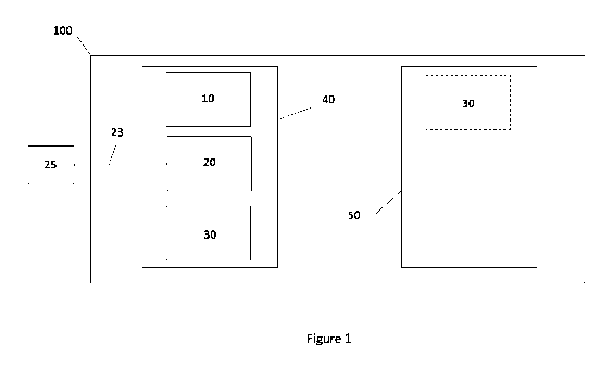
Figure 1 shows a conceptualized block diagram of a configuration of the
devices of the
present disclosure.
Figure 2 shows a schematic flow chart of the process of the present
disclosure.
CA 02921347 2016-02-12
WO 2014/201441
PCT/US2014/042434
DETAILED DESCRIPTION OF THE DISCLOSURE
The present disclosure provides devices and methods for ultra-rapid
prototyping of
5 three-dimensional constructs to be used during a surgery, thus limiting
the risk associated with
multiple surgeries and avoiding the currently necessary waiting period. The
devices used in the
present disclosure include an image acquisition device, a computer and image
manipulation/display device, and a printer or fabricator for printing the
three-dimensional
construct. The devices and methods used for acquiring and printing the three-
dimensional
io constructs, and in particular the printers, can be co-located with each
other and the location of
the surgery, meaning that they are in the same room or facility as the surgery
taking place. The
co-location or on-site presence of the printing devices enables the surgery,
when applicable or
desirable, to take place while the patient is under a single anesthetic. On-
site location of the
printing device, and single surgery or single anesthetic part replacement
prevents the
complications of longer or repeat surgery and also allows the surgeon more
intraoperative
tailoring of the part. Co-locating the scanning, computing, and/or printing
devices facilitates
the ability to perform the surgery quickly, even if adjustment of the
construct is needed, and
under a single anesthetic. This capability is currently impossible.
Thus, in one embodiment of the present disclosure, during a procedure, a
surgeon,
technician, or other user could acquire an image of a desired three-
dimensional construct for
use during the surgery. As described above, the three-dimensional construct
could be a
prosthetic or implant, in which case the user would acquire an image of area
the patient's body
to be operated on. (For example, a facial scan for an orbital bone implant or
replacement.) The
scanned image can then be sent to a viewing or computing device for review by
the user. Once
the image of the implement is acceptable, the user can send a command to the
printer to print
the implement. As the image acquisition device, display, and printer can be co-
located, the
user will have the implement while the patient is under a single anesthetic.
This presents a
dramatic improvement over currently available methods for printing surgical
implements.
CA 02921347 2016-02-12
WO 2014/201441
PCT/US2014/042434
6
By "co-located" or "on site", the present disclosure means that the scanning
device,
computer, and/or printer or fabricator, and in particular the printer, are
located within the
same room where the procedure is taking place, scaled to fit in the operating
room itself. "Co-
located" could signify that the image manipulation device and/or the
printer/fabricator may be
in separate rooms, but within the same hospital or medical care facility.
Either way, the devices
and methods of the present disclosure are co-located or "on site" with one
another in the
location where the surgical procedure is performed, so that ultra-rapid
prototyping is possible
during a single anesthetic, or single procedure, eliminating or significantly
reducing the amount
io of delay in obtaining a required three-dimensional construct. In one
embodiment, only the
printer is located on site at the location where the surgical procedure is
performed. Again, this
can be in the same operating room as the patient, or in the same facility, so
that ultra-rapid
prototyping is possible. This process substantially decreases the cost of
healthcare delivery, not
just for the individual three-dimensional construct, but it also obviates the
need for the current
massive warehoused stock kept by hospitals, doctor offices, and surgical
centers that is
maintained for potential use in countless sizes which are rarely customized to
the patient.
By "single anesthetic", the present disclosure means that the three-
dimensional
construct is printed or fabricated within the same operative procedure that
created the need
for the implant, either through tumor extirpation, fracture reduction,
resection of a
dysfunctional body part, identification of a missing part, creation of a hole,
debridement of
nonviable, infected, destroyed tissue or organ, or in the same operative
location as the location
where the image on which the three-dimensional construct is based is acquired.
Also, current
devices or methods may refer to "rapid-prototyping", but this typically means
that when the
image of a specific part is acquired, it is then sent off to be printed
remotely, in a process that
may take several days to weeks. With use of the terms "ultra-rapid
prototyping", "intra-
operative", and "single anesthetic", the present disclosure distinguishes over
these processes.
In the method of the present disclosure, the required three-dimensional
construct can be
CA 02921347 2016-02-12
WO 2014/201441
PCT/US2014/042434
7
provided during the same, single surgical procedure. This may all take place
while the patient is
under anesthesia.
Referring to Fig. 1, a representation of the devices of the present disclosure
is shown.
Image acquisition device or scanner 10, computer/image display device 20, and
printer 30 can
all be within the same operating room 40, within medical facility 100.
Alternatively one or
more of scanner 10, computer 20, and printer 30 could be in an adjacent room
50 (e.g., printer
30). As long as all three of devices 10, 20, and 30 are within the same
facility 100, they satisfy
the present disclosure's definition of "co-located" or "on-site". Computer 20
can communicate
io over a communications link 23 (e.g., broadband, wireless, cabled,
Ethernet connections) with a
server or database 25. Database 25 can store images and/or data relating to
three-dimensional
constructs that the user wishes to print out for use. Thus, computer 20 can
obtain image data
from scanner 10 or from database 25. This image data can then be sent to
printer 30 to print
the construct. Database 25 can be on site within facility 100, or be located
remotely, as shown.
Fig. 2 shows a flow chart of how the process of the present disclosure could
take place,
in several different embodiments. In the first step, image data relating to
the desired three-
dimensional construct is acquired. This can be via an image scanning device
(e.g. scanner 10),
or from a database (e.g., database 25) that has image data stored for any
number of implants,
prostheses, or surgical tools. The image can be pulled up on a computer or
display device (e.g.,
computer 20). Optionally, the image may be manipulated further by a doctor,
engineer,
technician, or other user/consultant that can be on- or off-site. After the
final image for the
construct is agreed upon, the image data is sent to a printer (e.g., printer
30), which fabricates
the three-dimensional construct. After this point, the construct can
optionally be verified
and/or sterilized if need be. The construct can then be placed in the patient,
or stored for later
use. Whether or not the construct needs sterilization can depend on the method
of fabrication
and the temperature maintained during fabrication, as well as the location to
be used (external
as a splinting device, intraoral, or implanted in a closed space). The
construct may or may not
require subsequent sterilization through autoclave, gas sterilization, etc.
CA 02921347 2016-02-12
WO 2014/201441
PCT/US2014/042434
8
The period of time that the printer or fabricator provides the three-
dimensional
construct after obtaining the final image can vary, depending on the
particular type of medical
procedure. This period of time can range from ten minutes to twelve hours, or
any subranges
there between.
As one example of how the devices and methods of the present disclosure can be
used,
when conducting surgery to remove a tumor or growth from the orbital cavity,
often times part
of the skull surrounding the cavity must be removed as well. In currently
available methods,
io surgeons use off-the-shelf replacement bone, which must be carved and
shaped before
placement in the patient. These off-the-shelf replacements are difficult to
work with, and very
costly to keep in stock. If the off-the-shelf implement cannot be adjusted
suitably (by whittling,
bending, cutting, or hitting, all of which induce stress that can lead to
device failure once
implanted) during the procedure, an additional replacement must be ordered,
which can add
several additional weeks to the procedure. In a more severe case, the surgical
wound in the
patient must be left open and bandaged while a customizable implement is
ordered. This is
obviously very psychologically damaging and dangerous for the patient, in
addition to
escalating costs associated with operating room time and hospital stays for
the patient.
Additionally, the wait for a suitable implant or prosthesis results in wound
contracture and loss
of soft tissue coverage and makes for a much more difficult ultimate
reconstruction. This
would also require the patient to undergo multiple rounds of anesthesia, which
carries its own
associated risks. Lastly, the cost of buying implements or prostheses under
currently available
methods can be extremely high, often as much as $10,000. The methods and
devices of the
present disclosure allow for producing a myriad of implements and implement
devices with
material costs of between $1-$100.
Additionally, the present disclosure addresses the deleterious effects of
wound
contracture, which would occur in the event that an additional or a more
definitive three-
dimensional construct needs to be designed and placed or replaced at a later
time. For
CA 02921347 2016-02-12
WO 2014/201441
PCT/US2014/042434
9
instance, in the setting of trauma or infection, a custom implant, which may
not be suitable as a
permanent device, is impregnated or bathed in antibiotics to allow delivery at
the wound site.
This implant, which serves as a stop-gap, maintains the soft tissue envelope,
thus allowing for
implant exchange at a later date.
By contrast, in the present disclosure, once the surgeon identifies the defect
or size
needed for the three-dimensional construct, a software program resident on the
computer
(which can be in the operating room itself) can manipulate a 3D image acquired
through
radiologic imaging, 3D photography, haptic input, 3D positional marker, laser
scanner, or via a
io database of stored images. As described above, the computer has the
added capability, in the
absence of imaging, to allow the surgeon to rely upon an anthropometric
dataset to select the
part. With simple intraoperative measurements, the surgeon, or other
professional/technician
can scale the obtained image as desired, to fit the patient in a nearly
identical manner to the
patient-specific techniques of the first embodiment, where the patient is
scanned.
The 3D rendering of the desired implement can be manipulated through CAD/CAM
(computer-aided design or manufacturing) software on the computer located in
the operating
room by the surgeon, a technician, or with the help of offsite biomedical
engineers in order to
facilitate production. Even if offsite engineers were used, however, the
methods and devices of
the present disclosure would work intraoperatively, or when the patient is
under a single
anesthetic.
Once the surgeon is satisfied with the construct, rapid prototyping commences.
The 3D
data is then rendered, sliced, and verified within minutes and output to the
prototyping
machine. The construct is created using fused-deposition modeling (FDM),
selective laser
sintering (SLS), stereolithography (SLA), electron beam melting (EBM), or any
other 3D printing
or additive manufacturing process.
CA 02921347 2016-02-12
WO 2014/201441
PCT/US2014/042434
The methods and devices of the present disclosure may also use subtractive
manufacturing. In this embodiment, the image acquisition device would send an
image of a
desired three-dimensional construct to the computer, as described above. The
final image, with
or without modification, is sent to a fabricator. The fabricator uses
subtractive methods to
5 produce the three-dimensional construct, where the three-dimensional
construct can be hewn
from a solid piece of implantable material. The subtractive methods may
include lathing the
three-dimensional construct, cutting with laser-blade, water-blade, or air-
blade-cutting tools,
stamping, grinding, or carving.
io The present disclosure also contemplates that the imaging of the three-
dimensional
construct and/or its printing can take place before the patient is placed
under anesthesia. For
example, in the application described above, an area of the orbital cavity of
a patient where
bone is missing can be imaged, the image sent to a printer or fabricator, and
an already-
customized replacement can be ultra-rapidly prototyped. In this example, the
turnaround time
would be on the lower end of the time period given above, since the image
would need to be
acquired and the surgery would need to be completed while the patient is still
under
anesthesia. In procedures where the imaging can take place while the patient
is alert before
surgery, such as a facial implant procedure, the turnaround time can be closer
to the higher end
of the range given above. The patient can sit for the construct imaging before
being placed
under anesthesia, the image can be sent to the printer or fabricator, and then
up to twelve
hours later the patient can come back to have the construct placed. The cost
of the procedure
is dramatically reduced as well, lowering the overall costs for the patient
and the healthcare
system as a whole.
The printer or fabricator of the present disclosure can also eliminate the
time associated
with sterilization of an implantable prosthesis constructs in currently
available devices and
methods. Currently, when the doctor or surgeon receives an implantable
prosthesis after the
printing delay, there is additional time associated with sterilization of the
prosthesis, which
further adds to the cost of the procedure and risk for the patient. With some
of the devices and
CA 02921347 2016-02-12
WO 2014/201441
PCT/US2014/042434
11
methods of the present disclosure, however, this time is significantly reduced
or eliminated
completely. The printer or fabricator can provide an already-sterilized
construct (depending on
the method of printing and the environment, as described above) for immediate
use, for
example, due to the high temperatures used in processes like fused deposition
modeling, which
produces the construct in a sterile manner. In the case of a construct
produced via computer-
guided lathe, the machining of the construct will still likely still require
sterilization, but the
lathing process can be more expeditious than printing, so the additional time
for sterilization
should not be impactive.
io The materials suitable for the constructs of the present disclosure may
vary. The
materials can include titanium, polylactic acid and acrylonitrile butadiene
styrene,
methylmethacrylate, porous polyethylene, which are approved by the United
States Food and
Drug Administration for implantable devices. Other materials contemplated may
include silk,
rubber, light-cured polymers, various metals, and implantable
antibioticimpregnated solids.
The present disclosure also contemplates the use of all available bio-
compatible materials,
including suitable plastic, metals, composites, and biologically fabricated
tissues.
In addition to being suitable for implanting constructs in patients, the
devices and
methods of the present disclosure can provide surgical planning models for the
doctor and
patient. The doctor can hold a model of a bone or skull, for example, and
develop a plan of
where incisions or bone removal are to take place. The doctor can also
illustrate the same to
the patient or the patient's caregiver or guardian. As previously mentioned,
there is no current
method for 3D surgical planning in the short window of composite tissue
allotransplantation
(i.e. hand transplantation and face transplantation). Due to limitations on
tissue survivability,
the surgeon must guess how much donor tissue will be required. Guesswork on
the recipient
tissue resection is the current standard of care, since time does not allow
for three dimensional
modeling. With this disclosure, the recipient tissue resection as well as the
donor resection
design could easily be customized to create cutting guides and jigs, allowing
for precision
CA 02921347 2016-02-12
WO 2014/201441
PCT/US2014/042434
12
cutting of soft tissue and bone. This would save precious time, on the order
of 30-60 minutes,
allowing the surgeon to focus on microsurgery and enhance rapid tissue
perfusion.
While the present disclosure has been described with reference to one or more
particular embodiments, it will be understood by those skilled in the art that
various changes
may be made and equivalents may be substituted for elements thereof without
departing from
the scope thereof. In addition, many modifications may be made to adapt a
particular situation
or material to the teachings of the disclosure without departing from the
scope thereof.
Therefore, it is intended that the disclosure not be limited to the particular
embodiment(s)
io disclosed as the best mode contemplated for carrying out this
disclosure.