Note: Descriptions are shown in the official language in which they were submitted.
CA 02922372 2016-02-24
English Translation of PCT/JP2013/074292 Your ref: K8001542W0CA
Applicant ref.: G113243CA: Our ref.: F-13P062SZ-CA
METHOD FOR PREPARING PEPTIDE FRAGMENTS, KIT FOR PREPARING
PEPTIDE FRAGMENTS TO BE USED THEREIN, AND ANALYSIS METHOD
TECHNICAL FIELD
[0001] The present invention relates to a method for preparing peptide
fragments
by site-selectively proteolyzing a protein, such as an antibody, using a
protease, and
a kit for preparing peptide fragments to be used therein. Further, the present
invention relates to a method for analyzing peptide fragments, prepared by the
method, by mass spectrometry or the like to detect or quantitate a protein.
BACKGROUND ART
[0002]
Demand for antibody drugs has rapidly grown and simple quantitation of
the concentration of an antibody in blood has become important in clinical
practice.
Heretofore, the accurate measurement of the concentration of an antibody drug
in
blood after administration has not been regarded as important. However,
measurement of the concentration of an antibody in blood has become essential
also
in clinical trials or the like for antibody drugs since it was found that, in
clinical trials
performed to expand the application of trastuzumab (trade name: Herceptin) to
gastric cancer, there were significant differences in the concentration of
trastuzumab
in blood and overall survival. For example, identification of an antibody drug
or
quantitation of the concentration of an antibody drug in blood is required
also in
quality control, such as pharmacokinetic determination, confirmation of
identity with
original drugs in clinical trials for generic drugs, or the like.
[0003]
ELISA (enzyme-linked immunosorbent assay) using antigen-antibody
reaction is excellent in specificity and quantitativity, and is therefore
widely used in
clinical trials or the like, for example, in detection and quantitation of an
antigen or
antibody in blood. However, ELISA is a method intended to detect a single
protein
(antigen or antibody), and therefore cannot detect many different proteins at
the
same time. In order to detect many proteins by ELISA, it is necessary to
prepare a
specific antibody for each detection target and to set conditions for
measuring the
concentration of the specific antibody, which requires much time and costs.
Further,
ELISA uses antigen-antibody reaction, and is therefore known to have a problem
that
cross-reactivity with a coadministered drug or metabolite may have an
influence on
1
CA 02922372 2016-02-24
English Translation of PCT/JP2013/074292 Your ref : K8001542W0CA
Applicant ref.: G113243CA; Our ref.: F-13P062SZ-CA
a measurement result, ELISA is difficult to be applied to some antibody drugs
to
analyze a stored specimen, or the like.
[0004] For this reason, there has been demand for development of an
analytical
method generally applicable to various antibodies. With the development of
proteomics, a technique has been developed in which proteins can be
comprehensively
detected and quantitated by mass spectrometry. In recent years, analysis of a
macrobiomolecule, such as an antibody, has also become possible. In mass
spectrometry, a molecule to be measured needs to be ionized. Therefore, a
macrobiomolecule, such as an antibody, is often difficult to be directly
analyzed by
mass spectrometry. For this reason, a method is adopted in which a protein is
proteolyzed (fragmented) by protease digestion, a peptide fragment having an
amino
acid sequence specific to the protein to be analyzed is selected from peptide
fragments, and the selected peptide fragment is detected and quantitated by a
mass
spectrometer. In order to improve the digestion efficiency, protease digestion
is
generally performed after a substrate protein is denatured in a high-
concentration
urea or guanidine solution.
[0005] When a protein, such as an antibody, is fragmented by protease
digestion
to perform mass spectrometry, it is important to selectively detect a subject
peptide
fragment. However, in some cases, it is difficult to detect a specific peptide
fragment
derived from a protein to be analyzed in a biological sample containing a wide
variety
of impurities. More specifically, a biological sample, such as blood, contains
a wide
variety of impurities, and therefore when a protease is added to a biological
sample,
proteins derived from impurities are also subjected to protease digestion so
that a
huge number of peptide fragments are produced. In order to selectively detect
and
quantitate, in such peptide fragments, a target peptide fragment derived from
a
detection target (e.g., a specific antibody), peptide fragments need to be,
for example,
separated or concentrated before subjected to mass spectrometry. Further, when
a
peptide fragment having an amino acid sequence in common with a peptide
fragment
derived from a protein to be detected is produced from a protein that is not a
detection
target, false detection or reduction in quantitativity may be caused. For this
reason,
selection of a peptide fragment to be detected tends to be more difficult as
the number
of peptide fragments produced from a biological sample increases.
2
CA 02922372 2016-02-24
English Translation of PCIVJP2013/074292 Your ref.: K8001542W0CA
Applicant ref.: G113243CA; Our ref.; F-13P062SZ-CA
[0006] In view of such complexity of a biological sample, a method has
been
developed in which a specific protein in a complex sample, such as a
biological sample,
is quantitated by subjecting the sample to a mass spectrometer after
purification by
liquid chromatography (LC), or by multistage mass spectrometry in which ionic
cleavage is caused by collision-induced dissociation (CID) or the like, or by
a
combination thereof (multiple reaction monitoring, MRM). However, when a
sample
becomes more complex, various separation modes need to be combined, which
causes
problems such that a high-accuracy experimental apparatus and much time for
setting of analytical conditions are required.
[0007] Further, even when a purified sample is used, a number of peptide
fragments are produced by protease digestion from a macroprotein such as an
antibody. Therefore, a peptide fragment having an amino acid sequence specific
to a
detection target tends to be difficult to be selectively detected and
quantitated in the
produced peptide fragments. In view of such problems, a method has been
developed
in which a protein to be analyzed is subjected to site-selective protease
proteolysis to
reduce the number (of types) of peptide fragments in a sample to improve the
accuracy of analysis and simplify the process of analysis. For example, Patent
Document 1 proposes a method in which an antibody is subjected to pepsin
digestion
to produce an F(ab')2 fragment, and then the F(ab')2 fragment is further
digested with
a protease such as trypsin to produce peptide fragments containing the
complementarity determining region (CDR) of the antibody, and the peptide
fragments containing CDR are detected and quantitated by mass spectrometry.
[0008] Hereinafter, the structure of an antibody will be described. All
antibodies
have two heavy chains (H chains) and two light chains (L chains). One light
chain
and one heavy chain are linked through a disulfide (S-S) bond to form a
heterodimer,
and the two heterodimers are further linked through two disulfide bonds to
form a
"Y÷-shaped heterotetramer (see FIG. 2). An antibody has one Fc (Fragment,
crystallizable) domain comprising heavy chains and two Fab (Fragment, antigen
binding) domains comprising a heavy chain and a light chain, and the Fc domain
and
the Fab domains are linked through a hinge region.
[0009] The Fc domain of an antibody mainly has the function of
initiating a
reaction after the antibody binds to an antigen (effector function), and most
of
antibodies derived from the same species have a common amino acid sequence in
the
3
CA 02922372 2016-02-24
English Translation of PCT/JP2013/074292 Your ref.: K8001542W0CA
Applicant ref.: G113243CA: Our ref.: F-13P062SZ-CA
Fc domain. On the other hand, the end (on the N-terminal side) of the Fab
domain
has the function of binding to an antigen. The N-terminal part of the Fab
domain
diversely changes in its amino acid sequence so as to be able to bind to
various
antigens. This region is called variable region (V region), and the variable
region of
the light chain and the variable region of the heavy chain are called VL
region and
VII region, respectively. The Fab and Fc domains other than the V region are
called
constant region (C region) that varies little in amino acid sequence. The
constant
region of the light chain is called CL region, and the constant region of the
heavy
chain is called CH region. The CH region is further divided into three
regions, CH1
region, CH2 region, and CH3 region. The Fab domain of the heavy chain
comprises
the VH region and the CH1 region, and the Fc domain of the heavy chain
comprises
CH2 and CH3. The hinge region is located between CH1 and CH2.
[0010] The
specificity (i.e., specific bindability to an antigen) of an antibody is
determined by the combination of amino acid sequences of the V region. The
light
chain and the heavy chain each have three complementarity determining regions
(CDRs) in the V region of the Fab domain. CDR is also called hypervariable
region,
and varies in amino acid sequence depending on the type of antibody. There are
3
CDRs on each of the heavy and light chains of an antibody (6 types of CDRs in
total),
which creates diversity that allows the antibody to bind to various antigens.
In other
words, CDRs are regions characterizing an antibody, and therefore an antibody
can
be identified by identifying the amino acid sequences of CDRs thereof.
[0011] As
described above, the Fab domains and Fc domain of an antibody are
linked through a hinge region. Papain which is a kind of protease proteolyzes
the
hinge region, and therefore two Fab domains and one Fc domain are produced by
papain digestion of an antibody. Further, pepsin which is a kind of protease
proteolyzes one of the two disulfide bonds, i.e., the Fc domain-side (C-
terminal side)
disulfide bond of the hinge region, and therefore an F(ab')2 domain having two
Fab
domains linked together and many Fc domain fragments are produced by pepsin
digestion.
[0012] In the method disclosed in Patent Document 1, a Fab domain or
F(ab')2
domain is produced by pepsin digestion or papain digestion, and the Fab domain
or
F(ab')2 domain is subjected to site-selective protease preteolysis. This
method can
reduce the number of peptides produced by protease preteolysis and therefore
can
4
CA 02922372 2016-02-24
English Translation of PCT/JP2013/074292 Your ref.: K8001542W0CA
Applicant ref.: G113243CA; Our ref F-13P062SZ-CA
efficiently produce peptides containing CDR, which simplifies the detection
and
quantitation of an antibody by mass spectrometry. Further, Patent Document 2
reports that combined use of pepsin or papain and a specific ion improves the
efficiency of proteolysis with such a protease.
[0013] A purification kit or the like having optimized protease, for the
purpose of
purifying the Fab domain of an antibody, is commercially available. However,
in the
method disclosed in Patent Document 1, protease treatment needs to be further
performed after purification of the Fab domain or F(ab')2 domain, and
therefore it
takes much time and costs to prepare a sample to be subjected to mass
spectrometry.
For this reason, it is difficult to say that this method is a simple method.
Further,
efficiency of protease proteolysis (fragmentation) varies depending on the
type of
substrate protein to be proteolyzed, and therefore it is important to improve
proteolysis efficiency to simplify the preparation of a peptide fragment
sample to be
subjected to mass spectrometry.
[0014] In recent years, a method in which protease digestion is performed
in a
microenvironment (microreactor) such as nanoparticles has attracted attention,
since
the method can improve protease digestion efficiency. For example, Non-Patent
Document 1 reports an example in which the efficiency of trypsin digestion of
albumin
is increased by allowing an albumin solution to pass through a nylon porous
membrane having trypsin immobilized in pores thereof. Non-Patent Document 2
reports that a protein having a small molecular weight can be selectively
subjected
to trypsin digestion by using mesoporous silica having trypsin immobilized in
pores
thereof. Both the methods are intended to react a substrate protein in a
liquid phase
with a protease immobilized in pores of a porous body as a solid phase. The
reason
why digestion efficiency is increased by immobilizing a protease on a solid
phase in a
microenvironment is considered to be due to an enhanced probability of
reaction
between the substrate protein and the protease. In a local and micro
environment
at the interface between the solid phase and the liquid phase, the protein is
likely to
be denatured and solubility, fluctuation range of three-dimensional structure,
or the
like is perturbed. Therefore, the opportunity of contact between the substrate
protein
and the protease is likely to be enhanced to increase the reaction
probability.
PRIOR ART DOCUMENTS
PATENT DOCUMENTS
5
CA 02922372 2016-02-24
English Translation of PCT/JP2013/074292 Your ref.: K8001542W0CA
Applicant ref.: G113243CA; Our ref.: F-13P062SZ-CA
[0015] Patent Document 1: WO 2008/079914
Patent Document 2: JP 2011-130749 A
NON-PATENT DOCUMENTS
[0016] Non-Patent Document 1: Fei Xu et. al, Anal. Chem., 2010, 82,
10045-10051
Non-Patent Document 2: Qianhao Min et. al., Chem. Commun., 2010,
46(33), 6144-6146
SUMMARY OF THE INVENTION
PROBLEMS TO BE SOLVED BY THE INVENTION
[0017] The method disclosed in Non-Patent Document 2 can achieve selective
protease digestion of a protein having a small molecular weight, but cannot be
applied
to selective digestion of a large protein such as an antibody. Further, no
method has
been developed for performing site-selective protease proteolysis using such a
microreactor as disclosed in Non-Patent Document 1 or 2.
[0018] As described above, in order to simply detect and quantitate a
protein by
mass spectrometry, the protein to be analyzed needs to be site-selectively
proteolyzed
to efficiently produce a peptide fragment specific to the protein to be
analyzed and to
reduce the amount of other peptide fragments produced. For example, in order
to
detect and quantitatively analyze an antibody, the Fab domain, especially, the
V
region of the Fab domain needs to be subjected to site-selective protease
proteolysis
to suppress proteolysis of the Fe domain.
MEANS FOR SOLVING THE PROBLEMS
[0019] The inventors have intensively studied to find that
immobilization of both
a substrate protein, such as an antibody, and a protease on solid phases makes
it
possible to achieve site-selective protease proteolysis of the substrate
protein. This
finding led to the present invention.
[0020] The present invention relates to a method for preparing peptide
fragments
by proteolyzing a protein with a protease. The method of the present invention
includes a step of proteolyzing a substrate protein with a protease by
bringing a
porous body in which a substrate protein to be proteolyzed is immobilized in
pores
thereof and microparticles having a protease immobilized on surface thereof
into
contact with each other in a liquid (proteolysis step). The porous body on
which the
6
CA 02922372 2016-02-24
English Translation of PCT/JP2013/074292 Your ref:: K8001542W0CA
Applicant ref.: G113243CA; Our ref.; F-13P062SZ-CA
substrate protein to be proteolyzed is immobilized in the pores thereof can be
obtained
by a step of immobilizing a substrate protein to be proteolyzed in pores of a
porous
body (substrate immobilization step). In the present invention, an average
particle
diameter of the microparticles is preferably larger than an average pore
diameter of
the porous body.
[0021]
When the particle diameter of the microparticles is larger than the average
pore diameter of the porous body, the protease immobilized on the surface of
the
microparticles can access the shallow parts of the pores and the vicinity of
the porous
body (the interface between the porous body and the liquid phase and its
vicinity),
but cannot access the deep parts of the pores. In this way, the accessible
region of the
protease is physically (spatially) limited, and therefore the protease
selectively
accesses a specific site of the substrate protein immobilized in the pores of
the porous
body.
This makes it possible to achieve site-selective protease proteolysis
(fragmentation) of the substrate protein.
[0022] In the present invention, a predetermined region of the substrate
protein
is preferably immobilized on the porous body. In this embodiment, the region
immobilized on the porous body is located in the deep parts of the pores so
that a
region different from the immobilized region is located near the shallow parts
of the
pores. When a region different from the selective proteolysis site of the
substrate
protein, i.e., a region preferred not to be subjected to protease proteolysis,
is
immobilized on the porous body, the protease immobilized on the surface of the
microparticles accesses the selective proteolysis site of the substrate
protein located
near the shallow parts of the pores so that protease proteolysis is performed.
This
allows site-selective protease proteolysis at a desired site in the substrate
protein.
[0023] A linker molecule capable of site-specific interaction with the
substrate
protein is preferably immobilized in the pores of the porous body. The
substrate
protein is immobilized in the pores of the porous body preferably through the
linker
molecule. Examples of the linker molecule used when the substrate protein is
an
antibody include Protein G and Protein A. Such a linker molecule site-
specifically
binds with the Fe region of the antibody, and therefore the Fe region of the
antibody
is immobilized on the porous body so that the Fab region of the antibody is
located
near the shallow parts of the pores. According to this embodiment, the Fab
region of
the antibody can be subjected to selective protease proteolysis.
7
CA 02922372 2016-02-24
English Translation of PCT/JP2013/074292 Your reP: K8001542W0CA
Applicant ref.: G113243CA; Our ref:: F-13P062SZ-CA
[0024] When the linker molecule is immobilized in the pores of the
porous body, a
molecule in which the linker molecule binds with the substrate protein
preferably has
a size 0.5 times to 1.5 times the average pore diameter of the porous body.
Such
molecular size adjustment makes it possible to increase the probability of
access of
the protease immobilized on the surface of the microparticles to the selective
cleavage
site of the substrate protein, thereby improving the site-selectivity of
protease
proteolysis.
[0025] It is preferred that the surface of the microparticles is
modified with a
spacer molecule capable of binding with the protease, and the protease is
immobilized
on the surface of the microparticles through the spacer molecule. The
immobilization
of the protease through the spacer molecule makes it possible to suppress the
detachment of the protease from the surface of the microparticles, thereby
improving
the site-selectivity of protease proteolysis. Further, the adjustment of
molecular size
of the spacer makes it possible also to allow the protease to selectively
access a desired
position in the substrate protein to improve the site-selectivity.
[0026] In the present invention, the protease to be immobilized on the
surface of
the microparticles is preferably trypsin or a combination of trypsin and
another
protease. When the substrate protein is an antibody, trypsin is preferably
used alone,
or in a combination of proteases. When proteases are used in a combination,
the
amount of trypsin is preferably 90% or more of the total amount of proteases.
Particularly, when the substrate protein is an antibody, the Fab domain tends
to be
subjected to selective protease proteolysis by using trypsin so that the
protease
proteolysis of the Fc domain is suppressed.
[0027] The average pore diameter of the porous body is preferably about
30 to 150
nm, and the average particle diameter of the microparticles is preferably 100
nm or
more. Particularly, when the substrate protein is an antibody and the average
pore
diameter and the average particle diameter are within the above ranges, the
Fab
region can be more reliably site-selectively proteolyzed.
[0028] The present invention also relates to a kit for peptide fragment
preparation
used for the above method. The peptide fragment preparation kit of the present
invention includes a porous body having pores capable of immobilizing a
substrate
protein, and microparticles capable of immobilizing a protease on surface
thereof.
The microparticles may be provided in a state where the protease is
immobilized on
8
CA 02922372 2016-02-24
English Translation of PCT/JP2013/074292 Your ref:: K8001542W0CA
Applicant ref.: G113243CA; Our ref: F-13P062SZ-CA
the surface thereof. The porous body of the kit and a sample (e.g., a specimen
such
as blood) are brought into contact with each other such that the subject
substance
(substrate protein such as an antibody) in the sample can be immobilized in
the pores
of the porous body. The substrate protein is subjected to site-selective
protease
proteolysis by bringing the porous body after immobilization of the substrate
protein
and the microparticles having the protease immobilized on the surface thereof
into
contact with each other in a liquid.
[0029] Peptide fragments obtained by the above method are analyzed by
mass
spectrometry or the like, which makes it possible to detect (identify) or
quantitate the
substrate protein. In the present invention, the substrate protein is
subjected to site-
selective protease proteolysis, and therefore the number of types of peptide
fragments
contained in a measurement sample can be significantly reduced. Therefore, the
setting of measurement conditions of mass spectrometry can be simplified, and
the
accuracy of analysis is also expected to be improved.
[0030] For example, when the substrate protein is an antibody, the method
according to the present invention can achieve site-selective protease
proteolysis of
the Fab region containing a complementarity determining region, and therefore
a
peptide fragment containing at least part of the sequence of complementarity
determining region of the antibody can be produced as a detection target. The
complementarity determining region has an amino acid sequence specific to each
antibody. Therefore, the antibody can be detected or quantitated by analyzing
the
peptide fragment containing the sequence of the complementarity determining
region.
EFFECTS OF THE INVENTION
[0031] According to the present invention, a protein, such as an
antibody, can be
subjected to site-selective protease proteolysis by a simple method to obtain
peptide
fragments. When the method according to the present invention is applied to an
antibody, proteolysis of Fc region of the antibody is suppressed, and the Fab
region
containing CDR is subjected to selective protease proteolysis, and therefore
the
concentration of the peptide fragment containing the amino acid sequence of
CDR,
which is important for identification of the antibody, in a sample is
increased.
9
CA 02922372 2016-02-24
English Translation of PCT/JP2013/074292 Your ref.: K8001542W0CA
Applicant ref.: G113243CA; Our ref.: F-13P062SZ-CA
[0032] The method according to the present invention makes it possible
to
significantly reduce the number of types of peptides contained in a
measurement
sample. Therefore, the setting of conditions of mass spectrometry can be
simplified,
and the accuracy of analysis can also be expected to be improved. The
concentration
of an antibody drug in blood can also be quantitated by analyzing obtained
peptide
fragments. Therefore, the method according to the present invention can also
be
applied as a pretreatment method for a system for measuring the concentration
of an
antibody drug in a preclinical or clinical trial.
[0033] Further, the method according to the present invention can be
applied not
only to antibody drugs but also to many proteins, and therefore can be
expected to be
extensively applied to pharmaceutical industry. In addition, the method
according to
the present invention can also be expected to be applied, for example, in the
field of
fundamental research such as interactive analysis of biomolecules.
BRIEF DESCRIPTION OF THE DRAWINGS
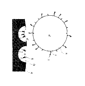
[0034] FIG. 1 is a conceptual diagram for illustrating the principle of
site-selective
proteolysis according to the present invention.
FIG. 2 is a schematic diagram for illustrating the structure of an antibody.
FIG. 3 is a schematic diagram of one embodiment of a kit for preparing
peptide fragments.
FIG. 4 shows electrophoretic patterns obtained in an experiment for
examining the quantitative ratio of a protease.
FIG. 5 shows mass spectra (MALDI-TOFMS) obtained in the experiment
for examining the quantitative ratio of a protease.
FIG. 6 shows mass spectra (MALDI-TOFMS) obtained in an experiment
for examining proteolysis time.
FIG. 7 shows the result of database analysis based on the result of mass
spectrometry of tryptic fragments of trastuzumab.
FIG. 8 shows a mass spectrum (MALDI-TOFMS) of tryptic fragments of
trastuzumab.
FIG. 9 shows chromatograms of LC-MS analysis of tryptic fragments of
trastuzumab.
FIG. 10 shows mass spectra (LS-MS) of tryptic fragments of trastuzumab.
CA 02922372 2016-02-24
English Translation of PCIVJP2013/074292 Your ref.: K8001542W0CA
Applicant ref.: G113243CA; Our ref.: F-13P062SZ-CA
FIGs. 11(A) and 11(B) show the amino acid sequences of heavy and light
chains of trastuzumab, respectively, wherein peptide fragments identified by
mass
spectrometry in this experiment are underlined.
FIG. 12 shows electrophoretic patterns obtained in an experiment for
studying mixed protease proteolysis.
FIG. 13 shows mass spectra (MALDI-TOFMS) obtained in the experiment
for studying mixed protease proteolysis.
MODE FOR CARRYING OUT THE INVENTION
[0035] According to a method for preparing peptide fragments of the present
invention, a substrate protein to be proteolyzed is immobilized in pores of a
porous
body, and the porous body having the substrate protein immobilized thereon is
brought, in a liquid, into contact with microparticles having a protease
immobilized
on a surface thereof. FIG. 1 is a conceptual diagram for illustrating the
principle of
protease proteolysis in the present invention.
[0036] On the surface of microparticles 10 (average particle diameter
Di), a
protease 15 is immobilized. A porous body 20 has a plurality of pores 29
(average
pore diameter D2), and a substrate protein 25 is immobilized in the pores. In
the
method according to the present invention, as described above, both the
protease 15
and the substrate protein 25 are immobilized on solid phases in a micro
region, and
protease proteolysis is performed by contact between the solid phases.
[0037] The average particle diameter Di of the microparticles 10 is
larger than
the average pore diameter D2 of the porous body 20. Therefore, the
microparticles 10
can access the shallow parts of the pores 29 and their vicinity, but cannot
access the
deep parts of the pores 29. As a result, the protease 15 immobilized on the
surface of
the microparticles 10 cannot access the deep parts of the pores 29. In FIG. 1,
a dotted
line near each of the pores 29 indicates the limit of the region accessible to
protease
15.
[0038] In this way, the accessibility of the protease 15 to the
substrate protein 25
in the pores 29 is site-selectively limited so that the relative probability
of
accessibility of the protease 15 to the liquid phase side ("r-shaped site in
FIG. 1) of
the substrate protein increases. This makes it possible to subject the
substrate
protein 25 to site-selective protease proteolysis to obtain peptide fragments.
11
CA 02922372 2016-02-24
English Translation of PCT/JP2013/074292 Your ref: K8001542W0CA
Applicant ref.: G113243CA; Our ref.: F-13P062SZ-CA
[0039] [Substrate Protein]
The substrate protein 25 is a protein to be analyzed. The type of the
substrate protein is not particularly limited. However, from the viewpoint of
performing site-selective proteolysis, the substrate protein preferably has a
molecular diameter larger than that of the protease 15. The substrate protein
may
be a protein complex. As the molecular diameter, a value determined based on
structural analysis by X-ray or NMR is available from various documents or
databases. For example, the molecular diameter of an antibody is about 15 nm.
Alternatively, the molecular diameter may be determined by, for example, X-ray
small
angle scattering or may be roughly estimated from a molecular weight. As
reference
examples, Table 1 shows molecular weights and molecular diameters of proteins
used
as marker molecules to determine the separation properties of an
ultrafiltration
membrane.
12
CA 02922372 2016-02-24
English Translation of PCT/JP2013/074292 Your ref.: K8001542W0CA
Applicant ref.: G113243CA: Our ref.: F-13P062SZ-CA
[0040] [Table 1]
protein molecular weight [Dal molecular diameter Inm1
sucrose 340 1.1
raffinase 590 1.3
vitamin B12 1,360 1.7
bacitracin 1,410 1.7
insulin 5,700 2.7
cytochrome C 13,400 3.8
myoglobin 17,000 4.0
a-chymotrysinogene 25,000 4.6
pepsin 35,000 5.0
ovalbumin 43,000 5.6
Bovine albumin 67,000 6.4
aldolase 142,000 8.2
y- globulin 150,000 8.4
[0041] The substrate protein 25 is preferably one that can site-
specifically bind
into the pores 29 of the porous body 20. Site-specific binding of the
substrate protein
25 allows a site other than the binding site to be subjected to selective
protease
proteolysis. For example, a protein bearing, at its C- or N-terminus, a tag
sequence
such as a His tag (tag peptide containing about 6 continuous histidine
residues) or a
biotinylated peptide, an enzyme that specifically binds with a specific
substrate, or
the like may also be used as the protein that can site-specifically bind.
[0042] In the present invention, an antibody is particularly preferably
used as the
substrate protein that can site-specifically bind into the pores of the porous
body.
Immobilization of the Fc domain of the antibody on the porous body 20 allows
the Fab
domain of the antibody to be subjected to selective protease proteolysis.
Although the
type of the antibody is not particularly limited, a monoclonal antibody is
preferred.
Examples of the monoclonal antibody include: human antibodies such as
panitumumab (Vectibix), ofatumumab (Arzerra), golimumab (Simponi), and
ipilimumab (Yervoy); humanized antibodies such as tocilizumab (Actemra),
trastuzumab (Herceptin), bevacizumab (Avastin), omalizumab (Xolair),
mepolizumab
(Bosatria), gemtuzumab ozogamicin (Mylotarg), palivizumab (Synagis),
ranibizumab
(Lucentis), certolizumab (Cimzia), ocrelizumab, mogamulizumab (Poteligeo), and
eculizumab (Soliris); and chimeric antibodies such as rituximab (Rituxan),
cetuximab
13
CA 02922372 2016-02-24
English Translation of PCT/JP2013/074292 Your ref: K8001542W0CA
Applicant ref.: G113243CA; Our ref.: F-13P062SZ-CA
(Erbitux), infliximab (Remicade), and basiliximab (Simulect). These antibodies
are
used as antibody drugs (molecularly-targeted drugs), and the concentrations of
the
antibodies in blood need to be quantitated in clinical trials or the like.
[0043] As will be described later with reference to Examples, according
to the
method of the present invention, the Fab domain of the monoclonal antibody can
be
subjected to site-selective protease proteolysis to obtain peptide fragments,
and the
antibody can be identified and quantitated by mass spectrometry of the
obtained
peptide fragments. The analysis method according to the present invention is a
method in which peptide fragments derived from the variable region of the
antibody
are detected to identify (detect) or quantitate the antibody, that is, a
method in which
peptide fragments derived from the antibody are directly measured. Therefore,
the
analysis method according to the present invention requires no specific
binding
substance such as an antigen, and therefore can be applied irrespective of the
type of
antibody. Therefore, the method according to the present invention can be
applied
not only to the above-mentioned antibodies but also to newly-developed
monoclonal
antibodies.
[0044] [Porous Body]
The material of the porous body 20 is not particularly limited as long as
the material has pores 29. Although the pores shown in FIG. 1 have a semi-
spherical
shape, the shape of the pores is not particularly limited. A porous body
having
through-holes, such as a porous membrane, may also be used.
[0045] For the porous body 20, activated carbon, a porous membrane,
porous resin
beads, metal particles, or the like may be used. Among them, one that can
specifically
bind with the substrate protein is preferred, and one that can site-
specifically bind
with the substrate protein is particularly preferred. For example, affinity
column
packing beads used to purify a specific protein or the like can satisfy such a
requirement.
[0046] The porous body 20 preferably used in the present invention is
one in which
a linker molecule 21 that can site-specifically interact with the substrate
protein 25
is immobilized in the pores 29 thereof. Examples of the interaction between
the
substrate protein and the linker molecule include chemical binding, hydrogen
binding, ion binding, complex formation, hydrophobic interaction, van der
Waals
interaction, electrostatic interaction, and stereoselective interaction.
14
CA 02922372 2016-02-24
English Translation of PCT/JP2013/074292 Your ref:: K8001542W0CA
Applicant ref.: G113243CA: Our ref.: F-13P062SZ-CA
[0047] The optimum linker molecule can be appropriately selected,
depending on
the type or binding site of the substrate protein, from functional groups such
as an
amino group, a carboxyl group, and an epoxy group; labeling compounds such as
biotin and digoxygenin; proteins such as avidin, streptoavidin, Protein A,
Protein G,
and immunoglobulin; various ligands; substrate compounds for enzymes; silica;
and
metal chelates.
[0048] Protein G, Protein A, or the like is preferably used as the
linker molecule
21, when the substrate protein 25 is an antibody. Protein A or Protein G site
specifically binds with the Fc domain of the antibody. The use of the porous
body 20
having the linker molecule 21, such as Protein A or Protein G, immobilized in
the
pores 29 allows the Fc domain of the antibody (substrate protein 25) to be
site-
specifically immobilized in the pores so that the Fab domain of the antibody
is located
on the liquid-phase side (near the shallow parts of the pores). Such
immobilization
of the antibody in the pores in a given direction controls the orientation of
the
antibody in the pores, and therefore the Fab domain can be site-selectively
proteolyzed with the protease.
[0049] Further, when the substrate protein is immobilized in the pores
so as to be
present in a microenvironment at the interface between the solid phase and the
liquid
phase, the substrate protein is likely to be denatured and molecular
fluctuations are
disturbed so that the probability of being attacked by the protease increases.
Further,
in the present invention, the protease is immobilized on the particles, and
therefore
an environment is created in which the protease is sterically stable and
autolysis is
less likely to occur. This is considered to increase the stability of the
protease.
Therefore, according to the method of the present invention, site-selective
protease
proteolysis can be performed, and in addition, high activity of the protease
can be
maintained.
[0050] The size of the pores 29 of the porous body 20 is not
particularly limited.
The size of the pores is preferably determined in consideration of the
molecular
diameter of the substrate protein etc. so that the tip of the substrate
protein, Le., the
site to be selectively proteolyzed, is located near the shallow parts of the
pores 29
when the substrate protein 25 is immobilized. The average pore diameter D2 of
the
porous body 20 is appropriately set to fall in the range of, for example,
about 10 nm
to 500 nm and to be smaller than the average particle diameter Di of the
CA 02922372 2016-02-24
English Translation of PCIVP2013/074292 Your ref.: K8001542W0CA
Applicant ref: G113243CA; Our ref: F-13P062SZ-CA
microparticles 10. The average pore diameter D2 of the porous body 20 is, for
example, preferably about 20 nm to 200 nm, more preferably about 30 nm to 150
nm.
Particularly, when the substrate protein 25 is an antibody, in order to
immobilize the
Fe domain of the antibody in the pores to subject the Fab domain of the
antibody to
site-selective protease proteolysis, the pore diameter of the porous body is
preferably
30 nm to 150 nm, more preferably 40 nm to 120 nm, further preferably 50 nm to
100
nm.
[0051] The size of the linker molecule is selected in consideration of
the size of the
pores or size of the substrate protein so that the selective proteolysis site
of the
substrate protein is located near the shallow parts of the pores. The size of
a molecule
in which the linker molecule binds with the substrate protein is preferably
about 0.5
to 1.5 times, more preferably about 0.6 to 1.2 times, further preferably about
0.7 to
1.1 times, particularly preferably about 0.8 to 1 times the pore diameter of
the porous
body. When the linker molecule is not immobilized on the porous body 20 and
the
substrate protein directly binds into the pores of the porous body, the
molecular
diameter of the substrate protein and the pore diameter of the porous body
preferably
satisfy the above relationship.
[0052] [Immobilization of Substrate Protein]
A method for immobilizing the substrate protein 25 in the pores 29 of the
porous body 20 is not particularly limited, and an appropriate method can be
adopted
depending on the properties of the substrate protein and the porous body (or
the
linker molecule immobilized on the porous body) etc. For example, when the
porous
body has Protein A or Protein G immobilized in the pores thereof, an antibody
can be
easily immobilized in the pores by mixing a suspension of the porous body and
a
solution containing the antibody.
[0053] The quantitative ratio between the porous body and the substrate
protein
can be appropriately set depending on the purpose. For example, in the case of
quantitative analysis of the substrate protein, it is desired that almost
entire amount
of the substrate protein in a sample should be immobilized on the porous body.
Therefore, the quantitative ratio is preferably set so that the amount of the
porous
body becomes higher than the estimated amount of the substrate protein
contained
in the sample.
[0054] [Protease]
16
CA 02922372 2016-02-24
English Translation of PCT/JP2013/074292 Your ref.: K8001542W0CA
Applicant ref.: G113243CA: Our ref.: F-13P062SZ-CA
The protease 15 recognizes the amino acid sequence of the substrate
protein and selectively proteolyzes a specific bond in a specific sequence. In
the
present invention, the substrate protein 25 is immobilized in the pores 29 of
the
porous body 20, and the protease 15 proteolyzes the substrate protein 25 at a
specific
amino acid sequence site, so that peptide fragments are obtained.
[0055] Examples of the protease include trypsin (which proteolyzes a
peptide at
the C-terminal side of basic amino acid residues (Arg and Lys)), lysyl
endopeptidase
(which proteolyzes a peptide at the C-terminal side of a Lys residue),
arginine
endopeptidase (which proteolyzes a peptide at the C-terminal side of an Arg
residue),
chymotrypsin (which proteolyzes a peptide at the C-terminal side of aromatic
amino
acid residues (Phe, Tyr, and Trp)), V8 protease (which proteolyzes a peptide
at the C-
terminal side of a Glu residue), pepsin, and papain. Two or more of these
proteases
may be used in combination.
[0056] When peptide fragments of the substrate protein after protease
proteolysis
are subjected to mass spectrometry as a measurement sample, the protease to be
used
is preferably one with low autolysis and high selectivity for a sequence to be
proteolyzed. When a commercially-available protease is used, a mass
spectrometry-
grade protease or a sequencing-grade protease is preferably used. For example,
it is
known that native trypsin derived from a living body has low specificity for a
proteolysis site because pseudo trypsin that exhibits chymotrypsin-like
activity is
generated due to autolysis. Therefore, mass spectrometry-grade trypsin is
commercially available which achieves high resistance to autolysis due to
reductive
methylation of lysine residues of trypsin.
[0057] In order to improve the site-selectivity of protease proteolysis
of the
substrate protease, it is important to limit the region where the protease can
access
the substrate protein. Therefore, the molecular diameter of the protease is
preferably
smaller than that of the substrate protein. More specifically, the molecular
diameter
of the protease is preferably 10 nm or less, more preferably 8 nm or less,
further
preferably 6 nm or less, particularly preferably 5 nm or less. A protein
having a
molecular weight of about 30 kDa, such as trypsin or lysyl endopeptidase, has
a
molecular diameter of about 4 nm (see Table 1 shown above).
[0058] Among the above-mentioned proteases, trypsin is particularly
preferably
used in the present invention. As described above, trypsin has a small
molecular
17
CA 02922372 2016-02-24
English Translation of PCT/JP2013/074292 Your ref : K8001542W0CA
Applicant ref.: G113243CA; Our ref.: F-13P062SZ-CA
diameter and its active site is present inside its molecule. This limits the
region
where the active site can access the substrate protein, which makes it
possible to
improve the site-selectivity of protease proteolysis. Particularly, when the
substrate
protein is an antibody, the protease to be used is preferably trypsin.
[0059] In proteome analysis study, digestion with a combination of trypsin
and
lysyl endopeptidase has attracted attention in recent years as a technique for
improving the recovery rate of peptide fragments (J. Proteome Res., 2012,
11(11),
5145-5156). The reason for this is considered to be that trypsin has the
property of
allowing a decomposition reaction to proceed in stages from the outside of a
steric
structure, and lysyl endopeptidase first proteolyzes mainly the hinge region
of an
antibody. In the present invention, on the other hand, it is preferred that
trypsin is
used alone or that even when lysyl endopeptidase or the like is used in
combination
with trypsin, the amount of trypsin is preferably 90% or higher of the total
amount
of proteases used, in order to suppress the proteolysis of the hinge region of
an
antibody and to selectively proteolyze the Fab domain (more preferably, the V
region
of the Fab domain) of the antibody.
[0060] [Microparticles]
The microparticles 10 are used for the purpose of immobilizing the protease
15 on the surface thereof to control the accessibility of the protease to the
substrate
protein 25 immobilized in the pores 29 of the porous body 20. Therefore, the
average
particle diameter Di of the microparticles 10 is preferably larger than the
average
pore diameter D2 of the porous body 20 so that the microparticles 10 do not
enter the
deep part of the pores 29 of the porous body 20. The average particle diameter
Di of
the microparticles 10 is more preferably 1.2 times or more, further preferably
1.5
times or more, particularly preferably L8 times or more the average pore
diameter
D2 of the porous body 20.
[0061] Although the shape of the microparticles 10 is not particularly
limited,
spherical microparticles are preferred from the viewpoint of equalizing the
accessibility of the protease to the pores 29 of the porous body 20. Further,
the
microparticles 10 preferably have a uniform average particle diameter.
[0062] When the average pore diameter of the porous body 20 is about 30
to 150
nm, the average particle diameter Di of the microparticles 10 is preferably
100 nm or
more, more preferably 150 nm or more. When the substrate protein 25 is an
antibody
18
CA 02922372 2016-02-24
English Translation of PCT/JP2013/074292 Your ref.: K8001542W0CA
Applicant ref.: G113243CA; Our ref.: F-13P062SZ-CA
and the average pore diameter of the porous body 20 is about 50 nm to 100 nm,
the
average particle diameter of the microparticles 10 is preferably 120 nm or
more, more
preferably 150 nm or more, particularly preferably 170 nm or more. The upper
limit
of the average particle diameter Di of the microparticles 10 is not
particularly limited,
but is preferably 1 p.m or less, more preferably 500 nm or less, further
preferably 300
nm or less, from the viewpoint of improving the efficiency of protease
proteolysis.
[0063]
The material of the microparticles 10 is not particularly limited as long as
the protease can be immobilized on the surface thereof, and a metal, a resin,
or the
like is appropriately used. Alternatively, a material obtained by coating the
surface
of a metal with a resin, a material obtained by coating the surface of a resin
with a
metal, or the like may be used.
[0064]
The microparticles 10 preferably have a surface capable of suppressing
nonspecific protein adsorption and of selectively immobilizing the protease
thereon.
For example, as shown in FIG. 1, microparticles whose surface is modified by a
spacer
11 that can specifically bind with the protease are appropriately used. The
spacer is
preferably one that can bind with the protease and does not deactivate the
protease.
[0065]
Further, from the viewpoint of controlling the range of accessibility of the
protease 15 immobilized on the surface of the microparticles 10, the spacer 11
preferably has a small molecular diameter. The molecular diameter of the
spacer is
preferably 5 nm or less, more preferably 3 nm or less, further preferably 2 nm
or less.
Further, the molecular weight of the spacer is preferably 2000 or less, more
preferably
1500 or less, further preferably 1000 or less, particularly preferably 800 or
less. The =
spacer molecule that has a molecular diameter in the above range and is
capable of
immobilizing the protease is preferably non-protein and preferably has a
functional
group such as an amino group, an amide group, an ester group, an epoxy group,
a
carboxyl group, biotin, avidin, or a chelate. For example, the spacer
preferably used
to immobilize trypsin has an ester group. Further, a molecule having an
activated
ester group is also preferably used as the spacer to improve the efficiency of
protease
immobilization.
[0066] In the present invention, commercially-available microparticles
modified
with a spacer molecule may also be used. For example, microparticles modified
with
a spacer molecule having an ester group activated by N-hydroxysuccinimide are
19
CA 02922372 2016-02-24
English Translation of PCT/JP2013/074292 Your ref.: K8001542W0CA
Applicant ref.: G113243CA: Our ref.: F-13P062SZ-CA
commercially available as microparticles for affinity purification under the
trade
name of "FG beads NHS".
[0067] [Preparation of Protease-Immobilized Microparticles]
A method for immobilizing the protease 15 on the surface of the
microparticles 10 is not particularly limited, and an appropriate method can
be
adopted depending on the properties of the protease and of the microparticles
(or the
spacer molecule modifying the surface of the microparticles) etc. For example,
when
trypsin is immobilized on the surface of the microparticles modified with the
spacer,
a suspension of the microparticles and a solution containing trypsin are mixed
together. In this way, the protease can be immobilized on the surface of the
microparticles.
[0068] After the protease is immobilized on the surface of the
microparticles,
active portions not binding with the protease on the surface of the
microparticles are
preferably deactivated. For example, if the spacer molecule not having the
protease
immobilized thereon is present on the surface of the microparticles, there is
a case
where a problem that the unbound spacer molecule binds with an impurity or the
like
in a sample so that protease proteolysis is adversely affected, a problem that
peptide
fragments produced by protease proteolysis are immobilized on the
microparticles, or
the like occurs. Such a problem is suppressed by blocking the unbound spacer
after
the immobilization of the protease. The deactivation of the active portions
not
binding with the protease is preferably performed by chemical modification.
For
example, an activated ester group is deactivated by forming an amide bond
through
a reaction with an amine.
[0069] [Protease Proteolysis]
The substrate protein is subjected to protease proteolysis by bringing the
porous body 20 having the substrate protein 25 immobilized thereon and the
microparticles 10 having the protease 15 immobilized on the surface thereof
into
contact with each other in a liquid so that peptide fragments are produced.
[0070] In the present invention, the condition of the protease
proteolysis are not
particularly limited, and conditions similar to those of general protease
digestion can
be appropriately adopted. For example, the protease proteolysis is preferably
performed by incubation in a buffer solution having a pH adjusted to about the
CA 02922372 2016-02-24
English Translation of PCT/JP2013/074292 Your ref K8001542W0CA
Applicant ref.: G113243CA; Our ref.: F-13P062SZ-CA
optimum pH value of the protease at a temperature of usually about 37 C for
about 4
hours to 20 hours.
[0071] The quantitative mixing ratio between the porous body having the
substrate protein immobilized thereon and the microparticles having the
protease
immobilized on the surface thereof is not particularly limited, either, and
may be set
so that the amount of the protease becomes appropriate for the amount of the
substrate protein. It is to be noted that protease digestion is generally
performed
under a condition where the ratio (weight ratio) of substrate protein protease
= about
100 1 to 20 1. On the other hand, in the present invention, the amount of
the
protease is preferably larger than that used in general protease digestion
because the
access between the substrate protein and the protease is physically limited
due to the
combined use of the porous body and the microparticles. For example, the ratio
of
substrate protein protease is preferably about 30 1 to 3 1, more preferably
about
1 to 4:1, further preferably about 10 1 to 5:1.
15 [0072] In general, when an antibody in a biological sample such as
blood is
subjected to selective protease digestion, the protease digestion needs to be
performed
after the sample is first mixed with particles having Protein G or the like
immobilized
thereon to immobilize the antibody to the particles, impurities are removed,
and then
the antibody is eluted from the particles and then denatured with urea or
guanidine.
In the method according to the present invention, in contrast, protease
proteolysis is
performed in a state where the antibody is kept immobilized on the porous
body.
Further, peptide fragments produced by protease proteolysis are present in a
liquid
phase, and therefore peptide fragments of the Fab domain of the antibody can
be site
selectively obtained without performing elution or denaturation of the
antibody. In
this way, according to the method of the present invention, peptide fragments
can be
site-selectively recovered by simpler operation as compared to the
conventional
method.
[0073] [Kit for Preparing Peptide Fragments]
Peptide fragments may be prepared using a previously-prepared kit for
preparing peptide fragments according to the present invention. The kit for
preparing peptide fragments according to the present invention comprises a
porous
body having pores capable of immobilizing a substrate protein and
microparticles
capable of immobilizing a protease on the surface thereof. The kit may further
21
CA 02922372 2016-02-24
English Translation of PCT/JP2013/074292 Your ref.: K8001542W0CA
Applicant ref.: G113243CA: Our ref.: F-13P062SZ-CA
comprise a protease. The microparticles may be provided in a state where a
protease
is immobilized on the surface thereof.
[0074] FIG. 3 is a diagram showing one embodiment of the kit for
preparing
peptide fragments according to the present invention. In FIG. 3,
microparticles 110
capable of immobilizing a protease on the surface thereof are provided as a
suspension 131. The kit may further comprise a protease. The microparticles
110
may be provided in a state where a protease is immobilized on the surface
thereof. A
spin column 132 comprises an inner container 135 and an outer container 136,
and
they are configured so as to be detachably attached to each other. At the
bottom of
the inner container 135, a porous membrane 120 is provided which has pores
capable
of immobilizing a substrate protein. The porous membrane 120 has such a pore
diameter in order that a liquid is prevented from permeating the porous
membrane
120 at an ordinary pressure.
[0075] When peptide fragments are prepared using such a spin column, a
sample
(e.g., a specimen such as blood) containing a substrate protein is first
placed in the
inner container 135 of the spin column to bring the sample into contact with
the
porous membrane. If necessary, the container may be shaken to bring the sample
into uniform contact with the porous membrane. This operation allows the
substrate
protein, such as an antibody, to be immobilized in the pores of the porous
membrane
120.
[0076] The sample liquid after immobilization of the substrate protein
on the
porous membrane is preferably discharged from the inner container 135. The
liquid
may be discharged from the opening of the inner container by manipulation such
as
pipetting or may be discharged from the bottom of the inner container through
the
porous membrane by centrifugation or the like. Then, if necessary, washing is
performed with an appropriate solution.
[0077] The microparticles 110 having a protease immobilized on the
surface
thereof are added to the inner container 135 provided with the porous membrane
120
having the substrate protein immobilized thereon. As described above, the
protease
may previously be immobilized on the microparticles or may be immobilized on
the
surface of the microparticles just before use.
[0078] If necessary, a solution, such as a buffer, may further be added
for the
purpose of, for example, optimizing the conditions of protease proteolysis.
The
22
CA 02922372 2016-02-24
English Translation of PCT/JP2013/074292 Your ref. K8001542W0CA
Applicant ref.: G113243CA; Our ref.: F-13P062SZ-CA
substrate protein immobilized on the porous membrane 120 in the inner
container is
proteolyzed by the protease immobilized on the surface of the microparticles
110. As
described above, the conditions of protease proteolysis can be appropriately
set.
Peptide fragments produced by protease proteolysis migrate into the liquid
phase.
[0079] The peptide fragments produced by site-selectively proteolyzing the
substrate protein are obtained by recovering the liquid phase after protease
proteolysis. A method for recovering the liquid phase is not particularly
limited. The
liquid phase can be simply recovered by centrifugation. In this case, the
liquid phase
is discharged from the bottom of the inner container 135 through the porous
membrane and recovered in the outer container 136. Then, operation such as
washing or elution may be performed for the purpose of, for example, elution
of the
peptide fragments held in the pores of the porous membrane.
[0080] As described above, the use of the kit makes it possible to more
simply
perform the operation of preparing peptide fragments according to the present
invention and to easily automate the operation using a device. Particularly,
trypsin
or the like can maintain its activity even in a state where it is immobilized
on the
surface of the microparticles. Therefore, the operation of preparing peptide
fragments can be further simplified by providing, as the component of the kit,
a
protease in a state where it is immobilized on the surface of the
microparticles.
[0081] [Analysis]
A sample containing the peptide fragments obtained above can be analyzed
by chromatography or mass spectrometry to identify or quantitate the substrate
protein. In the present invention, the substrate protein is subjected to site-
selective
protease treatment, and therefore the number of types of peptide fragments
contained in a sample is reduced. Therefore, the conditions of analysis by
mass
spectrometry or the like can be easily set. If necessary, the sample used for
analysis
may be subjected to pretreatment, such as desalting, solubilization,
extraction,
concentration, or drying, before analysis.
[0082] Mass spectrometry is suitable for identification or quantitation
of the
substrate protein from the peptide fragments produced by protease proteolysis.
Mass
spectrometry can determine the amino acid sequences of peptide fragments, and
therefore can determine whether or not the peptide fragments are derived from
a
23
CA 02922372 2016-02-24
English Translation of PCT/JP2013/074292 Your ref : K8001542W0CA
Applicant ref.: G113243CA; Our ref F-13P062SZ-CA
specific protein such as an antibody. Further, the concentrations of the
peptide
fragments in the sample can be determined based on peak intensities.
[0083] An ionization method used in mass spectrometry is not
particularly
limited, and may be, for example, electron ionization (El), chemical
ionization (CI),
field desorption (FD), fast atom bombardment (FAB), matrix-assisted laser
desorption ionization (MALDI), or electrospray ionization (ESI). A method for
analyzing the ionized sample is not particularly limited, and may be
appropriately
determined depending on the ionization method used. Examples of the method
include a magnetic deflection method, a quadrupole (Q) method, an ion trap
(IT)
method, a time-of-flight (TOF) method, and a Fourier transform ion cyclotron
resonance (FT-ICR) method. Alternatively, a triple quadrupole mass
spectrometer or
the like may be used to perform MS/MS analysis or multistage mass spectrometry
such as MS 3 or higher-order MS.
[0084] For the purpose of, for example, more reliably separating the
peptide
fragments to improve the accuracy of analysis, the sample may be separated and
concentrated by liquid chromatography (LC), solid phase extraction (SPE), or
the like
before subjected to mass spectrometry. When the sample is separated by LC,
LC/MS
including LC prior to mass spectrometry may be used so that an eluate from LC
is
directly ionized and subjected to mass spectrometry. The sample may be
analyzed by
LC/MS/MS or LC/MS n that is a combination of LC and tandem mass spectrometry.
The eluate from LC may be once fractionated before subjected to mass
spectrometry.
A column for LC or a carrier for SPE is not particularly limited and may be
appropriately selected. For example, a hydrophobic column, such as C30, C18,
C8, or
C4, generally used for peptide analysis or a carrier for hydrophilic affinity
chromatography may be used.
[0085] Existing databases may be used to identify the protein, such as
an
antibody, based on the result of mass spectrometry. In the present invention,
peptide
fragments obtained by site-selective protease proteolysis of the substrate
protein such
as an antibody are used, and therefore a hit rate in database search or data
accuracy
is increased. Further, the substrate protein can also be identified by
identifying the
amino acid sequences of the peptide fragments by multistage mass spectrometry
or
the like. For example, when the substrate protein is an antibody, the antibody
can
be identified by determining the sequence of a peptide fragment containing at
least
24
CA 02922372 2016-02-24
=
,
English Translation of PCT/JP2013/074292 Your ref: K8001542W0CA
Applicant ref.: G113243CA; Our ref: F-13P062SZ-CA
part of the amino acid sequence of a complementarity determining region (CDR)
having an amino acid sequence specific to the antibody.
[0086] When the antibody is detected or quantitated based on the
result of
detection of a specific peptide fragment containing the sequence of CDR, the
peptide
to be detected preferably has about 5 to 30 amino acid residues, more
preferably about
7 to 25 amino acid residues. If the number of amino acid residues is
excessively small,
the peptide to be detected is difficult to distinguish from peptide fragments
derived
from impurities or other sites of the same protein, which may cause false
detection
etc. On the other hand, if the number of amino acid residues is excessively
large, in
such cases where detection becomes difficult or quantitativity is reduced for
the
reason that ionization becomes difficult or the like.
[0087] When the concentration of the substrate protein is
quantitated, the
amount of the substrate protein can be calculated based on the peak areas or
peak
intensities of detected peptide fragment ions (in the case of multistage MS,
fragment
ions obtained by fragmentation of peptide fragment ions). For example, the
concentrations of the peptide fragments in the sample are calculated based on
the
association between a previously-determined calibration curve and peak areas,
the
association between peak areas derived from an internal standard added to the
sample and peak areas derived from the sample, or the like, and the amount or
concentration of the substrate protein is calculated based on the
concentration of the
peptide fragments.
[0088] As described above, according to the present invention,
both the substrate
protein and the protease are immobilized on solid phases to physically control
the
access between them so that a specific site in the substrate protein can be
subjected
to site-selective protease proteolysis. The peptide fragments so obtained can
be
analyzed by a known method such as mass spectrometry, and therefore the
protein
in the sample can be identified or quantitated without complicated processes.
[0089] The method according to the present invention is
particularly suitable for
detection or quantitation of an antibody. The sequence or amount of a peptide
fragment containing the amino acid sequence of a complementarity determining
region can be determined by mass spectrometry of a peptide fragment sample
obtained by subjecting the Fab region of an antibody to selective protease
proteolysis.
Further, the method according to the present invention can be implemented by
simple
= CA 02922372 2016-02-24
English Translation of PCT/JP2013/074292 Your ref.: K8001542W0CA
Applicant ref.: G113243CA: Our ref.: F-13P062SZ-CA
operation, can ensure reproducibility or quantitativity, and can also be
automated.
Therefore, the method can be applied also to fundamental research such as
pharmacokinetic analysis, interactive analysis using antigen-antibody
reaction,
various interactome analysis, and identification of immunoprecipitated
proteins. In
addition, the method according to the present invention can be expected to be
applied
to sequencing analysis of biomolecular drugs such as antibody drugs, quality
assurance, confirmation of identity of generic drugs, etc.
EXAMPLES
[0090] Hereinbelow, experimental examples will be described in which a
peptide
fragment sample obtained by subjecting human immunoglobulin G (IgG) or
trastuzumab (trade name: Herceptin) to protease proteolysis by the method
according
to the present invention was subjected to mass spectrometry. It is to be noted
that
the present invention is not limited to the following examples.
[0091] In the following description, % represents % by weight unless
otherwise
specified. Reagents and the like used in the experimental examples are as
follows.
Trypsin (sequencing grade, promega)
Lysyl endopeptidase (mass spectrometry grade, Wako Pure Chemical
Industries, Ltd.)
2-Morpholinoethanesulfonic acid (MES, DOJINDO LABORATORIES)
2- [4-(2-Hydroxyethy1)-1-piperazinyl]ethanesulfonic acid
(HEPES,
DOJINDO LABORATORIES)
Tris(hydroxymethypaminomethane (Tris, Wako Pure Chemical Industries,
Ltd.)
Reagents and the like other than those listed above, such as organic
solvents, were purchased from Wako Pure Chemical Industries, Ltd.
[0092] The following buffer solutions whose pH values were adjusted
with a
precise pH meter were used.
MES buffer: 25 mM MES-NaOH, pH 5.5
HEPES buffer: 25 mM HEPES-NaOH, pH 7.0
Ethanolamine buffer: 1M ethanolamine-HC1, pH 8.0
Tris buffer: 25 mM Tris-HC1, pH 8.0
[0093] <Preparation of Antibody-Immobilized Porous Body>
26
CA 02922372 2016-02-24
English Translation of PCT/JP2013/074292 Your ref : K8001542W0CA
Applicant ref.: G113243CA; Our ref.: F-13P062SZ-CA
A suspension of porous resin beads having Protein G bound to the surfaces
thereof (Pierce Biotechnology, Protein G UltraLink resin, average particle
diameter:
100 tm, pore diameter: 50 to 100 nm) of 54 was added to 200 of MES buffer, and
then an antibody solution was added thereto. Then, the resulting mixture was
gently
stirred at room temperature for about 1 hour so that an antibody was
immobilized by
binding to Protein G on the surfaces of the resin beads. Then, the resin beads
were
precipitated by centrifugation at 4 C (15000 rpm, 1 min) to remove the
supernatant.
Then, washing with Tris buffer and centrifugation were repeated twice, and the
porous beads were suspended in Tris buffer. (200 L). As the antibody
solution, a
human immonglobulin (IgG) solution (10 mg/mL, Sigma-Aldrich) or a trastuzumab
(Herceptin, 20 mg/mL, CHUGAI PHARMACEUTICAL CO., LTD.) solution was used.
[0094] <Preparation of Protease-Immobilized Microparticles>
Nanometer-sized microparticles for protease immobilization (TAMAGAWA
SEIKI CO., LTD., FG beads NHS) were used which were obtained by modifying the
surfaces of microparticles having an average particle diameter of 190 nm with
a
spacer whose carboxyl group was activated by N-hydroxysuccinimide (see the
following chemical formula, wherein L represents a binding site that binds to
the
surface of the microparticles), spacer length: 1 nm).
[0095]
OH OH 0 0
OH 0
0
[0096] Isopropanol suspension of FG beads of 50 [IL was centrifuged at 4
C (15000
rpm, 5 min) to precipitate the microparticles and remove the supernatant.
Then, the
microparticles were washed with methanol. A solution containing 501.1g of
protease
was dissolved in 2004 of HEPES buffer, and the resulting solution was added to
the
microparticles to obtain a suspension in which the microparticles were
suspended.
Herein, the suspension of the microparticles was performed by ultrasonic
treatment
for a few seconds to prevent the increase in temperature of the suspension.
[0097] The microparticle suspension was stirred at 4 C for 30 minutes
and then
centrifuged at 4 C (15000 rpm, 5 min) to precipitate the microparticles and
remove
27
CA 02922372 2016-02-24
English Translation of PCT/JP2013/074292 Your ref.: K8001542W0CA
Applicant ref.: G113243CA; Our ref.: F-13P062SZ-CA
the supernatant. Then, 200 pL of ethanolamine buffer was added to suspend the
beads, and the resulting suspension was stirred at 4 C for 30 minutes to block
redundant N-hydroxysuccinimide groups on the surface of the microparticles
with
ethanolamine. Then, the microparticles were precipitated by centrifugation at
4 C
(15000 rpm, 5 min) to remove the supernatant. Then, washing with Tris buffer
and
centrifugation were repeated twice, and the microparticles were suspended in
Tris
buffer (1001AL). The protease concentration of the suspension was 0.514/4.
[0098] [Experiment 1: Determination of Amount of Antibody Immobilized on
Porous Body]
In the preparation of the antibody-immobilized porous body, the amount of
the Protein G-binding resin bead suspension per 100 1.1g of IgG was changed in
the
range of 0 to 20 1.1L, and the resulting supernatant was analyzed by SDS-PAGE
electrophoresis. The approximate amount of unbound IgG remaining in the
supernatant (residual amount of antibody) was determined from the number of
pixels
per band in the resulting electrophoretic pattern. The residual amount of
antibody
tended to reduce as the amount of the Protein G-binding resin beads increased.
When
the amount of the Protein G-binding resin beads was 10 IAL, the residual
amount of
antibody was about 3%, from which it was confirmed that specifications given
in the
catalog of the Protein G-binding resin beads were almost reproduced (data not
shown).
[0099] [Experiment 2: Examination of Quantitative Ratio between Antibody
and
Protease]
The IgG-immobilized porous body suspension (Protein G-IgG) and the
protease-immobilized microparticles (FG beads-Trypsin) were mixed together,
and
the resulting mixture was gently stirred at 37 C for 15 hours to perform
protease
proteolysis. Then, the resin was precipitated by centrifugation at 4 C (15000
rpm, 5
min) to recover the liquid phase (supernatant). The above experiment was
performed
by changing the amount of the protease-immobilized microparticles so that the
amount of the protease was 5 g (Level 1), 10 pig (Level 2), or 2514 (Level
3). In the
case of Levels 4 to 6, the experiment was performed in the same manner except
that
a porous body on which no IgG was immobilized (Protein G UltraLink resin) was
directly used instead of the IgG-immobilized porous body suspension. Further,
in the
28
CA 02922372 2016-02-24
English Translation of PCT/JP2013/074292 Your ref: K8001542W0CA
Applicant ref.: G113243CA; Our ref: F-13P062SZ-CA
case of Levels 7 and 8, only the protease-immobilized microparticles (FG beads-
Trypsin) were incubated at 37 C for 15 hours without using a porous body.
[0100] The levels of the above experiments are shown in Table 2. The
weight (4
shown in Table 2 represents the amount of the protein (IgG or trypsin) in a
sample.
The SDS-PAGE electrophoretic patterns of the obtained supernatants are shown
in
FIG. 4. In FIG. 4, the leftmost lane is a molecular weight marker.
[0101] [Table 2]
Protein G-IgG FG beads-Trypsin
(pg) (hg)
1 100 5
2 100 10
3 100 25
4 Protein G only 5
5 Protein G only 10
6 Protein G only 25
7 5
8 10
[0102] (Mass Spectrometry)
The supernatants of Levels 1 to 6 were analyzed by MALDI-TOFMS
(AXIMA Resonance MALDI-QIT TOF MS, SHIMADZU CORPORATION). First,
trifluoroacetic acid was added to 20 [LL of the supernatant so that its final
concentration was 0.5%, and the resulting mixture was purified using a
hydrophobic
resin-packed tip (Millipore, ZipTip uC18). Then, elution was performed twice
with 1
L of an eluant. The resulting eluate was directly applied onto a MALDI
stainless
steel target and air-dried in a clean bench. After the air drying, 1 pL of a
10 mg/mL
2,5-dihydroxybenzoic acid solution (DHBA, SHIMADZU GLC Ltd.,
water/acetonitrile
= 50/50) was layered thereon to perform mass spectrometry. m/z of the
apparatus
was calibrated with Angiotensin II peptide (m/z = 1046.54, Sigma-Aldrich) and
ACTH
fragment peptide (m/z = 2465.20, Sigma-Aldrich). The resulting MS spectra are
shown in FIG. 5.
[0103] All the bands of Levels 4 to 6 shown in FIG. 4 were the same as
those of
Levels 7 and 8. Further, all the peaks at m/z = 842, 1045, 2211, and 2283
detected in
the case of Levels 4 to 6 shown in FIG. 5 are derived from fragments produced
by
autolysis of trypsin. As can be seen from these results, Protein G is not
proteolyzed
29
0
, CA 02922372 2016-02-24
English Translation of PCT/JP2013/074292 Your ref: K8001542W0CA
Applicant ref: G113243CA; Our ref : F-13P062SZ-CA
when the protease-immobilized microparticles and the Protein G-binding porous
body
are brought into contact with each other. The reason for this is considered to
be that
the pore diameter of the porous body is smaller than the particle diameter of
the
protease-immobilized microparticles, and therefore trypsin immobilized on the
surface of the microparticles cannot have accessibility to Protein G in the
pores.
[0104]
As shown in FIG. 5, in the case of Levels 1 to 3, peaks other than the
peaks
of fragments produced by autolysis of trypsin were detected at m/z = 835, 913,
1187,
1287, 1510, 1678, and 1946, and were all confirmed to be derived from peptide
fragments produced by trypsin proteolysis of IgG. This result showed that IgG
could
be selectively proteolyzed without proteolyzing Protein G immobilized on the
porous
body by bringing the IgG-immobilized porous and the trypsin-immobilized
microparticles into contact with each other.
[0105]
As can be seen from the comparison among the electrophoretic patterns of
Levels 1 to 3 shown in FIG. 4, as the amount of trypsin increases, high-
molecular
weight bands reduce, that is, the proteolysis reaction of the antibody more
efficiently
proceeds. On the other hand, as the amount of trypsin increases, autolysis
becomes
more pronounced. Based on these results, Level 2 (ratio of substrate : enzyme
= 10 :
1) was set as a standard condition to examine another condition that will be
described
later.
[0106] It is to
be noted that protease digestion is generally performed under the
condition where the ratio of substrate protein : protease = 100 : 1 to 20 : 1.
In the
method according to the present invention, the accessibility between the
substrate
protein and the protease is physically limited by the combined use of the
porous body
and the microparticles, and therefore the amount of the protease is larger
than that
in general protease digestion. For this reason, an optimum substrate-enzyme
ratio
is estimated to be about 10: 1 to 5: 1.
[0107]
[Experiment 3: Evaluation of Recovered Peptides Based on Proteolysis
Time]
The IgG-immobilized porous body suspension (amount of IgG on solid
phase: 100 pig) and the protease-immobilized microparticles (amount of trypsin
on
solid phase: 10 pig) were mixed so that the condition of Level 2 selected in
Experiment
2 was satisfied, and the time of proteolysis at 37 C was set to (1) 15 mins,
(2) 45 mins,
(3) 90 mins, (4) 180 mins, (5) 360 mins, and (6) 15 hrs (overnight, 0/N).
Trypsin
=
CA 02922372 2016-02-24
English Translation of PCT/JP2013/074292 Your ref.: K8001542W0CA
Applicant ref: G113243CA: Our ref.: F-13P062SZ-CA
proteolysis was performed in the same manner as in Experiment 2 except for the
above change, and obtained samples were subjected to mass spectrometry. The
resulting MS spectra are shown in FIG. 6.
[0108] As can be
seen from FIG. 6, as the proteolysis time increases, the peaks of
peptide fragments increase, that is, the amounts of recovered peptide
fragments
increase. The recovery rate of peptide fragments was higher in the case of
overnight
(6) than in the case of 360 min (5). Particularly, accumulation of fragments
easily
produced by protease proteolysis at m/z = 1187, 1510, 1678, etc. tended to be
more
pronounced. However, these fragments are derived from the C region of the
antibody,
and therefore do not contribute to the analysis of peptide fragments
containing CDR.
Therefore, the time of protease proteolysis was set to 6 hrs to make the
following
study.
[0109] [Experiment 4: Trypsin Proteolysis and Mass Spectrometry of
Trastuzumab]
An antibody-immobilized porous body was prepared using Herceptin
instead of IgG as an antibody and subjected to trypsin proteolysis under the
condition
selected in Experiments 2 and 3, that is, under the condition where the amount
of the
protease-immobilized microparticles per 100 lag of the amount of antibody on
solid
phase was 10 [tg and the proteolysis time was 6 hours. Then, mass spectrometry
was
performed and database (Mascot server) analysis was performed based on the
result
of mass spectrometry. As can be seen from FIG. 7, Herceptin (Chain B, X-Ray
Structures Of The Antigen-Binding Domains From Three Variants Of Humanized
Anti-P185-Her2 Antibody 4d5 And Comparison With Molecular Modeling) is
identified with a very high score.
[0110] In order
to confirm that peptide fragments of Herceptin were detected by
mass spectrometry, a more detailed analysis was performed in this experiment.
FIG.
8 shows the resulting mass spectrum (MALDI-TOFMS).
[0111] Further,
the resulting supernatant after proteolysis was analyzed by LC
MS (LCMS-8080 Triple-quadrupole ultra high-performance liquid chromatography
MS, SHIMADZU CORPORATION). FIG. 9 shows the resulting LC-MS
chromatogram and FIG. 10 shows the resulting MS spectra. LC-MS measurement
was performed using a sample prepared by adding formic acid to the supernatant
to
a final concentration of 0.5%, and LC was performed under the following
conditions.
31
CA 02922372 2016-02-24
English Translation of PCT/JP2013/074292 Your ref.: K8001542W0CA
Applicant ref.: G113243CA: Our ref.: F-13P062SZ-CA
[0112] <HPLC Solutions>
Solution A: 0.1% formic acid, 1% acetonitrile/aqueous solution
Solution B: 0.1% formic acid, acetonitrile solution
<Column>
ShimPack ODS XR-ODS II (inner diameter: 2 mm, column length: 50 mm)
Column Temperature: 40 C
Flow Rate: 0.4 mL/min
Injection Amount: 201AL
<Gradient Program>
0-2 min: %B = 0
2-10 min: %B = 0-40 gradient
10-11 min: %B = 40-98 gradient
11-13 min: %B = 98
13-13.5 min: %B = 98-0 gradient
13.5-15 min: %B = 0
[0113] Underlined peptide sequences in the heavy chain (FIG. 11(A), SEQ
ID No.
1 in Sequence Listing) and light chain (FIG. 11(B), SEQ ID No. 2 in Sequence
Listing)
of trastuzumab were detected and identified by the above mass spectrometry. As
can
be seen from FIG. 11(A), all the CDR1 (SEQ ID No. 3 in Sequence Listing), CDR2
(SEQ ID No. 4 in Sequence Listing), CDR3 (SEQ ID No. 5 in Sequence Listing) of
the
heavy chain were detected. Further, as shown in FIG. 11(B), the CDR1 (SEQ ID
No.
6 in Sequence Listing) and CDR2 (SEQ ID No. 7 in Sequence Listing) of the
light
chain were detected. A tryptic fragment containing the sequence of CDR3 (SEQ
ID
No. 8 in Sequence Listing) of the light chain has a length of 4 amino acid
residues or
37 amino acid residues, and is therefore not suitable for mass spectrometry.
This is
the reason that the CDR3 of the light chain could not be identified. However,
it is
considered that a peptide fragment that allows the CDR3 of the light chain to
be
detected by mass spectrometry can be prepared by changing the type of protease
used.
Further, in this experiment, 5 out of the total 6 CDRs of the heavy and light
chains
were detected, although the CDR3 of the light chain could not be detected.
Therefore,
as shown in FIG. 7, trastuzumab was identified by database analysis.
[0114] As can be seen from the above, since the peptide fragments
prepared by
the method according to the present invention are peptide fragments obtained
by site
32
CA 02922372 2016-02-24
English Translation of PCT/JP2013/074292 Your ref K8001542W0CA
Applicant ref.: G113243CA: Our ref.: F-13P062SZ-CA
selective protease proteolysis of the antibody, the antibody can be identified
by mass
spectrometry measurement without the need for setting complicated measurement
conditions.
[0115] [Experiment 5: Study of Mixed Protease Proteolysis]
In order to study the applicability of mixed protease proteolysis to a system
according to the present invention, the experiment of protease proteolysis was
performed using trypsin and lysyl endopeptidase (Lys-C) in combination.
[0116] An antibody (IgG or Herceptin) was subjected to protease
proteolysis in the
same manner as in each of the above experimental examples by stirring an
antibody-
immobilized porous body having 100 ,g of the antibody immobilized on Protein
G
bound thereto and microparticles having 10 g of protease immobilized thereon
at
37 C for 6 hours. The experiment was performed for both cases where IgG was
used
as an antibody and where Herceptin was used as an antibody under the condition
where the ratio (weight ratio) between trypsin and lysyl endopeptidase was set
to (1)
10 : 0, (2) 9 : 1, (3) 8 : 2, and (4) 0 :10. The resulting supernatant after
proteolysis
and the component immobilized on the surface of the porous body were analyzed
by
SDS-PAGE electrophoresis. The resulting electrophoretic patterns are shown in
FIG.
12. In FIG. 12, the leftmost lane is a molecular weight marker.
[0117] The supernatants after proteolysis of Levels 1 to 4 obtained when
Herceptin was used as an antibody were subjected to mass spectrometry in the
same
manner as in the above Experiment 2. The resulting MS spectra are shown in
FIG.
13. Further, database analysis was performed by Mascot server in the same
manner
as in Experiment 4 based on the result of mass spectrometry. The database
analysis
result of Level 1 (trypsin 100%) was the same as that obtained in the above
Experiment 4 (FIGs. 7 and 8). The database analysis results of Levels 2 to 4
are as
follows (data was not shown).
[0118] <Level 2: trypsin lysyl endopeptidase = 90: 10>
Mixture
gi I 442924 Mass: 23708 Score: 117 Expect: 4.9e-007 Matches: 13
Chain B, X-Ray Structures Of The Antigen-Binding Domains From Three Variants
Of
Humanized Anti-P185-Her2 Antibody 4d5 And Comparison With Molecular Modeling
gi 1184747 Mass: 36012 Score: 64 Expect: 0.11 Matches: 10
immunoglobulin gamma -1 heavy chain constant region [Homo sapiens]
33
. CA 02922372 2016-02-24
English Translation of PCT/JP2013/074292 Your ref : K8001542W0CA
Applicant ref.: G113243CA; Our ref : F-13P062SZ-CA
[0119] <Level 3: trypsin : lysyl endopeptidase = 80 : 20>
Mixture
gi I 442924 Mass: 23708 Score: 115 Expect: 7.8e-007 Matches: 13
Chain B, X-Ray Structures Of The Antigen-Binding Domains From Three Variants
Of
Humanized Anti-P185-Her2 Antibody 4d5 And Comparison With Molecular Modeling
gi I 184747 Mass: 36012 Score: 54 Expect: 1.1 Matches: 9
immunoglobulin gamma-1 heavy chain constant region [Homo sapiens]
[0120] <Level 4: lysyl endopeptidase 100%>
gi 1184747 Mass: 36012 Score: 71 Expect: 0.021 Matches: 8
immunoglobulin gamma-1 heavy chain constant region [Homo sapiens]
[0121] In the cases of Levels 2 and 3 in which trypsin and lysyl
endopeptidase
were used in combination, the analysis result showed that the constant region
of a
human-derived antibody as well as the antigen-binding region of Herceptin were
detected. In the case of Level 4 in which lysyl endopeptidase was used alone,
the
analysis result showed that Herceptin was not detected, and only the constant
region
of an antibody was dominantly detected.
[0122] As can be seen from the electrophoretic patterns shown in
FIG. 12, there
is a tendency that as the ratio of lysyl endopeptidase increases, the number
of peptide
fragments in the supernatant and the areas of bands also increase, that is,
proteolysis
efficiency and peptide recovery rate increase. Further, as shown in FIG. 13,
the types
or amounts of peptide fragments detected increase as the ratio of lysyl
endopeptidase
increases. However, most of the peptide fragments newly detected in the case
of
Levels 2 to 4 are derived from the Fe domain of Herceptin, from which it is
found that
the site-selectivity of protease proteolysis (i.e., the property of
selectively proteolyzing
the Fab domain) is reduced.
[0123] As can be seen from these results, as the amount of lysyl
endopeptidase
used increases, the proteolysis efficiency of the antibody increases, but the
site
selectivity of protease proteolysis reduces, and therefore the relative
production
amount of peptide fragments from the V region reduces as the production amount
of
peptide fragments from the constant region increases so that the accuracy of
detection and identification of the antibody tends to reduce. Therefore, from
the
viewpoint of site-selectively proteolyzing the Fab region of the antibody and
specifically detecting CDR by the method according to the present invention,
trypsin
34
k
CA 02922372 2016-02-24
English Translation of PCT/JP2013/074292 Your ref.: K8001542W0CA
Applicant ref.: G113243CA; Our ref.: F-13P062SZ-CA
is preferably used alone. When trypsin and lysyl endopeptidase are used in
combination, the amount of lysyl endopeptidase mixed is preferably 10% or
less.
[0124] It is to be noted that site-selective proteolysis was not
performed in the
case of Level 4 using lysyl endopeptidase alone, whereas CDRs were efficiently
detected by database analysis in the case of Level 1 (Experiment 4) using
trypsin
alone, from which it was found that the V region of the Fab domain was
subjected to
selective protease proteolysis. It can be said that the above results showed
that when
the substrate protein was an antibody, the steric access of the protease to
the antibody
was appropriately controlled by applying the present invention using trypsin
so that
site-selective protease proteolysis could be achieved.
[0125] Further, this experiment showed that when trypsin and lysyl
endopeptidase were used in combination, each of the proteases did not lose its
function and could proteolyze the substrate protein immobilized in the pores.
An
antibody has a V region at the end of its molecule, and therefore when the
amount of
used lysyl endopeptidase increases, site-selectivity tends to reduce. However,
it was
suggested that in the case where another protein was used as a substrate,
there was
a possibility that site-selectivity or proteolysis efficiency was improved by
using a
combination of proteases.
[0126] As can be seen from the above experimental examples, according
to the
present invention, a peptide fragment sample can be obtained by subjecting a
protein,
such as an antibody, to site-selective protease proteolysis by a simple
method, and the
obtained peptide fragment sample is suitable for identification or detection
of the
protein by mass spectrometry.
DESCRIPTION OF REFERENCE SIGNS
[0127]
10 microp article
11 spacer
15 protease
20 porous body
21 linker molecule
25 substrate protein
29 pore
CA 02922372 2016-02-24
English Translation of PCT/JP2013/074292 Your ref:: K8001542W0CA
Applicant ref.: G113243CA: Our ref.: F-13P062SZ-CA
110 microp article
120 porous membrane
131 microparticle suspension
132 spin column
135 inner container
136 outer container
36