Note: Descriptions are shown in the official language in which they were submitted.
CA 02922882 2016-03-01
Insertion and Release System for Implants
The invention relates to a device for the introduction of an implant into
blood vessels of
the human body, said device comprising an implant, a pusher or insertion wire,
and a
tube-like sheathing, wherein the implant is capable of being deformed inside a
microcatheter in a manner that allows it to assume a shape of reduced diameter
and, after
omission of such external constraint exerted by the nnicrocatheter, expand at
the
placement site and adapt to the blood vessel diameter, and wherein the implant
being
provided at the proximal end with connection elements attaching it to a
retaining element
by means of which the implant is coupled to the pusher wire, and wherein the
retaining
element is provided with peripheral cutouts into which the connecting elements
are fitted,
with the tube-like sheathing being drawn in a form-closed manner over the
retaining
element with fitted connection elements such that the connection elements are
secured
within the cutouts of the retaining element and the implant can be released by
the
retraction of the tube-like sheathing in proximal direction.
Arteriovenous malformation may significantly impair a patient and may even
result in fatal
risks. In particular, this applies to arteriovenous fistulas and aneurysms, in
particular when
these are found to exist in the cerebral region. Usually it is attempted to
occlude this kind
of malformations by means of implants. Such implants are as rule placed by
endovascular
methods using catheters.
Especially when treating aneurysms implanting platinum spirals has proven its
worth, said
spirals fill the aneurysm more or less completely, largely obstruct the blood
inflow and
enable a local thrombus or clot to form which fills and ultimately closes off
the aneurysm.
Nevertheless, this treatment approach only suits aneurysms that have a
relatively narrow
access to the vessel system, so-called aciniform aneurysms. In the event of
vessel
protuberances having a wide access to the blood vessel there is a risk that
the implanted
spirals may become flushed out and cause damage to other areas of the vascular
system.
In such cases it has already been proposed to place into position a kind of
stent that
õbars" the opening of the aneurysm and in this way prevents the occlusion
coils from
being flushed out. Stents of this nature that are provided with a wide-meshed
wall have
certain drawbacks, however.
On the one hand, this concerns the wide-meshed structure which does not
prevent blood
from entering the aneurysm. So if the occlusion means does not occupy the
aneurysm
CA 02922882 2016-03-01
2
space adequately the pressure exerted on the vessel wall persists unabated. An
aftertreatment in this case may be difficult, however, because the stent will
obstruct
access to the aneurysm and impair the placement of additional occlusion means.
Another drawback is that the stent cannot be adapted to its placement site. In
the interest
of functioning optimally the stent should have close contact with the vessel
wall but not
exert excessive pressure on the wall. Other than stents serving the purpose of
expanding
vessels to counteract stenoses this type of stent must rather be viewed as a
kind of
sleeve the influence of which on the vessel lumen and endothelium wall of the
vessel shall
be as slight as possible. It thus follows that this stent type is only of
limited use when it
comes to meet the respective requirements even if it has been selected
especially for the
envisaged purpose.
Stents consisting of wire braiding are known for a long time, particularly for
applications in
the coronary area. These stents are usually manufactured as a round braiding
structure
with the individual wire filaments forming the stent wall in layers of
oppositely running
spirally or helically shaped elements. In this way a mesh braiding is produced
that both
supports in radial direction and is permeable to blood.
Such stents of circular braiding design consisting of filaments are, when used
for the
treatment of stenoses, more often than not expanded hydraulically by means of
balloons
at the placement site and attached to the vessel wall. During placement the
balloon
attached to a pusher wire serves as transportation element onto which the
stent is crimp-
mounted. However, such a transportation element should not be used for
implants
intended to influence or channel the flow of blood in the cerebral region; on
the contrary,
an implant automatically adapting to the vessel diameter and leaning against
the vessel
wall is of advantage in this case.
Publication WO 2008/107172 Al describes an implant the braiding of which has
an
elongated shape of reduced diameter within a microcatheter and expands at the
placement site thus adapting to the vessel diameter and increasing its
braiding density,
wherein the filament ends projecting at the implant ends are brought together
at least in
pairs and connected with each other. In this manner, an implant was provided
that was
capable of adapting to the relevant vessel diameter and had atraumatically
designed
filament ends.
CA 02922882 2016-03-01
3
In accordance with this state of the art connecting elements are arranged on
the joined
filament ends that interact with retaining elements according to the key-and-
lock principle.
The retaining element via which the implant is coupled to a pusher wire has
cutouts
accommodating the fittingly designed connecting elements. The connecting
elements are
provided with thickenings, for example of ball shape, so that they are secured
in the
cutouts of the retaining element in a form-closed manner. Fixation of the
connecting
elements in the cutouts can be achieved with the help of a tube drawn in a
form-closed
manner over the retaining element with connecting elements in place. When the
implant
has reached its ultimate position this tube or hose is retracted in proximal
direction and in
this way liberates the implant. Following this, the pusher wire with retaining
element, tube
and catheter can be withdrawn and extracted from the body
For the introduction of such an implant into the blood vessel system it is of
advantage
when the overall system of the device, especially the pusher wire and tube-
like element, is
as flexibly designed as possible. This is particularly true for intracranial
areas where very
small blood vessels exist. In the interest of maximum flexibility a tube could
basically be
selected that has a low wall thickness and small outer diameter but it has
turned out in
this case that the tube may expand in longitudinal direction when retracted.
This results in
the tube movement at one end not being accurately translated to the other end
of the
tube. As a consequence, the connecting element may not be released as desired
from the
retaining element because the distal end of the tube still covers the cutouts
of the
retaining element in spite of the fact that a force is exerted at the proximal
end.
It is therefore the objective of the invention to provide an implant of the
kind first
mentioned above that on the one hand yields sufficient flexibility to be
guided also through
narrow-lumened blood vessels, respectively a microcatheter of small inner
diameter and
on the other enables the implant to be released without difficulty when the
tube is
retracted.
As proposed by the present invention this objective is accomplished by a
device for the
introduction of an implant into blood vessels of the human body, said device
comprising
an implant, a pusher or insertion wire, and a tube-like sheathing, wherein the
implant is
capable of being deformed inside a microcatheter in a manner that allows it to
assume a
shape of reduced diameter and, after omission of such external constraint
exerted by the
microcatheter, expand at the placement site and adapt to the blood vessel
diameter, and
CA 02922882 2016-03-01
4
wherein the implant being provided at the proximal end with connection
elements
attaching it to a retaining element by means of which the implant is coupled
to the pusher
wire, and wherein the retaining element is provided with peripheral cutouts
into which the
connecting elements are fitted, with the tube-like sheathing being drawn in a
form-closed
manner over the retaining element with fitted connection elements such that
the
connection elements are secured within the cutouts of the retaining element
and the
implant can be released by the retraction of the tube-like sheathing in
proximal direction,
with the outer diameter of the tube-like sheathing varying between the
proximal and the
distal end.
Varying the outer diameter of the tube-like sheathing between the proximal and
the distal
end makes it possible to advantageously combine high flexibility with a
problem-free and
predictable release capability. In certain sections of the sheathing,
especially in an area
proximally adjacent to the distal section that directly enwraps the retaining
element, high
flexibility is of great significance to enable the entire device when inserted
to also follow
smaller-sized vessel convolutions. For this reason, a small outer diameter is
regarded
expedient here. On the other hand, the segments of the tube-like sheathing
located
further in proximal direction should offer adequate resistance to avoid
undesirable
elongation. In the proximal segment this is an essential requirement to be met
as this
section constitutes the major part of the overall length of the sheathing
making it
necessary that its stretchability in longitudinal direction is kept to a
minimum, otherwise
the total elongation over the entire sheathing length may be undesirably high.
In the distal
segment covering the retaining element increased resistance against an
undesirable
elongation may be of advantage as well to make sure this segment of the
sheathing
actually moves proximally during retraction and does not just stretch in
longitudinal
direction. For that reason, the distal section as well may have an outer
diameter that is
greater than that of the middle section but this is not of absolute necessity.
The desirable
outer and inner diameter in the distal section also depends on the dimensions
of the
enwrapped retaining element.
For placement the implant is first moved forward through the microcatheter to
the desired
position by means of the pusher wire. The connecting elements secured within
the cutouts
of the retaining element are located at the proximal end of the implant that
is thus
enwrapped by the tube-like sheathing which also is true for the retaining
element itself
and often the entire pusher wire. When it is intended to release the implant
the
CA 02922882 2016-03-01
microcatheter is retracted initially. However, this alone does not result in a
complete
detachment because the tube-like sheathing of the retaining element continues
to hold in
place the connecting elements located in the cutouts of the retaining element.
The cutouts
are located in the outer zone of the retaining element; due to the expansion
of the implant
having been detached from the microcatheter there is a natural tendency for
the
connecting elements to move outwards and in this way disengage from the
cutouts.
However, this cannot be achieved before the tube-like sheathing has been
pulled back.
Therefore, even after the microcatheter has been drawn back there is still
sufficient time
available for the attending physician to analyze the prevailing situation and
then decide
whether to finalize the detachment of the implant by retracting the sheathing
in proximal
direction or, if the placement of the implant is not as desired, move the
implant back into
the microcatheter by pulling back the pusher wire and place it in position at
another site,
or if thought expedient remove the device altogether from the patient's body .
As soon as
the correctly placed implant has been successfully detached the pusher wire
together with
the retaining element as well as the tube-like sheathing can be retracted into
the
microcatheter and together with it removed from the blood vessel system.
In the framework of the description the term proximal shall be understood to
be situated
nearest to the attending physician, meaning the proximal end points into the
direction
external to the body. Vice versa, the distal end faces away from the
physician, i.e. points
zo towards the inside of the body.
Typically, the tube-like sheathing extends in proximal direction from the
retaining element
whose cutouts must be covered to enable the connecting element to be safely
secured
within the cutouts to the outside of the body. It is, however, also
conceivable for the
sheathing to not enwrap the entire pusher wire, with the sheathing just
covering the
retaining element being sufficient. In this case the sheathing is retracted
via a second wire
or thread running from the sheathing in proximal direction parallelly to the
pusher wire.
Accordingly, a tube-like sheathing is considered advantageous that comprises a
distal
section covering inter alia the retaining element, an adjacently arranged
middle section of
small outer diameter extending in proximal direction, and a proximal section
of large outer
diameter arranged adjacent to the middle section and extending in proximal
direction.
Moreover, it may be expedient for the distal section to have a large outer
diameter so as
to enwrap the retaining element with the connecting elements securely in
place. In other
CA 02922882 2016-03-01
6
words, the section covering the cutouts in the retaining element has a larger
outer
diameter and thus higher stiffness than the middle section adjoiningly
arranged in
proximal direction whose flexibility being of special significance for the
introduction of the
device. The by far longest section which is denoted here as proximal section
has a large
outer diameter to enable the sheathing to be introduced and retracted over
longer
distances as well.
Typically, the length of the middle section ranges between 50 and 500 mm, in
particular
between 80 and 120 mm, and especially preferred is approximately 100 mm. The
distal
section may, for example, have a length of between 2 and 10 mm; this will
usually be
sufficient to cover the cutouts in the retaining element. The total length of
the sheathing
may amount to between 1000 and 2000 mm, e.g. 1800 mm, with the proximal
section
normally being the longest having a length ranging between 500 and 1900 mm.
In the context of the invention the terms õlarge outer diameter" and õsmall
outer diameter"
shall be understood such that in areas where a large outer diameter exists the
outer
diameter is greater than in areas where a small outer diameter has been
arranged. The
exact dimensions may vary same as the proportional relation of the diameters,
in
particular depending on the conditions prevailing in the blood vessel system
and the
specific application. Typically, a large outer diameter ranges between 0.4 and
0.8 mm, in
particular between 0.5 and 0.7 mm, for example amounts to approx. 0.6 mm. A
typical
small outer diameter is in the range of between 0.3 and 0.55 mm, in particular
between
0.4 and 0.5 mm, for example amounts to approx. 0.45 mm.
Adjoining the proximal section of the tube-like sheathing usually having a
large outer
diameter a proximal end may also be arranged that again has a relatively small
outer
diameter. In this case, the tube-like sheathing is expediently clamped onto
the pusher
wire, for example by using a torquer, so as to produce a frictional connection
and in this
way rule out any undesirable mutual displacement between pusher wire and
sheathing.
During the application of the inventive implant a displacement may not occur
before the
implant has been released.
To facilitate the retraction of the tube-like sheathing with a view to
liberating the implant a
gripping means can be arranged at the proximal end of the sheathing
independently of the
outer diameter prevailing in this area. This can be provided in the form of a
thickening
CA 02922882 2016-03-01
7
element or as a sleeve surrounding the proximal end of the sheathing. If the
implant is
about to be released the torquer clamping the sheathing onto the pusher wire
is as a rule
slackened and, if thought expedient, newly clamped on the pusher wire with a
view to
improving the grip on the wire. Following this, the user can now take hold of
the sheathing
via the gripping means and pull it back in proximal direction.
The passage of the implant including pusher wire and surrounding tube-like
sheathing
through the catheter can be facilitated in such a way that the outside of the
tube-like
sheathing is provided with a coating that reduces the friction between
sheathing and
catheter. Preferably, this coating is of hydrophilic nature.
As regards the retraction of the tube-like sheathing it is also considered
meaningful to
keep the frictional forces arising between pusher wire and sheathing to a
minimum. For
this purpose and at least in partial areas a friction-abating coating may be
applied to the
outside of the pusher wire, respectively inside of the tube-like sheathing.
Preferred is the
use of polytetrafluoroethylene (PTFE). This applies particularly to areas
where the pusher
wire has been sanded back which is typically true for the proximal end so as
to enable
seizure by means of a torquer.
As per a preferred embodiment of the invention not only the outer diameter
varies but also
the wall thickness of the tube-like sheathing, i.e. in large diameter areas
the sheathing has
a greater wall thickness than in areas where a small diameter is provided.
Reducing the
wall thickness will result in even higher flexibility and pliability of the
sheathing so that
inside the microcatheter it can easily follow even fine ramifications of the
blood vessel
system.
As per an especially preferred embodiment the tube-like sheathing is produced
on the
basis of a uniformly structured sheathing having at least throughout the major
portion of
its length a constant outer and inner diameter and a constant wall thickness
as well. From
this sheathing and in the desired sections of it material is removed on the
outside which
results in the outer diameter to be reduced. Since no material is removed from
the
sheathing interior the wall thickness of it will decrease to the same extent.
In this way, a
tube-like sheathing is obtained that is of one-piece design comprising partial
sections,
particularly the middle section, where the outer diameter as well as the wall
thickness
have been reduced by material removal. In other partial sections, for example
in the
CA 02922882 2016-03-01
8
proximal and, as the case may be, distal sections, material will as a rule not
be removed
so that the original outer diameter is maintained in these areas.
The removal of material may basically be carried out by processes known in the
prior art,
for example by turning, grinding or shaving making use of mechanical tools or
with the aid
of laser techniques. Material may also be removed at the proximal end so as to
enable a
torquer to be properly mounted here.
Typically, the tube-like sheathing is made of plastic material. For this
purpose, polyimides
have turned out to be of special worth. However, other materials may be
employed here
as well, for example polypropylene or polytetrafluoroethylene (PTFE).
Combinations of
different plastic materials or multilayered, coextruded polymers may also be
used.
Moreover, the tube-like sheathing may be provided with additional reinforcing
measures
by embedding fibers into the sheathing, for example metal fibers. Conceivable
in this case
is, for example, a tube-like sheathing reinforced by a fabric or braiding.
Aside from this, the tube-like sheathing may also be made of metal, however it
should
have a thin-walled design to avoid its bending stiffness to be undesirably
high. As metal in
this case nickel-titanium alloys such as nitinol may in particular be
employed.
To enable the bending stiffness to be further reduced the tube-like sheathing
may be
provided with recesses or thinner material portions, for example in the form
of slits or
openings. This applies irrespective of the material used for the tube-like
sheathing, i.e.
both for plastic materials and for metals. These recesses/thinner material
portions may be
arranged especially in certain zones of the tube-like sheathing where a low
bending
stiffness is of great significance, for example in the distal area, but may
also be provided
over the entire length of the tube-like sheathing. In this manner, the
flexibility of the
sheathing is increased without the tensile strength of the sheathing being
influenced
negatively.
The removal of material may take place in such a way that the tube-like
sheathing has a
plurality of different outer diameters when the removal process has been
concluded. In
particular, there may be a gradual transition between sections of large outer
diameter and
those of small outer diameter and vice versa, for example by providing several
small steps
CA 02922882 2016-03-01
9
resulting in the different outer diameters to vary just slightly. Likewise, a
continuous
transition may be arranged for so that the outer diameter reduces or increases
in a
uniform manner. In this case the transition is of tapered design. When viewed
as a
longitudinal section, the sheathing wall at locations where the outer diameter
transitions
from large to small may be provided in the form of a bevel, an inclination or
have a round
or bow-shaped configuration.
Alternatively, the tube-like sheathing may also consist of a plurality of
individual parts. In
this case, the partial sections of the sheathing of different outer sections
are attached to
each other, usually by a bonding or fusing method. Expediently, the partial
sections can
io be attached to each other by using adhesives.
When joining the partial sections of different outer diameters said sections
should overlap
to ensure the connections are safely made, in particular the bonding surface
should be
adequately sized for the adhesive bond. If thought expedient, the inner
diameter of a
partial section of greater outer diameter can be widened to enable a partial
section of
smaller diameter to be partially inserted. Additionally, steps can be taken to
ensure the
transition between the partial sections extends as uniformly as possible and
arrange for
each outer diameter to be increased or reduced gradually and not abruptly or
step-like.
For this purpose the partial sections can be chamfered, however the material
may also be
removed in another way. Optionally, a certain additional amount of a suitable
material, for
example an adhesive, can be applied and in this way bring about a continuous
transitional
passage from a large to a small outer diameter.
Moreover, the partial sections may also overlap over longer distances, for
instance one
layer of the tube-like sheathing may run continuously over the major part of
the length of
the tube-like sheathing. A layer may be arranged that starts at the distal end
or slightly
proximal of the distal end of the sheathing and extends without interruption
up to the
proximal end of the sheathing which enables the sheathing inner diameter to be
kept
largely uniform in this manner. A uniform inner diameter offers manufacturing
advantages.
In certain sections, especially in the distal and proximal section, an outer
sheathing layer
is applied to the outside of the continuous layer of the sheathing. The inner
and outer
layers are bonded together, in particular by adhesive methods. In places where
the inner
and outer layers are bonded together a sheathing of greater outer diameter and
greater
total wall thickness is produced in this way, whereas in sections where no
outer layer
CA 02922882 2016-03-01
exists the outer diameter and the wall thickness are smaller. Surprisingly, it
has been
found in this context that a multi-layer design gives more flexibility also to
those sections
of the sheathing that have a large outer diameter, which is particularly true
for the
proximal section. As a result of the relatively great wall thickness and
associated large
5 cross-sectional area of the outer wall the tensile strength, however, is
high. In comparison
with a single-layer structure of the sheathing wall having an identical
overall wall
thickness, the flexibility will thus be higher while the tensile strength is
of comparable
magnitude.
With this embodiment as well the transitions between sections of large and
small outer
10 diameter may of course be of continuous configuration or provided in the
form of several
small steps. Aside from this and in addition to the inner and outer layer, the
tube-like
sheathing may be provided with further layers which means the sheathing may
basically
be formed of an optional number of layers.
Irrespective of thp specific design of the inventive sheathing the clearance
between the
pusher wire and the inner sheathing wall is of significance insofar as if
there is too great a
clearance when feeding in the microcatheter takes place bending or folding
over may
occur in the event the pusher wire is too thin in relation to the sheathing's
inner diameter
so that in the worst case any further forward movement is rendered impossible.
On the
other hand, any insufficient clearance between the inner wall of the sheathing
and the
pusher wire causes problems insofar as high frictional forces will arise when
relative
movement occurs that, for instance, may impede the retraction of the sheathing
when it is
intended to release the implant.
It is considered to be of advantage if an inner layer of the tube-like
sheathing extends at
least to a large extent continuously from distal to proximal. This means, the
inner layer
extends over at least 70 %, preferably at least 80 %, and especially preferred
at least
90 % of the length. The definition inner layer in this context does not only
refer to a layer
that initially is provided separately and subsequently bonded to an outer
layer but also to
the inner part of a sheathing of one-piece design as it has been described
hereinbefore. In
this way, not only a uniform inner sheathing diameter is achieved but
undesirable
elongation or stretching of the sheathing during retraction in proximal
direction will also be
avoided to a great extent. On the one hand, sections where flexibility is of
considerable
significance, in particular in the middle section, are designed to be
especially thin and
CA 02922882 2016-03-01
11
resilient so that the sheathing can be well navigated through narrow blood
vessels. On the
other hand, further sections, in particular the proximal and, as the case may
be, the distal
section, offer sufficient resistance to counteract an undesirable elongation
of the
sheathing in the event it is withdrawn in proximal direction. In this manner,
the implant can
be released safely and without difficulty.
Also the diameter of the pusher wire may vary for the respective sections. In
particular,
the diameter may distally be smaller than in the proximal section because a
low bending
stiffness of the pusher wire is also of advantage distally to enable it to
follow within the
microcatheter the configuration of the blood vessel as easily as possible.
However, if the
diameter is too small this may also lead to the pusher wire being bent when
moved
forward resulting in any feed motion to be impeded or even rendered
impossible. It is
therefore considered expedient for the pusher wire to be of smaller diameter
in the distal
section because especially in this zone the wire must carefully navigate
through the blood
vessel configuration whereas in the proximal section the undisturbed feed
movement is of
prime importance. The diameter may as well vary several times over the length
of the
pusher wire wherein it preferably increases or decreases uniformly in the
transition zones.
Therefore, the transitions are preferably of tapered design. Varying the
pusher wire
diameter may also take place independently of a variation of the outer
diameter of the
tube-like sheathing; accordingly, the invention also relates to a device as
explained by the
preamble of claim 1 providing for the diameter of the pusher wire to vary
between the
proximal end and the distal end.
Even if a small diameter is basically viewed as beneficial in the distal
section of the
pusher wire, individual areas of the pusher wire may again be of greater
diameter in the
distal section. This applies especially to the tip of the pusher wire.
However, when dividing
the pusher wire into a proximal and a distal half it is considered to be
expedient if, on
average, the diameter in the distal half is smaller than in the proximal half.
The areas of the pusher wire having a small diameter may be enwrapped in
polymeric
material, for example PTFE. This enables clearance between pusher wire and
tube-like
sheathing to be avoided preventing any undesirable deformation of the pusher
wire during
forward movement. Nonetheless, the pusher wire in this section maintains
sufficient
flexibility and pliability since the stiffness of the wire will hardly be
increased by the
polymeric material. The polymer may also be applied in the form of a spiral-
shaped coil
CA 02922882 2016-03-01
12
embracing the pusher wire wholly or in partial areas only. Said spiral-shaped
coil may also
consist of another material, particularly metal.
It is considered advantageous for the outer diameter of the tube-like
sheathing and the
diameter of the pusher wire to increase or decrease essentially in synchrony
with one
another. This is also viewed expedient as high flexibility is desirable in
identical sections
of sheathing on the one hand and pusher wire on the other. Moreover, it is
ensured in this
manner that the clearance between inner wall of the sheathing and pusher wire
remains
relatively constant. The diameter of the pusher wire may even considerably
decrease
distally so that the inner diameter of the sheathing may also be small in the
respective
sections; for example, it is thus conceivable that in the middle section the
sheathing inner
diameter is smaller than that of the pusher wire in the proximal section.
The pusher wire may not only extend through the tube-like sheathing but even
beyond it
through the implant itself which is intended to be released. The pusher wire
may, in
particular, extend in distal direction even beyond the distal end of the
implant when the
implant is in compressed state, i.e. is attached to the retaining element. In
other words,
the pusher wire tip is situated further distally than the distal end of the
implant as long as
this has not been detached from the retaining element. It is ensured in this
way that even
when the implant has been liberated an object still extends through the
interior of the
implant until the pusher wire is retracted. This makes it possible to probe
the vessel
respectively implant again, for example by passing a catheter over the pusher
wire and
over the adjoining pusher wire tip. The catheter is moved in this way through
the liberated
and expanded implant. Only when the pusher wire is finally retracted will the
pusher wire
tip be removed.
The pusher wire tip may be designed so as to be rotationally symmetric. Its
cross section
may be round, oval, rectangular or have another basically optional form. It is
moreover
considered expedient to visualize the pusher wire tip, for example by
manufacturing the
pusher wire tip itself at least to some extent of a radiopaque material and/or
by providing
the pusher wire tip with a radiopaque marker arranged at the tip's distal end.
The pusher
wire tip may be manufactured of stainless steel, nitinol, platinum,
platinum/iridium or other
metals.
CA 02922882 2016-03-01
13
The pusher wire tip and the pusher wire proper may be of one-piece design, in
which case
the wire in fact has a continuous form. However, the pusher wire tip and the
pusher wire
may as well be separately manufactured and only connected with each other
subsequently. In this case, beneficial characteristics of different materials
may be
combined with each other, for example the pusher wire itself may be made of
stainless
steel warranting ease of forward movement while the pusher wire tip may be of
a nickel-
titanium alloy such as nitinol offering increased flexibility.
The term pusher wire is to be understood broadly and must not always refer to
a wire
within the conventional sense of the word. For example, other elongated
insertion aids
having a hollow inner space may be employed as well. In such a case, the above
discussed pusher wire diameter corresponds with the outer diameter. It is
nevertheless of
importance that the pusher wire extends proximally sufficiently for the
attending physician
to be able to seize and move the wire.
The implant intended to be released preferably has a wall structure comprising
individual
filaments intersecting with each other and forming a tubular braiding or mesh.
The tubular
braiding is in most cases of round shape and has a circular cross section when
facing its
proximal or distal end. However, the braid may also have a shape other than
circular, for
example an oval cross section may be provided.
As filaments forming the braiding structure individual wires made of metal may
be
employed but it is also possible to provide strands, i.e. several wires of
small diameter
arranged so as to form a filament, preferably twisted around each other.
The implant is described hereinafter based on a flow diverter which is
suitably employed
to influence the blood flow in a vessel in such a manner that arteriovenous
malformations
are sealed off from the blood flow to the extent possible. The malformations
in this context
are usually aneurysms. However, use of the inventive device shall not be
limited in this
respect and the device is basically suitable for other implant types as well
which are
meant to be inserted into blood vessels and released there, for example
traditional stents
intended to have a supporting function. The inventive device offers special
advantages in
conjunction with implants that proximally do not only have a single but
several ends which
is primarily the case with implants designed in the form of a mesh or braided
structure
consisting of filaments joined with a view to forming a plurality of proximal
ends. These
CA 02922882 2016-03-01
14
ends of an implant should be released simultaneously which is achievable
without
difficulty by way of the present invention.
The implant may also serve the purpose of occluding vessels which are to be
separated
from the blood circulation system, e.g. because they feed blood to tumors. By
appropriately selecting the implant diameter to suit the respective vessel
diameter the
implant should be capable of adapting to the relevant vessel diameter. In the
area of
enlargements and protuberances it shall expand to its maximum nominal
diameter, i.e. the
diameter the implant takes up in the absence of any external constraint.
Placement of the implant should be effected in an atraumatic manner without a
balloon
being used. Via its connecting elements the retaining element reliably secures
the implant
until the same has finally been released from the microcatheter and until the
tube-like
sheathing has been retracted and in this way also enables the implant to be
drawn back
into the microcatheter as long as the liberation has not yet been completed.
Suitable materials for the implant are, in particular, those that have a high
restoring force
or spring action. These are especially materials having superelastic or shape-
memory
properties, for example nitinol. To form the individual filaments wires of
different diameter
may also be used. Such a design makes it possible to combine or counterbalance
the
advantages and drawbacks associated with wires of different cross sections. In
most
cases the wire cross section is round but wires having oval or square cross
sections or
combinations thereof may also be employed.
In any case, it is essential that the implant, on the one hand, is capable of
assuming a
compressed form so that it can pass through the microcatheter and, on the
other,
expanding automatically when released from the external force exerted by the
microcatheter and then leaning against the inner wall of the vessel at the
placement site.
The implant can also be manufactured from composite materials, for example
using
nickel-titanium wires coated with platinum or platinum-wires coated with
nickel-titanium.
This enables the shape-memory properties of the nickel-titanium alloy
(nitinol) to be
combined with the radiopacity of platinum.
CA 02922882 2016-03-01
The diameter of the implant in expanded state typically ranges between 2.5 and
5.0 mm
with its length for example amounting to between 20 and 40 mm.
The pusher wire may be manufactured of stainless steel or of a shape-memory
material,
in particular of a nickel-titanium alloy such as nitinol. In the event of
pusher wires the
5 diameters of which vary the pusher wire may be ground to the desired size
from a single
wire, i.e. material can be removed in areas of smaller diameter. Another
option is to join
several individual wires with a view to forming a pusher wire at the locations
where the
diameter of the pusher wire shall be varied. Different materials may be
employed in this
context. In particular, a pusher wire made of stainless steel may be provided
at the distal
10 end with a tip consisting of a nickel-titanium alloy.
In the event the implant serves as a flow diverter it must not necessarily
fulfil a supporting
function as is the case with common stents. The implant in this case rather
serves to
channelize the flow of blood in the area of malformations in the sense of a
kind of internal
sleeve. For example, it shall also prevent occlusion means placed in an
aneurysm from
15 being flushed out into the vascular pathway. Moreover, the inflow and/or
outflow of blood
in an aneurysm can be prevented.
The implants according to the invention are manufactured as braiding
consisting of a
multitude of filaments, wherein the braid basically forms an endless hose.
This endless
hose can then be cut to the length desired for the relevant implant. The
individual
zo filaments are wound spirally or in the form of a helix, with the
individual filaments being
intertwined to form a braiding, i.e. crossing one below and above the other.
For this
purpose, the individual filaments are as a rule wound in two directions thus
crossing each
other at a constant angle, with this angle of intersection being, for example,
90 . In normal
stress-free condition angles of more than 90 are preferable, especially those
ranging
between 90 and 160 , and the angles meant here are those which are open
towards the
axial ends of the implant. Provided it is sufficiently dense, such a steep
winding of the
individual filaments can produce a braiding of high surface density capable of
being
stretched in axial direction thus yielding significantly smaller diameters. If
the stretching
forces are omitted and the restoring force of the filament material is
sufficiently high the
braiding again approaches its nominal diameter, i.e. the originally existing
stress-free
condition, and expands which at the placement site leads to a close contact
with the
vessel wall and causes the mesh structure at the wall to become denser. This
also applies
CA 02922882 2016-03-01
16
particularly to areas where vessel enlargements exist. In addition, the
surface density of
the braid can also be varied by the braiding technique used. In the middle
area for
example where aneurysms are typically closed off the braided structure of the
implant
may be denser than in its end regions which ensures the neck of the aneurysm
is covered
to a great extent. On the other hand, if the surface density in the end
regions is reduced
this will yield adequate flexibility. Vessel branches (bifurcations) can be
taken into account
with the implants, for example, by providing areas of lower mesh density.
Typically, the
filament thickness amounts to 0.01 to 0.2 mm, in particular ranges between
0.02 and
0.05 mm.
In the braid the filament ends protruding from the ends of the implant are
joined at least in
pairs and connected with each other permanently. This may, for example, be
achieved by
welding or by a mechanical clasping method, twisting, soldering, or adhesive
bonding. A
connection of the filament ends may also be achieved by means of a mounted
sleeve.
Such a sleeve may be attached to the filament ends by a substance-to-substance
bond,
for example it may be connected by welding or also by crimping. As an
alternative the
sleeve may be suitably sized such that thicker slubs or nubs arranged at the
ends of the
filaments are prevented from passing or sliding through said sleeve. The
sleeve is thus
slidable in axial direction relative to the filaments but cannot be completely
pulled off. It is
moreover considered advantageous if the sleeves are of staggered arrangement
in axial
direction. Such an arrangement will ensure that the sleeves are not positioned
one over
the other when the implant is compressed so that a smaller overall implant
diameter can
be achieved.
Joining and connecting the filament ends is of importance, in particular at
the proximal
end of the implant; experience has shown that even free filament ends do not
cause
problems at the distal end of the implant. By joining the filament ends at the
proximal end
connecting elements may as well be created which are suitably secured within
the
retaining element of the pusher wire. However, it is nonetheless possible to
bring together
and connect the filament ends with each other also at the distal end of the
implant.
Also conceivable is to bring the filament ends together to form first braiding
ends which in
turn are joined to form second braiding ends, as has been described in DE 10
2009 006
180 Al.
CA 02922882 2016-03-01
17
During this process or additionally the joined filament ends may be formed
such that they
do not cause traumatic effects. In particular, the filament ends may be
provided distally
and/or proximally with a thicker atraumatic element of roughly spherical or
ball shape for
example. Such slubs/thickenings may be shaped out of the filament end or
attached to it
by laser welding, brazing, adhesive bonding, crimping or similar methods.
The slubs/thickenings may at the same time function as connecting elements
that fit into
the cutouts of the retaining element and are secured therein in a form-closing
manner.
The connecting elements are arranged at the proximal end of the implant where
they
serve the purpose of establishing the connection with the pusher wire via the
retaining
element.
The connecting elements may be formed in a manner that produces and arrange
for
thickenings of defined diameter at the proximal end of the implant, and said
thickenings
can be created by fusing with the help of a laser. The slubs/thickenings may
be of
spherical, oval, rectangular, square or similar shape.
Extensions may also be arranged at the proximal ends of the filaments, with
said
extensions extending further in proximal direction and having ends provided
with said
connecting elements. Such an extension element may, for example, consist of a
wire
arranged at the linkage point of two or more filament ends and further extends
in axial
direction.
Other than a ball shape the design of the connecting elements may also provide
for
shapes such as anchors, rectangles or other form pieces. The connecting
elements
function according to the key/lock principle, i.e. they interact with a
retaining element
being provided on its periphery with suitable recesses or receptacles. As long
as the
retaining element and the implant attached to it are moved along within a
microcatheter in
elongated and diameter-reduced form both are forcibly kept together due to the
restraint
of the catheter wall; and when the retaining element has exited the
microcatheter and the
tube-like sheathing has been drawn back in proximal direction the implant
expands until it
reaches its ultimate diameter and in this way disengages itself from the
receptacles
provided in the retaining element. The retaining element is usually of
rotationally
symmetric design and may, for example, be manufactured of stainless steel or
nitinol.
CA 02922882 2016-03-01
18
However, other embodiments are conceivable as well that are provided with
additional
connecting elements arranged at the distal end of the implant which are
secured by
another retaining element. A suitably designed object with two retaining
elements may
have both retaining elements connected to one and the same pusher wire at a
defined
distance so that it is ensured the implant of a given length also undergoes a
defined
elongation and tensioning. In this manner any excessive elongation is ruled
out and the
restoring forces that are exerted after the implant is liberated within the
vessel can be fully
effective. As an alternative, the retaining elements may also be attached to
two separate
pusher wires enabling the implant to be adjusted or elongated by the attending
physician
or by means of a suitably designed fixation device. The connecting elements
located in
the retaining element arranged further proximally are only disengaged when the
tube-like
sheathing has been retracted in proximal direction whereas the connecting
elements
located in the retaining element arranged further distally are also disengaged
either by
retracting the sheathing or already upon release from the microcatheter.
In actual practice placement of the inventive implants will be under
radiographic control.
The implant and, as the case may be, the pusher wire as well should therefore
be
provided with a radiopaque marker material or entirely consist of a radiopaque
material.
Such radiopaque materials are in particular tantalum, gold, tungsten, and
platinum metals,
for example Pt-Ir alloys, with the latter to be given preference. These
markers may, for
instance, be attached as marker elements to the ends of the filaments in a
manner known
per se or plaited into the braid structure of the implant as marker filaments.
Individual
filaments may as well be sheathed in a helix or enclosed in wire consisting of
radiopaque
material such as platinum. The helix or wire may be attached to the filaments
by welding,
adhesive bonding or the like. It is also possible to coat or fill the
filaments with a
radiopaque material.
Radiopaque markers in the form of sleeves surrounding the joined filaments may
also be
employed. These sleeves may be welded to or crimped onto the ends of the
filaments.
The radiopaque sleeves may be identical to the sleeves bringing the filament
ends
together as mentioned hereinbefore and thus fulfill a dual function. The
connecting
elements as well can be manufactured of a radiopaque material. Moreover, a
distal
section of the pusher wire may be provided with a helix/coil consisting of a
radiopaque
material, for example a Pt helix/coil. This is preferably located at a point
proximally
contiguous to the retaining element.
CA 02922882 2016-03-01
19
It is also conceivable to introduce radiopaque substances into the tube-like
sheathing.
These may be radiopaque particles as they are customarily employed as contrast
medium
for radiotechnological purposes. Such radiopaque substances are, for example,
heavy
metal salts such as barium sulfate or iodine compounds. A radiopaque sheathing
proves
beneficial during implant placement and for localization purposes and may be
used either
additionally to or instead of marker elements.
Basically, the braiding may be plaited in any known way. It may have a one-
plaited and/or
multi-plaited structure. Especially when used in a narrowly plaited
arrangement a dense
braiding will cause the individual filaments to be highly stressed. However,
while a multi-
plaited design is conducive to removing stresses from the braid, a too highly
plaited
arrangement on the other hand will cause the bond in the braid to deteriorate.
The plaiting
method indicates how many times a given filament passes crossing filaments on
the same
side of such filaments before it changes sides and subsequently passes on the
other side
of a corresponding number of crossing filaments. In case of a two-plaited
arrangement a
filament, for example, passes in succession over two crossing filaments and
then in
succession along the underside of two crossing filaments.
In particular, also multi-ply filaments may employed. The plying indicates the
number of
joined, parallelly arranged individual filaments. Single or multiple plying
may be provided
with one or several individual filaments extending in parallel. Since during
the braid
manufacturing process filaments are introduced into the process from bobbins,
one or
several individual filaments are fed from the respective bobbin simultaneously
to the
mandrel on which the braiding is produced. Each individual filament may
consist of a
single wire or of strands comprising several individual wires joined and
preferably twisted
together.
The individual wires may be of identical diameter and/or may have different
diameters.
The wires may also consist of different materials (nitinol, cobalt-chrome
alloys, platinum
alloys). Wires made of a radiopaque material, for example, enable the implant
to be
visible by radiographic methods.
As described hereinbefore, in regard to a stress-free arrangement of the
individual
filaments in the braiding it is essential for the implant surface to be
structured so as to be
as dense as possible. Since the flexibility of the braid must be maintained, a
100 %
CA 02922882 2016-03-01
coverage of the surface with filaments can at best be approached to some
extent only,
however. The surface coverage may also be reduced, however, and, depending on
the
relevant application, such a reduced surface coverage has also proved to be
sufficient.
Preferred is a surface coverage in the range of 30 to 80 %, preferably between
35 and 70
5 %.
To improve the surface coverage the braid may be coated with a film
consisting, for
example, of teflon, silicone or other biocompatible plastic material. To
increase flexibility
and expandability such a plastic film may be provided with slots which are of
staggered
arrangement, with the longitudinal direction of the slots extending along the
peripheral line
1.0 of the implant. Such a film may, for example, be produced by immersing
the implant into a
suitable liquid film medium (dispersion or solution) and subsequent provision
of slots, for
instance by means of laser equipment. By immersion the meshes may, for
example, be
filled fully or partly.
Alternatively, by immersion into a plastic dispersion or solution the
individual filaments of
15 the implant may be coated with such a plastic material and the filament
cross section
increased in this way. In this case, the mesh area remains open but the mesh
size is
significantly reduced.
The implant may be coated in a manner known per se. Suitable coating materials
are, in
particular, those described for stents, for example materials having
antiproliferative,
20 antiphlogistic, antithrombogeneous properties or hemocompatible
characteristics
conducive to ingrowth and/or preventing deposits. Preferred is a coating that
promotes the
ingrowth of the implant and the formation of neointima. It may be expedient to
provide the
implant externally with such a type of coating and inside use an agent that
inhibits
adherence, for example heparin or a derivative, ASS or oligosaccharides and
chitin
derivatives suitable for the purpose. Further suited in this context are
layers of
nanoparticles, for example ultra-thin layers of polymeric Si02 reducing
adherence.
As mentioned. above, the combination of pusher wire with retaining element,
tube-like
sheathing, and implant is moved through a microcatheter. The diameter of the
retaining
element as well as the sheathing is sized so as to enable both to be easily
guided
together through a customary microcatheter. Accordingly, the present invention
also
relates to a device that comprises in addition to the implant, the tube-like
sheathing, and
CA 02922882 2016-03-01
21
the pusher wire also a microcatheter through which the additional components
can be
brought to the placement site. Moreover, the device may comprise a storage
sleeve which
for storage purposes can accommodate the implant and, as the case may be, the
tube-
like sheathing and pusher wire. For application and by using the pusher wire
the implant is
pushed out of the storage sleeve and into the microcatheter for which purpose
a tapered
transition piece is typically employed.
Aside from the inventive implant the invention also relates to a method for
the
manufacture of a tube-like sheathing that may be used in conjunction with a
device as
described hereinbefore. Such manufacture may be effected such that based on a
sheathing of uniform outer diameter and uniform wall thickness in partial
sections of the
sheathing, in particular in the middle section, the outer diameter and the
wall thickness
are reduced by way of the removal of material. Alternatively, the sheathing
may also be
manufactured by attaching at least one partial section of the sheathing having
a small
outer diameter to partial sections of the sheathing having a large outer
diameter. The
attachment is advantageously made by an adhesive method.
The invention is explained in more detail by way of the following figures
where
Figures la,b show a device with distal pusher wire tip;
Figures 2a,b show a device without distal pusher wire
tip;
Figures 3a,b illustrate variants of joining the ends of
filaments;
Figure 4 shows the implant connection to and release from
the retaining element;
Figure 5 depicts an embodiment of the invention
wherein the
outer diameter of the tube-like sheathing varies with
step-like transitions;
Figure 6 depicts another embodiment of the invention
wherein the outer diameter of the tube-like sheathing
varies with transitions of tapered configuration;
CA 02922882 2016-03-01
22
Figure 7
illustrates another embodiment of the invention
wherein the outer diameter of the tube-like sheathing
varies with the sheathing comprising a plurality of
individual components;
Figure 8 shows another
embodiment of the invention wherein
the outer diameter of the tube-like sheathing is
larger in the proximal section only;
Figure 9
depicts another embodiment of the invention
wherein the outer diameter as well as the wall
thickness of the tube-like sheathing varies, and;
Figure 10
shows another embodiment of the invention with a
tapered tube-like sheathing.
Figure la illustrates the basic design of the inventive device in storage
condition wherein
the special features of the tube-like sheathing 13 are not visible in this
representation. The
device consists of an implant 1, a pusher wire 14, and a tube-like sheathing
13. The
implant 1 comprises a braiding in which individual wires 4 of a radiopaque
material are
interlaced to ensure the implant 1 is visible during radiography. At the
proximal end the
implant 1 is coupled to the pusher wire 14 which is provided with a retaining
element not
shown here in detail. Extending from the proximal end of the implant 1 the
connecting
elements are secured in the retaining element, with the tube-like sheathing 13
preventing
the connecting elements to become released from the retaining element. The
pusher wire
14 extends through the implant 1 in distal direction and is provided with a
pusher wire tip 9
located at the distal end. In the storage condition shown here the implant 1
is contained in
a storage sleeve 8 out of which implant 1 is pushed into the microcatheter for
application
purposes. At the proximal end pusher wire 14 and tube-like sheathing 13 are
held
together by a torquer 7.
In Figure lb the implant 1 shown in Figure la is illustrated in released
state. The tube-like
sheathing 13 has been retracted so that the connecting elements could
disengage from
the retaining element of pusher wire 14. The pusher wire tip 9 still extends
through the
implant 1 but may be withdrawn together with pusher wire 14 and sheathing 13.
CA 02922882 2016-03-01
23
Figures 2a and 2b illustrate an embodiment of the invention that is basically
identical with
the one shown in Figures la and 1 b, however, a distal pusher wire tip 9 has
been omitted
in this case.
From Figure 3a it can be seen how the ends of filaments 2 forming the braiding
of implant
1 and intersecting at crossing points 3 are kept together at the proximal end
by means of
a sleeve 5. Sleeve 5 may be attached to the filaments by welding or crimping.
Moreover,
sleeve 5 may at the same time serve to visualize the implantation process
provided said
sleeve consists of a radiopaque material.
As is shown in Figure 3b, the proximal filament ends are provided with
atraumatic
thickenings which serve as connecting elements 6. These may be formed out of
the
filament 2 or attached additionally. If thickening elements 6 are of
sufficient diameter this
alone will prevent sleeve 5 from sliding off the filament ends. However,
sleeve 5 may of
course also be retained/secured by crimping, welding, soldering, adhesive
bonding or the
like.
Figure 4 shows the fixation and detachment of implant 1 connected to pusher
wire 14 via
a retaining element 15. Retaining element 15 and pusher wire 14 are enclosed
in a tube-
like sheathing 13. Retaining element 15 is provided with cutouts in which the
connecting
elements 6 engage at the proximal end of implant 1. As long as the retaining
element 15
encloses sheathing 13 the thickening elements 6 are prevented from exiting the
retaining
element 15. As soon as sheathing 13 is retracted the implant 1 is capable of
expanding at
the proximal end, with the connecting elements disengaging from the cutouts
provided in
retaining element 15. Subsequently, the pusher wire 14 to which distal end the
retaining
element 15 is attached can also be retracted.
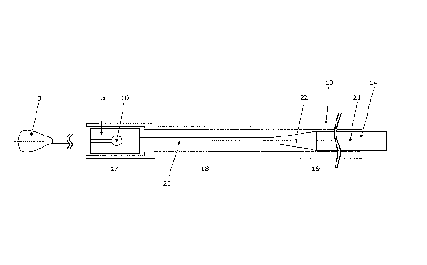
In Figure 5 an inventive embodiment of the device is depicted wherein for the
sake of
clarity the representation of the implant with its connecting elements has
been omitted. At
its distal end pusher wire 14 is provided with a pusher wire tip 9 as well as
a retaining
element 15 with cutouts 16 intended to accommodate the connecting elements
originating
from implant 1. A tube-like sheathing 13 encloses the retaining element 15
with
connecting elements fitted in place and thus prevents the implant from being
released.
CA 02922882 2016-03-01
24
According to the invention it is of significance that the outer diameter of
the tube-like
sheathing 13 varies. For this purpose, a distinction can roughly be made with
respect to
the sheathing 13 between a distal section 17 enclosing the retaining element
15, a middle
section 18 which should be highly flexible and pliable, and a significantly
longer proximal
section 19. In the interest of bringing about sufficient pliability, the
middle section 18 has
an outer diameter which is smaller than that of the two other sections 17, 19.
Additionally, the diameter of the pusher wire 14 varies as well and in the
proximal section
21 is larger than in the distal section 20. In this manner the flexibility of
the pusher wire 14
and thus the entire device increases which is of significance when advancing
it through
the microcatheter in narrow blood vessels. The transition 22 between the
proximal and
distal sections of the pusher wire is tapered, i.e. gradual, in this case,
whereas the
transitions between the individual sections of the tube-like sheathing 13 are
of step-like
configuration. This is produced by plastic deformation.
In Figure 6 a similar embodiment is shown wherein again the tube-like
sheathing 13 is
produced by plastic deformation. However, other than with the embodiment
illustrated in
Figure 5 the transitions between the distal section 17 and middle section 18
and between
the middle section 18 and proximal section 19 are tapered, that is they have a
more
gradual contour.
As per Figure 7 the tube-like sheathing 13 is designed to comprise a plurality
of parts and
is thus composed of several sheathing segments bonded in an overlapping
fashion. In this
context, the sheathing segment forming the middle section 18 of sheathing 13
has a
diameter lower than that of the sheathing segments of distal and proximal
sections 17, 19.
The individual sheathing segments may in particular be connected by adhesive
bonding.
Figure 8 illustrates an embodiment wherein the tube-like sheathing 13 is of
one-piece
configuration. From a sheathing having a uniform outer diameter and uniform
wall
thickness material is removed from the outside of the distal section 17 and
the middle
section 18 so that the outer diameter and the wall thickness are reduced in
these
sections. In this way, a tube-like sheathing 13 is obtained that has high
distal flexibility and
pliability.
CA 02922882 2016-03-01
Figure 9 also shows a one-piece tube-like sheathing 13. However, other than is
shown in
Figure 8 the distal section 17 has an outer diameter larger than that of
middle section 18.
This may prove especially expedient if the diameter of retaining element 15 is
larger.
Although the transitions between the individual sections 17, 18, and 19 are
shown in
5 Figures 8 and 9 to have a step-like contour, rounded or chamfered
transitions may of
course also be provided, however. Likewise, several steps may be arranged at
the
transitions.
In conclusion, Figure 10 serves to illustrate an embodiment wherein the tube-
like
sheathing 13 is also of one-piece design but has an outer diameter that
constantly
10 reduces from the proximal section 19 to distal section 17 so that the
sheathing 13 has a
moderately conical shape. In the distal and middle sections 17, 18 material is
removed
from the outside of the sheathing 13 by means of turning or grinding methods.
This also
results in the pliability of sheathing 13 to increase distally.