Note: Descriptions are shown in the official language in which they were submitted.
1
Novel Peptides and Analogs for Use in the Treatment of Oral Mucositis
Related Applications
This application claims priority from U.S. Provisional Application No.
61/877,767, filed
on September 13, 2013.
Introduction
Innate Immune System
The innate immune response is an evolutionarily conserved protective system
associated
with the barriers between tissues and the external environment, such as the
skin, the orogastric
mucosa and the airways. Providing rapid recognition and eradication of
invading pathogens as
well as a response to cellular damage, it is often associated with
inflammatory responses and is
a key contributor to the activation of adaptive immunity. Innate defenses are
triggered by the
binding of pathogen and/or damage associated molecules (PAMPs or DAMPs) to
pattern-
recognition receptors, including Toll-like receptors (TLRs). Pattern
recognition receptors are
found in and on many cell types, distributed throughout the body in both
circulating and tissue
resident compartments, and serve to provide early "danger" signals that lead
to the release of
non-specific antimicrobial molecules, cytokines, chemokines, and host defense
proteins and
peptides as well as the recruitment of immune cells (neutrophils, macrophages,
monocytes) in a
highly orchestrated fashion (Janeway 2002; Beutler 2003; Beutler 2004; Athman
2004; Tosi 2005;
Doyle 2006; Foster 2007; Matzinger 2002). Moreover the innate immune system is
directly
involved in the generation of tolerance to commensal microbiota in the
gastrointestinal tract and
in gastrointestinal repair and immune defense (Santaolalla, 2011; Molloy
2012).
Mucositis
CA 2923553 2018-09-20
CA 02923553 2016-03-07
WO 2015/038264 PCT/US2014/050516
2
Mucositis is the clinical term for damage done to the mucosa by anticancer
therapies. It
can occur in any mucosal region, but is most commonly associated with the
mouth, followed by
the small intestine. Though many mucositis scales are used clinically, the two
most commonly
used grading systems are the NO and WHO scales.
The mechanisms of mucositis have been extensively studied and have been
recently
linked to the interaction of chemotherapy and/or radiation therapy with the
innate defense
system (Sonis 2010). Bacterial infection of the ulcerative lesions is now
regarded as a secondary
consequence of dysregulated local inflammation triggered by therapy-induced
cell death, rather
than as the primary cause of the lesions. Mucositis affects 500,000 people in
the US per year and
occurs in 40% of patients receiving chemotherapy (Sonis 2010, Curie Op,).
Mucositis almost
always occurs in patients with head and neck cancer treated with radiation
therapy (>80%
incidence of severe mucositis) (Elting et al. 2008). Mucositis is common (40-
100% incidence) in
patients undergoing high dose chemotherapy and stern cell transplantation
(SCT) where the
incidence and severity of mucositis depends greatly on the nature of the
conditioning regimen
used for myeloahlation (Murphy 2007), Of well-established chemotherapy drugs,
5-FU and
irinotecan are particularly noted for causing mucositis but it also occurs
with newer agents such
as mTOR inhibitors and kinase inhibitors (Nlateus et al. 2009; Sankhala et al.
2009). Mucositis
can be seriously debilitating and can lead to infection, sepsis, the need for
parenteral nutrition
and narcotic analgesia. The intestinal damage causes severe diarrhea. These
symptoms can limit
the doses and duration of cancer treatment, thus leading to sub--optimal
treatment outcomes
including reduced survival. Direct and indirect consequences of mucositis have
been estimated
to add ¨$18K per patient to cancer treatment costs (Nonzee at al. 2008).
Mucositis occurs 3-12
weeks after the initiation of radiation, or 3-12 days after the initiation of
chemotherapy, and
resolves after 2-3 weeks, assuming no further chemotherapy or radiation
treatment is
undertaken.
R1VPA (SEQ ID NO. 5) is an 1DR (Innate Defense Regulator), a new class of
short, synthetic.
peptides with a novel mechanism. Designed to mimic one of the recently
discovered functions of
natural mucosal defense peptides, 1DRs have no direct antibiotic activity but
modulate host
SUBSTITUTE SHEET (RULE 26)
CA 02923553 2016-03-07
WO 2015/038264 PCT/US2014/050516
3
responses, increasing survival after infections with a broad range of
bacterial Gram-negative and
Gram-positive pathogens, as well as accelerating resolution of tissue damage
following exposure
to a variety of agents including bacterial pathogens, trauma and cherno- or
radiation-therapy.
Based on preclinicai data obtained in models of chemotherapy-induced
mucositis,
radiation-induced mucositis, neutropenic infection and colitis, oral mucositis
is a promising
indication for RIVPA (SEQ. ID NO, 5) and other IDR peptides. Since the drug
would be given soon
after the chemotherapy infusion or radiation, the IV dosage form of RIVPA
(SEQ. ID NO. 5) is well
suited to the mucositis indication. Preclinical efficacy results obtained with
RIVPA (SEQ ID NO. 5)
in mouse and hamster models of mucositis indicate that dosing every third day
should be able to
cover the mucositis "window" with seven to fourteen doses, depending on the
duration of
chemotherapy or radiation exposure.
With regard to breast cancer, -20% of patients receiving ACT therapy suffer
ulcerative
mucositis, during their first round of chemotherapy but ¨70% of that subset of
patients will have
ulcerative mucositis on their second round (Sonis 2010). This represents a
'high risk" patient
population that would benefit from RIVPA (HQ ID NO, 5) treatment, There are
currently no
systemic agents approved for the amelioration of mucositis in this population.
Patients undergoing high dose chemotherapy and SCT for the treatment of
hematologic
cancers are an immunosuppressed population at high risk of infection. In this
treatment, high
doses of chemotherapy (sometimes in combination with radiation), a
"conditioning regimen",
are used to di a large proportion of the cancer cells. These treatment levels
would cause lethal
myelosuppresion unless stem cells (from bone marrow or blood) are administered
afterwards to
allow reconstitution of blood cells. Autologous transplants use the patient's
own stern cells for
this purpose while allogeneic transplants use cells from a matched healthy
donor. Autologous
transplants are used most often in the treatment of Multiple Myelorna (MM) and
non-Hodgkins
Lymphoma (NHL), Allogeneic transplants are typically used to treat leukemias
such as AML.
With regard to SCT, until recently the various conditioning chemotherapy
regimens all
resulted in a relatively high rate of oral mucositis (40-100%) and in most US
centers these patients
are managed in-hosnital. Oral mucositis associated with radiation and/or
chemoradiation
SUBSTITUTE SHEET (RULE 26)
CA 02923553 2016-03-07
WO 2015/038264 PCT/US2014/050516
4
therapy for head & neck cancer is a major problem, with 85% of subjects
suffering some degree
of mucositis ¨ 42% being grade 3 or 4.
Acute Radiation Syndrome
Acute radiation syndrome (ARS) is a serious illness that occurs when the
entire body (or
most of it) receives a high dose of radiation, typically over a short period
of time. Many survivors
of the Hiroshima and Nagasaki atomic bombs in the 1940s and many of the
firefighters who first
responded after the Chernobyl Nuclear Power Plant accident in 1986 became ill
with ARS (CDC
2013).
Individuals exposed to radiation will get ARS only if the:
di the radiation dose was high (doses from medical procedures such as chest X-
rays are
too low to cause ARS),
.the radiation was penetrating (that is, able to reach internal organs),
othe person's entire body, or most of it, received the dose, and
*the radiation was received in a short time, usually within minutes.
Radiation induces dose-proportional injury to mammalian cells and tissues. At
low doses,
the injury may be limited to point mutations in somatic and/or germ-line DNA
that may be
associated with long-term effects such as an increased risk of cancer or birth
defects. At
intermediate doses, radiation induces chrornosomal abnormalities such as
breaks and
translocations, which again increases the risk of cancers and birth defects,
and if severe enough
will result in the death of rapidly dividing cells within hours of exposure.
At very high doses,
radiation can denature proteins, resulting in almost immediate death of cells
and tissues. The
tissues with rapidly dividing cells that are the most commonly affected by
moderate doses of
radiation include the bone marrow, the gastrointestinal tract and the testis
Exposure to radiation
is associated with acute effects, including skin rashes and burns, bone marrow
failure, including
anemia, depressed white blood cell counts, and thrombocytopenia, as well as
gastrointestinal
toxicity such as diarrhea, and more chronic effects such as the development of
tumors, especially
sarcomas and leukemias, and birth defects.
SUBSTITUTE SHEET (RULE 26)
CA 02923553 2016-03-07
WO 2015/038264 PCT/US2014/050516
The first symptoms of ARS typically are nausea, vomiting, and diarrhea. These
symptoms
will start within minutes to days after the exposure, will last for minutes up
to several days, and
may come and go, Then the person usually looks and feels healthy for a short
time, after which
he or she will become sick again with loss of appetite, fatigue, fever,
nausea, vomiting, diarrhea,
and possibly even seizures and coma. This seriously ill stage may last from a
few hours up to
several months,
People with ARS typically also have some skin damage. This damage can start to
show
within a few hours after exposure and can include swelling, itching, redness
of the skin and hair
loss. As with the other symptoms, the skin may heal for a short time, followed
by the return of
swelling, itching, and redness days or weeks later, Complete healing of the
skin may take from
several weeks up to a few years depending on the radiation dose the person's
skin received.
The gastrointestinal manifestation of ARS is referred to as gastrointestinal
acute radiation
syndrome or GI-ARS. GI-ARS consists of diarrhea, dehydration, enterobacterial
infection, and in
severe cases, septic shock and death (Patten 1990). Following radiation
exposure, GI-ARS is
thought to be caused by direct damage to stern cells within the base of the
crypts of Lieberkuhn,
resulting in mitotic cessation and death through apoptotic mechanisms (Pollen
1997a, Potten
1997b). The integrity of gastrointestinal mucosa depends on a rapid
proliferation of a pool of
pluripotent stern cells at the bottom of the crypts (Brittan 2002, Gordon
1994, Potten 1997b).
Thus, stem cell death is thought to be the critical element in this process,
since surviving intestinal
stem cells appear to be sufficient for reconstitution of a crypt-villus unit
(Potten 1990). Renewal
of the intestinal epithelial barrier depends upon an active stem cell
compartment similar to the
hematopoietic system. Intestinal crypt-villus precursor clonogen cells are
particularly sensitive to
ionizing radiation exposure such that with increasing radiation dose, crypt-
villus clonogen cells
cannot produce enough cells to repopulate the viHi. This results in blunting
and diminution in
villus height and eventual functional incapacity, leading to decreased
nutrient absorption and
barrier function, loss of fluid and electrolytes, and bacterial translocation
through the intestinal
barrier (Monti 2005, Zhao 2009). Above 8 Gray (Gy), dose-dependent stern cell
death leads to
reduction of crypt regeneration, until the level of regeneration is
insufficient to rescue the GI
SUBSTITUTE SHEET (RULE 26)
CA 02923553 2016-03-07
WO 2015/038264 PCT/US2014/050516
6
mucosa. From studies in mice, progressive denudation of the epithelium leads,
by day 6 to 7
after radiation, to death from the GI syndrome. When mitotic activity resumes,
precipitous
depletion of crypts ensues, presumably as a result of the onset of
reproductive death of crypt
clonogens (Withers 1971). At the lower-dose range (8-13 Gy), surviving
clonogens regenerate
the crypt system, leading to complete recovery of injured mucosa. At doses
exceeding 14 Gy,
massive clonogen loss causes collapse of the crypt-villus system, mucosal
denudation and animal
death from the gastrointestinal syndrome (Paris 2001; Potten 1990; Withers
1971; Withers
1969).
The intestinal stern cell compartment is not the only compartment sensitive to
ionizing
radiation. Another critical factor involving the response of the CI tract to a
major physical insult
is hypoperfusbn of the intestine. Persistent gut hypoperfusion is an important
inciting event in
the development of the systemic inflammatory response syndrome and multi-organ
failure
(MOP) (Moore 1999). increased intestinal vascular permeability together with
capillary leakage
has been observed in the early period after irradiation (Cockerham 1984; Eddy
1968, Willoughby
1960). Additional post-irradiation alterations include moderate dilatation and
tortuosity of small
arterial vessels, reduction in numbers and/or lengths of vessels followed by
later occurring
hemorrhagic patterns (Eddy 1968). There has been an ongoing controversy
concerning whether
the primary lesion after irradiation is intestinal epithelium stem cell death
or a result of
endothelial cell death (Kirsch 2010), Regardless of primary lesion, it is
clear that irradiation
results in a complex injury response including death of intestinal epithelial
cells, endothelial cells
and gut hypoperfusion (Williams 2010).
Treatment modalities such as hematopoietic growth factors, i.e., granulocyte-
and/or
granulocyte-macrophage colony stimulation factors (G-CSF and GNI-CSF) and
erythropoietin
(EPO), and hematopoietic stem cell/bone marrow transplantation, are available
to attenuate
mortality from hematopoietic failure.
The chance of survival for people with ARS decreases with increasing radiation
dose. The
cause of death within 15 days of radiation exposure is usually damage to the
GI tract whereas
SUBSTITUTE SHEET (RULE 26)
CA 02923553 2016-03-07
WO 2015/038264 PCT/US2014/050516
7
after 15 days death usually is a consequence of bone marrow injury. For the
survivors, the
recovery process may last from several weeks up to 2 years (CDC 2013).
There is an urgent need for the development of radiation mitigators, as there
currently
are none approved for the treatment of acute radiation syndrome. RIVPA (SEQ.
ID NO. 5) has the
potential to decrease the acute mortality in ARS, enabling supportive care
efforts, and to aid in
the recovery of skin damage,
Infection
A variety of microorganisms, including viruses, bacteria, fungi and parasites
can cause
disease, Microbial cells are distinct from cells of animais and plants that
are unable to live
alone in nature, existing only as parts of multicellular organisms. Microbial
cells can be
pathogenic or non-pathogenic, depending, in part, on the microorganism and the
status of the
host. For example, in an immunocompromised host, a normally harmless bacterium
can
become a pathogen. Entry into host cefis is critical for the survival of
bacterial pathogens that
replicate in an intracellular milieu. For organisms that replicate at
extraceilular sites,
significance of bacterial entry into host cells is less well defined.
Drug resistance remains an obstacle in the ongoing effort to fight infection.
For
example, penicillin was effective in treating Staphylococcus aureus until the
bacterium became
resistant. Throughout the second half of the 20th century, new antibiotics,
such as vancomycin
and methicillin, were developed; these successfully cured S. aureus infection.
However,
methicillin-resistant strain of S. aureus evolved in the 1970s, and have been
plaguing hospitals
worldwide ever since. More recently, vancomycin-resistant strains ot S. aureus
have surfaced.
With the increasing threat of resistance to antimicrobial drugs and the
emergence of
new infectious diseases, there exists a continuing need for novel therapeutic
compounds.
Therapeutics that act on the host, not the pathogen, are desirable, because
they do not
encourage pathogenic resistance. in particular, drugs that act on the host via
the innate
immune system provide a promising source of therapeutics. There is evidence to
indicate that
innate responses are instrumental in controlling most infections, and also
contribute to
SUBSTITUTE SHEET (RULE 26)
8
inflammatory responses. Inflammatory responses triggered by infection are
known to be
central components of disease pathogenesis. An ability to increase host
resistance to infection,
while controlling inflammation, would be very beneficial in the ongoing battle
against infection,
including infection caused by resistant organisms.
IDRs and the Innate Immune System
Innate Defense Regulators (IDRs) interact with intracellular signaling events
and modulate
the innate defense response. Whereas much of the initial work with the IDRs
focused on their
role in fighting infection while controlling inflammation, recent results in
animal models of
chemotherapy- or radiation-induced mucositis and wound healing suggest that
IDRs can be
beneficial during the responses to a broader range of damage-inducing agents
beyond
pathogens. IDRs treat and prevent infections by selectively modifying the
body's innate defense
responses when they are activated by PAMPs or DAMPs, without triggering
associated
inflammation responses (Matzinger 2002). The same mechanisms underlie positive
effects seen
in mucositis and wound healing models, where signaling downstream of the
recognition of
DAMPs is affected. RIVPA (SEQ ID NO. 5) has demonstrated safety in humans and
efficacy in
animal models of fractionated radiation-induced and chemotherapy-induced oral
mucositis, in
models of chemotherapy induced damage to the gastro-intestinal tract and in
models of local
and systemic Gram-positive and Gram-negative infection in immunocompetent and
immunocompromised hosts.
In accordance with an aspect of the present invention there is provided the
use of an
effective amount of a peptide for treating oral mucositis in a subject who has
been exposed to a
damaging amount of radiation or chemotherapeutic agent(s), wherein the peptide
comprises the
amino acid sequence of any one of SEQ ID NO: 5, SEQ ID NO: 91 and SEQ ID NO:
92, or a
pharmaceutical salt, ester or amide thereof.
In accordance with a further aspect of the present invention there is provided
an isolated
peptide comprising the amino acid of SEQ ID NO. 91 or SEQ ID NO. 92 or a
pharmaceutical salt,
ester or amide thereof.
Date Recue/Date Received 2022-04-14
8a
In accordance with a further aspect of the present invention there is provided
an isolated
peptide consisting of the amino acid sequence of:
R(tBg)V1KR(tBg)V2,
wherein tBg = tert-butyl glycine,
and further wherein R(tBg)V2 is linked via an amide bond between V2 and the
lysine amino group in the side chain (SEQ ID NO. 91).
In accordance with a further aspect of the present invention there is provided
an isolated
peptide consisting of the amino acid sequence:
RIV(mp2)A-NH2, wherein mp2 = 4-Amino-1-methyl-1H-pyrrole-2-carboxylic acid
CH3
1
5N)
COOH
NH2
(SEQ ID NO. 92).
In accordance with a further aspect of the present invention there is provided
the use of
an isolated peptide comprising the amino acid of SEQ ID NO. 91, or SEQ ID NO.
92 or a
pharmaceutical salt, ester or amide thereof.
In accordance with a further aspect of the present invention there is provided
the use of
a peptide consisting of an amino acid sequence selected from the group
consisting of SEQ ID
NO:5, SEQ ID NO:91 and SEQ ID NO:92 for treating an individual suffering from
mucositis, colitis
or acute radiation sickness.
BRIEF DESCRIPTION OF THE FIGURES
Figure 1. RIVPA (SEQ ID NO. 5) reduces the duration of severe oral mucositis
in a fractionated
radiation model.
Figure 2. RIVPA (SEQ ID NO. 5) reduces the duration of severe oral mucositis
in a fractionated
radiation model using an optimized dosing regimen.
Date Recue/Date Received 2022-04-14
8b
Figure 3. RIVPA (SEQ ID NO. 5) reduces the severity of DSS-induced colitis as
measured by
endoscopy on days 7, 14, and 21 (A) and histopathology on day 21 (B,C,D).
Date Recue/Date Received 2022-04-14
CA 02923553 2016-03-07
WO 2015/038264 PCT/US2014/050516
9
Figure 4. RIVPA (SEQ ID NO. 5) reduces the duration of severe oral mucositis
(A), severity of colitis
(B) and body weight loss (C) in a chemotherapy model (First study).
Figure 5. RIVPA (SEQ ID NO. 5) reduces the duration of severe oral mucositis
(A), severity of colitis
(B) and body weight loss (C) in a chemotherapy model (Second study).
Figure 6. RIVPA (SEQ ID NO. 5) reduces the duration of severe oral mucositis
(A), severity of colitis
(B) and body weight loss (C) in a chemotherapy model in a dose responsive
manner.
Figure 7. Combination of RIVPA (SEQ ID NO. 5) and Vancornycin treatment in an
MRSA IP
infection model.
Figure 8. RIVPA (SEO, ID NO. 5) activity in neutropenic mice in the thigh
abscess MRSA infection
model.
Figure 9. Dose response of RIVPA (HQ ID NO. 5) in the MRSA bacteremia model in
immunocom petent mice,
Figure 10. Dose response of RIVPA (SEQ ID NO. 5) in the MRSA bacteremia model
in mice lacking
T-cells.
Figure 11. Therapeutic RIVPA (SEQ ID NO. 5) efficacy in the S. aureus acute
peritoneal infection
model.
Figure 12. RIVPA (SEQ ID NO. 5) activity in neutopenic mice in the thigh
abscess .S. aureus
infection model.
Figure 13. RIVPA (SEQ ID NO. 5) efficacy in a Kiebsiella peritoneal infection
model with high (A)
and low (B) bacterial infection. * Absence of a bar in (A) indicates all mice
died (0% survival).
Figure 14. RIVPA (SEQ ID NO. 5) enhances resolution of tissue damage in
topically MRSA-infected
skin. 43h Bacterial Burden (A), 96h Scatter plot (B), Blinded Photographic
Scoring at 4811 (C),
Blinded Photographic Scoring at 96n (D). * Absence of a bar in (C) and (D)
indicates all mice had
a zero score, yielding a mean and standard error of the mean (SEM) of 0 0.
SUBSTITUTE SHEET (RULE 26)
CA 02923553 2016-03-07
WO 2015/038264 PCT/US2014/050516
Figure 15. Lack of RIVPA (SEQ ID NO, 5) on recovery of circulating blood cell
leukocytes (A) or
neutrophils (B) after induction of leukopenia in CD-1 mice.
Figure 16. R(tBg)V1KR(tBg)V2 (SEQ ID NO. 91) reduces the severity of oral
mucositis in a
chemotherapy model.
Figure 17. RIV(rnp2).A-NH2 (SEQ ID NO. 92) reduces the severity of oral
mucositis in a
chemotherapy model.
Figure 18. R(tBg)V1KR(t8g)V2 (SEQ ID NO, 91) enhances survival in an MRS,A
bacteremia model.
Figure 19. RIV(rnp2)A-NH2 (SEQ ID NO. 92) enhances survival in an MRSA
bactere.rnia model.
DETAILED DESCRIPTION OF THE INVENTION
It is an object of the present invention to provide an isolated peptide
consisting of the
amino acid sequence of R(tBg)V1KR(tBg)V2, wherein Iftg tert-butyl glycine and
hither wherein
R(tBg)V2 is linked via an amide bond between V1 and K.
It is an object of the present invention to provide An isolated peptide
consisting of the
amino acid sequence of RIV(rnp2)A-NH2,
wherein mp2 = 4-4mino4-methyl-1H-pyrrole-2-carboxylic acid
CH3
\?/----COOH
NH2
It is yet another object of the present invention to provide a method of
treating oral
mucositis in a subject who has been exposed to a damaging amount of radiation
or
chemotherapeutic agents, comprising administering to the patient an effective
amount of:
SUBSTITUTE SHEET (RULE 26)
CA 02923553 2016-03-07
WO 2015/038264 PCT/US2014/050516
11
a) a peptide comprising an amino acid sequence of Table 1; or
b) a peptide comprising the amino acid sequence of any of SEQ ID NOs: 5,7,
10, 14,
17, 18, 22, 23, 24, 27, 28, 31, 34, 35, 63, 64, 66-69, 72, 76, 77, 90, 91 and
92 or a pharmaceutical
salt, ester or amide thereof and a pharmaceutically-acceptable carrier,
diluent or excipient.
It is an object of the present invention to provide a method of treating oral
mucositis in
a subject who has been exposed to a damaging amount of radiation or
chemotherapeutic
agents, comprising administering to the patient an effective amount of:
a) a peptide comprising an amino acid sequence of up to 7 amino acids,
said
peptide comprising the amino acid sequence of X1X2X3P (SEQ ID NO: 56),
wherein:
X1 is R;
X2 is I or V. wherein X2 can be N-methylated;
X3 is 1 or V, wherein X3 can be N-methylated,
P is proline or a proline analogue;
wherein SEQ ID NO: 56 if the first four amino acids at the N-terminus of the
peptide, or a pharmaceutical salt, ester or amide thereof and a
pharmaceutically-acceptable
carrier, diluent, or excipient; or
b) a peptide comprising the amino acid sequence of any of SEQ ID NOs:
5,7, 10, 14,
17, 18, 22, 23, 24, 27, 28, 31, 34,, 35, 63, 64, 66-69, 72, 76, 77, 90 and 92
or a pharmaceutical
salt, ester or amide thereof and a pharmaceutically-acceptable carrier,
diluent or excipient,
It is another object of the present invention to provide a method of treating
oral
mucositis in a subject who has been exposed to a damaging amount of radiation
or
chemotherapeutic agents, wherein the peptide is SEQ ID NO: 5 or a
pharmaceutical salt, ester,
or amide thereof and a pharmaceutically-acceptable carrier, diluent, or
excipient.
SUBSTITUTE SHEET (RULE 26)
CA 02923553 2016-03-07
WO 2015/038264 PCT/US2014/050516
12
It is another object of the present invention to provide a method of treating
oral
mucositis in a subject who has been exposed to a damaging amount of radiation
or
chemotherapeutic agents, wherein the peptide is administered orally,
parenterally,
transdermally, intranasally.
It is yet another object of the present invention to provide a method of
treating oral
mucositis in a subject who has been exposed to a damaging amount of radiation
or
chemotherapeutic agents, wherein the effective amount of peptide administered
to a subject is
at least 1 mg/kg. In a preferred embodiment the effect amount of peptide
administered to a
subject is about 13 mg/kg to 6 mg/kg.
It is yet another object of the present invention to provide a method of
treating oral
mucositis in a subject who has been exposed to a damaging amount of radiation
or
chemotherapeutic agents, wherein the peptide is administered to the subject
every third day
during radiation or chemotherapeutic agent administration.
It is still another object of the present invention to provide a method of
treating oral
mucositis in a subject who has been exposed to a damaging amount of radiation
or
chemotherapeutic agents, wherein the peptide is administered in combination
with an oral
dosage form of a topically active corticosteroid or a metabolite thereof to
the subject, wherein
the oral dosage form is effective for topical or local treatment of the
gastrointestinal tract and
oral cavity of the subject and further wherein the subject exhibits symptoms
of inflammation
due to tissue damage arising from radiation or chemotherapy treatment.
Representative
topically active corticosteroids include, but are not limited to,
beclomethasone 17,21-
dipropionate, alclomethasone dipropionate, budesonide, 22S budesonide, 22R
budesonide,
beclornethasone-17-monopropionate, clobetasol propionate, diflorasone
diacetate, flunisolide,
flurandrenolide, fluticasone propionate, halobetasol propionate, halcinocicie,
rnometasone
furoate, and triamcinolone acetonide. In a preferred embodiment of this
invention, the
topically active corticosteroid is beclomethasone diproprionate. The effective
amount of
topically active corticosteroid in each dosage form may vary from patient to
patient, and may
be readily determined by one skilled in the art by well-known does-response
studies, Such
SUBSTITUTE SHEET (RULE 26)
CA 02923553 2016-03-07
WO 2015/038264 PCT/US2014/050516
13
effective amounts will generally range between about 0.1 mg/day to about 8
mg/day, and more
typically range from about 2 mg/day to about 4 mg/day,
It is still another object of the present invention to provide a method
mitigating the
gastrointestinal, hematopoietic and cutaneous impacts of acute radiation
syndrome in a subject
who has received a high, penetrating dose of radiation to a substantial
portion of their body in a
short period of time.
It is still another object of the present invention to provide a method of
treating acute
radiation syndrome in a subject who has received a high, penetrating dose of
radiation to a
substantial portion of their body in a short period of time, wherein the
peptide is administered
in combination with an oral dosage form of a topically active corticosteroid
or a metabolite
thereof to the subject, wherein the oral dosage form is effective for topical
or local treatment
of the gastrointestinal tract and oral cavity of the subject and further
wherein the subject
exhibits symptoms of inflammation due to tissue damage arising from radiation
or
chemotherapy treatment. Representative topically active corticosteroids
include, but are not
limited to, beclomethasone 17,21-dipropionate, alclomethasone dipropionate,
budesonide, 22S
budesonideõ 22R budesonideõ beclomethasone47-monopropionate, ciobetasol
propionate,
difiorasone diacetate, flunisolide, flurandrenolide, fluticasone propionate,
halobetasol
propionate, halcinocide, mometasone furcate, and triamcinolone acetonide. In a
preferred
embodiment of this invention, the topically active corticosteroid is
beclomethasone
diproprionate. The effective amount of topically active corticosteroid in each
dosage form may
vary from patient to patient, and may be readily determined by one skilled in
the art by well-
known does-response studies. Such effective amounts will generally range
between about 0,1
mg/day to about 8 mg,/day, and more typically range from about 2 mg/day to
about 4 mg/day.
It is still another object of the present invention to provide a method of
treating and/or
preventing infection (e.g., a microbial infection) in a subject, by
administering to the subject a
peptide having or comprising the amino acid sequence of TABLE 1 or an
analogue, derivative, or
variant thereof or obvious chemical equivalent thereof, By way of example, the
subject may
SUBSTITUTE SHEET (RULE 26)
CA 02923553 2016-03-07
WO 2015/038264 PCT/US2014/050516
14
have, or be at risk of having, infection. In one embodiment, the peptide
modulates innate
immunity in the subject, thereby treating and/or preventing the infection in
the subject.
Exemplary infections which may be treated and/or prevented by the method of
the
present invention include an infection by a bacterium (e.g., a Gram-positive
or Gram-negative
bacterium), an infection by a fungus, an infection by a parasite, and an
infection by a virus. In
one embodiment of the present invention, the infection is a bacterial
infection (e.g., infection
by E. coli, Klebsiella pneurnoniae, Pseudomonas aeruginosa, Salmonella spp.,
Staphylococcus
aureus, Streptococcus spp., or vancomycin-resistant enterococcus). In another
embodiment,
the infection is a fungal infection (e.g., infection by a mould, a yeast, or a
higher fungus). In still
another embodiment, the infection is a parasitic infection (e.g,, infection by
a single-celled or
rnulticellular parasite, including Giardia duodenal's, Cryptosporidium parvum,
Cyclospora
cayetanensis, and Toxoplasrna gondii). In yet another embodiment, the
infection is a viral
infection (e.g., infection by a virus associated with AIDS, avian flu,
chickenpox, cold sores,
common cold, gastroenteritis, glandular fever, influenza, measles, mumps,
pharyngitis,
pneumonia, rubella, SARSõ and lower or upper respiratory tract infection (e.g,
respiratory
syncytialvirus)).Formulation of the Dosage Form
The dosage form of RIVPA (SEC. ID NO. 5) is an aqueous, aseptically processed,
sterile
solution for injection. Each vial contains 5 rnt_ of a 60 mg/mL solution (300
mg of RIVPA (SEQ. ID
NO. 5)). RIVPA (SEQ ID NO. 5) is formulated in Water for Injection and pH
adjusted to a target
value of 6Ø The formulation contains no excipients and has an osmolality of
¨300 mOsm/kg,
Route of Administration
RIVPA (SEC. ID NO. 5) drug product will be diluted in sterile saline to the
appropriate
concentration for injection, determined on a mg/kg basis by the recipient's
weight and the
designated dose level. Diluted RIVPA (SEC. ID NO. 5) will be administered as
an intravenous (IV)
infusion in 25 mL over 4 minutes, once every third day.
EXAMPLES
Peptide Synthesis
SUBSTITUTE SHEET (RULE 26)
CA 02923553 2016-03-07
WO 2015/038264 PCT/US2014/050516
The peptides in Table 1 were synthesized using a solid phase peptide synthesis
technique.
All the required Fmoc-protected amino acids were weighed in three-fold molar
excess
relative to the 1 mmole of peptide desired. The amino acids were then
dissolved in
Dimethylformaide (DMF) (7.5 ml) to make a 3mMol solution. The appropriate
amount of Rink
amide MBHA resin was weighed taking in to account the resin's substitution.
The resin was then
transferred into the automated synthesizer reaction vessel and was pre-soaked
with
Dichloromethane (DCM) for 15 minutes.
The resin was de-protected by adding 25% piperidine in DMF (30 ml) to the
resin and
mixing for 20 minutes, After de-protection of the resin the first coupling was
made by mixing the
3mMol amino acid solution with 4mMol 2-(1H-benzitriazole-1-y1)-1,1,3,3-
tetramethyluronium
hexafluorophosphate (HBTU) and 8rnMol NN-diisopropylethylamine (DIEPA). The
solution was
allowed to pre-activate for 5 minutes before being added to the resin. The
amino acid was
allowed to couple for 45 minutes,
After coupling the resin was thoroughly rinsed with DMF and
Dimethylacetarnicie (DMA).
The attached Fmoc protected amino acid was deprotected in the same manner
described above
and the next amino acid was attached using the same coupling scheme
AA:HBTU:DIEPA.
After the completion of the synthesis the peptide was cleaved from the resin
with the use
of a cleavage cocktail containing 97,5 % Trifluoroacetic acid (TFA) and 2.5%
water. The resin was
allowed to swim in the cleavage cocktail for 11/2 hours, The solution was then
filtered by gravity
using a Buchner funnel and the filtrate was collected in a 50 ml
centrifugation tube. The peptide
was isolated by precipitating with chilled diethyl ether. After centrifuging
and decanting diethyl
ether the crude peptide was washed with diethyl ether once more before being
dried in a vacuum
desiccator for 2 hours, The peptide was then dissolved in de-ionized water (10
ml), frozen at
80cC and lyophilized. The dry peptide was then ready for HPLC purification.
Due to the hydrophilic nature of these peptides the diethyl ether peptide
isolation did not
work. Therefore a chloroform extraction was required. The TFA was evaporated
and the resulting
peptide residue was dissolved in 10% acetic acid (15 ml). The impurities and
scavengers were
SUBSTITUTE SHEET (RULE 26)
CA 02923553 2016-03-07
WO 2015/038264 PCT/US2014/050516
16
removed from the acetic acid peptide solution by washing the solution twice
with chloroform
(30m1). The aqueous peptide solution was then frozen at -80 C and lyophilized
resulting in a
powdered peptide ready for HPLC purification,
Peptides +RlxVPA (SEQ. ID NO. 33) and +RIVPAx (SEQ. ID NO. 34) each contained
one N-
methyl amino acid. This coupling was carried out by combining the N-methyl
amino acid, PyBroP
and N-hydroxybenzotriazole*H20 (HOBt) and DIEPA solutions together in the RV
containing the
resin. After allowing to couple for 45 minutes the N-methyl amino acid was
then doubled coupled
to ensure complete coupling. It was observed that the coupling following the N-
methyl amino
acid was not fully complete. Therefore this coupling was performed using
N,N,N1,N1-Tetramethy1-
0-(7-azabenzotriazol-1-yOuronium hexafluorophosphate (HAM) instead of HBTU,
This still
resulted in a crude peptide that typically contained two impurities totaling
30-40% of the total
purity. The peptide was purified under modified HPLC conditions to isolate the
pure peptide peak
away from the closely eluting impurities.
R(tBg)V1KR(tBg)V2 (SEQ. ID NO. 91) is an 8-residue peptide dendrimer with
symmetrical
branches occurring off of a fourth amino acid lysine that possesses two
functional amine groups.
The peptide has been synthesized with solid-phase peptide synthesis
techniques, utilizing a di-
Frnoc protected fourth amino acid to facilitate the coupling of the branches,
using the general
synthesis techniques described above.
In addition, these peptides can also be synthesized with solution phase
peptide synthesis
techniques (Tsuda et al. 2010) and commonly known to experts in the art.
SUBSTITUTE SHEET (RULE 26)
CA 02923553 2016-03-07
WO 2015/038264 PCT/US2014/050516
17
Efficacy in Oral Mucositis
RIVPA (SEQ. ID NO. 5) and other IDRs modulate the innate defense response to
tissue
injury, reducing the severity of damage caused by the inflammatory cascade and
enhancing
resolution of disease. This attribute of IDRs has been demonstrated in
chemotherapy-Induced
oral and GI mucositis in mice, in radiation-induced oral mucositis in hamsters
and in DSS-induced
colitis in mice. In each of these models, the initial damage is thought to
trigger a cascade of innate
defense signaling which increases the severity of the injury (Marks 2011;
Sonis 2010), RIVPA (SEQ.
ID NO. 5) and other IDRs offset the signaling cascade, reducing the resultant
severity of the injury
and reducing the duration of severe tissue damage.
The optimum dosing regimen for RIVPA (SEQ. ID NO. 5) and other IDRs identified
in the
M RSA bacteremia model has been further confirmed in injury models, where the
longer duration
of disease makes repeat dosing more informative. Dosing of 25 mg/kg every
third day was found
to be optimal, reflecting the durable pharmacodynarydc impact of RIVPA and
other IDRs (SEQ. ID
NO, 3) despite its rapid PK clearance (within minutes) from the circulation of
mice,
RIVPA (SEO ID NO. 5) significantly reduced the severity and duration of
rnucositis in a
model of radiation-induced oral mucositis in hamsters, particularly when
administered every
third day during the fractionated radiation therapy. These studies confirmed
that optimal dosing
of RIVPA (SEQ. ID NO. 5) involves dosing every third day and that the 25 mg/kg
dose level is
effective. In this model, cannuiated male Golden Syrian hamsters were treated
with 7.5 Gy of
radiation, directed at the everted left cheek pouch, on Days 0, 1, 2, 3, 6, 7,
8 and 9. Mucositis was
evaluated every second day between Days 7 and 35, with peak mucositis severity
generally
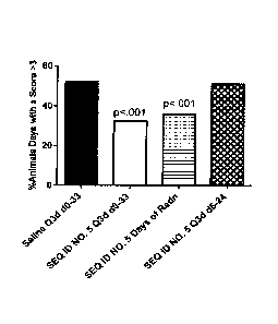
occurring around Day 19. In the first study, RIVPA (SEQ. ID NO. 5) (25 mg/kg
IV) was administered
either every third day starting on Day 0 and continuing until Day 33 (O3d d0-
33), or on days of
radiation therapy (Days 0, 1, 2, 3, 4, 7, 8, 9) or every third day starting on
Day 6 and continuing
to Day 24 (O3d d6-24), On days where both RIVPA (SEQ. ID NO. 5) and radiation
was administered,
RIVPA (SEQ. ID NO. 5) was given 2 hours after radiation. The results of this
study are shown in
Figure 1. RIVPA (SEQ ID NO. 5) treatment was most effective when administered
every third day
throughout the period or on days of radiation, whereas treatment starting 6
days after initiation
of radiation was not beneficial (i.e., Q3d d6-24). A follow-up study was
undertaken to evaluate
SUBSTITUTE SHEET (RULE 26)
CA 02923553 2016-03-07
WO 2015/038264 PCT/US2014/050516
18
dosing with 25 mg/kg RI RIVPA (SEC, ID NO. 5) Q3d d0-33, on days of radiation,
or every third day
during radiation treatment (Le., Days 0, 3, 6 and 9). The results of this
study are shown in Figure
2. Treatment every third day during radiation was found to be optimal, likely
reflecting the
durability of the RIVPA (SEQ ID NO. 5) pharrnacodynamic effect, coupled with
the reduction of
injection stress caused by fewer IV injections in these small rodents.
RIVPA (SEQ ID NO. 5) has also shown efficacy in mouse models of chemotherapy-
induced
oral and gastrointestinal mucositis, consistent with the response of the
innate immune response
to chemotherapy and / or radiation damage. In these studies, RIVPA (SEQ ID NO.
5)
administration was associated with a statistically significant reduction in
the duration of severe
oral mucositis in a model of chemotherapy-induced mucositis in the mouse. A
trend towards
reduced colitis was also observed, although the mild GI damage in the control
group rendered
the result not statistically significant. In each study, 5-fl IJO rouracil (60
mg/kg IP) was administered
to male C3F1/1-1eN mice on Days -4 and -2. On Day 0, a chemical burn was
applied to the underside
of the mouse tongue, inducing mucositis which generally peaked on Day 2. Mouse
tongues were
scored for mucositis daily from Days 1 to 14, with scores ?_3 representing
severe mucositis. Body
weights were also measured daily and colitis severity was determined by video
endoscopy on
Days 4 and 7. In the first study, RIVPA (HQ ID NO. 5) (25 mg/kg IV) was
administered either once
on Day -4 immediately prior to chemotherapy, twice on Days -4 and -2
immediately after
chemotherapy or 3 times on Days 4, 2 and 5, RIVPA (SEQ ID NO. 5)
administration on multiple
occasions throughout the period of peak mucositis damage was the most
effective (i.e., on Days
-1, 2 and 5). The results of this study are shown in Figure 4. In the second
study, RIVPA (SEQ ID
NO. 5) (25 mg/kg IV) was administered on either Days -1, 2 and 5, Days -1, 1
and 3 or Days 0, 2,
and 4. The results of this study are shown in Figure S. Statistically
significant changes in the
duration of severe mucositis (Figure 5 ¨ panel A), the severity of colitis on
Day 4 (Figure 5 ¨ panel
B) and the mean body weight loss (Figure 5 ¨ panel C) correlated among the
groups. In the third
study, RIVPA (SEQ ID NO. 5) (25 or 5 mg/kg IV, as indicated) was administered
either on Days -1,
2 and 5 or on Days 1 and 3. Again, the dosing regimen utilizing RIVPA (SE0, ID
NO. 5) on every
third day was most effective, with decreased dose levels resulting in
decreased efficacy. The
rnucositis, colitis and body weight results from this study are shown in
Figure 6 as A, B, and C,
SUBSTITUTE SHEET (RULE 26)
CA 02923553 2016-03-07
WO 2015/038264 PCT/US2014/050516
19
respectively. Statistical significance was assessed for oral mucositis using a
chi-square analysis
and for body weight area under the curve (AUC) with an ANOVA on ranks.
R(tBg)V1KR(tBg)V2
(SEQ ID NO. 91) and 92 also demonstrated efficacy in the mouse model of
chemotherapy-induced
mucositis, where the mucositis scores were evaluated for 4 days after
induction of mucositis
(Figure 16, Figure 17). Treatment with R(t8g)V1KR(tBg)V2 (SEQ ID NO. 91) and
RIV(rnp2)A-NH2
(SEQ ID NO. 92) was administered on Days -1 and 2 at a dose of 25 mg/kg IV.
Efficacy in Response to Radiation Damage
RIVPA (SEQ. ID NO. 5) and other IDRs modulate the innate defense response to
tissue
injury, reducing the severity of damage caused by the inflammatory cascade and
enhancing
resolution of disease. As described above, IDRs can mitigate the response to
radiation damage in
an oral mucositis model (Figure 1, Figure 2). In another model, assessing the
prevention of
radiation-induced mucositis (25 Gy administered to the mouse snout on Day 0),
RIVPA (SEQ ID
NO. 5) (5 doses of 25 mg/kg administered IV every second day) did not have any
significant impact
on disease progression. Progressive thinning of the mouse tongue was assessed
on Days 0, 2, 4,
6, 8, and 10 by histopathological analysis of the number of basal and
suprabasal apoptotic,
mitotic and total epithelial cells per unit area and per unit length. It is
noted that the dose of
radiation used (25 Gy) was chosen such that progressive thinning of the tongue
epithelium was
observed but no overt mucositis occurred. This result demonstrates the lack of
proliferative
potential of RIVPA (SEQ ID NO. 5), and suggests that RIVPA (SEQ ID NO. 5)
effects are only
observable once the relevant pathways are stimulated by overt tissue damage or
pathogen
invasion,
Efficacy in the Gastrointestinal Tract
The ability of IV RIVPA (SEQ. ID NO. 5), administered pre-emptively or
therapeutically, to
directly protect GI mucosal surfaces was confirmed in a DSS-induced colitis
model. In this model,
DSS was administered as a 3% DSS solution in the drinking water of male
C576116 mice from Days
0 to 5 of the study. Colitis was monitored by video endoscopy on Days 7, 14
and 21. RIVPA (SEQ
ID NO. 5) (25 mg/kg IV) was administered every third day from Days 0 to 18
(Q3d d0-18), from
Days 3 to 18 (03d d3-18) or from Days 6 to 18 (03d d6-18). The results of the
study are shown
in Figure 3. By Day 14, all RIVPA (SEQ ID NO. 5) treatment regimens
demonstrated a statistically
SUBSTITUTE SHEET (RULE 26)
CA 02923553 2016-03-07
WO 2015/038264 PCT/US2014/050516
significant reduction in endoscopic colitis severity score. However, reduction
in Day 7 scores was
only observed in groups which had received at least 2 doses of RIVPA (SEQ ID
NO. 5) by that time
(i.e., Q3d d0-18 and Q3d d348 but not Q3d d6-18). On Day 21, all 3 treatment
groups appeared
to be responding in a similar manner. Histopathology of the colon on Day 21
indicated that some
RIVPA ('SEQ ID NO. 5) treated groups had statistically significantly decreased
edema and necrosis,
whereas other RIVPA (SEQ ID NO. 5) treated groups had similar responses which
did not reach
statistical significance. Statistical analysis was undertaken using t-tests
and an asterisk indicates
statistically significant differences from control (p.05).
As described above, IDRs are also able to reduce the duration and I or
severity of
gastrointestintal mucositis in a chemotherapy-induced rnucositis model (Figure
4, Figure 5, Figure
6).
Efficacy in Infected Animals
RIVPA (SEQ ID NO. 5) reduces bacterial burden and improves survival in the
presence or
absence of antibiotic treatment in various murine infection models, with
consistent efficacy at
dose levels of 25 mg/kg IV and higher and with an enduring pharmacodynamic
effect of up to 5
days. RIVPA (SEQ ID NO. 5) efficacy is complementary to antibiotic treatment
in both normal
and immune compromised mice. Efficacy of RIVPA (SEQ ID NO, 5) has been
demonstrated against
disease caused by Gram-positive (S. oureus and MRSA) and Gram-negative
(Kiebsiella, E. coil and
B. pseudarnallei) infections.
S. aureus
RIVPA (SEQ ID NO, 5) has been tested both in combination with vancornycin
treatment
and as a stand-alone treatment.
RIVPA (SEQ ID NO, 5) treatment increased survival in a MRSA peritoneal
infection model
when administered in combination with a sub-optimal antibiotic dose of
vancomycin (Study tt: D--
7-E-11). RIVPA (SEQ ID NO. 5) (50 mg/kg) or saline treatment was administered
IV either 48 or
72 h prior to inoculation with MRSA (UC6685; 8.2 x 10⁷ colony forming units
[du]) to female CF-
1 mice (N-10,rgroup). Vancomycin treatment (3 mg/kg) was administered
subcutaneously (SC),
SUBSTITUTE SHEET (RULE 26)
CA 02923553 2016-03-07
WO 2015/038264 PCT/US2014/050516
21
1 and 5 h after infection. Survival was monitored once daily for 5 days. The
results of this study
are shown in Figure 7.
RIVPA (SEQ, ID NO. 5) is also effective when administered by itself. Multiple
studies with
IV administered RIVPA (SEQ ID NO. 5) were conducted in a MRSA bacteremia
model. RIVPA (SEQ
ID NO, 5) administration demonstrated a dose response in this model in either
immunocompetent Bali* mice or nu/nu mice lacking T-cells, with a single dose
of 50 mg/kg
resulting in statistically significant enhanced survival over the saline
control. In the first study,
MRSA (USA300õ 7.3log10 cfu) was administered via IV injection into the tail
vein of female Balb/c
mice at time 0. Four hours prior to infection, a single dose of saline or
RIVPA (SEQ ID NO. 5) at
the indicated dose levels was injected IV into the tail vein. Sub-optimal
antibiotic treatment
(linezolid, 6,25 mg/kg) was administered once orally immediately after
infection. Survival was
monitored for 21 days after the infection. The results of this study are shown
in Figure 9. In the
second study, RIVPA (SEQ ID NO. 5) (IV) or saline (IV) was administered once 4
h prior to infection
with MRSA (strain USA300, 7,0 logo cfu) via the tail vein into female nu/nu
mice. Survival was
monitored for '14 days, as shown in Figure 10. Statistically significant
differences (i.e., ps-0.05)
in survival were found with the 50 mg/kg dose level as assessed using Kaplan
Meier analysis of
each treatment group relative to the saline control,
In summary, investigations using stand-alone IV RIVP,4 (SEQ ID NO. 5)
treatment in various
S. aureus infection studies have demonstrated that:
= The effects of RIVPA (SEQ ID NO. 5) are dose dependent between 1 and SO
mg/kg in
mouse, with dose levels of 25 mg/kg and higher consistently demonstrating
efficacy (Table 2;
Figure 9 and Figure 10). This dose level was also effective in the more
chronic disease context
available in injury models (Figure Ito Figure 6).
SUBSTITUTE SHEET (RULE 26)
CA 02923553 2016-03-07
WO 2015/038264
PCT/US2014/050516
22
Table 2: Rate of Successful Treatment of S. aureus Infection with a Single
IV RIVPA (SEQ
ID NO. 5) Treatment as a Function of Dose Level
% Successful Treatments % Successful Treatments
(i)
RIVPA (SEQ ID NO. 5) Any Dose Schedule RIVPA (SEQ ID NO. 5) administered
Dose Level (mg/kg) (# tested groups) 4 h prior
to infection (# studies)
50(ii)
100 (N=4) 100 (N=2)
?5' 67 (N=6) 50 (N=4)
5'1v)
42 (N=12) 33 (N=6)
.1(v)
0 (N=2) 0 (N=2)
(i) Successful treatments demonstrated at least a 20% increase in survival
over the relevant
saline. control,
Study 4: TPS-8-B-100, TPS-8-B-150, TPS-8-B-116, D-7-6-9
Study 4: TPS-8-B-100, TPS-8-B-150, TPS-8-B-120, TPS-8-B412
Study 4: TPS-8-B-100, TPS-8-13-150, TPS-8-B416, TPS-8-B414, TPS-8-B420, TPS-8-
B401
(v) Study 4: TPS-8-B-100, TPS-8-B-150
O Daily dosing of RIVPA (SEQ ID NO. 5) is not required and dosing every rd
or 3rcj day is
sufficie.nt In the more chronic disease context available in injury models, it
was further
confirmed that dosing every 3 day appears to be optimal (data not shown).
= RIVPA (SEQ, ID NO. 5) can be administered up to 24 h after the initiation
of infection in the
MRSA bacteremia model and still confer a survival benefit (data not shown).
Hence its action
is rapid.
= Depending on dose level, a single dose of RIVPA (SEQ ID NO. 5) can be
administered up to
days prior to the initiation of infection and still confer a survival benefit
(data not shown),
reflecting the durable pharmacodynamic impact of RIVPA (SEQ ID NO. 5) despite
its rapid
pharmacokinetic (PK) clearance (within minutes) from the circulation of mice,
O The survival benefit conferred by RIVPA (SEQ ID NO. 5) treatment can be
sustained for at
least 21 days (Figure 9).
RlVPAY* (SEQ ID NO, 90) and R(tBg)V1KR(tBg)V2 (SEQ. ID NO. 91) (5 mg/kg
administered
4 hours prior to infection) also improve survival in an MRSA bacterernia model
(Figure 18; Figure
19),
SUBSTITUTE SHEET (RULE 26)
CA 02923553 2016-03-07
WO 2015/038264 PCT/US2014/050516
23
Local adminis,tration of RIVPA (SEQ ID NO. 5) has also been demonstrated to be
effective
when the administration is local to the site of infection. In a Gram-positive
peritoneal infection
model in mouse, RIVPA (SEQ ID NO. 5) significantly reduced the bacterial load
by over 7 logs
(Study #: D-7-E-14). ntraperitoneal (IP) injection of S. aureus (Catalog Na.
25923, ATCC, 6 x 10⁷
cfu) with 5% rnucin was administered IP to female CD-1 mice (N--,=8./group)
and RIVPA (SEC( ID NO.
5) (9.5 mg/kg) was injected IP 4 h later. Mice were sacrificed 24 h after
infection and peritoneal
lavage fluid was assessed for bacterial counts. The results of this study are
shown in Figure 11
(each data point represents the result from an individual mouse ¨ dead mice
were given the
highest bacterial count of any mouse obtained in the study and are represented
as open symbols
in the graph).
RIVPA (SEQ ID NO. 5) also significantly reduced bacterial load in neutropenic
mice in an S.
aureus thigh abscess infection model when administered as a local
intramuscular (1M) injection.
Female Swiss albino mice (N8/group) were rendered neutropenic by treatment
with Cp (100
mg/kg), 3 and 1 days before 1M infection with S. aureus (Catalog No. 2921.3,
ATCC, -9.5 x 10⁵
cfu). RIVPA (SEQ ID NO. 5) (50 mg/kg) was administered IM 24 h prior to
infection and
vancornycin (100 mg/kg) was administered SC at 1, 6 and 18 h after infection.
The number of
bacterial cfu present in the infected thigh was assessed 24 h after initiation
of infection in each
group. The results of this study are shown in Figure 12.
Kiebsiella
RIVPA (SEQ. ID NO, 5) increased survival in a Gram-negative peritoneal
infection model
when administered either locally (IP) or systemically (IV). Of note, systemic
administration
appeared as good or better than local administration. RIVPA (SEQ ID NO. 5)
treatment (24 mg/kg)
was administered either I P (24 h prior to infection or 4 h post-infection) or
IV (4 h post-infection)
to female Bak*: mice (N=8/group) inoculated with Kiebsietia pneurnoniae
(Catalog No. 4381.6.
ATCC) at either 2.8 x 10⁵ cfu (Figure 13 - panel A) or 5,3 x 10² cfu (Figure
13 - panel B) and survival
was monitored over 24 h. The protective effects of RIVPA (SEQ ID NO. 5) in
this context are
shown in Figure 13. A survival endpoint is shown for animals receiving the
higher inoculum of
bacteria (panel A). All animals receiving the lower inoculum survived in all
groups (panel B) and
SUBSTITUTE SHEET (RULE 26)
CA 02923553 2016-03-07
WO 2015/038264 PCT/US2014/050516
24
were assessed for clinical signs (e.g.: piloerection, decreased movement,
hunched abdomen, etc.)
24 h after infection; these are summarized as clinical scores
Efficacy in Skin Damage
Systemically administered RIVPA (SEQ ID NO. 5) is also efficacious in the case
of skin injury
and infection, accelerating skin healing in an MRSA skin infection model.
Infection was initiated
1 day after the hair was removed from the dorsal area of each mouse. RIVPA
(SEQ ID NO. 5) (25
mg/kg IV or 100 mg/kg SC) was administered 4 h prior to infection and at
various times after
infection as indicated. Oral line.zolid was used as the comparator and was
administered daily at
12.5 mg/kg. On Day 0 (at -1 h) each mouse was anesthetized using isoflurane
and the shaved
dorsal skin was damaged by 7 consecutive applications and removals of surgical
tape. This lesion
was then immediately infected by topical administration of 10 L. of the
bacterial suspension,
delivering a total challenge of 7.6 logo cfu per mouse. Efficacy was evaluated
by measurement
of the bacterial burden in punch biopsies of the skin at 48 h (Figure 14¨
panel A) and 95 h (Figure
3.4 ¨ panel B) following the bacterial challenge and by macroscopic assessment
of digital images
of the skin by a blinded, board-certified pathologist at 48 h (Figure 14 ¨
panel C) and 96 h (Figure
14¨ panel D) after infection. Of note, neither linezolid nor RIVPA (SEQ ID NO.
5) reduced bacterial
load in the biopsies at 48 or 96 h relative to control, although the
localization of any of the
isolated bacteria (i.e., on the skin surface or within the tissue) was not
determined. Nevertheless,
wound healing clearly occurred. The mean bacterial burden for each therapeutic
group was
statistically compared to that of its time-matched saline control through use
of a t-test
comparison of means, assuming unequal variances, performed on Excel,
Comparisons which
returned a p value 5. 0,05 were considered statistically different.
Safety Pharmacology in Healthy Animals:
Two pilot and 2 definitive repeat-dose toxicity studies were conducted with
RIVPA (SEQ,
ID NO. 5) in mice and cynomolgus monkeys using the intravenous (IV; slow
bolus) route of
administration. All studies were conducted by LAB Research Inc., Canada.
Non-GLP pilot toxicology studies indicated that the maximum tolerated dose
(MTD) of a
single administration of RIVPA (SEQ ID NO. 5), administered as an IV injection
over 30 to 60
seconds, is 88 mg/kg (actual dose) in mouse. In non-GLP pilot studies in
nonhuman primates
SUBSTITUTE SHEET (RULE 26)
CA 02923553 2016-03-07
WO 2015/038264 PCT/US2014/050516
(NHP), mild clinical signs (shallow/labored respiration, decreased activity,
partially closed eyes
and muscle twitches) were noted in 1. or both animals after administration of
90 (1 animals), 180
(both animals) and 220 (1 animal) mg/kg RIVPA (SEQ. ID NO. 5) during and
shortly after dosing.
These resolved within a few minutes without detectable residual effects.
The safety of multiple daily injections of RIVPA (SM. ID NO. 5) has also been
evaluated in
GLP studies in mice and cynornolgus monkeys. In mouse, doses of 20, 60, or 90
mg/kg/day were
given IV for 14 days, Deaths were observed at the high dose, preceded mainly
by labored
respiration and recumbancy. Lethality was also observed in 1_ animal given 60
mg/kg but no other
animals exhibited clinical signs at this close. No test article-related
mortality or clinical signs were
observed at 20 mg/kg. In survivors of all groups, there was no evidence of
toxicity in any organ
or abnormal biochemistry or hematology. No adverse effects were observed at 20
mg/kg for 14
days.
RIVPA (SEQ. ID NO. 5) at 20, 80, 160 mg/kg/clay was given IV to cynornolgus
monkeys for
14 days. Transient decreased activity and partially closed eyes continued to
be observed during
and shortly after dosing at 160 mg/kg for the first 3 days in most animals,
then sporadically
throughout the remaining dosing period. In all cases, these clinical signs
resolved within a few
minutes. No adverse effects were observed on any other measured parameter or
microscopically
in any tissue. The administration of RIVPA (SEQ. ID NO. 5) at doses of 20 and
80 mg/kg/day did
not result in any evidence of toxicity. A dose level of 80 mg/kg/day was
considered to be the No--
Observed-Adverse-Effect-Level (NOAEL) for this study.
No effects of RIVPA (SEQ ID NO, 5) have been observed on the central nervous
system
(CNS) in any study at any dose level and little or no radiolabelled RIVPA
(SEQ. ID NO, 5) was found
in the mouse CNS at dose levels of either 20 or 90 mg/kg. No interaction was
detected between
RIVPA (SEQ. ID NO. 5) and a battery of CNS receptors and ion channels in
vitro.
A cardiovascular (CV) / pulmonary study in cynomolgus monkey using single IV
doses of
20 or 80 mg/kg revealed no cardiovascular effects or changes in
electrocardiogram (ECG)
parameters. No respiratory effects were observed at doses of 20 or 80 mg/kg.
At a dose of 80
mg/kg, in this study. RIVPA (SEG ID NO. 5) was associated with transient
drooping eve lids and
SUBSTITUTE SHEET (RULE 26)
CA 02923553 2016-03-07
WO 2015/038264 PCT/US2014/050516
26
prostration during dosing. At 220 mg/kg, the administration of RIVPA (SEQ ID
NO. 5) was
associated with transient, severe clinical signs such as drooping eye lids,
tremor, prostration,
paleness, convulsion and collapse. In 1 animal, the high dose caused a marked
reduction in
respiratory rate followed by bradycardia, hypotension and death.
Overall, the NOAH_ is considered to be 80 mg/kg/day for cynomolgus monkeys
since
transient clinical signs were limited to a single study and occurred in only 2
instances of the 98
administrations of the drug at this dose level.
No carcinogenicity, mutagenicity or reproductive toxicity studies have been
conducted
with RIVPA (SEQ. ID NO. 5).
The effect of RIVPA (SEQ ID NO. 5) on the innate defense system is highly
selective.
Consistent with these findings, no changes were observed in immune-related
organ weights,
histopathology, hematology and clinical chemistry during mouse and NHP 14-
daytoxicity studies.
In the latter study, no effect on T-cell, B-cell or NK-cell counts was
observed after 14 days of
intravenous RIVPA (SEQ ID NO. 5) dosing in the NHP, RIVPA (SEQ ID NO, 5) did
not promote the
proliferation of either mouse or human normal blood cells in vitro, nor of
primary human
leukemia cells in vitro. Collectively, there is no indication of a potential
for RIVPA (SEQ ID NO. 5)
to cause immunotoxicity or non-specific immune activation. No hyperactivation
or suppression
of adaptive immune responses, or other impact on the phenotypes of cells
associated with
adaptive immunity, has been detected following RIVPA (SEQ, ID NO. 5)
administration.
In summary, the major toxicological finding was an acute-onset respiratory
depression,
accompanied by labored breathing, recumbency arid transient decreased
activity. At its most
severe, the acute toxicity resulted in death. Clinical signs were all
reversible when dosing was
discontinued and animals were observed to recover within minutes, with no
subsequent adverse
seouellae of clinical symptoms or toxicological findings. A
cardiovascular/pulmonary safety
pharmacology study in nonhuman primates confirmed no cardiac toxicity or QT
prolongation was
occurring.
SUBSTITUTE SHEET (RULE 26)
CA 02923553 2016-03-07
WO 2015/038264 PCT/US2014/050516
27
The observed respiratory depression occurred at different dose levels in
different species,
and was not predicted by allometric scaling. In particular, the mouse appeared
to be the most
sensitive species with acute toxicity occurring rarely at 60 mg/kg (HED: ¨5
mg/kg) and commonly
at 90 mg/kg (HED: ¨7 mg/kg). In contrast in NHP (cynomologus monkey), acute
toxicity occurred
occasionally at 160 mg/kg (HED: ¨50 mg/kg) and consistently at 240 mg/kg (HED:
¨78 mg/kg).
Further studies with RIVPA (SEQ. ID NO. 5) analogs in acute mouse toxicity
studies have indicated
that the toxicity is related to the charge but not the specific structure
(amino acid sequence) or
target protein binding status of the molecule, suggesting that the acute
toxicity is due to a high
instantaneous concentration of a charged molecule that scales with blood
volume as opposed to
allometrically. Moreover, mechanistic studies in mice have indicated that the
respiratory
depression is due to altered activity of the phrenic nerve.
Safety Pharmacology for Leukopenia and / or Infection:
In a non-GLP pharmacology study, RIVPA (SEQ. ID NO. 5) did not alter the
recovery of
circulating blood cell populations after the induction of leukopenia in CD-1
mice. Leukopenia was
induced with 2 IP injections of Cp (150 mg/kg on Day 1 and 100 mg/kg on Day
4), resulting in well-
established leukopenia by Day 4 that persisted until approximately Day 10.
Saline or RIVPA (SEQ.
ID NO. 5) (20 or 50 mg/kg) was administered IV on Days 5, 7, 9 and 11. Six
animals per group
were sacrificed on each of Days 6, 8, 10, 12 and 14 and evaluated for complete
blood count and
differential. Neither the levels nor dynamics of the total leukocyte and
differential white blood
cell counts were altered during the course of recovery when compared to the
vehicle control
group (Figure 15).
Infection studies in leukopenic animals have revealed no interference of RIVPA
(SEQ. ID
NO. 5) with antibiotic. efficacy.
The lack of RIVPA (SEQ. ID NO. 5) processing by, or inhibition of, CYP450
enzymes, the
primary metabolism of RIVPA (SEQ. ID NO. 5) by proteases throughout body
tissues and the very
minor role played by urine, feces and bile excretion in RIVPA (SEQ ID NO. 5)
clearance suggests
that pharmacokinetic drug-drug interactions will be minimal.
iv. Clinical Experience
SUBSTITUTE SHEET (RULE 26)
CA 02923553 2016-03-07
WO 2015/038264 PCT/US2014/050516
28
Clinical experience with RIVPA (SEQ ID NO. 5) was obtained in a Phase 1 Study.
The
primary objective of the study was to determine the maximum tolerated dose
(MID) of single
and repeat ascending doses of RIVPA (SEQ ID NO, 5) injectable solution
following IV
administration in healthy volunteers. The secondary objectives of this study
included the
assessment of the dose limiting toxicity (DLT), safety, PK and
pharmacoclynamic (PD) profiles of
RIVPA (SEQ ID NO. 5) after single and repeated ascending IV doses of RIVPA
(SEQ ID NO, 5). The
study was divided into 2 phases: a single-ascending dose (SAD) phase and a
multiple-ascending
dose (MAD) phase.
Human Safety
Single IV doses of RIVPA (SEQ ID NO. 5) were well tolerated up to the maximum
tested (8
mg/kg) and daily IV doses were well tolerated up to the maximum tested (6,5
mg/kg for 7 days).
There were no dose limiting toxicities (DLTs) and the MTD was not reached in
either phase of the
trial. There were no deaths and no clinically significant, severe, or serious
Adverse Events (AEs)
reported during the study, No safety concerns or significant differences in
mean values or
changes from baseline were observed for vital sign measurements, clinical
laboratory or
electrocardiogram (ECG) results between drug-treated and placebo control
subjects.
Single Ascending Dose Phase:
The incidence of TEAEs for those subjects who received RIVPA (SEQ ID NO, 5)
was not
dose-related and events did not occur at a clinically significant higher rate
for subjects who
received RIVPA (SEQ ID NO. 5) compared to those who received placebo. The most
frequently
reported TEAEs (observed in more than one subject who received RIVPA (SEQ ID
NO. 5) and in a
higher proportion (%) than placebo subjects) were study treatment procedure-
related events
(General Disorders and Administration Site Conditions) such as vessel puncture
site haematorria,
vessel puncture site reaction and vessel puncture site pain. All vessel
puncture-related events
were mild and determined to be unrelated to study treatment by the Ql. The
second most
frequently reported TEAEs were Nervous System Disorders, specifically headache
and dizziness;
these events were only mild to moderate. All other TEAEs were reported by only
1 subject at any
given dose level (maximum of 3 dose levels). No clinically significant trends
in the nature or
duration of TEAEs were demonstrated for any study cohort.
SUBSTITUTE SHEET (RULE 26)
CA 02923553 2016-03-07
WO 2015/038264 PCT/US2014/050516
29
Multiple Ascending Dose Phase:
The highest incidence of TEAEs was observed at the 2 highest dose levels (4.5
and
6,5 mg/kg/day), The incidence of "possibly-related" events was also higher in
the 2 highest dose
levels. However, due to the small sample sizes (4 subjects received active
treatment in each
cohort), it was not possible to conclude whether the results definitely
represented a dose-
response. The majority of the TEAEs were not related to study treatment and
were mild in
severity with only one event reported as moderate, The most frequently
reported TEAEs for
subjects who received RIVPA (SEQ ID NO. 5) were General Disorders and
Administration Site
Conditions (i.e., procedure-related events) such as vessel puncture site
haematoma, vessel
puncture site reaction, and vessel puncture site pain. All vessel puncture-
related events were
mild and judged to be unrelated to treatment. Increased alanine
aminotransferase (ALT) and back
pain were reported by 3 (15.0%) subjects who received RIVPA (HQ ID NO. 5) and
these events
were observed by only one (10,0%) subject who received the placebo.
Human Pharmacokinetics
Following IV administration in human subjects and consistent with findings in
animal
studies, RIVPA (SEQ. ID NO. 5) is cleared from the circulation within minutes,
in the single-dose
phase of a healthy volunteer Phase 1 trial, RIVPA (SEQ ID NO. 5) was rapidly
eliminated, with
plasma levels decreasing to less than 10 percent of the maximum concentration
(Cmax) within 9
min after the start of the 4-minute IV infusion. Following the rapid decline,
a slower elimination
phase was observed. The mean time of maximum concentration (Trnax) ranged
between ¨4 min
and ¨ 4.8 min after the start of infusion for the dose range of 0.15 mg/kg to
8 mg/kg. Maximum
plasma concentrations and total exposure levels were dose-proportional and
clearance of RIVPA
(SEQ ID NO. 5) from the circulation was rapid, consistent with the mouse and
NHP experience.
In light of the high clearance and short elimination half-life, accumulation
following daily
injection was not expected to occur. In the multiple-dose Phase 1 study, RIVPA
(SEQ ID NO. 5)
was administered daily for 7 days and the pre-dose concentrations measured on
Days 5, 6, 7, as
well as on Day 8 (24 h after the start of infusion on Day 7) were below the
lower limit of
qua ntitation (LLOQ) for all of the subjects,
SUBSTITUTE SHEET (RULE 26)
CA 02923553 2016-03-07
WO 2015/038264 PCT/US2014/050516
Human Pharmacodynamics
In ex vfvo investigations using blood samples obtained during the Phase 1
healthy human
volunteer study, a number of cytokine and chemokine analytes were quantified
after 4 hours of
in vitro stimulation of whole blood with [PS. The inter-individual variability
in analyte levels was
larger than any variation in time or response to RIVPA (SEQ. ID NO. 5) or
placebo administration
and the data were therefore self-normalized using the individual pre-dose
analyte level to
standardize ail responses for each individual subject (the Activity Ratio).
RIVPA (SEQ ID NO. 5)
effects on the analyte Activity Ratios (ARs) were neither constant throughout
time, nor linearly
dose responsive. Nevertheless, in the dose range 0.15 2 mg/kg, there was
evidence of an
increase in the "anti-inflammatory status" (i.e., higher anti-inflammatory TNF
RII and IL-1ra levels
coupled with iower TNFa and IL-113 levels after LPS stimulation of blood from
each individual).
b. Scientific rationale for IDR Injection
ucositis
Mucositis has been linked to the dysreg,ulation of the innate defense system,
resulting in
a cascade of inflammatory action which further damages the rnucosal lining and
leads to overt
mucositis (Solis, 2004). In particular, while the chemotherapy or radiation
treatment causes
damage to the underlying endothelium and epithelium, the response of the
innate defense
system to the resulting "DAMPS" results in an inflammatory cascade which
exacerbates this
damage. Recent studies evaluating gene expression in animals and humans pre-
disposed to
intense oral mucositis have supported the role of the innate defense system in
the disease (Sons,
2010). Moreover, lower gastrointestinal tract mucositis has also been
attributed to similar
mechanisms (Bowen, 2008).
Acute Radiation Syndrome
Acute radiation exposure is associated with damage to the epithelium (skin),
bone
marrow (he RI a topoietic syndrome) and gastrointestinal tract (GI). Moreover,
mortality becomes
increasingly acute as the radiation exposure increases, limiting the potential
for therapeutic
intervention. Early mortality (< 2 weeks) after acute radiation exposure is
associated with
SUBSTITUTE SHEET (RULE 26)
CA 02923553 2016-03-07
WO 2015/038264 PCT/US2014/050516
31
damage to the gastrointestinal tract. Acute radiation causes direct damage to
stem cells within
the base of the crypts of Lieberkuhn, resulting in mitotic cessation and their
death through
apoptotic mechanisms (Patten 1997a, Potten 19974 The recovery and / or long-
term sequellae
of this damage have been demonstrated to be related to both the GI microbiota
and the innate
immune repair response (Crawford 2005; Garg, 2010). Ongoing studies on the
radiobiology of
normal and oncogenic tissue have demonstrated a significant role for the
response of the innate
immune system to radiation (Schaue and McBride, 2010; Lauber 2012; Burnette
2012).
Moreover, agonists targeting the innate defense system (i.e., TLR-9 agonist)
have been shown to
be radioprotective in the GI ARS setting (Saha, 2012).
Oral and GI mucositis as a consequence of radiation tumor therapy serves as a
relevant
proxy for the GI component of ARS. Consistent with the function of IDRs and
the role of innate
defenses in mucositis, efficacy with IDRs has been demonstrated in a wide
array of mucosal
damage models. In particular, studies in both chemotherapy and radiation-
induced oral and
gastro-intestinal mucositis have revealed that IDRs can reduce the peak
intensity and duration of
mucositis yielding ¨50% reduction in the duration of severe mucositis (Figure
1, Figure 2, Figure
4, Figure 5, Figure 6, Figure 7, Figure 8, Figure 16, Figure 17),
Also consistent with the IDR impact on host innate immunity, efficacy in
infection models
has been demonstrated with both Gram-negative and Gram-positive (Figure 9,
Figure 10, Figure
18, Figure 19) bacterial pathogens in both immunocompetent (e.g., Figure 9)
and
immunocompromised (leukopenic or lacking T-cells) mice.
lDRs are systemically administered and impact mucosa! surfaces (e.g., oral
mucosa, colon
[Figure 3]) as well as the skin and is effective in both systemic (Figure 7,
Figure 8, Figure 9 and
Figure 10) and local infections.
In summary, IDRs modulates the innate defense response to damage (Figure 1 to
Figure
8, Figure 16, Figure 17). Specifically, RIVPA (SEQ. ID NO. 5) mitigates damage
incurred by radiation
(Figure 1, Figure 2) and is systemically active, with significant protective
effects observed in the
gastrointestinal tract in response to chemotherapy (Figure 3, Figure 4, Figure
5 and Figure 6).
Moreover, IDRs are efficacious in both immunocompetent and leukobenic animals
¨ suggesting
SUBSTITUTE SHEET (RULE 26)
CA 02923553 2016-03-07
WO 2015/038264 PCT/US2014/050516
32
that the hematopoietic impacts which occur concomitantly with the GI component
of ARS will
not impair the efficacy of RIVPA (HQ ID NO. 5). Given its anti-infective role,
RIVPA (SEQ. ID NO.
5) may also be efficacious in the hematopoietic subsyndrorne of ARS, but the
only direct
evaluations of this have utilized sub-optimal intraperitoneal dosing. RIVPA
(SEQ ID NO. 5) is not
expected to pharmacokinetically interfere with concomitant therapies and has
demonstrated no
interference with the major classes of antibiotics (data not shown), RIVPA
(SEQ ID NO. 5) does
not interfere with recovery from leukopenia (Figure 15). In combination with
the demonstrated
safety of RIVPA (SEQ ID NO. .5) in human volunteer studies (See 4.a.iv above),
these studies
strongly support the use of RIVPA (SEQ ID NO. 5) and other other IDRs
treatment in response to
acute radiation exposure to reduce acute mortality.
References
Athrnan R, Philpott D. Innate immunity via Toll-like receptors and Nod
proteins. Curr Opin
Microbiol, 2004;7,25-32,
Beutler B. Innate immunity: an overview. Moifimmtinoi. 2004;40,845-59.
Beutler B, Hoehe K, Du X, Ulevitch RJ. How we detect microbes and respond to
them: the Toll-
like receptors and their transducers. J Leukoc Biol. 2003;74,479-35_
Bowen J, Keefe D. New Pathways for Alimentary Mucositis. J Oncology. 2008;1-7.
Burnette B, Fu Y, Weichselbaum R. The confluence of radiotherapy and
immunotherapy.
Fontiers in Oncology, 2012; 2, article 143.
Crawford P, Gordon J. Microbial Regulation of Intestinal Radiosensitivity.
PNAS.
2005;102(37)43254-13259.
Doyle SL, O'Neill LA. Toll-like receptors: from the discovery of NFkappaB to
new insights into
transcriptional regulations in innate immunity. Biochem Phorrnacol.
2006;72,1102-13.
Elting et al. Cancer, 2008;113(10)2704-2713.
Foster SL, Hargreaves DC, Medzhitov R. Gene-specific control of inflammation
by TLR-induced
chromatin modifications. Nature. 2007;447,972-8.
SUBSTITUTE SHEET (RULE 26)
CA 02923553 2016-03-07
WO 2015/038264 PCT/US2014/050516
33
Garg S, Boerma M, Wang J, Fu Q Loose D, Kumar K, Hauer-Jensen M. Influence of
a Sublethaal
Total-Body Irradiation on Immune Cell Populations in the Intestinal Mucosa,
Radial-. Res,
2010;173(4),169-78.
Janeway CA, Jr, Medzhitov R. Innate immune recognition. Annu Rev lmmunol.
2002;20,197-
216,
Lauber K, Ernst A, Orth M, Herrmann M, Belka C. Dying cell clearance and its
impact on the
outcome of tumor radiotherapy. Frontiers in Oncology. 2012; 2, Article 116.
Mateus et al. Br. I. Dermatol, 2009; 161.(3),515-521.
Matzinger, P. The Danger Model : A renewed sense of self. Science, 2002;
296(5566), 301-305.
Molloy M, Bouladoux N, Beikaid V. Intestinal Microbiota: shaping local and
systemic immune
responses. Semin immunol, 2012; 24(1), 58-66.
Murphy. I Supportive Oncology, 2007;5(9) supplement 4, 13-21.
Nonzee. NJ, Dandade NA, Markossian T, Ag,ulnik M, Argiris A, Patel JD, Kern
RC, Munshi HG,
Calhoun EA, Bennett CL. Evaluating the supportive care costs of severe
radiochemotherapy-
induced muccsitis and pharyngitis. Cancer. 2008; 113: 1446-52.
Paris F, et al. Endothelial apoptosis as the primary lesion initiating
intestinal radiation damage in
mice. Science. 2001; 293(5528):293-297.
Potten CS, A comprehensive study of the radiobiological response of the murine
(BDF1) small
intestine. int J Radiat Biol. 1990; 58(5):925-973.
Potten CS ard Booth C. The role of radiation-induced and spontaneous apoptosis
in the
homeostasis of the gastrointestinal epithelium: a brief review. Comp Biochern
Physiol B. 1997a;
118(3)473-478,
Potten CS, Booth C and Pritchard DM. The intestinal epithelial stem cell: the
mucosal governor.
int J Exp Pathol. 1997b; 78(4):219-243.
SUBSTITUTE SHEET (RULE 26)
CA 02923553 2016-03-07
WO 2015/038264 PCT/US2014/050516
34
Saha S, Bhanja P, Liu L, Alfieri A, Yu 0, Kandimalla F, Agrawal F, Guha C.
TLR9 Agonist Protects
Mice from Radiation-Induced Gastrointestinal Syndrome. PLOS one. 2012; 7(1),
e29357.
Sankhala et al. Target Oncol. 2009; 4(2),135442.
Santaolalla R, Fukata M, Abreu M. innate immunity in the small intestine. Curr
Opin
Gastroenteral. 2011; 27(4125-31.
Schaue 0, McBride W. Links between innate Immunity and Normal Tissue
Radiobiology, Radiat
Res. 2010; 173(4406-17.
Scott MG, Dullaghan E, !vlookherje.e. N, Glavas N, Waldbrook M, Thompson A,
Wang A, Lee K,
Doria S. Hamill P. Yu ii, Li V, Donini 0, Guarna MM, Finlay BF, North IR.,
Hancock RE.W. An anti-
infective peptide that selectively modulates the innate immune response. Nat
Biotechnol 2007;
25: 465-72.
Seibenhener ML, Geetha T, Wooten MW. Sequestosome lip62--more than just a
scaffold. FEBS
Lett. 2007;581,175-9
Sonis ST, A Biological Approach to Mucositis,JSupp Ot7C. 2004; 2(1), 21-36.
Sonis. Oral Diseases in press & personal communication. 2010,
Sonis. Current Opinions in Supportive and palliative care. 2010; 4, 29-34.
Tosi MF. Innate immune responses to infection. /Allergy Ciin immunol.
2005;116,241-9; quiz
50.
Tsuda, Y. and Okada, Y, Solution-Phase Peptide Synthesis, in Amino Acids,
Peptides and Proteins
in Organic Chemistry; Buliding Blocks; Catalysis and Coupling Chemistry,
Volume 3 (eci A. B.
Hughes) 2010, Wiley-VCH Verlag GmbH & Co. KGaA, Weinheim, Germany.
Williams JP, et al. Animal models for medical countermeasures to radiation
exposure, Radiat
Res, 2010, 173(4): 55T78.
Withers HR. Regeneration of intestinal mucosa after irradiation. Cancer, 1971;
28(1):75-
81..Withers HR and Flkind MM. Radiosensitiyity and fractionation response of
crypt cells of
mouse jejunum. Radiat Res. 1969; 38(3):598-613.
SUBSTITUTE SHEET (RULE 26)
CA 02923553 2016-03-07
WO 2015/038264 PCT/US2014/050516
Wurthvvein C, Rohdewald P. Activation of heciornelhasone dipropionate by
hydrolysis to
beclornethasone-17-monooropionate. Biopharm Drug Dispos. 1990; 11:381-394,
Yu FIB, Kielcze,õvska A, Rozek A, Takenaka S, Li Y, Thorson L, et al.
Sequestosorne-1/p62 is the
key intracellular target of innate defense regulator peptide.1 Biol Chem.
2009;284,36007-11.
SUBSTITUTE SHEET (RULE 26)
CA 02923553 2016-03-07
WO 2015/038264
PCMS2014/050516
36
Table 1
al/ C-terrn;nal amidaled unless othenvlse Indicated"¨
SEQ ID No:,, 1 2 3 4 5 8 7 8 9 10 11 12
13 14 15 ' Length Net cilarge
¨
RIIIV P i I 3
'
AC:;4;1C165'.
2 AKSRIIIV P I 0 2
3 +SR I ' V P A 6 2
--- ------ -------------
4 ; +SR IV P 5 2
, R I V P A __________________ 2
6 + K I i V PA 5 2
= -ienctes D-
7 amino add +RIV P A* 5 2
8 +Ri_ VPA 4 2
= 9 +RI PA 4 n
.,
'0 Free add +. Ft I V P _A OH 5 I
+RAV P A 5 2
'2 ................................... +RRIV PA 3
13 +RKV PA 5 3
14 +RIV P K 5 3
IRPV PIA
1111 5 2
16 ........................ +1RIP PIA 2
17 + 1 R I V P I P 5 n
.
18 4- 1 R I V P 1G GA 7 2
-:
19 +GGIV PIA ,3 1
+ GIV P I A .,.
.., 1
----------------------------------------------
t 4-
21 +RGV P : A 5 ,
.:.
22 +RIV 1:,G 4 ............. 5 2
23 +RI I V P I S 1 5 2
, 24 + R I I V P =4. L.-1-
5 2 ..
,,,
+ +- .
26 1-RHV P 5 2? __
26 _________________________ +..RI P VA 5 2
27 +RVI P A 5 2
28 +Ft..1 I PA 5 __ 2
29 +AV P IR 5 2
+A P VIR 5 2
ic :ved-to-
31 tea ___________________ -RIV P A- 5 1
ofdiu - marls
32 link -C R I V P AC- 7 1
X denoies N.
mei* e:
33 bockbato ''' RIxV P A. 5 2 ..
,.' mots N-
metryl In
:34 bac:Kix:vie +...R I V P Ax 7; 2
IS +RIV P F 2
36 + at I V P A ' 1
37 +RLV P A 1 5 2
38 4.HIV PA 5 1?
1 +.1R RV PA 3
+ARV PA '.; 2
4
+ I RV PA 5 2
1
42 I 11' 0 I V P A I 2
1- 1- 1- -I' 4- -F 4
43 : :r:SII!V P A1 5 1
44 ' P S I L 13 3
45 +=i= .PAK.ti R V P S : 7 3
i
46 i + 'R : V P S Lit. 6 2
--1
47 + K P R i A V P *1 6 3
48 = P A I R V P = .
. -i 5 2
49 + I I R V P =
:
. -i 4 2
50 + R i V P S i E; 2
-i
51 + R11 V P 3 2
L= --
52 I .. 4.1 + P S Nfl P 3 5 I 6
1
'
S t
4
IV P A IPVSLL 11 2
55 Soa Nve 1 _. 1 [ i 1 XiX2 P I , ,
3 I
SUBSTITUTE SHEET (RULE 26)
CA 02923553 2016-03-07
WO 2015/038264 PCPUS2014/050516
37
Table 1
nil C-term;rial dinictatM unless otherwise i*rdicatea"***
SEQ ID 801. 1 2 3 4 5 6 7 8 9 10 11 12 13 14 15 ' Length Net
charge
!.16 .,369 NZA3 2 : I xl X2: X31 P : f 4
57 Saa Noit 3 a Xi X2' X3 P i . ' 5
58 Se N1 Xi X2 X3 P b 5
............................ q- +
59 Sao Ncla 5 al 82 Xi 4 Xa P 6
60 Sea No*6 a XI X2 X3 P b 6
61 + RIV PAC 6 2
62 + I r r V P 4 3
4- -i
ooroxtmc
63 acid + R I V P A HOH 5 2
64 +R IV P PA 6 2
65 +RIG P Al5 2
66 + i R I V Pip Al 5 2
4-
67 +RIVTIvA 5 2
68 +RI VFproAl 5 2
69 +RI VDhpAl 5 2
70 + , R I H P A 5 2
71 +--r R 1W PTA 5 2
72 +RIV P W 5 2
73 + di P V I R-1-1-I 6 2
74 + C P V I R H 6 2
75 RIE PA 5 1
76 + R I V P LE 5 1
77 + ! R I V PH 5 1
78 , RI.S1V P A ,
4 2
79 I- ERIV P AG 7 1
80 + I< V I PS 5 2
81 + K ',,.. ',,, PS 5 2
82 i + K P RP 4 3
-4
83 + R : I P 3 2
84 i= _ o VP 3 2
85 +S VP .=
. 3 1
86 + I K VP 3 2
--I- _.
87 + I R R P1 3 3
88 +II ? VP 3 1
0 KH P 3 2
' dance* D- i
N amino add I II I V P I A r 6 2
. 1
91 _______________________ L AR 4Bg V K : R Mg V- 8
= = D 1- RI I : V mp2 I A Ni42
5
, Ac indcatos acotylated. 0 indicatud Ornithina, Cit indicated Citronino,
backbone (versus amide backbone).
SUBSTITUTE SHEET (RULE 26)