Note: Descriptions are shown in the official language in which they were submitted.
CA 02925677 2016-03-29
WO 2015/091738
PCT/EP2014/078375
-1-
BISPECIFIC HER2 ANTIBODIES AND METHODS OF USE
FIELD OF THE INVENTION
The present invention relates to bispecific HER2 antibodies, novel HER2
variants,
methods for their production, pharmaceutical compositions containing said
antibodies, and uses
thereof.
BACKGROUND
Antibodies specific for tumor-associated antigens are a valuable approach in
cancer therapy
because they mediate selective destruction of tumor cells, while leaving
healthy cells and tissues
undamaged.
Members of the ErbB family of receptor tyrosine kinases are important
mediators of cell growth,
differentiation and survival. The receptor family includes four distinct
members, including epidermal
growth factor receptor (EGFR or ErbB1), HER2 (ErbB2 or p185"e"), HER3 (ErbB3)
and HER4 (ErbB4 or
tyro2). HER2 is a transmembrane surface-bound receptor tyrosine kinase and is
normally involved in the
signal transduction pathways leading to cell growth and differentiation. HER2
is a promising target for
treatment of breast cancer as it was found to be overexpressed in about one-
quarter of breast cancer
patients (Bange et al, 2001, Nature Medicine 7:548).
The murine monoclonal antibody 4D5 is targeting HER2 specifically in HER2
overexpressing
cancer cells, while having no effect on cells expressing physiological levels
of HER2. The humanized (4D5)
monoclonal antibody (hu4D5) is commercially known as the drug Herceptin
(trastuzumab, rhuMAb
HER2, US Patent No 5,821,337), which gained FDA marketing approval in late
1998.
Herceptin was the first monoclonal antibody developed for the treatment of
HER2-positive
breast cancer and has increased survival times for patients so that they are
now the same as for
patients with HER2-negative breast cancer. Before Herceptin treatment, shorter
survival
outcomes were expected for patients diagnosed with HER2-positive breast
cancer, compared to
patients with HER2-negative disease. In the CLEOPATRA study, PERJETA in
combination
with Herceptin and chemotherapy has shown the extension of survival times for
patients with
this aggressive disease even further than Herceptin.
CA 02925677 2016-03-29
WO 2015/091738
PCT/EP2014/078375
-2-
Pertuzumab (PERJETA, rhuMab 2C4, US Patent No. 7,862,817) is a humanized
monoclonal antibody, which is designed specifically to prevent the HER2
receptor from pairing
(dimerising) with other HER receptors (EGFR/HER1, HER3 and HER4) on the
surface of cells,
a process that is believed to play a role in tumor growth and survival. The
combination of
PERJETA, Herceptin and chemotherapy is thought to provide a more comprehensive
blockade
of HER signaling pathways. PERJETA is approved in combination with Herceptin
(trastuzumab)
and docetaxel in adult patients with HER2-positive metastatic or locally
recurrent unresectable
breast cancer and gained FDA approval for neoadjuvant breast cancer treatment
in September
2013. Pertuzumab binds to domain II of HER2, essential for dimerization, while
Ttrastuzumab
binds to extracellular domain IV of HER2.
Li et al (Cancer Research. 2013) describe bispecific, bivalent antibodies to
ErbB2 that
overcome trastuzumab resistance. The bispecific, bivalent antibodies described
therein are based
on the native Trastuzumab and Pertuzumab sequences.
Surprisingly the inventors of the present application found that optimizing
the native
Trastuzumab and Pertuzumab sequences and combining these optimized variants in
two different
improved bispecific, monovalent antibody formats leads to improved properties
as compared to
the combination of the monospecific antibodies rhuMab 2C4 and hu 4D5. Further
the antibodies
are superior to the bivalent antibody formats disclosed in Li et al, as they
are monovalent and
have the same molecular weight as the two monospecific antibodies Pertuzumab
and
Trastuzumab. Hence the new bispecific format combines the superior
characteristics of the
bispecific HER2 antibodies known in the art with the advantages of a classical
monospecific
antibody: The novel bispecific HER2 antibodies of the present invention are
monovalent for the
two different HER2 epitopes, resulting in the same avidity effect as the
bivalent parental
antibodies. In contrast, tetravalent antibodies may differ in their avidity
for HER2 on cells. The
avidity effect of the novel bispecific HER2 antibodies may result in a
superior safety window on
cell types with low HER2 expression such as in normal tissues or cardiac
tissues where
inhibition of HER2 and/or ADCC may not be desired.
Furthermore, the bispecific antibodies described herein have the same
diffusion constants
as the bivalent parental antibodies due to their natural IgG architecture that
matches to the
diffusion constant of the parental 150KD IgG1 antibody. Due to the natural IgG
architecture
furthermore the risk for immunogenicity and the formation of anti-drug
antibodies can be
expected to be reduced. Last but not least as compared to tetravalent
bispecific antibodies the
risk for the formation of immune complexes with shed HER2 extracellular domain
is reduced by
being monovalent and comparable to the parental antibodies. Immune complexes
may result in
CA 02925677 2016-03-29
WO 2015/091738
PCT/EP2014/078375
-3-
the enhanced immunogenicity of the complex taken up by antigen presenting
cells and ultimately
can induce kidney toxicity if immune complexes are deposited in the kidney.
In one aspect of the invention a monovalent bispecific antibody is provided,
wherein one
of the Fab fragments of an IgG molecule is replaced by a crossover Fab
fragment. Crossover Fab
fragments are Fab fragments wherein either the variable regions or the
constant regions of the
heavy and light chain are exchanged. Bispecific antibody formats comprising
crossover Fab
fragments have been described, for example, in W02009080252, W02009080253,
W02009080251, W02009080254, W02010/136172, W02010/145792 and W02013/026831.
The native Trastuzumab sequences has been optimized in their CDRs to improve
the stability of
the antibody CDRs against spontaneous chemical modification, the resulting
sequences
framework-grafted to avoid mispairing, and the bispecific antibody
glycoengineered, resulting in
highly potent bispecific antibodies that specifically bind to HER2 with
enhanced FcgRIII
binding resulting in enhanced recruitment of immune effector cells such as NK
cells or
monocytes/macrophages; finally they can be produced with high yield and only
low percentage
of side products comparable to the conventional parental Her2 antibodies. In
the case of the
HER2 bispecific CrossMAb antibody chain misparing of light chains resulting
from the fact that
both pertuzumab and trastuzumab are based on a comparable framework region has
been
overcome by grafting the CDRs on a completely novel antibody framework.
In another aspect of the invention monovalent bispecific antibodies
specifically binding to the
extracellular domains IV and II of HER2 are provided wherein the two binding
moieties
comprise identical light chains based on a consensus of the parental
trastuzumab and pertuzumab
light chains and the corresponding pertuzumab heavy chain has been remodeled.
The use of this
so-called 'common light chain' principle, i.e. combining two binders that
share one light chain
but still have separate specificities, prevents light chain mispairing and in
this particular case
retains the epitope specificity of the parental antibodies. As a consequence,
there are less side
products during production, facilitating the homogenous preparation of HER2
bispecific antigen
binding molecules at high yields. Surprisingly the inventors of the present
invention found that
the bispecific HER2 antibodies in the monovalent common light chain format
have an increased
affinity to the pertuzumab epitope, and show superior inhibitory effects on
cell proliferation and
induction of cell dependent cytotoxicity (CDC) as compared to the combination
of the parental
antibodies. Complement dependent cytotoxicity (CDC) is very important for the
optimal
therapeutic monoclonal antibodies (mAb) function and is totally conserved even
after a
CA 02925677 2016-03-29
WO 2015/091738
PCT/EP2014/078375
-4-
chemotherapy treatment. However, this activity is generated by some antibodies
but not all of
them.
SUMMARY
The present invention relates to bispecific antibodies specifically binding to
HER2 comprising a
first antigen binding site specific for the extracellular domain II of HER2
and a second antigen
binding site specific for the extracellular domain IV of HER2, wherein the
bispecific antibody is
monovalent for both the extracellular domain II and IV of HER2. In one
embodiment the
bispecific antibody induces complement-dependent cytotoxicity (CDC) to a
higher degree than
the combination of Pertuzumab or Trastuzumab. In one such embodiment the
complement
dependent cytotoxicity of the bispecific antibody is determined by a LDH assay
or a complement
assay and compared to the complement dependent cytotoxicity of the combination
of
Pertuzumab and Trastuzumab as determined by the same assay. In one embodiment
the
complement dependent cytotoxicity is determined in vitro on cancer cells,
preferably on breast
cancer cells. In one aspect the bispecific antibody specifically binding to
HER2, comprises a first
Fab molecule capable of specific binding to extracellular domain II of HER2
and a second Fab
molecule capable of specific binding to extracellular domain IV of HER2,
wherein the sequence
of the variable light chain of the first Fab molecule is identical to the
sequence of the variable
light chain of the second Fab molecule. In one aspect the bispecific antibody
specifically binding
to HER2 comprises (a) a first heavy chain comprising a heavy chain CDR1
selected from the
group consisting of SEQ ID NO: 55, SEQ ID NO: 58 and SEQ ID NO: 14; a heavy
chain CDR 2
selected from the group consisting of SEQ ID NO: 77; SEQ ID NO: 15 and SEQ ID
NO: 60 and
a heavy chain CDR 3 selected from the group consisting of SEQ ID NO: 56 or SEQ
ID NO: 59
and SEQ ID NO: 16, and (b) a second heavy chain comprising a heavy chain CDR1
of SEQ ID
NO: 20, a heavy chain CDR2 of SEQ ID NO: 29 and a heavy chain CDR3 selected
from the
group consisting of SEQ ID NO: 30 and SEQ ID NO: 79; and (c) a first and a
second light chain,
wherein the variable light chains of the first and second light chain comprise
the CDRs of SEQ
ID NO: 89, SEQ ID NO: 90 and SEQ ID NO: 19. In one aspect the bispecific
antibody
specifically binding to HER2 comprises two variable light chains comprising an
amino acid
sequence of SEQ ID NO: 54, a first heavy chain comprising a variable heavy
chain comprising
an amino acid sequence selected from the group consisting of SEQ ID NO: 64,
SEQ ID NO: 70
and SEQ ID NO: 68, and a second heavy chain comprising a variable heavy chain
comprising an
CA 02925677 2016-03-29
WO 2015/091738
PCT/EP2014/078375
-5-
amino acid sequence selected from the group consisting of SEQ ID NO: 92 and
SEQ ID NO:
117 . In one aspect the bispecific antibody specifically binding to HER2
comprises a first Fab
molecule capable of specific binding to extracellular domain II of HER2 and a
second Fab
molecule capable of specific binding to extracellular domain IV of HER2,
wherein either the
variable regions or the constant regions of the heavy and light chain of at
least one Fab fragment
are exchanged. In one aspect the bispecific antibody specifically binding to
HER2 comprises a
first Fab molecule comprising a heavy chain CDR1 of SEQ ID NO: 14, a heavy
chain CDR2 of
SEQ ID NO: 15 and a heavy chain CDR3 of SEQ ID NO: 16; and a light chain CDR1
of SEQ ID
NO: 11; a light chain CDR2 of SEQ ID NO: 12 and a light chain CDR3 of SEQ ID
NO: 13, and
a second Fab molecule comprising a heavy chain CDR1 of SEQ ID NO: 20; a heavy
chain
CDR2 of SEQ ID NO: 108; a heavy chain CDR3 of SEQ ID NO: 79; and a light chain
CDR1 of
SEQ ID NO: 107, a light chain CDR2 of SEQ ID NO: 18 and a light chain CDR3 of
SEQ ID
NO: 19. In one aspect the bispecific antibody specifically binding to HER2
comprises a first Fab
molecule comprising a heavy chain CDR1 of SEQ ID NO: 14, a heavy chain CDR2 of
SEQ ID
NO: 15 and a heavy chain CDR3 of SEQ ID NO: 16; and a light chain CDR1 of SEQ
ID NO: 11;
a light chain CDR2 of SEQ ID NO: 12 and a light chain CDR3 of SEQ ID NO: 13,
and a second
Fab molecule comprising a heavy chain CDR1 of SEQ ID NO: 20, a heavy chain
CDR2 of SEQ
ID NO: 29, and a heavy chain CDR3 selected from the group consisting of SEQ ID
NO: 79, SEQ
ID NO: 78, SEQ ID NO: 80, SEQ ID NO: 87, SEQ ID NO: 88; and a light chain CDR1
selected
from the group consisting of SEQ ID NO: 104, SEQ ID NO: 103 and SEQ ID NO:
158; a light
chain CDR2 of SEQ ID NO: 18 and a light chain CDR3 of SEQ ID NO: 19. In one
aspect the
bispecific antibody specifically binding to HER2 comprises a first Fab
molecule comprising a
variable heavy chain comprising an amino acid sequence of SEQ ID NO: 22 and a
variable light
chain comprising an amino acid sequence of SEQ ID NO: 24 and wherein a second
Fab
molecule comprising an amino acid sequence of SEQ ID NO: 105 and a light chain
variable
region comprising an amino acid sequence of SEQ ID NO: 106.
In a second object the present invention relates to a pharmaceutical
composition
comprising a bispecific antibody of the present invention.
In a third object the present invention relates to a bispecific antibody of
the present
invention for the treatment of cancer. In another embodiment, use of the
bispecific antibody as a
medicament is provided. Preferably said use is for the treatment of cancer.
In further objects the present invention relates to a nucleic acid sequence
comprising a
sequence encoding a heavy chain of a bispecific antibody of the present
invention, a nucleic acid
CA 02925677 2016-03-29
WO 2015/091738
PCT/EP2014/078375
-6-
sequence comprising a sequence encoding a light chain of a bispecific antibody
of the present
invention, an expression vector comprising a nucleic acid sequence of the
present invention and
to a prokaryotic or eukaryotic host cell comprising a vector of the present
invention. In addition a
method of producing an antibody comprising culturing the host cell so that the
antibody is
produced is provided.
BRIEF DESCRIPTION OF THE FIGURES
Figure 1: Schematic drawing of Trastuzumab and Pertuzumab bispecific
antibodies in a
2+2 IgG-scFv format. The antibodies are bivalent for each antigen binding site
and are able to
bind two different paratopes in the ErbB2/HER2 receptor (antigenl =
trastuzumab specificity, i.e.
extracellular domain IV of HER2; antigen2 = pertuzumab specificity
extracellular domain II of
HER2) (A): The single chain Fv (scFv) is fused C-terminally to the heavy chain
in the order VH-
VL (TvAB12, SEQ ID NOs 123 and 124). (B): The single chain Fv (scFv) is fused
N-terminally
to the light chain in the order VL-VH (TvAB13, SEQ ID NOs 125 and 126). (C)
The single
chain Fv (scFv) is fused C-terminally to the light chain in the order VL-VH
(TvAB16: SEQ ID
NOs 127 and 128, TvAB20: SEQ ID NOs 131 and 132). (D): The single chain Fv
(scFv) is fused
C-terminally to the heavy chain in the order VL-VH (TvAB17: SEQ ID NOs 129 and
130).
Figure 2: Purification of Trastuzumab and Pertuzumab bispecific antibodies in
a 2+2 IgG-
scFv format. (A): Size-exclusion purification of TvAbl2 (SEQ ID NOs 123 and
124) on a 26/60
Superdex 200 column. (B): SDS-Page analysis of main peak fraction originating
from size-
exclusion chromatography (NR = non-reducing, R = reducing conditions).
Figure 3: Purification of Trastuzumab and Pertuzumab bispecific antibodies in
a 2+2 IgG-
scFv format. (A): Size-exclusion purification of TvAbl6 (SEQ ID NOs 127 and
128) on a 26/60
Superdex 200 column. (B): SDS-Page analysis of main peak fraction originating
from size-
exclusion chromatography (NR = non-reducing, R = reducing conditions).
CA 02925677 2016-03-29
WO 2015/091738
PCT/EP2014/078375
-7-
Figure 4: Purification of Trastuzumab and Pertuzumab bispecific antibodies in
a 2+2 IgG-
scFv format. (A): Size-exclusion purification of TvAb20 (SEQ ID NOs 131 and
132) on a 26/60
Superdex 200 column. Main product peak marked with "1". (B) SDS-Page analysis
of main peak
fraction originating from size-exclusion chromatography (NR = non-reducing, R
= reducing
conditions).
Figure 5: Off-rates of Trastuzumab variants as determined by SPR method
(ProteOn
instrument) after incubating the samples for 1, 2, or 3 months at 40 in
buffer 40mM Histidin,
150mM NaC1, pH5Ø The off rates of the variant does not change over the
investigated time
period. "602": D98E mutation in heavy chain and T31V mutation in light chain.
Figure 6: Off-rates of Trastuzumab variants as determined by SPR method
(ProteOn
instrument) after incubating the samples for 1, 2, or 3 months at 40 C in
40mM Histidin,
150mM NaC1, at different pH. The off rates of the N305 variant were very slow,
and therefore
contain a high degree of uncertainty. "602": D98E mutation in heavy chain and
T31V mutation
in light chain, "N3OT": D98E mutation in heavy chain and N3OT mutation in
light chain,
"N305": D98E mutation in heavy chain and N305 mutation in light chain. (A):
pH5Ø (B):
pH6.0, (C): pH7.4.
Figure 7: Binding of Trastuzumab and Trastuzumab stabilization variants after
stress to
KPL-4 cells. Trastuzumab and 3 different stabilized Trastuzumab variants were
incubated for
one, two and three month in buffer with different pH values at 40 C. The
stressed antibodies
were tested compared to the antibody at time point zero for binding to KPL-4
cells by flow
cytometry. "602": D98E mutation in heavy chain and T31V mutation in light
chain, "N3OT":
D98E mutation in heavy chain and N3OT mutation in light chain, "N305": D98E
mutation in
heavy chain and N305 mutation in light chain.
Figure 8: Binding of Trastuzumab and Trastuzumab stabilization variants after
stress to
KPL-4 cells. Trastuzumab and the 2 stabilization variants GA602 (D98E mutation
in heavy
chain and T31V mutation in light chain) and GA603 (D98E mutation in heavy
chain and T31V
mutation in light chain and FcRN mutation T307Q und N434A) were incubated for
one, two and
three month in buffer 1 (40 mM Histidin 150 mM NaC1, pH5.0) or buffer 2 (2. 40
mM Histidin
CA 02925677 2016-03-29
WO 2015/091738
PCT/EP2014/078375
-8-
150 mM NaC1, pH6.0) at 40 C. The stressed antibodies were tested compared to
the antibody at
time point zero for binding to KPL-4 cells by flow cytometry.
Figure 9: ADCC induction with Trastuzumab, GA602 and GA603 after stress on KPL-
4
cells. Trastuzumab and the 2 stabilization variants GA602 (D98E mutation in
heavy chain and
T31V mutation in light chain) and GA603 (D98E mutation in heavy chain and T31V
mutation in
light chain and FcRN mutation T307Q und N434A) were incubated for one, two and
three month
in buffer 1 (40 mM Histidin 150 mM NaC1, pH 5.0) or buffer 2 (2. 40 mM
Histidin 150 mM
NaC1, pH6.0) at 40 C. The stressed antibodies were tested compared to the
antibody at time
point zero for ADCC induction after 4 h on KPL-4 cells.
Figure 10: Schematic drawing of Trastuzumab and Pertuzumab bispecific
antibodies in a
1+1 format. (A): single chain Fab (scFab) based molecules (B): cross-over Fab
(xFab) based
molecules.
Figure 11: Purification of CrossMab-XPer (SEQ ID NOs 109, 110, 96, 86). (A):
SDS-PAGE
showing the purified antibody molecule under reduced and non-reduced
conditions. (B): HP-
SEC analysis of purified CrossMab-XPer.
Figure 12: Q-TOF mass spectrometry comparison of the spectra of CrossMab-XTra
(top,
SEQ ID NOs 119, 120, 121,122) and CrossMab-CDRG (bottom, SEQ ID NOs 109, 110,
111,
112) estimating the integrity and purity of the antibody molecules.
Figure 13: Proliferation inhibition by non-glycoengineered HER2 CrossMab (SEQ
ID NOs
119, 120, 121,122) after 5 days of incubation as measured in an AlamarBlue
assay. (A) BT474
cells (B) N87 cells.
Figure 14: ADCC induced by different HER2 specific antibodies using (A) KPL-4,
(B) T47D
and (C) Calu-3 as target cells (E:T = 25:1, effectors human PBMCs, incubation
time 4 h).
"HER2 crossmab wt": SEQ ID NOs 119, 120, 121,122, non glycoengineered; "HER2
crossmab
g2": SEQ ID NOs 119, 120, 121,122, glycoengineered.
CA 02925677 2016-03-29
WO 2015/091738
PCT/EP2014/078375
-9-
Figure 15: Antitumor activity of different anti-Her2 antibodies in the Calu3
non-small cell
lung cancer xenograft (Experiment: BispecHer2_PZ_Calu3_001). SCID beige mice
with
Calu3 xenograft tumors were treated i.p. once weekly at the indicated dosages
for 7 weeks.
Xolair a humanized IgG1 antibody targeting human IgE was used as a control.
Statistical
analysis based on medians at endpoint (day 85) reveals that compared to Xolair
the bispecific
HER2 antibodies suppressed tumor growth by 87.5% (s.); OmniE (SEQ ID NOs 145,
146) by
43.7 % (n.s.); Crossmab_003 (SEQ ID NOs 119, 120, 121,122, non
glycoengineered) by 92.1%
(s.); TvAbl2 (SEQ ID NOs 123 and 124) by 59.8% (n.s.) and TvAb20 (SEQ ID NOs
131 and
132) by 12.6% (n.s.). Tumor growth curves are depicted as mean +/- SEM (n=8 in
each group).
Figure 16: Antitumor activity of different anti-Her2 antibodies in the KPL-4
breast cancer
xenograft (Experiment: Bispec.Her2_PZ_KPL-4_002). SCID beige mice with KPL-4
xenograft tumors were treated i.p. once weekly at the indicated dosages for 5
weeks. Xolair a
humanized IgG1 antibody targeting human IgE was used as a control. Statistical
analysis based
on medians at endpoint (day 59) reveals that compared to Xolair the bispecific
HER2 antibodies
suppressed tumor growth by 120.8% (s.); Crossmab_003 (SEQ ID NOs 119, 120,
121,122, non
glycoengineered) by 120.6% (s.); TvAbl2 (SEQ ID NOs 123 and 124) by 70.1%
(s.); TvAb20
(SEQ ID NOs 131 and 132) by 83.4% (s.). OmniE (SEQ ID NOs 145, 146) had no
significant
effect on tumor growth. Tumor growth curves are depicted as mean +/- SEM (n=9
in each group).
Figure 17: Antitumor activity of anti-Her2_005 crossmab antibody (SEQ ID NOs
119, 120,
121,122, non glycoengineered) in the KPL-4 breast cancer xenograft
(Experiment:
Bispec.Her2_PZ_KPL-4_003). SCID beige mice with KPL-4 xenograft tumors were
treated i.p.
once weekly with escalating dosages of the crossmab ranging from 1 to 20 mg/kg
for 5 weeks.
Xolair a humanized IgG1 antibody targeting human IgE was used as a control.
Statistical
analysis based on medians at endpoint (day 70) reveals that compared to Xolair
the bispecific
HER2 antibodies suppressed tumor growth by 121.8% (s.); The Her2 crossmab_005
suppressed
tumor growth at a dosage of 1 mg/kg by 25.1% (n.s.); at 5 mg/kg by 112.3%
(s.); at 10 mg/kg by
109.5% (s.) and by 20 mg/kg by 121.8% (s.). Tumor growth curves are depicted
as mean +/-
SEM (n=10 in each group).
Figure 18: Antitumor activity of different anti-Her2 antibodies in the KPL-4
breast cancer
xenograft (Experiment: Bispec.Her2_PZ_KPL-4_009). SCID beige mice with KPL-4
CA 02925677 2016-03-29
WO 2015/091738
PCT/EP2014/078375
-10-
xenograft tumors were treated i.p. once weekly with the different compounds
for 4 weeks. Xolair
a humanized IgG1 antibody targeting human IgE was used as a control.
Statistical analysis based
on medians at endpoint (day 70) reveals that compared to Xolair the bispecific
HER2 antibodies
(each dosed at 5 mg/kg) suppressed tumor growth by 83.2% (s.) and both given
at a dosage of 10
mg/kg each by 109.5% (s.). TvAb 16 (SEQ ID NOs 127 and 128) given at two
different dosages
(5 mg/kg and 10 mg/kg) had no significant anti-tumoral effect. TvAb20 (SEQ ID
NOs 131 and
132), at a dosage of 5 mg/kg, suppressed tumor growth by 75.3% (s.) and at a
dosage of 10
mg/kg by 59.8% (n.s.). Tumor growth curves are depicted as mean +/- SEM (n=10
in each
group).
Figure 19: SPR analysis of initial Pertuzumab / Trastuzumab hybrid light
chains. SPR-
based kinetic analyses of Pertuzumab, Trastuzumab, and sequence combinations
with the initial
Pertuzumab hybrid LCs harboring amino acid residues of the Trastuzumab LCDR3
region.
Smooth lines represent a global fit of the data to a 1:1 interaction model.
PertuzumabTrasL3:
SEQ ID No: 26, PertuzumabTras Y91H: SEQ ID No: 28.
Figure 20: SPR analysis of the Pertuzumab and Trastuzumab HCs in combination
with the
newly identified common light chain Pertuzumab (Tras.L3)(QM), SEQ ID No: 54.
Shown is
the binding of both antibodies to Her2 at different concentrations. Smooth
lines represent a
global fit of the data to a 1:1 interaction model.
Figure 21: Characterization of the affinity-matured Pertuzumab clones
identified by phage
display. SPR analysis of the identified affinity-matured clones. Shown is the
binding of bacterial
Fabs to Her2 at different concentrations. Smooth lines represent a global fit
of the data to a 1:1
interaction model. B2: SEQ ID No: 66, Dl: SEQ ID No: 62, El: SEQ ID No: 68,
C8: SEQ ID
No: 72, G2: SEQ ID No: 70, Al: SEQ ID No: 74.
Figure 22: Schematic drawing of the bi-specific HER2 antibodies with a common
light
chain.
Figure 23: Purification and analytical characterization of the bi-specific
HER2 antibodies
with a common light chain. The purification method involved an affinity step
(protein A)
followed by size exclusion chromatography (Superdex 200, GE Healthcare). The
final product
CA 02925677 2016-03-29
WO 2015/091738
PCT/EP2014/078375
-11-
was analyzed and characterized by analytical size exclusion chromatography
(Superdex 200
column). (A): comprising Dlder (SEQ ID NO: 64), (B): comprising G2 (SEQ ID NO:
70), (C):
comprising El(SEQ ID NO: 68).
Figure 24: SPR analysis of the Her2 knock-out variants. Shown are the
sensograms of
Trastuzumab and Pertuzumab binding to both knock-out variants. Smooth lines
represent a
global fit of the data to a 1:1 interaction model.
Figure 25: Binding of bi-specific HER2 antibodies with a common light chain
clone
variants to KPL-4 cells. KPL-4 cells were stained with increasing
concentrations of the
indicated antibodies. The antibodies were detected with a FITC labeled anti-
human secondary
and the fluorescence was determined by flow cytometry. "Herceptarg CLC Dl-
der": SEQ ID
NOs 64, 54, 92, "Herceptarg CLC G2/2": SEQ ID NOs 70, 54, 92, "Herceptarg CLC
E1/1":
SEQ ID NOs 68, 54, 92; "GA 604": SEQ ID NOs 109, 110, 111, 112.
Figure 26: Proliferation inhibition of BT474, N87, and SkBr3 cells with bi-
specific HER2
antibodies with common light chain clone variants. BT474 (A), N87 (B), and
SkBr3 (C) cells
were treated with the three different Herceptarg variants. As controls
Trastuzumab, Pertuzumab
and the combination of both were included. After 5 days, proliferation
inhibition was determined
with CellTiter Glo. "Herceptarg CLC Dl-der": SEQ ID NOs 64, 54, 92,
"Herceptarg CLC G2/2":
SEQ ID NOs 70, 54, 92, "Herceptarg CLC E1/1": SEQ ID NOs 68, 54, 92; "GA 604":
SEQ ID
NOs 109, 110, 111, 112.
Figure 27: Killing of KPL-4 cells and MDA-MB 231 with bi-specific HER2
antibodies with
common light chain variants. (A) Antibody dependent killing of KPL-4 cells
with PBMCs
(E:T 25:1) or was determined by measuring LDH release after 4 h. (B)Antibody
dependent
killing of MDA-MB 231 cells with PBMCs (E:T 5:1) was determined by measuring
LDH release
after 24 h. "Herceptarg CLC Dl-der": SEQ ID NOs 64, 54, 92, "Herceptarg CLC
G2/2": SEQ
ID NOs 70, 54, 92, "Herceptarg CLC E1/1": SEQ ID NOs 68, 54, 92; "GA 604": SEQ
ID NOs
109, 110, 111, 112.
Figure 28: Proliferation inhibition of BT474 cells with bi-specific HER2
antibodies with
common light chain clone variants. BT474 cells were treated with the different
Herceptarg
CA 02925677 2016-03-29
WO 2015/091738
PCT/EP2014/078375
-12-
variants. As controls Trastuzumab, Pertuzumab and the combination of both were
included.
After 6 days, proliferation inhibition was determined with CellTiter Glo.
"Herceptarg CLC D1-
der wt": SEQ ID NOs 64, 54, 92, Herceptarg CLC Di-der G2": SEQ ID NOs 64, 54,
92
(glycoengineered variant) "Herceptarg CrossMab": SEQ ID NOs 109, 110, 111,
112.
Figure 29: Clq binding of Her2 antibodies on BT-474 cells. BT474 cells were
incubated with
the three Herceptarg variants. As controls Trastuzumab, Pertuzumab and the
combination of both
were included. "Herceptarg CLC Dl-der wt": SEQ ID NOs 64, 54, 92, Herceptarg
CLC Dl-der
G2": SEQ ID NOs 64, 54, 92 (glycoengineered variant) "Herceptarg CrossMab":
SEQ ID NOs
109, 110, 111, 112.
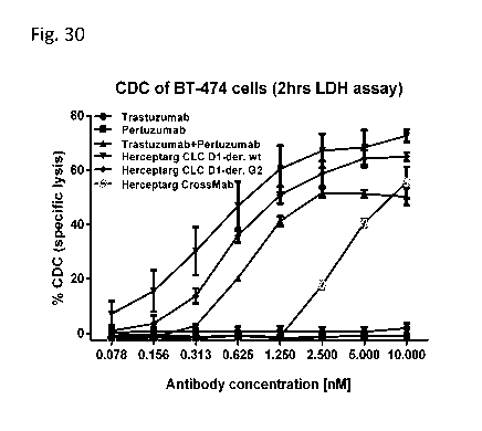
Figure 30: CDC activation on BT-474 cells (LDH release). BT474 cells were
incubated with
the three Herceptarg variants. As controls Trastuzumab, Pertuzumab and the
combination of both
were included. "Herceptarg CLC Dl-der wt": SEQ ID NOs 64, 54, 92, Herceptarg
CLC Dl-der
G2": SEQ ID NOs 64, 54, 92 (glycoengineered variant) "Herceptarg CrossMab":
SEQ ID NOs
109, 110, 111, 112.
Figure 31: CDC mediated killing of BT-474 cells (ACEA). BT474 cells were
incubated with
the three Herceptarg variants. As controls Trastuzumab, Pertuzumab and the
combination of both
were included. "Herceptarg CLC Dl-der wt": SEQ ID NOs 64, 54, 92, Herceptarg
CLC Dl-der
G2": SEQ ID NOs 64, 54, 92 (glycoengineered variant) "Herceptarg CrossMab":
SEQ ID NOs
109, 110, 111, 112.
Figure 32: In vivo activity of bispecific antibodies. Tumor volume in mouse
xenograft models
after treatment with different Her2 bispecific molecules (10 mg/kg) was
compared to treatment
with Trastuzumab, Pertuzumab and the combination of both. "Herceptarg": SEQ ID
NOs 64, 54,
92. "Control": Xolair, a non Her2 binding antibody.
DETAILED DESCRIPTION OF EMBODIMENTS OF THE INVENTION
I. DEFINITIONS
Throughout the disclosure, the terms "ErbB2", "ErbB2 receptor", "c-Erb-B2",
and
"HER2" are used interchangeably, and, unless otherwise indicated, refer to a
native sequence
CA 02925677 2016-03-29
WO 2015/091738
PCT/EP2014/078375
-13-
ErbB2 human polypeptide, or a functional derivative thereof. "ber2", "erbB2"
and "c-erb-B2"
refer to the corresponding human gene.
An "acceptor human framework" for the purposes herein is a framework
comprising the
amino acid sequence of a light chain variable domain (VL) framework or a heavy
chain variable
domain (VH) framework derived from a human immunoglobulin framework or a human
consensus framework, as defined below. An acceptor human framework "derived
from" a human
immunoglobulin framework or a human consensus framework may comprise the same
amino
acid sequence thereof, or it may contain amino acid sequence changes. In some
embodiments,
the number of amino acid changes are 10 or less, 9 or less, 8 or less, 7 or
less, 6 or less, 5 or less,
4 or less, 3 or less, or 2 or less. In some embodiments, the VL acceptor human
framework is
identical in sequence to the VL human immunoglobulin framework sequence or
human
consensus framework sequence.
"Affinity" refers to the strength of the sum total of noncovalent interactions
between a
single binding site of a molecule (e.g., an antibody) and its binding partner
(e.g., an antigen).
Unless indicated otherwise, as used herein, "binding affinity" refers to
intrinsic binding affinity
which reflects a 1:1 interaction between members of a binding pair (e.g.,
antibody and antigen).
The affinity of a molecule X for its partner Y can generally be represented by
the dissociation
constant (Kd). Affinity can be measured by common methods known in the art,
including those
described herein. Specific illustrative and exemplary embodiments for
measuring binding
affinity are described in the following.
An "affinity matured" antibody refers to an antibody with one or more
alterations in one
or more hypervariable regions (HVRs), compared to a parent antibody which does
not possess
such alterations, such alterations resulting in an improvement in the affinity
of the antibody for
antigen.
The terms "a bispecific HER2 antibody" and "a bispecific antibody that
specifically
binds to HER2" are used interchangeably and refer to a bispecific antibody
that is capable of
binding HER2 on both extracellular domains II and IV, respectively, with
sufficient affinity such
that the antibody is useful as a diagnostic and/or therapeutic agent in
targeting cells expressing
HER2. In one embodiment, the extent of binding of a bispecific antibody that
specifically binds
to HER2 on both extracellular domains II and IV to an unrelated, non-HER2
protein is less than
about 10% of the binding of the antibody to HER2 as measured, e.g., by a
Enzyme-linked
immunosorbent assay (ELISA), surface plasmon resonance (SPR) based assays
(e.g. Biacore) or
flow cytometry (FACS). In certain embodiments, a bispecific antibody that
specifically binds to
CA 02925677 2016-03-29
WO 2015/091738
PCT/EP2014/078375
-14-
HER2 has a dissociation constant (Kd) of < li.tM, < 100 nM, < 10 nM, < 1 nM, <
0.1 nM, < 0.01
nM, or < 0.001 nM (e.g. 10-8M or less, e.g. from 10-8M to 10-13M, e.g., from
10-9M to 10-13 M).
The term "antibody" herein is used in the broadest sense and encompasses
various
antibody structures, including but not limited to monoclonal antibodies,
polyclonal antibodies,
multispecific antibodies (e.g., bispecific antibodies), and antibody fragments
so long as they
exhibit the desired antigen-binding activity.
An "antibody fragment" refers to a molecule other than an intact antibody that
comprises
a portion of an intact antibody that binds the antigen to which the intact
antibody binds.
Examples of antibody fragments include but are not limited to Fv, Fab, Fab',
Fab'-SH, F(abt)2;
diabodies, cross-Fab fragments; linear antibodies; single-chain antibody
molecules (e.g. scFv);
and multispecific antibodies formed from antibody fragments. scFv antibodies
are, e.g. described
in Houston, J.S., Methods in Enzymol. 203 (1991) 46-96). In addition, antibody
fragments
comprise single chain polypeptides having the characteristics of a VH domain,
namely being
able to assemble together with a VL domain, or of a VL domain, namely being
able to assemble
together with a VH domain to a functional antigen binding site and thereby
providing the antigen
binding property of full length antibodies.
As used herein, "Fab fragment" refers to an antibody fragment comprising a
light chain
fragment comprising a VL domain and a constant domain of a light chain (CL),
and a VH
domain and a first constant domain (CH1) of a heavy chain. In one embodiment
the bispecific
antibodies of the invention comprise at least one Fab fragment, wherein either
the variable
regions or the constant regions of the heavy and light chain are exchanged.
Due to the exchange
of either the variable regions or the constant regions, said Fab fragment is
also referred to as
"cross-Fab fragment" or "xFab fragment" or "crossover Fab fragment". Two
different chain
compositions of a crossover Fab molecule are possible and comprised in the
bispecific antibodies
of the invention: On the one hand, the variable regions of the Fab heavy and
light chain are
exchanged, i.e. the crossover Fab molecule comprises a peptide chain composed
of the light
chain variable region (VL) and the heavy chain constant region (CH1), and a
peptide chain
composed of the heavy chain variable region (VH) and the light chain constant
region (CL). This
crossover Fab molecule is also referred to as CrossFab (vLvH). On the other
hand, when the
constant regions of the Fab heavy and light chain are exchanged, the crossover
Fab molecule
comprises a peptide chain composed of the heavy chain variable region (VH) and
the light chain
constant region (CL), and a peptide chain composed of the light chain variable
region (VL) and
CA 02925677 2016-03-29
WO 2015/091738
PCT/EP2014/078375
-15-
the heavy chain constant region (CH1). This crossover Fab molecule is also
referred to as
Cros sFab (CLCH1)=
A "single chain Fab fragment" or "scFab" is a polypeptide consisting of an
antibody
heavy chain variable domain (VH), an antibody constant domain 1 (CH1), an
antibody light
chain variable domain (VL), an antibody light chain constant domain (CL) and a
linker, wherein
said antibody domains and said linker have one of the following orders in N-
terminal to C-
terminal direction:
a) VH-CH1-linker-VL-CL, b) VL-CL-linker-VH-CH1, c) VH-CL-linker-VL-CH1 or d)
VL-
CH1-linker-VH-CL; and wherein said linker is a polypeptide of at least 30
amino acids,
preferably between 32 and 50 amino acids. Said single chain Fab fragments a)
VH-CH1-linker-
VL-CL, b) VL-CL-linker-VH-CH1, c) VH-CL-linker-VL-CH1 and d) VL-CH1-linker-VH-
CL,
are stabilized via the natural disulfide bond between the CL domain and the
CH1 domain. In
addition, these single chain Fab molecules might be further stabilized by
generation of interchain
disulfide bonds via insertion of cysteine residues (e.g. position 44 in the
variable heavy chain and
positionn 100 in the variable light chain according to Kabat numbering). The
term "N-terminus
denotes the last amino acid of the N-terminus. The term "C-terminus denotes
the last amino acid
of the C-terminus.
By "fused" or "connected" is meant that the components (e.g. a Fab molecule
and an Fc domain
subunit) are linked by peptide bonds, either directly or via one or more
peptide linkers.
The term "linker" as used herein refers to a peptide linker and is preferably
a peptide with
an amino acid sequence with a length of at least 5 amino acids, preferably
with a length of 5 to
100, more preferably of 10 to 50 amino acids. In one embodiment said peptide
linker is (GxS)n
or (GxS)nGm with G = glycine, S = serine, and (x = 3, n= 3, 4, 5 or 6, and m=
0, 1, 2 or 3) or (x
= 4,n= 2, 3, 4 or 5 and m= 0, 1, 2 or 3), preferably x = 4 and n= 2 or 3, more
preferably with x =
4, n= 2. In one embodiment said peptide linker is (G45)2.
The term "immunoglobulin molecule" refers to a protein having the structure of
a
naturally occurring antibody. For example, immunoglobulins of the IgG class
are
heterotetrameric glycoproteins of about 150,000 daltons, composed of two light
chains and two
heavy chains that are disulfide-bonded. From N- to C-terminus, each heavy
chain has a variable
region (VH), also called a variable heavy domain or a heavy chain variable
domain, followed by
three constant domains (CH1, CH2, and CH3), also called a heavy chain constant
region.
Similarly, from N- to C-terminus, each light chain has a variable region (VL),
also called a
variable light domain or a light chain variable domain, followed by a constant
light (CL) domain,
CA 02925677 2016-03-29
WO 2015/091738
PCT/EP2014/078375
-16-
also called a light chain constant region. The heavy chain of an
immunoglobulin may be
assigned to one of five types, called a (IgA), 6 (IgD), 8 (IgE), y (IgG), or
IA (IgM), some of which
may be further divided into subtypes, e.g. yi (IgGO, y2 (IgG2), y3 (IgG3), y4
(IgG4), ai (IgAi) and
U2 (IgA2). The light chain of an immunoglobulin may be assigned to one of two
types, called
kappa (lc) and lambda (X), based on the amino acid sequence of its constant
domain. An
immunoglobulin essentially consists of two Fab molecules and an Fc domain,
linked via the
immunoglobulin hinge region.
An "antibody that binds to the same epitope" as a reference antibody refers to
an
antibody that blocks binding of the reference antibody to its antigen in a
competition assay by
50% or more, and conversely, the reference antibody blocks binding of the
antibody to its
antigen in a competition assay by 50% or more. An exemplary competition assay
is provided
herein.
The term "antigen binding domain" refers to the part of an antigen binding
molecule that
comprises the area which specifically binds to and is complementary to part or
all of an antigen.
Where an antigen is large, an antigen binding molecule may only bind to a
particular part of the
antigen, which part is termed an epitope. An antigen binding domain may be
provided by, for
example, one or more antibody variable domains (also called antibody variable
regions).
Preferably, an antigen binding domain comprises an antibody light chain
variable region (VL)
and an antibody heavy chain variable region (VH).
The term "chimeric" antibody refers to an antibody in which a portion of the
heavy
and/or light chain is derived from a particular source or species, while the
remainder of the heavy
and/or light chain is derived from a different source or species, usually
prepared by recombinant
DNA techniques. Chimeric antibodies comprising a rabbit variable region and a
human constant
region are preferred. Other preferred forms of "chimeric antibodies"
encompassed by the present
invention are those in which the constant region has been modified or changed
from that of the
original antibody to generate the properties according to the invention,
especially in regard to
Clq binding and/or Fc receptor (FcR) binding. Such chimeric antibodies are
also referred to as
"class-switched antibodies". Chimeric antibodies are the product of expressed
immunoglobulin
genes comprising DNA segments encoding immunoglobulin variable regions and DNA
segments encoding immunoglobulin constant regions. Methods for producing
chimeric
antibodies involve conventional recombinant DNA and gene transfection
techniques are well
known in the art. See e.g. Morrison, S.L., et al., Proc. Natl. Acad. Sci. USA
81(1984) 6851-
6855; US Patent Nos. 5,202,238 and 5,204,244.
CA 02925677 2016-03-29
WO 2015/091738
PCT/EP2014/078375
-17-
The term "cytotoxic agent" as used herein refers to a substance that inhibits
or prevents a
cellular function and/or causes cell death or destruction. Cytotoxic agents
include, but are not
limited to, radioactive isotopes (e.g., At211, 1131, 1125, y90, Re186, Re188,
sm153, Bi212, p32, pb212 and
radioactive isotopes of Lu); chemotherapeutic agents or drugs (e.g.,
methotrexate, adriamicin,
vinca alkaloids (vincristine, vinblastine, etoposide), doxorubicin, melphalan,
mitomycin C,
chlorambucil, daunorubicin or other intercalating agents); growth inhibitory
agents; enzymes and
fragments thereof such as nucleolytic enzymes; antibiotics; toxins such as
small molecule toxins
or enzymatically active toxins of bacterial, fungal, plant or animal origin,
including fragments
and/or variants thereof; and the various antitumor or anticancer agents
disclosed below.
"Effector functions" refer to those biological activities attributable to the
Fc region of an
antibody, which vary with the antibody isotype. Examples of antibody effector
functions include:
Clq binding and complement dependent cytotoxicity (CDC); Fc receptor binding;
antibody-
dependent cell-mediated cytotoxicity (ADCC); antibody-dependent cellular
phagocytosis
(ADCP), cytokine secretion, immune complex-mediated antigen uptake by antigen
presenting
cells; down regulation of cell surface receptors (e.g. B cell receptor); and B
cell activation.
As used herein, the terms "engineer, engineered, engineering", are considered
to include
any manipulation of the peptide backbone or the post-translational
modifications of a naturally
occurring or recombinant polypeptide or fragment thereof. Engineering includes
modifications of
the amino acid sequence, of the glycosylation pattern, or of the side chain
group of individual
amino acids, as well as combinations of these approaches.
The term "amino acid mutation" as used herein is meant to encompass amino acid
substitutions, deletions, insertions, and modifications. Any combination of
substitution, deletion,
insertion, and modification can be made to arrive at the final construct,
provided that the final
construct possesses the desired characteristics, e.g., reduced binding to an
Fc receptor, or
increased association with another peptide. Amino acid sequence deletions and
insertions include
amino- and/or carboxy-terminal deletions and insertions of amino acids.
Particular amino acid
mutations are amino acid substitutions. For the purpose of altering e.g. the
binding
characteristics of an Fc region, non-conservative amino acid substitutions,
i.e. replacing one
amino acid with another amino acid having different structural and/or chemical
properties, are
particularly preferred. Amino acid substitutions include replacement by non-
naturally occurring
amino acids or by naturally occurring amino acid derivatives of the twenty
standard amino acids
(e.g. 4-hydroxyproline, 3-methylhistidine, ornithine, homoserine, 5-
hydroxylysine). Amino acid
mutations can be generated using genetic or chemical methods well known in the
art. Genetic
CA 02925677 2016-03-29
WO 2015/091738
PCT/EP2014/078375
-18-
methods may include site-directed mutagenesis, PCR, gene synthesis and the
like. It is
contemplated that methods of altering the side chain group of an amino acid by
methods other
than genetic engineering, such as chemical modification, may also be useful.
Various
designations may be used herein to indicate the same amino acid mutation. For
example, a
substitution from proline at position 329 of the Fc domain to glycine can be
indicated as 329G,
G329, G329, P329G, or Pro329Gly.
An "effective amount" of an agent, e.g., a pharmaceutical formulation, refers
to an
amount effective, at dosages and for periods of time necessary, to achieve the
desired therapeutic
or prophylactic result.
The term "Fe domain" or "Fe region" herein is used to define a C-terminal
region of an
immunoglobulin heavy chain that contains at least a portion of the constant
region. The term
includes native sequence Fc regions and variant Fc regions. Although the
boundaries of the Fc
region of an IgG heavy chain might vary slightly, the human IgG heavy chain Fc
region is
usually defined to extend from Cys226, or from Pro230, to the carboxyl-
terminus of the heavy
chain. However, the C-terminal lysine (Lys447) of the Fc region may or may not
be present.
Unless otherwise specified herein, numbering of amino acid residues in the Fc
region or constant
region is according to the EU numbering system, also called the EU index, as
described in Kabat
et al., Sequences of Proteins of Immunological Interest, 5th Ed. Public Health
Service, National
Institutes of Health, Bethesda, MD, 1991. A "subunit" of an Fc domain as used
herein refers to
one of the two polypeptides forming the dimeric Fc domain, i.e. a polypeptide
comprising C-
terminal constant regions of an immunoglobulin heavy chain, capable of stable
self-association.
For example, a subunit of an IgG Fc domain comprises an IgG CH2 and an IgG CH3
constant
domain.
A "modification promoting the association of the first and the second subunit
of the Fc
domain" is a manipulation of the peptide backbone or the post-translational
modifications of an
Fc domain subunit that reduces or prevents the association of a polypeptide
comprising the Fc
domain subunit with an identical polypeptide to form a homodimer. A
modification promoting
association as used herein particularly includes separate modifications made
to each of the two
Fc domain subunits desired to associate (i.e. the first and the second subunit
of the Fc domain),
wherein the modifications are complementary to each other so as to promote
association of the
two Fc domain subunits. For example, a modification promoting association may
alter the
structure or charge of one or both of the Fc domain subunits so as to make
their association
sterically or electrostatically favorable, respectively. Thus,
(hetero)dimerization occurs between
CA 02925677 2016-03-29
WO 2015/091738
PCT/EP2014/078375
-19-
a polypeptide comprising the first Fc domain subunit and a polypeptide
comprising the second
Fc domain subunit, which might be non-identical in the sense that further
components fused to
each of the subunits (e.g. antigen binding moieties) are not the same. In some
embodiments the
modification promoting association comprises an amino acid mutation in the Fc
domain,
specifically an amino acid substitution. In a particular embodiment, the
modification promoting
association comprises a separate amino acid mutation, specifically an amino
acid substitution, in
each of the two subunits of the Fc domain.
"Framework" or "FR" refers to variable domain residues other than
hypervariable region
(HVR) residues. The FR of a variable domain generally consists of four FR
domains: FR1, FR2,
FR3, and FR4. Accordingly, the HVR and FR sequences generally appear in the
following
sequence in VH (or VL): FR1-H1(L1)-FR2-H2(L2)-FR3-H3(L3)-FR4.
The terms "full length antibody," "intact antibody," and "whole antibody" are
used
herein interchangeably to refer to an antibody having a structure
substantially similar to a native
antibody structure or having heavy chains that contain an Fc region as defined
herein.
The terms "host cell," "host cell line," and "host cell culture" are used
interchangeably
and refer to cells into which exogenous nucleic acid has been introduced,
including the progeny
of such cells. Host cells include "transformants" and "transformed cells,"
which include the
primary transformed cell and progeny derived therefrom without regard to the
number of
passages. Progeny may not be completely identical in nucleic acid content to a
parent cell, but
may contain mutations. Mutant progeny that have the same function or
biological activity as
screened or selected for in the originally transformed cell are included
herein.
A "human antibody" is one which possesses an amino acid sequence which
corresponds
to that of an antibody produced by a human or a human cell or derived from a
non-human source
that utilizes human antibody repertoires or other human antibody-encoding
sequences. This
definition of a human antibody specifically excludes a humanized antibody
comprising non-
human antigen-binding residues. As also mentioned for chimeric and humanized
antibodies
according to the invention the term "human antibody" as used herein also
comprises such
antibodies which are modified in the constant region to generate the
properties according to the
invention, especially in regard to Clq binding and/or FcR binding, e.g. by
"class switching" i.e.
change or mutation of Fc parts (e.g. from IgG1 to IgG4 and/or IgGl/IgG4
mutation.)
The term "recombinant human antibody", as used herein, is intended to include
all human
antibodies that are prepared, expressed, created or isolated by recombinant
means, such as
antibodies isolated from a host cell such as a NSO or CHO cell or from an
animal (e.g. a mouse)
CA 02925677 2016-03-29
WO 2015/091738
PCT/EP2014/078375
-20-
that is transgenic for human immunoglobulin genes or antibodies expressed
using a recombinant
expression vector transfected into a host cell. Such recombinant human
antibodies have variable
and constant regions in a rearranged form. The recombinant human antibodies
according to the
invention have been subjected to in vivo somatic hypermutation. Thus, the
amino acid sequences
of the VH and VL regions of the recombinant antibodies are sequences that,
while derived from
and related to human germ line VH and VL sequences, may not naturally exist
within the human
antibody germ line repertoire in vivo.
A "human consensus framework" is a framework which represents the most
commonly
occurring amino acid residues in a selection of human immunoglobulin VL or VH
framework
sequences. Generally, the selection of human immunoglobulin VL or VH sequences
is from a
subgroup of variable domain sequences. Generally, the subgroup of sequences is
a subgroup as
in Kabat et al., Sequences of Proteins of Immunological Interest, Fifth
Edition, NIH Publication
91-3242, Bethesda MD (1991), vols. 1-3. In one embodiment, for the VL, the
subgroup is
subgroup kappa I as in Kabat et al., supra. In one embodiment, for the VH, the
subgroup is
subgroup III as in Kabat et al., supra.
A "humanized" antibody refers to a chimeric antibody comprising amino acid
residues
from non-human HVRs and amino acid residues from human FRs. In certain
embodiments, a
humanized antibody will comprise substantially all of at least one, and
typically two, variable
domains, in which all or substantially all of the HVRs (e.g., CDRs) correspond
to those of a non-
human antibody, and all or substantially all of the FRs correspond to those of
a human antibody.
A humanized antibody optionally may comprise at least a portion of an antibody
constant region
derived from a human antibody. A "humanized form" of an antibody, e.g., a non-
human
antibody, refers to an antibody that has undergone humanization. Other forms
of "humanized
antibodies" encompassed by the present invention are those in which the
constant region has
been additionally modified or changed from that of the original antibody to
generate the
properties according to the invention, especially in regard to Clq binding
and/or Fc receptor
(FcR) binding.
The term "hypervariable region" or "HVR," as used herein refers to each of the
regions
of an antibody variable domain which are hypervariable in sequence and/or form
structurally
defined loops ("hypervariable loops"). Generally, native four-chain antibodies
comprise six
HVRs; three in the VH (H1, H2, H3), and three in the VL (L1, L2, L3). HVRs
generally
comprise amino acid residues from the hypervariable loops and/or from the
"complementarity
determining regions" (CDRs), the latter being of highest sequence variability
and/or involved in
CA 02925677 2016-03-29
WO 2015/091738
PCT/EP2014/078375
-21-
antigen recognition. Exemplary hypervariable loops occur at amino acid
residues 26-32 (L1), 50-
52 (L2), 91-96 (L3), 26-32 (H1), 53-55 (H2), and 96-101 (H3). (Chothia and
Lesk, J. Mol. Biol.
196:901-917 (1987).) Exemplary CDRs (CDR-L1, CDR-L2, CDR-L3, CDR-H1, CDR-H2,
and
CDR-H3) occur at amino acid residues 24-34 of Li, 50-56 of L2, 89-97 of L3, 31-
35B of H1,
50-65 of H2, and 95-102 of H3. (Kabat et al., Sequences of Proteins of
Immunological Interest,
5th Ed. Public Health Service, National Institutes of Health, Bethesda, MD
(1991).)
Hypervariable regions (HVRs) are also referred to as complementarity
determining regions
(CDRs), and these terms are used herein interchangeably in reference to
portions of the variable
region that form the antigen binding regions. This particular region has been
described by Kabat
et al., U.S. Dept. of Health and Human Services, "Sequences of Proteins of
Immunological
Interest" (1983) and by Chothia et al., J. Mol. Biol. 196:901-917 (1987),
where the definitions
include overlapping or subsets of amino acid residues when compared against
each other.
Nevertheless, application of either definition to refer to a CDR of an
antibody or variants thereof
is intended to be within the scope of the term as defined and used herein. The
appropriate amino
acid residues which encompass the CDRs as defined by each of the above cited
references are set
forth below in Table A as a comparison. The exact residue numbers which
encompass a
particular CDR will vary depending on the sequence and size of the CDR. Those
skilled in the
art can routinely determine which residues comprise a particular CDR given the
variable region
amino acid sequence of the antibody.
TABLE A. CDR Definitions'
CDR Kabat Chothia AbM2
VH CDR1 31-35 26-32 26-35
VH CDR2 50-65 52-58 50-58
VH CDR3 95-102 95-102 95-102
VL CDR1 24-34 26-32 24-34
VL CDR2 50-56 50-52 50-56
VL CDR3 89-97 91-96 89-97
Numbering of all CDR definitions in Table A is according to the numbering
conventions set forth by
Kabat et al. (see below).
2 "AbM" with a lowercase "b" as used in Table A refers to the CDRs as defined
by
Oxford Molecular's "AbM" antibody modeling software.
Kabat et al. also defined a numbering system for variable region sequences
that is
applicable to any antibody. One of ordinary skill in the art can unambiguously
assign this system
of "Kabat numbering" to any variable region sequence, without reliance on any
experimental
CA 02925677 2016-03-29
WO 2015/091738
PCT/EP2014/078375
-22-
data beyond the sequence itself. As used herein, "Kabat numbering" refers to
the numbering
system set forth by Kabat et al., U.S. Dept. of Health and Human Services,
"Sequence of
Proteins of Immunological Interest" (1983). Unless otherwise specified,
references to the
numbering of specific amino acid residue positions in an antibody variable
region are according
to the Kabat numbering system.
With the exception of CDR1 in VH, CDRs generally comprise the amino acid
residues
that form the hypervariable loops. CDRs also comprise "specificity determining
residues," or
"SDRs," which are residues that contact antigen. SDRs are contained within
regions of the CDRs
called abbreviated-CDRs, or a-CDRs. Exemplary a-CDRs (a-CDR-L1, a-CDR-L2, a-
CDR-L3, a-
CDR-H1, a-CDR-H2, and a-CDR-H3) occur at amino acid residues 31-34 of Li, 50-
55 of L2,
89-96 of L3, 31-35B of H1, 50-58 of H2, and 95-102 of H3. (See Almagro and
Fransson, Front.
Biosci. 13:1619-1633 (2008).) Unless otherwise indicated, HVR residues and
other residues in
the variable domain (e.g., FR residues) are numbered herein according to Kabat
et al., supra.
An "immunoconjugate" is an antibody conjugated to one or more heterologous
molecule(s), including but not limited to a cytotoxic agent.
An "individual" or "subject" is a mammal. Mammals include, but are not limited
to,
domesticated animals (e.g., cows, sheep, cats, dogs, and horses), primates
(e.g., humans and non-
human primates such as monkeys), rabbits, and rodents (e.g., mice and rats).
In certain
embodiments, the individual or subject is a human.
An "isolated" antibody is one which has been separated from a component of its
natural
environment. In some embodiments, an antibody is purified to greater than 95%
or 99% purity as
determined by, for example, electrophoretic (e.g., SDS-PAGE, isoelectric
focusing (IEF),
capillary electrophoresis) or chromatographic (e.g., ion exchange or reverse
phase HPLC). For
review of methods for assessment of antibody purity, see, e.g., Flatman et
al., J. Chromatogr. B
848:79-87 (2007).
An "isolated" nucleic acid refers to a nucleic acid molecule that has been
separated from
a component of its natural environment. An isolated nucleic acid includes a
nucleic acid
molecule contained in cells that ordinarily contain the nucleic acid molecule,
but the nucleic acid
molecule is present extrachromosomally or at a chromosomal location that is
different from its
natural chromosomal location.
"Isolated nucleic acid encoding a bispecific antibody that specifically binds
HER2" refers
to one or more nucleic acid molecules encoding antibody heavy and light chains
(or fragments
CA 02925677 2016-03-29
WO 2015/091738
PCT/EP2014/078375
-23-
thereof), including such nucleic acid molecule(s) in a single vector or
separate vectors, and such
nucleic acid molecule(s) present at one or more locations in a host cell.
The term "monoclonal antibody" as used herein refers to an antibody obtained
from a
population of substantially homogeneous antibodies, i.e., the individual
antibodies comprising
the population are identical and/or bind the same epitope, except for possible
variant antibodies,
e.g., containing naturally occurring mutations or arising during production of
a monoclonal
antibody preparation, such variants generally being present in minor amounts.
In contrast to
polyclonal antibody preparations, which typically include different antibodies
directed against
different determinants (epitopes), each monoclonal antibody of a monoclonal
antibody
preparation is directed against a single determinant on an antigen. Thus, the
modifier
"monoclonal" indicates the character of the antibody as being obtained from a
substantially
homogeneous population of antibodies, and is not to be construed as requiring
production of the
antibody by any particular method. For example, the monoclonal antibodies to
be used in
accordance with the present invention may be made by a variety of techniques,
including but not
limited to the hybridoma method, recombinant DNA methods, phage-display
methods, and
methods utilizing transgenic animals containing all or part of the human
immunoglobulin loci,
such methods and other exemplary methods for making monoclonal antibodies
being described
herein.
A "naked antibody" refers to an antibody that is not conjugated to a
heterologous moiety
(e.g., a cytotoxic moiety) or radiolabel. The naked antibody may be present in
a pharmaceutical
formulation.
"Native antibodies" refer to naturally occurring immunoglobulin molecules with
varying
structures. For example, native IgG antibodies are heterotetrameric
glycoproteins of about
150,000 daltons, composed of two identical light chains and two identical
heavy chains that are
disulfide-bonded. From N- to C-terminus, each heavy chain has a variable
region (VH), also
called a variable heavy domain or a heavy chain variable domain, followed by
three constant
domains (CH1, CH2, and CH3). Similarly, from N- to C-terminus, each light
chain has a
variable region (VL), also called a variable light domain or a light chain
variable domain,
followed by a constant light (CL) domain. The light chain of an antibody may
be assigned to one
of two types, called kappa (lc) and lambda (X), based on the amino acid
sequence of its constant
domain.
The term "package insert" is used to refer to instructions customarily
included in
commercial packages of therapeutic products, that contain information about
the indications,
CA 02925677 2016-03-29
WO 2015/091738
PCT/EP2014/078375
-24-
usage, dosage, administration, combination therapy, contraindications and/or
warnings
concerning the use of such therapeutic products.
"No substantial cross-reactivity" means that a molecule (e.g., an antibody)
does not
recognize or specifically bind an antigen different from the actual target
antigen of the molecule
(e.g. an antigen closely related to the target antigen), particularly when
compared to that target
antigen. For example, an antibody may bind less than about 10% to less than
about 5% to an
antigen different from the actual target antigen, or may bind said antigen
different from the
actual target antigen at an amount consisting of less than about 10%, 9%, 8%
7%, 6%, 5%, 4%,
3%, 2%, 1%, 0.5%, 0.2%, or 0.1%, preferably less than about 2%, 1%, or 0.5%,
and most
preferably less than about 0.2% or 0.1% antigen different from the actual
target antigen.
"Percent (%) amino acid sequence identity" with respect to a reference
polypeptide
sequence is defined as the percentage of amino acid residues in a candidate
sequence that are
identical with the amino acid residues in the reference polypeptide sequence,
after aligning the
sequences and introducing gaps, if necessary, to achieve the maximum percent
sequence identity,
and not considering any conservative substitutions as part of the sequence
identity. Alignment
for purposes of determining percent amino acid sequence identity can be
achieved in various
ways that are within the skill in the art, for instance, using publicly
available computer software
such as BLAST, BLAST-2, ALIGN or Megalign (DNASTAR) software. Those skilled in
the art
can determine appropriate parameters for aligning sequences, including any
algorithms needed to
achieve maximal alignment over the full length of the sequences being
compared. For purposes
herein, however, % amino acid sequence identity values are generated using the
sequence
comparison computer program ALIGN-2. The ALIGN-2 sequence comparison computer
program was authored by Genentech, Inc., and the source code has been filed
with user
documentation in the U.S. Copyright Office, Washington D.C., 20559, where it
is registered
under U.S. Copyright Registration No. TXU510087. The ALIGN-2 program is
publicly available
from Genentech, Inc., South San Francisco, California, or may be compiled from
the source
code. The ALIGN-2 program should be compiled for use on a UNIX operating
system, including
digital UNIX V4.0D. All sequence comparison parameters are set by the ALIGN-2
program and
do not vary.
In situations where ALIGN-2 is employed for amino acid sequence comparisons,
the %
amino acid sequence identity of a given amino acid sequence A to, with, or
against a given
amino acid sequence B (which can alternatively be phrased as a given amino
acid sequence A
CA 02925677 2016-03-29
WO 2015/091738
PCT/EP2014/078375
-25-
that has or comprises a certain % amino acid sequence identity to, with, or
against a given amino
acid sequence B) is calculated as follows:
100 times the fraction X/Y
where X is the number of amino acid residues scored as identical matches by
the sequence
alignment program ALIGN-2 in that program's alignment of A and B, and where Y
is the total
number of amino acid residues in B. It will be appreciated that where the
length of amino acid
sequence A is not equal to the length of amino acid sequence B, the % amino
acid sequence
identity of A to B will not equal the % amino acid sequence identity of B to
A. Unless
specifically stated otherwise, all % amino acid sequence identity values used
herein are obtained
as described in the immediately preceding paragraph using the ALIGN-2 computer
program.
The term "pharmaceutical formulation" refers to a preparation which is in such
form as to
permit the biological activity of an active ingredient contained therein to be
effective, and which
contains no additional components which are unacceptably toxic to a subject to
which the
formulation would be administered.
A "pharmaceutically acceptable carrier" refers to an ingredient in a
pharmaceutical
formulation, other than an active ingredient, which is nontoxic to a subject.
A pharmaceutically
acceptable carrier includes, but is not limited to, a buffer, excipient,
stabilizer, or preservative.
As used herein, "treatment" (and grammatical variations thereof such as
"treat" or
"treating") refers to clinical intervention in an attempt to alter the natural
course of the individual
being treated, and can be performed either for prophylaxis or during the
course of clinical
pathology. Desirable effects of treatment include, but are not limited to,
preventing occurrence or
recurrence of disease, alleviation of symptoms, diminishment of any direct or
indirect
pathological consequences of the disease, preventing metastasis, decreasing
the rate of disease
progression, amelioration or palliation of the disease state, and remission or
improved prognosis.
In some embodiments, antibodies of the invention are used to delay development
of a disease or
to slow the progression of a disease.
The term cancer as used herein refers to proliferative diseases, such as
lymphomas,
lymphocytic leukemias, lung cancer, non small cell lung (NSCL) cancer,
bronchioloalviolar cell
lung cancer, bone cancer, pancreatic cancer, skin cancer, cancer of the head
or neck, cutaneous
or intraocular melanoma, uterine cancer, ovarian cancer, rectal cancer, cancer
of the anal region,
stomach cancer, gastric cancer, colon cancer, breast cancer, uterine cancer,
carcinoma of the
CA 02925677 2016-03-29
WO 2015/091738
PCT/EP2014/078375
-26-
fallopian tubes, carcinoma of the endometrium, carcinoma of the cervix,
carcinoma of the
vagina, carcinoma of the vulva, Hodgkin's Disease, cancer of the esophagus,
cancer of the small
intestine, cancer of the endocrine system, cancer of the thyroid gland, cancer
of the parathyroid
gland, cancer of the adrenal gland, sarcoma of soft tissue, cancer of the
urethra, cancer of the
penis, prostate cancer, cancer of the bladder, cancer of the kidney or ureter,
renal cell carcinoma,
carcinoma of the renal pelvis, mesothelioma, hepatocellular cancer, biliary
cancer, neoplasms of
the central nervous system (CNS), spinal axis tumors, brain stem glioma,
glioblastoma
multiforme, astrocytomas, schwanomas, ependymonas, medulloblastomas,
meningiomas,
squamous cell carcinomas, pituitary adenoma and Ewings sarcoma, including
refractory versions
of any of the above cancers, or a combination of one or more of the above
cancers.
The term "variable region" or "variable domain" refers to the domain of an
antibody
heavy or light chain that is involved in binding the antibody to antigen. The
variable domains of
the heavy chain and light chain (VH and VL, respectively) of a native antibody
generally have
similar structures, with each domain comprising four conserved framework
regions (FRs) and
three hypervariable regions (HVRs). (See, e.g., Kindt et al. Kuby Immunology,
6th ed., W.H.
Freeman and Co., page 91 (2007).) A single VH or VL domain may be sufficient
to confer
antigen-binding specificity. Furthermore, antibodies that bind a particular
antigen may be
isolated using a VH or VL domain from an antibody that binds the antigen to
screen a library of
complementary VL or VH domains, respectively. See, e.g., Portolano et al., J.
Immunol.
150:880-887 (1993); Clarkson et al., Nature 352:624-628 (1991).
The term "antigen-binding site of an antibody" when used herein refer to the
amino acid
residues of an antibody which are responsible for antigen-binding. The antigen-
binding portion
of an antibody comprises amino acid residues from the "complementary
determining regions" or
"CDRs". "Framework" or "FR" regions are those variable domain regions other
than the
hypervariable region residues as herein defined. Therefore, the light and
heavy chain variable
domains of an antibody comprise from N- to C-terminus the domains FR1, CDR1,
FR2, CDR2,
FR3, CDR3, and FR4. Especially, CDR3 of the heavy chain is the region which
contributes most
to antigen binding and defines the antibody's properties. CDR and FR regions
are determined
according to the standard definition of Kabat et al., Sequences of Proteins of
Immunological
Interest, 5th ed., Public Health Service, National Institutes of Health,
Bethesda, MD (1991)
and/or those residues from a "hypervariable loop".
Antibody specificity refers to selective recognition of the antibody for a
particular
epitope of an antigen. Natural antibodies, for example, are monospecific. The
term
CA 02925677 2016-03-29
WO 2015/091738
PCT/EP2014/078375
-27-
"monospecific" antibody as used herein denotes an antibody that has one or
more binding sites
each of which bind to the same epitope of the same antigen.
"Bispecific antibodies" according to the invention are antibodies which have
two
different antigen-binding specificities. Antibodies of the present invention
are specific for two
different epitopes of HER2, i.e. the extracellular domains II and IV of HER2.
The term
"bispecific" antibody as used herein denotes an antibody that has at least two
binding sites each
of which bind to different epitopes of the same antigen.
Bispecific antibodies may also be used to localize cytotoxic agents to cells
which express
HER2. Bispecific antibodies can be prepared as full length antibodies or
antibody fragments.
The term "valent" as used within the current application denotes the presence
of a
specified number of binding sites in an antibody molecule. As such, the terms
"bivalent",
"tetravalent", and "hexavalent" denote the presence of two binding sites, four
binding sites, and
six binding sites, respectively, in an antibody molecule. The bispecific
antibodies according to
the invention are at least "bivalent" and may be "trivalent" or "multivalent"
(e.g."tetravalent" or
"hexavalent").
Antibodies of the present invention have two or more binding sites and are
bispecific.
That is, the antibodies may be bispecific even in cases where there are more
than two binding
sites (i.e. that the antibody is trivalent or multivalent). Bispecific
antibodies of the invention
include, for example, multivalent single chain antibodies, diabodies and
triabodies, as well as
antibodies having the constant domain structure of full length antibodies to
which further
antigen-binding sites (e.g., single chain Fv, a VH domain and/or a VL domain,
Fab, or (Fab)2)
are linked via one or more peptide-linkers. The antibodies can be full length
from a single
species, or be chimerized or humanized.
The term "vector," as used herein, refers to a nucleic acid molecule capable
of
propagating another nucleic acid to which it is linked. The term includes the
vector as a self-
replicating nucleic acid structure as well as the vector incorporated into the
genome of a host cell
into which it has been introduced. Certain vectors are capable of directing
the expression of
nucleic acids to which they are operatively linked. Such vectors are referred
to herein as
"expression vectors."
The term "amino acid" as used within this application denotes the group of
naturally
occurring carboxy a-amino acids comprising alanine (three letter code: ala,
one letter code: A),
arginine (arg, R), asparagine (asn, N), aspartic acid (asp, D), cysteine (cys,
C), glutamine (gln,
Q), glutamic acid (glu, E), glycine (gly, G), histidine (his, H), isoleucine
(ile, I), leucine (leu, L),
CA 02925677 2016-03-29
WO 2015/091738
PCT/EP2014/078375
-28-
lysine (lys, K), methionine (met, M), phenylalanine (phe, F), proline (pro,
P), serine (ser, S),
threonine (thr, T), tryptophan (trp, W), tyrosine (tyr, Y), and valine (val,
V).
As used herein, the expressions "cell", "cell line", and "cell culture" are
used
interchangeably and all such designations include progeny. Thus, the words
"transfectants" and
"transfected cells" include the primary subject cell and cultures derived
there from without
regard for the number of transfers. It is also understood that all progeny may
not be precisely
identical in DNA content, due to deliberate or inadvertent mutations. Variant
progeny that have
the same function or biological activity as screened for in the originally
transformed cell are
included.
"Affinity" refers to the strength of the sum total of noncovalent interactions
between a
single binding site of a molecule (e.g., an antibody) and its binding partner
(e.g., an antigen).
Unless indicated otherwise, as used herein, "binding affinity" refers to
intrinsic binding affinity
which reflects a 1:1 interaction between members of a binding pair (e.g.,
antibody and antigen).
The affinity of a molecule X for its partner Y can generally be represented by
the dissociation
constant (Kd). Affinity can be measured by common methods known in the art,
including those
described herein. Specific illustrative and exemplary embodiments for
measuring binding
affinity are described in the following.
As used herein, the term "binding" or "specifically binding" refers to the
binding of the
antibody to an epitope of the antigen in an in-vitro assay, preferably in a
surface plasmon
resonance assay (SPR, BIAcore, GE-Healthcare Uppsala, Sweden). The affinity of
the binding is
defined by the terms ka (rate constant for the association of the antibody
from the
antibody/antigen complex), kD (dissociation constant), and KD (kD/ka). Binding
or specifically
binding means a binding affinity (KD) of 10-8 mo1/1 or less, preferably 10-9 M
to 10-13 mo1/1.
Binding of the antibody to the death receptor can be investigated by a BIAcore
assay
(GE-Healthcare Uppsala, Sweden). The affinity of the binding is defined by the
terms ka (rate
constant for the association of the antibody from the antibody/antigen
complex), kD (dissociation
constant), and KD (kD/ka)
The term "epitope" includes any polypeptide determinant capable of specific
binding to
an antibody. In certain embodiments, epitope determinant include chemically
active surface
groupings of molecules such as amino acids, sugar side chains, phosphoryl, or
sulfonyl, and, in
certain embodiments, may have specific three dimensional structural
characteristics, and or
specific charge characteristics. An epitope is a region of an antigen that is
bound by an antibody.
CA 02925677 2016-03-29
WO 2015/091738
PCT/EP2014/078375
-29-
As used herein, the terms "engineer, engineered, engineering," particularly
with the
prefix "glyco-," as well as the term "glycosylation engineering" are
considered to include any
manipulation of the glycosylation pattern of a naturally occurring or
recombinant polypeptide or
fragment thereof. Glycosylation engineering includes metabolic engineering of
the glycosylation
machinery of a cell, including genetic manipulations of the oligosaccharide
synthesis pathways
to achieve altered glycosylation of glycoproteins expressed in cells.
Furthermore, glycosylation
engineering includes the effects of mutations and cell environment on
glycosylation. In one
embodiment, the glycosylation engineering is an alteration in
glycosyltransferase activity. In a
particular embodiment, the engineering results in altered
glucosaminyltransferase activity and/or
fucosyltransferase activity.
II. COMPOSITIONS AND METHODS
In one aspect, the invention is based on bispecific antibodies specifically
binding to
HER2. Antibodies of the invention are useful, e.g., for the treatment or
diagnosis of cancer.
A. Exemplary bispecific antibodies specifically binding to HER2 with a
common
light chain
In one aspect of the invention, a bispecific antibody specifically binding to
HER2 is provided,
wherein the antibody comprises a first binding moiety that specifically binds
the extracellular
domain II of HER2 and a second binding moiety that specifically binds the
extracellular domain
IV of HER2. The inventors of the present invention surprisingly generated a
monovalent
bispecific antibody wherein both binding moieties share a common light chain
that retains the
efficacy of the parent monospecific antibodies in terms of inhibition of tumor
cell proliferation
and has an increased affinity to the extracellular domain II of HER2. The
generation of a
bispecific molecule with a common light chain that retains the binding
properties of both of the
parent antibodies is not straight-forward as the common CDRs of the hybrid
light chain have to
retain the binding specifity for both the extracellular domains II and IV of
HER2. The use of this
so-called 'common light chain' principle, i.e. combining two binders that
share one light chain
but still have separate specificities, prevents light chain mispairing and in
this particular case
retains the epitope specificity of the parental antibodies. As a consequence,
there are less side
products during production, facilitating the homogenous preparation of HER2
bispecific antigen
binding molecules at high yields. The heavy chain of Pertuzumab was further
optimized,
CA 02925677 2016-03-29
WO 2015/091738
PCT/EP2014/078375
-30-
resulting in a more potent molecule in terms of affinity to the extracellular
domain II of HER2.
In addition, the Trastuzumab heavy chain has been stabilized by introducing
certain mutations
into the CDRs . The resulting molecule is superior to the parent Pertuzumab
and Trastuzumab
monospecific antibodies. The bispecific HER2 antibodies in the monovalent
common light chain
format have an increased affinity to the pertuzumab epitope, and show superior
inhibitory effects
on cell proliferation as compared to the combination of the parental
antibodies.
In one embodiment a bispecific antibody is provided, comprising a first Fab
molecule
capable of specific binding to extracellular domain II of HER2 and a second
Fab molecule
capable of specific binding to extracellular domain IV of HER2, wherein the
sequence of the
variable light chain of the first Fab molecule is identical to the sequence of
the variable light
chain of the second Fab molecule (i.e. the first and the second Fab molecule
comprise a common
light chain).
In one embodiment, the invention provides a bispecific antibody that
specifically binds to
extracellular domains II and IV of HER2 comprising
a first heavy chain comprising
(a) a heavy chain CDR1 of SEQ ID NO: 55;
(b) a heavy chain CDR2 of SEQ ID NO: 77;
(c) a heavy chain CDR3 of SEQ ID NO: 56;
and a second heavy chain comprising
(a) a heavy chain CDR1 of SEQ ID NO: 20;
(b) a heavy chain CDR2 of SEQ ID NO: 29;
(c) a heavy chain CDR3 of SEQ ID NO: 30;
and a first and a second light chain comprising
(a) a light chain CDR1 of SEQ ID NO: 89;
(b) a light chain CDR2 of SEQ ID NO: 90;
(c) a light chain CDR3 of SEQ ID NO: 19.
In one embodiment, the invention provides a bispecific antibody that
specifically binds to
extracellular domains II and IV of HER2 comprising
a first heavy chain comprising
(a) a heavy chain CDR1 of SEQ ID NO: 14;
CA 02925677 2016-03-29
WO 2015/091738
PCT/EP2014/078375
-31-
(b) a heavy chain CDR2 of SEQ ID NO: 60;
(c) a heavy chain CDR3 of SEQ ID NO: 16;
and a second heavy chain comprising
(a) a heavy chain CDR1 of SEQ ID NO: 20;
(b) a heavy chain CDR2 of SEQ ID NO: 29;
(c) a heavy chain CDR3 of SEQ ID NO: 30;
and a first and a second light chain comprising
(a) a light chain CDR1 of SEQ ID NO: 89;
(b) a light chain CDR2 of SEQ ID NO: 90;
(c) a light chain CDR3 of SEQ ID NO: 19.
In one embodiment, the invention provides a bispecific antibody that
specifically binds to
extracellular domains II and IV of HER2 comprising
a first heavy chain comprising
(a) a heavy chain CDR1 of SEQ ID NO: 58;
(b) a heavy chain CDR2 of SEQ ID NO: 15;
(c) a heavy chain CDR3 of SEQ ID NO: 59;
and a second heavy chain comprising
(a) a heavy chain CDR1 of SEQ ID NO: 20;
(b) a heavy chain CDR2 of SEQ ID NO: 29;
(c) a heavy chain CDR3 of SEQ ID NO: 30;
and a first and a second light chain comprising
(a) a light chain CDR1 of SEQ ID NO: 89;
(b) a light chain CDR2 of SEQ ID NO: 90;
(c) a light chain CDR3 of SEQ ID NO: 19.
In one embodiment the second heavy chain of any of the embodiments above bears
at
least one modification in the amino acid sequence that confers higher chemical
stability to the
CDR , resulting in retained binding to HER2 under stress conditions.
Modifications useful herein
are e.g. D98E, D98N, D98T, G99A or G995. Surpringly the inventors found that
some
modifications of the CDRs did not only improve the stability of the molecule
but also improved
the binding affinity to HER2.
CA 02925677 2016-03-29
WO 2015/091738
PCT/EP2014/078375
-32-
Hence in one embodiment, the invention provides a bispecific antibody that
specifically binds to
extracellular domains II and IV of HER2 comprising
a first heavy chain comprising
(a) a heavy chain CDR1 of SEQ ID NO: 55;
(b) a heavy chain CDR2 of SEQ ID NO: 77;
(c) a heavy chain CDR3 of SEQ ID NO: 56;
and a second heavy chain comprising
(a) a heavy chain CDR1 of SEQ ID NO: 20;
(b) a heavy chain CDR2 of SEQ ID NO: 29;
(c) a heavy chain CDR3 of SEQ ID NO: 79;
and a first and a second light chain comprising
(a) a light chain CDR1 of SEQ ID NO: 89;
(b) a light chain CDR2 of SEQ ID NO: 90;
(c) a light chain CDR3 of SEQ ID NO: 19.
In one embodiment, the invention provides a bispecific antibody that
specifically binds to
extracellular domains II and IV of HER2 comprising
a first heavy chain comprising
(a) a heavy chain CDR1 of SEQ ID NO: 14;
(b) a heavy chain CDR2 of SEQ ID NO: 60;
(c) a heavy chain CDR3 of SEQ ID NO: 16;
and a second heavy chain comprising
(a) a heavy chain CDR1 of SEQ ID NO: 20;
(b) a heavy chain CDR2 of SEQ ID NO: 29;
(c) a heavy chain CDR3 of SEQ ID NO: 79;
and a first and a second light chain comprising
(a) a light chain CDR1 of SEQ ID NO: 89;
(b) a light chain CDR2 of SEQ ID NO: 90;
(c) a light chain CDR3 of SEQ ID NO: 19.
.
In one embodiment, the invention provides a bispecific antibody that
specifically binds to
extracellular domains II and IV of HER2 comprising
CA 02925677 2016-03-29
WO 2015/091738
PCT/EP2014/078375
-33-
a first heavy chain comprising
(a) a heavy chain CDR1 of SEQ ID NO: 58;
(b) a heavy chain CDR2 of SEQ ID NO: 15;
(c) a heavy chain CDR3 of SEQ ID NO: 59;
and a second heavy chain comprising
(a) a heavy chain CDR1 of SEQ ID NO: 20;
(b) a heavy chain CDR2 of SEQ ID NO: 29;
(c) a heavy chain CDR3 of SEQ ID NO: 79;
and a first and a second light chain comprising
(a) a light chain CDR1 of SEQ ID NO: 89;
(b) a light chain CDR2 of SEQ ID NO: 90;
(c) a light chain CDR3 of SEQ ID NO: 19.
In one embodiment, the bispecific antibody comprises two variable light chains
comprising an
amino acid sequence of SEQ ID NO 54 (i.e. a common light chain), a first heavy
chain
comprising a variable heavy chain comprising an amino acid sequence of SEQ ID
NO: 64, and a
second heavy chain comprising a variable heavy chain comprising an amino acid
sequence of
SEQ ID NO 92.
In one embodiment, the bispecific antibody comprises two variable light chains
comprising an
amino acid sequence of SEQ ID NO 54 (i.e. a common light chain), a first heavy
chain
comprising a variable heavy chain comprising an amino acid sequence of SEQ ID
NO: 70, and a
second heavy chain comprising a variable heavy chain comprising an amino acid
sequence of
SEQ ID NO 92.
In one embodiment, the bispecific antibody comprises two variable light chains
comprising an
amino acid sequence of SEQ ID NO 54 (i.e. a common light chain), a first heavy
chain
comprising a variable heavy chain comprising an amino acid sequence of SEQ ID
NO: 68, and a
second heavy chain comprising a variable heavy chain comprising an amino acid
sequence of
SEQ ID NO: 92.
In one embodiment the second heavy chain of any of the embodiments above bears
at
least one modification in the amino acid sequence that confers stability to
the CDRs and the
binding to the target, e.g. D98E, D98N, D98T, G99A or G995.
CA 02925677 2016-03-29
WO 2015/091738
PCT/EP2014/078375
-34-
Hence in one embodiment, the bispecific antibody comprises two variable light
chains
comprising an amino acid sequence of SEQ ID NO: 54 (i.e. a common light
chain), a first heavy
chain comprising a variable heavy chain comprising an amino acid sequence of
SEQ ID NO: 64,
and a second heavy chain comprising a variable heavy chain comprising an amino
acid sequence
of SEQ ID NO: 117.
In one embodiment, the bispecific antibody comprises two variable light chains
comprising an
amino acid sequence of SEQ ID NO:54 (i.e. a common light chain), a first heavy
chain
comprising a variable heavy chain comprising an amino acid sequence of SEQ ID
NO: 70, and a
second heavy chain comprising a variable heavy chain comprising an amino acid
sequence of
SEQ ID NO: 117.
In one embodiment, the bispecific antibody comprises two variable light chains
comprising an
amino acid sequence of SEQ ID NO:54 (i.e. a common light chain), a first heavy
chain
comprising a variable heavy chain comprising an amino acid sequence of SEQ ID
NO: 68, and a
second heavy chain comprising a variable heavy chain comprising an amino acid
sequence of
SEQ ID NO: 117.
In one embodiment, the bispecific antibody of the invention comprises a first
heavy chain
constant region comprising the amino acid sequence of SEQ ID NO: 114.
In one embodiment, the bispecific antibody of the invention comprises a second
heavy
chain constant region comprising the amino acid sequence of SEQ ID NO: 115.
In another embodiment the bispecific antibody of the invention comprises a
first light
chain constant region comprising the amino acid sequence of SEQ ID NO: 113.
In another embodiment the bispecific antibody of the invention comprises a
second light
chain constant region comprising the amino acid sequence of SEQ ID NO: 116.
In one embodiment a bispecific antibody is provided comprising SEQ ID NOs: 54,
113, 64, 114,
82, 116, 92 and 115.
In one embodiment a bispecific antibody is provided comprising SEQ ID NOs: 54,
113, 64, 114,
82, 116, 96 and 115.
CA 02925677 2016-03-29
WO 2015/091738
PCT/EP2014/078375
-35-
In one embodiment the bispecific antibody of any of the above embodiments is
glycoengineered,
as outlined in section F below. In one embodiment the bispecific antibody of
any of the above
embodiments comprises a Fc domain modification that promotes
heterodimerization as outlined
in section D below.
B. Exemplary HER2 bispecific antibodies comprising a crossover Fab
fragment
In one embodiment of the invention, a bispecific antibody specifically binding
to HER2 is
provided, wherein the antibody comprises a first binding moiety that
specifically binds to the
extracellular domain II of HER2 and a second binding moiety that specifically
binds to the
extracellular domain IV of HER2. The inventors of the present invention
generated a second
monovalent bispecific antibody format wherein one of the binding moieties is a
crossover Fab
fragment. In one aspect of the invention a monovalent bispecific antibody is
provided, wherein
one of the Fab fragments of an IgG molecule is replaced by a crossover Fab
fragment. Crossover
Fab fragments are Fab fragments wherein either the variable regions or the
constant regions of
the heavy and light chain are exchanged. Bispecific antibody formats
comprising crossover Fab
fragments have been described, for example, in W02009080252, W02009080253,
W02009080251, W02009080254, W02010/136172, W02010/145792 and W02013/026831.
The native Trastuzumab sequence has been optimized by introducing
modifications into the
CDRs of both the variable heavy chain and the variable light chain to improve
stability and
affinity, the resulting sequences framework-grafted to avoid mispairing of the
light chains in the
bispecific molecule, and the bispecific antibody glycoengineered, resulting in
highly potent
bispecific antibodies that target HER2 that can be produced with high yield
and only low
percentage of side products. In addition it shows superior inhibition of tumor
cell proliferation
as compared to the combination of the respective parentnal antibodies.
In one embodiment, the invention provides a bispecific antibody specifically
binding to
HER2 comprising
a first antigen binding site specific for extracellular domain II of HER2,
comprising
(a) a heavy chain CDR1 of SEQ ID NO: 14;
(b) a heavy chain CDR2 of SEQ ID NO: 15;
(c) a heavy chain CDR3 of SEQ ID NO: 16;
(d) a light chain CDR1 of SEQ ID NO: 11;
(e) a light chain CDR2 of SEQ ID NO: 12;
(f) a light chain CDR3 of SEQ ID NO: 13;
CA 02925677 2016-03-29
WO 2015/091738
PCT/EP2014/078375
-36-
And a second antigen binding site specific for extracellular domain IV of HER2
comprising
(a) a heavy chain CDR1 of SEQ ID NO: 20;
(b) a heavy chain CDR2 of SEQ ID NO: 108;
(c) a heavy chain CDR3 of SEQ ID NO: 79;
(d) a light chain CDR1 of SEQ ID NO: 107;
(e) a light chain CDR2 of SEQ ID NO: 18;
(f) a light chain CDR3 of SEQ ID NO: 19;
In one embodiment, the bispecific antibody comprises a first antigen binding
site specific
for the extracellular domain II of HER2 comprising a variable heavy chain
comprising an amino
acid sequence of SEQ ID NO: 22 and a variable light chain comprising an amino
acid sequence
of SEQ ID NO: 24; and a second antigen binding site specific for the
extracellular domain IV of
HER2, comprising a heavy chain variable region comprising an amino acid
sequence of SEQ ID
NO: 105 and a light chain variable region comprising an amino acid sequence of
SEQ ID NO:
106.
In one embodiment, the invention provides a bispecific antibody that
specifically binds to
HER2 comprising
a first antigen binding site specific for extracellular domain II of HER2
comprising
(a) a heavy chain CDR1 of SEQ ID NO: 14;
(b) a heavy chain CDR2 of SEQ ID NO: 15;
(c) a heavy chain CDR3 of SEQ ID NO: 16;
(d) a light chain CDR1 of SEQ ID NO: 11;
(e) a light chain CDR2 of SEQ ID NO: 12;
(f) a light chain CDR3 of SEQ ID NO: 13.
and a second antigen binding site specific for extracellular domain IV of
HER2,
comprising
(a) a heavy chain CDR1 of SEQ ID NO: 20;
(b) a heavy chain CDR2 of SEQ ID NO: 29;
(c) a heavy chain CDR3 of SEQ ID NO: 79;
CA 02925677 2016-03-29
WO 2015/091738
PCT/EP2014/078375
-37-
(d) a light chain CDR1 of SEQ ID NO: 104;
(e) a light chain CDR2 of SEQ ID NO: 18;
(f) a light chain CDR3 of SEQ ID NO: 19.
In one embodiment, the bispecific antibody comprises a first antigen binding
site specific
for the extracellular domain II of HER2 comprising a variable heavy chain
comprising an amino
acid sequence of SEQ ID NO: 22 and a variable light chain comprising an amino
acid sequence
of SEQ ID NO: 24; and a second antigen binding site specific for the
extracellular domain IV of
HER2, comprising a heavy chain variable region comprising an amino acid
sequence of SEQ ID
NO: 117 and a light chain variable region comprising an amino acid sequence of
SEQ ID NO:
118.
In one embodiment, the invention provides a bispecific antibody that
specifically binds to
HER2 comprising
a first antigen binding site specific for extracellular domain II of HER2
comprising
(a) a heavy chain CDR1 of SEQ ID NO: 14;
(b) a heavy chain CDR2 of SEQ ID NO: 15;
(c) a heavy chain CDR3 of SEQ ID NO: 16;
(d) a light chain CDR1 of SEQ ID NO: 11;
(e) a light chain CDR2 of SEQ ID NO: 12;
(f) a light chain CDR3 of SEQ ID NO: 13.
and a second antigen binding site specific for extracellular domain IV of
HER2, comprising
(a) a heavy chain CDR1 of SEQ ID NO: 20;
(b) a heavy chain CDR2 of SEQ ID NO: 29;
(c) a heavy chain CDR3 selected from the group consisting of SEQ ID NO: 79,
SEQ ID NO: 78,
SEQ ID NO: 80, SEQ ID NO: 87, SEQ ID NO: 88;
(d) a light chain CDR1 selected from the group consisting of SEQ ID NO: 104,
SEQ ID NO:
103 and SEQ ID NO: 158;
(e) a light chain CDR2 of SEQ ID NO: 18;
(f) a light chain CDR3 of SEQ ID NO: 19;
In one embodiment, the bispecific antibody of the invention comprises a first
heavy chain
constant region comprising the amino acid sequence of SEQ ID NO: 114.
CA 02925677 2016-03-29
WO 2015/091738
PCT/EP2014/078375
-38-
In one embodiment, the bispecific antibody of the invention comprises a first
heavy chain
constant region comprising the amino acid sequence of SEQ ID NO: 114, wherein
the C-terminal
Lysine has been removed.
In one embodiment, the bispecific antibody of the invention comprises a second
heavy
chain constant region comprising the amino acid sequence of SEQ ID NO: 115.
In one embodiment, the bispecific antibody of the invention comprises a second
heavy
chain constant region comprising the amino acid sequence of SEQ ID NO: 115,
wherein the C-
terminal Lysine has been removed.
In another embodiment the bispecific antibody of the invention comprises a
first light
chain constant region comprising the amino acid sequence of SEQ ID NO: 113.
In another embodiment the bispecific antibody of the invention comprises a
second light
chain constant region comprising the amino acid sequence of SEQ ID NO: 116.
In one embodiment a bispecific antibody is provided comprising SEQ ID NOs:
109, 110, 111
and 112.
In one embodiment a bispecific antibody that specifically binds to HER2
according to
any of the above embodiments comprises
an Fc domain,
one Fab fragment comprising a first antigen binding site specific for the
extracellular
domain II of HER2,
and one Fab fragment comprising a second antigen binding site specific for the
extracellular domain IV of HER2,
wherein either the variable regions or the constant regions of the heavy and
light chain of at least
one Fab fragment are exchanged.
Since the above bispecific antibody is bivalent with one binding site for the
extracellular
domain II of HER2 and one binding site for the extracellular domain IV of
HER2, this format is
also referred to as "1+1" format. Hence the bispecific antibodies described in
this section are
monovalent for the extracellular domain II of HER2 and monovalent for the
extracellular domain
IV of HER2. An exemplary structure of a bispecific antibody with a 1 +1 format
is depicted in
CA 02925677 2016-03-29
WO 2015/091738
PCT/EP2014/078375
-39-
Figure 10. Due to the exchange of either the variable regions or the constant
regions, the Fab
fragment above is also referred to as "cross-Fab fragment" or "xFab fragment"
or "crossover Fab
fragment". The IgG molecule in a 1+1 format is also referred to as Crossmab
format (see
Schaefer et al. Proc Natl Acad Sci USA 2011; 108:11187-92).
In one embodiment a bispecific antibody that specifically binds to HER2
according to
any of the above embodiments comprises
an Fc domain,
one Fab fragment comprising an antigen binding site specific for the
extracellular domain
II of HER2, wherein either the variable regions or the constant regions of the
heavy and light
chain are exchanged.
and one Fab fragment comprising an antigen binding site specific for the
extracellular
domain IV of HER2.
In one embodiment a bispecific antibody that specifically binds to HER2
according to
any of the above embodiments comprises
an Fc domain,
one Fab fragment comprising an antigen binding site specific for the
extracellular domain
II of HER2,
and one Fab fragment comprising an antigen binding site specific for the
extracellular
domain IV of HER2, wherein either the variable regions or the constant regions
of the heavy and
light chain are exchanged.
In one embodiment a bispecific antibody that specifically binds to HER2
according to
any of the above embodiments comprises
an Fc domain,
one Fab fragment comprising an antigen binding site specific for the
extracellular domain
II of HER2,
and one Fab fragment comprising an antigen binding site specific for the
extracellular
domain IV of HER2 , wherein the variable regions of the heavy and light chain
of the Fab
fragment are exchanged.
In one embodiment a bispecific antibody that specifically binds to HER2
according to
any of the above embodiments comprises
CA 02925677 2016-03-29
WO 2015/091738
PCT/EP2014/078375
-40-
an Fc domain,
one Fab fragments comprising an antigen binding site specific for the
extracellular
domain II of HER2,
and one Fab fragment comprising an antigen binding site specific for the
extracellular
domain IV of HER2, wherein the constant regions of the heavy and light chain
are exchanged.
In one embodiment said bispecific antibody that specifically binds to HER2
according to
any of the above embodiments comprises an Fc domain to which two Fab fragments
are fused to
the N-terminus, wherein either the variable regions or the constant regions of
the heavy and light
chain of at least one Fab fragment are exchanged. In one embodiment the two
Fab fragments are
fused to the N-terminus of the Fc domain through an immunoglobulin hinge
region. In one
embodiment, the immunoglobulin hinge region is a human IgG1 hinge region. In
one
embodiment the Fab fragment comprising an antigen binding site specific for
the extracellular
domain II of HER2, the Fab fragment comprising an antigen binding site
specific for the
extracellular domain IV of HER2 and the Fc domain are part of an
immunoglobulin molecule. In
a particular embodiment the immunoglobulin molecule is an IgG class
immunoglobulin. In an
even more particular embodiment the immunoglobulin is an IgG1 subclass
immunoglobulin. In
another embodiment the immunoglobulin is an IgG4 subclass immunoglobulin. In a
further
particular embodiment the immunoglobulin is a human immunoglobulin. In other
embodiments
the immunoglobulin is a chimeric immunoglobulin or a humanized immunoglobulin.
In one embodiment a bispecific antibody that specifically binds to HER2
according to
any of the above embodiments comprises an Immunoglobulin G (IgG) molecule with
one
binding site specific for the extracellular domain II of HER2 and one binding
site specific for the
extracellular domain IV of HER2, wherein either the variable regions or the
constant regions of
the heavy and light chain of one arm (Fab fragment) of the IgG molecule are
exchanged.
In one embodiment a bispecific antibody that specifically binds to HER2
according to
any of the above embodiments comprises an Immunoglobulin G (IgG) molecule with
one
binding site specific for the extracellular domain II of HER2 and one binding
site specific for the
extracellular domain IV of HER2, wherein the variable regions of the heavy and
light chain of
one arm (Fab fragment) of the IgG molecule are exchanged. This antibody format
is also referred
to as CrossMab(vHvL).
CA 02925677 2016-03-29
WO 2015/091738
PCT/EP2014/078375
-41-
In one embodiment the variable regions of the heavy and light chain of the one
arm (Fab
fragment) of the IgG molecule which comprises the binding site specific for
the extracellular
domain IV of HER2 are exchanged.
In one embodiment a bispecific antibody that specifically binds to HER2
according to
any of the above embodiments comprises an Immunoglobulin G (IgG) molecule with
one
binding site specific for the extracellular domain II of HER2 and one binding
site specific for the
extracellular domain IV of HER2, wherein the constant regions of the heavy and
light chain of
one arm (Fab fragment) of the IgG molecule are exchanged. This antibody format
is also
referred to as CrossMab(CH1CL)
In one embodiment the constant regions of the heavy and light chain of the one
arm (Fab
fragment) of the IgG molecule which comprises the binding site specific for
the extracellular
domain IV of HER2 are exchanged.
In one embodiment a bispecific antibody that specifically binds to HER2
according to
any of the above embodiments comprises an Immunoglobulin G (IgG) molecule with
one
binding site specific for the extracellular domain II of HER2 and one binding
site specific for the
extracellular domain IV of HER2, wherein the complete VH-CH1 and VL-CL domains
of one
arm (Fab fragment) of the IgG molecule are exchanged. This means that at least
one of the Fab
fragments is fused to the N-terminus of the Fc domain via the light chain
(VLCL). In one
embodiment the other Fab fragment is fused to the the N-terminus of the Fc
domain via the
heavy chain (VHCH1).
This antibody format is also referred to as CrossMabFab. In one embodiment
both Fab fragments
are are fused to the N-terminus of the Fc domain through an immunoglobulin
hinge region.
In one embodiment the bispecific antibody of any of the above embodiments is
glycoengineered,
as outlined in section F below. In one embodiment the bispecific antibody of
any of the above
embodiments comprises a Fc domain modification that promotes
heterodimerization as outlined
in section D below.
C. Fe domain modifications promoting heterodimerization
The bispecific HER2 antibodies of the invention comprise different antigen
binding moieties,
fused to one or the other of the two subunits of the Fc domain, thus the two
subunits of the Fc
domain are typically comprised in two non-identical polypeptide chains.
Recombinant co-
CA 02925677 2016-03-29
WO 2015/091738
PCT/EP2014/078375
-42-
expression of these polypeptides and subsequent dimerization leads to several
possible
combinations of the two polypeptides. To improve the yield and purity of the
bispecific
antibodies of the invention in recombinant production, it will thus be
advantageous to introduce
in the Fc domain of the bispecific antibodies of the invention a modification
promoting the
association of the desired polypeptides.
Accordingly, in particular embodiments the Fc domain of the bispecific
antibodies of the
invention comprises a modification promoting the association of the first and
the second subunit
of the Fc domain. The site of most extensive protein-protein interaction
between the two subunits
of a human IgG Fc domain is in the CH3 domain of the Fc domain. Thus, in one
embodiment
said modification is in the CH3 domain of the Fc domain.
In a specific embodiment said modification is a so-called "knob-into-hole"
modification,
comprising a "knob" modification in one of the two subunits of the Fc domain
and a "hole"
modification in the other one of the two subunits of the Fc domain.
The knob-into-hole technology is described e.g. in US 5,731,168; US 7,695,936;
Ridgway et al.,
Prot Eng 9, 617-621 (1996) and Carter, J Immunol Meth 248, 7-15 (2001).
Generally, the
method involves introducing a protuberance ("knob") at the interface of a
first polypeptide and a
corresponding cavity ("hole") in the interface of a second polypeptide, such
that the
protuberance can be positioned in the cavity so as to promote heterodimer
formation and hinder
homodimer formation. Protuberances are constructed by replacing small amino
acid side chains
from the interface of the first polypeptide with larger side chains (e.g.
tyrosine or tryptophan).
Compensatory cavities of identical or similar size to the protuberances are
created in the
interface of the second polypeptide by replacing large amino acid side chains
with smaller ones
(e.g. alanine or threonine).
Accordingly, in a particular embodiment, in the CH3 domain of the first
subunit of the Fc
domain of the bispecific antibodies of the invention an amino acid residue is
replaced with an
amino acid residue having a larger side chain volume, thereby generating a
protuberance within
the CH3 domain of the first subunit which is positionable in a cavity within
the CH3 domain of
the second subunit, and in the CH3 domain of the second subunit of the Fc
domain an amino acid
residue is replaced with an amino acid residue having a smaller side chain
volume, thereby
generating a cavity within the CH3 domain of the second subunit within which
the protuberance
within the CH3 domain of the first subunit is positionable.
The protuberance and cavity can be made by altering the nucleic acid encoding
the polypeptides,
e.g. by site-specific mutagenesis, or by peptide synthesis.
CA 02925677 2016-03-29
WO 2015/091738
PCT/EP2014/078375
-43-
In a specific embodiment, in the CH3 domain of the first subunit of the Fc
domain the threonine
residue at position 366 is replaced with a tryptophan residue (T366W), and in
the CH3 domain of
the second subunit of the Fc domain the tyrosine residue at position 407 is
replaced with a valine
residue (Y407V). In one embodiment, in the second subunit of the Fc domain
additionally the
threonine residue at position 366 is replaced with a serine residue (T366S)
and the leucine
residue at position 368 is replaced with an alanine residue (L368A).
In yet a further embodiment, in the first subunit of the Fc domain
additionally the serine residue
at position 354 is replaced with a cysteine residue (S354C), and in the second
subunit of the Fc
domain additionally the tyrosine residue at position 349 is replaced by a
cysteine residue
(Y349C). Introduction of these two cysteine residues results in formation of a
disulfide bridge
between the two subunits of the Fc domain, further stabilizing the dimer
(Carter, J Immunol
Methods 248, 7-15 (2001)).
In an alternative embodiment a modification promoting association of the first
and the second
subunit of the Fc domain comprises a modification mediating electrostatic
steering effects, e.g.
as described in PCT publication WO 2009/089004. Generally, this method
involves replacement
of one or more amino acid residues at the interface of the two Fc domain
subunits by charged
amino acid residues so that homodimer formation becomes electrostatically
unfavorable but
heterodimerization electrostatically favorable.
In one embodiment a bispecific antibody that specifically binds to HER2
according to
any of the above embodiments comprises an Immunoglobulin G (IgG) molecule
comprising a
first antigen binding site specific for extracellular domain II of HER2 and a
second antigen
binding site specific for extracellular domain IV of HER2, wherein the Fc part
of the first heavy
chain comprises a first dimerization module and the Fc part of the second
heavy chain comprises
a second dimerization module allowing a heterodimerization of the two heavy
chains of the IgG
molecule.
In a further preferred embodiment, the first dimerization module comprises
knobs and the
second dimerization module comprises holes according to the knobs into holes
strategy (see
Carter P.; Ridgway J.B.B.; Presta L.G.: Immunotechnology, Volume 2, Number 1,
February
1996 , pp. 73-73(1)).
D. Nucleic Acid sequences, vectors and methods of
The invention further provides isolated polynucleotides encoding a bispecific
antibody
specifically binding to HER2 as described herein or a fragment thereof. The
polynucleotides
CA 02925677 2016-03-29
WO 2015/091738
PCT/EP2014/078375
-44-
encoding bispecific antibodies of the invention may be expressed as a single
polynucleotide that
encodes the entire bispecific antigen binding molecule or as multiple (e.g.,
two or more)
polynucleotides that are co-expressed. Polypeptides encoded by polynucleotides
that are co-
expressed may associate through, e.g., disulfide bonds or other means to form
a functional
bispecific antibody. For example, the light chain portion of a Fab fragment
may be encoded by a
separate polynucleotide from the portion of the bispecific antibody comprising
the heavy chain
portion of the Fab fragment, an Fc domain subunit and optionally (part of)
another Fab fragment.
When co-expressed, the heavy chain polypeptides will associate with the light
chain
polypeptides to form the Fab fragment. In another example, the portion of the
bispecific antibody
provided therein comprising one of the two Fc domain subunits and optionally
(part of) one or
more Fab fragments could be encoded by a separate polynucleotide from the
portion of the
bispecific antibody provided therein comprising the other of the two Fc domain
subunits and
optionally (part of) a Fab fragment. When co-expressed, the Fc domain subunits
will associate to
form the Fc domain.
In one embodiment, the present invention is directed to an isolated
polynucleotide encoding a
bispecific antibody of the invention or a fragment thereof, wherein the
polynucleotide comprises
a sequence that encodes a first variable heavy chain sequence as shown in SEQ
ID NOs 63, 67
and 69. In one embodiment, the present invention is directed to an isolated
polynucleotide
encoding a bispecific antibody of the invention or a fragment thereof, wherein
the polynucleotide
comprises a sequence that encodes a second variable heavy chain sequence as
shown in SEQ ID
NOs 91 and 133.
In one embodiment, the present invention is directed to an isolated
polynucleotide encoding a
bispecific antibody of the invention or a fragment thereof, wherein the
polynucleotide comprises
a sequence that encodes a variable light chain sequence as shown in SEQ ID NO:
53.
In another embodiment, the present invention is directed to an isolated
polynucleotide encoding
a bispecific antibody or fragment thereof, wherein the polynucleotide
comprises a sequence that
encodes a polypeptide sequence as shown in SEQ ID NOs 83, 85, 91, 93, 95, 97,
99, 101, 63, 67,
69, 53, 21 and 23.
In another embodiment, the invention is directed to an isolated polynucleotide
encoding a
bispecific antibody of the invention or a fragment thereof, wherein the
polynucleotide comprises
a sequence that encodes a first variable heavy chain sequence that is at least
about 80%, 85%,
CA 02925677 2016-03-29
WO 2015/091738
PCT/EP2014/078375
-45-
90%, 95%, 96%, 97%, 98%, or 99% identical to an amino acid sequence in SEQ ID
NOs 63, 67
and 69.
In another embodiment, the invention is directed to an isolated polynucleotide
encoding a
bispecific antibody of the invention or a fragment thereof, wherein the
polynucleotide comprises
a sequence that encodes a second variable heavy chain sequence that is at
least about 80%, 85%,
90%, 95%, 96%, 97%, 98%, or 99% identical to an amino acid sequence in SEQ ID
NOs 91 and
133.
In another embodiment, the invention is directed to an isolated polynucleotide
encoding a
bispecific antibody of the invention or a fragment thereof, wherein the
polynucleotide comprises
a sequence that encodes a variable light chain sequence that is at least about
80%, 85%, 90%,
95%, 96%, 97%, 98%, or 99% identical to an amino acid sequence in SEQ ID NO:
53.
In certain embodiments the polynucleotide or nucleic acid is DNA. In other
embodiments,
a polynucleotide of the present invention is RNA, for example, in the form of
messenger RNA
(mRNA). RNA of the present invention may be single stranded or double
stranded.
In further objects the present invention relates to an expression vector
comprising a nucleic
acid sequence of the present invention and to a prokaryotic or eukaryotic host
cell comprising a
vector of the present invention. In addition a method of producing an antibody
comprising
culturing the host cell so that the antibody is produced is provided.
E. Fe domain modifications reducing Fe receptor binding and/or effector
function
In one aspect a bispecific antibody that specifically binds to HER2 according
to any of the
above embodiments comprises an Immunoglobulin G (IgG) molecule wherein the Fc
part is
modified. The modified Fc part has a reduced binding affinity for the Fcy
receptors compared to
a wildtype Fc part.
The Fc domain of the bispecific antibodies of the invention consists of a pair
of polypeptide
chains comprising heavy chain domains of an immunoglobulin molecule. For
example, the Fc
domain of an immunoglobulin G (IgG) molecule is a dimer, each subunit of which
comprises the
CH2 and CH3 IgG heavy chain constant domains. The two subunits of the Fc
domain are
capable of stable association with each other.
CA 02925677 2016-03-29
WO 2015/091738
PCT/EP2014/078375
-46-
In one embodiment according the invention the Fc domain of the bispecific
antibodies of the
invention is an IgG Fc domain. In a particular embodiment the Fc domain is an
IgGi Fc domain.
In another embodiment the Fc domain is an IgG4 Fc domain. In a more specific
embodiment, the
Fc domain is an IgG4 Fc domain comprising an amino acid substitution at
position S228 (Kabat
numbering), particularly the amino acid substitution S228P. In a more specific
embodiment, the
Fc domain is an IgG4 Fc domain comprising amino acid substitutions L235E and
S228P and
P329G. This amino acid substitution reduces in vivo Fab arm exchange of IgG4
antibodies (see
Stubenrauch et al., Drug Metabolism and Disposition 38, 84-91 (2010)). In a
further particular
embodiment the Fc domain is human.
The Fc domain confers favorable pharmacokinetic properties to the bispecific
antibodies of the
invention, including a long serum half-life which contributes to good
accumulation in the target
tissue and a favorable tissue-blood distribution ratio. At the same time it
may, however, lead to
undesirable targeting of the bispecific antibodies of the invention to cells
expressing Fc receptors
rather than to the preferred antigen-bearing cells. Accordingly, in particular
embodiments the Fc
domain of the the bispecific antibodies of the invention exhibits reduced
binding affinity to an Fc
receptor and/or reduced effector function, as compared to a native IgGi Fc
domain. In one such
embodiment the Fc domain (or the bispecific antibodies of the invention
comprising said Fc
domain) exhibits less than 50%, preferably less than 20%, more preferably less
than 10% and
most preferably less than 5% of the binding affinity to an Fc receptor, as
compared to a native
IgGi Fc domain (or a bispecific antibodies of the invention comprising a
native IgGi Fc domain),
and/or less than 50%, preferably less than 20%, more preferably less than 10%
and most
preferably less than 5% of the effector function, as compared to a native IgGi
Fc domain domain
(or a bispecific antibodies of the invention comprising a native IgGi Fc
domain). In one
embodiment, the Fc domain (or the bispecific antibodies of the invention
comprising said Fc
domain) does not substantially bind to an Fc receptor and/or induce effector
function. In a
particular embodiment the Fc receptor is an Fcy receptor. In one embodiment
the Fc receptor is a
human Fc receptor. In one embodiment the Fc receptor is an activating Fc
receptor. In a specific
embodiment the Fc receptor is an activating human Fcy receptor, more
specifically human
FcyRIIIa, FcyRI or FcyRIIa, most specifically human FcyRIIIa. In one
embodiment the Fc
receptor is an inhibitory Fc receptor. In a specific embodiment the Fc
receptor is an inhibitory
human Fcy receptor, more specifically human FcgRIIB. In one embodiment the
effector function
is one or more of CDC, ADCC, ADCP, and cytokine secretion. In a particular
embodiment the
effector function is ADCC. In one embodiment the Fc domain domain exhibits
substantially
CA 02925677 2016-03-29
WO 2015/091738
PCT/EP2014/078375
-47-
similar binding affinity to neonatal Fc receptor (FcRn), as compared to a
native IgGi Fc domain
domain. Substantially similar binding to FcRn is achieved when the Fc domain
(or the bispecific
antibodies of the invention comprising said Fc domain) exhibits greater than
about 70%,
particularly greater than about 80%, more particularly greater than about 90%
of the binding
affinity of a native IgGi Fc domain (or the bispecific antibodies of the
invention comprising a
native IgGi Fc domain) to FcRn.
In certain embodiments the Fc domain is engineered to have reduced binding
affinity to an Fc
receptor and/or reduced effector function, as compared to a non-engineered Fc
domain. In
particular embodiments, the Fc domain of the bispecific antibodies of the
invention comprises
one or more amino acid mutation that reduces the binding affinity of the Fc
domain to an Fc
receptor and/or effector function. Typically, the same one or more amino acid
mutation is
present in each of the two subunits of the Fc domain. In one embodiment the
amino acid
mutation reduces the binding affinity of the Fc domain to an Fc receptor. In
one embodiment the
amino acid mutation reduces the binding affinity of the Fc domain to an Fc
receptor by at least 2-
fold, at least 5-fold, or at least 10-fold. In embodiments where there is more
than one amino acid
mutation that reduces the binding affinity of the Fc domain to the Fc
receptor, the combination of
these amino acid mutations may reduce the binding affinity of the Fc domain to
an Fc receptor
by at least 10-fold, at least 20-fold, or even at least 50-fold. In one
embodiment the bispecific
antibodies of the invention comprising an engineered Fc domain exhibits less
than 20%,
particularly less than 10%, more particularly less than 5% of the binding
affinity to an Fc
receptor as compared to a bispecific antibodies of the invention comprising a
non-engineered Fc
domain. In a particular embodiment the Fc receptor is an Fcy receptor. In some
embodiments the
Fc receptor is a human Fc receptor. In one embodiment the Fc receptor is an
inhibitory Fc
receptor. In a specific embodiment the Fc receptor is an inhibitory human Fcy
receptor, more
specifically human FcgRIIB. In some embodiments the Fc receptor is an
activating Fc receptor.
In a specific embodiment the Fc receptor is an activating human Fcy receptor,
more specifically
human FcyRIIIa, FcyRI or FcyRIIa, most specifically human FcyRIIIa.
Preferably, binding to
each of these receptors is reduced. In some embodiments binding affinity to a
complement
component, specifically binding affinity to Clq, is also reduced. In one
embodiment binding
affinity to neonatal Fc receptor (FcRn) is not reduced. Substantially similar
binding to FcRn, i.e.
preservation of the binding affinity of the Fc domain to said receptor, is
achieved when the Fc
domain (or the bispecific antibodies of the invention comprising said Fc
domain) exhibits greater
than about 70% of the binding affinity of a non-engineered form of the Fc
domain (or the
CA 02925677 2016-03-29
WO 2015/091738
PCT/EP2014/078375
-48-
bispecific antibodies of the invention comprising said non-engineered form of
the Fc domain) to
FcRn. The Fc domain, or the bispecific antibodies of the invention of the
invention comprising
said Fc domain, may exhibit greater than about 80% and even greater than about
90% of such
affinity. In certain embodiments the Fc domain of the bispecific antibodies of
the invention is
engineered to have reduced effector function, as compared to a non-engineered
Fc domain. The
reduced effector function can include, but is not limited to, one or more of
the following:
reduced complement dependent cytotoxicity (CDC), reduced antibody-dependent
cell-mediated
cytotoxicity (ADCC), reduced antibody-dependent cellular phagocytosis (ADCP),
reduced
cytokine secretion, reduced immune complex-mediated antigen uptake by antigen-
presenting
cells, reduced binding to NK cells, reduced binding to macrophages, reduced
binding to
monocytes, reduced binding to polymorphonuclear cells, reduced direct
signaling inducing
apoptosis, reduced dendritic cell maturation, or reduced T cell priming. In
one embodiment the
reduced effector function is one or more of reduced CDC, reduced ADCC, reduced
ADCP, and
reduced cytokine secretion. In a particular embodiment the reduced effector
function is reduced
ADCC. In one embodiment the reduced ADCC is less than 20% of the ADCC induced
by a non-
engineered Fc domain (or a bispecific antibody of the invention comprising a
non-engineered Fc
domain).
In one embodiment the amino acid mutation that reduces the binding affinity of
the Fc domain to
an Fc receptor and/or effector function is an amino acid substitution. In one
embodiment the Fc
domain comprises an amino acid substitution at a position of E233, L234, L235,
N297, P331 and
P329. In a more specific embodiment the Fc domain comprises an amino acid
substitution at a
position of L234, L235 and P329. In some embodiments the Fc domain comprises
the amino
acid substitutions L234A and L235A. In one such embodiment, the Fc domain is
an IgGi Fc
domain, particularly a human IgGi Fc domain. In one embodiment the Fc domain
comprises an
amino acid substitution at position P329. In a more specific embodiment the
amino acid
substitution is P329A or P329G, particularly P329G. In one embodiment the Fc
domain
comprises an amino acid substitution at position P329 and a further amino acid
substitution at a
position selected from E233, L234, L235, N297 and P331. In a more specific
embodiment the
further amino acid substitution is E233P, L234A, L235A, L235E, N297A, N297D or
P33 1S. In
particular embodiments the Fc domain comprises amino acid substitutions at
positions P329,
L234 and L235. In more particular embodiments the Fc domain comprises the
amino acid
mutations L234A, L235A and P329G ("P329G LALA"). In one such embodiment, the
Fc
domain is an IgGi Fc domain, particularly a human IgGi Fc domain. The "P329G
LALA"
CA 02925677 2016-03-29
WO 2015/091738
PCT/EP2014/078375
-49-
combination of amino acid substitutions almost completely abolishes Fcy
receptor binding of a
human IgGi Fc domain, as described in PCT patent application no.
PCT/EP2012/055393,
incorporated herein by reference in its entirety. PCT/EP2012/055393 also
describes methods of
preparing such mutant Fc domains and methods for determining its properties
such as Fc
receptor binding or effector functions.
IgG4 antibodies exhibit reduced binding affinity to Fc receptors and reduced
effector functions as
compared to IgGi antibodies. Hence, in some embodiments the Fc domain of the
bispecific
antibodies of the invention is an IgG4 Fc domain, particularly a human IgG4 Fc
domain. In one
embodiment the IgG4 Fc domain comprises amino acid substitutions at position
S228,
specifically the amino acid substitution S228P. To further reduce its binding
affinity to an Fc
receptor and/or its effector function, in one embodiment the IgG4 Fc domain
comprises an amino
acid substitution at position L235, specifically the amino acid substitution
L235E. In another
embodiment, the IgG4 Fc domain comprises an amino acid substitution at
position P329,
specifically the amino acid substitution P329G. In a particular embodiment,
the IgG4 Fc domain
comprises amino acid substitutions at positions S228, L235 and P329,
specifically amino acid
substitutions S228P, L235E and P329G. Such IgG4 Fc domain mutants and their
Fcy receptor
binding properties are described in PCT patent application no.
PCT/EP2012/055393,
incorporated herein by reference in its entirety.
In a particular embodiment the Fc domain exhibiting reduced binding affinity
to an Fc receptor
and/or reduced effector function, as compared to a native IgGi Fc domain, is a
human IgGi Fc
domain comprising the amino acid substitutions L234A, L235A and optionally
P329G, or a
human IgG4 Fc domain comprising the amino acid substitutions 5228P, L235E and
optionally
P329G.
In certain embodiments N-glycosylation of the Fc domain has been eliminated.
In one such
embodiment the Fc domain comprises an amino acid mutation at position N297,
particularly an
amino acid substitution replacing asparagine by alanine (N297A) or aspartic
acid (N297D).
In addition to the Fc domains described hereinabove and in PCT patent
application no.
PCT/EP2012/055393, Fc domains with reduced Fc receptor binding and/or effector
function also
include those with substitution of one or more of Fc domain residues 238, 265,
269, 270, 297,
327 and 329 (U.S. Patent No. 6,737,056). Such Fc mutants include Fc mutants
with substitutions
at two or more of amino acid positions 265, 269, 270, 297 and 327, including
the so-called
"DANA" Fc mutant with substitution of residues 265 and 297 to alanine (US
Patent No.
7,332,581).
CA 02925677 2016-03-29
WO 2015/091738
PCT/EP2014/078375
-50-
Mutant Fc domains can be prepared by amino acid deletion, substitution,
insertion or
modification using genetic or chemical methods well known in the art. Genetic
methods may
include site-specific mutagenesis of the encoding DNA sequence, PCR, gene
synthesis, and the
like. The correct nucleotide changes can be verified for example by
sequencing.
Binding to Fc receptors can be easily determined e.g. by ELISA, or by Surface
Plasmon
Resonance (SPR) using standard instrumentation such as a BIAcore instrument
(GE Healthcare),
and Fc receptors such as may be obtained by recombinant expression. A suitable
such binding
assay is described herein. Alternatively, binding affinity of Fc domains or
cell activating
bispecific antigen binding molecules comprising an Fc domain for Fc receptors
may be evaluated
using cell lines known to express particular Fc receptors, such as human NK
cells expressing
FcyllIa receptor.
Effector function of an Fc domain, or bispecific antibodies of the invention
comprising an Fc
domain, can be measured by methods known in the art. A suitable assay for
measuring ADCC is
described herein. Other examples of in vitro assays to assess ADCC activity of
a molecule of
interest are described in U.S. Patent No. 5,500,362; Hellstrom et al. Proc
Natl Acad Sci USA 83,
7059-7063 (1986) and Hellstrom et al., Proc Natl Acad Sci USA 82, 1499-1502
(1985); U.S.
Patent No. 5,821,337; Bruggemann et al., J Exp Med 166, 1351-1361 (1987).
Alternatively, non-
radioactive assays methods may be employed (see, for example, ACTIrm non-
radioactive
cytotoxicity assay for flow cytometry (CellTechnology, Inc. Mountain View,
CA); and CytoTox
96 non-radioactive cytotoxicity assay (Promega, Madison, WI)). Useful
effector cells for such
assays include peripheral blood mononuclear cells (PBMC) and Natural Killer
(NK) cells.
Alternatively, or additionally, ADCC activity of the molecule of interest may
be assessed in vivo,
e.g. in a animal model such as that disclosed in Clynes et al., Proc Natl Acad
Sci USA 95, 652-
656 (1998).
In some embodiments, binding of the Fc domain to a complement component,
specifically to
Clq, is reduced. Accordingly, in some embodiments wherein the Fc domain is
engineered to
have reduced effector function, said reduced effector function includes
reduced CDC. Clq
binding assays may be carried out to determine whether the bispecific
antibodies of the invention
is able to bind Clq and hence has CDC activity. See e.g., Clq and C3c binding
ELISA in WO
2006/029879 and WO 2005/100402. To assess complement activation, a CDC assay
may be
performed (see, for example, Gazzano-Santoro et al., J Immunol Methods 202,
163 (1996);
Cragg et al., Blood 101, 1045-1052 (2003); and Cragg and Glennie, Blood 103,
2738-2743
(2004)).
CA 02925677 2016-03-29
WO 2015/091738
PCT/EP2014/078375
-51-
The following section describes preferred embodiments of the bispecific
antibodies of the
invention comprising Fc domain modifications reducing Fc receptor binding
and/or effector
function.
F. Antibody Variants
In certain embodiments, amino acid sequence variants of the bispecific
antibodies
provided herein are contemplated, in addition to those described above. For
example, it may be
desirable to improve the binding affinity and/or other biological properties
of the bispecific
antibody. Amino acid sequence variants of a bispecific antibody may be
prepared by introducing
appropriate modifications into the nucleotide sequence encoding the bispecific
antibody, or by
peptide synthesis. Such modifications include, for example, deletions from,
and/or insertions into
and/or substitutions of residues within the amino acid sequences of the
antibody. Any
combination of deletion, insertion, and substitution can be made to arrive at
the final construct,
provided that the final construct possesses the desired characteristics, e.g.,
antigen-binding.
1. Substitution, Insertion, and Deletion Variants
In certain embodiments, antibody variants having one or more amino acid
substitutions
are provided. Sites of interest for substitutional mutagenesis include the
HVRs and FRs.
Conservative substitutions are shown in Table B under the heading of
"conservative
substitutions." More substantial changes are provided in Table B under the
heading of
"exemplary substitutions," and as further described below in reference to
amino acid side chain
classes. Amino acid substitutions may be introduced into an antibody of
interest and the products
screened for a desired activity, e.g., retained/improved antigen binding,
decreased
immunogenicity, or improved ADCC or CDC.
TABLE B
Original Exemplary Preferred
Residue Substitutions
Substitutions
Ala (A) Val; Leu; Ile Val
Arg (R) Lys; Gin; Asn Lys
Asn (N) Gin; His; Asp, Lys; Arg Gin
Asp (D) Glu; Asn Glu
Cys (C) Ser; Ala Ser
Gin (Q) Asn; Glu Asn
CA 02925677 2016-03-29
WO 2015/091738
PCT/EP2014/078375
-52-
Original Exemplary Preferred
Residue Substitutions
Substitutions
Glu (E) Asp; Gin Asp
Gly (G) Ala Ala
His (H) Asn; Gin; Lys; Arg Arg
Ile (I) Leu; Val; Met; Ala; Phe; Norleucine Leu
Leu (L) Norleucine; Ile; Val; Met; Ala; Phe Ile
Lys (K) Arg; Gin; Asn Arg
Met (M) Leu; Phe; Ile Leu
Phe (F) Trp; Leu; Val; Ile; Ala; Tyr Tyr
Pro (P) Ala Ala
Ser (S) Thr Thr
Thr (T) Val; Ser Ser
Trp (W) Tyr; Phe Tyr
Tyr (Y) Trp; Phe; Thr; Ser Phe
Val (V) Ile; Leu; Met; Phe; Ala; Norleucine Leu
Amino acids may be grouped according to common side-chain properties:
(1) hydrophobic: Norleucine, Met, Ala, Val, Leu, Ile;
(2) neutral hydrophilic: Cys, Ser, Thr, Asn, Gln;
(3) acidic: Asp, Glu;
(4) basic: His, Lys, Arg;
(5) residues that influence chain orientation: Gly, Pro;
(6) aromatic: Trp, Tyr, Phe.
Non-conservative substitutions will entail exchanging a member of one of these
classes
for another class.
One type of substitutional variant involves substituting one or more
hypervariable region
residues of a parent antibody (e.g. a humanized or human antibody). Generally,
the resulting
variant(s) selected for further study will have modifications (e.g.,
improvements) in certain
biological properties (e.g., increased affinity, reduced immunogenicity)
relative to the parent
antibody and/or will have substantially retained certain biological properties
of the parent
antibody. An exemplary substitutional variant is an affinity matured antibody,
which may be
conveniently generated, e.g., using phage display-based affinity maturation
techniques such as
CA 02925677 2016-03-29
WO 2015/091738
PCT/EP2014/078375
-53-
those described herein. Briefly, one or more HVR residues are mutated and the
variant
antibodies displayed on phage and screened for a particular biological
activity (e.g. binding
affinity).
Alterations (e.g., substitutions) may be made in HVRs, e.g., to improve
antibody affinity.
Such alterations may be made in HVR "hotspots," i.e., residues encoded by
codons that undergo
mutation at high frequency during the somatic maturation process (see, e.g.,
Chowdhury,
Methods Mol. Biol. 207:179-196 (2008)), and/or SDRs (a-CDRs), with the
resulting variant VH
or VL being tested for binding affinity. Affinity maturation by constructing
and reselecting from
secondary libraries has been described, e.g., in Hoogenboom et al. in Methods
in Molecular
Biology 178:1-37 (O'Brien et al., ed., Human Press, Totowa, NJ, (2001).) In
some embodiments
of affinity maturation, diversity is introduced into the variable genes chosen
for maturation by
any of a variety of methods (e.g., error-prone PCR, chain shuffling, or
oligonucleotide-directed
mutagenesis). A secondary library is then created. The library is then
screened to identify any
antibody variants with the desired affinity. Another method to introduce
diversity involves HVR-
directed approaches, in which several HVR residues (e.g., 4-6 residues at a
time) are
randomized. HVR residues involved in antigen binding may be specifically
identified, e.g., using
alanine scanning mutagenesis or modeling. CDR-H3 and CDR-L3 in particular are
often
targeted.
In certain embodiments, substitutions, insertions, or deletions may occur
within one or
more HVRs so long as such alterations do not substantially reduce the ability
of the antibody to
bind antigen. For example, conservative alterations (e.g., conservative
substitutions as provided
herein) that do not substantially reduce binding affinity may be made in HVRs.
Such alterations
may be outside of HVR "hotspots" or SDRs. In certain embodiments of the
variant VH and VL
sequences provided above, each HVR either is unaltered, or contains no more
than one, two or
three amino acid substitutions.
A useful method for identification of residues or regions of an antibody that
may be
targeted for mutagenesis is called "alanine scanning mutagenesis" as described
by Cunningham
and Wells (1989) Science, 244:1081-1085. In this method, a residue or group of
target residues
(e.g., charged residues such as arg, asp, his, lys, and glu) are identified
and replaced by a neutral
or negatively charged amino acid (e.g., alanine or polyalanine) to determine
whether the
interaction of the antibody with antigen is affected. Further substitutions
may be introduced at
the amino acid locations demonstrating functional sensitivity to the initial
substitutions.
Alternatively, or additionally, a crystal structure of an antigen-antibody
complex to identify
CA 02925677 2016-03-29
WO 2015/091738
PCT/EP2014/078375
-54-
contact points between the antibody and antigen. Such contact residues and
neighboring residues
may be targeted or eliminated as candidates for substitution. Variants may be
screened to
determine whether they contain the desired properties.
Amino acid sequence insertions include amino- and/or carboxyl-terminal fusions
ranging
in length from one residue to polypeptides containing a hundred or more
residues, as well as
intrasequence insertions of single or multiple amino acid residues. Examples
of terminal
insertions include an antibody with an N-terminal methionyl residue. Other
insertional variants
of the antibody molecule include the fusion to the N- or C-terminus of the
antibody to an enzyme
(e.g. for ADEPT) or a polypeptide which increases the serum half-life of the
antibody.
2. Glycosylation variants
In certain embodiments, a bispecific antibody provided herein is altered to
increase or
decrease the extent to which the antibody is glycosylated. Addition or
deletion of glycosylation
sites to an antibody may be conveniently accomplished by altering the amino
acid sequence such
that one or more glycosylation sites is created or removed.
Where the bispecific antibody comprises an Fc region, the carbohydrate
attached thereto
may be altered. Native antibodies produced by mammalian cells typically
comprise a branched,
biantennary oligosaccharide that is generally attached by an N-linkage to
Asn297 of the CH2
domain of the Fc region. See, e.g., Wright et al. TIB TECH 15:26-32 (1997).
The oligosaccharide
may include various carbohydrates, e.g., mannose, N-acetyl glucosamine
(G1cNAc), galactose,
and sialic acid, as well as a fucose attached to a GlcNAc in the "stem" of the
biantennary
oligosaccharide structure. In some embodiments, modifications of the
oligosaccharide in a
bispecific antibody of the invention may be made in order to create antibody
variants with
certain improved properties.
In one embodiment, bispecific antibody variants are provided having a
carbohydrate
structure that lacks fucose attached (directly or indirectly) to an Fc region.
For example, the
amount of fucose in such antibody may be from 1% to 80%, from 1% to 65%, from
5% to 65%
or from 20% to 40%. The amount of fucose is determined by calculating the
average amount of
fucose within the sugar chain at Asn297, relative to the sum of all
glycostructures attached to
Asn 297 (e. g. complex, hybrid and high mannose structures) as measured by
MALDI-TOF mass
spectrometry, as described in WO 2008/077546, for example. Asn297 refers to
the asparagine
residue located at about position 297 in the Fc region (Eu numbering of Fc
region residues);
however, Asn297 may also be located about 3 amino acids upstream or
downstream of
CA 02925677 2016-03-29
WO 2015/091738
PCT/EP2014/078375
-55-
position 297, i.e., between positions 294 and 300, due to minor sequence
variations in antibodies.
Such fucosylation variants may have improved ADCC function. See, e.g., US
Patent Publication
Nos. US 2003/0157108 (Presta, L.); US 2004/0093621 (Kyowa Hakko Kogyo Co.,
Ltd).
Examples of publications related to "defucosylated" or "fucose-deficient"
antibody variants
include: US 2003/0157108; WO 2000/61739; WO 2001/29246; US 2003/0115614; US
2002/0164328; US 2004/0093621; US 2004/0132140; US 2004/0110704; US
2004/0110282; US
2004/0109865; WO 2003/085119; WO 2003/084570; WO 2005/035586; WO 2005/035778;
W02005/053742; W02002/031140; Okazaki et al. J. Mol. Biol. 336:1239-1249
(2004);
Yamane-Ohnuki et al. Biotech. Bioeng. 87: 614 (2004). Examples of cell lines
capable of
producing defucosylated antibodies include Lec13 CHO cells deficient in
protein fucosylation
(Ripka et al. Arch. Biochem. Biophys. 249:533-545 (1986); US Pat Appl No US
2003/0157108
Al, Presta, L; and WO 2004/056312 Al, Adams et al., especially at Example 11),
and knockout
cell lines, such as alpha-1,6-fucosyltransferase gene, FUT8, knockout CHO
cells (see, e.g.,
Yamane-Ohnuki et al. Biotech. Bioeng. 87: 614 (2004); Kanda, Y. et al.,
Biotechnol. Bioeng.,
94(4):680-688 (2006); and W02003/085107).
Bispecific antibodies variants are further provided with bisected
oligosaccharides, e.g., in
which a biantennary oligosaccharide attached to the Fc region of the
bispecific antibody is
bisected by GlcNAc. Such bispecific antibody variants may have reduced
fucosylation and/or
improved ADCC function. Examples of such antibody variants are described,
e.g., in WO
2003/011878 (Jean-Mairet et al.); US Patent No. 6,602,684 (Umana et al.); and
US
2005/0123546 (Umana et al.). Antibody variants with at least one galactose
residue in the
oligosaccharide attached to the Fc region are also provided. Such antibody
variants may have
improved CDC function. Such antibody variants are described, e.g., in WO
1997/30087 (Patel et
al.); WO 1998/58964 (Raju, S.); and WO 1999/22764 (Raju, S.).
3. Cysteine engineered antibody variants
In certain embodiments, it may be desirable to create cysteine engineered
bispecific
antibodies, e.g., "thioMAbs," in which one or more residues of a bispecific
antibody are
substituted with cysteine residues. In particular embodiments, the substituted
residues occur at
accessible sites of the bispecific antibody. By substituting those residues
with cysteine, reactive
thiol groups are thereby positioned at accessible sites of the antibody and
may be used to
conjugate the antibody to other moieties, such as drug moieties or linker-drug
moieties, to create
an immunoconjugate. In certain embodiments, any one or more of the following
residues may be
CA 02925677 2016-03-29
WO 2015/091738
PCT/EP2014/078375
-56-
substituted with cysteine: V205 (Kabat numbering) of the light chain; A118 (EU
numbering) of
the heavy chain; and S400 (EU numbering) of the heavy chain Fc region.
Cysteine engineered
antibodies may be generated as described, e.g., in U.S. Patent No. 7,521,541.
G. Recombinant Methods and Compositions
Bispecific antibodies of the invention may be obtained, for example, by solid-
state peptide
synthesis (e.g. Merrifield solid phase synthesis) or recombinant production.
For recombinant
production one or more polynucleotide encoding the bispecific antibodies (or
fragments), e.g., as
described above, is isolated and inserted into one or more vectors for further
cloning and/or
expression in a host cell. Such polynucleotide may be readily isolated and
sequenced using
conventional procedures. In one embodiment a vector, preferably an expression
vector,
comprising one or more of the polynucleotides of the invention is provided.
Methods which are
well known to those skilled in the art can be used to construct expression
vectors containing the
coding sequence of a bispecific antibody (fragment) along with appropriate
transcriptional/translational control signals. These methods include in vitro
recombinant DNA
techniques, synthetic techniques and in vivo recombination/genetic
recombination. See, for
example, the techniques described in Maniatis et al., MOLECULAR CLONING: A
LABORATORY
MANUAL, Cold Spring Harbor Laboratory, N.Y. (1989); and Ausubel et al.,
CURRENT
PROTOCOLS IN MOLECULAR BIOLOGY, Greene Publishing Associates and Wiley
Interscience,
N.Y (1989). The expression vector can be part of a plasmid, virus, or may be a
nucleic acid
fragment. The expression vector includes an expression cassette into which the
polynucleotide
encoding the bispecific antibody (fragment) (i.e. the coding region) is cloned
in operable
association with a promoter and/or other transcription or translation control
elements. As used
herein, a "coding region" is a portion of nucleic acid which consists of
codons translated into
amino acids. Although a "stop codon" (TAG, TGA, or TAA) is not translated into
an amino acid,
it may be considered to be part of a coding region, if present, but any
flanking sequences, for
example promoters, ribosome binding sites, transcriptional terminators,
introns, 5' and 3'
untranslated regions, and the like, are not part of a coding region. Two or
more coding regions
can be present in a single polynucleotide construct, e.g. on a single vector,
or in separate
polynucleotide constructs, e.g. on separate (different) vectors. Furthermore,
any vector may
contain a single coding region, or may comprise two or more coding regions,
e.g. a vector of the
present invention may encode one or more polypeptides, which are post- or co-
translationally
separated into the final proteins via proteolytic cleavage. In addition, a
vector, polynucleotide, or
nucleic acid of the invention may encode heterologous coding regions, either
fused or unfused to
CA 02925677 2016-03-29
WO 2015/091738
PCT/EP2014/078375
-57-
a polynucleotide encoding the bispecific antibody (fragment) of the invention,
or variant or
derivative thereof. Heterologous coding regions include without limitation
specialized elements
or motifs, such as a secretory signal peptide or a heterologous functional
domain. An operable
association is when a coding region for a gene product, e.g. a polypeptide, is
associated with one
or more regulatory sequences in such a way as to place expression of the gene
product under the
influence or control of the regulatory sequence(s). Two DNA fragments (such as
a polypeptide
coding region and a promoter associated therewith) are "operably associated"
if induction of
promoter function results in the transcription of mRNA encoding the desired
gene product and if
the nature of the linkage between the two DNA fragments does not interfere
with the ability of
the expression regulatory sequences to direct the expression of the gene
product or interfere with
the ability of the DNA template to be transcribed. Thus, a promoter region
would be operably
associated with a nucleic acid encoding a polypeptide if the promoter was
capable of effecting
transcription of that nucleic acid. The promoter may be a cell-specific
promoter that directs
substantial transcription of the DNA only in predetermined cells. Other
transcription control
elements, besides a promoter, for example enhancers, operators, repressors,
and transcription
termination signals, can be operably associated with the polynucleotide to
direct cell-specific
transcription. Suitable promoters and other transcription control regions are
disclosed herein. A
variety of transcription control regions are known to those skilled in the
art. These include,
without limitation, transcription control regions, which function in
vertebrate cells, such as, but
not limited to, promoter and enhancer segments from cytomegaloviruses (e.g.
the immediate
early promoter, in conjunction with intron-A), simian virus 40 (e.g. the early
promoter), and
retroviruses (such as, e.g. Rous sarcoma virus). Other transcription control
regions include those
derived from vertebrate genes such as actin, heat shock protein, bovine growth
hormone and
rabbit 5.-globin, as well as other sequences capable of controlling gene
expression in eukaryotic
cells. Additional suitable transcription control regions include tissue-
specific promoters and
enhancers as well as inducible promoters (e.g. promoters inducible
tetracyclins). Similarly, a
variety of translation control elements are known to those of ordinary skill
in the art. These
include, but are not limited to ribosome binding sites, translation initiation
and termination
codons, and elements derived from viral systems (particularly an internal
ribosome entry site, or
IRES, also referred to as a CITE sequence). The expression cassette may also
include other
features such as an origin of replication, and/or chromosome integration
elements such as
retroviral long terminal repeats (LTRs), or adeno-associated viral (AAV)
inverted terminal
repeats (ITRs).
CA 02925677 2016-03-29
WO 2015/091738
PCT/EP2014/078375
-58-
Polynucleotide and nucleic acid coding regions of the present invention may be
associated with
additional coding regions which encode secretory or signal peptides, which
direct the secretion
of a polypeptide encoded by a polynucleotide of the present invention. For
example, if secretion
of the bispecific antibody is desired, DNA encoding a signal sequence may be
placed upstream
of the nucleic acid encoding a bispecific antibody of the invention or a
fragment thereof.
According to the signal hypothesis, proteins secreted by mammalian cells have
a signal peptide
or secretory leader sequence which is cleaved from the mature protein once
export of the
growing protein chain across the rough endoplasmic reticulum has been
initiated. Those of
ordinary skill in the art are aware that polypeptides secreted by vertebrate
cells generally have a
signal peptide fused to the N-terminus of the polypeptide, which is cleaved
from the translated
polypeptide to produce a secreted or "mature" form of the polypeptide. In
certain embodiments,
the native signal peptide, e.g. an immunoglobulin heavy chain or light chain
signal peptide is
used, or a functional derivative of that sequence that retains the ability to
direct the secretion of
the polypeptide that is operably associated with it. Alternatively, a
heterologous mammalian
signal peptide, or a functional derivative thereof, may be used. For example,
the wild-type leader
sequence may be substituted with the leader sequence of human tissue
plasminogen activator
(TPA) or mouse 13-glucuronidase.
DNA encoding a short protein sequence that could be used to facilitate later
purification (e.g. a
histidine tag) or assist in labeling the bispecific antibody may be included
within or at the ends of
the bispecific antibody (fragment) encoding polynucleotide.
In a further embodiment, a host cell comprising one or more polynucleotides of
the invention is
provided. In certain embodiments a host cell comprising one or more vectors of
the invention is
provided. The polynucleotides and vectors may incorporate any of the features,
singly or in
combination, described herein in relation to polynucleotides and vectors,
respectively. In one
such embodiment a host cell comprises (e.g. has been transformed or
transfected with) a vector
comprising a polynucleotide that encodes (part of) a bispecific antibody of
the invention. As
used herein, the term "host cell" refers to any kind of cellular system which
can be engineered to
generate the bispecific antibodies of the invention or fragments thereof. Host
cells suitable for
replicating and for supporting expression of bispecific antibodies are well
known in the art. Such
cells may be transfected or transduced as appropriate with the particular
expression vector and
large quantities of vector containing cells can be grown for seeding large
scale fermenters to
obtain sufficient quantities of the bispecific antibody for clinical
applications. Suitable host cells
include prokaryotic microorganisms, such as E. coli, or various eukaryotic
cells, such as Chinese
CA 02925677 2016-03-29
WO 2015/091738
PCT/EP2014/078375
-59-
hamster ovary cells (CHO), insect cells, or the like. For example,
polypeptides may be produced
in bacteria in particular when glycosylation is not needed. After expression,
the polypeptide may
be isolated from the bacterial cell paste in a soluble fraction and can be
further purified. In
addition to prokaryotes, eukaryotic microbes such as filamentous fungi or
yeast are suitable
cloning or expression hosts for polypeptide-encoding vectors, including fungi
and yeast strains
whose glycosylation pathways have been "humanized", resulting in the
production of a
polypeptide with a partially or fully human glycosylation pattern. See
Gerngross, Nat Biotech
22, 1409-1414 (2004), and Li et al., Nat Biotech 24, 210-215 (2006). Suitable
host cells for the
expression of (glycosylated) polypeptides are also derived from multicellular
organisms
(invertebrates and vertebrates). Examples of invertebrate cells include plant
and insect cells.
Numerous baculoviral strains have been identified which may be used in
conjunction with insect
cells, particularly for transfection of Spodoptera frugiperda cells. Plant
cell cultures can also be
utilized as hosts. See e.g. US Patent Nos. 5,959,177, 6,040,498, 6,420,548,
7,125,978, and
6,417,429 (describing PLANTIBODIESrTh4 technology for producing antibodies in
transgenic
plants). Vertebrate cells may also be used as hosts. For example, mammalian
cell lines that are
adapted to grow in suspension may be useful. Other examples of useful
mammalian host cell
lines are monkey kidney CV1 line transformed by 5V40 (COS-7); human embryonic
kidney line
(293 or 293T cells as described, e.g., in Graham et al., J Gen Virol 36, 59
(1977)), baby hamster
kidney cells (BHK), mouse sertoli cells (TM4 cells as described, e.g., in
Mather, Biol Reprod 23,
243-251 (1980)), monkey kidney cells (CV1), African green monkey kidney cells
(VERO-76),
human cervical carcinoma cells (HELA), canine kidney cells (MDCK), buffalo rat
liver cells
(BRL 3A), human lung cells (W138), human liver cells (Hep G2), mouse mammary
tumor cells
(MMT 060562), TRI cells (as described, e.g., in Mather et al., Annals N.Y.
Acad Sci 383, 44-68
(1982)), MRC 5 cells, and F54 cells. Other useful mammalian host cell lines
include Chinese
hamster ovary (CHO) cells, including dhfr- CHO cells (Urlaub et al., Proc Natl
Acad Sci USA
77, 4216 (1980)); and myeloma cell lines such as YO, NSO, P3X63 and Sp2/0. For
a review of
certain mammalian host cell lines suitable for protein production, see, e.g.,
Yazaki and Wu,
Methods in Molecular Biology, Vol. 248 (B.K.C. Lo, ed., Humana Press, Totowa,
NJ), pp. 255-
268 (2003). Host cells include cultured cells, e.g., mammalian cultured cells,
yeast cells, insect
cells, bacterial cells and plant cells, to name only a few, but also cells
comprised within a
transgenic animal, transgenic plant or cultured plant or animal tissue. In one
embodiment, the
host cell is a eukaryotic cell, preferably a mammalian cell, such as a Chinese
Hamster Ovary
(CHO) cell, a human embryonic kidney (HEK) cell or a lymphoid cell (e.g., YO,
NSO, Sp20 cell).
CA 02925677 2016-03-29
WO 2015/091738
PCT/EP2014/078375
-60-
Standard technologies are known in the art to express foreign genes in these
systems. Cells
expressing a polypeptide comprising either the heavy or the light chain of an
antigen binding
domain such as an antibody, may be engineered so as to also express the other
of the antibody
chains such that the expressed product is an antibody that has both a heavy
and a light chain.
In one embodiment, a method of producing a bispecific antibody according to
the invention is
provided, wherein the method comprises culturing a host cell comprising a
polynucleotide
encoding the bispecific antibody, as provided herein, under conditions
suitable for expression of
the bispecific antibody, and recovering the bispecific antibody from the host
cell (or host cell
culture medium).
The components of the bispecific antibody are genetically fused to each other.
Bispecific
antibodies can be designed such that its components are fused directly to each
other or indirectly
through a linker sequence. The composition and length of the linker may be
determined in
accordance with methods well known in the art and may be tested for efficacy.
Examples of
linker sequences between different components of bispecific antibodies are
found in the
sequences provided herein. Additional sequences may also be included to
incorporate a cleavage
site to separate the individual components of the fusion if desired, for
example an endopeptidase
recognition sequence.
In certain embodiments the Fab fragments forming part of the bispecific
antibody comprise at
least an antibody variable region capable of binding an antigenic determinant.
Variable regions
can form part of and be derived from naturally or non-naturally occurring
antibodies and
fragments thereof. Methods to produce polyclonal antibodies and monoclonal
antibodies are well
known in the art (see e.g. Harlow and Lane, "Antibodies, a laboratory manual",
Cold Spring
Harbor Laboratory, 1988). Non-naturally occurring antibodies can be
constructed using solid
phase-peptide synthesis, can be produced recombinantly (e.g. as described in
U.S. patent No.
4,186,567) or can be obtained, for example, by screening combinatorial
libraries comprising
variable heavy chains and variable light chains (see e.g. U.S. Patent. No.
5,969,108 to
McCafferty).
Any animal species of antibody, antibody fragment, antigen binding domain or
variable region
can be used in the bispecific antibodies of the invention. Non-limiting
antibodies, antibody
fragments, antigen binding domains or variable regions useful in the present
invention can be of
murine, primate, or human origin. If the bispecific antibody is intended for
human use, a
chimeric form of antibody may be used wherein the constant regions of the
antibody are from a
human. A humanized or fully human form of the antibody can also be prepared in
accordance
CA 02925677 2016-03-29
WO 2015/091738
PCT/EP2014/078375
-61-
with methods well known in the art (see e. g. U.S. Patent No. 5,565,332 to
Winter).
Humanization may be achieved by various methods including, but not limited to
(a) grafting the
non-human (e.g., donor antibody) CDRs onto human (e.g. recipient antibody)
framework and
constant regions with or without retention of critical framework residues
(e.g. those that are
important for retaining good antigen binding affinity or antibody functions),
(b) grafting only the
non-human specificity-determining regions (SDRs or a-CDRs; the residues
critical for the
antibody-antigen interaction) onto human framework and constant regions, or
(c) transplanting
the entire non-human variable domains, but "cloaking" them with a human-like
section by
replacement of surface residues. Humanized antibodies and methods of making
them are
reviewed, e.g., in Almagro and Frans son, Front Biosci 13, 1619-1633 (2008),
and are further
described, e.g., in Riechmann et al., Nature 332, 323-329 (1988); Queen et
al., Proc Natl Acad
Sci USA 86, 10029-10033 (1989); US Patent Nos. 5,821,337, 7,527,791,
6,982,321, and
7,087,409; Jones et al., Nature 321, 522-525 (1986); Morrison et al., Proc
Natl Acad Sci 81,
6851-6855 (1984); Morrison and 0i, Adv Immunol 44, 65-92 (1988); Verhoeyen et
al., Science
239, 1534-1536 (1988); Padlan, Molec Immun 31(3), 169-217 (1994); Kashmiri et
al., Methods
36, 25-34 (2005) (describing SDR (a-CDR) grafting); Padlan, Mol Immunol 28,
489-498 (1991)
(describing "resurfacing"); Dall'Acqua et al., Methods 36, 43-60 (2005)
(describing "FR
shuffling"); and Osbourn et al., Methods 36, 61-68 (2005) and Klimka et al.,
Br J Cancer 83,
252-260 (2000) (describing the "guided selection" approach to FR shuffling).
Human antibodies
and human variable regions can be produced using various techniques known in
the art. Human
antibodies are described generally in van Dijk and van de Winkel, Curr Opin
Pharmacol 5, 368-
74 (2001) and Lonberg, Curr Opin Immunol 20, 450-459 (2008). Human variable
regions can
form part of and be derived from human monoclonal antibodies made by the
hybridoma method
(see e.g. Monoclonal Antibody Production Techniques and Applications, pp. 51-
63 (Marcel
Dekker, Inc., New York, 1987)). Human antibodies and human variable regions
may also be
prepared by administering an immunogen to a transgenic animal that has been
modified to
produce intact human antibodies or intact antibodies with human variable
regions in response to
antigenic challenge (see e.g. Lonberg, Nat Biotech 23, 1117-1125 (2005). Human
antibodies and
human variable regions may also be generated by isolating Fv clone variable
region sequences
selected from human-derived phage display libraries (see e.g., Hoogenboom et
al. in Methods in
Molecular Biology 178, 1-37 (O'Brien et al., ed., Human Press, Totowa, NJ,
2001); and
McCafferty et al., Nature 348, 552-554; Clackson et al., Nature 352, 624-628
(1991)). Phage
CA 02925677 2016-03-29
WO 2015/091738
PCT/EP2014/078375
-62-
typically display antibody fragments, either as single-chain Fv (scFv)
fragments or as Fab
fragments.
In certain embodiments, the Fab fragments useful in the present invention are
engineered to have
enhanced binding affinity according to, for example, the methods disclosed in
U.S. Pat. Appl.
Publ. No. 2004/0132066, the entire contents of which are hereby incorporated
by reference. The
ability of the bispecific antibody of the invention to bind to a specific
antigenic determinant can
be measured either through an enzyme-linked immunosorbent assay (ELISA) or
other techniques
familiar to one of skill in the art, e.g. surface plasmon resonance technique
(analyzed on a
BIACORE T100 system) (Liljeblad, et al., Glyco J 17, 323-329 (2000)), and
traditional binding
assays (Heeley, Endocr Res 28, 217-229 (2002)). Competition assays may be used
to identify an
antibody, antibody fragment, antigen binding domain or variable domain that
competes with a
reference antibody for binding to a particular antigen. In certain
embodiments, such a competing
antibody binds to the same epitope (e.g. a linear or a conformational epitope)
that is bound by the
reference antibody. Detailed exemplary methods for mapping an epitope to which
an antibody
binds are provided in Morris (1996) "Epitope Mapping Protocols," in Methods in
Molecular
Biology vol. 66 (Humana Press, Totowa, NJ). In an exemplary competition assay,
immobilized
antigen is incubated in a solution comprising a first labeled antibody that
binds to the antigen and
a second unlabeled antibody that is being tested for its ability to compete
with the first antibody
for binding to the antigen. The second antibody may be present in a hybridoma
supernatant. As a
control, immobilized antigen is incubated in a solution comprising the first
labeled antibody but
not the second unlabeled antibody. After incubation under conditions
permissive for binding of
the first antibody to the antigen, excess unbound antibody is removed, and the
amount of label
associated with immobilized antigen is measured. If the amount of label
associated with
immobilized antigen is substantially reduced in the test sample relative to
the control sample,
then that indicates that the second antibody is competing with the first
antibody for binding to
the antigen. See Harlow and Lane (1988) Antibodies: A Laboratory Manual ch.14
(Cold Spring
Harbor Laboratory, Cold Spring Harbor, NY).
Bispecific antibodies prepared as described herein may be purified by art-
known techniques such
as high performance liquid chromatography, ion exchange chromatography, gel
electrophoresis,
affinity chromatography, size exclusion chromatography, and the like. The
actual conditions
used to purify a particular protein will depend, in part, on factors such as
net charge,
hydrophobicity, hydrophilicity etc., and will be apparent to those having
skill in the art. For
affinity chromatography purification an antibody, ligand, receptor or antigen
can be used to
CA 02925677 2016-03-29
WO 2015/091738
PCT/EP2014/078375
-63-
which the bispecific antibody binds. For example, for affinity chromatography
purification of
bispecific antibodies of the invention, a matrix with protein A or protein G
may be used.
Sequential Protein A or G affinity chromatography and size exclusion
chromatography can be
used to isolate a bispecific antibody essentially as described in the
Examples. The purity of the
bispecific antibody can be determined by any of a variety of well known
analytical methods
including gel electrophoresis, high pressure liquid chromatography, and the
like.
H. Assays
Bispecific antibodies that specifically bind to HER2 provided herein may be
identified, screened
for, or characterized for their physical/chemical properties and/or biological
activities by various
assays known in the art.
1. Affinity assays
The affinity of the bispecific antibody specifically binding to HER2 can be
determined in
accordance with the methods set forth in the Examples by surface plasmon
resonance (SPR),
using standard instrumentation such as a BIAcore instrument (GE Healthcare),
and receptors or
target proteins such as may be obtained by recombinant expression.
Alternatively, binding of
bispecific antibody provided therein to HER2 may be evaluated using cell lines
expressing the
particular receptor or target antigen, for example by flow cytometry (FACS). A
specific
illustrative and exemplary embodiment for measuring binding affinity is
described in the
following and in the Examples below.
According to one embodiment, KD is measured by surface plasmon resonance using
a
BIACORE T100 machine (GE Healthcare) at 25 C.
To analyze the interaction between the Fc-portion and Fc receptors, His-tagged
recombinant Fc-
receptor is captured by an anti-Penta His antibody (Qiagen) immobilized on CM5
chips and the
bispecific constructs are used as analytes. Briefly, carboxymethylated dextran
biosensor chips
(CM5, GE Healthcare) are activated with N-ethyl-N'-(3-dimethylaminopropy1)-
carbodiimide
hydrochloride (EDC) and N-hydroxysuccinimide (NHS) according to the supplier's
instructions.
Anti Penta-His antibody is diluted with 10 mM sodium acetate, pH 5.0, to 40
[tg/m1 before
injection at a flow rate of 5 p1/min to achieve approximately 6500 response
units (RU) of
coupled protein. Following the injection of the ligand, 1 M ethanolamine is
injected to block
unreacted groups. Subsequently the Fc-receptor is captured for 60 s at 4 or 10
nM. For kinetic
measurements, four-fold serial dilutions of the bispecific construct (range
between 500 nM and
CA 02925677 2016-03-29
WO 2015/091738
PCT/EP2014/078375
-64-
4000 nM) are injected in HBS-EP (GE Healthcare, 10 mM HEPES, 150 mM NaC1, 3 mM
EDTA,
0.05 % Surfactant P20, pH 7.4) at 25 C at a flow rate of 30 i.t1/min for 120
s.
To determine the affinity to the target antigen, bispecific constructs are
captured by an anti
human Fab specific antibody (GE Healthcare) that is immobilized on an
activated CM5-sensor
chip surface as described for the anti Penta-His antibody. The final amount of
coupled protein is
is approximately 12000 RU. The bispecific constructs are captured for 90 s at
300 nM. The
target antigens are passed through the flow cells for 180 s at a concentration
range from 250 to
1000 nM with a flowrate of 30 p1/min. The dissociation is monitored for 180 s.
Bulk refractive index differences are corrected for by subtracting the
response obtained on
reference flow cell. The steady state response was used to derive the
dissociation constant KD by
non-linear curve fitting of the Langmuir binding isotherm. Association rates
(k011) and
dissociation rates (koff) are calculated using a simple one-to-one Langmuir
binding model
(BIACORE T100 Evaluation Software version 1.1.1) by simultaneously fitting
the association
and dissociation sensorgrams. The equilibrium dissociation constant (KD) is
calculated as the
ratio koff/kon. See, e.g., Chen et al., J Mol Biol 293, 865-881 (1999).
2. Binding assays and other assays
In one aspect, a bispecific antibody of the invention is tested for its
antigen binding
activity, e.g., by known methods such as ELISA, Western blot, etc.
In another aspect, competition assays may be used to identify an antibody that
competes
with a specific anti-HER2 for binding to HER2. In certain embodiments, such a
competing
antibody binds to the same epitope (e.g., a linear or a conformational
epitope) that is bound by a
specific anti-HER2 antibody. Detailed exemplary methods for mapping an epitope
to which an
antibody binds are provided in Morris (1996) "Epitope Mapping Protocols," in
Methods in
Molecular Biology vol. 66 (Humana Press, Totowa, NJ). Further methods are
described in the
example section.
3. Activity assays
In one aspect, assays are provided for identifying bispecific antibodies that
bind to HER2
thereof having biological activity. Biological activity may include, e.g., DNA
fragmentation,
induction of apoptosis and lysis of targeted cells. Antibodies having such
biological activity in
vivo and/or in vitro are also provided. In one embodiment said activity is
induction of
complement-dependent cytotoxicity (CDC). In one embodiment said bispecific
antibodies induce
CA 02925677 2016-03-29
WO 2015/091738
PCT/EP2014/078375
-65-
CDC to a higher degree than pertuzumab or trastuzumab alone. In one embodiment
said
bispecific antibodies induce CDC to at least an about 10 times higher degree,
an about 20 times
higher degree or an about 30 times higher degree than pertuzumab or
trastuzumab alone. In
another embodiment said bispecific antibodies induce CDC to a higher degree
than the
combination of pertuzumab or trastuzumab. In another embodiment said
bispecific antibodies
induce CDC to about a 30%, 40%, 50 % or 60 %, or at least a 30% to 70 %, or at
least a 40% to
60 % higher degree than the combination of pertuzumab or trastuzumab. The
complement-
dependent cytotoxicity (CDC) assay can be performed with non heat-treated
serum or
commercially available complement fractions (see e.g. Lazar, G. A. et al.
Engineered antibody
Fc variants with enhanced effector function. Proc. Natl Acad. Sci. USA 103,
4005-4010 (2006)).
Target cell killing can be assessed by several cell viability reagents such as
Alamar Blue (Lazar,
G. A. et al. Engineered antibody Fc variants with enhanced effector function.
Proc. Natl Acad.
Sci. USA 103, 4005-4010 (2006), Idusogie, E. E. et al. Engineered antibodies
with increased
activity to recruit complement. J. Immunol. 166, 2571-2575 (2001)), CellTiter-
Glo (see e.g.
Zhao, X. et al. Targeting C-type lectin-like molecule-1 for antibody-mediated
immunotherapy in
acute myeloid leukemia. Haematologica 95, 71-78 (2009)), LDH release (see e.g.
Konishi, E.,
Kitai, Y. & Kondo, T. Utilization of complement-dependent cytotoxicity to
measure low levels
of antibodies: application to nonstructural protein 1 in a model of Japanese
encephalitis virus.
Clin. Vaccine Immunol. 15, 88-94 (2008) and the examples disclosed herein) or
calcein-AM
release. In some embodiments said degree of induction of CDC is determined by
a LDH release
assay or a complement assay measuring binding of complement protein Clq to the
antibodies of
the invention bound to a cellular antigen. The CDC induction of the bispecific
antibody is then
compared to the CDC induction of either Pertuzumab or Trastuzumab alone, or
the combination
of Pertuzumab and Trastuzumab, with all values for CDC induction being assayed
in the same
assay, with the same cell line and the same respective antibody concentration.
If performed in a
microtiterplate, the capability of the bispecific antibodies and the controls
(either Pertuzumab or
Trastuzumab alone, or the combination of Pertuzumab and Trastuzumab) to induce
CDC are
preferably measured in the same microtiterplate using the same assay.
Exemplary assays are
disclosed, e.g. in example 18 or 19. In one embodiment said induction of CDC
is determined on
cancer cells, e.g. breast cancer cells.
In certain embodiments, a bispecific antibody of the invention is tested for
such
biological activity. Assays for detecting cell lysis (e.g. by measurement of
LDH release) or
apoptosis (e.g. using the TUNEL assay) are well known in the art. Assays for
measuring ADCC
CA 02925677 2016-03-29
WO 2015/091738
PCT/EP2014/078375
-66-
or CDC are also described in WO 2004/065540 (see Example 1 therein), the
entire content of
which is incorporated herein by reference.
I. Pharmaceutical Formulations
Pharmaceutical formulations of a bispecific antibody that specifically binds
to HER2 as
described herein are prepared by mixing such bispecific antibody having the
desired degree of
purity with one or more optional pharmaceutically acceptable carriers
(Remington's
Pharmaceutical Sciences 16th edition, Osol, A. Ed. (1980)), in the form of
lyophilized
formulations or aqueous solutions. Pharmaceutically acceptable carriers are
generally nontoxic to
recipients at the dosages and concentrations employed, and include, but are
not limited to:
buffers such as phosphate, citrate, and other organic acids; antioxidants
including ascorbic acid
and methionine; preservatives (such as octadecyldimethylbenzyl ammonium
chloride;
hexamethonium chloride; benzalkonium chloride; benzethonium chloride; phenol,
butyl or
benzyl alcohol; alkyl parabens such as methyl or propyl paraben; catechol;
resorcinol;
cyclohexanol; 3-pentanol; and m-cresol); low molecular weight (less than about
10 residues)
polypeptides; proteins, such as serum albumin, gelatin, or immunoglobulins;
hydrophilic
polymers such as polyvinylpyrrolidone; amino acids such as glycine, glutamine,
asparagine,
histidine, arginine, or lysine; monosaccharides, disaccharides, and other
carbohydrates including
glucose, mannose, or dextrins; chelating agents such as EDTA; sugars such as
sucrose, mannitol,
trehalose or sorbitol; salt-forming counter-ions such as sodium; metal
complexes (e.g. Zn-protein
complexes); and/or non-ionic surfactants such as polyethylene glycol (PEG).
Exemplary
pharmaceutically acceptable carriers herein further include insterstitial drug
dispersion agents
such as soluble neutral-active hyaluronidase glycoproteins (sHASEGP), for
example, human
soluble PH-20 hyaluronidase glycoproteins, such as rHuPH20 (HYLENEX , Baxter
International, Inc.). Certain exemplary sHASEGPs and methods of use, including
rHuPH20, are
described in US Patent Publication Nos. 2005/0260186 and 2006/0104968. In one
aspect, a
sHASEGP is combined with one or more additional glycosaminoglycanases such as
chondroitinases.
Exemplary lyophilized antibody formulations are described in US Patent No.
6,267,958.
Aqueous antibody formulations include those described in US Patent No.
6,171,586 and
W02006/044908, the latter formulations including a histidine-acetate buffer.
The formulation herein may also contain more than one active ingredients as
necessary
for the particular indication being treated, preferably those with
complementary activities that do
CA 02925677 2016-03-29
WO 2015/091738
PCT/EP2014/078375
-67-
not adversely affect each other. Such active ingredients are suitably present
in combination in
amounts that are effective for the purpose intended.
Active ingredients may be entrapped in microcapsules prepared, for example, by
coacervation techniques or by interfacial polymerization, for example,
hydroxymethylcellulose
or gelatin-microcapsules and poly-(methylmethacylate) microcapsules,
respectively, in colloidal
drug delivery systems (for example, liposomes, albumin microspheres,
microemulsions, nano-
particles and nanocapsules) or in macroemulsions. Such techniques are
disclosed in Remington's
Pharmaceutical Sciences 16th edition, Osol, A. Ed. (1980).
Sustained-release preparations may be prepared. Suitable examples of sustained-
release
preparations include semipermeable matrices of solid hydrophobic polymers
containing the
antibody, which matrices are in the form of shaped articles, e.g. films, or
microcapsules.
The formulations to be used for in vivo administration are generally sterile.
Sterility may
be readily accomplished, e.g., by filtration through sterile filtration
membranes.
J. Therapeutic Methods and Compositions
Any of the bispecific antibodies that bind to HER2 provided herein may be used
in
therapeutic methods.
In one aspect, a bispecific antibody that specifically binds to HER2 for use
as a
medicament is provided. In further aspects, a bispecific antibody that
specifically binds to HER2
for use in treating cancer is provided. In certain embodiments, a bispecific
antibody that
specifically binds to HER2 for use in a method of treatment is provided. In
certain embodiments,
the invention provides a bispecific antibody that specifically binds to HER2
for use in a method
of treating an individual having cancer comprising administering to the
individual an effective
amount of the bispecific antibody that specifically binds to HER2. In one such
embodiment, the
method further comprises administering to the individual an effective amount
of at least one
additional therapeutic agent, e.g., as described below. An "individual"
according to any of the
above embodiments is preferably a human.
In a further aspect, the invention provides for the use of a bispecific
antibody that
specifically binds to HER2 in the manufacture or preparation of a medicament.
In one
embodiment, the medicament is for treatment of cancer. In a further
embodiment, the
medicament is for use in a method of treating cancer comprising administering
to an individual
having cancer an effective amount of the medicament. In one such embodiment,
the method
further comprises administering to the individual an effective amount of at
least one additional
CA 02925677 2016-03-29
WO 2015/091738
PCT/EP2014/078375
-68-
therapeutic agent, e.g., as described below. An "individual" according to any
of the above
embodiments may be a human.
In a further aspect, the invention provides a method for treating cancer. In
one
embodiment, the method comprises administering to an individual having cancer
an effective
amount of a bispecific antibody that binds to HER2. In one such embodiment,
the method further
comprises administering to the individual an effective amount of at least one
additional
therapeutic agent, as described below. An "individual" according to any of the
above
embodiments may be a human.
In a further aspect, the invention provides pharmaceutical formulations
comprising any of
the bispecific antibodies that bind to HER2 provided herein, e.g., for use in
any of the above
therapeutic methods. In one embodiment, a pharmaceutical formulation comprises
any of the
bispecific antibodies that bind to HER2 provided herein and a pharmaceutically
acceptable
carrier. In another embodiment, a pharmaceutical formulation comprises any of
the bispecific
antibodies that bind to HER2 provided herein and at least one additional
therapeutic agent, e.g.,
as described below.
A bispecific antibody of the invention can be administered by any suitable
means,
including parenteral, intrapulmonary, and intranasal, and, if desired for
local treatment,
intralesional administration. Parenteral infusions include intramuscular,
intravenous, intraarterial,
intraperitoneal, or subcutaneous administration. Dosing can be by any suitable
route, e.g. by
injections, such as intravenous or subcutaneous injections, depending in part
on whether the
administration is brief or chronic. Various dosing schedules including but not
limited to single or
multiple administrations over various time-points, bolus administration, and
pulse infusion are
contemplated herein.
Bispecific antibodies of the invention would be formulated, dosed, and
administered in a
fashion consistent with good medical practice. Factors for consideration in
this context include
the particular disorder being treated, the particular mammal being treated,
the clinical condition
of the individual patient, the cause of the disorder, the site of delivery of
the agent, the method of
administration, the scheduling of administration, and other factors known to
medical
practitioners. The bispecific antibody need not be, but is optionally
formulated with one or more
agents currently used to prevent or treat the disorder in question. The
effective amount of such
other agents depends on the amount of antibody present in the formulation, the
type of disorder
or treatment, and other factors discussed above. These are generally used in
the same dosages
and with administration routes as described herein, or about from 1 to 99% of
the dosages
CA 02925677 2016-03-29
WO 2015/091738
PCT/EP2014/078375
-69-
described herein, or in any dosage and by any route that is
empirically/clinically determined to
be appropriate.
For the prevention or treatment of disease, the appropriate dosage of a
bispecific antibody
of the invention will depend on the type of disease to be treated, the type of
antibody, the
severity and course of the disease, whether the bispecific antibody is
administered for preventive
or therapeutic purposes, previous therapy, the patient's clinical history and
response to the
bispecific antibody, and the discretion of the attending physician. The
bispecific antibody is
suitably administered to the patient at one time or over a series of
treatments. Depending on the
type and severity of the disease, about 1 jug/kg to 15 mg/kg (e.g. 0.1mg/kg-
10mg/kg) of the
bispecific antibody can be an initial candidate dosage for administration to
the patient, whether,
for example, by one or more separate administrations, or by continuous
infusion. One typical
daily dosage might range from about 1 jug/kg to 100 mg/kg or more, depending
on the factors
mentioned above. For repeated administrations over several days or longer,
depending on the
condition, the treatment would generally be sustained until a desired
suppression of disease
symptoms occurs. One exemplary dosage of the bispecific antibody would be in
the range from
about 0.05 mg/kg to about 10 mg/kg. Thus, one or more doses of about 0.5
mg/kg, 2.0 mg/kg,
4.0 mg/kg or 10 mg/kg may be administered to the patient. Such doses may be
administered
intermittently, e.g. every week or every three weeks (e.g. such that the
patient receives from
about two to about twenty, or e.g. about six doses of the bispecific
antibody). An initial higher
loading dose, followed by one or more lower doses may be administered.
However, other dosage
regimens may be useful. The progress of this therapy is easily monitored by
conventional
techniques and assays.
It is understood that any of the above formulations or therapeutic methods may
be carried
out using an immunoconjugate of the invention in place of or in addition to a
bispecific antibody
that specifically binds to HER2 of the invention.
K. Articles of Manufacture
In another aspect of the invention, an article of manufacture containing
materials useful
for the treatment, prevention and/or diagnosis of the disorders described
above is provided. The
article of manufacture comprises a container and a label or package insert on
or associated with
the container. Suitable containers include, for example, bottles, vials,
syringes, IV solution bags,
etc. The containers may be formed from a variety of materials such as glass or
plastic. The
container holds a composition which is by itself or combined with another
composition effective
for treating, preventing and/or diagnosing the condition and may have a
sterile access port (for
CA 02925677 2016-03-29
WO 2015/091738
PCT/EP2014/078375
-70-
example the container may be an intravenous solution bag or a vial having a
stopper pierceable
by a hypodermic injection needle). At least one active agent in the
composition is a bispecific
antibody of the invention. The label or package insert indicates that the
composition is used for
treating the condition of choice. Moreover, the article of manufacture may
comprise (a) a first
container with a composition contained therein, wherein the composition
comprises a bispecific
antibody of the invention; and (b) a second container with a composition
contained therein,
wherein the composition comprises a further cytotoxic or otherwise therapeutic
agent. The article
of manufacture in this embodiment of the invention may further comprise a
package insert
indicating that the compositions can be used to treat a particular condition.
Alternatively, or
additionally, the article of manufacture may further comprise a second (or
third) container
comprising a pharmaceutically-acceptable buffer, such as bacteriostatic water
for injection
(BWFI), phosphate-buffered saline, Ringer's solution and dextrose solution. It
may further
include other materials desirable from a commercial and user standpoint,
including other buffers,
diluents, filters, needles, and syringes.
It is understood that any of the above articles of manufacture may include an
immunoconjugate of the invention in place of or in addition to a bispecific
antibody that
specifically binds to HER2 of the invention.
L. Immunoconjugates
The invention also provides immunoconjugates comprising an bispecific antibody
that
specifically binds to HER2 herein conjugated to one or more cytotoxic agents,
such as
chemotherapeutic agents or drugs, growth inhibitory agents, toxins (e.g.,
protein toxins,
enzymatically active toxins of bacterial, fungal, plant, or animal origin, or
fragments thereof), or
radioactive isotopes.
In one embodiment, an immunoconjugate is an antibody-drug conjugate (ADC) in
which
an antibody is conjugated to one or more drugs, including but not limited to a
maytansinoid (see
U.S. Patent Nos. 5,208,020, 5,416,064 and European Patent EP 0 425 235 B1); an
auristatin such
as monomethylauristatin drug moieties DE and DF (MMAE and MMAF) (see U.S.
Patent Nos.
5,635,483 and 5,780,588, and 7,498,298); a dolastatin; a calicheamicin or
derivative thereof (see
U.S. Patent Nos. 5,712,374, 5,714,586, 5,739,116, 5,767,285, 5,770,701,
5,770,710, 5,773,001,
and 5,877,296; Hinman et al., Cancer Res. 53:3336-3342 (1993); and Lode et
al., Cancer Res.
58:2925-2928 (1998)); an anthracycline such as daunomycin or doxorubicin (see
Kratz et al.,
Current Med. Chem. 13:477-523 (2006); Jeffrey et al., Bioorganic & Med. Chem.
Letters
CA 02925677 2016-03-29
WO 2015/091738
PCT/EP2014/078375
-71-
16:358-362 (2006); Torgov et al., Bioconj. Chem. 16:717-721 (2005); Nagy et
al., Proc. Natl.
Acad. Sci. USA 97:829-834 (2000); Dubowchik et al., Bioorg. & Med. Chem.
Letters 12:1529-
1532 (2002); King et al., J. Med. Chem. 45:4336-4343 (2002); and U.S. Patent
No. 6,630,579);
methotrexate; vindesine; a taxane such as docetaxel, paclitaxel, larotaxel,
tesetaxel, and
ortataxel; a trichothecene; and CC1065.
In another embodiment, an immunoconjugate comprises an antibody as described
herein
conjugated to an enzymatically active toxin or fragment thereof, including but
not limited to
diphtheria A chain, nonbinding active fragments of diphtheria toxin, exotoxin
A chain (from
Pseudomonas aeruginosa), ricin A chain, abrin A chain, modeccin A chain, alpha-
sarcin,
Aleurites fordii proteins, dianthin proteins, Phytolaca americana proteins
(PAPI, PAPII, and
PAP-S), momordica charantia inhibitor, curcin, crotin, sapaonaria officinalis
inhibitor, gelonin,
mitogellin, restrictocin, phenomycin, enomycin, and the tricothecenes.
In another embodiment, an immunoconjugate comprises an antibody as described
herein
conjugated to a radioactive atom to form a radioconjugate. A variety of
radioactive isotopes are
available for the production of radioconjugates. Examples include At211, 1131,
1125, y90, Re186,
Re188, sm153, Bi212, P32, Pb 212
and radioactive isotopes of Lu. When the radioconjugate is used
for detection, it may comprise a radioactive atom for scintigraphic studies,
for example tc99m or
1123, or a spin label for nuclear magnetic resonance (NMR) imaging (also known
as magnetic
resonance imaging, mri), such as iodine-123 again, iodine-131, indium-111,
fluorine-19, carbon-
13, nitrogen-15, oxygen-17, gadolinium, manganese or iron.
Conjugates of an antibody and cytotoxic agent may be made using a variety of
bifunctional protein coupling agents such as N-succinimidy1-3-(2-
pyridyldithio) propionate
(SPDP), succinimidy1-4-(N-maleimidomethyl) cyclohexane-l-carboxylate (SMCC),
iminothiolane (IT), bifunctional derivatives of imidoesters (such as dimethyl
adipimidate HC1),
active esters (such as disuccinimidyl suberate), aldehydes (such as
glutaraldehyde), bis-azido
compounds (such as bis (p-azidobenzoyl) hexanediamine), bis-diazonium
derivatives (such as
bis-(p-diazoniumbenzoy1)-ethylenediamine), diisocyanates (such as toluene 2,6-
diisocyanate),
and bis-active fluorine compounds (such as 1,5-difluoro-2,4-dinitrobenzene).
For example, a
ricin immunotoxin can be prepared as described in Vitetta et al., Science
238:1098 (1987).
Carbon-14-labeled 1-isothiocyanatobenzy1-3-methyldiethylene
triaminepentaacetic acid (MX-
DTPA) is an exemplary chelating agent for conjugation of radionucleotide to
the antibody. See
W094/11026. The linker may be a "cleavable linker" facilitating release of a
cytotoxic drug in
the cell. For example, an acid-labile linker, peptidase-sensitive linker,
photolabile linker,
CA 02925677 2016-03-29
WO 2015/091738
PCT/EP2014/078375
-72-
dimethyl linker or disulfide-containing linker (Chari et al., Cancer Res.
52:127-131(1992); U.S.
Patent No. 5,208,020) may be used.
The immunuoconjugates or ADCs herein expressly contemplate, but are not
limited to
such conjugates prepared with cross-linker reagents including, but not limited
to, BMPS, EMCS,
GMBS, HBVS, LC-SMCC, MBS, MPBH, SBAP, SIA, STAB, SMCC, SMPB, SMPH, sulfo-
EMCS, sulfo-GMBS, sulfo-KMUS, sulfo-MBS, sulfo-SIAB, sulfo-SMCC, and sulfo-
SMPB, and
SVSB (succinimidy1-(4-vinylsulfone)benzoate) which are commercially available
(e.g., from
Pierce Biotechnology, Inc., Rockford, IL., U.S.A).
III. EXAMPLES
The following are examples of methods and compositions of the invention. It is
understood that various other embodiments may be practiced, given the general
description
provided above.
Although the foregoing invention has been described in some detail by way of
illustration
and example for purposes of clarity of understanding, the descriptions and
examples should not
be construed as limiting the scope of the invention. The disclosures of all
patent and scientific
literature cited herein are expressly incorporated in their entirety by
reference.
Example 1: Materials and Methods
Unless stated otherwise the following general methods have been applied:
Recombinant DNA techniques
Standard methods were used to manipulate DNA as described in Sambrook, J. et
al., Molecular
cloning: A laboratory manual; Cold Spring Harbor Laboratory Press, Cold Spring
Harbor, New
York, 1989. The molecular biological reagents were used according to the
manufacturer's
instructions.
DNA and protein sequence analysis and sequence data management
General information regarding the nucleotide sequences of human
immunoglobulins light and
heavy chains is given in: Kabat, E., A., et al., (1991) Sequences of Proteins
of Immunological
Interest, Fifth Ed., NIH Publication No 91-3242. Amino acids of antibody
chains are numbered
according to EU numbering (Edelman, G.M., et al., PNAS 63 (1969) 78-85; Kabat,
E.A., et al.,
(1991) Sequences of Proteins of Immunological Interest, Fifth Ed., NIH
Publication No 91-3242).
CA 02925677 2016-03-29
WO 2015/091738
PCT/EP2014/078375
-73-
The GCG's (Genetics Computer Group, Madison, Wisconsin) software package
version 10.2 and
Infomax's Vector NTI Advance suite version 8.0 was used for sequence creation,
mapping,
analysis, annotation and illustration.
DNA sequencing
DNA sequences were determined by double strand sequencing performed at
SequiServe
(Vaterstetten, Germany) and Geneart AG (Regensburg, Germany).
Example 2: Generation of Trastuzumab and Pertuzumab bispecific antibodies in a
2+2
IgG-scFv format
Gene synthesis
Desired gene segments were prepared by Geneart AG (Regensburg, Germany) from
synthetic
oligonucleotides and PCR products by automated gene synthesis. The gene
segments which are
flanked by singular restriction endonuclease cleavage sites were cloned into
pGA18 (ampR)
plasmids. The plasmid DNA was purified from transformed bacteria and
concentration
determined by UV spectroscopy. The DNA sequence of the subcloned gene
fragments was
confirmed by DNA sequencing.
Construction of the expression plasmids
The following expression vector was used for the construction of all heavy and
light chain
encoding expression plasmids. The vector is composed of the following
elements:
- a hygromycin resistance gene as a selection marker,
- an origin of replication, oriP, of Epstein-Ban virus (EBV),
- an origin of replication from the vector pUC18 which allows replication
of this
plasmid in E. coli
- a beta-lactamase gene which confers ampicillin resistance in E. coli,
- the immediate early enhancer and promoter from the human cytomegalovirus
(HCMV),
- the human 1-immunoglobulin polyadenylation ("poly A") signal sequence,
and
The immunoglobulin genes comprising the heavy or light chain were prepared by
gene synthesis
and cloned into pGA18 (ampR) plasmids as described above. Variable heavy chain
constructs
were constructed by directional cloning using unique restriction sites.
Variable light chain
constructs were ordered as gene synthesis comprising VL and CL and constructed
by directional
CA 02925677 2016-03-29
WO 2015/091738
PCT/EP2014/078375
-74-
cloning using unique restriction sites. The final expression vectors were
transformed into E. coli
cells, expression plasmid DNA was isolated (Miniprep) and subjected to
restriction enzyme
analysis and DNA sequencing. Correct clones were grown in 150 ml LB-Amp
medium, again
plasmid DNA was isolated (Maxiprep) and sequence integrity confirmed by DNA
sequencing.
Transient expression of immunoglobulin variants in HEK293 cells
Recombinant immunoglobulin variants were expressed by transient transfection
of human
embryonic kidney 293-F cells using the FreeStyleTM 293 Expression System
according to the
manufacturer's instruction (Invitrogen, USA). For small scale test expressions
30 ml of 0.5 x 10⁶
HEK293F cells/ml were seeded one day prior to transfection. The next day,
plasmid DNA (1 jig
DNA per ml culture volume) was mixed with 1.2 ml Opti-MEM I Reduced Serum
Medium
(Invitrogen, Carlsbad, CA, USA) followed by addition of 40 jul of 293FectinTm
Transfection
Reagent (Invitrogen, Carlsbad, CA, USA). The mixture was incubated for 15 min
at room
temperature and added drop wise to the cells. One day post-transfection each
flask was fed with
300 jul L-Glutamine (200 mM, Sigma-Aldrich, Steinheim, Germany) and 600 jul
feed7
containing L-asparagine, amino acids, trace elements, ammonium-Fe(III)
citrate, ethanolamine,
trace elements, D-glucose, FreeStyle medium without RPMI. Three days post-
transfection cell
concentration, viability and glucose concentration in the medium were
determined using an
automated cell viability analyzer (Vi-CELL Th4 XR, Beckman Coulter, Fullerton,
CA, USA) and a
glucose meter (Accu-CHEK Sensor comfort, Roche Diagnostics GmbH, Mannheim,
Germany).
In addition each flask was fed with 300 jul of L-glutamine, 300 jul non-
essential amino acids
solution (PAN Tm Biotech, Aidenbach, Germany), 300 jul sodium pyruvate (100
mM, Gibco,
Invitrogen), 1.2 ml feed7 and ad 5 g/L glucose (D-(+)-Glucose solution 45%,
Sigma). Finally,
six days post-transfection antibodies were harvested by centrifugation at 3500
rpm in a X3R
Multifuge (Heraeus, Buckinghamshire, England) for 15 min at ambient
temperature, the
supernatant was sterile filtered through a Steriflip filter unit (0.22 mm
Millipore Express PLUS
PES membrane, Millipore, Bedford, MA) and stored at -20 C until further use.
Large scale
transfections up to 5 L were scaled linearly.
Purification of bispecific and control antibodies
Bispecific antibodies were purified from cell culture supernatants by affinity
chromatography
using Protein A-SepharoseTm (GE Healthcare, Sweden) and Superdex200 size
exclusion
chromatography. Briefly, sterile filtered cell culture supernatants were
applied on a HiTrap
ProteinA HP (5 ml) column equilibrated with PBS buffer (10 mM Na2HPO4, 1 mM
KH2PO4,
CA 02925677 2016-03-29
WO 2015/091738
PCT/EP2014/078375
-75-
137 mM NaC1 and 2.7 mM KC1, pH 7.4). Unbound proteins were washed out with
equilibration
buffer. Antibody and antibody variants were eluted with 0.1 M citrate buffer,
pH 2.8, and the
protein containing fractions were neutralized with 0.1 ml 1 M Tris, pH 8.5.
Eluted protein
fractions were pooled, concentrated with an Amicon Ultra centrifugal filter
device (MWCO: 30
K, Millipore) to a volume of 3 ml and loaded on a Superdex200 HiLoad 120 ml
16/60 gel
filtration column (GE Healthcare, Sweden) equilibrated with 20mM Histidin, 140
mM NaC1, pH
6Ø Fractions containing purified bispecific and control antibodies with less
than 5 % high
molecular weight aggregates were pooled and stored as 1.0 mg/ml aliquots at -
80 C.
Protein Quantification
Proteins were quantified by affinity chromatography using the automated
Ultimate 3000 system
(Dionex, Idstein, Germany) with a pre-packed Poros A protein A column
(Applied Biosystems,
Foster City, CA, USA). All samples were loaded in buffer A (0.2 M Na2HPO4.
[2H20], pH 7.4)
and eluted in buffer B (0.1 M citric acid, 0.2 M NaC1, pH 2.5). In order to
determine the protein
concentration an extinction coefficient of 1.62 was used for all samples.
Analysis of purified proteins
The protein concentration of purified protein samples was determined by
measuring the optical
density (OD) at 280 nm, using the molar extinction coefficient calculated on
the basis of the
amino acid sequence. Purity and molecular weight of bispecific and control
antibodies were
analyzed by SDS-PAGE in the presence and absence of a reducing agent (5 mM 1,4-
dithiotreitol)
and staining with Coomassie brilliant blue. The NuPAGE Pre-Cast gel system
(Invitrogen,
USA) was used according to the manufacturer's instruction (4-20 % Tris-Glycine
gels). The
aggregate content of bispecific and control antibody samples was analyzed by
high-performance
SEC using a Superdex 200 analytical size-exclusion column (GE Healthcare,
Sweden) in 200
mM KH2PO4, 250 mM KC1, pH 7.0 running buffer at 25 C. 25 jug protein were
injected on the
column at a flow rate of 0.5 ml/min and eluted isocratic over 50 minutes.
Integrity of the amino
acid backbone of reduced bispecific antibody light and heavy chains was
verified by
NanoElectrospray Q-TOF mass spectrometry after removal of N-glycans by
enzymatic treatment
with Peptide-N-Glycosidase F (Roche Molecular Biochemicals).
CA 02925677 2016-03-29
WO 2015/091738
PCT/EP2014/078375
-76-
Analytical HPLC
Antibodies were analyzed using a Agilent HPLC 1100 (Agilent Technologies, Palo
Alto, CA,
USA) with a TSK-GEL G3000SW gel filtration column (7.5 mm ID x 30 cm, TosoHaas
Corp.,
Montgomeryville, PA, USA). 18 jul of the eluted proteins were loaded onto the
column in Buffer
A (0.05 M K2HPO4/KH2PO4 in 300 mM NaC1, pH 7.5) and separated based on size.
Reducing and Non-Reducing SDS-PAGE
7 jul of the eluted proteins were mixed with 2 x sample buffer (NuPAGE LDS
Sample buffer,
Invitrogen, Carlsbad, CA, USA) and another 7 jul were mixed with 2 x sample
buffer containing
10% reducing agent (NuPAGE Sample Reducing Agent, Invitrogen, Carlsbad, CA,
USA).
Samples were heated to 70 for 10 min and loaded onto a pre-cast NuPAGE 4-12%
BisTris
Gel (Invitrogen, Carlsbad, CA, USA). The gel was run for 45 min at 200V and
125 mA.
Afterwards the gel was washed three times with Millipore water and stained
with SimplyBluem4
SafeStain (Invitrogen, Carlsbad, CA, USA). The gel was destained overnight in
Millipore water.
Figure 1 a) to d) depict schematically the different variants of Trastuzumab
and Pertuzumab
bispecific antibodies in a 2+2 IgG-scFv format. All bispecific antibodies are
bivalent for each
antigen binding site and bind two different paratopes in the ErbB2/HER2
receptor (antigenl =
trastuzumab specificity; antigen2 = pertuzumab specificity). All bispecific
antibodies in a 2+2
IgG-scFv format described herein are non frame-work grafted, non-CDR
optimized, not
glycoengineered and do not bear any mutation in the Fc part.
Figures 2 to 4 show exemplary size-exclusion purification graphs, SDS-PAGE
analysis and
analytical HPLC of variants of Trastuzumab and Pertuzumab bispecific
antibodies in a 2+2 IgG-
scFv format. No data shown for TvAB17, TvAB13 and variants Herceptin-scFv_A to
E; all
variants of Trastuzumab and Pertuzumab bispecific antibodies in a 2+2 IgG-scFv
format were
produced with the same quality.
Table la: Sequences of Trastuzumab and Pertuzumab bispecific antibodies in a
2+2 IgG-scFv
format
SEQ Name Sequence
ID
NO
TvAB12_2431_TrastuzumabHCscFvOmnitarg(HC_LC)
123 Light chain diqmtqsps sls asvgdrvtitcras qdvntavawyqqkp gkapklliys
as flys gvp srfs gs
(kappa)
rsgtdftltisslqpedfatyycqqhyttpptfgqgtkveikrtvaapsvfifppsdeqlksgtasvv
[Tras tuzum cllnnfypreakvqwkvdnalqsgnsqesvteqdskdstysls stltlskadyekhkvyacevt
ab, 1016] hqgls spvtksfnrgec
CA 02925677 2016-03-29
WO 2015/091738 PCT/EP2014/078375
-77-
124 Heavy evqlvesggglvqpggslrlscaasgfnikdtyihwvrqapgkglewvariyptngytryadsv
chain kgrftisadtskntaylqmnslraedtavyyc
srwggdgfyamdywgqgtivtvssastkgps
[Tras tuzum vfplaps skstsggtaalgelvkdyfpepvtvswnsgaltsgvhtfpavlqssglysls
svvtvps
ab + seFv s
slgtqtyienvnhkpsntkvdkkvepksedkthteppepapellggpsvflfppkpkdtlmis
Omnitarg, rtpevtevvvdvshedpevkfnwyvdgvevhnaktkpreeqynstyrvvsyltvlhqdwing
RB40] keykckv snkalp apiektiskakgqprepqvytlpp sreemtknqv
sltclvkgfyp sdiave
wesngqpennykttppvldsdgsfflyskltvdksrwqqgnvfsesvmhealhnhytqksls1
spgkggggsggggsggggsevqlvesggglvqpggslrlscaasgftftdytmdwvrqapgk
clewvadvnpnsggsiynqrfkgrftlsvdrskntlylqmnslraedtavyyearnlgpsfyfdy
wgqgtivtvssggggsggggsggggsggggsdiqmtqspsslsasvgdrvtitekasqdvsig
vawyqqkp gkapklliys as yrytgvp srfs gs g s gtdftltis slqpedfatyycqqyyiyp ytf
gcgtkveik
TvAB13 [TvAb13_1330scFvTrastuzumab(L C_HC)OmnitargLC_IntronA_cDNA]
125 Light chain diqmtqsps sls asvgdrvtitcras qdvntavawyqqkp gkapklliys as
flys gvp srfs gs
[seFv
rsgtdftltisslqpedfatyycqqhyttpptfgqgtkveikggggsggggsggggsevqlvesg
Trastuzuma g glvqp g g slrls c aas gfnikdtyihwvrq ap
gkglewvariyptngytryadsvkgrftis ad
b + tskntaylqmnslraedtavyyc
srwggdgfyamdywgqgtivtvssggggsggggsgggg
Omnitarg, sdiqmtqsp s slsas vgdrvtitckasqdvsigvawyqqkp gkapklliys as
yrytgvp srfs g
RB34]
sgsgtdftltisslqpedfatyycqqyyiypytfgqgtkveikrtvaapsvfifppsdeqlksgtas
vvellnnfypreakvqwkvdnalqsgnsqesvteqdskdstysls stltlskadyekhkvyace
vthqgls spvtksfnrgec
126 Heavy evqlvesggglvqpggslrlscaasgftftdytmdwvrqapgkglewvadvnpnsggsiynqr
chain fkgrftls vdrskntlylqmnslraedtavyyc arnlgp s fyfdyw gqgtivtv
s s as tkgp s vfp
(Omnitarg, lap s sksts ggtaalgelvkdyfpepvtvswnsgalts gvhtfpavlqs
sglysls svvtvps sslg
RB33)
tqtyienvnhkpsntkvdkkvepksedkthteppepapellggpsvflfppkpkdtlmisrtpe
vtevvvdvshedpevkfnwyvdgvevhnaktkpreeqynstyrvvsyltvlhqdwingkey
kckv snkalp apiektiskakgqprepqvytlpp srdeltknqv sltclvkgfyp s diavewesn
gqpennykttppvldsdgsfflyskltvdksrwqqgnvfsesvmhealhnhytqksls1spgk
TvAB16 [TVAb16_2330TrastuzumabL CscFvOmnitarg(LC_HC)]
127 Light chain diqmtqsps sls asvgdrvtitcras qdvntavawyqqkp gkapklliys as
flys gvp srfs gs
[Tras tuzum
rsgtdftltisslqpedfatyycqqhyttpptfgqgtkveikrtvaapsvfifppsdeqlksgtasvv
ab + cllnnfypreakvqwkvdnalq s gnsqes vteqdskds tysls
stltlskadyekhkvyacevt
scFvOmnita hqgls spvtksfnrgecggggsggggsggggsdiqmtqsps slsasvgdrvtitckasqdvsig
rg, RB35] vawyqqkp gkapklliys as yrytgvp srfs gs g s gtdftltis
slqpedfatyycqqyyiyp ytf
gqgtkveikggggsggggsggggsevqlvesggglvqpggslrlscaasgftftdytmdwvrq
ap gkglewvadvnpns ggsiynqrfkgrftlsvdrskntlylqmn slraedtavyyc arnlgp s
fyfdywgqgtivtvss
128 Heavy evqlvesggglvqpggslrlscaasgfnikdtyihwvrqapgkglewvariyptngytryadsv
chain kgrftisadtskntaylqmnslraedtavyyc
srwggdgfyamdywgqgtivtvssastkgps
[Tras tuzum vfplaps skstsggtaalgelvkdyfpepvtvswnsgaltsgvhtfpavlqssglysls
svvtvps
ab, 1036] s
slgtqtyienvnhkpsntkvdkkvepksedkthteppepapellggpsvflfppkpkdtlmis
rtpevtevvvdvshedpevkfnwyvdgvevhnaktkpreeqynstyrvvsyltvlhqdwing
keykckv snkalp apiektiskakgqprepqvytlpp srdeltknqv sltclvkgfyp s diave
wesngqpennykttppvldsdgsfflyskltvdksrwqqgnvfsesvmhealhnhytqksls1
spgk
TvAB17 [TvAb17_2431_TrastuzumabHCscFvOmnitarg(L C_HC)]
129 Light chain diqmtqsps sls asvgdrvtitcras qdvntavawyqqkp gkapklliys as
flys gvp srfs gs
(kappa)
rsgtdftltisslqpedfatyycqqhyttpptfgqgtkveikrtvaapsvfifppsdeqlksgtasvv
[Tras tuzum cllnnfypreakvqwkvdnalq s gnsqes vteqdskds tysls
stltlskadyekhkvyacevt
CA 02925677 2016-03-29
WO 2015/091738 PCT/EP2014/078375
-78-
ab, 1016] hqglsspvtksfnrgec
130 Heavy evqlvesggglvqpggslrlscaasgfnikdtyihwvrqapgkglewvariyptngytryadsv
chain kgrftisadtskntaylqmnslraedtavyyc
srwggdgfyamdywgqgtivtvssastkgps
[Tras tuzum vfplaps skstsggtaalgclvkdyfpepvtvswnsgaltsgvhtfpavlqssglysls
svvtvps
ab + s
slgtqtyicnvnhkpsntkvdkkvepkscdkthtcppcpapellggpsvflfppkpkdtlmis
scFvOmnita rtpevtcvvvdv shedpevkfnwyvdgvevhnaktkpreeqyn styrvv s vltvlhqdwlng
rg, RB43]
keykckvsnkalpapiektiskakgqprepqvytlppsreemtknqvsltclvkgfypsdiave
wesngqpennykttppvldsdgsfflyskltvdksrwqqgnvfscsvmhealhnhytqks1s1
spgkggggsggggsggggsdiqmtqsps slsasvgdrvtitckasqdvsigvawyqqkpgka
pklliys as yrytgvp srfs g s gs gtdftltis slqpedfatyycqqyyiypytfgcgtkveikggg
gsggggsggggsggggsevqlvesggglvqpggslrlscaasgftftdytmdwvrqapgkcle
wvadvnpnsggsiynqrfkgrftlsvdrskntlylqmnslraedtavyycarnlgpsfyfdywg
qgtivtvs s
TvAB20 [TVAb20_4441TrastuzumabL CscFvOmnitarg(L C_H C)]
131 Light chain diqmtqsps sls asvgdrvtitcras qdvntavawyqqkp gkapklliys as
flys gvp srfs gs
[Tras tuzum
rsgtdftltisslqpedfatyycqqhyttpptfgqgtkveikrtvaapsvfifppsdeqlksgtasvv
ab + scFv cllnnfypreakvqwkvdnalqsgnsqesvteqdskdstysls
stltlskadyekhkvyacevt
Omnitarg, hqgls
spvtksfnrgecggggsggggsggggsggggsdiqmtqspsslsasvgdrvtitckasq
RB61] dv sigvawyqqkp gkapklliys as yrytgvp srfs g s gs gtdftltis
slqpedfatyycqqyyi
ypytfgcgtkveikggggsggggsggggsggggsevqlvesggglvqpggslrlscaasgftft
dytmdwvrqapgkclewvadvnpnsggsiynqrfkgrftlsvdrskntlylqmnslraedtav
yycarnlgpsfyfdywgqgtivtvs s
132 Heavy evqlvesggglvqpggslrlscaasgfnikdtyihwvrqapgkglewvariyptngytryadsv
chain kgrftisadtskntaylqmnslraedtavyyc
srwggdgfyamdywgqgtivtvssastkgps
[Tras tuzum vfplaps skstsggtaalgclvkdyfpepvtvswnsgaltsgvhtfpavlqssglysls
svvtvps
ab, 1036] s
slgtqtyicnvnhkpsntkvdkkvepkscdkthtcppcpapellggpsvflfppkpkdtlmis
rtpevtcvvvdv shedpevkfnwyvdgvevhnaktkpreeqyn styrvv s vltvlhqdwlng
keykckvsnkalpapiektiskakgqprepqvytlppsrdeltknqvsltclvkgfypsdiave
wesngqpennykttppvldsdgsfflyskltvdksrwqqgnvfscsvmhealhnhytqks1s1
spgk
Herceptarg 2+2 OmniE
145 Heavy chain evqlvesggg Ivqpggslrlscaasgftftdytmdwvrqa pg kg
lewvadvnpnsggsiynqrfkg rftls
with scFv vd rskntlylqmnslraedtavyyca rnlg psfyfdywgqgtivtvssastkg
psvfpla psskstsggtaa I
Trastuzu ma b gclvkdyfpepvtvswnsga Itsgvhtfpavlqssg
lysIssvvtvpsssIgtqtyicnvnhkpsntkvd kkv
stabilized epkscdkthtcppcpa pellgg psvflfppkpkdtlm
isrtpevtcvvvdvshedpevkfnwyvdgvevh
with na ktkpreeqynstyrvvsvItvl hqdwl ng keykckvsn ka I pa
piektiska kgq prepqvytlppsrde
disulphide Itknqvsltclvkgfypsd iavewesngq pennykttppvldsdg sfflyskltvd
ksrwqqg nvfscsvm h
bonding ea I hn hytq ksIsIspg kggggsggggsevqlvesggg
Ivqpggslrlscaasgfni kdtyi hwvrqa pg k
g lewva riyptngytryadsvkg rftisadtskntaylqm nslraedtavyycsrwggegfya mdywgcg
tivtvssgggg sgggg sgggg sd iq mtqspsslsasvgd rvtitcrasqdvnvavawyqq kpg
kcpkIliy
sasflysgvpsrfsgsrsgtdftltisslqpedfatyycqqhyttpptfgqgtkveik
146 Pertuzumab diqmtqspsslsasvgd rvtitckasqdvsigvawyqq kpg ka pkI I
iysasyrytgvpsrfsgsgsgtdftlt
light chain isslqpedfatyycqqyyiypytfgqgtk\reikrtvaa psvfifppsdeq I
ksgtasvvcl I nnfyprea kvqw
kvd na lqsg nsqesvteqdskdstysIsstItIskadyekhkvyacevthqg Isspvtksfnrgec
Table 1 b): Variants of TvAB12_2431_TrastuzumabHCscFvOmnitarg(HC_LC) with a
stabilizing disulphide bond in the scFv part to reduce aggregation levels
Name of molecule Disulphide position (VH-VL)
Trastuzumab_scFv_WT WT
CA 02925677 2016-03-29
WO 2015/091738
PCT/EP2014/078375
-79-
Trastuzumab_scFv_A 44-100
Trastuzumab_scFv_B 45-98
Trastuzumab_scFv_C 101-46
Trastuzumab_scFv_D 103-44
Trastuzumab_scFv_E 105-43
Example 3: Proliferation inhibition assay with Trastuzumab and Pertuzumab
bispecific
antibodies in a 2+2 IgG-scFv format
Cell lines
MDA-MB 175 VII cells were maintained in DMEM/F12 medium (Gibco) supplemented
with
10% fetal calf serum and 2mL L-glutamine. Propagation of cell lines followed
standard cell
culture protocols.
The ability of the bispecific antibodies to inhibit proliferation was assessed
in the cell line MDA-
MB-175 VII. MDA-MB-175 VII were cultured in DMEM/F12 medium (Gibco)
supplemented
with 10% fetal calf serum, 2 mM L-glutamine. Cells in the logarithmic growth
phase were
detached, counted and 2x10e4 cells were seeded in 100 ILEL medium per well of
a 96-well cell
culture plate. Cells were maintained overnight in the incubator and the
following day 100 ILEL of
the respective antibodies diluted in medium were added in form of a dilution
series to the cells.
After a total incubation time of 6 days cell growth was assessed in an Alamar
Blue (Invitrogen)
assay. The assay was performed as recommended by the manufacturer.
Table 2 shows the potency of selected bispecific antibodies in the
proliferation assay.
Antibody SEQ ID NO: EC50 [nM]
Herceptarg WT 1,84
Herceptarg A 1,89
Herceptarg B 2,66
Herceptarg C 2,56
Herceptarg D 1,63
Herceptarg E 1,75
TvAbl2 123/124 4,90
CrossMab 119/ 4,75
120/121/122
Pertuzumab 2,11
Pertuzumab + Herceptin 1,70
Herceptin 2,92
CA 02925677 2016-03-29
WO 2015/091738
PCT/EP2014/078375
-80-
Example 4: Herceptarg Stabilisation
The aspartate isomerization site at position 98 of the heavy chain and the
asparagine deamidation
site at position 30 of the light chain are stability hotspots of trastuzumab.
Those two positions
affect the stability and integrity of the antigen binding capacity of the
antibody. This problem
was overcome by introducing the lyophilized formulations using either sodium
succinate or
histidine buffer. In order to increase stability and storage half-life we
intended to replace those
known sources of instability by amino acids, or amino acid streches that
should have a higher
intrinsic stability. We tested herefore the replacement of Asp98 by Glu in the
heavy chain and
the replacement of Asn30 by Ser, as well Thr31 by Val in the light chain.
Those mutations were
abbreviated D98E, N30S, and T31V. T31V does not directly influence the
deamidation of N30,
but it was assumed that the residue adjacent on the C-terminal side of an
Asparagine would
influence the stability properties of a polypeptide chain.
The antibody samples were incubated over a period of either 1, 2, or 3 months
at 40 C in one of
the three buffers: 40mM Histidin, 150mM NaC1, pH5.0, 40mM Histidin, 150mM
NaC1, pH6.0,
or 40mM Histidin, 150mM NaCL, pH7.4. The protein concentration during this
period was
always 1 mg/ml. After the indicated time points, samples were taken and shock
frozen in liquid
nitrogen, and then kept at -80 C until further analysis. This analysis was
carried out on a
ProteOn XPR36 instrument (BioRad). Approximately 700 RU of Her2, respectively,
were
immobilized on 2 channels of a GLM chip using amine coupling (vertical
orientation).
Trastuzumab variants were measured in duplicates at 6 different analyte
concentrations (100, 50,
25, 12.5, 6.25, 0 nM) by injections in horizontal orientation at 100 i.t1/min.
Association rate were
recorded for 180s, the dissociation rate for 600s. Regeneration was performed
by two pulses of
10mM glycine pH 1.5 and 50mM NaOH for 60s at 1501,t1/min (horizontal
orientation). In total
four different Trastuzumab variants were measured. In the first experiment
(Tables 3 and 4),
only the unmodified Trastuzumab and variant 602 (D98E of heavy chain and T31V
of light chain)
were tested. In the second experiment the unmodified Trastuzumab, variant 602
(D98E of heavy
chain and T31V of light chain) and variants VH:D98E/VL: N3OS VH:D98E/VL: N3OT
were
tested (Table 5, 6, 7).
Results:
All variants show the same affinity towards recombinant Her2 antigen as the
parental
Trastuzumab molecule. After exposure to pH5 or pH6 at 40 C, Trastuzumab lost
affinity by a
factor ¨5, mainly driven by increasing the off-rate and keeping the on-rate
unchanged. Variant
CA 02925677 2016-03-29
WO 2015/091738 PCT/EP2014/078375
-81-
602 showed almost undistinguishable affinities before and after pH stress.
Variant N3OS had a
higher affinity from the beginning compared to the parental Trastuzumab, which
stayed
approximately constant during the stress conditions. The variants D98E (VH)
together with
either N30S, N30T, or T31V (VL) were used in the further experiments. Results
are shown in
Figures 5 and 6 and the tables below.
0
tO ti t2
t3 t..)
o
,-,
u,
p115 ka ka kd KD ka kd KD ka kd KD ka kd KD
,o
,-,
-4
cio
1
2.4E+05 1.1E-04 4.3E-10 3.3E+05 1.6E-04 5.0E-10 2.9E+05 2.6E-
04 9.2E-10 2.8E+05 3.4E-04 1.2E-09
wt
2
2.4E+05 4.4E-05 1.8E-10 2.8E+05 1.9E-04 7.0E-10 2.8E+05 2.8E-
04 9.9E-10 2.9E+05 3.6E-04 1.3E-09
1
2.6E+05 1.2E-04 4.6E-10 2.7E+05 2.2E-04 8.0E-10 2.9E+05 1.7E-
04 5.8E-10 3.0E+05 1.7E-04 5.8E-10
602
2
2.3E+05 1.3E-04 5.4E-10 2.8E+05 1.4E-04 5.2E-10 2.9E+05 1.6E-
04 5.3E-10 3.1E+05 1.7E-04 5.5E-10 P
,9
g
oo
,
Table 3. Kinetic affinity parameters of Trastuzumab variants as determined by
SPR method (ProteOn instrument) after incubating the samples for 1, 2,
or 3 months at 40 in 40mM Histidin, 150mM NaC1, pH5.0 buffer. Each
measurement was done in duplicate and both experimental values are shown.
Tested were the resynthesized Trastuzumab (wt) and compared to the D98E T31V
variant in the heavy and light chain, respectively (named clone 602).
1-d
n
1-i
m
Iv
t..)
o
,-,
.6.
O-
-4
oo
c,.
-4
u,
0
tO ti t2
t3 t..)
o
,-,
u,
p116 ka ka kd KD ka kd KD ka kd KD ka kd KD
,o
,-,
-4
cio
1
2.8E+05 9.5E-05 3.4E-10 3.0E+05 1.5E-04 5.1E-10 2.5E+05 3.1E-
04 1.2E-09 2.3E+05 4.0E-04 1.7E-09
wt
2
2.8E+05 8.6E-05 3.1E-10 2.8E+05 2.1E-04 7.7E-10 2.6E+05 3.3E-
04 1.3E-09 2.4E+05 4.1E-04 1.8E-09
1
2.9E+05 1.4E-04 4.8E-10 3.0E+05 1.6E-04 5.2E-10 3.0E+05 1.6E-
04 5.3E-10 3.0E+05 1.8E-04 5.9E-10
602
2
2.8E+05 1.4E-04 5.0E-10 2.9E+05 1.5E-04 5.1E-10 2.9E+05 1.6E-
04 5.5E-10 2.9E+05 1.7E-04 5.7E-10 P
2
oe
2
Table 4. Kinetic affinity parameters of Trastuzumab variants similar to table
3. Here the samples are incubated at 40 in 40mM Histidin, 150mM NaC1,
pH6.0 for the same time intervals as before.
,
."
1-d
n
1-i
m
Iv
t..)
o
,-,
.6.
O-
-4
oo
-4
u,
0
tO ti
t2 t3 t..)
o
u,
p115 ka ka kd KD ka kd KD ka kd
KD ka kd KD ,.tD
,-,
-4
cio
602 1
2.20E+05 1.40E-04 6.34E-10 2.32E+05 9.14E-05
3.95E-10 2.32E+05 2.36E-04 1.02E-09 2.01E+05 9.67E-05 4.81E-10
CHO
2
2.32E+05 2.13E-04 9.21E-10 2.10E+05 1.54E-04 7.35E-10
2.37E+05 2.35E-04 9.89E-10 2.08E+05 1.30E-04 6.25E-10
1
2.19E+05 2.11E-04 9.64E-10 2.21E+05 1.46E-04 6.61E-10
2.20E+05 1.77E-04 8.05E-10 2.31E+05 2.87E-04 1.24E-09
N3OT
2
2.18E+05 2.61E-04 1.20E-09 2.05E+05 1.85E-04 9.02E-10
2.01E+05 2.02E-04 1.00E-09 2.27E+05 2.82E-04 1.24E-09 P
,9
1
2.59E+05 2.59E-19 1.00E-24 2.54E+05 1.93E-05 7.58E-11
2.49E+05 1.16E-16 4.64E-22 2.54E+05 4.11E-18 1.62E-23
N3OS *
,9
2
2.44E+05 2.36E-17 9.68E-23 2.42E+05 3.47E-05 1.43E-10
2.37E+05 3.71E-16 1.57E-21 2.37E+05 6.06E-17 2.55E-22 21:
1
2.51E+05 1.60E-04 6.39E-10 2.30E+05 3.62E-04 1.58E-09
2.38E+05 6.47E-04 2.72E-09 2.33E+05 7.72E-04 3.32E-09
wt
2
2.29E+05 1.93E-04 8.43E-10 2.13E+05 2.66E-04 1.25E-09
2.26E+05 6.88E-04 3.05E-09 2.18E+05 7.91E-04 3.62E-09
Table 5. Kinetic affinity parameters of Trastuzumab variants similar to table
3. Here the samples are incubated at 40 in 40mM Histidin, 150mM NaC1, A
,-i
pH5.0 for the same time intervals as before. Samples included are the
resynthesized Trastuzumab (wt), the D98E T31V variant in the heavy and light
4
w
chain, respectively (named clone 602. This was produced in CHO instead of
HEK). Also the light chain variants N305, and N30T (both have the D98E E
-a
heavy chain variant)
-4
oo
-4
u,
* Due to slow dissociation rates, the off-rates and the dissociation constant
contain a high degree of uncertainty.
0
tO ti
t2 t3 t..)
o
u,
p116 ka ka kd KD ka kd KD ka kd KD
ka kd KD ,.tD
,-,
-4
cio
602 1
2.07E+05 9.33E-05 4.51E-10 2.30E+05 1.37E-04 5.94E-10 2.19E+05 1.29E-04
5.87E-10 2.14E+05 1.50E-04 7.01E-10
CHO
2 2.16E+05 1.97E-
04 9.12E-10 2.16E+05 2.02E-04 9.32E-10 2.07E+05 1.52E-04 7.31E-10 2.13E+05
1.60E-04 7.50E-10
1 2.24E+05 2.45E-
04 1.09E-09 2.03E+05 1.24E-04 6.10E-10 2.11E+05 2.27E-04 1.08E-09 2.08E+05
2.01E-04 9.67E-10
N3OT
2 2.31E+05 2.71E-
04 1.17E-09 1.93E+05 1.57E-04 8.13E-10 2.05E+05 2.55E-04 1.24E-09 2.03E+05
1.98E-04 9.75E-10 P
2
1 2.56E+05 1.18E-
17 4.62E-23 2.65E+05 2.77E-05 1.04E-10 2.38E+05 8.97E-19 3.77E-24 2.36E+05
1.84E-05 7.80E-11
N3OS *
0"
,
2 2.48E+05 8.14E-
06 3.28E-11 2.51E+05 4.23E-05 1.68E-10 2.25E+05 4.30E-17 1.91E-22 2.33E+05
3.41E-05 1.46E-10 2
1 2.23E+05 7.43E-
05 3.33E-10 2.22E+05 4.60E-04 2.07E-09 2.31E+05 9.24E-04 4.00E-09 1.89E+05
9.91E-04 5.24E-09
wt
2 2.23E+05 7.98E-
05 3.59E-10 2.21E+05 4.03E-04 1.82E-09 2.20E+05 9.31E-04 4.24E-09 1.78E+05
9.95E-04 5.58E-09
Table 6: Kinetic affinity parameters of Trastuzumab variants similar to table
3. Here the samples are incubated at 40 in 40mM Histidin, 150mM NaC1, A
,-i
pH6.0 for the same time intervals as before. Samples included are the
resynthesized Trastuzumab (wt), the D98E T31V variant in the heavy and light
4
w
chain, respectively (named clone 602. This was produced in CHO instead of
HEK). Also the light chain variants N305, and N30T (both have the D98E E
-a
heavy chain variant)
-4
oo
-4
u,
* Due to slow dissociation rates, the off-rates and the dissociation constant
contain a high degree of uncertainty.
0
tO ti
t2 t3 t..)
o
u,
p117.4 ka ka kd KD ka kd KD ka
kd KD ka kd KD ,.tD
,-,
-4
cio
602 1 2.15E+05 1.23E-04
5.71E-10 2.32E+05 1.80E-04 7.78E-10 1.97E+05 1.05E-04 5.34E-10 2.02E+05
2.24E-04 1.11E-09
CHO
2 2.17E+05 2.08E-04
9.55E-10 2.19E+05 2.31E-04 1.06E-09 1.82E+05 1.33E-04 7.30E-10 2.00E+05
2.14E-04 1.07E-09
1 2.46E+05 2.19E-04
8.87E-10 2.15E+05 1.86E-04 8.65E-10 2.12E+05 2.64E-04 1.25E-09 1.94E+05
1.41E-04 7.26E-10
N3OT
2 2.20E+05 2.30E-04
1.04E-09 2.00E+05 2.03E-04 1.02E-09 1.98E+05 2.84E-04 1.44E-09
1.73E+05 1.54E-04 8.87E-10 P
,9
1 2.58E+05 1.35E-06
5.25E-12 2.60E+05 2.55E-05 9.82E-11 2.43E+05 7.28E-21 2.99E-26
2.37E+05 6.30E-05 2.66E-10 cg
N3OS
,9
2 2.36E+05 2.24E-05
9.46E-11 2.49E+05 6.31E-18 2.53E-23 2.27E+05 1.24E-05 5.46E-11
2.21E+05 6.55E-05 2.97E-10 21:
1 2.25E+05 7.49E-05
3.33E-10 1.99E+05 8.80E-04 4.43E-09 1.64E+05 1.35E-03 8.27E-09 1.55E+05
1.79E-03 1.15E-08
wt
2 2.12E+05 9.85E-05
4.65E-10 2.00E+05 8.78E-04 4.40E-09 1.67E+05 1.29E-03 7.72E-09 1.45E+05
1.63E-03 1.12E-08
Table 7: Kinetic affinity parameters of Trastuzumab variants similar to table
3. Here the samples are incubated at 40 in 40mM Histidin, 150mM NaC1,
pH7.4 for the same time intervals as before. Samples included are the
resynthesized Trastuzumab (wt), the D98E T31V variant in the heavy and light
A
,-i
chain, respectively (named clone 602. This was produced in CHO instead of
HEK). Also the light chain variants N305, and N30T (both have the D98E 4
w
heavy chain variant)
.6.
O-
* Due to slow dissociation rates, the off-rates and the dissociation constant
contain a high degree of uncertainty. -4
oo
-4
u,
CA 02925677 2016-03-29
WO 2015/091738
PCT/EP2014/078375
-87-
Example 5: Binding of Trastuzumab and Trastuzumab stabilization variants after
stress to
KPL-4 cells
Binding
KPL-4 cells were harvested and resuspended in FACS buffer. 0.2 Mio cells were
seeded into a
96 well round bottom plate. The plate was centrifuged at 400 g for 3 min to
pellet the cells. The
supernatant was removed and the cells were resuspended in 40 jul of the
diluted antibodies. The
plate was incubated for 30 min at 4 C to allow binding of the antibodies. To
remove unbound
antibodies the cells were centrifuged again and washed twice with FACS buffer.
To detect the
antibodies the cells were resuspended in 12 jul diluted secondary goat anti-
human Fc specific
FITC-labeled secondary antibody (Jackson ImmunoResearch # 109-096-098) and
incubated
again for 30 min at 4 C. Afterwards the cells were washed twice with FACS
buffer, resuspended
in 200 jul FACS buffer and the fluorescence was measured with BD CantoII.
ADCC
Target cells were harvested, washed, stained with calcein (Invitrogen),
resuspended in AIM V
medium (Life Technologies), and plated at a concentration of 3 x 10⁴
cells/well. The respective
antibody dilutions were added in triplicates to the cells and incubated for 10
min before addition
of the effector cells (peripheral blood mononuclear effector cells [PBMCs]).
Effector (E) and
target (T) cells were then incubated for the indicated time at 37 C at the
indicated E:T ratio
(triplicates for all samples). After incubation the cells were washed once
with PBS and then
lysed with borate buffer. Calcein retention was measured in a Wallac Victor3
1420 Multilabel
Counter. ADCC was calculated using the following formula:
(- -
sample release ¨ spontaneous release
Percentage ADCC = x100.
maximal release ¨ spontaneous release
_ _2
Spontaneous release, corresponding to target cells incubated with effector
cells without antibody,
was defined as 0% cytotoxicity, and maximal release (target cells lysed with
1% Triton X-100)
was defined as 100% cytotoxicity. The average percentage of ADCC and standard
deviations of
the triplicates of each experiment were calculated.
Results are shown in Figures 7 to 9.
CA 02925677 2016-03-29
WO 2015/091738
PCT/EP2014/078375
-88-
Example 6: Generation of Herceptarg CrossMab and framework grafting on novel
LCO6
based framework to achieve less mispairing
Gene synthesis
Desired gene segments were prepared by Geneart AG (Regensburg, Germany) from
synthetic
oligonucleotides and PCR products by automated gene synthesis. The gene
segments encoding
heavy or light chains with C-terminal attachment of scFv antibody fragments,
"knobs-into-hole"
antibody heavy chains carrying S354C and T366W mutations and "knobs-into-hole"
heavy
chains carrying Y349C, T366S, L368A and Y407V mutations in the CH3 domain in
combination with unmodified VH domains, crossed C kappa domains or scFab
antibody
fragments as well as unmodified antibody light chains or CH1 domain exchanged
light chains
are flanked by singular restriction endonuclease cleavage sites (BamHI ¨ XbaI,
BamHI ¨ XmnI
or BamHI ¨ KpnI) and were cloned into pGA18 (ampR) plasmids. The plasmid DNA
was
purified from transformed bacteria and concentration determined by UV
spectroscopy. The DNA
sequence of the subcloned gene fragments was confirmed by DNA sequencing. All
constructs
were designed with a 5'-end DNA sequence coding for a leader peptide
(MGWSCIILFLVATATGVHS), which targets proteins for secretion in eukaryotic
cells.
Construction of the expression plasmids
The expression vector that was used for the construction of all "knobs-into-
hole" heavy chain as
well as antibody light chain encoding expression plasmids comprises the
following elements:
- a hygromycin resistance gene as a selection marker,
- an origin of replication, oriP, of Epstein-Ban virus (EBV),
- an origin of replication from the vector pUC18 which allows replication
of this
plasmid in E. coli
- a beta-lactamase gene which confers ampicillin resistance in E. coli,
- the immediate early enhancer and promoter from the human cytomegalovirus
(HCMV),
- the human 1-immunoglobulin polyadenylation ("poly A") signal sequence,
and
- unique BamHI and XbaI restriction sites.
The immunoglobulin genes comprising heavy or light chains with C-terminal
attachment of scFv
antibody fragments, "knobs-into-hole" heavy chains with unmodified VH domains,
crossed C
kappa domains or scFab fragments as well as unmodified light chains or CH1
domain exchanged
CA 02925677 2016-03-29
WO 2015/091738
PCT/EP2014/078375
-89-
light chains were prepared by gene synthesis and cloned into pGA18 (ampR)
plasmids as
described. The pG18 (ampR) plasmids carrying the synthesized DNA segments and
the
expression vector were digested with BamHI and XbaI, BamHI and XmnI or BamHI
and KpnI
restriction enzymes (Roche Molecular Biochemicals) and subjected to agarose
gel
electrophoresis. Purified heavy or light chains with C-terminal attachment of
scFv antibody
fragments, "knobs-into-hole" heavy and unmodified or domain exchanged light
chain encoding
DNA segments were then ligated to the isolated expression vector BamHI/XbaI,
BamHI/XmnI
or BamHI/KpnI fragment resulting in the final expression vectors. The final
expression vectors
were transformed into E. coli cells, expression plasmid DNA was isolated
(Miniprep) and
subjected to restriction enzyme analysis and DNA sequencing. Correct clones
were grown in 150
ml LB-Amp medium, again plasmid DNA was isolated (Maxiprep) and sequence
integrity
confirmed by DNA sequencing.
Transient expression of bispecific antibodies in HEK293 cells
Recombinant bispecific antibodies were expressed by transient transfection of
human embryonic
kidney 293-F cells using the FreeStyleTM 293 Expression System according to
the manufacturer's
instruction (Invitrogen, USA). Briefly, suspension FreeStyleTM 293-F cells
were cultivated in
FreeStyleTM 293 Expression medium at 37 C/8 % CO2 and the cells were seeded in
fresh
medium at a density of lx106 viable cells/ml one day before transfection. For
transfection, DNA
was prepared in 10 ml Dulbecco's PBS (PAA, Austria) using 162.5 jul of 293-
FreeTM
Transfection Reagent (Merck, USA) and 125 jig of heavy with N-terminal
attachment of scFab
encoding DNA in a plasmid ratio of 1:1 with "Knobs-into-hole" heavy chain 1
and 2 and light
chain plasmid DNA in a 1:1:1 molar ratio in 250 ml final transfection volume.
For transfection
of Cross Mabs, a plasmid ratio of 1:1:1:1, 1:1:1:2, 1:1:1:4, 1:1:1:8 of "Knobs-
into-hole" heavy
chain 1: unmodified light chain : C kappa domain exchanged "Knobs-into-hole"
heavy chain 2:
CH1 domain exchanged light chain was prepared. The CH1-VL light chain plasmid
ration was
used at lx, 2x, 4x and 8x molar ratios to assess the optimization of chain
pairing using a
combination of CE-SDS and Q-TOF spectrometry. Antibody containing cell culture
supernatants
were harvested 7 days after transfection by centrifugation at 14000 g for 30
minutes and filtered
through a sterile filter (0.22 gm). Supernatants were stored at -20 C until
purification. The
sequences of the resulting antibodies are shown below in table 8.
CA 02925677 2016-03-29
WO 2015/091738
PCT/EP2014/078375
-90-
Preparation of the glycoengineered derivatives of bispecific <Her2GlyMab>
antibodies
Glycoengineered derivatives of bispecific <Her2GlyMab> antibodies were
produced by co-
transfecting HEK293-EBNA cells with the mammalian antibody heavy and light
chain
expression vectors using a calcium phosphate-transfection approach.
Exponentially growing
HEK293-EBNA cells were transfected by the calcium phosphate method. For the
production of
the glycoengineered antibody, the cells were co-transfected with a plasmid for
a fusion GnTIII
polypeptide expression and a second plasmid for mannosidase II expression,
respectively.
Plasmid ratios of bispecific antibodies were added as described in the
material and methods
section above. Cells were grown as adherent monolayer cultures in T flasks
using DMEM
culture medium supplemented with 10 % FCS, and were transfected when they were
between 50
and 80 % confluent. For the transfection of a T75 flask, 7.5 (to 8) million
cells were seeded 24
hours before transfection in ca 14 ml DMEM culture medium supplemented with
FCS (at 10 %
V/V final), (eventually 250 jug/mlneomycin,) and cells were placed at 37 C in
an incubator with
a 5 % CO2 atmosphere overnight. For each T75 flask to be transfected, a
solution of DNA,
CaC12 and water was prepared by mixing 47 jig total plasmid vector DNA, 235
jul of a 1M
CaC12 solution, and adding water to a final volume of 469 pl. To this
solution, 469 jul of a
50mM HEPES, 280 mM NaC1, 1.5 mM Na2HPO4 solution at pH 7.05 were added, mixed
immediately for 10 sec and left to stand at room temperature for 20 sec. The
suspension was
diluted with ca. 12 ml of DMEM supplemented with 2 % FCS, and added to the T75
in place of
the existing medium. The cells were incubated at 37 C, 5 % CO2 for about 17 to
20 hours, then
medium was replaced with ca. 12 ml DMEM, 10 % FCS. The conditioned culture
medium was
harvested 5 to 7 days post-transfection centrifuged for 5 min at 210 ¨ 300 *g,
sterile filtered
through a 0.22 gm filter (or alternatively centrifuged for 5 min at 1200 rpm,
followed by a
second centrifugation for 10 min at 4000 rpm) and kept at 4 C.
Glycoengineered antibodies were purified and formulated as described above for
the non-
glycoengineered antibodies. The oligosaccharides attached to the Fc region of
the antibodies
were analysed as described below to determine the amount of fucose.
Framework grafting
Rationale: Similar framework of trastuzumab and pertuzumab allows mis-pairing
of light chains:
Both Trastuzumab and Pertuzumab have a VHIII VikI framework and therefore the
light chain
interface affinity to both heavy chains is identical.
CA 02925677 2016-03-29
WO 2015/091738
PCT/EP2014/078375
-91-
In order to avoid mispairing of light chains in the crossMab Herceptarg
bispecific antibody, the
Trastuzumab framework of "CrossMabXPer Her2GlyMab" was exchanged with the
framework
of the non- related antibody LC06. Trastuzumab is related to germlines hVH3_66
and
hVK1D_39 whereas the antibody LC06 corresponds to germlines hVbase_VH1_1
germline and
hVL_3, respectively. Pertuzumab is related to hVH3_23 and hVK1D_13 showing a
very similar
framework system compared to Trastuzumab.
Both germline acceptor frameworks of LC06 are different from the Trastuzumab
framework,
especially, the LC06 lambda light chain, compared to the Trastzumab kappa
light chain. The
antibody Fab crystal structure of Trastuzumab has been superimposed and the
compatibility of
the framewords have been structrally evaluated. CDRI, II and III of the
Trastuzumab light chain
were grafted onto the new Lambda framework of LC06. Mutations included were
D98E and
N3OS, the N3OS was used in favour of T31V because of the increase in affinity
to the Her2
extracellular domain with the N3OS modification. It was thought that any
reduction of the Kd
caused by the CDR grafting could be compensated by the use of the N3OS
mutation. Some
accomodations in the acceptor frameworks were required in order to get the
CDRs in their
biological active conformation; for example A(LCO6 lambda) at the Kabat
position 71 of the
light chain has been backmutated to F(Trastuzumab kappa). The original VHIII -
VLkI (Kappa I
family) framework of Pertuzumab was maintained. The resulting bispecific
antibody is depicted
as "CrossMab-CDRG Her2GlyMab", with sequences as shown in Table 8 below.
Table 8 : Sequences of Herceptarg CrossMab bispecific antibodies.
Construct SEQ ID NO Sequence
0AseFabl Her2GlyMab
scFab 133
diqmtqspsslsasvgdrvtiterasqdvnvavawyqqkpgkapklliysasflysgvpsrfsg
Trastuzumab
srsgtdftltisslqpedfatyycqqhyttpptfgqgtkveikrtvaapsvfifppsdeqlksgtas
vvellnnfypreakvqwkvdnalqsgnsqesvteqdskdstysls stltlskadyekhkvyac
heavy chain 1
evthqglsspvtksfnrgeeggggsggggsggggsggggsggggsggggsggevqlvesgg
glvqpggslrlscaasgfnikdtyihwvrqapgkglewvariyptngytryadsvkgrftisadt
skntaylqmnslraedtavyycsrwggegfyamdywgqgtivtvssastkgpsvfplapssk
stsggtaalgelvkdyfpepvtvswnsgaltsgvhtfpavlqssglyslssvvtvpssslgtqtyi
envnhkpsntkvdkkvepksedkthteppepapellggpsvflfppkpkdtlmisrtpevtev
vvdvshedpevkfnwyvdgvevhnaktkpreeqynstyrvvsyltvlhqdwlngkeykek
vsnkalpapiektiskakgqprepqvytlpperdeltknqvslwelvkgfypsdiavewesng
qpennykttppvldsdgsfflyskltvdksrwqqgnvfsesvmhealhnhytqksls1spgk
Pertuzumab 134
evqlvesggglvqpggslrlscaasgftftdytmdwvrqapgkglewvadvnpnsggsiyn
h
qrfkgrftlsvdrskntlylqmnslraedtavyyearnlgpsfyfdywgqgtivtvssastkgps
eavy chain 2
vfplapsskstsggtaalgelvkdyfpepvtvswnsgaltsgvhtfpavlqssglyslssvvtvp
ssslgtqtyienvnhkpsntkvdkkvepksedkthteppepapellggpsvflfppkpkdtlm
CA 02925677 2016-03-29
WO 2015/091738 PCT/EP2014/078375
-92-
isrtpevtevvvdvshedpevkfnwyvdgvevhnaktkpreeqynstyrvvsyltvlhqdwl
ngkeykekvsnkalpapiektiskakgqprepqvctlppsrdeltknqvslscavkgfypsdi
avewesngqpennykttppvldsdg sfflvskltvdksrwqqgnvfsc svmhealhnhytq
ks1s1spgk
Pertuzumab 135 diqmtqsp s sls asvgdrvtitckasqdvsigvawyqqkp
gkapklliys as yrytgvp srfs g
li ht chain 1
sgsgtdftltisslqpedfatyycqqyyiypytfgqgtkveikrtvaapsvfifppsdeqlksgtas
vvellnnfypreakvqwkvdnalqsgnsqesvteqdskdstysls stltlskadyekhkvyac
evthqglsspvtksfnrgec
0AseFab2 Her2GlyMab
Pertuzumab 136
evqlvesggglvqpggslrlscaasgftftdytmdwvrqapgkglewvadvnpnsggsiyn
h qrfkgrftlsvdrskntlylqmnslraedtavyyearnlgp
sfyfdywgqgtivtvs s astkgp s
eavy chain 1
vfplapsskstsggtaalgelvkdyfpepvtvswnsgaltsgvhtfpavlqssglyslssvvtvp
ssslgtqtyienvnhkpsntkvdkkvepksedkthteppepapellggpsvflfppkpkdtlm
isrtpevtevvvdvshedpevkfnwyvdgvevhnaktkpreeqynstyrvvsyltvlhqdwl
ngkeykekvsnkalpapiektiskakgqprepqvctlppsrdeltknqvslscavkgfypsdi
avewesngqpennykttppvldsdg sfflvskltvdksrwqqgnvfsc svmhealhnhytq
ks1s1spgk
Pertuzumab 137 diqmtqsp s sls asvgdrvtitckasqdvsigvawyqqkp
gkapklliys asyrytgvp srfs g
li ht chain 1
sgsgtdftltisslqpedfatyycqqyyiypytfgqgtkveikrtvaapsvfifppsdeqlksgtas
vvellnnfypreakvqwkvdnalqsgnsqesvteqdskdstysls stltlskadyekhkvyac
evthqglsspvtksfnrgec
scFab 138
evqlvesggglvqpggslrlscaasgfnikdtyihwvrqapgkglewvariyptngytryads
Trastuzumab
vkgrftisadtskntaylqmnslraedtavyycsrwggegfyamdywgqgtivtvssastkg
psvfplapsskstsggtaalgelvkdyfpepvtvswnsgaltsgvhtfpavlqssglyslssvvt
heavy chain 2
vpssslgtqtyienvnhkpsntkvdkkvepksedkthteppepapellggpsvflfppkpkdt
lmisrtpevtevvvdvshedpevkfnwyvdgvevhnaktkpreeqynstyrvvsyltvlhq
dwlngkeykekvsnkalpapiektiskakgqprepqvytlpperdeltknqvslwelvkgfy
psdiavewesngqpennykttppvldsdgsfflyskltvdksrwqqgnvfsesvmhealhn
hytqks1s1spgkggggsggggsggggsggggsggggsggggsggdiqmtqspsslsasvg
drvtiterasqdvnvavawyqqkpgkapklliysasflysgvpsrfsgsrsgtdftltisslqped
fatyycqqhyttpptfgqgtkveikrtvaapsvfifppsdeqlksgtasvvellnnfypreakvq
wkvdnalqsgnsqesvteqdskdstyslsstltlskadyekhkvyacevthqglsspvtksfnr
gec
0AseFabPerl Her2GlyMab
scFab 139
evqlvesggglvqpggslrlscaasgftftdytmdwvrqapgkglewvadvnpnsggsiyn
Pertuzumab qrfkgrftlsvdrskntlylqmnslraedtavyyearnlgp
sfyfdywgqgtivtvs s astkgp s
vfplapsskstsggtaalgelvkdyfpepvtvswnsgaltsgvhtfpavlqssglyslssvvtvp
heavy chain 1
ssslgtqtyienvnhkpsntkvdkkvepksedkthteppepapellggpsvflfppkpkdtlm
isrtpevtevvvdvshedpevkfnwyvdgvevhnaktkpreeqynstyrvvsyltvlhqdwl
ngkeykekvsnkalpapiektiskakgqprepqvctlppsrdeltknqvslscavkgfypsdi
avewesngqpennykttppvldsdg sfflvskltvdksrwqqgnvfsc svmhealhnhytq
ks1s1spgkggggsggggsggggsggggsggggsggggsggdiqmtqspsslsasvgdrvt
itekasqdvsigvawyqqkpgkapklliysasyrytgvpsrfsgsgsgtdftltisslqpedfaty
yeqqyyiypytfgqgtkveikrtvaapsvfifppsdeqlksgtasvvellnnfypreakvqwk
vdnalqsgnsqesvteqdskdstysls stltlskadyekhkvyacevthqgls spvtksfnrgec
Trastuzumab 140
evqlvesggglvqpggslrlscaasgfnikdtyihwvrqapgkglewvariyptngytryads
CA 02925677 2016-03-29
WO 2015/091738 PCT/EP2014/078375
-93-
heavy chain 2 vkgrftisadtskntaylqmnslraedtavyyc
srwggegfyamdywgqgtivtvs s as tkg
psvfplapsskstsggtaalgelvkdyfpepvtvswnsgaltsgvhtfpavlqssglyslssvvt
vpss slgtqtyienvnhkpsntkvdkkvepksedkthteppepapellggpsvflfppkpkdt
lmisrtpevtevvvdvshedpevkfnwyvdgvevhnaktkpreeqynstyrvvsyltvlhq
dwlngkeykekvsnkalpapiektiskakgqprepqvytlpperdeltknqvslwelvkgfy
psdiavewesngqpennykttppvldsdgsfflyskltvdksrwqqgnvfsesvmhealhn
hytqks1s1spgk
Trastuzumab 141 diqmtqsps
slsasvgdrvtiterasqdvnvavawyqqkpgkapklliysasflysgvpsrfsg
li ht chain 2 srsgtdftltis slqpedfatyycqqhyttpptfgqgtkveikrtvaap
svfifppsdeqlksgtas
vvellnnfypreakvqwkvdnalqsgnsqesvteqdskdstysls stltlskadyekhkvyac
evthqglsspvtksfnrge
0AseFabPer2 Her2GlyMab
Trastuzumab 142
evqlvesggglvqpggslrlscaasgfnikdtyihwvrqapgkglewvariyptngytryads
heav chain 1 vkgrftisadtskntaylqmnslraedtavyyc
srwggegfyamdywgqgtivtvs s as tkg
y
psvfplapsskstsggtaalgelvkdyfpepvtvswnsgaltsgvhtfpavlqssglyslssvvt
vpss slgtqtyienvnhkpsntkvdkkvepksedkthteppepapellggpsvflfppkpkdt
lmisrtpevtevvvdvshedpevkfnwyvdgvevhnaktkpreeqynstyrvvsyltvlhq
dwlngkeykekvsnkalpapiektiskakgqprepqvytlpperdeltknqvslwelvkgfy
psdiavewesngqpennykttppvldsdgsfflyskltvdksrwqqgnvfsesvmhealhn
hytqks1s1spgk
Trastuzumab 143 diqmtqsps
slsasvgdrvtiterasqdvnvavawyqqkpgkapklliysasflysgvpsrfsg
li ht chain 1 srsgtdftltis slqpedfatyycqqhyttpptfgqgtkveikrtvaap
svfifppsdeqlksgtas
vvellnnfypreakvqwkvdnalqsgnsqesvteqdskdstysls stltlskadyekhkvyac
evthqglsspvtksfnrgec
scFab 144
evqlvesggglvqpggslrlscaasgftftdytmdwvrqapgkglewvadvnpnsggsiyn
Pertuzumab qrfkgrftlsvdrskntlylqmnslraedtavyyc arnlgp s
fyfdywgqgtivtv s s astkgp s
vfplapsskstsggtaalgelvkdyfpepvtvswnsgaltsgvhtfpavlqssglyslssvvtvp
heavy chain 2 s
sslgtqtyienvnhkpsntkvdkkvepksedkthteppepapellggpsvflfppkpkdtlm
isrtpevtevvvdvshedpevkfnwyvdgvevhnaktkpreeqynstyrvvsyltvlhqdwl
ngkeykekvsnkalpapiektiskakgqprepqvctlppsrdeltknqvslscavkgfypsdi
avewesngqpennykttppvldsdgsfflvskltvdksrwqqgnvfsesvmhealhnhytq
ks1s1spgkggggsggggsggggsggggsggggsggggsggdiqmtqspsslsasvgdrvt
itekasqdvsigvawyqqkpgkapklliysasyrytgvpsrfsgsgsgtdftltisslqpedfaty
yeqqyyiypytfgqgtkveikrtvaapsvfifppsdeqlksgtasvvellnnfypreakvqwk
vdnalqsgnsqesvteqdskdstysls stltlskadyekhkvyacevthqgls spvtksfnrgec
CrossMab-XPer Her2GlyMab
XPertuzumab 109
evqlvesggglvqpggslrlscaasgftftdytmdwvrqapgkglewvadvnpnsggsiyn
heav chain qrfkgrftlsvdrskntlylqmnslraedtavyyc arnlgp s
fyfdywgqgtivtv s s asvaap s
y
vfifppsdeqlksgtasvvellnnfypreakvqwkvdnalqsgnsqesvteqdskdstyslsst
ltlskadyekhkvyacevthqglsspvtksfnrgeedkthteppepapellggpsvflfppkpk
dtlmisrtpevtevvvdvshedpevkfnwyvdgvevhnaktkpreeqynstyrvvsyltvlh
qdwlngkeykekvsnkalpapiektiskakgqprepqvytlpperdeltknqvslwelvkgf
ypsdiavewesngqpennykttppvldsdgsfflyskltvdksrwqqgnvfsesvmhealh
nhytqks1s1spgk
XPertuzumab 110 diqmtqsps sls asvgdrvtitckas qdv sigvawyqqkp
gkapklliys as yrytgvp srfs g
sgsgtdftltisslqpedfatyycqqyyiypytfgqgtkveikssastkgp svfplaps skstsggt
light chain
aalgelvkdyfpepvtvswnsgaltsgvhtfpavlqs sglyslssvvtvps s slgtqtyicnvnh
kpsntkvdkkvepksc
CA 02925677 2016-03-29
WO 2015/091738 PCT/EP2014/078375
-94-
Trastuzumab 96 evqlvesggglvqpgg slrl se aas gfnikdtyihwvrqap gkglewv
ariyptng ytryads
heav chain vkgrftisadtskntaylqmnslraedtavyyc
srwggegfyamdywgqgtivtvs s as tkg
y
p svfplap s sksts g gtaalgelvkdyfpepvtv s wns gaits gvhtfpavlqs sglysls svvt
(VHD98E CH1) vpss
slgtqtyienvnhkpsntkvdkkvepksedkthteppepapellggpsvflfppkpkdt
lmisrtpevtevvvdvshedpevkfnwyvdgvevhnaktkpreeqynstyrvvsyltvlhq
dwlngkeykekv snkalp apiekti skakgqprepqvctlpp srdeltknqv sl se avkgfyp
s diavewe s ngqpennykttppvlds dg s fflv skltvdksrwqq gnvfs c svmhealhnh
ytqks1s1spgk
Trastuzumab 86 diqmtqsps
slsasvgdrvtiterasqdvnvavawyqqkpgkapklliysasflysgvpsrfsg
li ht chain srsgtdftltis slqpedfatyycqqhyttpptfgqgtkveikrtvaap
svfifppsdeqlksgtas
vvellnnfypreakvqwkvdnalqsgnsqesvteqdskdstysls stltlskadyekhkvyac
(VLT31v CL) evthqgls spvtksfnrgec
Cros sMab-XTra Her2GlyMab
Pertuzumab 119 evqlvesggglvqpgg slrl se aas gftftdytmdwvrqap
gkglewvadvnpns g g siyn
heav chain qrfkgrftl svdrskntlylqmn s lraedtavyyc arnl gp s
fyfdywgqgtivtv s s asvaap s
y
vfifppsdeqlksgtasvvellnnfypreakvqwkvdnalqsgnsqesvteqdskdstyslsst
ltlskadyekhkvyacevthqglsspvtksfnrgeedkthteppepapellggpsvflfppkpk
dtlmisrtpevtevvvdvshedpevkfnwyvdgvevhnaktkpreeqynstyrvvsyltvlh
qdwlngkeykekv snkalp apiekti skakgqprepqvctlpp srdeltknqv sl se avkgfy
p sdiavewe s ngqpennykttppvlds dg s fflv skltvdksrwqqgnvfs c svmhealhn
hytqks1s1spgk
Pertuzumab 120 diqmtqsps sl s asvgdrvtitckas qdv sigvawyqqkp gkapklliys as
yrytgvpsrfsg
li ht chain
sgsgtdftltisslqpedfatyycqqyyiypytfgqgtkveikrtvaapsvfifppsdeqlksgtas
vvellnnfypreakvqwkvdnalqsgnsqesvteqdskdstysls stltlskadyekhkvyac
evthqgls spvtksfnrgec
XTrastuzuma 121 evqlvesggglvqpgg slrl se aas gfnikdtyihwvrqap
gkglewvariyptng ytryads
b h vkgrftisadtskntaylqmnslraedtavyyc
srwggegfyamdywgqgtivtvs s as tkg
eavy chain
p svfplap s sksts g gtaalgelvkdyfpepvtv s wns gaits gvhtfpavlqs sglysls svvt
vpss slgtqtyienvnhkpsntkvdkkvepksedkthteppepapellggpsvflfppkpkdt
lmisrtpevtevvvdvshedpevkfnwyvdgvevhnaktkpreeqynstyrvvsyltvlhq
dwlngkeykekvsnkalpapiektiskakgqprepqvytlpperdeltknqvslwelvkgfy
p sdiavewe s ngqpennykttppvlds dg s fflyskltvdksrwqqgnvfs c svmhealhn
hytqks1s1spgk
XTrastuzuma 122 diqmtqsps
slsasvgdrvtiterasqdvnvavawyqqkpgkapklliysasflysgvpsrfsg
b l ht chain srsgtdftltis slqpedfatyycqqhyttpptfgqgtkveiks s as
tkgp s vfplap s sksts g gta
algelvkdyfpepvtv s wn s gaits gvhtfp avlq s sglyslssvvtvpss slgtqtyicnvnhk
p sntkvdkkvepks c
Optimized Trastuzumab sequences: CrossMab-XTra Her2GlyMab and CrossMab-XPer
Her2GlyMab
Trastuzumab 117 evqlvesggglvqpgg slrl se aas gfnikdtyihwvrqap
gkglewvariyptng ytryads
VH (D98E) vkgrftisadtskntaylqmnslraedtavyyc
srwggegfyamdywgqgtivtvs s
Trastuzumab 115 astkgpsvfplaps skstsg gtaalgelvkdyfpepvtv s wns gaits
gvhtfpavlqs s glysl
CH1 s svvtvpss slgtqtyicnvnhkp sntkvdkkvepks cdkth
Trastuzumab 118 diqmtqsps
slsasvgdrvtiterasqdvnvavawyqqkpgkapklliysasflysgvpsrfsg
VL (T31V) srsgtdftltis slqpedfatyycqqhyttpptfgqgtkveikr
CA 02925677 2016-03-29
WO 2015/091738 PCT/EP2014/078375
-95-
Trastuzumab 116
tvaapsvfifppsdeqlksgtasvvellnnfypreakvqwkvdnalqsgnsqesvteqdskds
CL tyslsstltlskadyekhkvyacevthqglsspvtksfnrgec
Trastuzumab 20 gfnikdtyih
VH CDR1
Trastuzumab 29 riyptngytryadsvkg
VH CDR2
Trastuzumab 79 wggegfyamdy
VH CDR3
Trastuzumab 104 rasqdvnvava
VL CDR1
Trastuzumab 18 sasflys
VL CDR2
Trastuzumab 19 qqhyttppt
VL CDR3
CrossMab-CDRG Her2GlyMab
XPertuzumab 109
evqlvesggglvqpggslrlscaasgftftdytmdwvrqapgkglewvadvnpnsggsiyn
h qrfkgrftlsvdrskntlylqmnslraedtavyyc arnlgp s
fyfdywgqgtivtv s s asvaap s
eavy chain
vfifppsdeqlksgtasvvellnnfypreakvqwkvdnalqsgnsqesvteqdskdstyslsst
(VHCL)
ltlskadyekhkvyacevthqglsspvtksfnrgeedkthteppepapellggpsvflfppkpk
dtlmisrtpevtevvvdvshedpevkfnwyvdgvevhnaktkpreeqynstyrvvsyltvlh
qdwlngkeykekvsnkalpapiektiskakgqprepqvytlpperdeltknqvslwelvkgf
ypsdiavewesngqpennykttppvldsdgsfflyskltvdksrwqqgnvfsesvmhealh
nhytqks1s1spgk
Pertuzumab 110 diqmtqsps sls asvgdrvtitckas qdv sigvawyqqkp
gkapklliys as yrytgvpsrfsg
li ht chain sgsgtdftltisslqpedfatyycqqyyiypytfgqgtkveikssastkgp
svfplaps skstsggt
aalgelvkdyfpepvtvswnsgaltsgvhtfpavlqs sglyslssvvtvps s slgtqtyicnvnh
(VLCH1) kpsntkvdkkvepksc
Trastuzumab 111 qvqlvqsg aevkkp gas vkv sckasgfnikdtyihwvrqap gq
glewmgriyptng ytrya
CDRG heavy
qkfqgrvtmtrdtsistaymelsrlrsddtavyycsrwggegfyamdywgqgtmvtvssast
kgpsvfplapsskstsggtaalgelvkdyfpepvtvswnsgaltsgvhtfpavlqssglyslssv
chain vtvpss
slgtqtyienvnhkpsntkvdkkvepksedkthteppepapellggpsvflfppkpk
(VHCH1)
dtlmisrtpevtevvvdvshedpevkfnwyvdgvevhnaktkpreeqynstyrvvsyltvlh
qdwlngkeykekvsnkalpapiektiskakgqprepqvctlppsrdeltknqvslscavkgfy
psdiavewesngqpennykttppvldsdgsfflvskltvdksrwqqgnvfsesvmhealhn
hytqks1s1spgk
Trastuzumab 112 diqltqpp sv s vap gqtaritc g as qdv stavawyqqkp
gqapvlvvys as flys gip srfs g s
CDRG light
rsgtdftltisrveagdeadyycqqhyttpptfgtgtkvtvlrtvaapsvfifppsdeqlksgtasv
vellnnfypreakvqwkvdnalqsgnsqesvteqdskdstysls stltlskadyekhkvyace
chain (VLCL) vthqglsspvtksfnrgec
CA 02925677 2016-03-29
WO 2015/091738 PCT/EP2014/078375
-96-
Trastuzumab 105
evqlvqsgaevkkpgasvkvsekasgfnikdtyihwvrqapgqglewmgriyptngytrya
CDRG VH
qkfqgrvtmtrdtsistaymelsrlrsddtavyycsrwggegfyamdywgqgtmvtvss
(D98E,
CDRG)
Trastuzumab 115
astkgpsvfplapsskstsggtaalgelvkdyfpepvtvswnsgaltsgvhtfpavlqssglysl
CH1 ssvvtvpssslgtqtyienvnhkpsntkvdkkvepksedkth
Trastuzumab 106
diqltqppsvsvapgqtaritegasqdvstavawyqqkpgqapvlvvysasflysgipsrfsgs
CDRG VL rsgtdftltisrveagdeadyycqqhyttpptfgtgtkvtvlr
(N30T,
CDRG)
Trastuzumab 116
tvaapsvfifppsdeqlksgtasvvellnnfypreakvqwkvdnalqsgnsqesvteqdskds
CL tyslsstltlskadyekhkvyacevthqglsspvtksfnrgec
Trastuzumab 20 gfnikdtyih
CDRG VH
CDR1
Trastuzumab 108 riyptngytryaqkfqg
CDRG VH
CDR2
Trastuzumab 79 wggegfyamdy
CDRG VH
CDR3
Trastuzumab 107 gasqdvstava
CDRG VL
CDR1
Trastuzumab 18 sasflys
CDRG VL
CDR2
Trastuzumab 19 qqhyttppt
CDRG VL
CDR3
Pertuzumab sequences in CrossMab-CDRG Her2GlyMab, CrossMab-XTra Her2GlyMab and
CrossMab-XPer Her2GlyMab: see Pertuzumab wt (parent) sequences Table 32
CA 02925677 2016-03-29
WO 2015/091738
PCT/EP2014/078375
-97-
Purification of bispecific antibodies
Bispecific antibodies were purified from cell culture supernatants by affinity
chromatography
using MabSelectSure-SepharoseTm (GE Healthcare, Sweden) and Superdex 200 size
exclusion
(GE Healthcare, Sweden) chromatography. Briefly, sterile filtered cell culture
supernatants were
captured on a MabSelect SuRe resin equilibrated with PBS buffer (10 mM
Na2HPO4, 1 mM
KH2PO4, 137 mM NaC1 and 2.7 mM KC1, pH 7.4), washed with equilibration buffer
and eluted
with 25 mM sodium citrate at pH 3Ø The eluted protein fractions were pooled,
neutralized with
2M Tris, pH 9.0 and further purified by size exclusion chromatography using a
Superdex 200
26/60 GL (GE Healthcare, Sweden) column equilibrated with 20 mM histidine, 140
mM NaC1,
pH 6Ø Size exclusion chromatography fractions were analysed by CE-SDS
(Caliper Life
Science, USA) and bispecific antibody containing fractions were pooled and
stored at -80 C.
Analysis of purified proteins
The protein concentration of purified protein samples was determined by
measuring the optical
density (OD) at 280 nm, using the molar extinction coefficient calculated on
the basis of the
amino acid sequence. Purity, antibody integrity and molecular weight of
bispecific and control
antibodies were analyzed by CE-SDS using microfluidic Labchip technology
(Caliper Life
Science, USA). 5 jul of protein solution was prepared for CE-SDS analysis
using the HT Protein
Express Reagent Kit according manufacturer's instructions and analysed on
LabChip GXII
system using a HT Protein Express Chip. Data were analyzed using LabChip GX
Software
version 3Ø618Ø The aggregate content of bispecific and control antibody
samples was
analyzed by high-performance SEC using a Superdex 200 analytical size-
exclusion column (GE
Healthcare, Sweden) in 200 mM KH2PO4, 250 mM KC1, pH 7.0 running buffer at 25
C. 25 jug
protein were injected on the column at a flow rate of 0.5 ml/min and eluted
isocratic over 50
minutes.
Mass spectrometry
The integrity of the amino acid backbone of reduced bispecific antibody light
and heavy chains
was verified by NanoElectrospray Q-TOF mass spectrometry after removal of N-
glycans by
enzymatic treatment with Peptide-N-Glycosidase F (Roche Molecular
Biochemicals). The
amount of heavy and light chain mis-pairing was also quantified.
CA 02925677 2016-03-29
WO 2015/091738
PCT/EP2014/078375
-98-
Surface Plasmon Resonance
Instrument: BIAacore T100 (GE Healthcare)
Software: Biacore T100 Control, Version 2.02/ 2.03
Biacore T100 Evaluation, Version 2.02/ 2.03
Biacore B3000 (Biacore)
Software: Biacore B3000 Control, Version 4.1.2
BIAEvaluation, Version 4.1.1
Assayformat Chip: CM5-Chip
Kinetic constants and resulting affinities of <Her2GlyMab> -molecules were
measured for both
"Trastuzumab"- and "Pertuzumab"- functionalities respecitively. These two
functionalities were
distinguished via pre-complexation of Her2 with either amine coupled parental
MAb
"Trastuzumab" (FC1/2) or "Pertuzumab" FC3/4). Complex formation of parental
MAb and Her2
commensed after injection of Her2 ECD.
As a consequence of pre-complexed parental MAb "Trastuzumab"/Her2 all
"Trastuzumab"-
binding sites are saturated, but all "Pertuzumab"-binding sites are available
and vice versa.
Finally the binding of the "<Her2GlyMab> "-molecules to be analyzed was
measured via
injections using increasing concentrations with each cycle. Association and
dissociation
observed were calculated with a Langmuir 1:1 binding model. To minimize
dissociation of Her2
during the measurements, the kinetic constants were measured at T = 25 C.
Amine coupling of capture molecules
Standard amine coupling on flow cells 1 to 4 according to the manufacturer's
instructions: CMS
Chip, T = 25 C, running buffer: HBS-N buffer, activation by mixture of
EDC/NHS, aimed at
800 RU; the parental Abs "Trastuzumab" or "Pertuzumab" were diluted in
coupling buffer
sodium acetate, pH 4.5, c = 2-3 jug/mL; finally remaining activated carboxyl
groups were
blocked by injection of 1 M Ethanolamine.
Chip surface on flow cell 1 and 3 (with either amine coupled parental mAb
"Trastuzumab" or
"Pertuzumab") were used as reference control surface for correction of
possible buffer-effects or
non-specific binding.
CA 02925677 2016-03-29
WO 2015/091738
PCT/EP2014/078375
-99-
Kinetic characterization of <Her2GlyMab> molecules at 25 C
Running buffer: PBS
All samples were diluted with running buffer + 1 mg/mL BSA.
Capturing of HER2 ECD on flow cells 2 and 4: c = 100 nM, flow 5 1/min, time
120 sec.
Analyte samples: A classical concentration series of the <Her2GlyMab> -
molecules were
analyzed at five concentrations (c = 300, 100, 33.33, 11.11 und 3,7 nM.) at a
flow rate of
50 1/min was injected. Singles for each concentration, one as a duplicate;
association time: 180
sec., dissociation time: 900 sec.
Final regeneration was performed after each cycle using 10 mM Glycin pH 2.5
for amine
coupled Mab "Trastuzumab" and 25 mM NaOH for amine coupled Mab "Pertuzumab",
contact
time each 60 sec, flow rate 30 1/min.
Kinetic parameters were calculated by using double referencing (control
reference: binding of
analyte to Mabs Trastuzumab and Pertuzumab respectively; Flow Cell: 1
respectively 3),
concentration "0" used as the blank. Calculations were performed with model
'Langmuir binding
1:1, RI (refractive index) = 0.
Results: Expression & purification bispecific, bivalent <Her2GlyMab> antibody
molecules
According the procedures described in the materials and methods above, the
bispecific, bivalent
<Her2GlyMab> antibody molecules 0AscFabl, 0AscFab2, 0AscFabPerl, 0AscFabPer2,
CrossMab-XPer, CrossMab-XTra and CrossMab-CDRG were expressed and purified. In
each
molecule the VH and VL of part are based on optimized Trastuzumab sequences
with mutations
T31V or N3OT in the variable light chain and mutation D98E in the heavy chain
and the
Pertuzumab parent sequence respectively. The schematic structure of the
antibodies is shown in
Figure 10. The sequences are shown in Table 8 above.
Expression of <Her2GlyMab> antibody molecules 0AscFabl Her2 GlyMab, 0AscFab2
Her2
GlyMab, 0AscFabPerl Her2 GlyMab, 0AscFabPer2 Her2 GlyMab (all glycoengineered,
Knobs-into-holes, with the Trastuzumab scFab bearing D98E and T31V mutations),
CrossMab-
XPer Her2 GlyMab , CrossMab-XTra Her2 GlyMab (both glycoengineered, Knobs-into-
holes,
with the Trastuzumab cross-Fab bearing D98E and T31V mutations), and CrossMab-
CDRG
Her2 GlyMab (glycoengineered, Knobs-into-holes, CDR grafted, with the
Trastuzumab cross-
Fab bearing D98E and N305 mutations) was confirmed by western blotting and HP-
SEC (Figure
11). Purification of 0AscFabl, 0AscFab2, 0AscFabPerl, 0AscFabPer2, CrossMab-
XPer,
CrossMab XTra and CrossMab-CDRG led to the yields shown in Table 9. All
0AscFab
CA 02925677 2016-03-29
WO 2015/091738
PCT/EP2014/078375
-100-
constructs showed less than 90% monomer post purification, the reduced purity
of these
molecules could not be increased by optimization of the plasmid ratios in
expression (data not
shown). However, optimization of the plasmid chain ratios in expression proved
to increase the
monomeric fraction present post purification of the CrossMab antibodies as
described below.
Name Purity Expression Purity
Expression
post yield post post yield
post
protein A protein A SEC SEC
(mg/L)
(%) (mg/L) (%)
0AscFabl 48.4 144 90 31.6
(SEQ ID NOs
133, 134, 135)
0AscFab2 41 42.9 93.6 12.6
(SEQ ID NOs
136, 137, 138)
0AscFabPerl 41.6 43.4 88.8 4.6
(SEQ ID NOs
139, 140, 141)
0AscFabPer2 38.2 34.2 86.7 6.6
(SEQ ID NOs
142, 143, 144)
CrossMab-XTra 65.1 28.4 73 10.6
(SEQ ID NOs
119, 120, 121,
122)
CrossMab-XPer 76.4 30.9 85 17.9
(SEQ ID NOs
109, 110, 96, 86)
CrossMab- 73 31.5 95 11.9
CDRG
(SEQ ID NOs
109, 110, 111,
112)
Table 9¨ Her2GlyMab Purification - Analysis of the percentage aggregation post
protein A
and SEC purification using HP-SEC and the respective protein yields calculated
by UV
spectroscopy A280 of the glycoengineered constructs.
CA 02925677 2016-03-29
WO 2015/091738
PCT/EP2014/078375
-101-
Example 7: Expression & Purification bispecific, bivalent <Her2GlyMab>
antibody
molecules optimization of the plasmid ratios used in expression
CrossMab-XTra
According the procedures described in the materials and methods above, the
bispecific, bivalent
<Her2GlyMab> antibody molecule CrossMab-XTra, was expressed with molar plasmid
ratios of
1:1:1:1, 1:1:1:2, 1:1:1:4 and 1:1:1:8 and purified. Expression of CrossMab-
XTra was confirmed
by Western blot. After Protein A purification of cell culture supernatants the
construct showed at
a 1:1:1:1 equimolar plasmid ratio approximately 73% of bispecific antibody
with the expected
molecular weight of approximately 148 kDa as detected by Q-TOF MS; 11%
containing 2 x
Pertuzumab light chains paired with Pertuzumab and XTrastuzumab heavy chains;
9% intact
XTrastuzumab antibodies (both heavy and light chains originating from
XTrastuzumab) with the
formation of the heavy chain hole-hole association; 4% XTrastuzumab heavy
chain hole-hole
association combined with Pertuzumab light chains only; and 3% XTrastuzumab
heavy chain
hole-hole association with 1 x XTrastuzumab light chain and 1 x Pertuzumab
light chain.
The 1:1:1:2 plasmid ratio where the molar ratio of the crossed Trastuzumab
light chain (XHerLC)
was expressed 2-fold, showed approximately 81% of bispecific antibody with the
expected
molecular weight of approximately 148 kDa as detected by Q-TOF MS; 1%
containing 2 x
Pertuzumab light chains paired with Pertuzumab and XTrastuzumab heavy chains;
16% intact
XTrastuzumab antibodies (both heavy and light chains originating from
XTrastuzumab) with the
formation of the heavy chain hole-hole association; XTrastuzumab heavy chain
hole-hole
association combined with Pertuzumab light chains only were not detected; and
1%
XTrastuzumab heavy chain hole-hole association with 1 x XTrastuzumab light
chain and 1 x
Pertuzumab light chain.
The 1:1:1:4 plasmid ratio where the molar ratio of the crossed Trastuzumab
light chain was
expressed 4-fold, showed approximately 64% of bispecific antibody with the
expected molecular
weight of approximately 148 kDa as detected by Q-TOF MS; 2 x Pertuzumab light
chains paired
with Pertuzumab and XTrastuzumab heavy chains was not detected, however, 12% 2
x
XTrastuzumab light chains paired with Pertuzumab and XTrastuzumab heavy chains
was
detected for the first time at this ratio; 24% intact XTrastuzumab antibodies
(both heavy and
light chains originating from XTrastuzumab) with the formation of the heavy
chain hole-hole
association; XTrastuzumab heavy chain hole-hole association combined with
Pertuzumab light
CA 02925677 2016-03-29
WO 2015/091738
PCT/EP2014/078375
-102-
chains only were not detected; XTrastuzumab heavy chain hole-hole association
with 1 x
XTrastuzumab light chain and 1 x Pertuzumab light chain were not detected.
The 1:1:1:8 plasmid ratio where the molar ratio of the crossed Trastuzumab
light chain was
expressed 8-fold, showed approximately 45% of bispecific antibody with the
expected molecular
weight of approximately 148 kDa as detected by Q-TOF MS; 2 x Pertuzumab light
chains paired
with Pertuzumab and XTrastuzumab heavy chains was not detected, however, 28% 2
x
XTrastuzumab light chains paired with Pertuzumab and XTrastuzumab heavy chains
was
detected for the first time at this ratio; 27% intact XTrastuzumab antibodies
(both heavy and
light chains originating from XTrastuzumab) with the formation of the heavy
chain hole-hole
association; XTrastuzumab heavy chain hole-hole association combined with
Pertuzumab light
chains only were not detected; XTrastuzumab heavy chain hole-hole association
with 1 x
XTrastuzumab light chain and 1 x Pertuzumab light chain were not detected.
Detected protein CrossMab- CrossMab- CrossMab- CrossMab-
species in MS XTra XTra XTra XTra
Her2GlyMab Her2GlyMab Her2GlyMab Her2GlyMab
1:1:1:1 1:1:1:2 1:1:1:4 1:1:1:8
1 x XHer HC; lx
Per HC; 2 x XHer N. D. N. D. -12% -28%
LC
2 x XHer HC; 2 x
-9% -16% -24% -27%
XHer LC
CrossMab-XTra
Her2GlyMab -73% 1 -81% -64% -45%
(100%)
2 x XHer HC; lx
XHer LC; 1 x Per -3% -1% N. D. N. D.
HC
1 x XHer HC; lx
Per HC; 2 x Per -11% -1% N. D. N. D.
LC
2 x XHer HC; 2 x
-4% N. D. N. D. N. D.
Per LC
Table 10- CrossMab-XTra Her2GlyMab - Q-TOF analysis and quantification of the
by-
product profile of the Her2GlyMab antibody constructs comparing the
optimization plasmid
titration of the crossed Trastuzumab light chain (XHer LC). N.D. - Not
Detected
CA 02925677 2016-03-29
WO 2015/091738
PCT/EP2014/078375
-103-
CrossMab-XPer
After Protein A purification of cell culture supernatants the construct showed
at a 1:1:1:1
equimolar plasmid ratio approximately 85% of bispecific antibody with the
expected molecular
weight of approximately 148 kDa as detected by Q-TOF MS; 2% containing 2 x
XPertuzumab
light chains paired with XPertuzumab and Trastuzumab heavy chains; 1% intact
Trastuzumab
antibodies (both heavy and light chains originating from Trastuzumab) with the
formation of the
heavy chain hole-hole association; 12% 2 x Trastuzumab light chains paired
with XPertuzumab
and Trastuzumab heavy chains. No further species were detected.
The 1:1:1:2 plasmid ratio where the molar ratio of the crossed Pertuzumab
light chain (XPerLC)
was expressed 2-fold, showed approximately 89% of bispecific antibody with the
expected
molecular weight of approximately 148 kDa as detected by Q-TOF MS; 7%
containing 2 x
XPertuzumab light chains paired with XPertuzumab and Trastuzumab heavy chains;
intact
Trastuzumab antibodies (both heavy and light chains originating from
Trastuzumab) with the
formation of the heavy chain hole-hole association were not detected; 4% 2 x
Trastuzumab light
chains paired with XPertuzumab and Trastuzumab heavy chains.
The 1:1:1:4 plasmid ratio where the molar ratio of the crossed Pertuzumab
light chain (XPerLC)
was expressed 4-fold, showed approximately 74% of bispecific antibody with the
expected
molecular weight of approximately 148 kDa as detected by Q-TOF MS; 25%
containing 2 x
XPertuzumab light chains paired with XPertuzumab and Trastuzumab heavy chains;
intact
Trastuzumab antibodies (both heavy and light chains originating from
Trastuzumab) with the
formation of the heavy chain hole-hole association were not detected; 1% 2 x
Trastuzumab light
chains paired with XPertuzumab and Trastuzumab heavy chains.
The 1:1:1:8 plasmid ratio where the molar ratio of the crossed Pertuzumab
light chain (XPerLC)
was expressed 8-fold, showed approximately 52% of bispecific antibody with the
expected
molecular weight of approximately 148 kDa as detected by Q-TOF MS; 48%
containing 2 x
XPertuzumab light chains paired with XPertuzumab and Trastuzumab heavy chains;
intact
Trastuzumab antibodies (both heavy and light chains originating from
Trastuzumab) with the
formation of the heavy chain hole-hole association were not detected; 2 x
Trastuzumab light
chains paired with XPertuzumab and Trastuzumab heavy chains were not detected.
CrossMab-CDRG
After Protein A purification of cell culture supernatants the construct showed
at a 1:1:1:1
equimolar plasmid ratio approximately 95% of bispecific antibody with the
expected molecular
CA 02925677 2016-03-29
WO 2015/091738
PCT/EP2014/078375
-104-
weight of approximately 148 kDa as detected by Q-TOF MS and 5% containing 2 x
XPertuzumab light chains paired with XPertuzumab and Trastuzumab (HerCDRG)
heavy chains.
No further species were detected, therefore, further optimization of the
plamid ratios was not
performed. The MS spectra performed on the antibodies CrossMab-XHer and
CrossMab-CDRG
to compare the byproduct profile can be seen in Figure 12.
Detected protein
CrossMab-XPer CrossMab-XPer CrossMab-XPer CrossMab-XPer
species in MS
Her2GlyMab Her2GlyMab Her2GlyMab Her2GlyMab
1:1:1:1 1:1:1:2 1:1:1:4 1:1:1:8
1 x Her HC; lx
XPer HC; 2 x -2% -7% -25% -48%
XPer LC
2 xHerHC; 2x
-1% n.d. n.d. n.d.
Her LC
CrossMab-XPer
Her2GlyMab -85% -89% -74% -52%
(100%) .
1 x Her HC; lx
XPer HC; 2 x Her -12% -4% -1% n.d.
LC
Table 11- CrossMab-XPer Her2GlyMab - Q-TOF analysis and quantification of the
by-
product profile of the Her2GlyMab antibody constructs comparing the
optimization plasmid
titration of the crossed Trastuzumab light chain (XPer LC). N.D. - Not
Detected.
Detected protein species in MS CrossMab-
CDRG
Her2GlyMab
1:1:1:1
2 x XPer HC; 2 x XPer LC N. D.
2 x HerCDRG HC; 2 x HerCDRG LC N. D.
CrossMab-CDRG Her2GlyMab (100%) -95%
1 x HerCDRG HC; 1 x XPer HC; 2 x XPer LC -5%
Table 12- CrossMab-CDRG Her2GlyMab - Q-TOF analysis and quantification of the
by-
product profile of the Her2GlyMab antibody construct detecting the presence of
mispaired
antibody heavy and light chains. N.D. - Not Detected
CA 02925677 2016-03-29
WO 2015/091738
PCT/EP2014/078375
-105-
Example 8: Simultaneous binding of bispecific antibodies to both antigens
The binding of the bispecific antibody was analyzed via BIAcore as described
above.
In separate assay format samples (CrossMabXPer as well as for 0AscFabl and
0AscFab2) were
proven to be functional for Trastuzumab as well as Pertuzumab specificity.
CrossMabXPer
showed kinetic constants and resulting affinities in the same order of
magnitude as the positive
controls. Except for a slightly reduced ka-rate constant for the Trastuzumab
mediated binding
0AscFabl and 0AscFab2 showed kinetic constants and resulting affinities in the
same order of
magnitude as the positive controls i.e the parental Mabs.
Partly bivalent binding of the positive controls -depending on the ligand
density on the CM5-
Chip may cause the variation of the dissociation rate constants in the two
experiments depicted
in this example.
T = 25 C ka kd t(1/2) KD
analyzed
rvc.s_1} [s-1} [mini m
Experiment function analyte
UJ2530 "Trastuzumab" Trastuzumab 3.9E+05 8.7E-05 132.5
2.2E-10
UJ2530 "Trastuzumab" Pertuzumab no binding as expected
UJ2530 "Trastuzumab" CrossMabXPer 1.0E+05 7.6E-05 151.9
7.4E-10
UJ2530 "Pertuzumab" Trastuzumab no binding as expected
UJ2530 "Pertuzumab" Pertuzumab 4.7E+05
9.8E-05 118.1 2.1E-10
UJ2530 "Pertuzumab" CrossMabXPer 2.0E+05 1.8E-04 66.0 8.7E-10
UJ2530_b "Trastuzumab" Trastuzumab 7.0E+05
3.0E-05 382.9 4.3E-11
UJ2530_b "Trastuzumab" Pertuzumab no binding as expected
UJ2530_b "Trastuzumab" 0AscFab 1 1.6E+04 8.7E-05 133.5
5.4E-09
UJ2530_b "Trastuzumab" 0AscFab2 1.8E+04 1.1E-04 100.6
6.2E-09
UJ2530_b "Pertuzumab" Trastuzumab no binding as expected
UJ2530_b "Pertuzumab" Pertuzumab 2.4E+05 2.6E-04 44.8 1.1E-09
UJ2530_b "Pertuzumab" 0AscFabl 1.2E+05 2.6E-04 44.3 2.2E-09
UJ2530_b "Pertuzumab" 0AscFab2 1.7E+05 2.6E-04 44.8 1.5E-09
Table 13 - SPR analysis of the Her2GlyMab affinities - The association and
dissociation rates
of the antibodies were measure using a BIAcoreT100 with a CM5-Chip at 25 C.
CA 02925677 2016-03-29
WO 2015/091738
PCT/EP2014/078375
-106-
Example 9: In vitro evaluation of 1+1 Herceptarg CrossMAb and glyocoengineered
Herceptarg Crossmab
Proliferation inhibition assay
AlamarBlue (Invitrogen) was used for the measurement of the metabolic
activity and
proliferation of (A) BT474 and (B) N87 cells after a 5 day incubation in
presence of HER2
CrossMab (CrossMab-XTra Her2GlyMab, SEQ ID NOs 119, 120, 121, 122),
Trastuzumab,
Pertuzumab or the combination of Trastuzumab/Pertuzumab. The bioreduction of
the dye
reduces the amount of the oxidized form (blue) and concomitantly increases the
fluorescent
intermediate (red).
Target cells were harvested, washed, resuspended in RPMI 1640 (Gibco) + 10 %
FCS + 1 %
GlutaMAXTm (Gibco) and plated at a concentration of 1 x 10⁴ cells/well. Cells
were incubated
for 3 hours in the cell incubator before respective antibody dilutions were
added. Plates were
gently shaked and incubated for 5 days in the cell incubator.
25 1/well of Alamar Blue were added to the plate and incubated for 7 h in the
incubator.
Absorbance was monitored at 584 nm and 612 nm in a Wallac Victor3 1420
Multilabel Counter.
For calculation of the percentage of proliferation inhibition, non-treated
controls samples were
included in the assay and defined as 100% proliferation. The average
percentage of proliferation
inhibition of the triplicates of each experiment was calculated.
Results are shown in Figure 13.
ADCC assay
ADCC mediated by HER2 CrossMab (CrossMab-XTra Her2GlyMab, SEQ ID NOs 119, 120,
121, 122), Trastuzumab, Pertuzumab or the combo of Trastuzumab/Pertuzumab was
assessed on
KPL-4 (A), T47D (B) and Calu-3 (C) cells.
Target cells were harvested, washed, resuspended in AIM V medium (Life
Technologies), and
plated at a concentration of 3 x 10⁴ cells/well. The respective antibody
dilutions were added in
triplicates to the cells and incubated for 10 min before addition of the
effector cells (peripheral
blood mononuclear effector cells [PBMCs]). Effector (E) and target (T) cells
were then
incubated for 4 h at 37 C at an E:T ratio of 25:1 (triplicates for all
samples). Lactate
dehydrogenase (LDH) release was measured using the LDH Cytotoxicity Detection
Kit (Roche
Applied Science). ADCC was calculated using the following formula:
CA 02925677 2016-03-29
WO 2015/091738
PCT/EP2014/078375
-107-
(- -
sample release ¨spontaneous release
Percentage ADCC = __________________________________________ x100.
maximal release ¨ spontaneous release
_ _2
Spontaneous release, corresponding to target cells incubated with effector
cells without antibody,
was defined as 0% cytotoxicity, with maximal release (target cells lysed with
1% Triton X-100)
defined as 100% cytotoxicity. The average percentage of ADCC and standard
deviations of the
triplicates of each experiment were calculated. Results are shown in Figure
14.
Example 10: In vivo characterization of HER2 CrossMab: Effect of bispecific
antibodies
targeting HER2 on tumor growth in Calu3 lung cancer and KPL4 breast cancer
xenograft
In vitro cultured cells - Calu3
This human lung adenocarcinoma cancer cell line has been established from a
human caucasian
male with lung cancer. Cells were obtained from Chugai Pharmaceuticals Co.,
Ltd. and passaged
in house for working cell bank. Tumor cells are routinely cultured in RPMI
medium (PAN
Biotech, Germany) supplemented with 10 % fetal bovine serum glutamine (PAN
Biotech,
Germany) at 37 C in a water-saturated atmosphere at 5 % CO2. Culture passage
is performed
with trypsin / EDTA lx (PAN) splitting twice / week. Cell passage P6 is used
for in vivo study.
In vitro cultured cells - KPL-4
This human breast cancer cell line has been established from the malignant
pleural effusion of a
breast cancer patient with an inflammatory skin metastasis. Cells have been
provided by
Professor J. Kurebayashi (Kawasaki Medical School, Kurashiki, Japan).Tumor
cells are
routinely cultured in DMEM medium (PAN Biotech, Germany) supplemented with 10
% fetal
bovine serum (PAN Biotech, Germany) and 2 mM L-glutamine (PAN Biotech,
Germany) at 37
C in a water-saturated atmosphere at 5 % CO2. Culture passage is performed
with trypsin /
EDTA lx (PAN) splitting twice / week. Cell passage P6 is used for in vivo
study.
Animals
Female SCID beige (C.B.-17) mice; age 10-12 weeks; body weight 18-20 g
(Charles River
Germany, Sulzfeld) or female BALB/C nu/nu mice; age 8-10 weeks; body weight
>20 g
(Bomholtgard, Denmark) are maintained under specific-pathogen-free condition
with daily
cycles of 12 h light /12 h darkness according to international guidelines (GV-
Solas; Felasa;
TierschG). After arrival animals are housed in the quarantine part of the
animal facility for one
CA 02925677 2016-03-29
WO 2015/091738
PCT/EP2014/078375
-108-
week to get accustomed to new environment and for observation. Continuous
health monitoring
is carried out on regular basis. Diet food (Alltromin) and water (acidified pH
2.5-3) are provided
ad libitum. The experimental study was reviewed and approved by local
government; registration
no. 55.2-1-54-2531.2-3-08 Scid-beige orthotop rodent breast cancer model and
211-2531.2-
16/00. 1.2.2 subcutan tumor model.
Tumor cell injection
At the day of injection tumor cells are harvested (trypsin-EDTA) from culture
flasks (Greiner
TriFlask) and transferred into 50 ml culture medium, washed once and
resuspended in PBS.
After an additional washing step with PBS and filtration (cell strainer;
Falcon 0 1001.tm) the final
cell titer is adjusted to 1.5 x 10⁸ / ml. Tumor cell suspension is carefully
mixed with transfer
pipette to avoid cell aggregation. Anesthesia is performed using a Stephens
inhalation unit for
small animals with preincubation chamber (plexiglas), individual mouse nose-
mask (silicon) and
not flammable or explosive anesthesia compound Isoflurane (Pharmacia-Upjohn,
Germany) in a
closed circulation system. Two days before injection, coat of the SCID beige
mice are shaved.
For subcutaneous injection of Calu3 cells, skin of anaesthetized animals is
carefully lifted up
with an anatomic forceps and 100 i.il cell suspension (= 5.0 x 10e6 cells) is
injected
subcutaneously in the right flank of the animals. Cell suspension is filled
into a 1.0 ml tuberculin
syringe (Braun, Melsungen) using a wide injection needle (0.45 x 25 mm). KPL-4
cells (3 x 10e6
cells) are injected orthotopically in a volume of 20 i.il into the right
penultimate inguinal
mammary fat pad of each anesthetized mouse. For the orthotopic implantation,
the cell
suspension is injected through the skin under the nipple using a using a
Hamilton microliter
syringe and a 30Gx1/2" needle.
Monitoring
Animals are controlled daily for detection of clinical symptoms of adverse
effects. For
monitoring throughout the experiment the body weight of the animals is
documented two times
weekly and the tumor volume is measured by caliper twice weekly. Tumor volume
was
calculated according to NCI protocol (Tumor weight = 1/2ab2, where "a" and "b"
are the long
and the short diameters of the tumor, respectively).Termination criteria were
the critical tumor
mass (up to 1.7 g or 0 > 1.5 cm), body weight loss more than 20% from
baseline, tumor
ulceration or poor general condition of the animals. Study exclusion criteria
for the animals are
described and approved in the corresponding "Tierversuchsanzeige".
CA 02925677 2016-03-29
WO 2015/091738
PCT/EP2014/078375
-109-
Treatment of animals
Mice were randomized for tumor volume, for KPL-4 a mean of 80mm3, for Calu3 a
mean of
100mm3. Mice were treated once weekly with a volume of 10m1/kg intra
peritoneal. For
combination treatment Trastuzumab was given first and Pertuzumab was given 24
hrs thereafter.
Results are shown in tables 14 to 17 and figures 15 to 18.
roup No of Compound Dose Route/Mode of No of
Cumulative
animals (mg/kg) administration treatments
dose
(mg/kg)
1 8 negative control 10
i.p. once weekly 7 70
2 8 Trastuzumab + 10 i.p. once
weekly 7 70
Pertuzumab 10 i.p. once weekly 7
70
3 8 Herceptarg 2+2 13,5 i.p. once
weekly 7 94,5
OmniE
4 8 CrossMAb_003 20 i.p. once
weekly 7 140
non-ge
5 8 TvAbl2 11,3 i.p. once weekly 7
79,1
6 8 TvAb20 11,4 i.p. once weekly 7
79,8
Table 14: BispecHer2_Pz_Calu3_001 (Figure 15): CrossMAb_003 non-ge: non
glycoengineered CrossMab-XTra Her2GlyMab (SEQ ID NOs 119, 120, 121, 122),
negative
control: anti-IgE antibody (Omalizumab), Herceptarg 2+2 OmniE: Pertuzumab
antibody with
the Trastuzumab scFV added onto the c-terminus of the heavy chains. The
trastuzumab scFv
contains a stabilizing disulphide bond between VH 105-VL 43 (SEQ ID NOs: 145,
146),
TvAbl2 and TvAb20: scFv 2+2 HER2 bispecific antibodies, see example 2.
roup No of Compound Dose Route/Mode of No of
Cumulat
animals
(mg/kg) administration treatments ive dose
(mg/kg)
1 9 Negative control 10 i.p. once weekly 5
50
2 9 Trastuzumab + 10 i.p. once
weekly 5 50
Pertuzumab 10 i.p. once weekly
50
3 9 Herceptarg 2+2 13.5 i.p. once weekly 5
67,5
OmniE
4 9 CrossMAb_003 20 i.p. once weekly 5
100
non-ge
5 9 TvAbl2 11,3 i.p. once weekly 5
56,5
6 9 TvAb20 11,42 i.p. once weekly 5
57,1
Table 15: BispecHER2_PZ_KPL-4_002 (Figure 16): CrossMAb_003 non-ge: non
glycoengineered CrossMab-XTra Her2GlyMab (SEQ ID NOs 119, 120, 121, 122),
negative
control: anti-IgE antibody (Omalizumab), Herceptarg 2+2 OmniE: Pertuzumab
antibody with
the Trastuzumab scFV added onto the c-terminal of the heavy chains. The
trastuzumab scFv
CA 02925677 2016-03-29
WO 2015/091738
PCT/EP2014/078375
-110-
contains a stabilizing disulphide bond between VH 105-VL 43 (SEQ ID NOs: 145,
146),
TvAbl2 and TvAb20: scFv 2+2 HER2 bispecific antibodies, see example 2.
Group No of Compound Dose Route/Mode
of No of Cumulat
animals
(mg/kg) administration treatments ive dose
(mg/kg)
1 10 Negative control 10 i.p. once
weekly 5 50
2 10 Trastuzumab + 10 i.p. once
weekly 5 50
Pertuzumab 10 i.p. once
weekly 50
3 10 Her2 Crossmab 005
_ _ 20 i.p. once
weekly 5 100
4 10 Her2 Crossmab 005
_ _ 10 i.p. once
weekly 5 50
10 Her2 Crossmab 005
_ _ 5 i.p. once
weekly 5 25
6 10 Her2 Crossmab 005
_ _ 1 i.p. once
weekly 5 5
Table 16: Bispec.HER2_PZ_KPL-4_003 (Figure 17): CrossMAb_005 non
glycoengineered
5
CrossMab-XTra Her2GlyMab (SEQ ID NOs 119, 120, 121, 122), negative control:
anti-IgE
antibody (Omalizumab).
'Group No of Compound Dose Route/Mode of No of
Cumulative
animal (mg/kg) administration treatments dose
s
(mg/kg)
1 10 Negative 10 i.p. once weekly 4 40
control
2 10 Trastuzumab + 10 i.p. once weekly 4 40
Pertuzumab 10 i.p. once weekly 4 40
3 10 Trastuzumab + 5 i.p. once weekly 4 20
Pertuzumab 5 i.p. once weekly 4 20
4 10 TvAbl6 10 i.p. once weekly 4 40
5 10 TvAbl6 5 i.p. once weekly 4 20
6 10 TvAb20 10 i.p. once weekly 4 40
7 10 TvAb20 5 i.p. once weekly 4 20
Table 17: Exploratory_PZ_KPL-4_009 (Figure 18): negative control: anti-IgE
antibody
(Omalizumab), TvAbl6 and TvAb20: scFv 2+2 HER2 bispecific antibodies, see
example 2.
Example 11: Generation of a common light chain for Trastuzumab and Pertuzumab
Gene Synthesis
Desired gene segments, where required, were either generated by PCR using
appropriate
templates or were synthesized at Geneart AG (Regensburg, Germany) from
synthetic
oligonucleotides and PCR products by automated gene synthesis. In cases where
no exact gene
sequence was available, oligonucleotide primers were designed based on
sequences from closest
CA 02925677 2016-03-29
WO 2015/091738
PCT/EP2014/078375
-111-
homologues and the genes were isolated by RT-PCR from RNA originating from the
appropriate
tissue. The gene segments flanked by singular restriction endonuclease
cleavage sites were
cloned into standard cloning / sequencing vectors. The plasmid DNA was
purified from
transformed bacteria and concentration determined by UV spectroscopy. The DNA
sequence of
the subcloned gene fragments was confirmed by DNA sequencing. Gene segments
were
designed with suitable restriction sites to allow subcloning into the
respective expression vectors.
All constructs were designed with a 5'-end DNA sequence coding for a leader
peptide which
targets proteins for secretion in eukaryotic cells. SEQ ID NOs: 155, 156, and
157 give exemplary
leader peptides.
Cloning of antigen expression vectors
A DNA fragment encoding amino acids 1 to 629 of matured Tyrosine kinase-type
cell surface
receptor HER2 (Her2, Uniprot: P04626) was cloned in frame into a mammalian
recipient vector
containing an N-terminal leader sequence. In addition, the construct contains
a C-terminal avi-
tag allowing specific biotinylation during co-expression with Bir A biotin
ligase and a His-tag
used for purification by immobilized-metal affinity chromatography (IMAC) (SEQ
ID NOs 1
and 2).
The antigen expression is generally driven by an MPSV promoter and
transcription is terminated
by a synthetic polyA signal sequence located downstream of the CDS. In
addition to the
expression cassette, each vector contains an EBV oriP sequence for autonomous
replication in
EBV-EBNA expressing cell lines.
Production and purification of antigens and antibodies
Both antigens and antibodies were transiently transfected into HEK 293 cells,
stably expressing
the EBV-derived protein EBNA. A simultaneously co-transfected plasmid encoding
biotin ligase
Bir A allowed avi tag-specific biotinlylation in vivo. The proteins were then
purified using a
protein A column followed by gel filtration.
Design of Trastuzumab/Pertuzumab common light chains
For the generation of a common light chain (CLC) for Trastuzumab and
Pertuzumab, the
individual light chains (LC) were analyzed and compared. Sequence analysis
revealed that both
variable domains originate from the same germline sequence. Given that the
Trastuzumab
LCDR3 in general and residue H91 in particular interact specifically with the
Trastuzumab-
CA 02925677 2016-03-29
WO 2015/091738
PCT/EP2014/078375
-112-
specific epitope on Her2, the first attempt to create a Trastuzumab/Pertuzumab
CLC was done as
follows: Either the complete LCDR3 region or only residue H91 of Trastuzumab
substituted the
corresponding positions in the Pertuzumab LC. As a result, a hybrid LC
construct was created
encoding Pertuzumab-derived LCDR1 and 2 but harboring Trastuzumab-derived
LCDR3 amino
acid residues. The resulting CLCs, named either "Pertuzumab (Tras.L3) LC" (DNA
sequence of
variable domains listed as SEQ ID NO: 25) or "Pertuzumab (Tras.Y91H) LC" (SEQ
ID NO: 27),
were co-expressed with either the Trastuzumab or the Pertuzumab HC. The
resulting four
antibodies (protein sequence of variable domains listed as SEQ ID NOs: 22 and
26, "Pertuzumab
HC" x "Pertuzumab (Tras.L3) LC"; SEQ ID NO: 92 and 26, "Trastuzumab HC" x
"Pertuzumab
(Tras.L3) LC"; SEQ ID NOs: 22 and 28, "Pertuzumab HC" x "Pertuzumab
(Trast.Y91H) LC";
SEQ ID NO: 92 and 28, "Trastuzumab HC" x "Pertuzumab (Tras.Y91H) LC") were
purified
from mammalian-derived cell culture supernatant and binding to Her2 was
measured and
compared with the respectiv parental antibodies (SEQ ID NOs: 22 and 24,
"Pertuzumab HC" x
"Pertuzumab LC"; SEQ ID NO: 92 and 82, "Trastuzumab HC" x "Trastuzumab LC") by
SPR.
Affinity-determination by SPR using BioRad's ProteOn XPR36 biosensor
The Affinity (KD) of the new antibody chain combinations was measured by
surface plasmon
resonance using a ProteOn XPR36 instrument (Biorad) at 25 C. In a first step,
6500 RU of anti-
human IgG (Sigma 12136, polyclonal goat antibody) recognizing hu IgG (Fc-
specific) was
immobilized on all 6 channels of a GLM chip by Amine coupling( NaAcetate pH4,
301,t1/min,
300s) (vertical orientation).
Each antibody was diluted with PBST (10 mM phosphate, 150 mM sodium chloride
pH 7.4,
0.005% Tween 20) to 2 jig/ml, and then injected for 60s at 30 IA/minute to
achieve
immobilization levels of about 400 response units (RU) in vertical
orientation. Injection of Her2:
For one-shot kinetics measurements, injection direction was changed to
horizontal orientation,
three-fold dilution series of purified Her2 (varying concentration ranges
between 300 and 3.7
nM) were injected simultaneously at 1001,t1/min along separate channels 1-5,
with association
times of 180s, and dissociation times of 600s. Buffer (PBST) was injected
along the sixth
channel to provide an "in-line" blank for referencing. Regeneration was
performed by two pulses
of 10mM glycine pH 1.5 and 50mM NaOH for 30s at 1001,t1/min (horizontal
orientation).
Association rate constants (icon) and dissociation rate constants (koff) were
calculated using a
simple one-to-one Langmuir binding model in ProteOn Manager v3.1 software by
CA 02925677 2016-03-29
WO 2015/091738
PCT/EP2014/078375
-113-
simultaneously fitting the association and dissociation sensorgrams. The
equilibrium dissociation
constant (KD) was calculated as the ratio koff/kon.
As expected, both control antibodies Trastuzumab and Pertuzumab recognized
Her2 with their
known affinities in the low nanomolar or sub-nanomolar range. However, while
the affinity of
Pertuzumab HC in combination with the newly designed "Pertuzumab (Tras.Y91H)
LC" was
slightly reduced, the same light chain in combination with Trastuzumab HC did
not yield any
detectable binding. This indicates that a single Trastuzumab-derived point
mutation (Y91H) in
the Pertuzumab LC is not sufficient translate binding to Her2 in this chain
combination. This is
in contrast to the second CLC variant "Pertuzumab LC (Trast. L3)" which
resulted in weak
binding when combined with Trastuzumab HC, while binding was was reduced but
clearly
visible when co-expressed with the Pertuzumab HC. A summary of the kinetic and
thermodynamic measurements is given in Figure 19 and table 18. Based on this
finding, the CLC
"Pertuzumab LC (Trast. L3)" was further modified in order to restore Her2
binding in
combination with Trastuzumab HC while affecting the affinity as little as
possible when co-
expressed with Pertuzumab HC.
Table 18: Kinetic and thermodynamic parameters of Trastuzumab, Pertuzumab, and
hybrid
molecules thereof
Clone ka [1/Ms] kd [Vs] KD [M]
Trastuzumab 1.01E+05 2.24E-04 2.21E-09
Pertuzumab 8.08E+04 6.69E-05 1.47E-10
Pertuzumab HC 6.47E+04 1.73E-03 2.67E-08
Pertuzumab (Tras.L3) LC
Pertuzumab HC 8.99E+04 1.98E-04 2.21E-09
Pertuzumab(Tras.Y91H) LC
Trastuzumab HC 4.19E+04 0.05 1.09E-06
Pertuzumab(Tras.L3) LC
Trastuzumab HC N/A N/A N/A
Pertuzumab(Tras.Y91H) LC
CA 02925677 2016-03-29
WO 2015/091738
PCT/EP2014/078375
-114-
Example 12: Affinity-Improvement of the common light chain
Introduction and characterization of additional Trastuzumab-specific L CDR
residues into
the Pertuzumab (Tras.L3) LC
Based on the results of the previous SPR measurement, binding of the designed
CLC
"Pertuzumab (Trast. L3) LC" in combination with the Pertuzumab HC led to a
reduced but still
well detectable binding. In contrast, co-expression of the CLC with
Trastuzumab HC resulted in
a very weak affinity in the micromolar range and was significantly reduced
compared to the
parental Trastuzumab antibody.
In order to further evolve the generation of a CLC and improve the binding in
combination with
the Trastuzumab HC, additional Trastuzumab-specific amino acid of the LCDR1,
2, and 3 as
well in framework 3 region that are specific for Trastuzumab were individually
introduced into
the sequence of the previously designed "Pertuzumab (Trast.L3) LC" (DNA
sequence of variable
domains listed as SEQ ID NO: 31, 33, 35, 37, 39, 41, 43, 45, 47, 49, and 51).
The resulting
constructs (protein sequence of variable domains listed as SEQ ID NO: 32, 34,
36, 38, 40, 42, 44,
46, 48, 50, and 52) were co-expressed with the Trastuzumab HC (SEQ ID NO: 92)
or the
Pertuzumab HC (SEQ ID NO: 22) and purified from mammalian-derived cell culture
supernatant.
Binding to Her2 was measured and compared with the respective parental
antibodies.
Affinity-determination of the "Pertuzumab (Trast.L3) LC" variants by SPR
The Affinity (KD) of the new antibody chain combinations was measured by
surface plasmon
resonance using a ProteOn XPR36 instrument (Biorad) and was performed as
described before.
A summary of the kinetic and thermodynamic measurements is given in table 19.
Interestingly, none of the additional single mutations in the "Pertuzumab
(Tras.L3) LC" variants
significantly affected the affinity when combined with the Pertuzumab and the
affinity was
comparable to "Pertuzumab HC"x"Pertuzumab (Tras.L3) LC". However, combination
of the
"Pertuzumab (Tras.L3) LC" variants with the Trastuzumab HC resulted in
significant differences.
While back mutations to the Pertuzumab sequence in the LCDR3 region (P94Y and
T96Y)
(protein sequence of variable domains listed as SEQ ID NOs: 50 and 52)
completely abolished
binding to Her2, introduction of Pertuzumab-specific mutations (SEQ ID NOs:
32, 34, 36, 38, 40,
42, 44, 46, and 48) yielded an equal or improved affinity compared to the
initial combination
"Trastuzumab HC" x "Pertuzumab (Tras.L3) LC". In particular, introduction of
the mutations
131T, G32A, Y53F, and G66R led to the most significant improvement in binding.
CA 02925677 2016-03-29
WO 2015/091738
PCT/EP2014/078375
-115-
Simultaneous introduction of most significant Trastuzumab-specific residues
into the
"Pertuzumab (Tras.L3) LC"
Based on the binding measurement described above, introduction of the 4 most
relevant
Trastuzumab-specific mutations into the "Pertuzumab (Tras.L3) LC" was
combined. The protein
sequences of the variable domains containing either the individual mutations
are listed as SEQ
ID NOs: 36, 40, 42, and 48. ) The chain containing the "quadruple mutation"
was named
"Pertuzumab (Tras.L3) (QM) LC" (SEQ ID NOs: 54).
Affinity-determination of the "Pertuzumab (Trast.L3)(QM) LC" by SPR
In order to characterize the new "Pertuzumab (Tras.L3) (QM) LC" and to
determine the binding
affinity when combined with either Pertuzumab HC or Trastuzumab HC, a further
SPR
experiment was performed. The Affinity (KD) of the new antibody chain
combinations was
measured using a ProteOn XPR36 instrument (Biorad) and was performed as
described before.
A summary of the kinetic and thermodynamic measurements is given in Figure 20
and table 20.
Similar to the individual mutations that were introduced and characterized
before, the affinity of
the antibody comprising the Pertuzumab HC and the "Pertuzumab (Tras.L3) (QM)
LC" did not
significantly decrease compared to the initial chain combination "Pertuzumab
HC" x
"Pertuzumab (Tras.L3) LC". For both chain combinations, the affinity was in
the range of 26 to
34 nM. Interestingly, the combination of the "Trastuzumab HC" with the
"Pertuzumab (Tras.L3)
(QM) LC" leads to a very strong improvement of the affinity to Her2.
Furthermore, its affinity of
200 pM is even better than the measured affinity of the parental antibody
Trastuzumab (2.2 nM).
Given that the combination of "Pertuzumab (Tras.L3) (QM) LC" with Trastuzumab
HC fully
restores (or even exceedes) binding to Her2 and considering that the
combination with the
Pertuzumab HC diminishes but not abrogates binding to Her2, it was apparent to
use the
"Pertuzumab (Tras.L3) (QM) LC" as a CLC for both Her2 specificities and
restore binding to the
Pertuzumab-specific epitope by affinity-maturation of the Pertuzumab HC.
CA 02925677 2016-03-29
WO 2015/091738 PCT/EP2014/078375
-116-
Table 19: Kinetic and thermodynamic parameters of antibodies comprising either
a Trastuzumab
or Pertuzumab HC in combination of additional CLC hybrid molecules
Pertuzumab HC Trastuzumab HC
Clone
ka [1/1\'ls] kd [1/s] KD [M] ka [1/1\/ls] kd [1/s] KD
[M]
Tras.L3 4.57E+05 2.11E-03 4.62E-09 3.77E+05 2.52E-02
6.68E-08
(1(24R)
Tras.L3 5.30E+05 1.80E-03 3.40E-09 3.96E+05 2.56E-03
6.46E-08
(S3ON)
Tras.L3 4.31E+05 1.75E-03 4.06E-09 4.73E+05 3.41E-03
7.21E-09
(131T)
Tras.L3 4.40E+05 1.92E-03 4.35E-09 4.13E+05 1.16E-02
2.80E-08
(131V)
Tras.L3 5.11E+05 1.87E-03 3.66E-09 6.75E+05 2.68E-03
3.97E-09
(G32A)
Tras.L3 3.16E+05 2.54E-03 8.03E-09 3.81E+05 1.71E-02
4.49E-08
(Y53F)
Tras.L3 4.64E+05 2.26E-03 4.87E-09 4.16E+05 2.97E-02
7.15E-08
(R54L)
Tras.L3 4.43E+05 2.45E-03 5.52E-09 4.70E+05 2.32E+02
4.94E-08
(T56S)
Tras.L3 3.58E+05 2.47E-03 6.91E-09 5.14E+05 5.58E-03
1.09E-08
(G66R)
Tras.L3 8.05E+05 6.30E-04 7.82E-10 N/A N/A N/A
(T94Y)
Tras.L3 3.44E+05 9.70E-04 2.82E-09 N/A N/A N/A
(P96Y)
Tras.L3 4.22E+05 2.09E-03 4.96E-09 3.25E+05 2.36E-02
7.25E-08
CA 02925677 2016-03-29
WO 2015/091738
PCT/EP2014/078375
-117-
Table 20: Kinetic and thermodynamic parameters of antibodies comprising either
a Trastuzumab
or Pertuzumab HC and the final CLC
Clone ka [1/1V1s] kd [Vs] KD [M]
Pertuzumab HC 9.46E+04 3.24E-03 3.42E-08
Pertuzumab (Tras.L3)(QM) LC
Trastuzumab HC 2.69E+05 5.42E-05 2.02E-10
Pertuzumab (Tras.L3)(QM) LC
Example 13: Affinity maturation of the Pertuzumab heavy chain
Generation of Pertuzumab-based H1/H3 and H2 affinity maturation libraries
Generation of affinity-matured Pertutzumab-derived heavy chains was carried
out by phage
display using standard protocols (Silacci et al, 2005).
For the generation of Pertuzumab-derived HCs with improved affinity when
jointly expressed
with "Pertuzumab (Tras.L3) (QM) LC" a maturation library randomized in CDR1
and 3 or in
CDR2 was generated. The sequence of the Pertuzumab HC (SEQ ID NO: 22) and of
the
"Pertuzumab (Tras.L3) (QM) LC" (SEQ ID NO: 54) was cloned into a phagemid and
used as a
template for the randomization. For the generation of the Pertuzumab HC
affinity maturation
library randomized in CDR1 and 3, three fragments were assembled by "splicing
by overlapping
extension" (SOE) PCR and cloned into the phage vector. The following primer
combinations
were used to generate the library fragments: fragment 1 (LMB3 (SEQ ID NO: 147)
and
AM_omni_Hl_TN-ba (SEQ ID NO: 148), fragment 2 (RJH108 (omni_3'Hl_fo) (SEQ ID
NO:
149) and RJH109 (omni_5'H3_re) (SEQ ID NO: 150), and fragment 3
(AM_omni_H3_TN_fo
(SEQ ID NO: 151) and RJH99 (SEQ ID NO: 152). After assembly of sufficient
amounts of full
length randomized fragment, it was digested with MunlINhel alongside with
identically treated
acceptor phagemid vector. 6ug of Fab library insert were ligated with 24ug of
phagemid vector.
Purified ligations were used for 60 transformations resulting in 6 x 10 exp9
transformants.
Phagemid particles displaying the Pertuzumab affinity maturation library were
rescued and
purified by PEG/NaC1 purification to be used for selections.
The generation of the CDR2-randomized Pertuzumab HC affinity maturation
library was done
similarly, but only 2 fragments were generated, assembled and cloned into the
phagemid using
the same restriction enzymes as before. The following primer combinations were
used to
CA 02925677 2016-03-29
WO 2015/091738
PCT/EP2014/078375
-118-
generate the library fragments: fragment 1 (LMB3 (SEQ ID NO: 147) and RJH110
(omni_5'H2_ba) (SEQ ID NO: 153) and fragment 2 (AM_omni_h2_TN_fo (SEQ ID NO:
154)
and RJH99 (SEQ ID NO:152). Purified ligations were used for 60 transformations
resulting in 4
x 10 exp9 transformants. Phagemid particles displaying the Pertuzumab affinity
maturation
library were rescued and purified by PEG/NaC1 purification to be used for
selections.
Table 21 a): Primer combinations for the generation of the CDR1 and 3-
randomized Pertuzumab
affinity-maturation library
Pertuzumab HC affinity maturation (CDR1 and 3)
fragment 5' Primer 3' Primer
PCR1 LMB3 AM omni H1 TN-ba
PCR2 RJH108 (omni_3'Hl_fo) RJH109 (omni_5'H3_re)
PCR3 AM omni H3 TN fo RJH99
Table 21 b): Primer sequences for the generation of the CDR1 and 3-randomized
Pertuzumab
affinity-maturation library
Pertuzumab HC affinity maturation (CDR1 and 3)
SEQ ID Name Sequence
147 LMB3 CAGGAAACAGC TAT GACCAT GAT TAC
148 AM omni CCGGTGCCTGACGAACCCAATCCAT 4 3 2 1
H1 TN-ba AAAGGTAAAACCGCTTGCTGCACAGCTC
1 T=60%, S/G/R/N/D=20% (4% each), rest=20% (1.7% each)
2 D=60%, S/N/T/A/R/E/Q/G=30% (3.8% each), rest=10% (1.1%
each)
3 Y=60%, F/S/H/N/D/T=30% (5.0% each), rest=10% (0.9%)
4 T=60%, A/G/V/S/P/D/N=30% (4.3% each), rest=10% (1.0%)
149 RJH1 08 ATGGATTGGGTTCGTCAGGCACCGGGTAAAGG
( omni 3 '
Hi _f o )
150 RJH1 09 AT TACGTGCACAATAATACACTGCGGTATCCTC
( omni 5 '
H3 re )
151 AM omni TACCGCAGTGTATTATTGTGCACGT 4a 5 5a 6 7 TTC
H3 TN fo 8 TTTGATTATTGGGGTCAGGGCACCCTGGTTAC
CA 02925677 2016-03-29
WO 2015/091738
PCT/EP2014/078375
-119-
4a N=60%, G/D/E/Q/V/S/A/P/R/L/T/Y=40% (3.3% each)
L=60%, G/Y/S/A/D/T/R/P/V/N/W/F/I/E=40% (2.9% each)
5a G=60%, Y/S/A/D/T/R/P/L/V/N/W/F/I/E=40% (2.9% each)
6 P=60%, G/Y/S/A/D/T/R/L/V/N/W/F/I/E=40% (2.9% each)
7 S=60%, G/Y/P/A/D/T/R/L/V/N/W/F/I/E=40% (2.9% each)
8 Y=60%, G/A/P/W/S/D/T/F/R/K/H=40% (3.6% each)
152 RJH99 GGCTGAGACTCCTCAAGAGAAGGATTAG
Table 22 a): Primer combinations for the generation of the CDR2-randomized
Pertuzumab
affinity-maturation library
Pertuzumab HC affinity maturation (CDR2)
fragment 5' Primer 3' Primer
PCR1 LMB3 RJH110(omni_5'H2_ba)
5 Table 22 b): Primer combinations for the generation of the CDR2-
randomized Pertuzumab
affinity-maturation library
Pertuzumab HC affinity maturation (CDR2)
SEQ ID Name Sequence
153 RJH110(o ATTAACATCTGCAACCCATTCCAGACCTTTAC
mni 5' H2
_b a)
154 AM omni GGTCTGGAATGGGTTGCAGATGTTAAT 9 10 11 12
h2 TN fo GGT 13 ATT 14 AAC 15
CGTTTTAAAGGTCGTTTTACCCTGAG
9 P=60%, G/A/S/T/D/N/F/Y=30% (3.8% each), rest=10% (1.0%
each)
N=60%, S/D/G/T/R/A=30% (5.0% each), rest=10% (0.8%)
11 S=60%, G=10%, rest=30% (1.8% each)
Selection of Affinity matured Pertuzumab HC-derived clones
Selections against the extracellular domain (ECD) of human Her2 were carried
out using
10 HEK293-expressed protein. The antigen was enzymatically biotinylated by
co-expression of the
biotin ligase Bir A via an N-terminal avi-tag. Panning rounds were performed
in solution
according to the following pattern: 1. binding of ¨ 1012 phagemid particles to
lOnM biotinylated
Her2 ECD for 0.5 h in a total volume of lml, 2. capture of biotinylated Her2
ECD and
specifically bound phage particles by addition of 5.4 x 10⁷ streptavidin-
coated magnetic beads
CA 02925677 2016-03-29
WO 2015/091738
PCT/EP2014/078375
-120-
for 10 min, 3. washing of beads using 5x lml PBS/Tween20 and 5x lml PBS, 4.
elution of phage
particles by addition of lml 100mM TEA for 10 min and neutralization by adding
5001,t1 1M
Tris/HC1 pH 7.4, 5. re-infection of exponentially growing E. coli TG1
bacteria, and 6.infection
with helperphage VCSM13 and subsequent PEG/NaC1 precipitation of phagemid
particles to be
used in subsequent selection rounds. Selections were carried out over 3 rounds
using decreasing
(from 20x10-9M to 1x10⁻⁹M) antigen concentrations. In round 2 and 3, capture
of antigen:phage
complexes was performed using neutravidin plates instead of streptavidin
beads. In addition,
neutravidin plates were washed for 3h in 2 1 PBS. Specific binders were
identified by ELISA as
follows: 100 i.il of 30nM biotinylated Her2 ECD per well were coated on
neutravidin plates. Fab-
containing bacterial supernatants were added and binding Fabs were detected
via their Flag-tags
by using an anti-Flag/HRP secondary antibody. ELISA-positive clones were
bacterially
expressed as soluble Fab fragments in 96-well format and supernatants were
subjected to a
kinetic screening experiment by SPR-analysis using Proteon XPR36.
Affinity-determination of affinity-matured Pertuzumab HC variants by SPR
The Affinity (KD) of the new Pertuzumab HC variants was measured by surface
plasmon
resonance. In a first step, 7000 RU of polyclonal anti-human Fab antibody were
immobilized on
all 6 channels of a GLM chip by Amine coupling (NaAcetate pH4.5, 301,t1/min,
300s) (vertical
orientation).
Each antibody-containing bacterial supernatant was filtered and 3-fold diluted
with PBS, and
then injected for 180s at 30 IA/minute to achieve immobilization levels of
between 100 and 400
response units (RU) in vertical orientation. Injection of Her2: For one-shot
kinetics
measurements, injection direction was changed to horizontal orientation, two-
fold dilution series
of purified Her2 (varying concentration ranges between 100 and 6.25 nM) were
injected
simultaneously at 1001,t1/min along separate channels 1-5, with association
times of 180s, and
dissociation times of 1000s. Buffer (PBST) was injected along the sixth
channel to provide an
"in-line" blank for referencing. Regeneration was performed by two pulses of
10mM glycine pH
1.5 and 50mM NaOH for 30s at 1001,t1/min (horizontal orientation). Association
rate constants
(kon) and dissociation rate constants (koff) were calculated using a simple
one-to-one Langmuir
binding model in ProteOn Manager v3.1 software by simultaneously fitting the
association and
dissociation sensorgrams. The equilibrium dissociation constant (KD) was
calculated as the ratio
koffikon = Clones expressing Fabs with the highest affinity constants were
identified and the heavy
chains of the corresponding phagemids were sequenced. The thermodynamic
measurement of
CA 02925677 2016-03-29
WO 2015/091738
PCT/EP2014/078375
-121-
the most affine Pertuzumab HC variants (protein sequence of variable domains
listed as SEQ ID
NOs: 62, 66, 68, 70, 72, and 74) is summarized in table 23 and Figure 21.
Table 23: Affinity of selected affinity matured Pertuzumab clone variants in
combination with
the common light chain (CLC)
Pertuzumab heavy chain affinity (nM)
Pertuzumab 34
Aff.mat. clone D1 1
Aff.mat. clone B2 1
Aff.mat. clone El 0.5
Aff.mat. clone G2 1
Aff.mat. clone C8 3
Aff.mat. clone Al 1
Example 14: Characterization of the Trastuzumab /Pertuzumab bispecific
antibodies
Generation of theTrastuzumab /Pertuzumab bi-specific anti-Her antibodies with
a CLC
In the following step, the affinity-matured Pertuzumab HC as well as the
Trastuzumab HC were
expressed in combination with the CLC named "Pertuzumab (Tras.L3) (QM) LC" in
a bispecific
antibody format. For the generation of such bispecific antibodies with a CLC,
heterodimerization
of the 2 different HCs was achieved by application of the knob-into-hole
technology. Variable
domains of the Pertuzumab affinity-matured clones El and G2 (protein sequence
of variable
domains listed as SEQ ID NOs: 68 and 70) as well as a clone "Dl-derived" (DI-
der) sequence
(SEQ ID NOs: 64) were cloned into a human IgG1 HC containing the "hole"
mutations in
domain CH3. A schematic overview of the bispecific antibody with a CLC is
shown in Figure 22.
Affinity-maturation clone "Dl-der" combines the CDR1 and 3 mutations of clone
D1 (SEQ ID
NOs: 62) with additional CDR2 mutations found in other selected clones. The
variable domain
of the Tratuzumab HC (SEQ ID NO: 92) was cloned into a human IgG1 HC harboring
the
"knob" mutations in domain CH3. The resulting constructs, called "Herceptarg"
constructs, were
CA 02925677 2016-03-29
WO 2015/091738
PCT/EP2014/078375
-122-
co-expressed and purified from mammalian-derived cell culture supernatant. A
summary of the
analytical data for all three bi-specific antibodies is shown clones in figure
23 and table 24.
Table 24: Production of HER2 antibody with common light chain
Antibody yield / liter % Monomer
(mg/I)
Herceptarg CLC G2 41.3 100
Herceptarg CLC E1 72.1 96
Herceptarg CLC D1- 20.1 100
der.
Generation of Her2 knock-out antigen variants
In order to analyze and characterize binding of the Herceptarg bi-specific
antibodies to each of
both epitopes on Her2, Her2 knock-out variants were designed. In these
variants, either of the
two specific epitopes was deleted by mutation of the amino acids that interact
with the respective
antibody chains.
For expression and purification, the respective DNA fragments were fused in
frame to an N-
terminal leader sequence and a C-terminal human IgG1 Fc coding fragment
serving as solubility-
and purification tag. An avi-tag at the C-terminal end of the Fc fragment
allowed in vivo
biotinylation. In order to express the antigen in a monomeric form, these Fc
chains contained the
"knob" mutations (SEQ ID NOs: 5 and 6, Her2 ECD-Fc(knob); SEQ ID NOs: 7 and 8,
Her2
ECD (Pertuzumab KO)-Fc(knob); SEQ ID NOs: 9 and 10, Her2 ECD (Trastuzumab KO)-
Fc(knob)) and was co-expressed in combination with an "Fc-hole" counterpart
(SEQ ID NOs: 3
and 4).
Affinity-determination of the Herceptarg bi-specific antibodies by SPR
The Affinity (KD) of the new bi-specific antibodies to each of their epitopes
in her2 was
measured by SPR using a ProteOn XPR36 instrument (Biorad) at 25 C. In a first
step, 11000 RU
of a polyclonal goat anti-human IgG (Sigma 121360 recognizing human IgG (Fc-
specific) was
immobilized on all 6 channels of a GLM chip by Amine coupling( NaAcetate pH4,
300min,
300s) (vertical orientation).
Each antibody was diluted with PBST (10 mM phosphate, 150 mM sodium chloride
pH 7.4,
0.005% Tween 20) to 2 jig/ml, and then injected for 60s at 30 i.iliminute to
achieve
CA 02925677 2016-03-29
WO 2015/091738
PCT/EP2014/078375
-123-
immobilization levels of about 400 response units (RU) in vertical
orientation. Injection of Her2:
For one-shot kinetics measurements, injection direction was changed to
horizontal orientation,
two-fold dilution series of purified monovalent Her2-Fc protein constructs
(varying
concentration ranges between 100 and 6.25 nM) were injected simultaneously at
1000min
along separate channels 1-5, with association times of 180s, and dissociation
times of 600s.
Buffer (PBST) was injected along the sixth channel to provide an "in-line"
blank for referencing.
Regeneration was performed by two pulses of 10mM glycine pH 1.5 and 50mM NaOH
for 30s at
1000min (horizontal orientation). Association rate constants (kon) and
dissociation rate
constants (koff) were calculated using a simple one-to-one Langmuir binding
model in ProteOn
Manager v3.1 software by simultaneously fitting the association and
dissociation sensorgrams.
The equilibrium dissociation constant (KD) was calculated as the ratio kodkon.
All antibodies including Trastuzumab, Pertuzumab as well as three bi-specific
Herceptarg
constructs comprising the affinity-matured Pertuzumab HCs, the Trastuzumab HC,
and the CLC
"Pertuzumab (Trast.L3) (QM)" were tested for binding to both Her2 knock-out
variants. As
expected, both Trastuzumab and Pertuzumab bind to their respective Her2
epitope with the
excepted affinity. Binding to their corresponding knock-out variant was
abolished (Figure 24).
These results demonstrate that the use of Her2 knock-out epitope variants
allows dissecting the
binding of the bispecific antibodies and analyzing the individual affinity of
both specificities.
Determination of the individual KD values of each Herceptarg clone variant
revealed a constant
binding affinity to the Trastuzumab epitope in the expected range of 0.6 to
1.8 nM. In contrast,
binding to the Pertuzumab epitope depends on the affinity-matured Pertuzumab
HC. Among the
three Herceptarg clone variants, clone "Dl-der" was found to have the highest
affinity (0.16 nM).
A summary of the Thermodynamic data is shown in table 25. In summary, this
experiment
confirms that we were able to generate a bi-specific antibody with a CLC and
specific for the
epitopes Trastuzumab and Pertuzumab.
Binding analysis of the Herceptarg clone variants to KPL-4 cells
KPL-4 cells were harvested and resuspended in FACS buffer. 0.2 Mio cells were
seeded into a
96 well round bottom plate. The plate was centrifuged at 400 g for 3 min to
pellet the cells. The
supernatant was removed and the cells were resuspended in 40 jul of the
diluted antibodies. The
plate was incubated for 30 min at 4 C to allow binding of the antibodies. To
remove unbound
antibodies the cells were centrifuged again and washed twice with FACS buffer.
To detect the
antibodies the cells were resuspended in 12 jul diluted secondary goat anti-
human Fc specific
CA 02925677 2016-03-29
WO 2015/091738
PCT/EP2014/078375
-124-
FITC-labeled secondary antibody (Jackson ImmunoResearch # 109-096-098) and
incubated
again for 30 min at 4 C. Afterwards the cells were washed twice with FACS
buffer, resuspended
in 200 jul FACS buffer and the fluorescence was measured with BD CantoII.
Results are shown
in Figure 25.
Table 25: Affinity of selected bi-specific antibodies affinity matured
Pertuzumab clone variants
in combination with the CLC
clone KD (nM) Trastuzumab KD (nM) Pertuzumab
Epitope Epitope
Trastuzumab 2.17 N/A
Pertuzumab N/A 0.62
Herceptarg El 0.72 1.32
Herceptarg G2 1.82 9.65
Herceptarg Dl-derived 0.6 0.16
Herceptarg crossmab 0.8 0.84
Table 26: Proliferation inhibition of various cell types (IC50 values with
confidence
interval)
Trastuzumab
cell Herceptarg Herceptarg Herceptarg
Trastuzumab Pertuzumab + GA604
line CLC E1/1 CLC Dl-der CLC G2/2
Pertuzumab
110.8 205.4 222.3 114.1 78.38 89.99 125.4
BT474
(96.7 -127.0) (156-270.7) (176.5-280.1) (106-122.7)
(69.03- 89) (82.3-98.4) (113.6-138.4)
83.13 205.8 312.9 136.5 92.7 119.1 109.6
N87
(59.7-115.7) (126.3-335) (156.5-625.6) (126.6-147) (80.7-106.5) (107.6-132)
(98.3-122.2)
73.73 55.63 69.01 47.54 92.59 59.84
SkBr3 nd
(43.54-124.8) (25.97-119.2) (55.78-85.4) (26.61-84.9) (70.1-
122.3) (50.2-71.4)
Proliferation inhibition mediated by the Herceptarg binding variants
Target cells were harvested, washed, resuspended in RPMI 1640 (Gibco) + 10 %
FCS + 1 %
GlutaMAXTm (Gibco) and plated at a concentration of 5 x 10³ cells/well. Cells
were incubated
for 4 hours in the cell incubator before respective antibody dilutions were
added. Plates were
gently shaked and incubated for 5 days in the cell incubator. The plates were
equilibrated to
CA 02925677 2016-03-29
WO 2015/091738
PCT/EP2014/078375
-125-
room temperature and 100 1/well of the freshly prepared CellTiter Glo
(Promega) substrate
were added to each well. Luminescence was measured in a Wallac Victor3 1420
Multilabel
Counter. Results are shown in Table 26 and Figure 26.
Herceptarg clones-mediated antibody-dependent cell-mediated cytotoxicity
(ADCC)
Target cells were harvested, washed, resuspended in AIM V medium (Life
Technologies), and
plated at a concentration of 3 x 10⁴ cells/well. The respective antibody
dilutions were added in
triplicates to the cells and incubated for 10 min before addition of the
effector cells (peripheral
blood mononuclear effector cells [PBMCs]). Effector (E) and target (T) cells
were then
incubated for the indicated time at 37 C at the indicated E:T ratio
(triplicates for all samples).
Lactate dehydrogenase (LDH) release was measured using the LDH Cytotoxicity
Detection Kit
(Roche Applied Science). ADCC was calculated using the following formula:
(- -
sample release ¨ spontaneous release
Percentage ADCC = __________________________________________ x100.
maximal release ¨ spontaneous release
_ _2
Spontaneous release, corresponding to target cells incubated with effector
cells without antibody,
was defined as 0% cytotoxicity, and maximal release (target cells lysed with
1% Triton X-100)
was defined as 100% cytotoxicity. The average percentage of ADCC and standard
deviations of
the triplicates of each experiment were calculated. Results are shown in
Figure 27.
Table 27: Leader Peptides
SEQ Protein sequence
ID
155 MDWTWRI LFLVAAATGAHS
156 MDMRVPAQLLGLLLLWFPGARC
157 MGWSC I I LFLVATATGVHS
Example 15: Proliferation assay
4
lx10 BT-474 cells/well were cultured in RPMI/10%FCS in a 96-well flat bottom
plate. After 24
hrs growth medium was removed and titrated amounts of indicated antibodies
were added
(premixed in culture medium) to a final volume of 100 1.
To determine the number of viable cells in culture, the CellTiter-Glo
Luminescent Cell Viability
Assay was performed by quantifying the present ATP levels as an indicator of
metabolically
CA 02925677 2016-03-29
WO 2015/091738
PCT/EP2014/078375
-126-
active cells. Thus, after six days of culture, 100 1 CellTiter-Glo Reaction
Mix (Promega, cat.no.
#G7571) was added to the cells, shook for 2 min before 75 1 of the lysate was
transferred to a
separate 96-well flat bottom titer plate (Costar, cat.no. #3917). After
additional mixing,
luminescence was assed according to the manufacturer's instructions using a
Tecan Infinite
Reader.
Results are shown in table 28 and Figure 28.
In the proliferation assay it was shown that the bispecific Her2 antibodies
inhibited proliferation
of BT-474 cells more potently than Pertuzumab or Trastuzumab alone or in
combination. The
following bispecific Her2 antibodies were tested: Herceptarg CLC Dl-der wt":
SEQ ID NOs 64,
54, 92, Herceptarg CLC Dl-der G2": SEQ ID NOs 64, 54, 92 (glycoengineered
variant)
"Herceptarg CrossMab": SEQ ID NOs 109, 110, 111, 112.
Table 28: IC50 BT474 proliferation assay
Antibody treatment 1050 Proliferation [nM]
Trastuzumab n.d.
Pertuzumab n.d.
Trastuzumab+ Pertuzumab 6.20
Herceptarg CLC Dl-der. wt 3.31
Herceptarg CLC Dl-der. G2 3.93
Herceptarg CrossMab 4.75
Example 16: Clq FACS binding assay
3x10⁵ BT-474 cells were incubated with 10 g/m1 of indicated antibody on ice.
The following
bispecific Her2 antibodies were tested: Herceptarg CLC Dl-der wt": SEQ ID NOs
64, 54, 92,
Herceptarg CLC Dl-der G2": SEQ ID NOs 64, 54, 92 (glycoengineered variant)
"Herceptarg
CrossMab": SEQ ID NOs 109, 110, 111, 112.
After 30 min, 10 g/m1Clq (Sigma, C1740) was added and additionally incubated
for 20 min.
After washing, cells were counterstained with commercial PE-labeled anti-Clq
antibody
CA 02925677 2016-03-29
WO 2015/091738
PCT/EP2014/078375
-127-
(Cedarlane, CL7611PE-SP). After further incubation (30 min, ice), cells were
washed twice and
analyzed on a FACS Canto II.
Results are shown in Figure 29 and table 29. This Clq assay illustrates the
binding of
recombinant complement factor Clq to different Her antibodies on BT-474 cells.
It was shown
that the highest Clq binding resulted upon treatment with the combination of
Trastuzumab and
Pertuzumab, followed by the two CLC bispecific Her2 antibodies. Treatment with
the Crossmab
resulted only in a slightly elevated Clq binding.
Table 29: Clq binding assay
antibody/antibodies PE-signal
(geomean)
trastuzumab 282
pertuzumab 344
combination of trastuzumab and pertuzumab 2157
bispecific anti-HER2 antibody, common light chain 1439
bispecific anti-HER2 antibody, common light chain, 1036
glycoengineered
bispecific anti-HER2 antibody, CrossMab format 489
Example 17: LDH assay with Baby Rabbit complement (BRC)
CHO-Kl Nxrel9 cells (IL15R transfected CHO-K1) were seeded at 10,000
cells/well on 96-well
flat bottom cell culture plates (NUNC, 100 !LEL/well) and cultivated
overnight. 1L15-Fc fusion
polypeptide was added (25 ILEL/well in 5-fold end-concentration) and incubated
for one hour.
Thereafter, one vial of Baby Rabbit complement (Cedarlane, Cat. No. CL3441)
was
reconstituted with 1 mL of Aqua bidest. The complement solution was diluted
with medium and
!LEL added to the wells. After four hours the plates were centrifuged at 200 g
and 100 ILEL/well
20 were transferred to another 96-well flat bottom plate. Thereafter 100
!LEL of LDH reaction mix
(Cytotoxicity Detection Kit, Roche Diagnostic GmbH, Mannheim, Germany) was
added. After
an incubation time of 20 min. at 37 C optical density (OD) was measured at
492/690 nm on a
Tecan Sunrise reader.
25 Table 30: LDH assay with BRC
sample signal [OD]
BRC 1/40 BRC 1/30
9000 ng/ml 1L15-Fc-fusion with HUC 11.3 12.3
CA 02925677 2016-03-29
WO 2015/091738
PCT/EP2014/078375
-128-
3000 ng/ml 1L15-Fc-fusion with HUC 12.3 17.0
1000 ng/ml 1L15-Fc-fusion with HUC 10.2 13.6
333.3 ng/ml 1L15-Fc-fusion with HUC 7.8 12.2
111.1 ng/ml 1L15-Fc-fusion with HUC 8.3 13.0
37.04 ng/ml 1L15-Fc-fusion with HUC 14.9 19.7
12.35 ng/ml 1L15-Fc-fusion with HUC 43.2 53.0
4.12 ng/ml 1L15-Fc-fusion with HUC 41.5 63.8
0 ng/ml 1L15-Fc-fusion with HUC 42.4 48.4
It can be seen that BRC has a low background toxicity and shows dose dependent
complement
toxicity.
Example 18: CDC (complement dependent cytotoxicity) activation on BT-474 cells
(LDH
release)
lx104cells/well were incubated with 10 g/m1 of the indicated antibodies for 30
min at 37 C in
150 1. The following bispecific Her2 antibodies were tested: Herceptarg CLC Dl-
der wt": SEQ
ID NOs 64, 54, 92, Herceptarg CLC Di-der G2": SEQ ID NOs 64, 54, 92
(glycoengineered
variant) "Herceptarg CrossMab": SEQ ID NOs 109, 110, 111, 112.
Then 50 1 Baby Rabbit Complement (Cedarlane, cat. no. CL3441, batch no. 6312)
was added
and incubated for further 2 hrs. Then, the s/n was transferred and mixed with
50 1 LDH Reaction
Mix (Roche) and, after a further incubation of 15 min, extinktion (Ex.) at
490/620 nm was
analyzed on a Tecan Sunrise Reader. The specific antibody dependent toxicity
(mean +/- SD of
n=4) on BT-474 cell was calculated as follows:
(Ex. sample - Ex. spontanous lysis / Ex. maximal lysis- spontanous lysis) x
100.
Results are shown in Figure 30.
This CDC assay shows the release of LDH as a marker for dying/dead cells upon
treatment of
different anti-Her2 antibodies (formats, combination) in the presence of baby
rabbit complement.
Here, the combination of Trastuzumab and Pertuzumab resulted in a significant
induction of
CDC, whereas the parental antibodies alone did not. Surprisingly, both CLC
Herceptarg variants
provoked even superior CDC effects, whereas the Herceptarg Crossmab treatment
results in a
CDC reaction less effective than the combination of the parental antibodies.
Example 19: CDC (complement dependent cytotoxicity)- mediated killing of BT-
474 cells
(ACEA)
CA 02925677 2016-03-29
WO 2015/091738 PCT/EP2014/078375
-129-
4
1x10 BT-474 cells/well were seeded on 96-well E-Plates (ACEA Biosciences Inc.)
and grown
overnight in an Xcelligence device. Growth medium was removed and cells were
washed once
with serum-free AIM-V medium (Gibco). 50 1/well AIM-V medium and 50 1 antibody
in AIM-
V (3-fold end concentration) were added and incubated for 20min. 50 1 Baby
Rabbit
Complement (Cedarlane) was added and CellIndex (CI; as representative for the
viability of the
cells) was measured every 5 minutes (see curve). The following bispecific Her2
antibodies were
tested: Herceptarg CLC Dl-der wt": SEQ ID NOs 64, 54, 92, Herceptarg CLC Dl-
der G2":
SEQ ID NOs 64, 54, 92 (glycoengineered variant) "Herceptarg CrossMab": SEQ ID
NOs 109,
110, 111, 112.
Specific CDC was calculated according following formula, whereas CI is the
normalized cell
index:
Cl Complement control- Cl sample
% CDC - ---------------------------- x 100
Cl Complement control
At two representative time points (1 hr and 2 hrs after starting the reaction,
specific lysis
(=CDC-induced cell death) was calculated and shown in the diagram (mean+/SEM
of n=4).
Results are shown in Figure 31 and table 31. This CDC assay illustrates a
change in the cell
index as a marker for dying/dead cells upon treatment with different anti-Her2
antibodies
(formats, combination) in the presence of baby rabbit complement: Here, the
combination of
Trastuzumab and Pertuzumab resulted in a significant induction of CDC, whereas
the parental
antibodies alone did not. Surprisingly, both CLC Herceptarg variants provoked
even superior
CDC effects, whereas the Herceptarg Crossmab treatment results in a CDC
reaction less
effective than the combination of the parental antibodies.
One possible reason for the superiority of Herceptarg CLC Dl-der may be the
slightly higher
affinity to the Trastuzumab epitope as well as the significantly higher
affinity to the Pertuzumab
epitope (see also table 25).
Table 31: CDC (complement dependent cytotoxicity)- mediated killing of BT-474
cells (ACEA)
antibody/antibodies specific lysis [% cell index
ACEA]
1 hour 2 hours
trastuzumab -3.5 0.6 -6.5 0.8
pertuzumab -5.3 1.0 -8.3 2.1
CA 02925677 2016-03-29
WO 2015/091738
PCT/EP2014/078375
-130-
antibody/antibodies specific lysis [% cell index
ACEA]
1 hour 2 hours
combination of trastuzumab and
20.9 6.7 26.3 7.0
pertuzumab
bispecific anti-HER2 antibody, common
31.8 3.4 38.9 3.7
light chain (D1 der)
bispecific anti-HER2 antibody, common
28.8 2.6 35.8 2.6
light chain, glycoengineered (D1 der)
bispecific anti-HER2 antibody,
12.9 1.4 22.7 1.6
CrossMab format
Example 20: Mouse xenograft studies
Cell line KPL4
This human breast cancer cell line has been established from the malignant
pleural effusion of a
breast cancer patient with an inflammatory skin metastasis. Cells have been
provided by
Professor J. Kurebayashi (Kawasaki Medical School, Kurashiki, Japan).Tumor
cells were
routinely cultured in DMEM medium (PAN Biotech, Germany) supplemented with 10
% fetal
bovine serum (PAN Biotech, Germany) and 2 mM L-glutamine (PAN Biotech,
Germany) at 37
C in a water-saturated atmosphere at 5 % CO2. Culture passage was performed
with trypsin /
EDTA lx (PAN) splitting twice / week. Cell passage P6 was used for in vivo
study.
Mice
Female SCID beige (C.B.-17) mice; age 10-12 weeks; body weight 18-20 g
(Charles River
Germany, Sulzfeld); body weight >20 g are maintained under specific-pathogen-
free condition
with daily cycles of 12 h light /12 h darkness according to international
guidelines (GV-Solas;
Felasa; TierschG). After arrival animals were housed in the quarantine part of
the animal facility
for one week to get accustomed to new environment and for observation.
Continuous health
monitoring was carried out on regular basis. Diet food (Alltromin) and water
were provided ad
libitum. The experimental study was reviewed and approved by local government.
Tumor cell injection
At the day of injection tumor cells were harvested (trypsin-EDTA) from culture
flasks (Greiner
TriFlask) and transferred into 50 ml culture medium, washed once and
resuspended in PBS.
After an additional washing step with PBS and filtration (cell strainer;
Falcon 0 1001.tm) the final
cell titer was adjusted to 1.5 x 10e8 / ml. Tumor cell suspension was
carefully mixed with
CA 02925677 2016-03-29
WO 2015/091738
PCT/EP2014/078375
-131-
transfer pipette to avoid cell aggregation. Anesthesia was performed using a
Stephens inhalation
unit for small animals with preincubation chamber (plexiglas), individual
mouse nose-mask
(silicon) and not flammable or explosive anesthesia compound Isoflurane
(Pharmacia-Upjohn,
Germany) in a closed circulation system. Two days before injection, coat of
the SCID beige mice
were shaved and KPL-4 cells (3 x 10e6 cells) were injected orthotopically in a
volume of 20 i.il
(using a Hamilton microliter syringe and a 30Gx1/2" needle) into the right
penultimate inguinal
mammary fat pad of each anesthetized mouse. The cell suspension was injected
through the skin
under the nipple.
Monitoring
Animals were controlled daily for detection of clinical symptoms of adverse
effects. For
monitoring throughout the experiment the body weight of the animals was
documented two
times weekly and the tumor volume was measured by caliper twice weekly. Tumor
volume was
calculated according to NCI protocol (Tumor weight = 1/2ab2, where "a" and "b"
are the long
and the short diameters of the tumor, respectively). Termination criteria were
the critical tumor
mass (up to 1.7 g or 0> 1.5 cm), body weight loss more than 20% from baseline,
tumor
ulceration or poor general condition of the animals. Study exclusion criteria
for the animals are
described and approved in the corresponding "Tierversuchsanzeige".
Treatment
Mice were randomized for tumor volume of 80mm3 and subsequently treated once
weekly with a
volume of 10m1/kg intra peritoneal. For combination treatment Herceptin was
given first and
Perjeta was given 24 hrs thereafter.
The following bispecific Her2 antibodies was tested: Herceptarg CLC-Dl-der:
SEQ ID NOs 64,
54, 92. Results are shown in Figure 32.
CA 02925677 2016-03-29
WO 2015/091738 PCT/EP2014/078375
-132-
oup No of Compound Dose (mg/kg)
Route/Mode of
animals
administration
1 9 Control (Xolair) 10 i.p.
once weekly
2 9 Herceptin 10 i.p.
once weekly
3 9 Perjeta 10 i.p.
once weekly
4 9 Herceptin plus 10 plus 10 i.p.
once weekly
Perjeta
8 Herceptarg CLC- 10 i.p. once weekly
D1-der
Table 32: Parent sequences of Pertuzumab and Trastuzumab
SEQ Name Sequence
ID NO
Pertuzumab wt (parent) sequences
22 Pertuzumab EVQLVESGGGLVQPGGSLRLSCAASGFTFTDYTMDWVRQAPGKGLEW
wt VH
(10289) VADVNPNSGGSIYNQRFKGRFTLSVDRSKNTLYLQMNSLRAEDTAVY
YCARNLGPSFYFDYWGQGTLVTVSS
21 Pertuzumab GAGGTGCAGCTGGTCGAGTCTGGCGGCGGACTGGTGCAGCCTGGCGG
wt VH
(10289) DNA CAGCCTGAGACTGAGCTGCGCCGCCAGCGGCTTCACCTTCACCGACT
ACACCATGGACTGGGTGCGGCAGGCCCCTGGCAAGGGCCTGGAATGG
GTGGCCGACGTGAACCCCAACAGCGGCGGCAGCATCTACAACCAGCG
GTTCAAGGGCCGGTTCACCCTGAGCGTGGACAGAAGCAAGAACACCC
TGTACCTCCAGATGAACAGCCTGCGGGCCGAGGACACCGCCGTGTAC
TACTGCGCCCGGAACCTGGGCCCCAGCTTCTACTTCGACTACTGGGG
CCAGGGCACCCTGGTGACCGTGAGCAGCGCTAGCACCAAGGGCCCAT
CGGTCTTCCCCCTGGCACCCTCCTCCAAGAGCACCTCTGGGGGCACA
GCGGCCCTGGGCTGCCTGGTCAAGGACTACTTCCCCGAACCGGTGAC
GGTGTCGTGGAACTCAGGCGCCCTGACCAGCGGCGTGCACACCTTCC
CGGCTGTCCTACAGTCCTCAGGACTCTACTCCCTCAGCAGC
14 Pertuzumab GFTFTDYTMD
wt VH CDR1
Pertuzumab DVNP NS GGS I YNQFtF KG
wt VH CDR2
16 Pertuzumab NLGP SFYFDY
wt VH CDR3
114 Pertuzumab AS TKGP S'VFP LAP S SKS T S GGTAALGCLVKDYFP EPVTVSWNS GALT
wt CH1
SGVHTFPAVLQSSGLYSLSSVVTVP S S SLGTQTY I CNVNHKP SNTKV
DKKV
24 Pertuzumab DIQMTQSPSSLSASVGDRVTITCKASQDVSIGVAWYQQKPGKAPKLL
wt VL
(10290) IYSASYRYTGVPSRFSGSGSGTDFTLTISSLQPEDFATYYCQQYYIY
PYTFGQGTKVEIK
23 Pertuzumab GACATCCAGATGACCCAGAGCCCCAGCAGCCTGAGCGCCAGCGTGGG
wt VL
(10290) DNA CGACAGAGTGACCATCACCTGCAAGGCCAGCCAGGACGTGTCCATCG
GCGTGGCCTGGTATCAGCAGAAGCCCGGCAAGGCCCCCAAGCTGCTG
ATCTACAGCGCCAGCTACCGGTACACAGGCGTGCCCAGCCGGTTCAG
CGGCAGCGGCTCCGGCACCGACTTCACCCTGACCATCAGCAGCCTGC
AGCCCGAGGACTTCGCCACCTACTACTGCCAGCAGTACTACATCTAC
CA 02925677 2016-03-29
WO 2015/091738 PCT/EP2014/078375
-133-
SEQ Name Sequence
ID NO
CCCTACACCTTCGGCCAGGGCACCAAGGTGGAGATCAAG
11 Pertuzumab KASQDVSIGVA
wt VL CDR1
12 Pertuzumab SASYRYT
wt VL CDR2
13 Pertuzumab QQYYIYPYT
wt VL CDR3
113 Pertuzumab RTVAAPSVFIFPPSDEQLKSGTASVVCLLNNFYPFtEAKVQWKVDNAL
wt CL
QSGNSQESVTEQDSKDSTYSLSSTLTLSKADYEKHKVYACEVTHQGL
SSPVTKSFNRGEC
Trastuzumab wt (parent) sequences
82 Trastuzumab D I QMTQSP S SLSASVGDRVT I TCRASQDVNTAVAWYQQKPGKAPKLL
VL (4245)
IYSASFLYSGVPSRFSGSRSGTDFTLTISSLQPEDFATYYCQQHYTT
PP TFGQGTKVEIK
81 Trastuzumab GACATCCAGATGACCCAGAGCCCAAGCTCTCTGTCTGCCTCTGTGGG
VL (4245) DNA
CGACAGAGTGACCATCACCTGCAGAGCCAGCCAGGACGTGAACACAG
CCGTGGCCTGGTATCAGCAGAAGCCAGGCAAGGCCCCAAAGCTGCTG
ATCTACAGCGCCAGCTTCCTGTACAGCGGCGTGCCAAGCAGATTCAG
CGGCAGCAGAAGCGGCACAGACTTCACCCTGACCATCAGCAGCCTGC
AGCCAGAGGACTTCGCCACCTACTACTGCCAGCAGCACTACACCACC
CCACCAACCTTCGGACAGGGCACCAAGGTGGAGATCAAG
17 Trastuzumab RASQDVNTAVA
wt VL CDR1
18 Trastuzumab SASFLYS
wt VL CDR2
19 Trastuzumab QQHYTTPPT
wt VL CDR3
116 Trastuzumab RTVAAPSVFIFPPSDEQLKSGTASVVCLLNNFYPFtEAKVQWKVDNAL
wt CL
QSGNSQESVTEQDSKDSTYSLSSTLTLSKADYEKHKVYACEVTHQGL
SSPVTKSFNRGEC
92 Trastuzumab EVQLVE SGGGLVQPGGSLRLSCAASGFNIKDTY I HWVRQAPGKGLEW
VH
(11345) VARIYP TNGYTRYADSVKGRFT I SADTSKNTAYLQMNSLRAEDTAVY
YCSRWGGDGFYAMDYWGQGTLVTVS S
91 Trastuzumab GAAGTGCAATTGGTGGAAAGCGGCGGCGGCCTGGTGCAACCGGGCGG
VH
(11345) DNA CAGCCTGCGTCTGAGCTGCGCGGCCTCCGGATTTAACATAAAGGACA
CATACATCCACTGGGTGCGCCAAGCACCTGGGAAGGGTCTCGAGTGG
GTGGCTCGGATTTACCCAACAAATGGCTACACCAGGTATGCGGATAG
CGTGAAAGGCCGTTTTACCATTTCAGCTGATACTTCGAAGAACACCG
CCTATCTGCAAATGAACAGCCTGCGTGCGGAAGATACGGCCGTGTAT
TAT T GCTCGCGT T GGGGAGGAGACGGGT TCTAT GC TAT GGAT TACTG
GGGCCAAGGCACCCTGGTGACGGTTAGCTCA
20 Trastuzumab GFNIKDTYIH
wt VH CDR1
29 Trastuzumab RIYPTNGYTRYADSVKG
wt VH CDR2
30 Trastuzumab WGGDGFYAMDY
wt VH CDR3
115 Trastuzumab ASTKGPS'VFPLAPSSKSTSGGTAALGCLVKDYFPEPVTVSWNSGALT
wt CH1
SGVHTFPAVLQSSGLYSLSSVVTVPSSSLGTQTYICNVNHKPSNTKV
DKKV
CA 02925677 2016-03-29
WO 2015/091738 PCT/EP2014/078375
-134-
Table 33: Sequences of antibodies with common light chain
SEQ Name Sequence
ID NO
Pertuzumab / Trastuzumab hybrid light chains
26 Pertuzumab D I QMTQ SP S SLSASVGDRVT I TCKASQDVS I GVAWYQQKP GKAPKLL
VL (Trast.
IY SAS YRYTGVP SRF SGSGSGTDFTLT I S SLQPEDFATYYCQQHYTT
L3)
(10403) PP TFGQGTKVE IK
25 Pertuzumab GACATCCAGATGACCCAGAGCCCCAGCAGCCTGAGCGCCAGCGTGGG
VL (Trast.
CGACAGAGTGACCATCACATGCAAGGCCAGCCAGGACGTGTCCATCG
L3)
(10403)DNA GCGTGGCCTGGTATCAGCAGAAGCCCGGCAAGGCCCCCAAGCTGCTG
ATCTACAGCGCCAGCTACCGGTACACCGGCGTGCCCAGCAGATTCAG
CGGCAGCGGCTCCGGCACCGACTTCACCCTGACCATCAGCAGCCTGC
AGCCCGAGGACTTCGCCACCTACTACTGCCAGCAGCACTACACCACC
CCCCCCACCTTCGGCCAGGGCACCAAGGTGGAAATCAAG
28 Pertuzumab D I QMTQ SP S SLSASVGDRVT I TCKASQDVS I GVAWYQQKP GKAPKLL
VL (Trast.
IY SAS YRYTGVP SRF SGSGSGTDFTLT I S SLQPEDFATYYCQQHYIY
(10404) PYTFGQGTKVE IK
27 Pertuzumab GACATCCAGATGACCCAGAGCCCCAGCAGCCTGAGCGCCAGCGTGGG
VL (Trast.
CGACAGAGTGACCATCACCTGCAAGGCCAGCCAGGACGTGTCCATCG
H91)
(10404) DNA GCGTGGCCTGGTATCAGCAGAAGCCCGGCAAGGCCCCCAAGCTGCTG
ATCTACAGCGCCAGCTACCGGTACACAGGCGTGCCCAGCCGGTTCAG
CGGCAGCGGCTCCGGCACCGACTTCACCCTGACCATCAGCAGCCTGC
AGCCCGAGGACTTCGCCACCTACTACTGCCAGCAGCACTACATCTAC
CCCTACACCTTCGGCCAGGGCACCAAGGTGGAGATCAAG
32 Pertuzumab D I QMTQ SP S SLSASVGDRVT I TCRASQDVS I GVAWYQQKP GKAPKLL
VL
(Tras.L3) IY SAS YRYTGVP SRF SGSGSGTD¨FTLT I S SLQPEDFATYYCQQHYTT
K24R PP TFGQGTKVE IK
(10949)
31 Pertuzumab GACATCCAGATGACCCAGAGCCCCAGCAGCCTGAGCGCCAGCGTGGG
VL
(Tras L3) CGACAGAGTGACCATCACATGCCGGGCCAGCCAGGACGTGTCCATCG
.
K24R GCGTGGCCTGGTATCAGCAGAAGCCCGGCAAGGCCCCCAAGCTGCTG
(10949)DNA ATCTACAGCGCCAGCTACCGGTACACCGGCGTGCCCAGCAGATTCAG
CGGCAGCGGCTCCGGCACCGACTTCACCCTGACCATCAGCAGCCTGC
AGCCCGAGGACTTCGCCACCTACTACTGCCAGCAGCACTACACCACC
CCCCCCACCTTCGGCCAGGGCACCAAGGTGGAAATCAAG
34 Pertuzumab D I QMTQ SP S SLSASVGDRVT I TCKASQDVNIGVAWYQQKPGKAPKLL
VL _
(Tras.L3) IY SAS YRYTGVP SRF SGSGSGTDFTLT I S SLQPEDFATYYCQQHYTT
S3ON PP TFGQGTKVE IK
(10950)
33 Pertuzumab GACATCCAGATGACCCAGAGCCCCAGCAGCCTGAGCGCCAGCGTGGG
VL(Tras.L3)
CGACAGAGTGACCATCACATGCAAGGCCAGCCAGGACGTGAACATCG
S3ON
(10950) DNA GCGTGGCCTGGTATCAGCAGAAGCCCGGCAAGGCCCCCAAGCTGCTG
ATCTACAGCGCCAGCTACCGGTACACCGGCGTGCCCAGCAGATTCAG
CGGCAGCGGCTCCGGCACCGACTTCACCCTGACCATCAGCAGCCTGC
AGCCCGAGGACTTCGCCACCTACTACTGCCAGCAGCACTACACCACC
CCCCCCACCTTCGGCCAGGGCACCAAGGTGGAAATCAAG
36 Pertuzumab D I QMTQ SP S SLSASVGDRVT I TCKASQDVSTGVAWYQQKPGKAPKLL
VL
(Tras.L3) IY SAS YRYTGVP SRF SGSGSGTDF TL T I S S¨LQPEDFATYYCQQHYTT
CA 02925677 2016-03-29
WO 2015/091738 PCT/EP2014/078375
-135-
SEQ Name Sequence
ID NO
I31T PP TFGQGTKVE IK
(10951)
35 Pertuzumab GACATCCAGATGACCCAGAGCCCCAGCAGCCTGAGCGCCAGCGTGGG
VL
(Tras L3) CGACAGAGTGACCATCACATGCAAGGCCAGCCAGGACGTGTCCACCG
.
I31T GCGTGGCCTGGTATCAGCAGAAGCCCGGCAAGGCCCCCAAGCTGCTG
(10951)DNA ATCTACAGCGCCAGCTACCGGTACACCGGCGTGCCCAGCAGATTCAG
CGGCAGCGGCTCCGGCACCGACTTCACCCTGACCATCAGCAGCCTGC
AGCCCGAGGACTTCGCCACCTACTACTGCCAGCAGCACTACACCACC
CCCCCCACCTTCGGCCAGGGCACCAAGGTGGAAATCAAG
38 Pertuzumab D I QMTQSP S SLSASVGDRVT I TCKASQDVSVGVAWYQQKPGKAPKLL
VL _
(Tras L3) IYSASYRYTGVP SRFSGSGSGTDFTLT I SSLQPEDFATYYCQQHYTT
.
I31V PP TFGQGTKVE IK
(10952)
37 Pertuzumab GACATCCAGATGACCCAGAGCCCCAGCAGCCTGAGCGCCAGCGTGGG
VL
(Tras L3) CGACAGAGTGACCATCACATGCAAGGCCAGCCAGGACGTGTCCGTCG
.
I31V GCGTGGCCTGGTATCAGCAGAAGCCCGGCAAGGCCCCCAAGCTGCTG
(10952)DNA ATCTACAGCGCCAGCTACCGGTACACCGGCGTGCCCAGCAGATTCAG
CGGCAGCGGCTCCGGCACCGACTTCACCCTGACCATCAGCAGCCTGC
AGCCCGAGGACTTCGCCACCTACTACTGCCAGCAGCACTACACCACC
CCCCCCACCTTCGGCCAGGGCACCAAGGTGGAAATCAAG
40 Pertuzumab D I QMTQSP S SLSASVGDRVT I TCKASQDVS IAVAWYQQKPGKAPKLL
VL _
(Tras L3) IYSASYRYTGVP SRFSGSGSGTDFTLT I SSLQPEDFATYYCQQHYTT
.
G32A PP TFGQGTKVE IK
(10953)
39 Pertuzumab GACATCCAGATGACCCAGAGCCCCAGCAGCCTGAGCGCCAGCGTGGG
VL
(Tras L3) CGACAGAGTGACCATCACATGCAAGGCCAGCCAGGACGTGTCCATCG
.
G32A CCGTGGCCTGGTATCAGCAGAAGCCCGGCAAGGCCCCCAAGCTGCTG
(10953)DNA ATCTACAGCGCCAGCTACCGGTACACCGGCGTGCCCAGCAGATTCAG
CGGCAGCGGCTCCGGCACCGACTTCACCCTGACCATCAGCAGCCTGC
AGCCCGAGGACTTCGCCACCTACTACTGCCAGCAGCACTACACCACC
CCCCCCACCTTCGGCCAGGGCACCAAGGTGGAAATCAAG
42 Pertuzumab D I QMTQSP S SLSASVGDRVT I TCKASQDVS I GVAWYQQKP GKAPKLL
VL
(Tras L3) IYSASFRYTGVP SRFSGSGSGTDFTLT I SSLQPEDFATYYCQQHYTT
. ¨
Y53F PP TFGQGTKVE IK
(10954)
41 Pertuzumab GACATCCAGATGACCCAGAGCCCCAGCAGCCTGAGCGCCAGCGTGGG
VL(Tras.L3)
CGACAGAGTGACCATCACATGCAAGGCCAGCCAGGACGTGTCCATCG
Y53F
(10954) DNA GCGTGGCCTGGTATCAGCAGAAGCCCGGCAAGGCCCCCAAGCTGCTG
ATCTACAGCGCCAGCTTCCGGTACACCGGCGTGCCCAGCAGATTCAG
CGGCAGCGGCTCCGGCACCGACTTCACCCTGACCATCAGCAGCCTGC
AGCCCGAGGACTTCGCCACCTACTACTGCCAGCAGCACTACACCACC
CCCCCCACCTTCGGCCAGGGCACCAAGGTGGAAATCAAG
44 Pertuzumab D I QMTQSP S SLSASVGDRVT I TCKASQDVS I GVAWYQQKP GKAPKLL
VL
(Tras L3) IYSASYLYTGVP SRFSGSGSGTDFTLT I SSLQPEDFATYYCQQHYTT
. ¨
R54L PP TFGQGTKVE IK
(10955)
43 Pertuzumab GACATCCAGATGACCCAGAGCCCCAGCAGCCTGAGCGCCAGCGTGGG
(Tras.L3)
CGACAGAGTGACCATCACATGCAAGGCCAGCCAGGACGTGTCCATCG
R54L
(10955) DNA GCGTGGCCTGGTATCAGCAGAAGCCCGGCAAGGCCCCCAAGCTGCTG
ATCTACAGCGCCAGCTACCTGTACACCGGCGTGCCCAGCAGATTCAG
CA 02925677 2016-03-29
WO 2015/091738 PCT/EP2014/078375
-136-
SEQ Name Sequence
ID NO
CGGCAGCGGCTCCGGCACCGACTTCACCCTGACCATCAGCAGCCTGC
AGCCCGAGGACTTCGCCACCTACTACTGCCAGCAGCACTACACCACC
CCCCCCACCTTCGGCCAGGGCACCAAGGTGGAAATCAAG
46 Pertuzumab DIQMTQSP SSLSASVGDRVT I TCKASQDVS IGVAWYQQKPGKAPKLL
(Tras.L3)
IYSASYRYSGVPSRFSGSGSGTDFTLTISSLQPEDFATYYCQQHYTT
T56S ¨
(10956) PP TFGQGTKVE IK
45 Pertuzumab GACATCCAGATGACCCAGAGCCCCAGCAGCCTGAGCGCCAGCGTGGG
(Tras.L3)
CGACAGAGTGACCATCACATGCAAGGCCAGCCAGGACGTGTCCATCG
T56S
(10956) DNA GCGTGGCCTGGTATCAGCAGAAGCCCGGCAAGGCCCCCAAGCTGCTG
ATCTACAGCGCCAGCTACCGGTACAGCGGCGTGCCCAGCAGATTCAG
CGGCAGCGGCTCCGGCACCGACTTCACCCTGACCATCAGCAGCCTGC
AGCCCGAGGACTTCGCCACCTACTACTGCCAGCAGCACTACACCACC
CCCCCCACCTTCGGCCAGGGCACCAAGGTGGAAATCAAG
48 Pertuzumab DIQMTQSP SSLSASVGDRVT I TCKASQDVS IGVAWYQQKPGKAPKLL
(Tras.L3)
IYSASYRYTGVP SRFSGSRSGTDFTLT I SSLQPEDFATYYCQQHYTT
G66R ¨
(10957) PP TFGQGTKVE IK
47 Pertuzumab GACATCCAGATGACCCAGAGCCCCAGCAGCCTGAGCGCCAGCGTGGG
(Tras.L3)
CGACAGAGTGACCATCACATGCAAGGCCAGCCAGGACGTGTCCATCG
G66R
(10957) DNA GCGTGGCCTGGTATCAGCAGAAGCCCGGCAAGGCCCCCAAGCTGCTG
ATCTACAGCGCCAGCTACCGGTACACCGGCGTGCCCAGCAGATTCAG
CGGCAGCCGCTCCGGCACCGACTTCACCCTGACCATCAGCAGCCTGC
AGCCCGAGGACTTCGCCACCTACTACTGCCAGCAGCACTACACCACC
CCCCCCACCTTCGGCCAGGGCACCAAGGTGGAAATCAAG
50 Pertuzumab DIQMTQSP SSLSASVGDRVT I TCKASQDVS IGVAWYQQKPGKAPKLL
(Tras.L3)
IYSASYRYTGVPSRFSGSGSGTDFTLTISSLQPEDFATYYCQQHYTY
T94Y
(10958) PP TFGQGTKVE IK
49 Pertuzumab ACATCCAGATGACCCAGAGCCCCAGCAGCCTGAGCGCCAGCGTGGGC
(Tras.L3)
GACAGAGTGACCATCACATGCAAGGCCAGCCAGGACGTGTCCATCGG
T94Y
(10958)DNA CGTGGCCTGGTATCAGCAGAAGCCCGGCAAGGCCCCCAAGCTGCTGA
TCTACAGCGCCAGCTACCGGTACACCGGCGTGCCCAGCAGATTCAGC
GGCAGCGGCTCCGGCACCGACTTCACCCTGACCATCAGCAGCCTGCA
GCCCGAGGACTTCGCCACCTACTACTGCCAGCAGCACTACACCTACC
CCCCCACCTTCGGCCAGGGCACCAAGGTGGAAATCAAG
52 Pertuzumab DIQMTQSP SSLSASVGDRVT I TCKASQDVS IGVAWYQQKPGKAPKLL
(Tras.L3)
IYSASYRYTGVPSRFSGSGSGTDFTLTISSLQPEDFATYYCQQHYTT
P96Y
(10959) PYTFGQGTKVEIK
_
51 Pertuzumab GACATCCAGATGACCCAGAGCCCCAGCAGCCTGAGCGCCAGCGTGGG
(Tras.L3)
CGACAGAGTGACCATCACATGCAAGGCCAGCCAGGACGTGTCCATCG
P96Y
(10959) DNA GCGTGGCCTGGTATCAGCAGAAGCCCGGCAAGGCCCCCAAGCTGCTG
ATCTACAGCGCCAGCTACCGGTACACCGGCGTGCCCAGCAGATTCAG
CGGCAGCGGCTCCGGCACCGACTTCACCCTGACCATCAGCAGCCTGC
AGCCCGAGGACTTCGCCACCTACTACTGCCAGCAGCACTACACCACC
CCCTACACCTTCGGCCAGGGCACCAAGGTGGAAATCAAG
54 Pertuzumab DIQMTQSP SSLSASVGDRVT I TCKASQDVSTAVAWYQQKPGKAPKLL
(Tras.L3)
IYSASFRYTGVPSRFSGSRSGTDFTLTISSLQPEDFATYYCQQHYTT
(Qm)
(11055) PP TFGQGTKVE IK
53 Pertuzumab GACATCCAGATGACCCAGAGCCCCAGCAGCCTGTCTGCCAGCGTGGG
(Tras.L3)
CGACAGAGTGACCATCACATGCAAGGCCAGCCAGGACGTGTCCACAG
(Qm)
CA 02925677 2016-03-29
WO 2015/091738 PCT/EP2014/078375
-137-
SEQ Name Sequence
ID NO
(11055)DNA CCGTGGCCTGGTATCAGCAGAAGCCTGGCAAGGCCCCCAAGCTGCTG
ATCTACAGCGCCAGCTTCCGGTACACCGGCGTGCCCAGCAGATTCAG
CGGCAGCAGATCCGGCACCGACTTCACCCTGACCATCAGCTCCCTGC
AGCCCGAGGACTTCGCCACCTACTACTGCCAGCAGCACTACACCACC
CCCCCCACATTTGGCCAGGGCACCAAGGTGGAAATCAAG
89 Pertuzumab KASQDVSTAVA
(Tras.L3)
(QM)-CDR1
90 Pertuzumab SASFRYT
(Tras.L3)
(QM)-CDR2
19 Pertuzumab QQHYTTPPT
(Tras.L3)
(QM)-CDR3
Pertuzumab / Trastuzumab hybrid light chain affinity matured VH
clones
62 Pertuzumab EVQLVESGGGLVQPGGSLRLSCAASGFTFNDYTMDWVRQAPGKGLEW
aff.mat.
VADVNPNSGGSIYNQRFKGRFTLSVDRSKNTLYLQMNSLRAEDTAVY
clone D1
YCARNLGPFFYFDYWGQGTLVTVSS
61 Pertuzumab GAGGTGCAATTGGTTGAAAGCGGTGGTGGTCTGGTTCAGCCTGGTGG
aff.mat.
TAGCCTGCGTCTGAGCTGTGCAGCAAGCGGTTTTACCTTTAACGATT
clone D1
DNA ATACCATGGATTGGGTTCGTCAGGCACCGGGTAAAGGTCTGGAATGG
GTTGCAGATGTTAATCCGAATAGCGGTGGTAGCATTTATAACCAGCG
TTTTAAAGGTCGTTTTACCCTGAGCGTTGATCGTAGCAAAAATACCC
TGTATCTGCAAATGAATAGTCTGCGTGCAGAGGATACCGCAGTGTAT
TATTGTGCACGTAACCTGGGTCCGTTCTTCTACTTTGATTATTGGGG
TCAGGGCACCCTGGTTACCGTTAGCAGC
55 Pertuzumab GFTFNDYTMD
aff.mat.
clone D1-
VH CDR1
15 Pertuzumab DVNPNSGGSIYNQRFKG
aff.mat.
clone D1-
VH CDR2
56 Pertuzumab NLGPFFYFDY
aff.mat.
clone D1-
VH CDR3
64 Pertuzumab EVQLVESGGGLVQPGGSLRLSCAASGFTFNDYTMDWVRQAPGKGLEW
aff.mat.
VADVNPNSGGSIVNRRFKGRFTLSVDRSKNTLYLQMNSLRAEDTAVY
clone D1-
derived YCARNLGPFFYFDYWGQGTLVTVSS
63 Pertuzumab GAGGTGCAATTGGTTGAAAGCGGTGGTGGTCTGGTTCAGCCTGGTGG
aff.mat.
TAGCCTGCGTCTGAGCTGTGCAGCAAGCGGTTTTACCTTTAACGATT
clone D1-
derived, ATACCATGGATTGGGTTCGTCAGGCACCGGGTAAAGGTCTGGAATGG
DNA GTTGCAGATGTTAATCCGAATAGCGGTGGTAGCATTGTTAACCGTCG
TTTTAAAGGTCGTTTTACCCTGAGCGTTGATCGTAGCAAAAATACCC
TGTATCTGCAAATGAATAGTCTGCGTGCAGAGGATACCGCAGTGTAT
TATTGTGCACGTAACCTGGGTCCGTTCTTCTACTTTGATTATTGGGG
TCAGGGCACCCTGGTTACCGTTAGCAGC
55 Pertuzumab GFTFNDYTMD
aff.mat.
clone D1-
derived VH
CDR1
CA 02925677 2016-03-29
WO 2015/091738 PCT/EP2014/078375
-138-
SEQ Name Sequence
ID NO
77 Pertuzumab DVNP NS GGS IVNRRF KG
aff.mat.
clone D1-
derived VH
CDR2
56 Pertuzumab NLGPFFYFDY
aff.mat.
clone D1-
derived VH
CDR3
66 Pertuzumab EVQLVESGGGLVQPGGSLRLSCAASGFTFNDYTMDWFRQAPGKGLEW
aff.mat.
VADVNPNSGGSIYNQRFKGRFTLSVDRSKNTLYLQMNSLRAEDTAVY
clone B2
YCARNLGPNFYFDYWGQGTLVTVSS
65 Pertuzumab GAGGTGCAATTGGTTGAAAGCGGTGGTGGTCTGGTTCAGCCTGGTGG
aff.mat.
TAGCCTGCGTCTGAGCTGTGCAGCAAGCGGTTTTACCTTTAACGATT
clone B2 ,
DNA ATACCATGGATTGGTTTCGTCAGGCACCGGGTAAAGGTCTGGAATGG
GTTGCAGATGTTAATCCGAATAGCGGTGGTAGCATTTATAACCAGCG
TTTTAAAGGTCGTTTTACCCTGAGCGTTGATCGTAGCAAAAATACCC
TGTATCTGCAAATGAATAGTCTGCGTGCAGAGGATACCGCAGTGTAT
TATTGTGCACGTAATCTGGGTCCGAACTTCTACTTTGATTATTGGGG
TCAGGGCACCCTGGTTACCGTTAGCAGC
55 Pertuzumab GF TFNDYTMD
aff.mat.
clone B2 -
VH CDR1
15 Pertuzumab DVNP NS GGS I YNQRF KG
aff.mat.
clone B2 -
VH CDR2
57 Pertuzumab NLGPNFYFDY
aff.mat.
clone B2 -
VH CDR3
68 Pertuzumab EVQLVESGGGLVQPGGSLRLSCAASGFTFADYTMDWVRQAPGKGLEW
aff.mat.
VADVNPNSGGSIYNQRFKGRFTLSVDRSKNTLYLQMNSLRAEDTAVY
clone El
YCARNLGPWFYFDYWGQGTLVTVSS
67 Pertuzumab GAGGTGCAATTGGTTGAAAGCGGTGGTGGTCTGGTTCAGCCTGGTGG
aff.mat.
TAGCCTGCGTCTGAGCTGTGCAGCAAGCGGTTTTACCTTTGCAGATT
clone El ,
DNA ATACCATGGATTGGGTTCGTCAGGCACCGGGTAAAGGTCTGGAATGG
GTTGCAGATGTTAATCCGAATAGCGGTGGTAGCATTTATAACCAGCG
TTTTAAAGGTCGTTTTACCCTGAGCGTTGATCGTAGCAAAAATACCC
TGTATCTGCAAATGAATAGTCTGCGTGCAGAGGATACCGCAGTGTAT
TATTGTGCACGTAATCTGGGTCCGTGGTTCTACTTTGATTATTGGGG
TCAGGGCACCCTGGTTACCGTTAGCAGC
58 Pertuzumab GF TFADYTMD
aff.mat.
clone El-
VH CDR1
15 Pertuzumab DVNP NS GGS I YNQRF KG
aff.mat.
clone El-
VH CDR2
59 Pertuzumab NLGPWFYFDY
aff.mat.
clone El -
VH CDR3
70 Pertuzumab EVQLVESGGGLVQPGGSLRLSCAASGFTFTDYTMDWVRQAPGKGLEW
CA 02925677 2016-03-29
WO 2015/091738 PCT/EP2014/078375
-139-
SEQ Name Sequence
ID NO
aff.mat. VADVNPNSGGYIVNRRFKGRFTLSVDRSKNTLYLQMNSLRAEDTAVY
clone G2
YCARNLGPSFYFDYWGQGTLVTVSS
69 Pertuzumab GAGGTGCAATTGGTTGAAAGCGGTGGTGGTCTGGTTCAGCCTGGTGG
aff.mat.
TAGCCTGCGTCTGAGCTGTGCAGCAAGCGGTTTTACCTTTACCGATT
clone G2 ,
DNA ACACAATGGATTGGGTTCGTCAGGCACCGGGTAAAGGTCTGGAATGG
GTTGCAGATGTTAATCCGAACTCTGGTGGTTACATTGTTAACCGTCG
TTTTAAAGGTCGTTTTACCCTGAGCGTTGATCGTAGCAAAAATACCC
TGTATCTGCAAATGAATAGTCTGCGTGCAGAGGATACCGCAGTGTAT
TATTGTGCACGTAATCTGGGTCCGAGCTTCTATTTTGATTATTGGGG
TCAGGGCACCCTGGTTACCGTTAGCAGC
14 Pertuzumab GFTFTDYTMD
aff.mat.
clone G2 -
VH CDR1
60 Pertuzumab DVNP NS GGY IVNRRF KG
aff.mat.
clone G2-
VH CDR2
16 Pertuzumab NLGP SFYFDY
aff.mat.
clone G2 -
VH CDR3
72 Pertuzumab EVQLVESGGGLVQPGGSLRLSCAASGFTFTDYTMDWVRQAPGKGLEW
aff.mat.
VADVNPNSGGSIMNRRFKGRFTLSVDRSKNTLYLQMNSLRAEDTAVY
clone C8
YCARNLGPSFYFDYWGQGTLVTVSS
71 Pertuzumab GAGGTGCAATTGGTTGAAAGCGGTGGTGGTCTGGTTCAGCCTGGTGG
aff.mat.
TAGCCTGCGTCTGAGCTGTGCAGCAAGCGGTTTTACCTTTACCGATT
clone C8 ,
DNA ACACAATGGATTGGGTTCGTCAGGCACCGGGTAAAGGTCTGGAATGG
GTTGCAGATGTTAATCCGAACTCTGGTGGTTCTATTATGAACCGTCG
TTTTAAAGGTCGTTTTACCCTGAGCGTTGATCGTAGCAAAAATACCC
TGTATCTGCAAATGAATAGTCTGCGTGCAGAGGATACCGCAGTGTAT
TATTGTGCACGTAATCTGGGTCCGAGCTTCTATTTTGATTATTGGGG
TCAGGGCACCCTGGTTACCGTTAGCAGC
14 Pertuzumab GFTFTDYTMD
aff.mat.
clone C8 -
VH CDR1
75 Pertuzumab DVNP NS GGS IMNRRF KG
aff.mat.
clone C8-
VH CDR2
16 Pertuzumab NLGP SFYFDY
aff.mat.
clone C8 -
VH CDR3
74 Pertuzumab EVQLVESGGGLVQPGGSLRLSCAASGFTFTDYTMDWVRQAPGKGLEW
aff.mat.
VADVNPNSGGSIVNQRFKGRFTLSVDRSKNTLYLQMNSLRAEDTAVY
clone Al
YCARNLGPWFYFDYWGQGTLVTVSS
73 Pertuzumab GAGGTGCAATTGGTTGAAAGCGGTGGTGGTCTGGTTCAGCCTGGTGG
aff.mat.
TAGCCTGCGTCTGAGCTGTGCAGCAAGCGGTTTTACCTTTACCGATT
clone Al ,
DNA ACACAATGGATTGGGTTCGTCAGGCACCGGGTAAAGGTCTGGAATGG
GTTGCAGATGTTAATCCGAACTCTGGTGGTTCTATTGTTAACCAGCG
TTTTAAAGGTCGTTTTACCCTGAGCGTTGATCGTAGCAAAAATACCC
TGTATCTGCAAATGAATAGTCTGCGTGCAGAGGATACCGCAGTGTAT
CA 02925677 2016-03-29
WO 2015/091738 PCT/EP2014/078375
-140-
SEQ Name Sequence
ID NO
TATTGTGCACGTAATCTGGGTCCGTGGTTCTACTTTGATTATTGGGG
TCAGGGCACCCTGGTTACCGTTAGCAGC
14 Pertuzumab GFTFTDYTMD
aff.mat.
clone Al-
VH CDR1
76 Pertuzumab DVNPNSGGSIVNQRFKG
aff.mat.
clone Al-
VH CDR2
59 Pertuzumab NLGPWFYFDY
aff.mat.
clone Al-
VH CDR3
Trastuzumab Stabilization Variants
84 Trastuzumab DIQMTQSPSSLSASVGDRVTITCRASQDVNAAVAWYQQKPGKAPKLL
VL T31A
IYSASFLYSGVPSRFSGSRSGTDFTLTISSTQPEDFATYYCQQHYTT
(6641)
PPTFGQGTKVEIK
83 Trastuzumab GATATCCAGATGACCCAGTCTCCATCCTCCCTGTCTGCATCTGTAGG
VL T31A
AGACAGAGTCACCATCACTTGCCGGGCAAGTCAGGACGTGAACGCCG
(6641)
CTGTAGCGTGGTACCAGCAGAAACCAGGTAAGGCACCGAAGCTATTA
ATTTATAGTGCGAGCTTCCTGTACAGTGGGGTCCCGTCGCGTTTTAG
CGGCTCTCGATCCGGCACGGATTTTACCCTGACCATTAGCAGCCTGC
AGCCTGAAGACTTTGCGACATATTATTGCCAACAGCACTACACAACT
CCTCCCACCTTTGGCCAGGGTACGAAAGTTGAAATTAAA
103 Trastuzumab RASQDVNAAVA
VL T31A
(6641)CDR1
18 Trastuzumab SASFLYS
VL T31A
(6641)CDR2
19 Trastuzumab QQHYTTPPT
VL T31A
(6641)CDR3
86 Trastuzumab DIQMTQSPSSLSASVGDRVTITCRASQDVNVAVAWYQQKPGKAPKLL
VL
642)T31V
IYSASFLYSGVPSRFSGSRSGTDFTLTISSTQPEDFATYYCQQHYTT
(6
PPTFGQGTKVEIK
85 Trastuzumab GATATCCAGATGACCCAGTCTCCATCCTCCCTGTCTGCATCTGTAGG
VL T31V
(6642)DNA AGACAGAGTCACCATCACTTGCCGGGCAAGTCAGGACGTGAACGTGG
CTGTAGCGTGGTACCAGCAGAAACCAGGTAAGGCACCGAAGCTATTA
ATTTATAGTGCGAGCTTCCTGTACAGTGGGGTCCCGTCGCGTTTTAG
CGGCTCTCGATCCGGCACGGATTTTACCCTGACCATTAGCAGCCTGC
AGCCTGAAGACTTTGCGACATATTATTGCCAACAGCACTACACAACT
CCTCCCACCTTTGGCCAGGGTACGAAAGTTGAAATTAAAG
104 Trastuzumab RASQDVNVAVA
VL T31V
(6642)CDR1
18 Trastuzumab SASFLYS
VL T31V
(6642)CDR2
19 Trastuzumab QQHYTTPPT
VL T31V
(6642)CDR3
158 Trastuzumab RASQDVSTAVA
VL N3OS
CDR1
CA 02925677 2016-03-29
WO 2015/091738 PCT/EP2014/078375
-141-
SEQ Name Sequence
ID NO
94 Trastuzumab EVQLVE SGGGLVQPGGSLRLSCAASGFNIKDTY I HWVRQAPGKGLEW
VH (D98N)
VARIYP TNGYTRYADSVKGRFT I SADTSKNTAYLQMNSLRAEDTAVY
(6636)
YCSRWGGNGFYAMDYWGQGTLVTVSS
_
93 Trastuzumab GAAGTGCAATTGGTGGAAAGCGGCGGCGGCCTGGTGCAACCGGGCGG
VH (D98N)
(6636)DNA CAGCCTGCGTCTGAGCTGCGCGGCCTCCGGATTTAACATAAAGGACA
CATACATCCACTGGGTGCGCCAAGCACCTGGGAAGGGTCTCGAGTGG
GTGGCTCGGATTTACCCAACAAATGGCTACACCAGGTATGCGGATAG
CGTGAAAGGCCGTTTTACCATTTCAGCTGATACTTCGAAGAACACCG
CCTATCTGCAAATGAACAGCCTGCGTGCGGAAGATACGGCCGTGTAT
TAT TGCTCGCGT TGGGGAGGAAACGGGT TCTATGCTATGGAT TACTG
GGGCCAAGGCACCCTGGTGACGGTTAGCTCA
20 Trastuzumab GFNIKDTYIH
VH (D98N)
(6636) CDR1
29 Trastuzumab RIYPTNGYTRYADSVKG
VH (D98N)
(6636) CDR2
78 Trastuzumab WGGNGFYAMDY
VH (D98N)
(6636) CDR3
96 Trastuzumab EVQLVE SGGGLVQPGGSLRLSCAASGFNIKDTY I HWVRQAPGKGLEW
VH (D98E)
(6637) VARIYP TNGYTRYADSVKGRFT I SADTSKNTAYLQMNSLRAEDTAVY
YCSRWGGEGFYAMDYWGQGTLVTVSS
_
95 Trastuzumab GAAGTGCAATTGGTGGAAAGCGGCGGCGGCCTGGTGCAACCGGGCGG
VH (D98E)
(6637)DNA CAGCCTGCGTCTGAGCTGCGCGGCCTCCGGATTTAACATAAAGGACA
CATACATCCACTGGGTGCGCCAAGCACCTGGGAAGGGTCTCGAGTGG
GTGGCTCGGATTTACCCAACAAATGGCTACACCAGGTATGCGGATAG
CGTGAAAGGCCGTTTTACCATTTCAGCTGATACTTCGAAGAACACCG
CCTATCTGCAAATGAACAGCCTGCGTGCGGAAGATACGGCCGTGTAT
TAT TGCTCGCGT TGGGGAGGAGAGGGGT TCTATGCTATGGAT TACTG
GGGCCAAGGCACCCTGGTGACGGTTAGCTCA
20 Trastuzumab GFNIKDTYIH
VH (D98E)
(6637) CDR1
29 Trastuzumab RIYPTNGYTRYADSVKG
VH (D98E)
(6637) CDR2
79 Trastuzumab WGGEGFYAMDY
VH (D98E)
(6637) CDR3
98 Trastuzumab EVQLVE SGGGLVQPGGSLRLSCAASGFNIKDTY I HWVRQAPGKGLEW
VH (D98T)
VARIYP TNGYTRYADSVKGRFT I SADTSKNTAYLQMNSLRAEDTAVY
(6638)
YCSRWGGTGFYAMDYWGQGTLVTVSS
97 Trastuzumab GAAGTGCAATTGGTGGAAAGCGGCGGCGGCCTGGTGCAACCGGGCGG
VH (D98T)
(6638)DNA CAGCCTGCGTCTGAGCTGCGCGGCCTCCGGATTTAACATAAAGGACA
CATACATCCACTGGGTGCGCCAAGCACCTGGGAAGGGTCTCGAGTGG
GTGGCTCGGATTTACCCAACAAATGGCTACACCAGGTATGCGGATAG
CGTGAAAGGCCGTTTTACCATTTCAGCTGATACTTCGAAGAACACCG
CCTATCTGCAAATGAACAGCCTGCGTGCGGAAGATACGGCCGTGTAT
TAT TGCTCGCGT TGGGGAGGAACCGGGT TCTATGCTATGGAT TACTG
GGGCCAAGGCACCCTGGTGACGGTTAGCTCA
20 Trastuzumab GFNIKDTYIH
VH (D98T)
CA 02925677 2016-03-29
WO 2015/091738 PCT/EP2014/078375
-142-
SEQ Name Sequence
ID NO
(6638) CDR1
29 Trastuzumab RI YP TNGYTRYAD SVKG
VH (D98T)
(6638) CDR2
80 Trastuzumab WGGTGFYAMDY
VH (D98T)
(6638) CDR3
100 Trastuzumab EVQLVE SGGGLVQP GGSLRL SCAASGFNIKDTY I HWVRQAP GKGLEW
VH (G99A)
VARIYP TNGYTRYADSVKGRFT I SADTSKNTAYLQMNSLRAEDTAVY
(6639)
YCSRWGGDAFYAMDYWGQGTLVTVS S
_
99 Trastuzumab GAAGTGCAATTGGTGGAAAGCGGCGGCGGCCTGGTGCAACCGGGCGG
VH (G99A)
(6639)DNA CAGCCTGCGTCTGAGCTGCGCGGCCTCCGGATTTAACATAAAGGACA
CATACATCCACTGGGTGCGCCAAGCACCTGGGAAGGGTCTCGAGTGG
GTGGCTCGGATTTACCCAACAAATGGCTACACCAGGTATGCGGATAG
CGTGAAAGGCCGTTTTACCATTTCAGCTGATACTTCGAAGAACACCG
CC TAT CT GCAAAT GAACAGCC T GCGT GCGGAAGATACGGCCGT GTAT
TAT T GCT CGCGT T GGGGAGGAGACGCCT IC TAT GC TAT GGAT TACTG
GGGCCAAGGCACCCTGGTGACGGTTAGCTCA
20 Trastuzumab GFNIKD TY I H
VH (G99A)
(6639) CDR1
29 Trastuzumab RI YP TNGYTRYAD SVKG
VH (G99A)
(6639)CDR2
87 Trastuzumab WGGDAFYAMDY
VH (G99A)
(6639)CDR3
102 Trastuzumab EVQLVE SGGGLVQP GGSLRL SCAASGFNIKDTY I HWVRQAP GKGLEW
VH (G99S)
VARIYP TNGYTRYADSVKGRFT I SADTSKNTAYLQMNSLRAEDTAVY
(6640)
YCSRWGGDSFYAMDYWGQGTLVTVS S
_
101 Trastuzumab GAAGTGCAATTGGTGGAAAGCGGCGGCGGCCTGGTGCAACCGGGCGG
VH (G99S)
(6640)DNA CAGCCTGCGTCTGAGCTGCGCGGCCTCCGGATTTAACATAAAGGACA
CATACATCCACTGGGTGCGCCAAGCACCTGGGAAGGGTCTCGAGTGG
GTGGCTCGGATTTACCCAACAAATGGCTACACCAGGTATGCGGATAG
CGTGAAAGGCCGTTTTACCATTTCAGCTGATACTTCGAAGAACACCG
CC TAT CT GCAAAT GAACAGCC T GCGT GCGGAAGATACGGCCGT GTAT
TAT T GCT CGCGT T GGGGAGGAGACAGCT TC TAT GC TAT GGAT TACTG
GGGCCAAGGCACCCTGGTGACGGTTAGCTCA
20 Trastuzumab GFNIKD TY I H
VH (G99S)
(6640) CDR1
29 Trastuzumab RI YP TNGYTRYAD SVKG
VH (G99S)
(6640) CDR2
88 Trastuzumab WGGD SF YAMD Y
VH (G99S)
(6640) CDR3
Table 34: Antigens
SEQ Name Sequence
ID NO
2 Her2 ECD MGWSC I ILFLVATATGVHSTQVCTGTDMKLRLPASPETHLDMLRHLY
QGCQVVQGNLELTYLP TNASLSFLQD I QEVQGYVL IAHNQVRQVPLQ
CA 02925677 2016-03-29
WO 2015/091738 PCT/EP2014/078375
-143-
SEQ Name Sequence
ID NO
RLRIVRGTQLFEDNYALAVLDNGDPLNNTTPVTGASPGGLRELQLRS
LTEILKGGVLIQRNPQLCYQDTILWKDIFHKNNQLALTLIDTNRSRA
CHPCSPMCKGSRCWGESSEDCQSLTRTVCAGGCARCKGPLPTDCCHE
QCAAGCTGPKHSDCLACLHFNHSGICELHCPALVTYNTDTFESMPNP
EGRYTFGASCVTACPYNYLSTDVGSCTLVCPLHNQEVTAEDGTQRCE
KCSKPCARVCYGLGMEHLREVRAVTSANIQEFAGCKKIFGSLAFLPE
SFDGDPASNTAPLQPEQLQVFETLEEITGYLYISAWPDSLPDLSVFQ
NLQVIRGRILHNGAYSLTLQGLGISWLGLRSLRELGSGLALIHHNTH
LCFVHTVPWDQLFRNPHQALLHTANRPEDECVGEGLACHQLCARGHC
WGPGPTQCVNCSQFLRGQECVEECRVLQGLPREYVNARHCLPCHPEC
QPQNGSVTCFGLEADQCVACAHYKDPPFCVARCPSGVKPDLSYMPIW
KFPDEEGACQPCPINCTHSCVDLDDKGCPAEQRASPLTVDEQLYFQG
GSGLNDIFEAQKIEWHEARAHHHHHH
1 Her2 ECD ACCCAAGTGTGCACCGGCACAGACATGAAGCTGCGGCTCCCTGCCAG
DNA
TCCCGAGACCCACCTGGACATGCTCCGCCACCTCTACCAGGGCTGCC
AGGTGGTGCAGGGAAACCTGGAACTCACCTACCTGCCCACCAATGCC
AGCCTGTCCTTCCTGCAGGATATCCAGGAGGTGCAGGGCTACGTGCT
CATCGCTCACAACCAAGTGAGGCAGGTCCCACTGCAGAGGCTGCGGA
TTGTGCGAGGCACCCAGCTCTTTGAGGACAACTATGCCCTGGCCGTG
CTAGACAATGGAGACCCGCTGAACAATACCACCCCTGTCACAGGGGC
CTCCCCAGGAGGCCTGCGGGAGCTGCAGCTTCGAAGCCTCACAGAGA
TCTTGAAAGGAGGGGTCTTGATCCAGCGGAACCCCCAGCTCTGCTAC
CAGGACACGATTTTGTGGAAGGACATCTTCCACAAGAACAACCAGCT
GGCTCTCACACTGATAGACACCAACCGCTCTCGGGCCTGCCACCCCT
GTTCTCCGATGTGTAAGGGCTCCCGCTGCTGGGGAGAGAGTTCTGAG
GATTGTCAGAGCCTGACGCGCACTGTCTGTGCCGGTGGCTGTGCCCG
CTGCAAGGGGCCACTGCCCACTGACTGCTGCCATGAGCAGTGTGCTG
CCGGCTGCACGGGCCCCAAGCACTCTGACTGCCTGGCCTGCCTCCAC
TTCAACCACAGTGGCATCTGTGAGCTGCACTGCCCAGCCCTGGTCAC
CTACAACACAGACACGTTTGAGTCCATGCCCAATCCCGAGGGCCGGT
ATACATTCGGCGCCAGCTGTGTGACTGCCTGTCCCTACAACTACCTT
TCTACGGACGTGGGATCCTGCACCCTCGTCTGCCCCCTGCACAACCA
AGAGGTGACAGCAGAGGATGGAACACAGCGGTGTGAGAAGTGCAGCA
AGCCCTGTGCCCGAGTGTGCTATGGTCTGGGCATGGAGCACTTGCGA
GAGGTGAGGGCAGTTACCAGTGCCAATATCCAGGAGTTTGCTGGCTG
CAAGAAGATCTTTGGGAGCCTGGCATTTCTGCCGGAGAGCTTTGATG
GGGACCCAGCCTCCAACACTGCCCCGCTCCAGCCAGAGCAGCTCCAA
GTGTTTGAGACTCTGGAAGAGATCACAGGTTACCTATACATCTCAGC
ATGGCCGGACAGCCTGCCTGACCTCAGCGTCTTCCAGAACCTGCAAG
TAATCCGGGGACGAATTCTGCACAATGGCGCCTACTCGCTGACCCTG
CAAGGGCTGGGCATCAGCTGGCTGGGGCTGCGCTCACTGAGGGAACT
GGGCAGTGGACTGGCCCTCATCCACCATAACACCCACCTCTGCTTCG
TGCACACGGTGCCCTGGGACCAGCTCTTTCGGAACCCGCACCAAGCT
CTGCTCCACACTGCCAACCGGCCAGAGGACGAGTGTGTGGGCGAGGG
CCTGGCCTGCCACCAGCTGTGCGCCCGAGGGCACTGCTGGGGTCCAG
GGCCCACCCAGTGTGTCAACTGCAGCCAGTTCCTTCGGGGCCAGGAG
TGCGTGGAGGAATGCCGAGTACTGCAGGGGCTCCCCAGGGAGTATGT
GAATGCCAGGCACTGTTTGCCGTGCCACCCTGAGTGTCAGCCCCAGA
CA 02925677 2016-03-29
WO 2015/091738 PCT/EP2014/078375
-144-
SEQ Name Sequence
ID NO
ATGGCTCAGTGACCTGTTTTGGACTGGAGGCTGACCAGTGTGTGGCC
TGTGCCCACTATAAGGACCCTCCCTTCTGCGTGGCCCGCTGCCCCAG
CGGTGTGAAACCTGACCTCTCCTACATGCCCATCTGGAAGTTTCCAG
ATGAGGAGGGCGCATGCCAGCCTTGCCCCATCAACTGCACCCACTCC
TGTGTGGACCTGGATGACAAGGGCTGCCCCGCCGAGCAGAGAGCCAG
CCCTCTGACGGTCGACGAACAGTTATATTTTCAGGGCGGCTCAGGCC
TGAACGACATCTTCGAGGCCCAGAAGATCGAGTGGCACGAGGCTCGA
GC T CACCACCAT CACCAT CAC
4 Fc (hole) DKTHTCPPCPAPELLGGP SVFLFPPKPKDTLMI SRTPEVTCVVVDVS
HEDPEVKFNWYVDGVEVHNAKTKPREEQYNSTYRVVSVLTVLHQDWL
NGKEYKCKVSNKALPAP IEKT I SKAKGQPREPQVCTLPP SRDELTKN
QVSLSCAVKGFYP SD IAVEWESNGQPENNYKTTPPVLDSDGSFFLVS
KLTVDKSRWQQGNVF SCSVMHEALHNRF TQKSL SL SP GK
3 Fc (hole) DNA GACAAAACTCACACATGCCCACCGTGCCCAGCACCTGAACTCCTGGG
GGGACCGTCAGTCTTCCTCTTCCCCCCAAAACCCAAGGACACCCTCA
TGATCTCCCGGACCCCTGAGGTCACATGCGTGGTGGTGGACGTGAGC
CACGAAGACCCTGAGGTCAAGTTCAACTGGTACGTGGACGGCGTGGA
GGTGCATAATGCCAAGACAAAGCCGCGGGAGGAGCAGTACAACAGCA
CGTACCGTGTGGTCAGCGTCCTCACCGTCCTGCACCAGGACTGGCTG
AATGGCAAGGAGTACAAGTGCAAGGTCTCCAACAAAGCCCTCCCAGC
CCCCATCGAGAAAACCATCTCCAAAGCCAAAGGGCAGCCCCGAGAAC
CACAGGTGTGCACCCTGCCCCCATCCCGGGATGAGCTGACCAAGAAC
CAGGT CAGCC TCT CGT GCGCAGT CAAAGGC T IC TAT CCCAGCGACAT
CGCCGTGGAGTGGGAGAGCAATGGGCAGCCGGAGAACAACTACAAGA
CCACGCCTCCCGTGCTGGACTCCGACGGCTCCTTCTTCCTCGTGAGC
AAGCTCACCGTGGACAAGAGCAGGTGGCAGCAGGGGAACGTCTTCTC
ATGCTCCGTGATGCATGAGGCTCTGCACAACCGCTTCACGCAGAAGA
GCCTCT CCC T GT CT CCGGGTAAA
6 Her2 ECD- TQVCTGTDMKLRLPASPETHLDMLRHLYQGCQVVQGNLELTYLP TNA
Fc (knob)
SLSFLQD I QEVQGYVL IAHNQVRQVPLQRLRIVRGTQLFEDNYALAV
LDNGDPLNNTTPVTGASPGGLRELQLRSLTE ILKGGVL I QRNPQLCY
QDT ILWKD IFHKNNQLALTLIDTNRSRACHPCSPMCKGSRCWGES SE
DCQSLTRTVCAGGCARCKGPLP TDCCHEQCAAGCTGPKHSDCLACLH
FNHSGI CELHCPALVTYNTDTFE SMPNPEGRYTFGASCVTACP YNYL
STDVGSCTLVCPLHNQEVTAEDGTQRCEKCSKPCARVCYGLGMEHLR
EVRAVT SANI QEFAGCKKIFGSLAFLPE SFDGDPASNTAPLQPEQLQ
VFETLEE I TGYLY I SAWPDSLPDLSVFQNLQVIRGRILHNGAYSLTL
QGLGI SWLGLRSLRELGSGLALIHHNTHLCFVHTVPWDQLFRNPHQA
LLHTANRPEDECVGEGLACHQLCARGHCWGP GP TQCVNCSQFLRGQE
CVEECRVLQGLPREYVNARHCLPCHPECQPQNGSVTCFGLEADQCVA
CAHYKDPPFCVARCP SGVKPDLSYMP IWKFPDEEGACQPCP INCTHS
CVDLDDKGCPAEQRASPLTVDGGSP TPP TPGGGSADKTHTCPPCPAP
ELLGGP SVFLFPPKPKDTLMI SRTPEVTCVVVDVSHEDPEVKFNWYV
DGVEVHNAKTKPREEQYNSTYRVVSVLTVLHQDWLNGKEYKCKVSNK
ALPAP IEKT I SKAKGQPREPQVYTLPPCRDELTKNQVSLWCLVKGFY
P SD IAVEWESNGQPENNYKTTPPVLDSDGSFFLYSKLTVDKSRWQQG
CA 02925677 2016-03-29
WO 2015/091738 PCT/EP2014/078375
-145-
SEQ Name Sequence
ID NO
NVFSCSVMHEALHNHYTQKSLSLSPGKSGGLNDIFEAQKIEWHE
Her2 ECD- ACCCAAGTGTGCACCGGCACAGACATGAAGCTGCGGCTCCCTGCCAG
Fc (knob) DNA
TCCCGAGACCCACCTGGACATGCTCCGCCACCTCTACCAGGGCTGCC
AGGTGGTGCAGGGAAACCTGGAACTCACCTACCTGCCCACCAATGCC
AGCCTGTCCTTCCTGCAGGATATCCAGGAGGTGCAGGGCTACGTGCT
CATCGCTCACAACCAAGTGAGGCAGGTCCCACTGCAGAGGCTGCGGA
TTGTGCGAGGCACCCAGCTCTTTGAGGACAACTATGCCCTGGCCGTG
CTAGACAATGGAGACCCGCTGAACAATACCACCCCTGTCACAGGGGC
CTCCCCAGGAGGCCTGCGGGAGCTGCAGCTTCGAAGCCTCACAGAGA
TCTTGAAAGGAGGGGTCTTGATCCAGCGGAACCCCCAGCTCTGCTAC
CAGGACACGATTTTGTGGAAGGACATCTTCCACAAGAACAACCAGCT
GGCTCTCACACTGATAGACACCAACCGCTCTCGGGCCTGCCACCCCT
GTTCTCCGATGTGTAAGGGCTCCCGCTGCTGGGGAGAGAGTTCTGAG
GATTGTCAGAGCCTGACGCGCACTGTCTGTGCCGGTGGCTGTGCCCG
CTGCAAGGGGCCACTGCCCACTGACTGCTGCCATGAGCAGTGTGCTG
CCGGCTGCACGGGCCCCAAGCACTCTGACTGCCTGGCCTGCCTCCAC
TTCAACCACAGTGGCATCTGTGAGCTGCACTGCCCAGCCCTGGTCAC
CTACAACACAGACACGTTTGAGTCCATGCCCAATCCCGAGGGCCGGT
ATACATTCGGCGCCAGCTGTGTGACTGCCTGTCCCTACAACTACCTT
TCTACGGACGTGGGATCCTGCACCCTCGTCTGCCCCCTGCACAACCA
AGAGGTGACAGCAGAGGATGGAACACAGCGGTGTGAGAAGTGCAGCA
AGCCCTGTGCCCGAGTGTGCTATGGTCTGGGCATGGAGCACTTGCGA
GAGGTGAGGGCAGTTACCAGTGCCAATATCCAGGAGTTTGCTGGCTG
CAAGAAGATCTTTGGGAGCCTGGCATTTCTGCCGGAGAGCTTTGATG
GGGACCCAGCCTCCAACACTGCCCCGCTCCAGCCAGAGCAGCTCCAA
GTGTTTGAGACTCTGGAAGAGATCACAGGTTACCTATACATCTCAGC
ATGGCCGGACAGCCTGCCTGACCTCAGCGTCTTCCAGAACCTGCAAG
TAATCCGGGGACGAATTCTGCACAATGGCGCCTACTCGCTGACCCTG
CAAGGGCTGGGCATCAGCTGGCTGGGGCTGCGCTCACTGAGGGAACT
GGGCAGTGGACTGGCCCTCATCCACCATAACACCCACCTCTGCTTCG
TGCACACGGTGCCCTGGGACCAGCTCTTTCGGAACCCGCACCAAGCT
CTGCTCCACACTGCCAACCGGCCAGAGGACGAGTGTGTGGGCGAGGG
CCTGGCCTGCCACCAGCTGTGCGCCCGAGGGCACTGCTGGGGTCCAG
GGCCCACCCAGTGTGTCAACTGCAGCCAGTTCCTTCGGGGCCAGGAG
TGCGTGGAGGAATGCCGAGTACTGCAGGGGCTCCCCAGGGAGTATGT
GAATGCCAGGCACTGTTTGCCGTGCCACCCTGAGTGTCAGCCCCAGA
ATGGCTCAGTGACCTGTTTTGGACTGGAGGCTGACCAGTGTGTGGCC
TGTGCCCACTATAAGGACCCTCCCTTCTGCGTGGCCCGCTGCCCCAG
CGGTGTGAAACCTGACCTCTCCTACATGCCCATCTGGAAGTTTCCAG
ATGAGGAGGGCGCATGCCAGCCTTGCCCCATCAACTGCACCCACTCC
TGTGTGGACCTGGATGACAAGGGCTGCCCCGCCGAGCAGAGAGCCAG
CCCTCTGACGGTCGACGGTGGTAGTCCGACACCTCCGACACCCGGGG
GTGGTTCTGCAGACAAAACTCACACATGCCCACCGTGCCCAGCACCT
GAACTCCTGGGGGGACCGTCAGTCTTCCTCTTCCCCCCAAAACCCAA
GGACACCCTCATGATCTCCCGGACCCCTGAGGTCACATGCGTGGTGG
TGGACGTGAGCCACGAAGACCCTGAGGTCAAGTTCAACTGGTACGTG
GACGGCGTGGAGGTGCATAATGCCAAGACAAAGCCGCGGGAGGAGCA
GTACAACAGCACGTACCGTGTGGTCAGCGTCCTCACCGTCCTGCACC
CA 02925677 2016-03-29
WO 2015/091738 PCT/EP2014/078375
-146-
SEQ Name Sequence
ID NO
AGGACTGGCTGAATGGCAAGGAGTACAAGTGCAAGGTCTCCAACAAA
GCCCTCCCAGCCCCCATCGAGAAAACCATCTCCAAAGCCAAAGGGCA
GCCCCGAGAACCACAGGTGTACACCCTGCCCCCATGCCGGGATGAGC
TGACCAAGAACCAGGTCAGCCTGTGGTGCCTGGTCAAAGGCTTCTAT
CCCAGCGACATCGCCGTGGAGTGGGAGAGCAATGGGCAGCCGGAGAA
CAACTACAAGACCACGCCTCCCGTGCTGGACTCCGACGGCTCCTTCT
TCCTCTACAGCAAGCTCACCGTGGACAAGAGCAGGTGGCAGCAGGGG
AACGTCTTCTCATGCTCCGTGATGCATGAGGCTCTGCACAACCACTA
CACGCAGAAGAGCCTCTCCCTGTCTCCGGGTAAATCCGGAGGCCTGA
ACGACATCTTCGAGGCCCAGAAGATTGAATGGCACGAG
8 Her2 TQVCTGTDMKLRLPASPETHLDMLRHLYQGCQVVQGNLELTYLP TNA
ECD (pertuzu
SLSFLQD I QEVQGYVL IAHNQVRQVPLQRLRIVRGTQLFEDNYALAV
mab KO) -
Fc (knob) LDNGDPLNNTTPVTGASPGGLRELQLRSLTE I LKGGVL I QRNP QLCY
QDT I LWKD IFHKNNQLALTL ID TNRSRACHP CSPMCKGSRCWGE S SE
DCQSLTRTVCAGGCARCKGP LP TDCCHEQCAAGCTGPKHSDCLACLH
FNHSGI CELHCPALVTYNTD TRE SMPNPEGRYRFGASCVTACP YNYL
STDRGSCTLVCPLANQEVTAEDGTQRCEKCSKPCARVCYGLGMEHLR
EVRAVTSANIQEFAGCKKIFGSLAFLPESFDGDPASNTAPLQPEQLQ
VFETLEE I TGYLY I SAWPDSLPDLSVFQNLQVIRGRILHNGAYSLTL
QGLGI SWLGLRSLRELGSGLAL I HHNTHLCFVHTVPWDQLFRNP HQA
LLHTANRPEDECVGEGLACHQLCARGHCWGP GP TQCVNCSQFLRGQE
CVEECRVLQGLPREYVNARHCLPCHPECQPQNGSVTCFGLEADQCVA
CAHYKDPPFCVARCP SGVKPDLSYMP IWKFPDEEGACQP CP INCTHS
CVDLDDKGCPAEQRASPLTVDGGSP TPP TPGGGSADKTHTCPPCPAP
ELLGGP SVFLFPPKPKDTLMI SRTPEVTCVVVDVSHEDPEVKFNWYV
DGVEVHNAKTKPREEQYNS TYRVVSVLTVLHQDWLNGKEYKCKVSNK
ALPAP IEKT I SKAKGQPREPQVYTLPPCRDELTKNQVSLWCLVKGFY
P SD IAVEWESNGQPENNYKTTPPVLDSDGSFFLYSKLTVDKSRWQQG
NVF SCSVMHEALHNHYTQKSL SL SP GKSGGLND IFEAQKIEWHE
7 Her2 ACCCAAGTGTGCACCGGCACAGACATGAAGCTGCGGCTCCCTGCCAG
ECD (pertuzu
TCCCGAGACCCACCTGGACATGCTCCGCCACCTCTACCAGGGCTGCC
mab KO) -
Fc (knob) DNA AGGTGGTGCAGGGAAACCTGGAACTCACCTACCTGCCCACCAATGCC
AGCCTGTCCTTCCTGCAGGATATCCAGGAGGTGCAGGGCTACGTGCT
CATCGCTCACAACCAAGTGAGGCAGGTCCCACTGCAGAGGCTGCGGA
TTGTGCGAGGCACCCAGCTCTTTGAGGACAACTATGCCCTGGCCGTG
CTAGACAATGGAGACCCGCTGAACAATACCACCCCTGTCACAGGGGC
CTCCCCAGGAGGCCTGCGGGAGCTGCAGCTTCGAAGCCTCACAGAGA
TCTTGAAAGGAGGGGTCTTGATCCAGCGGAACCCCCAGCTCTGCTAC
CAGGACACGATTTTGTGGAAGGACATCTTCCACAAGAACAACCAGCT
GGCTCTCACACTGATAGACACCAACCGCTCTCGGGCCTGCCACCCCT
GTTCTCCGATGTGTAAGGGCTCCCGCTGCTGGGGAGAGAGTTCTGAG
GATTGTCAGAGCCTGACGCGCACTGTCTGTGCCGGTGGCTGTGCCCG
CTGCAAGGGGCCACTGCCCACTGACTGCTGCCATGAGCAGTGTGCTG
CCGGCTGCACGGGCCCCAAGCACTCTGACTGCCTGGCCTGCCTCCAC
TTCAACCACAGTGGCATCTGTGAGCTGCACTGCCCAGCCCTGGTCAC
CTACAACACAGACACGCGGGAGTCCATGCCCAATCCCGAGGGCCGGT
ATAGATTCGGCGCCAGCTGTGTGACTGCCTGTCCCTACAACTACCTT
CA 02925677 2016-03-29
WO 2015/091738 PCT/EP2014/078375
-147-
SEQ Name Sequence
ID NO
TCTACGGACCGGGGATCCTGCACCCTCGTCTGCCCCCTGGCCAACCA
AGAGGTGACAGCAGAGGATGGAACACAGCGGTGTGAGAAGTGCAGCA
AGCCCTGTGCCCGAGTGTGCTATGGTCTGGGCATGGAGCACTTGCGA
GAGGTGAGGGCAGTTACCAGTGCCAATATCCAGGAGTTTGCTGGCTG
CAAGAAGATCTTTGGGAGCCTGGCATTTCTGCCGGAGAGCTTTGATG
GGGACCCAGCCTCCAACACTGCCCCGCTCCAGCCAGAGCAGCTCCAA
GTGTTTGAGACTCTGGAAGAGATCACAGGTTACCTATACATCTCAGC
ATGGCCGGACAGCCTGCCTGACCTCAGCGTCTTCCAGAACCTGCAAG
TAATCCGGGGACGAATTCTGCACAATGGCGCCTACTCGCTGACCCTG
CAAGGGCTGGGCATCAGCTGGCTGGGGCTGCGCTCACTGAGGGAACT
GGGCAGTGGACTGGCCCTCATCCACCATAACACCCACCTCTGCTTCG
TGCACACGGTGCCCTGGGACCAGCTCTTTCGGAACCCGCACCAAGCT
CTGCTCCACACTGCCAACCGGCCAGAGGACGAGTGTGTGGGCGAGGG
CCTGGCCTGCCACCAGCTGTGCGCCCGAGGGCACTGCTGGGGTCCAG
GGCCCACCCAGTGTGTCAACTGCAGCCAGTTCCTTCGGGGCCAGGAG
TGCGTGGAGGAATGCCGAGTACTGCAGGGGCTCCCCAGGGAGTATGT
GAATGCCAGGCACTGTTTGCCGTGCCACCCTGAGTGTCAGCCCCAGA
ATGGCTCAGTGACCTGTTTTGGACTGGAGGCTGACCAGTGTGTGGCC
TGTGCCCACTATAAGGACCCTCCCTTCTGCGTGGCCCGCTGCCCCAG
CGGTGTGAAACCTGACCTCTCCTACATGCCCATCTGGAAGTTTCCAG
ATGAGGAGGGCGCATGCCAGCCTTGCCCCATCAACTGCACCCACTCC
TGTGTGGACCTGGATGACAAGGGCTGCCCCGCCGAGCAGAGAGCCAG
CCCTCTGACGGTCGACGGTGGTAGTCCGACACCTCCGACACCCGGGG
GTGGTTCTGCAGACAAAACTCACACATGCCCACCGTGCCCAGCACCT
GAACTCCTGGGGGGACCGTCAGTCTTCCTCTTCCCCCCAAAACCCAA
GGACACCCTCATGATCTCCCGGACCCCTGAGGTCACATGCGTGGTGG
TGGACGTGAGCCACGAAGACCCTGAGGTCAAGTTCAACTGGTACGTG
GACGGCGTGGAGGTGCATAATGCCAAGACAAAGCCGCGGGAGGAGCA
GTACAACAGCACGTACCGTGTGGTCAGCGTCCTCACCGTCCTGCACC
AGGACTGGCTGAATGGCAAGGAGTACAAGTGCAAGGTCTCCAACAAA
GCCCTCCCAGCCCCCATCGAGAAAACCATCTCCAAAGCCAAAGGGCA
GCCCCGAGAACCACAGGTGTACACCCTGCCCCCATGCCGGGATGAGC
TGACCAAGAACCAGGTCAGCCTGTGGTGCCTGGTCAAAGGCTTCTAT
CCCAGCGACATCGCCGTGGAGTGGGAGAGCAATGGGCAGCCGGAGAA
CAACTACAAGACCACGCCTCCCGTGCTGGACTCCGACGGCTCCTTCT
TCCTCTACAGCAAGCTCACCGTGGACAAGAGCAGGTGGCAGCAGGGG
AACGTCTTCTCATGCTCCGTGATGCATGAGGCTCTGCACAACCACTA
CACGCAGAAGAGCCTCTCCCTGTCTCCGGGTAAATCCGGAGGCCTGA
ACGACATCTTCGAGGCCCAGAAGATTGAATGGCACGAG
Her2 TQVCTGTDMKLRLPASPETHLDMLRHLYQGCQVVQGNLELTYLPTNA
ECD (trastuz
SLSFLQDIQEVQGYVLIAHNQVRQVPLQRLRIVRGTQLFEDNYALAV
umab KO) -
Fc (knob) LDNGDPLNNTTPVTGASPGGLRELQLRSLTEILKGGVLIQRNPQLCY
QDTILWKDIFHKNNQLALTLIDTNRSRACHPCSPMCKGSRCWGESSE
DCQSLTRTVCAGGCARCKGPLPTDCCHEQCAAGCTGPKHSDCLACLH
FNHSGICELHCPALVTYNTDTFESMPNPEGRYTFGASCVTACPYNYL
STDVGSCTLVCPLHNQEVTAEDGTQRCEKCSKPCARVCYGLGMEHLR
EVRAVTSANIQEFAGCKKIFGSLAFLPESFDGDPASNTAPLQPEQLQ
VFETLEEITGYLYISAWPDSLPDLSVFQNLQVIRGRILHNGAYSLTL
CA 02925677 2016-03-29
WO 2015/091738 PCT/EP2014/078375
-148-
SEQ Name Sequence
ID NO
QGLGISWLGLRSLRELGSGLALIHHNTHLCFVHTVPWDQLFRNPHQA
LLHTANRPEDECVGEGLACHQLCARGHCWGPGPTQCVNCSQFLRGQE
CVEECRVLQGLPREYVNARHCLPCHPECQPQNGSVTCFGLEARQCVA
CAHYKDRRCVARCPSGVKPDLSYMPIWKFPDEEGACQPCPINCTHSC
VDLDDKGCPAEQRASPLTVDGGSPTPPTPGGGSADKTHTCPPCPAPE
LLGGPSVFLFPPKPKDTLMISRTPEVTCVVVDVSHEDPEVKFNWYVD
GVEVHNAKTKPREEQYNSTYRVVSVLTVLHQDWLNGKEYKCKVSNKA
LPAPIEKTISKAKGQPREPQVYTLPPCRDELTKNQVSLWCLVKGFYP
SDIAVEWESNGQPENNYKTTPPVLDSDGSFFLYSKLTVDKSRWQQGN
VFSCSVMHEALHNHYTQKSLSLSPGKSGGLNDIFEAQKIEWHE
9 Her2 ACCCAAGTGTGCACCGGCACAGACATGAAGCTGCGGCTCCCTGCCAG
ECD (trastuz
TCCCGAGACCCACCTGGACATGCTCCGCCACCTCTACCAGGGCTGCC
umab KO) -
Fc (knob) DNA AGGTGGTGCAGGGAAACCTGGAACTCACCTACCTGCCCACCAATGCC
AGCCTGTCCTTCCTGCAGGATATCCAGGAGGTGCAGGGCTACGTGCT
CATCGCTCACAACCAAGTGAGGCAGGTCCCACTGCAGAGGCTGCGGA
TTGTGCGAGGCACCCAGCTCTTTGAGGACAACTATGCCCTGGCCGTG
CTAGACAATGGAGACCCGCTGAACAATACCACCCCTGTCACAGGGGC
CTCCCCAGGAGGCCTGCGGGAGCTGCAGCTTCGAAGCCTCACAGAGA
TCTTGAAAGGAGGGGTCTTGATCCAGCGGAACCCCCAGCTCTGCTAC
CAGGACACGATTTTGTGGAAGGACATCTTCCACAAGAACAACCAGCT
GGCTCTCACACTGATAGACACCAACCGCTCTCGGGCCTGCCACCCCT
GTTCTCCGATGTGTAAGGGCTCCCGCTGCTGGGGAGAGAGTTCTGAG
GATTGTCAGAGCCTGACGCGCACTGTCTGTGCCGGTGGCTGTGCCCG
CTGCAAGGGGCCACTGCCCACTGACTGCTGCCATGAGCAGTGTGCTG
CCGGCTGCACGGGCCCCAAGCACTCTGACTGCCTGGCCTGCCTCCAC
TTCAACCACAGTGGCATCTGTGAGCTGCACTGCCCAGCCCTGGTCAC
CTACAACACAGACACGTTTGAGTCCATGCCCAATCCCGAGGGCCGGT
ATACATTCGGCGCCAGCTGTGTGACTGCCTGTCCCTACAACTACCTT
TCTACGGACGTGGGATCCTGCACCCTCGTCTGCCCCCTGCACAACCA
AGAGGTGACAGCAGAGGATGGAACACAGCGGTGTGAGAAGTGCAGCA
AGCCCTGTGCCCGAGTGTGCTATGGTCTGGGCATGGAGCACTTGCGA
GAGGTGAGGGCAGTTACCAGTGCCAATATCCAGGAGTTTGCTGGCTG
CAAGAAGATCTTTGGGAGCCTGGCATTTCTGCCGGAGAGCTTTGATG
GGGACCCAGCCTCCAACACTGCCCCGCTCCAGCCAGAGCAGCTCCAA
GTGTTTGAGACTCTGGAAGAGATCACAGGTTACCTATACATCTCAGC
ATGGCCGGACAGCCTGCCTGACCTCAGCGTCTTCCAGAACCTGCAAG
TAATCCGGGGACGAATTCTGCACAATGGCGCCTACTCGCTGACCCTG
CAAGGGCTGGGCATCAGCTGGCTGGGGCTGCGCTCACTGAGGGAACT
GGGCAGTGGACTGGCCCTCATCCACCATAACACCCACCTCTGCTTCG
TGCACACGGTGCCCTGGGACCAGCTCTTTCGGAACCCGCACCAAGCT
CTGCTCCACACTGCCAACCGGCCAGAGGACGAGTGTGTGGGCGAGGG
CCTGGCCTGCCACCAGCTGTGCGCCCGAGGGCACTGCTGGGGTCCAG
GGCCCACCCAGTGTGTCAACTGCAGCCAGTTCCTTCGGGGCCAGGAG
TGCGTGGAGGAATGCCGAGTACTGCAGGGGCTCCCCAGGGAGTATGT
GAATGCCAGGCACTGTTTGCCGTGCCACCCTGAGTGTCAGCCCCAGA
ATGGCTCAGTGACCTGTTTTGGACTGGAGGCTCGGCAGTGTGTGGCC
TGTGCCCACTATAAGGACAGACGGTGCGTGGCCCGCTGCCCCAGCGG
TGTGAAACCTGACCTCTCCTACATGCCCATCTGGAAGTTTCCAGATG
CA 02925677 2016-03-29
WO 2015/091738 PCT/EP2014/078375
-149-
SEQ Name Sequence
ID NO
AGGAGGGCGCATGCCAGCCTTGCCCCATCAACTGCACCCACTCCTGT
GTGGACCTGGATGACAAGGGCTGCCCCGCCGAGCAGAGAGCCAGCCC
TCTGACGGTCGACGGTGGTAGTCCGACACCTCCGACACCCGGGGGTG
GT TCTGCAGACAAAACTCACACATGCCCACCGTGCCCAGCACCTGAA
CT CC T GGGGGGACCGT CAGT CT T CCTCT T CCCCCCAAAACCCAAGGA
CACCCTCATGATCTCCCGGACCCCTGAGGTCACATGCGTGGTGGTGG
ACGTGAGCCACGAAGACCCTGAGGTCAAGTTCAACTGGTACGTGGAC
GGCGTGGAGGTGCATAATGCCAAGACAAAGCCGCGGGAGGAGCAGTA
CAACAGCACGTACCGTGTGGTCAGCGTCCTCACCGTCCTGCACCAGG
ACTGGCTGAATGGCAAGGAGTACAAGTGCAAGGTCTCCAACAAAGCC
CTCCCAGCCCCCATCGAGAAAACCATCTCCAAAGCCAAAGGGCAGCC
CCGAGAACCACAGGTGTACACCCTGCCCCCATGCCGGGATGAGCTGA
CCAAGAACCAGGT CAGCC T GT GGT GCC T GGT CAAAGGC T IC TAT CCC
AGCGACATCGCCGTGGAGTGGGAGAGCAATGGGCAGCCGGAGAACAA
CTACAAGACCACGCCTCCCGTGCTGGACTCCGACGGCTCCTTCTTCC
TCTACAGCAAGCTCACCGTGGACAAGAGCAGGTGGCAGCAGGGGAAC
GTCTTCTCATGCTCCGTGATGCATGAGGCTCTGCACAACCACTACAC
GCAGAAGAGCCTCTCCCTGTCTCCGGGTAAATCCGGAGGCCTGAACG
ACATCTTCGAGGCCCAGAAGATTGAATGGCACGAG
Table 35: Full-length antibody sequences of common light chain antibody "Dl
der"
SEQ Name Sequence
ID NO
159 Trastuzumab EVQLVE SGGGLVQP GGSLRL SCAASGFNIKD TY I HWVRQAP GKGLEW
VHCH1- Fc
VARIYP TNGYTRYADSVKGRFT I SAD T SKNTAYLQMNSLRAEDTAVY
KNOB
YCSRWGGDGFYAMDYWGQGTLVTVS SAS TKGP SVFPLAP S SKS TSGG
TAALGCLVKDYFPEPVTVSWNSGALTSGVHTFPAVLQS SGLYSLS SV
VTVP S S SLGTQTY I CNVNHKP SNTKVDKKVEPKSCDKTHTCPPCPAP
ELLGGP SVFLFPPKPKDTLMI SRTPEVTCVVVDVSHEDPEVKFNWYV
DGVEVHNAKTKPREEQYNS TYRVVSVLTVLHQDWLNGKEYKCKVSNK
ALPAP IEKT I SKAKGQPREPQVYTLPPCRDELTKNQVSLWCLVKGFY
P SD IAVEWESNGQPENNYKTTPPVLDSDGSFFLYSKLTVDKSRWQQG
NVF SCSVMHEALHNHYTQKSL SL SP GK
160 Trastuzumab GAAGTGCAATTGGTGGAAAGCGGCGGCGGCCTGGTGCAACCGGGCGGCAGCC
VHCH1- Fc TGCGTCTGAGCTGCGCGGCCTCCGGATTTAACATAAAGGACACATACATCCA
KNOB DNA
CTGGGTGCGCCAAGCACCTGGGAAGGGTCTCGAGTGGGTGGCTCGGATTTAC
CCAACAAAT GGCTACACCAGGTATGCGGATAGCGT GAAAGGCCGT TT TACCA
TTT CAGC TGATAC TT CGAAGAACACCGCC TATC TGCAAATGAACAGCCT GCG
TGCGGAAGATACGGCCGTGTATTATTGCTCGCGTTGGGGAGGAGACGGGTTC
TATGCTATGGATTACTGGGGCCAAGGCACCCTGGTGACGGTTAGCTCAGCTA
GCACCAAGGGCCCATCGGTCTTCCCCCTGGCACCCTCCTCCAAGAGCACCTC
TGGGGGCACAGCGGCCC TGGGCT GC CT GGT CAAGGAC TACT TCCCCGAACCG
GTGACGGTGTCGTGGAACTCAGGCGCCCTGACCAGCGGCGTGCACACCTTCC
CGGCTGTCCTACAGTCCTCAGGACTCTACTCCCTCAGCAGCGTGGTGACCGT
GCCCT CCAGCAGC TT GGGCACCCAGACCTACAT CT GCAACGTGAATCACAAG
CCCAGCAACACCAAGGT GGACAAGAAAGT TGAGCCCAAATC TT GT GACAAAA
CTCACACATGCCCACCGTGCCCAGCACCTGAACTCCTGGGGGGACCGTCAGT
CTTCCTCTTCCCCCCAAAACCCAAGGACACCCTCATGATCTCCCGGACCCCT
CA 02925677 2016-03-29
WO 2015/091738 PCT/EP2014/078375
-150-
SEQ Name Sequence
ID NO
GAGGTCACATGCGTGGTGGTGGACGTGAGCCACGAAGACCCTGAGGTCAAGT
TCAACTGGTACGTGGACGGCGTGGAGGTGCATAATGCCAAGACAAAGCCGCG
GGAGGAGCAGTACAACAGCACGTACCGTGTGGTCAGCGTCCTCACCGTCCTG
CACCAGGACTGGCTGAATGGCAAGGAGTACAAGTGCAAGGTCTCCAACAAAG
CCCTCCCAGCCCCCATCGAGAAAACCATCTCCAAAGCCAAAGGGCAGCCCCG
AGAACCACAGGTGTACACCCTGCCCCCATGCCGGGATGAGCTGACCAAGAAC
CAGGTCAGCCTGTGGTGCCTGGTCAAAGGCTTCTATCCCAGCGACATCGCCG
TGGAGTGGGAGAGCAATGGGCAGCCGGAGAACAACTACAAGACCACGCCTCC
CGTGCTGGACTCCGACGGCTCCTTCTTCCTCTACAGCAAGCTCACCGTGGAC
AAGAGCAGGTGGCAGCAGGGGAACGTCTTCTCATGCTCCGTGATGCATGAGG
CTCTGCACAACCACTACACGCAGAAGAGCCTCTCCCTGTCTCCGGGTAAA
161 Common DIQMTQSPSSLSASVGDRVTITCKASQDVSTAVAWYQQKPGKAPKLLIYSAS
light chain
FRYTGVPSRFSGSRSGTDFTLTISSLQPEDFATYYCQQHYTTPPTFGQGTKV
VLCL
EIKRTVAAPSVFIFPPSDEQLKSGTASVVCLLNNFYPREAKVQWKVDNALQS
GNSQESVTEQDSKDSTYSLSSTLTLSKADYEKHKVYACEVTHQGLSSPVTKS
FNRGEC
162 Common GACATCCAGATGACCCAGAGCCCCAGCAGCCTGTCTGCCAGCGTGGGCGACAGAGTG
light chain ACCATCACATGCAAGGCCAGCCAGGACGTGTCCACAGCCGTGGCCTGGTATCAGCAG
VLCL - DNA AAGCCTGGCAAGGCCCCCAAGCTGCTGATCTACAGCGCCAGCTTCCGGTACACCGGC
GTGCCCAGCAGATTCAGCGGCAGCAGATCCGGCACCGACTTCACCCTGACCATCAGC
TCCCTGCAGCCCGAGGACTTCGCCACCTACTACTGCCAGCAGCACTACACCACCCCC
CCCACATTTGGCCAGGGCACCAAGGTGGAAATCAAGCGTACGGTGGCTGCACCATCT
GTCTTCATCTTCCCGCCATCTGATGAGCAGTTGAAATCTGGAACTGCCTCTGTTGTG
TGCCTGCTGAATAACTTCTATCCCAGAGAGGCCAAAGTACAGTGGAAGGTGGATAAC
GCCCTCCAATCGGGTAACTCCCAGGAGAGTGTCACAGAGCAGGACAGCAAGGACAGC
ACCTACAGCCTCAGCAGCACCCTGACGCTGAGCAAAGCAGACTACGAGAAACACAAA
GICTACGCCTGCGAAGTCACCCATCAGGGCCTGAGCTCGCCCGTCACAAAGAGCTIC
AACAGGGGAGAGTGT
163 Pertuzumab EVQLVESGGGLVQPGGSLRLSCAASGFTFNDYTMDWVRQAPGKGLEWVADVN
VHCH1 Fc
PNSGGSIVNRRFKGRFTLSVDRSKNTLYLQMNSLRAEDTAVYYCARNLGPFF
hole
YFDYWGQGTLVTVSSASTKGPSVFPLAPSSKSTSGGTAALGCLVKDYFPEPV
TVSWNSGALTSGVHTFPAVLQSSGLYSLSSVVTVPSSSLGTQTYICNVNHKP
SNTKVDKKVEPKSCDKTHTCPPCPAPELLGGPSVFLFPPKPKDTLMISRTPE
VTCVVVDVSHEDPEVKFNWYVDGVEVHNAKTKPREEQYNSTYRVVSVLTVLH
QDWLNGKEYKCKVSNKALPAP IEKTISKAKGQPREPQVCTLPPSRDELTKNQ
VSLSCAVKGFYPSDIAVEWESNGQPENNYKTTPPVLDSDGSFFLVSKLTVDK
SRWQQGNVFSCSVMHEALHNHYTQKSLSLSPGK
164 Pertuzumab GAAGTTCAGCTGGTTGAAAGCGGTGGTGGTCTGGTTCAGCCTGGTGGTAGCC
VHCH1 Fc
TGCGTCTGAGCTGTGCAGCAAGCGGTTTTACCTTTAACGATTATACCATGGA
hole DNA
TTGGGTTCGTCAGGCACCGGGTAAAGGTCTGGAATGGGTTGCAGATGTTAAT
CCGAATAGCGGTGGTAGCATTGTTAACCGTCGTTTTAAAGGTCGTTTTACCC
TGAGCGTTGATCGTAGCAAAAATACCCTGTATCTGCAAATGAATAGTCTGCG
TGCAGAGGATACCGCAGTGTATTATTGTGCACGTAACCTGGGTCCGTTCTTC
TACTTTGATTATTGGGGTCAGGGCACCCTGGTTACCGTTAGCAGCGCTAGCA
CCAAGGGCCCAAGCGTGTTCCCTCTGGCCCCCAGCAGCAAGAGCACAAGCGG
CGGAACAGCCGCCCTGGGCTGCCTGGTCAAGGACTACTTCCCCGAGCCCGTG
ACAGTGTCCTGGAACAGCGGAGCCCTGACCAGCGGCGTGCACACCTTTCCAG
CCGTGCTGCAGAGCAGCGGCCTGTACAGCCTGAGCAGCGTGGTCACAGTGCC
TAGCAGCAGCCTGGGCACCCAGACCTACATCTGCAACGTGAACCACAAGCCC
AGCAACACCAAGGTGGACAAGAAGGTGGAGCCCAAGAGCTGCGACAAGACCC
ACACCTGTCCCCCTTGTCCTGCCCCTGAGCTGCTGGGCGGACCCAGCGTGTT
CCTGTTCCCCCCAAAGCCCAAGGACACCCTGATGATCAGCCGGACCCCCGAA
CA 02925677 2016-03-29
WO 2015/091738 PCT/EP2014/078375
-151-
SEQ Name Sequence
ID NO
GTGACCTGCGTGGTGGTGGACGTGTCCCACGAGGACCCTGAAGTGAAGTTCA
ATTGGTACGTGGACGGCGTGGAGGTGCACAATGCCAAGACCAAGCCCCGGGA
GGAACAGTACAACAGCACCTACCGGGTGGTGTCCGTGCTGACCGTGCTGCAC
CAGGACTGGCTGAACGGCAAAGAGTACAAGTGCAAGGTCTCCAACAAGGCCC
TGCCTGCCCCCATCGAGAAAACCATCAGCAAGGCCAAGGGCCAGCCCAGAGA
ACCCCAGGTGTGCACCCTGCCCCCCAGCAGAGATGAGCTGACCAAGAACCAG
GTGTCCCTGAGCTGTGCCGTCAAGGGCTTCTACCCCAGCGATATCGCCGTGG
AGTGGGAGAGCAACGGCCAGCCTGAGAACAACTACAAGACCACCCCCCCTGT
GCTGGACAGCGACGGCAGCTTCTTCCTGGTGTCCAAACTGACCGTGGACAAG
AGCCGGTGGCAGCAGGGCAACGTGTTCAGCTGCAGCGTGATGCACGAGGCCC
TGCACAACCACTACACCCAGAAGTCCCTGAGCCTGAGCCCCGGCAAG